Embed Size (px)
Citation preview
Nanomed Res J 1(2): 59-68, Autumn 2016
MINI-REVIEW
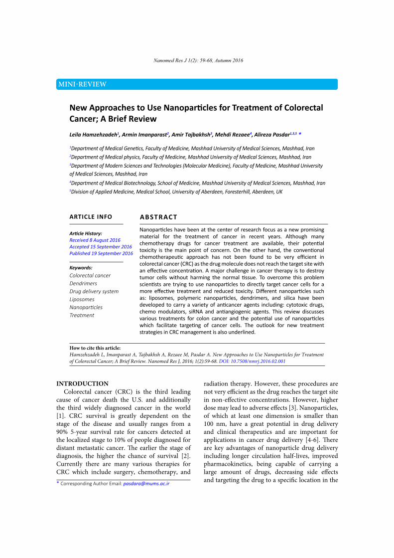
New Approaches to Use Nanoparticles for Treatment of Colorectal Cancer; A Brief Review
Leila Hamzehzadeh1, Armin Imanparast2, Amir Tajbakhsh3, Mehdi Rezaee4, Alireza Pasdar1,3,5 *
1Department of Medical Genetics, Faculty of Medicine, Mashhad University of Medical Sciences, Mashhad, Iran 2Department of Medical physics, Faculty of Medicine, Mashhad University of Medical Sciences, Mashhad, Iran 3Department of Modern Sciences and Technologies (Molecular Medicine), Faculty of Medicine, Mashhad University of Medical Sciences, Mashhad, Iran4Department of Medical Biotechnology, School of Medicine, Mashhad University of Medical Sciences, Mashhad, Iran 5Division of Applied Medicine, Medical School, University of Aberdeen, Foresterhill, Aberdeen, UK
* Corresponding Author Email: [email protected]
ARTICLE INFO
Article History:Received 8 August 2016Accepted 15 September 2016Published 19 September 2016
Keywords:Colorectal cancerDendrimersDrug delivery systemLiposomesNanoparticlesTreatment
ABSTRAC T
Nanoparticles have been at the center of research focus as a new promising material for the treatment of cancer in recent years. Although many chemotherapy drugs for cancer treatment are available, their potential toxicity is the main point of concern. On the other hand, the conventional chemotherapeutic approach has not been found to be very efficient in colorectal cancer (CRC) as the drug molecule does not reach the target site with an effective concentration. A major challenge in cancer therapy is to destroy tumor cells without harming the normal tissue. To overcome this problem scientists are trying to use nanoparticles to directly target cancer cells for a more effective treatment and reduced toxicity. Different nanoparticles such as: liposomes, polymeric nanoparticles, dendrimers, and silica have been developed to carry a variety of anticancer agents including: cytotoxic drugs, chemo modulators, siRNA and antiangiogenic agents. This review discusses various treatments for colon cancer and the potential use of nanoparticles which facilitate targeting of cancer cells. The outlook for new treatment strategies in CRC management is also underlined.
How to cite this article: Hamzehzadeh L, Imanparast A, Tajbakhsh A, Rezaee M, Pasdar A. New Approaches to Use Nanoparticles for Treatment of Colorectal Cancer; A Brief Review. Nanomed Res J, 2016; 1(2):59-68. DOI: 10.7508/nmrj.2016.02.001
INTRODUCTIONColorectal cancer (CRC) is the third leading
cause of cancer death the U.S. and additionally the third widely diagnosed cancer in the world [1]. CRC survival is greatly dependent on the stage of the disease and usually ranges from a 90% 5-year survival rate for cancers detected at the localized stage to 10% of people diagnosed for distant metastatic cancer. The earlier the stage of diagnosis, the higher the chance of survival [2]. Currently there are many various therapies for CRC which include surgery, chemotherapy, and
radiation therapy. However, these procedures are not very efficient as the drug reaches the target site in non-effective concentrations. However, higher dose may lead to adverse effects [3]. Nanoparticles, of which at least one dimension is smaller than 100 nm, have a great potential in drug delivery and clinical therapeutics and are important for applications in cancer drug delivery [4-6]. There are key advantages of nanoparticle drug delivery including longer circulation half-lives, improved pharmacokinetics, being capable of carrying a large amount of drugs, decreasing side effects and targeting the drug to a specific location in the
60
L. Hamzehzadeh et al. / Nanoparticles in cancer treatment
Nanomed Res J 1(2): 59-68, Autumn 2016
body (Table 1)[7, 8]. This article briefly reviews the nanoparticle-assisted co-delivery of drugs for CRC therapy.
Drug delivery system with nanoparticlesNanoparticle drug delivery platforms have
been in center of focus of researchers. Many solid tumors such as breast, lung, prostate, and colon cancers have unique structural features including the hyper permeable vasculature and impaired lymphatic drainage, hence, tumor tissues are quite permeable to macromolecules and nanocarriers [23, 24]. There are two major mechanisms for cell-specific targeting with nanocarriers: active and passive. The first strategy depends on the interaction between the nanocarriers and receptors on the target cell. Passive targeting involves
mechanisms to increase vascular permeability and also to retain long-circulating nanocarriers at tumor sites in their flow to impaired lymphatic system [25]. Enhanced permeability and retention (EPR) effect, nanoparticle clearance by the mononuclear phagocyte system (MPS), and desirable nanoparticle characteristics for cancer applications are important concepts in nanoparticle drug delivery. The EPR effect has a critical role in determining the efficacy of the nanoparticle-based drug delivery system [26]. There is however, a common problem among nanoparticles where they are quickly absorbed by macrophages, so-called MPS. The MPS (also known as the Reticulo Endothelial System (RES)) is mostly responsible for clearing macromolecules from circulation [27]. One of the major programs
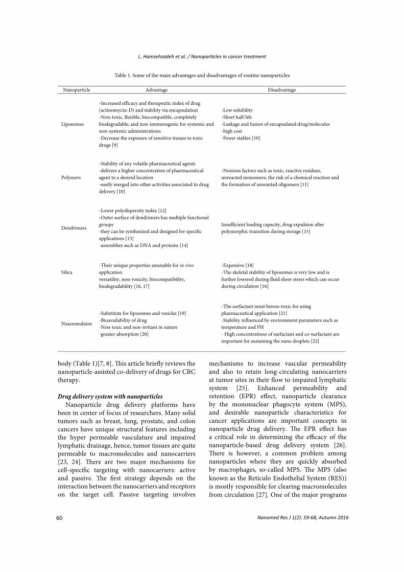
Table 1. Some of the main advantages and disadvantages of routine nanoparticles
Nanoparticle Advantage Disadvantage
Liposomes
-Increased efficacy and therapeutic index of drug (actinomycin-D) and stability via encapsulation -Non-toxic, flexible, biocompatible, completelybiodegradable, and non-immunogenic for systemic and non-systemic administrations -Decrease the exposure of sensitive tissues to toxic drugs [9]
-Low solubility-Short half-life -Leakage and fusion of encapsulated drug/molecules -high cost -Fewer stables [10]
Polymers
-Stability of any volatile pharmaceutical agents-delivers a higher concentration of pharmaceuticalagent to a desired location -easily merged into other activities associated to drug delivery [10]
-Noxious factors such as toxic, reactive residues, unreacted monomers, the risk of a chemical reaction and the formation of unwanted oligomers [11]
Dendrimers
-Lower polydispersity index [12] -Outer surface of dendrimers has multiple functionalgroups -they can be synthesized and designed for specific applications [13] -assemblies such as DNA and proteins [14]
Insufficient loading capacity, drug expulsion after polymorphic transition during storage [15]
Silica -Their unique properties amenable for in vivo application versatility, non-toxicity, biocompatibility, biodegradability [16, 17]
-Expensive [18] -The skeletal stability of liposomes is very low and is further lowered during fluid sheer stress which can occurduring circulation [16]
Nanoemulsion
-Substitute for liposomes and vesicles [19] -Bioavailability of drug -Non-toxic and non-irritant in nature -greater absorption [20]
-The surfactant must benon-toxic for using pharmaceutical application [21] -Stability influenced by environment parameters such astemperature and PH - High concentrations of surfactant and co-surfactant are important for sustaining the nano droplets [22]
1
Table 1. Some of the main advantages and disadvantages of routine nanoparticles
61
L. Hamzehzadeh et al. / Nanoparticles in cancer treatment
to prevent the rapid RES uptake is coating of the particles with surfactants or covalent linkage of polyoxyethylene [27, 28]. There a re d ifferent characteristics for delivering conventional therapeutics to solid tumors; life-size (less than 200nm), spherical shape and a smooth texture. Although particles larger than 500 nm are rapidly eliminated from the circulation [29].
LiposomesIn 1961, Bangham described liposomes as the
first n anoparticle p latform a pplied i n m edicine [30]. Liposomes were the first d rug-delivery system approved for clinical purposes. One of the most used delivery systems for small molecules, peptides, small and long nucleic acids, and proteins are liposomes and particularly nanoliposomes [31]. Liposomes are small, spherical artificial
Nanomed Res J 1(2): 59-68, Autumn 2016
carriers with an aqueous core and are naturally non-toxic [32]. Due to their phospholipid bilayer, their size and their ability to incorporate various substances liposomes are the most effective drug delivery systems into cells with slow-releasing and targeting characteristics and the ability to decrease side effects [33, 34].
Liposomes according to their different properties are divided into 3 groups:
1) Long-circulating liposomes (stealthliposomes): The conventional liposome surface is strongly affected by opsonization and the opsonized liposomes are subjected to uptake by MPS and subsequent clearance. Phospholipid bilayer structure of the liposome is modified by adding gangliosides or a polyethylene glycol (PEG) which tends to avoid blood plasma opsonins binding to the liposome surface. Subsequently, PEG causes
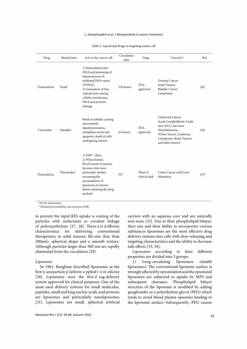
Table2 Liposomal drugs in targeting tumor cell
Drug Brand name Acts in the cancer cell Circulation
time Stage Cancer(s) Ref.
Doxorubicin Doxil®
1) Intercalation into DNA and poisoning oftopoisomerase-II-mediated DNA repair (TOP2A) 2) Generation of free radicals and causing cellular membranes, DNA and proteins damage
350 hours FDA approved
Ovarian Cancer Solid Tumors Bladder Cancer Lymphoma
[45]
Vincristine Marqibo®
Binds to tubulin causing microtubule depolymerization, metaphase arrest and apoptotic death of cells undergoing mitosis
6.6 hours FDA approved
Colorectal Cancer, Acute Lymphoblastic Leukemia (ALL), Sarcoma, Neuroblastoma, Wilms Tumor, Leukemia, Lymphoma, Brain Tumors and other tumors
[46]
Doxorubicin Thermodox®
1) EPR** effect 2) When heated, blood vessels in tumors become even more permeable, further increasing the accumulation of liposomes in tumors before releasing the drug payload
NI* Phase II clinical trial
Colon Cancer with Liver Metastasis
[47]
*NI: No information **Enhanced permeability and retention (EPR)
2
Table 2. Liposomal drugs in targeting tumor cell
62
L. Hamzehzadeh et al. / Nanoparticles in cancer treatment
Nanomed Res J 1(2): 59-68, Autumn 2016
a decrease in recognition of liposomes by the mononuclear phagocyte system and enables liposomes to stay stable in the circulation and maintain a prolonged half-life [35-37];
2) Active targeting liposomes: Liposomestargeting antibodies, glycoside residues, receptors, hormones and peptides;
3) Liposomes with special properties includethermo-sensitive, pH-sensitive, magnetic and positive;
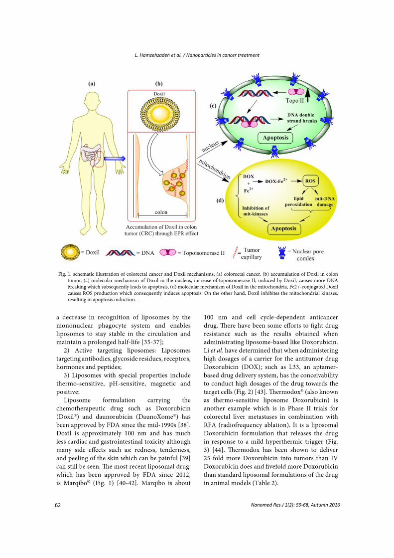
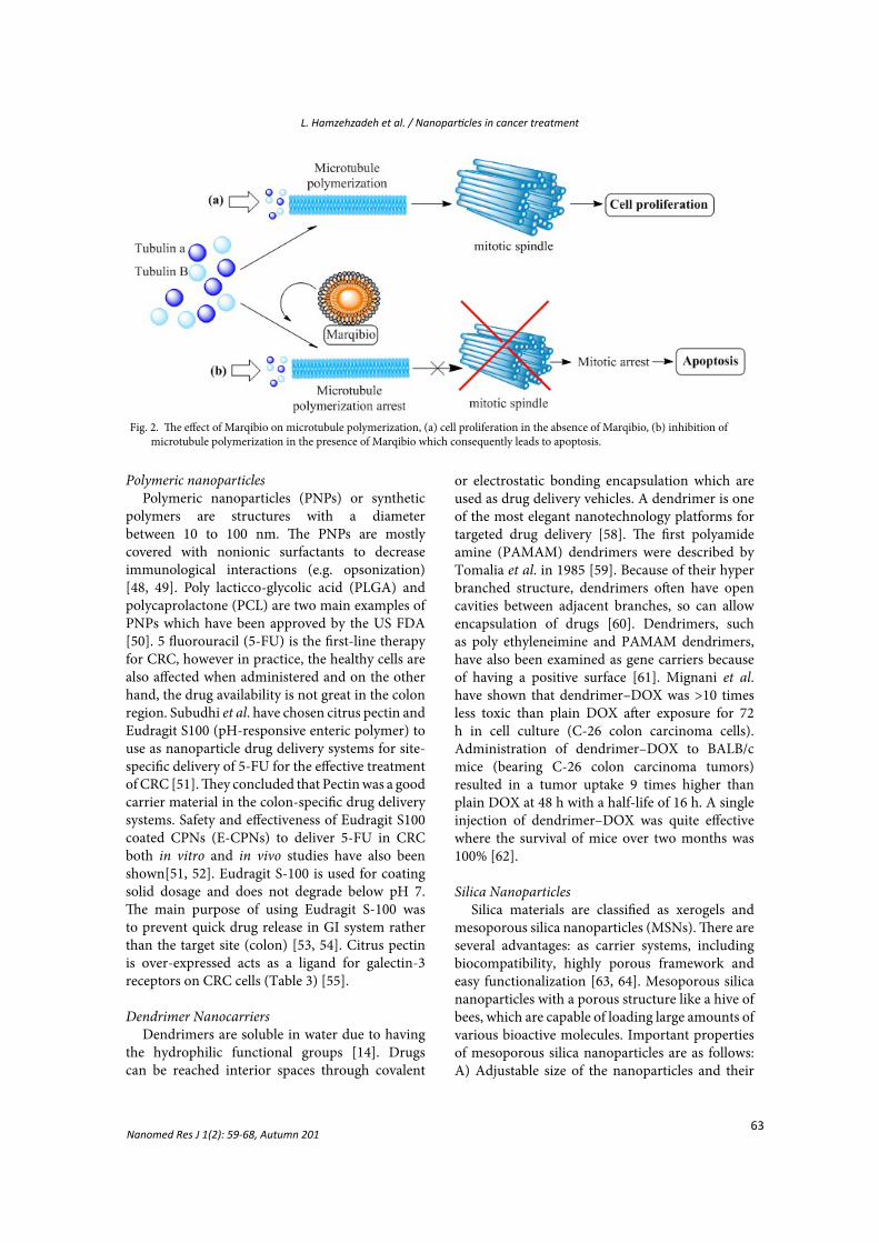
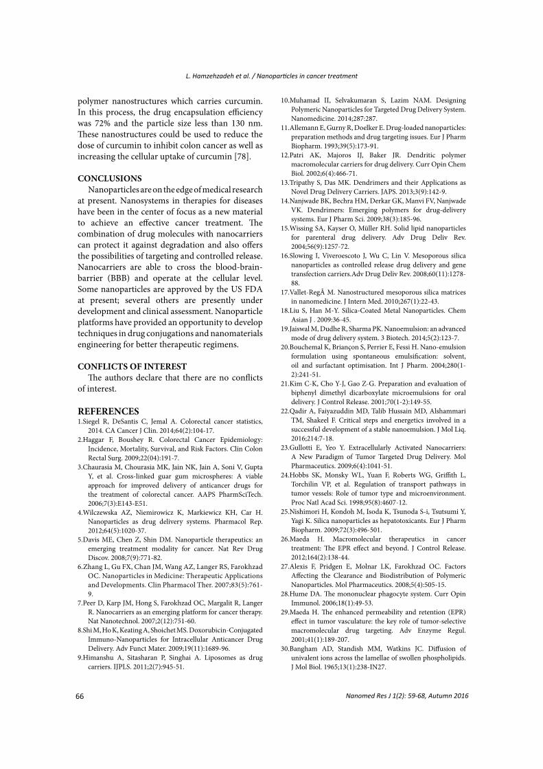
Liposome formulation carrying the chemotherapeutic drug such as Doxorubicin (Doxil®) and daunorubicin (DaunoXome®) has been approved by FDA since the mid-1990s [38]. Doxil is approximately 100 nm and has much less cardiac and gastrointestinal toxicity although many side effects such as: redness, tenderness, and peeling of the skin which can be painful [39] can still be seen. The most recent liposomal drug, which has been approved by FDA since 2012, is Marqibo® (Fig. 1) [40-42]. Marqibo is about
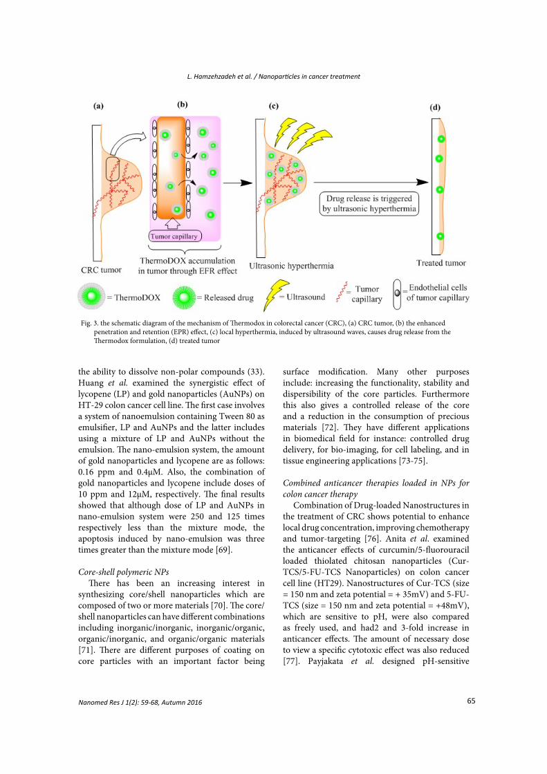
100 nm and cell cycle-dependent anticancer drug. There have been some efforts to fight drug resistance such as the results obtained when administrating liposome-based like Doxorubicin. Li et al. have determined that when administering high dosages of a carrier for the antitumor drug Doxorubicin (DOX); such as L33, an aptamer-based drug delivery system, has the conceivability to conduct high dosages of the drug towards the target cells (Fig. 2) [43]. Thermodox® (also known as thermo-sensitive liposome Doxorubicin) is another example which is in Phase II trials for colorectal liver metastases in combination with RFA (radiofrequency ablation). It is a liposomal Doxorubicin formulation that releases the drug in response to a mild hyperthermic trigger (Fig. 3) [44]. Thermodox has been shown to deliver25 fold more Doxorubicin into tumors than IVDoxorubicin does and fivefold more Doxorubicinthan standard liposomal formulations of the drugin animal models (Table 2).
Fig. 1. schematic illustration of colorectal cancer and Doxil mechanisms, (a) colorectal cancer, (b) accumulation of Doxil in colon tumor, (c) molecular mechanism of Doxil in the nucleus, increase of topoisomerase II, induced by Doxil, causes more DNA breaking which subsequentlyleads to apoptosis, (d) molecular mechanism of Doxil in the mitochondria, Fe2+-conjugated Doxil causes ROS production which consequentlyinduces apoptosis. On the other hand, Doxil inhibites the mitochondrial kinases, resulting in apoptosis induction.
1
Fig. 1. schematic illustration of colorectal cancer and Doxil mechanisms, (a) colorectal cancer, (b) accumulation of Doxil in colontumor, (c) molecular mechanism of Doxil in the nucleus, increase of topoisomerase II, induced by Doxil, causes more DNA breaking which subsequently leads to apoptosis, (d) molecular mechanism of Doxil in the mitochondria, Fe2+-conjugated Doxil causes ROS production which consequently induces apoptosis. On the other hand, Doxil inhibites the mitochondrial kinases, resulting in apoptosis induction.
63
L. Hamzehzadeh et al. / Nanoparticles in cancer treatment
Polymeric nanoparticlesPolymeric nanoparticles (PNPs) or synthetic
polymers are structures with a diameter between 10 to 100 nm. The PNPs are mostly covered with nonionic surfactants to decrease immunological interactions (e.g. opsonization) [48, 49]. Poly lacticco-glycolic acid (PLGA) and polycaprolactone (PCL) are two main examples of PNPs which have been approved by the US FDA [50]. 5 fluorouracil (5-FU) is the first-line therapy for CRC, however in practice, the healthy cells are also affected when administered and on the other hand, the drug availability is not great in the colon region. Subudhi et al. have chosen citrus pectin and Eudragit S100 (pH-responsive enteric polymer) to use as nanoparticle drug delivery systems for site-specific delivery of 5-FU for the effective treatment of CRC [51]. They concluded that Pectin was a good carrier material in the colon-specific drug delivery systems. Safety and effectiveness of Eudragit S100 coated CPNs (E-CPNs) to deliver 5-FU in CRC both in vitro and in vivo studies have also been shown[51, 52]. Eudragit S-100 is used for coating solid dosage and does not degrade below pH 7. The main purpose of using Eudragit S-100 was to prevent quick drug release in GI system rather than the target site (colon) [53, 54]. Citrus pectin is over-expressed acts as a ligand for galectin-3 receptors on CRC cells (Table 3) [55].
Dendrimer NanocarriersDendrimers are soluble in water due to having
the hydrophilic functional groups [14]. Drugs can be reached interior spaces through covalent
or electrostatic bonding encapsulation which are used as drug delivery vehicles. A dendrimer is one of the most elegant nanotechnology platforms for targeted drug delivery [58]. The first polyamide amine (PAMAM) dendrimers were described by Tomalia et al. in 1985 [59]. Because of their hyper branched structure, dendrimers often have open cavities between adjacent branches, so can allow encapsulation of drugs [60]. Dendrimers, such as poly ethyleneimine and PAMAM dendrimers, have also been examined as gene carriers because of having a positive surface [61]. Mignani et al. have shown that dendrimer–DOX was >10 times less toxic than plain DOX after exposure for 72 h in cell culture (C-26 colon carcinoma cells). Administration of dendrimer–DOX to BALB/c mice (bearing C-26 colon carcinoma tumors) resulted in a tumor uptake 9 times higher than plain DOX at 48 h with a half-life of 16 h. A single injection of dendrimer–DOX was quite effective where the survival of mice over two months was 100% [62].
Silica NanoparticlesSilica materials are classified as xerogels and
mesoporous silica nanoparticles (MSNs). There are several advantages: as carrier systems, including biocompatibility, highly porous framework and easy functionalization [63, 64]. Mesoporous silica nanoparticles with a porous structure like a hive of bees, which are capable of loading large amounts of various bioactive molecules. Important properties of mesoporous silica nanoparticles are as follows: A) Adjustable size of the nanoparticles and their
Fig. 2. The effect of Marqibio on microtubule polymerization, (a) cell proliferation in the absence of Marqibio, (b) inhibition of microtubulepolymerization in the presence of Marqibio which consequently leads to apoptosis.
2
Fig. 2. The effect of Marqibio on microtubule polymerization, (a) cell proliferation in the absence of Marqibio, (b) inhibition of microtubule polymerization in the presence of Marqibio which consequently leads to apoptosis.
Nanomed Res J 1(2): 59-68, Autumn 201
64
L. Hamzehzadeh et al. / Nanoparticles in cancer treatment
Nanomed Res J 1(2): 59-68, Autumn 2016
cavities in the range of 50 to 300 and 2 to 6 nm, respectively [44].
B) Very low toxicity, easy endocytosis, theability of extensive loading of the drug
C) Resistance to heat and pH [65].Radhakrishnan et al. used mesoporous silica
nanoparticle (MSN) -protamine hybrid system (MSN−PRM) to selectively release the drugs in the proximity of cancer cells where specific enzymes can trigger the drug activity [66]. Drug-induced cell death in CRC cells was also significantly enhanced when the hydrophobic drug was encapsulated in the MSN–PRM system in comparison to the free drug (P< 0.05) [66]. Yu M et al. showed that conjugation of hyaluronic acid to MSNs, the amount of DOX loading into HA-MSNs increases than bare MSNs [67]. Cellular uptake of DOX-HA-MSNs was also increased and was shown that DOX-HA-MSNs more cytotoxicity to HCT-116 cell lines (human colon carcinoma) than free
DOX [46]. In another work, Hanafi-Bojd et al. showed that when MSNs were functionalized with polyethylene glycol (PEG) and polyethylenimine-polyethylene glycol (PEI-PEG) groups, the amount of Epirubicin hydrochloride (EPI) loading into MSN was increased and produced an improved antitumor efficiency. The antitumor activity in C-26 colon carcinoma model was higher due toenhanced accumulation of MSN-PEI-PEG-EPIcompared to free EPI [68].
Nanoemulsion systemNanoemulsion is a transparent solution
including water, oil and surfactant with thermodynamically stable and uniform physical properties. Important features of nanoemulsion are as follows: a) facilitate the process of transferring drugs and drug combinations protect against external factors (such as heat, pH) [48] b) high stability, low toxicity and efficiency and finally c)
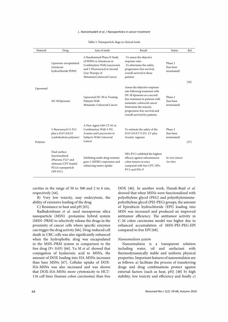
Table3. Nanoparticle dugs in clinical trials
Material Drug Aim of study Result Status Ref.
Liposomal
Liposome-encapsulated irinotecan hydrochloride PEP02
A Randomized Phase II Study of PEP02 or Irinotecan in Combination With Leucovorin and 5-Fluorouracil in Second Line Therapy of MetastaticColorectal Cancer
-To assess the objective response rates -To determine the safety, progression-free survival, overall survival in these patients
Phase 2 (has been terminated)
[56]
SN-38 liposome Liposomal SN-38 in Treating Patients With Metastatic Colorectal Cancer
Assess the objective response rate following treatment with SN-38 liposome as a second-line treatment in patients with metastatic colorectal cancer Determine the toxicity, progression-free survival and overall survival for patients.
Phase 2 (has been terminated)
Polymer
5-fluorouracil (5-FU) plus a DAVANAT (carbohydrate polymer)
A New Agent GM-CT-01 in Combination With 5-FU, Avastin and Leucovorin in Subjects With Colorectal Cancer
To estimate the safety of the DAVANAT®/5-FU, LV plus Avastin® regimen
Phase 2 (has been terminated)
[57]
Dual-surface-functionalized (Pluronic F127 and chitosan) CPT-loaded PLGA nanoparticle (NP-P/C)
Inhibiting multi-drug resistant gene 1 (MDR1) expression and enhancing tumor uptake
NPs-P/C1 exhibited the highest efficacy against subcutaneous colon tumors in mice compared with free CPT, NPs-PVA and NPs-P
In vivo (mice) /in vitro
3
Table 3. Nanoparticle dugs in clinical trials
65
L. Hamzehzadeh et al. / Nanoparticles in cancer treatment
the ability to dissolve non-polar compounds (33). Huang et al. examined the synergistic effect of lycopene (LP) and gold nanoparticles (AuNPs) on HT-29 colon cancer cell line. The first case involves a system of nanoemulsion containing Tween 80 as emulsifier, LP and AuNPs and the latter includes using a mixture of LP and AuNPs without the emulsion. The nano-emulsion system, the amount of gold nanoparticles and lycopene are as follows: 0.16 ppm and 0.4μM. Also, the combination of gold nanoparticles and lycopene include doses of 10 ppm and 12μM, respectively. The final results showed that although dose of LP and AuNPs in nano-emulsion system were 250 and 125 times respectively less than the mixture mode, the apoptosis induced by nano-emulsion was three times greater than the mixture mode [69].
Core-shell polymeric NPsThere has been an increasing interest in
synthesizing core/shell nanoparticles which are composed of two or more materials [70]. The core/shell nanoparticles can have different combinations including inorganic/inorganic, inorganic/organic, organic/inorganic, and organic/organic materials [71]. There are different purposes of coating on core particles with an important factor being
surface modification. Many other purposes include: increasing the functionality, stability and dispersibility of the core particles. Furthermore this also gives a controlled release of the core and a reduction in the consumption of precious materials [72]. They have different applications in biomedical field for instance: controlled drug delivery, for bio-imaging, for cell labeling, and in tissue engineering applications [73-75].
Combined anticancer therapies loaded in NPs for colon cancer therapy
Combination of Drug-loaded Nanostructures in the treatment of CRC shows potential to enhance local drug concentration, improving chemotherapy and tumor-targeting [76]. Anita et al. examined the anticancer effects of curcumin/5-fluorouracil loaded thiolated chitosan nanoparticles (Cur-TCS/5-FU-TCS Nanoparticles) on colon cancer cell line (HT29). Nanostructures of Cur-TCS (size = 150 nm and zeta potential = + 35mV) and 5-FU-TCS (size = 150 nm and zeta potential = +48mV), which are sensitive to pH, were also compared as freely used, and had2 and 3-fold increase in anticancer effects. The amount of necessary dose to view a specific cytotoxic effect was also reduced [77]. Payjakata et al. designed pH-sensitive
Fig. 3. the schematic diagram of the mechanism of Thermodox in colorectal cancer (CRC), (a) CRC tumor, (b) the enhanced penetration and retention (EPR) effect, (c) local hyperthermia, induced by ultrasound waves, causes drug release from the Thermodox formulation, (d) treatedtumor
3
Fig. 3. the schematic diagram of the mechanism of Thermodox in colorectal cancer (CRC), (a) CRC tumor, (b) the enhanced penetration and retention (EPR) effect, (c) local hyperthermia, induced by ultrasound waves, causes drug release from the Thermodox formulation, (d) treated tumor
Nanomed Res J 1(2): 59-68, Autumn 2016
66
L. Hamzehzadeh et al. / Nanoparticles in cancer treatment
Nanomed Res J 1(2): 59-68, Autumn 2016
polymer nanostructures which carries curcumin. In this process, the drug encapsulation efficiency was 72% and the particle size less than 130 nm. These nanostructures could be used to reduce the dose of curcumin to inhibit colon cancer as well as increasing the cellular uptake of curcumin [78].
CONCLUSIONSNanoparticles are on the edge of medical research
at present. Nanosystems in therapies for diseases have been in the center of focus as a new material to achieve an effective cancer treatment. The combination of drug molecules with nanocarriers can protect it against degradation and also offers the possibilities of targeting and controlled release. Nanocarriers are able to cross the blood-brain-barrier (BBB) and operate at the cellular level. Some nanoparticles are approved by the US FDA at present; several others are presently under development and clinical assessment. Nanoparticle platforms have provided an opportunity to develop techniques in drug conjugations and nanomaterials engineering for better therapeutic regimens.
CONFLICTS OF INTERESTThe authors declare that there are no conflicts
of interest.
REFERENCES1.Siegel R, DeSantis C, Jemal A. Colorectal cancer statistics,
2014. CA Cancer J Clin. 2014;64(2):104-17.2.Haggar F, Boushey R. Colorectal Cancer Epidemiology:
Incidence, Mortality, Survival, and Risk Factors. Clin ColonRectal Surg. 2009;22(04):191-7.
3.Chaurasia M, Chourasia MK, Jain NK, Jain A, Soni V, GuptaY, et al. Cross-linked guar gum microspheres: A viableapproach for improved delivery of anticancer drugs for the treatment of colorectal cancer. AAPS PharmSciTech. 2006;7(3):E143-E51.
4.Wilczewska AZ, Niemirowicz K, Markiewicz KH, Car H.Nanoparticles as drug delivery systems. Pharmacol Rep.2012;64(5):1020-37.
5.Davis ME, Chen Z, Shin DM. Nanoparticle therapeutics: anemerging treatment modality for cancer. Nat Rev DrugDiscov. 2008;7(9):771-82.
6.Zhang L, Gu FX, Chan JM, Wang AZ, Langer RS, FarokhzadOC. Nanoparticles in Medicine: Therapeutic Applicationsand Developments. Clin Pharmacol Ther. 2007;83(5):761-9.
7.Peer D, Karp JM, Hong S, Farokhzad OC, Margalit R, LangerR. Nanocarriers as an emerging platform for cancer therapy. Nat Nanotechnol. 2007;2(12):751-60.
8.Shi M, Ho K, Keating A, Shoichet MS. Doxorubicin-Conjugated Immuno-Nanoparticles for Intracellular Anticancer DrugDelivery. Adv Funct Mater. 2009;19(11):1689-96.
9.Himanshu A, Sitasharan P, Singhai A. Liposomes as drugcarriers. IJPLS. 2011;2(7):945-51.
10.Muhamad II, Selvakumaran S, Lazim NAM. DesigningPolymeric Nanoparticles for Targeted Drug Delivery System. Nanomedicine. 2014;287:287.
11.Allemann E, Gurny R, Doelker E. Drug-loaded nanoparticles: preparation methods and drug targeting issues. Eur J Pharm Biopharm. 1993;39(5):173-91.
12.Patri AK, Majoros IJ, Baker JR. Dendritic polymermacromolecular carriers for drug delivery. Curr Opin Chem Biol. 2002;6(4):466-71.
13.Tripathy S, Das MK. Dendrimers and their Applications asNovel Drug Delivery Carriers. JAPS. 2013;3(9):142-9.
14.Nanjwade BK, Bechra HM, Derkar GK, Manvi FV, Nanjwade VK. Dendrimers: Emerging polymers for drug-deliverysystems. Eur J Pharm Sci. 2009;38(3):185-96.
15.Wissing SA, Kayser O, Müller RH. Solid lipid nanoparticlesfor parenteral drug delivery. Adv Drug Deliv Rev.2004;56(9):1257-72.
16.Slowing I, Viveroescoto J, Wu C, Lin V. Mesoporous silicananoparticles as controlled release drug delivery and genetransfection carriers.Adv Drug Deliv Rev. 2008;60(11):1278-88.
17.Vallet-Regà M. Nanostructured mesoporous silica matricesin nanomedicine. J Intern Med. 2010;267(1):22-43.
18.Liu S, Han M-Y. Silica-Coated Metal Nanoparticles. ChemAsian J . 2009:36-45.
19.Jaiswal M, Dudhe R, Sharma PK. Nanoemulsion: an advanced mode of drug delivery system. 3 Biotech. 2014;5(2):123-7.
20.Bouchemal K, Briançon S, Perrier E, Fessi H. Nano-emulsion formulation using spontaneous emulsification: solvent,oil and surfactant optimisation. Int J Pharm. 2004;280(1-2):241-51.
21.Kim C-K, Cho Y-J, Gao Z-G. Preparation and evaluation ofbiphenyl dimethyl dicarboxylate microemulsions for oraldelivery. J Control Release. 2001;70(1-2):149-55.
22.Qadir A, Faiyazuddin MD, Talib Hussain MD, AlshammariTM, Shakeel F. Critical steps and energetics involved in asuccessful development of a stable nanoemulsion. J Mol Liq. 2016;214:7-18.
23.Gullotti E, Yeo Y. Extracellularly Activated Nanocarriers:A New Paradigm of Tumor Targeted Drug Delivery. MolPharmaceutics. 2009;6(4):1041-51.
24.Hobbs SK, Monsky WL, Yuan F, Roberts WG, Griffith L,Torchilin VP, et al. Regulation of transport pathways intumor vessels: Role of tumor type and microenvironment.Proc Natl Acad Sci. 1998;95(8):4607-12.
25.Nishimori H, Kondoh M, Isoda K, Tsunoda S-i, Tsutsumi Y,Yagi K. Silica nanoparticles as hepatotoxicants. Eur J Pharm Biopharm. 2009;72(3):496-501.
26.Maeda H. Macromolecular therapeutics in cancertreatment: The EPR effect and beyond. J Control Release.2012;164(2):138-44.
27.Alexis F, Pridgen E, Molnar LK, Farokhzad OC. FactorsAffecting the Clearance and Biodistribution of PolymericNanoparticles. Mol Pharmaceutics. 2008;5(4):505-15.
28.Hume DA. The mononuclear phagocyte system. Curr OpinImmunol. 2006;18(1):49-53.
29.Maeda H. The enhanced permeability and retention (EPR)effect in tumor vasculature: the key role of tumor-selectivemacromolecular drug targeting. Adv Enzyme Regul.2001;41(1):189-207.
30.Bangham AD, Standish MM, Watkins JC. Diffusion ofunivalent ions across the lamellae of swollen phospholipids.J Mol Biol. 1965;13(1):238-IN27.
67
L. Hamzehzadeh et al. / Nanoparticles in cancer treatment
31.Abreu AS, Castanheira EMS, Queiroz M-JRP, Ferreira PMT,Vale-Silva LA, Pinto E. Nanoliposomes for encapsulationand delivery of the potential antitumoral methyl 6-methoxy-3-(4-methoxyphenyl)-1H-indole-2-carboxylate. NanoscaleRes Lett. 2011;6(1):482.
32.Silva R, Ferreira H, Cavaco-Paulo A. Sonoproduction ofLiposomes and Protein Particles as Templates for DeliveryPurposes. Biomacromolecules. 2011;12(10):3353-68.
33.Patil YP, Jadhav S. Novel methods for liposome preparation.Chem Phys Lipids. 2014;177:8-18.
34.Suntres ZE. Liposomal Antioxidants for Protection againstOxidant-Induced Damage. J Toxicol. 2011;2011:1-16.
35.Nag O, Awasthi V. Surface Engineering of Liposomes forStealth Behavior. Pharmaceutics. 2013;5(4):542-69.
36.Noble GT, Stefanick JF, Ashley JD, Kiziltepe T, BilgicerB. Ligand-targeted liposome design: challenges andfundamental considerations. Trends Biotechnol.2014;32(1):32-45.
37.Akbarzadeh A, Rezaei-Sadabady R, Davaran S, Joo SW,Zarghami N, Hanifehpour Y, et al. Liposome: classification,preparation, and applications. Nanoscale Res Lett.2013;8(1):102.
38.Barenholz Y. Doxil® — The first FDA-approved nano-drug:Lessons learned. J Control Release. 2012;160(2):117-34.
39.Rivera E. Liposomal Anthracyclines in Metastatic BreastCancer: Clinical Update. Oncologist. 2003;8(Supp-2):3-9.
40.Allen TM, Cullis PR. Liposomal drug delivery systems:From concept to clinical applications. Adv Drug Deliv Rev.2013;65(1):36-48.
41.Lam R, Ho D. Nanodiamonds as vehicles for systemicand localized drug delivery. Expert Opin Drug Deliv.2009;6(9):883-95.
42.Lammers T, Hennink WE, Storm G. Tumour-targetednanomedicines: principles and practice. Br J Cancer, BJC.2008;99(3):392-7.
43.Li W, Chen H, Yu M, Fang J. Targeted Delivery of Doxorubicin Using a Colorectal Cancer-Specific ssDNA Aptamer. AnatRec. 2014;297(12):2280-8.
44.Stang J, Haynes M, Carson P, Moghaddam M. A PreclinicalSystem Prototype for Focused Microwave Thermal Therapyof the Breast. IEEE T Bio-Med Eng. 2012;59(9):2431-8.
45.Thorn CF, Oshiro C, Marsh S, Hernandez-BoussardT, McLeod H, Klein TE, et al. Doxorubicin pathways:pharmacodynamics and adverse effects. PharmacogenetGenomics. 2011;21(7):440.
46.Gidding CEM, Kellie SJ, Kamps WA, de Graaf SSN. Vincristine revisited. Crit Rev Oncol Hematol. 1999;29(3):267-87.
47.Celsion. Phase 2 Study of Thermodox as Adjuvant TherapyWith Thermal Ablation (RFA) in Treatment of MetastaticColorectal Cancer(mCRC) (ABLATE). In: ClinicalTrials.gov [Internet] [updated March 4, 2016; cited November1, 2011]. Available from:https://clinicaltrials.gov/ct2/show/NCT01464593
48.Bilensoy E, Sarisozen C, Esendağlı G, Doğan AL, Aktaş Y,Şen M, et al. Intravesical cationic nanoparticles of chitosanand polycaprolactone for the delivery of Mitomycin C tobladder tumors. Int J Pharm. 2009;371(1-2):170-6.
49.Torchilin V. Multifunctional pharmaceutical nanocarriers.New York: Springer Science & Business Media; 2008.
50.Zhang L, Radovic-Moreno AF, Alexis F, Gu FX, BastoPA, Bagalkot V, et al. Co-Delivery of Hydrophobicand Hydrophilic Drugs from Nanoparticle–AptamerBioconjugates. ChemMedChem. 2007;2(9):1268-71.
51.Subudhi M, Jain A, Jain A, Hurkat P, Shilpi S, Gulbake A,et al. Eudragit S100 Coated Citrus Pectin Nanoparticles forColon Targeting of 5-Fluorouracil. Materials. 2015;8(3):832-49.
52.Tummala S, Satish Kumar MN, Prakash A. Formulationand characterization of 5-Fluorouracil enteric coatednanoparticles for sustained and localized release in treatingcolorectal cancer. Saudi Pharm J. 2015;23(3):308-14.
53.Asghar LFA, Chandran S. Design and evaluation of matrices of Eudragit with polycarbophil and carbopol for colon-specific delivery. J Drug Target. 2008;16(10):741-57.
54.Obeidat WM, Price JC. Preparation and evaluation ofEudragit S 100 microspheres as pH-sensitive releasepreparations for piroxicam and theophylline using theemulsion-solvent evaporation method. J Microencapsul.2006;23(2):195-202.
55.Leclere L, Cutsem PV, Michiels C. Anti-cancer activities ofpH- or heat-modified pectin. Front Pharmacol. 2013;4.
56. US National Institute of Health. ClinicalTrial.gov [website].USA [updated 11/9/2016]. Available from: https://clinicaltrials.gov/.
57.Xiao B, Zhang M, Viennois E, Zhang Y, Wei N, Baker MT,et al. Inhibition of MDR1 gene expression and enhancingcellular uptake for effective colon cancer treatment usingdual-surface-functionalized nanoparticles. Biomaterials.2015;48:147-60.
58.Semwal R, Semwal D, Madan A, Paul P, Mujaffer F, Badoni R. Dendrimers: A novel approach for drug targeting. J PharmRes. 2010;3:2238-47.
59.Tomalia DA, Baker H, Dewald J, Hall M, Kallos G, MartinS, et al. A New Class of Polymers: Starburst-DendriticMacromolecules. Polym J. 1985;17(1):117-32.
60.Bhadra D, Bhadra S, Jain S, Jain NK. A PEGylated dendriticnanoparticulate carrier of fluorouracil. Int J Pharm.2003;257(1-2):111-24.
61.Xu Q, Wang C-H, Wayne Pack D. Polymeric Carriers for Gene Delivery: Chitosan and Poly(amidoamine) Dendrimers.Curr Pharm Des. 2010;16(21):2350-68.
62.Mignani S, Majoral JP. Dendrimers as macromoleculartools to tackle from colon to brain tumor types: a conciseoverview. New J Chem. 2013;37(11):3337.
63.Amato G. Silica-Encapsulated Efficient and Stable SiQuantum Dots with High Biocompatibility. Nanoscale ResLett. 2010;5(7):1156-60.
64.Wei L, Hu N, Zhang Y. Synthesis of Polymer—MesoporousSilica Nanocomposites. Materials. 2010;3(7):4066-79.
65.Bharti C, Gulati N, Nagaich U, Pal A. Mesoporous silicananoparticles in target drug delivery system: A review. Int JPharma Investig. 2015;5(3):124.
66.Radhakrishnan K, Gupta S, Gnanadhas DP, RamamurthyPC, Chakravortty D, Raichur AM. Protamine-CappedMesoporous Silica Nanoparticles for Biologically TriggeredDrug Release. Part Part Syst Char. 2013;31(4):449-58.
67.Yu M, Jambhrunkar S, Thorn P, Chen J, Gu W, Yu C.Hyaluronic acid modified mesoporous silica nanoparticlesfor targeted drug delivery to CD44-overexpressing cancercells. Nanoscale. 2013;5(1):178-83.
68.Hanafi-Bojd MY, Jaafari MR, Ramezanian N, Xue M,Amin M, Shahtahmassebi N, et al. Surface functionalizedmesoporous silica nanoparticles as an effective carrier forepirubicin delivery to cancer cells. Eur J Pharm Biopharm.2015;89:248-58.
69.Chen B-H, Huang R-FS, Wei Y-J, Stephen Inbaraj B. Inhibition
Nanomed Res J 1(2): 59-68, Autumn 2016
68
L. Hamzehzadeh et al. / Nanoparticles in cancer treatment
Nanomed Res J 1(2): 59-68, Autumn 2016
of colon cancer cell growth by nanoemulsion carrying gold nanoparticles and lycopene. Int J Nanomedicine. 2015;10(1):2823-46.
70.Zhou HS, Sasahara H, Honma I, Komiyama H, HausJW. Coated Semiconductor Nanoparticles: The CdS/PbSSystem’s Photoluminescence Properties. Chem Mater.1994;6(9):1534-41.
71.Ghosh Chaudhuri R, Paria S. Core/Shell Nanoparticles:Classes, Properties, Synthesis Mechanisms, Characterization, and Applications. Chem Rev. 2012;112(4):2373-433.
72.Kalele S, Gosavi S, Urban J, Kulkarni S. Nanoshellparticles: synthesis, properties and applications. Curr Sci.2006;91(8):1038-52.
73.Bai Y, Teng B, Chen S, Chang Y, Li Z. Preparation ofMagnetite Nanoparticles Coated with an AmphiphilicBlock Copolymer: A Potential Drug Carrier with a Core-Shell-Corona Structure for Hydrophobic Drug Delivery.Macromol Rapid Commun. 2006;27(24):2107-12.
74.Sounderya N, Zhang Y. Use of Core/Shell StructuredNanoparticles for Biomedical Applications. Recent PatBiomed Eng. 2008;1(1):34-42.
75.Stanciu L, Won Y-H, Ganesana M, Andreescu S. MagneticParticle-Based Hybrid Platforms for Bioanalytical Sensors.Sensors. 2009;9(4):2976-99.
76.Ma Y, Coombes AGA. Designing colon-specific deliverysystems for anticancer drug-loaded nanoparticles: Anevaluation of alginate carriers. J Biomed Mater Res A.2013;102(9):3167-76.
77.Anitha A, Deepa N, Chennazhi KP, Lakshmanan V-K,Jayakumar R. Combinatorial anticancer effects of curcuminand 5-fluorouracil loaded thiolated chitosan nanoparticlestowards colon cancer treatment. BBA-Gen Subjects.2014;1840(9):2730-43.
78.Prajakta D, Ratnesh J, Chandan K, Suresh S, Grace S, MeeraV, et al. Curcumin Loaded pH-Sensitive Nanoparticles forthe Treatment of Colon Cancer. J Biomed Nanotechnol.2009;5(5):445-55.