Embed Size (px)
Citation preview

New Advances in the Treatment of Autoimmune
Diseases
蔡長祐
台北榮民總醫院過敏免疫風濕科
國立陽明交通大學
CYT Aug 31, 2021
Pharmacist Workshop
Characteristic of Arthritis

Pathophysiological Role of Cytokines & Other Mediators & Their Inhibitors in RA & Other Autoimmune Inflammatory Diseases
This figure summarizes the cellular interactions believed to be of importance in the pathogenesis of RA and describes the interaction among macrophages, T cells, B cells, and nonhematopoietic cells (fibroblasts, connective tissue cells, and bone). These interactions are facilitated by the actions of cytokines released from the activated cells that then, through both autocrine (feedback on same cell) and paracrine (via other cell types) mechanisms, induce the production of other proinflammatory cytokines, which together contribute to the pathogenesis of this disease. Based on ex vivo studies from diseased tissue and in vivo studies on animal models, those cytokines with pathogenic potential have been identified and biological therapies developed to block their action. This figure identifies those therapeutic modalities and the stage in clinical development that these interventions have reached. sIL-6R, soluble IL-6 receptor.
Rituximab
ObinutuzumabCD20
Secukinumab
Ixekizumab
Brodalumab
Golimumab,
Certolizumab pegol,
Risankizumab
Guselkumab
Adapted from J Clin Invest. 2008;118(11):3537-3545. doi:10.1172/JCI36389.
Miscellaneous:CD152-IgG1: abatacept
Anti-integrin a4b7 (LPAM-1):vedolizumab
Anti-interferon-g:fontolizumab
Anti-Complement C5:euclizumab
Anti-type 1 IFN-R:Anifrolumab
Ustekizumab
RilonaceptCanakinumab
tadekinig
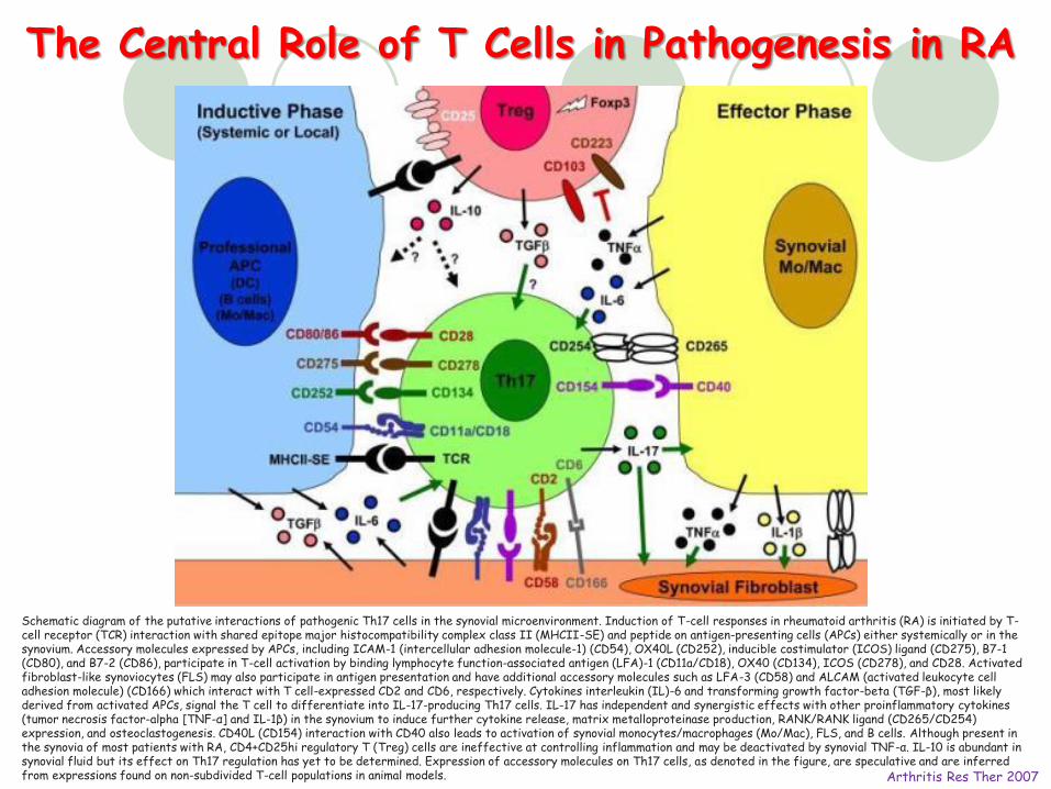
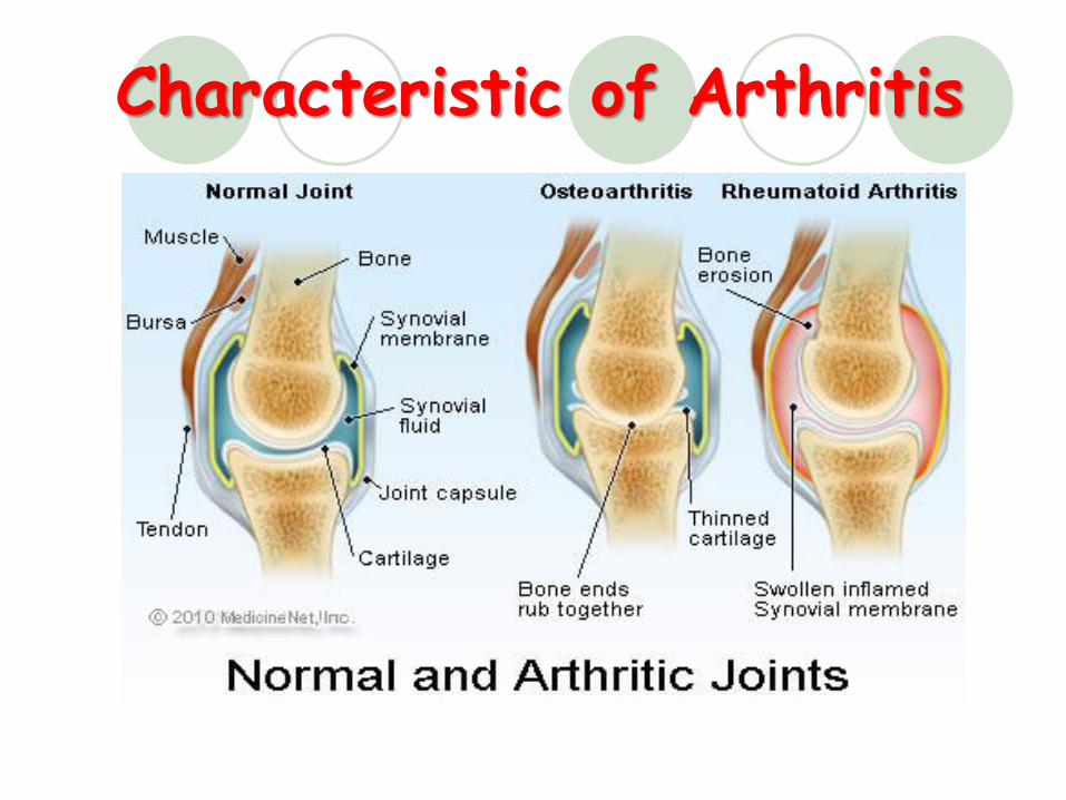
The Central Role of T Cells in Pathogenesis in RA
Arthritis Res Ther 2007
Schematic diagram of the putative interactions of pathogenic Th17 cells in the synovial microenvironment. Induction of T-cell responses in rheumatoid arthritis (RA) is initiated by T-cell receptor (TCR) interaction with shared epitope major histocompatibility complex class II (MHCII-SE) and peptide on antigen-presenting cells (APCs) either systemically or in the synovium. Accessory molecules expressed by APCs, including ICAM-1 (intercellular adhesion molecule-1) (CD54), OX40L (CD252), inducible costimulator (ICOS) ligand (CD275), B7-1 (CD80), and B7-2 (CD86), participate in T-cell activation by binding lymphocyte function-associated antigen (LFA)-1 (CD11a/CD18), OX40 (CD134), ICOS (CD278), and CD28. Activated fibroblast-like synoviocytes (FLS) may also participate in antigen presentation and have additional accessory molecules such as LFA-3 (CD58) and ALCAM (activated leukocyte cell adhesion molecule) (CD166) which interact with T cell-expressed CD2 and CD6, respectively. Cytokines interleukin (IL)-6 and transforming growth factor-beta (TGF-β), most likely derived from activated APCs, signal the T cell to differentiate into IL-17-producing Th17 cells. IL-17 has independent and synergistic effects with other proinflammatory cytokines (tumor necrosis factor-alpha [TNF-α] and IL-1β) in the synovium to induce further cytokine release, matrix metalloproteinase production, RANK/RANK ligand (CD265/CD254) expression, and osteoclastogenesis. CD40L (CD154) interaction with CD40 also leads to activation of synovial monocytes/macrophages (Mo/Mac), FLS, and B cells. Although present in the synovia of most patients with RA, CD4+CD25hi regulatory T (Treg) cells are ineffective at controlling inflammation and may be deactivated by synovial TNF-α. IL-10 is abundant in synovial fluid but its effect on Th17 regulation has yet to be determined. Expression of accessory molecules on Th17 cells, as denoted in the figure, are speculative and are inferred from expressions found on non-subdivided T-cell populations in animal models.
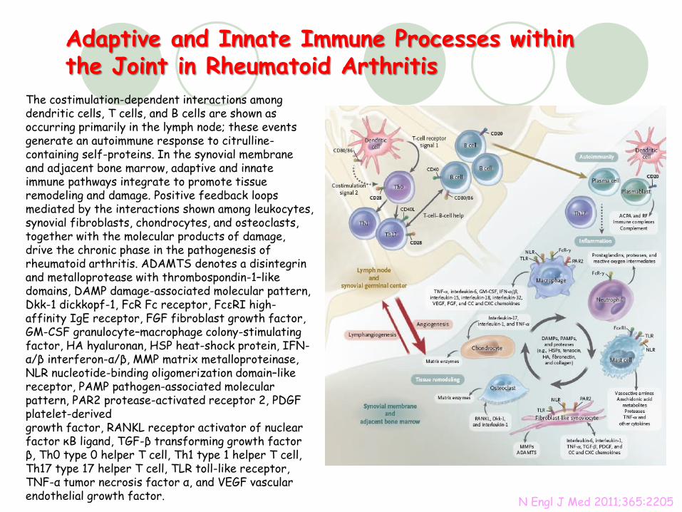
Adaptive and Innate Immune Processes within the Joint in Rheumatoid Arthritis
N Engl J Med 2011;365:2205
The costimulation-dependent interactions among dendritic cells, T cells, and B cells are shown as occurring primarily in the lymph node; these events generate an autoimmune response to citrulline-containing self-proteins. In the synovial membrane and adjacent bone marrow, adaptive and innate immune pathways integrate to promote tissue remodeling and damage. Positive feedback loops mediated by the interactions shown among leukocytes, synovial fibroblasts, chondrocytes, and osteoclasts, together with the molecular products of damage, drive the chronic phase in the pathogenesis of rheumatoid arthritis. ADAMTS denotes a disintegrin and metalloprotease with thrombospondin-1–like domains, DAMP damage-associated molecular pattern, Dkk-1 dickkopf-1, FcR Fc receptor, FcεRI high-affinity IgE receptor, FGF fibroblast growth factor, GM-CSF granulocyte–macrophage colony-stimulating factor, HA hyaluronan, HSP heat-shock protein, IFN-α/β interferon-α/β, MMP matrix metalloproteinase, NLR nucleotide-binding oligomerization domain–like receptor, PAMP pathogen-associated molecular pattern, PAR2 protease-activated receptor 2, PDGF platelet-derivedgrowth factor, RANKL receptor activator of nuclear factor κB ligand, TGF-β transforming growth factor β, Th0 type 0 helper T cell, Th1 type 1 helper T cell, Th17 type 17 helper T cell, TLR toll-like receptor, TNF-α tumor necrosis factor α, and VEGF vascular endothelial growth factor.
6
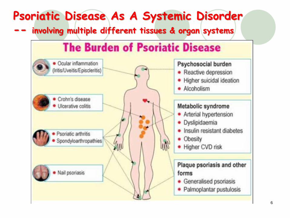
Psoriatic Disease As A Systemic Disorder -- involving multiple different tissues & organ systems
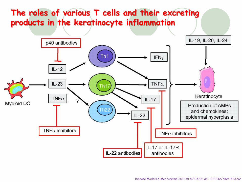
Disease Models & Mechanisms 2012 5: 423-433; doi: 10.1242/dmm.009092
The roles of various T cells and their excreting products in the keratinocyte inflammation
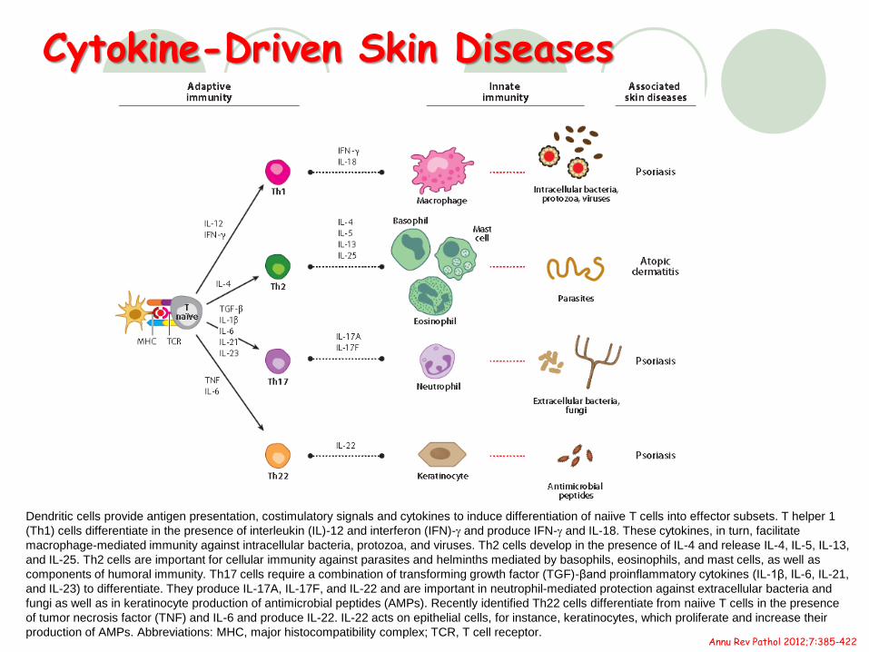
Cytokine-Driven Skin Diseases
Annu Rev Pathol 2012;7:385-422
Dendritic cells provide antigen presentation, costimulatory signals and cytokines to induce differentiation of naiive T cells into effector subsets. T helper 1
(Th1) cells differentiate in the presence of interleukin (IL)-12 and interferon (IFN)-g and produce IFN-g and IL-18. These cytokines, in turn, facilitate
macrophage-mediated immunity against intracellular bacteria, protozoa, and viruses. Th2 cells develop in the presence of IL-4 and release IL-4, IL-5, IL-13,
and IL-25. Th2 cells are important for cellular immunity against parasites and helminths mediated by basophils, eosinophils, and mast cells, as well as
components of humoral immunity. Th17 cells require a combination of transforming growth factor (TGF)-βand proinflammatory cytokines (IL-1β, IL-6, IL-21,
and IL-23) to differentiate. They produce IL-17A, IL-17F, and IL-22 and are important in neutrophil-mediated protection against extracellular bacteria and
fungi as well as in keratinocyte production of antimicrobial peptides (AMPs). Recently identified Th22 cells differentiate from naiive T cells in the presence
of tumor necrosis factor (TNF) and IL-6 and produce IL-22. IL-22 acts on epithelial cells, for instance, keratinocytes, which proliferate and increase their
production of AMPs. Abbreviations: MHC, major histocompatibility complex; TCR, T cell receptor.
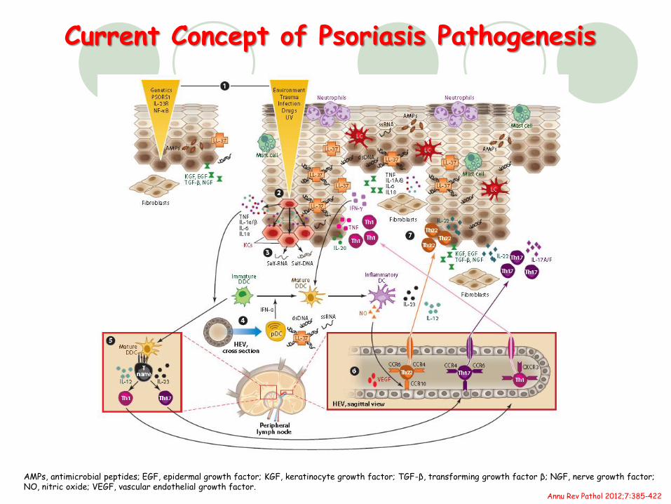
Current Concept of Psoriasis Pathogenesis
Annu Rev Pathol 2012;7:385-422
AMPs, antimicrobial peptides; EGF, epidermal growth factor; KGF, keratinocyte growth factor; TGF-β, transforming growth factor β; NGF, nerve growth factor; NO, nitric oxide; VEGF, vascular endothelial growth factor.
2021/9/23 10
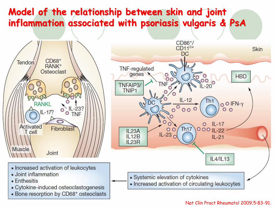
Model of the relationship between skin and joint inflammation associated with psoriasis vulgaris & PsA
Nat Clin Pract Rheumatol 2009;5:83-91.
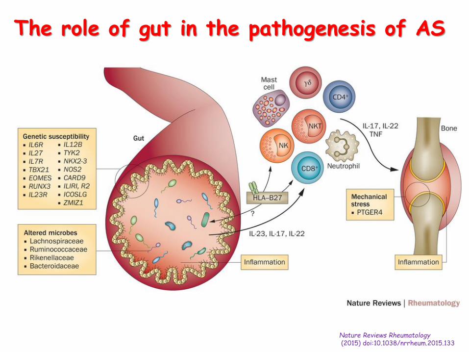
The role of gut in the pathogenesis of AS
Nature Reviews Rheumatology(2015) doi:10.1038/nrrheum.2015.133
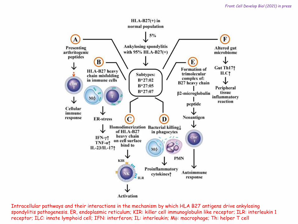
Intracellular pathways and their interactions in the mechanism by which HLA B27 antigens drive ankylosing spondylitis pathogenesis. ER, endoplasmic reticulum; KIR: killer cell immunoglobulin like receptor; ILR: interleukin 1 receptor; ILC: innate lymphoid cell; IFN: interferon; IL: interleukin; Mf: macrophage; Th: helper T cell
Front Cell Develop Biol (2021) in press
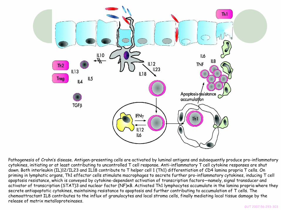
Pathogenesis of Crohn’s disease. Antigen-presenting cells are activated by luminal antigens and subsequently produce pro-inflammatory cytokines, initiating or at least contributing to uncontrolled T cell response. Anti-inflammatory T cell cytokine responses are shut down. Both interleukin (IL)12/IL23 and IL18 contribute to T helper cell 1 (Th1) differentiation of CD4 lamina propria T cells. On priming in lymphatic organs, Th1 effector cells stimulate macrophages to secrete further pro-inflammatory cytokines, inducing T cell apoptosis resistance, which is conveyed by cytokine-dependent activation of transcription factors—namely, signal transducer and activator of transcription (STAT)3 and nuclear factor (NF)κB. Activated Th1 lymphocytes accumulate in the lamina propria where they secrete antiapoptotic cytokines, maintaining resistance to apoptosis and further contributing to accumulation of T cells. The chemoattractant IL8 contributes to the influx of granulocytes and local stroma cells, finally mediating local tissue damage by the release of matrix metalloproteinases.
GUT 2007;56:293-303
2021/9/23 14
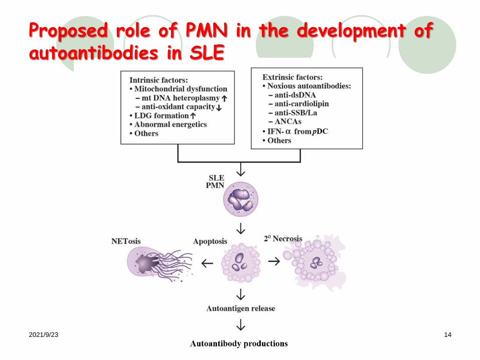
Proposed role of PMN in the development of autoantibodies in SLE
2021/9/23 15
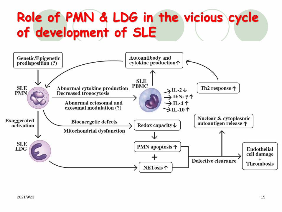
Role of PMN & LDG in the vicious cycle of development of SLE
2021/9/23 16
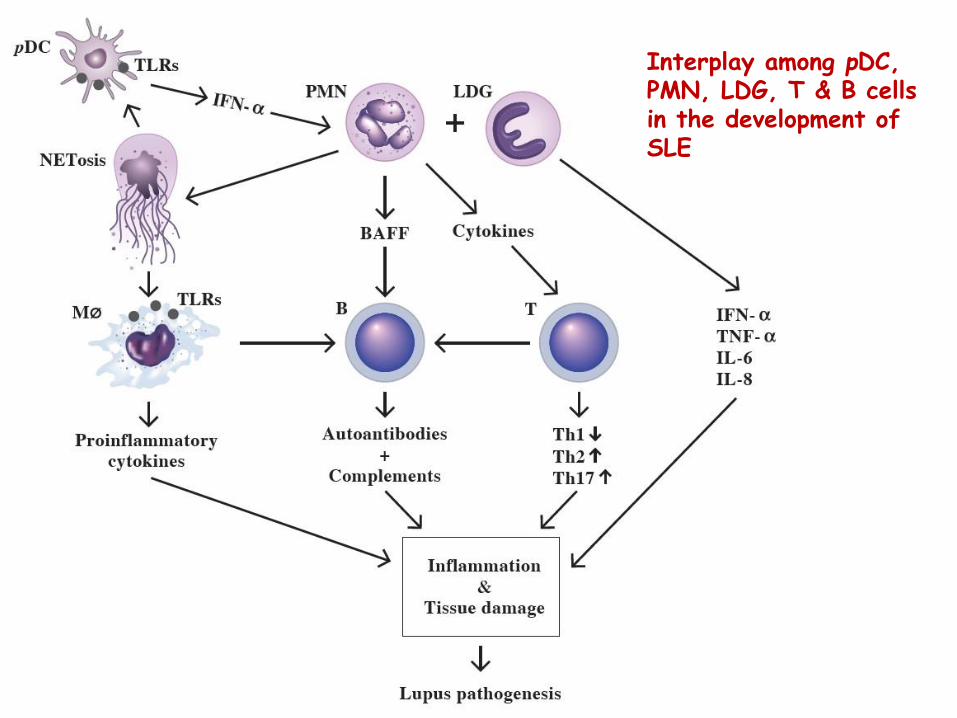
Interplay among pDC, PMN, LDG, T & B cells in the development of SLE
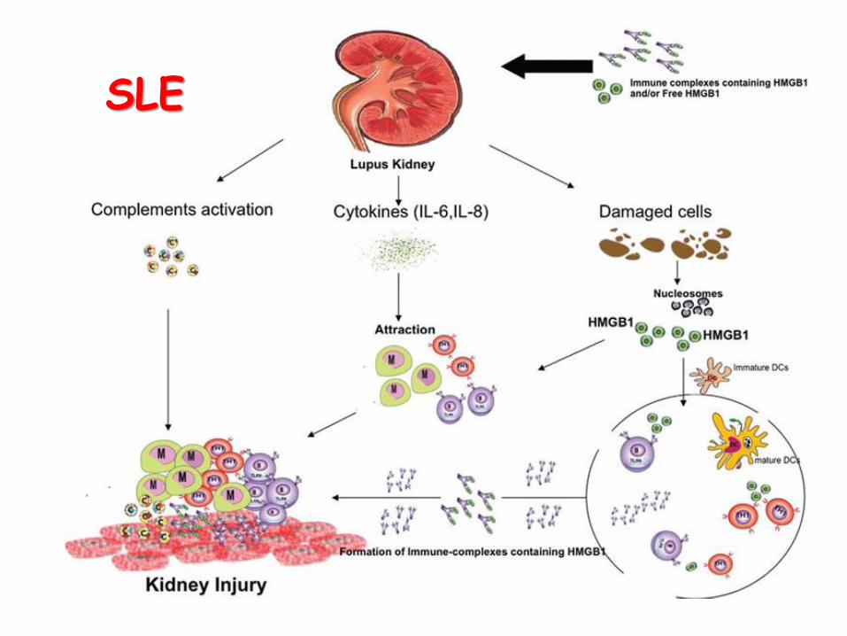
SLE
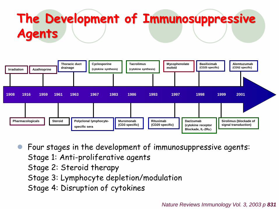
Nature Reviews Immunology Vol. 3, 2003 p 831
Irradiation Azathioprine
Thoracic duct
drainage
1908 1916 1959 1961 1963 1967 1983 1986 1993 1997 1998 1999 2001
Cyclosporine
(cytokine synthesis)
Pharmacologicals Steroid Polyclonal lymphocyte-
specific sera
Muromonab
(CD3 specific)
Tacrolimus
(cytokine synthesis)
Mycophenolate
mofetil
Rituximab
(CD20 specific)
Basiliximab(CD25 specific)
Alemtuzumab(CD52 specific)
Daclzumab
(cytokine receptor
Blockade, IL-2Ra)
Sirolimus (blockade of
signal transduction)
The Development of Immunosuppressive Agents
Four stages in the development of immunosuppressive agents:Stage 1: Anti-proliferative agentsStage 2: Steroid therapyStage 3: Lymphocyte depletion/modulationStage 4: Disruption of cytokines
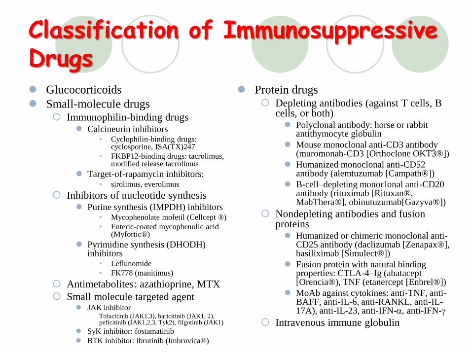
Classification of Immunosuppressive Drugs Glucocorticoids
Small-molecule drugs Immunophilin-binding drugs
Calcineurin inhibitors• Cyclophilin-binding drugs:
cyclosporine, ISA(TX)247
• FKBP12-binding drugs: tacrolimus, modified release tacrolimus
Target-of-rapamycin inhibitors: • sirolimus, everolimus
Inhibitors of nucleotide synthesis Purine synthesis (IMPDH) inhibitors
• Mycophenolate mofetil (Cellcept ® )
• Enteric-coated mycophenolic acid (Myfortic® )
Pyrimidine synthesis (DHODH) inhibitors
• Leflunomide
• FK778 (manitimus)
Antimetabolites: azathioprine, MTX
Small molecule targeted agent JAK inhibitor
Tofacitinib (JAK1,3), baricitinib (JAK1, 2), peficitinib (JAK1,2,3, Tyk2), filgotinib (JAK1)
SyK inhibitor: fostamatinib
BTK inhibitor: ibrutinib (Imbruvica® )
Protein drugs Depleting antibodies (against T cells, B
cells, or both) Polyclonal antibody: horse or rabbit
antithymocyte globulin
Mouse monoclonal anti-CD3 antibody (muromonab-CD3 [Orthoclone OKT3® ])
Humanized monoclonal anti-CD52 antibody (alemtuzumab [Campath® ])
B-cell–depleting monoclonal anti-CD20 antibody (rituximab [Rituxan® , MabThera® ], obinutuzumab[Gazyva® ])
Nondepleting antibodies and fusion proteins Humanized or chimeric monoclonal anti-
CD25 antibody (daclizumab [Zenapax® ], basiliximab [Simulect® ])
Fusion protein with natural binding properties: CTLA-4–Ig (abatacept[Orencia® ), TNF (etanercept [Enbrel® ])
MoAb against cytokines: anti-TNF, anti-BAFF, anti-IL-6, anti-RANKL, anti-IL-17A), anti-IL-23, anti-IFN-a, anti-IFN-g
Intravenous immune globulin
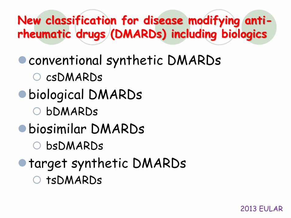
New classification for disease modifying anti-rheumatic drugs (DMARDs) including biologics
conventional synthetic DMARDs csDMARDs
biological DMARDs bDMARDs
biosimilar DMARDs bsDMARDs
target synthetic DMARDs tsDMARDs
2013 EULAR
TNF-a as a Target
Infliximab (Remicade® )
Etanercept (Enbel® )
Adalimumab (Humira® )
Golimumab (Simponi® )
Certolizumab pegol (Cimzia® )
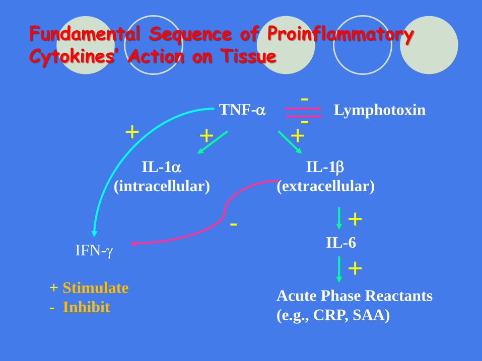
Fundamental Sequence of ProinflammatoryCytokines’ Action on Tissue
TNF-a
IL-1a
(intracellular)
IL-1b
(extracellular)
IL-6
Acute Phase Reactants
(e.g., CRP, SAA)
IFN-g
+
-
++
+
+
+ Stimulate
- Inhibit
Lymphotoxin--
IL-1b as A Target
Anakinra
Rilonacept
Canakinumab
……….
………….
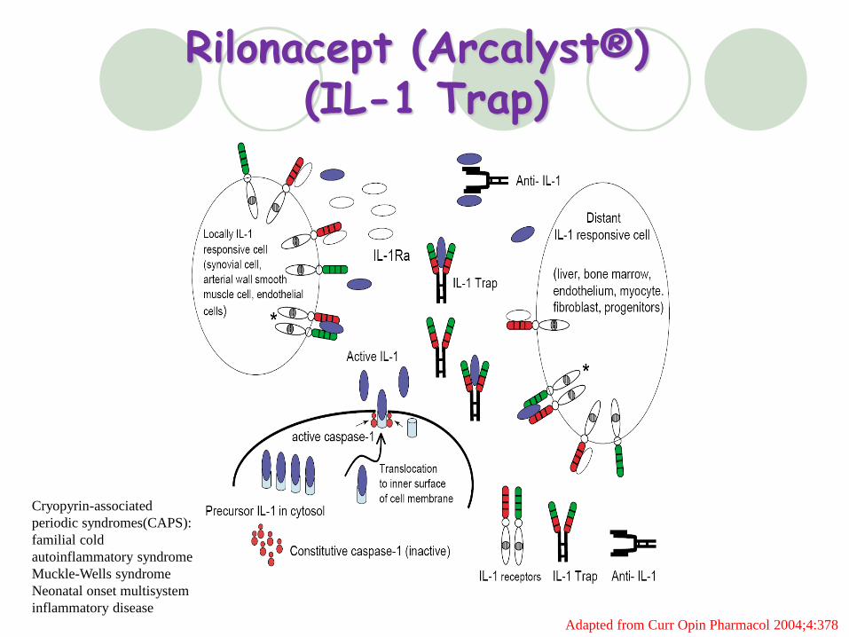
Adapted from Curr Opin Pharmacol 2004;4:378
Rilonacept (Arcalyst® ) (IL-1 Trap)
Cryopyrin-associated
periodic syndromes(CAPS):
familial cold
autoinflammatory syndrome
Muckle-Wells syndrome
Neonatal onset multisystem
inflammatory disease
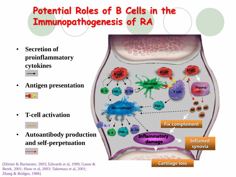
Potential Roles of B Cells in the Immunopathogenesis of RA
• Secretion of
proinflammatory
cytokines
• Antigen presentation
• T-cell activation
• Autoantibody production
and self-perpetuation
(Dörner & Burmester, 2003; Edwards et al, 1999; Gause &
Berek, 2001; Shaw et al, 2003; Takemura et al, 2001;
Zhang & Bridges, 1986)
Cartilage loss
IL-6
B cell
T cell
Macrophage
Dendritic cell
IL-10TNF-a
TNF-aIL-10
RF
Fix complement
Inflamed synovia
TNF-a
B cell
IL-6
B cell
Plasma cell
RF RF
RF
RF
IL-1
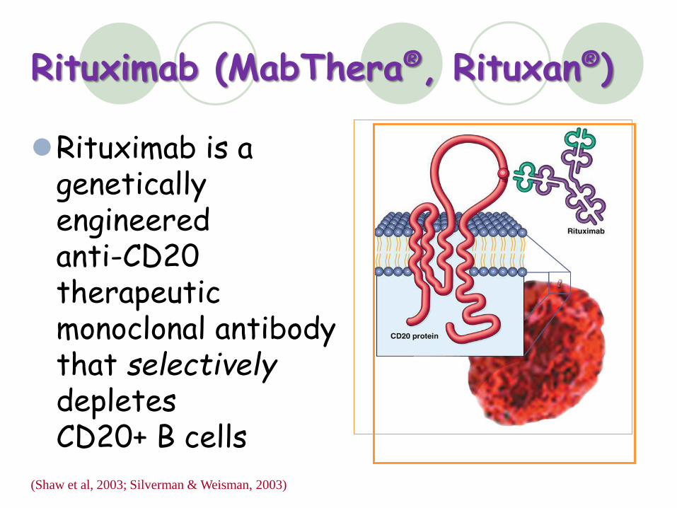
Rituximab (MabThera® , Rituxan® )
Rituximab is agenetically engineered anti-CD20 therapeutic monoclonal antibody that selectivelydepletes CD20+ B cells
(Shaw et al, 2003; Silverman & Weisman, 2003)
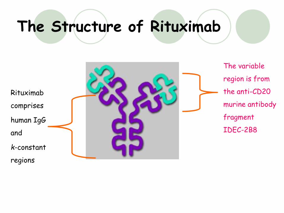
The Structure of Rituximab
Rituximab
comprises
human IgG
and
k-constant
regions
The variable
region is from
the anti-CD20
murine antibody
fragment
IDEC-2B8
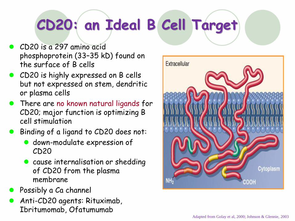
CD20: an Ideal B Cell Target
CD20 is a 297 amino acid phosphoprotein (33–35 kD) found on the surface of B cells
CD20 is highly expressed on B cells but not expressed on stem, dendriticor plasma cells
There are no known natural ligands for CD20; major function is optimizing B cell stimulation
Binding of a ligand to CD20 does not:
down-modulate expression of CD20
cause internalisation or shedding of CD20 from the plasma membrane
Possibly a Ca channel
Anti-CD20 agents: Rituximab, Ibritumomab, Ofatumumab
Adapted from Golay et al, 2000; Johnson & Glennie, 2003
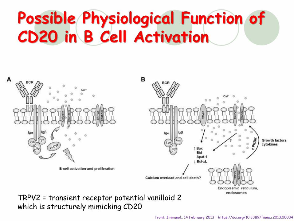
Possible Physiological Function of CD20 in B Cell Activation
TRPV2 = transient receptor potential vanilloid 2which is structurely mimicking CD20
Front. Immunol., 14 February 2013 | https://doi.org/10.3389/fimmu.2013.00034
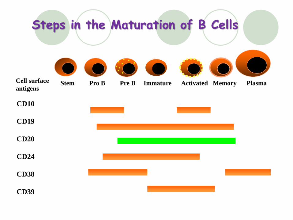
Steps in the Maturation of B Cells
Stem Pro B Pre B Immature Activated Memory Plasma
CD10
CD19
CD20
CD24
CD38
CD39
Cell surface
antigens
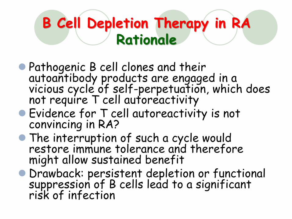
B Cell Depletion Therapy in RARationale
Pathogenic B cell clones and their autoantibody products are engaged in a vicious cycle of self-perpetuation, which does not require T cell autoreactivity
Evidence for T cell autoreactivity is not convincing in RA?
The interruption of such a cycle would restore immune tolerance and therefore might allow sustained benefit
Drawback: persistent depletion or functional suppression of B cells lead to a significant risk of infection
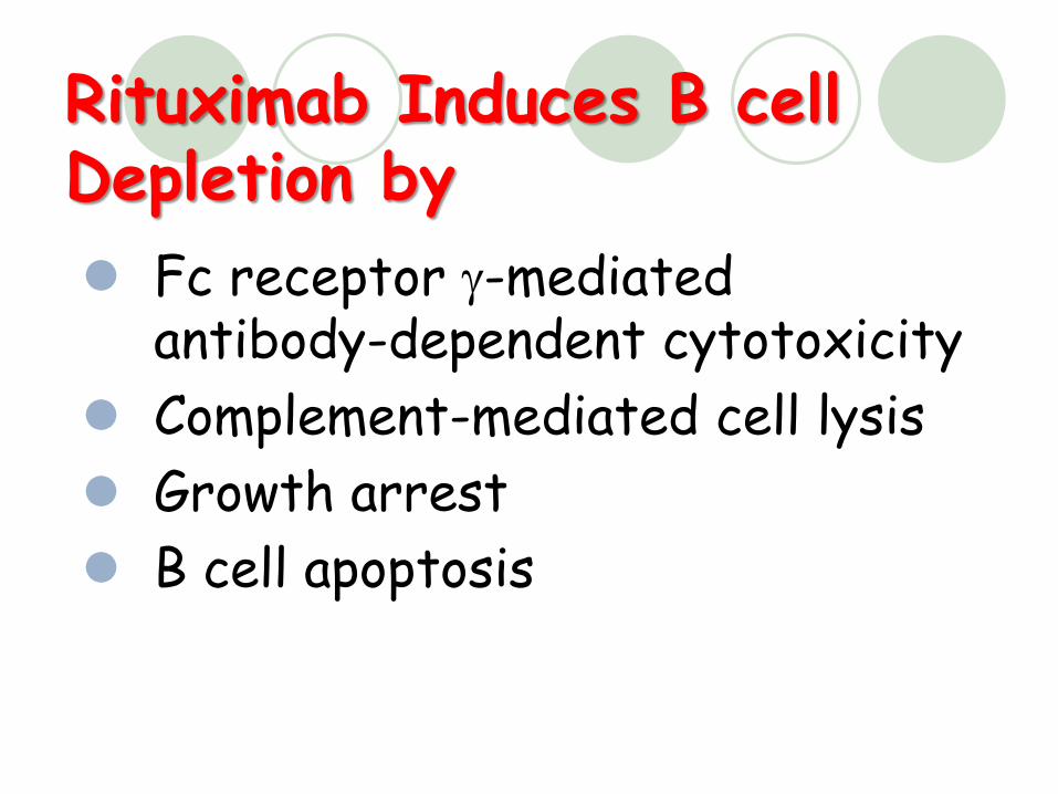
Rituximab Induces B cell Depletion by
Fc receptor g-mediated antibody-dependent cytotoxicity
Complement-mediated cell lysis
Growth arrest
B cell apoptosis
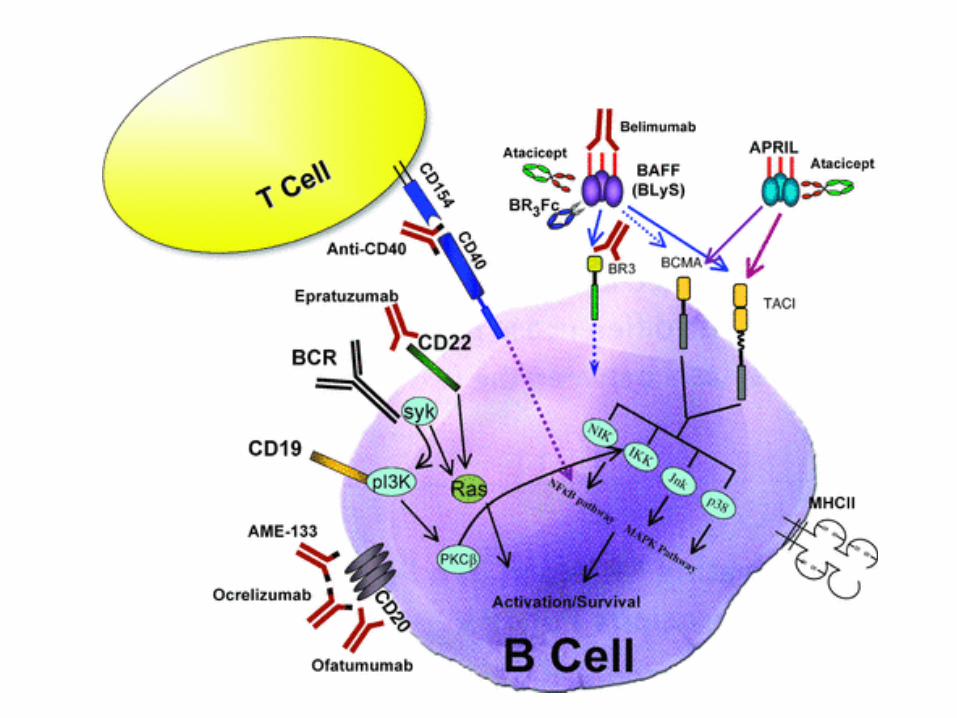
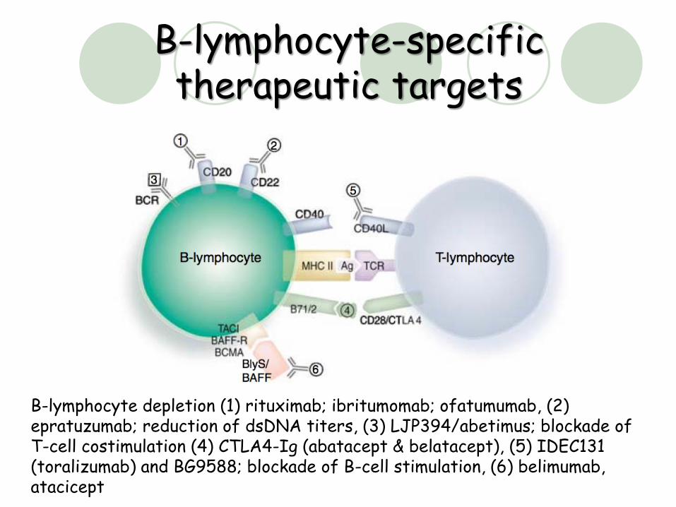
B-lymphocyte-specific therapeutic targets
B-lymphocyte depletion (1) rituximab; ibritumomab; ofatumumab, (2) epratuzumab; reduction of dsDNA titers, (3) LJP394/abetimus; blockade of T-cell costimulation (4) CTLA4-Ig (abatacept & belatacept), (5) IDEC131 (toralizumab) and BG9588; blockade of B-cell stimulation, (6) belimumab, atacicept
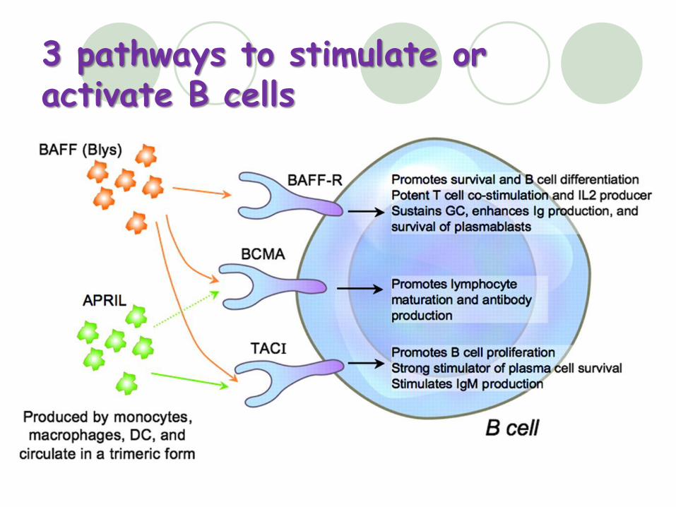
3 pathways to stimulate or activate B cells
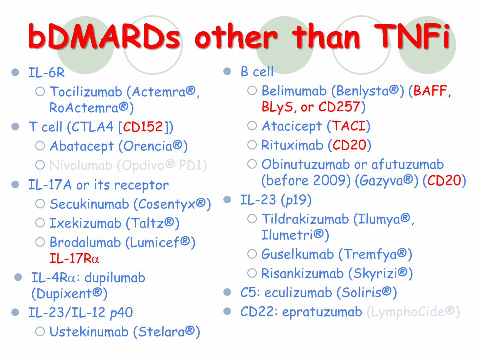
bDMARDs other than TNFi IL-6R
Tocilizumab (Actemra® , RoActemra® )
T cell (CTLA4 [CD152])
Abatacept (Orencia® )
Nivolumab (Opdivo® PD1)
IL-17A or its receptor
Secukinumab (Cosentyx® )
Ixekizumab (Taltz® )
Brodalumab (Lumicef® )IL-17Ra
IL-4Ra: dupilumab(Dupixent® )
IL-23/IL-12 p40
Ustekinumab (Stelara® )
B cell
Belimumab (Benlysta® ) (BAFF,BLyS, or CD257)
Atacicept (TACI)
Rituximab (CD20)
Obinutuzumab or afutuzumab (before 2009) (Gazyva® ) (CD20)
IL-23 (p19)
Tildrakizumab (Ilumya® , Ilumetri® )
Guselkumab (Tremfya® )
Risankizumab (Skyrizi® )
C5: eculizumab (Soliris® )
CD22: epratuzumab (LymphoCide® )
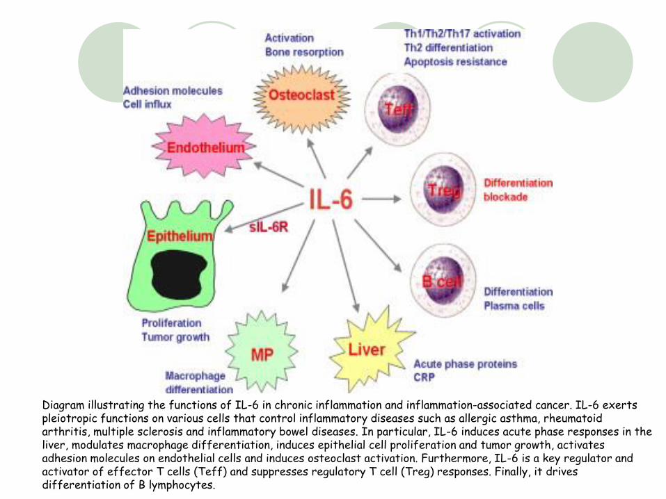
Diagram illustrating the functions of IL-6 in chronic inflammation and inflammation-associated cancer. IL-6 exerts pleiotropic functions on various cells that control inflammatory diseases such as allergic asthma, rheumatoid arthritis, multiple sclerosis and inflammatory bowel diseases. In particular, IL-6 induces acute phase responses in the liver, modulates macrophage differentiation, induces epithelial cell proliferation and tumor growth, activates adhesion molecules on endothelial cells and induces osteoclast activation. Furthermore, IL-6 is a key regulator and activator of effector T cells (Teff) and suppresses regulatory T cell (Treg) responses. Finally, it drives differentiation of B lymphocytes.
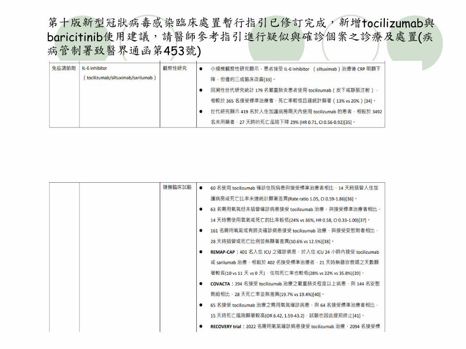
第十版新型冠狀病毒感染臨床處置暫行指引已修訂完成,新增tocilizumab與baricitinib使用建議,請醫師參考指引進行疑似與確診個案之診療及處置(疾病管制署致醫界通函第453號)
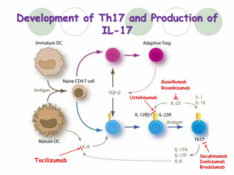
Development of Th17 and Production of IL-17
Tocilizumab
Ustekinumab
Guselkumab
SecukinumabIxekizumabBrodalumab
Risankizumab
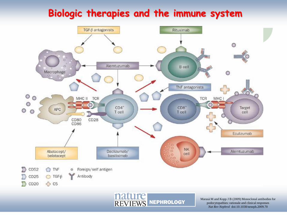
Biologic therapies and the immune system
Marasà M and Kopp J B (2009) Monoclonal antibodies for
podocytopathies: rationale and clinical responses
Nat Rev Nephrol doi:10.1038/nrneph.2009.70
Small Molecules as Targets
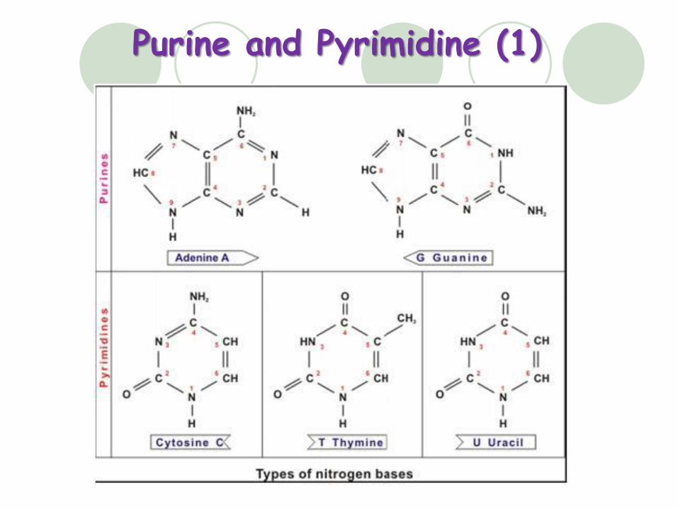
Purine and Pyrimidine (1)
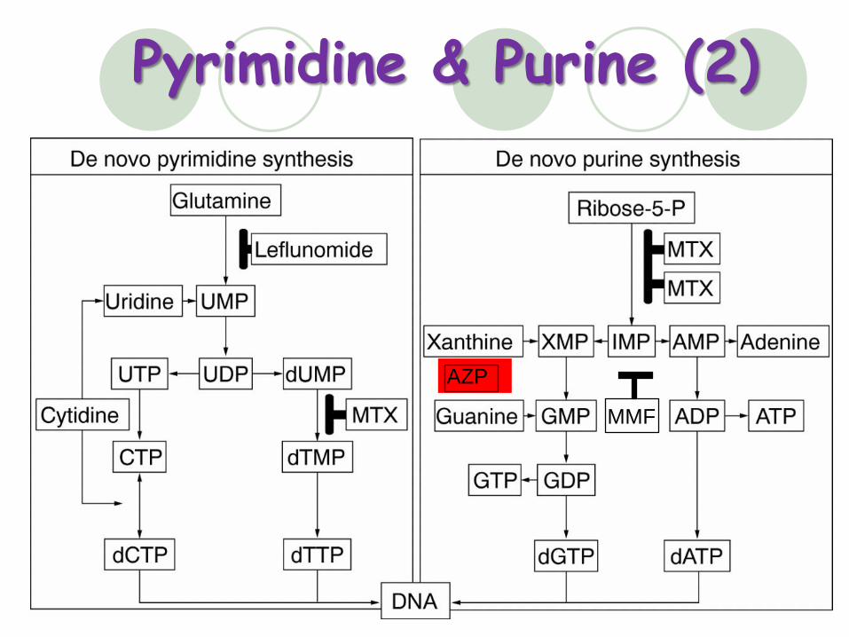
Pyrimidine & Purine (2)
MMF
AZP

Metabolism of Azathioprine
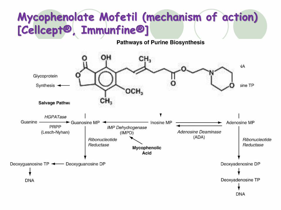
Mycophenolate Mofetil (mechanism of action)[Cellcept® , Immunfine® ]
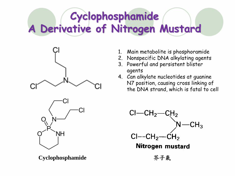
CyclophosphamideA Derivative of Nitrogen Mustard
1. Main metabolite is phosphoramide2. Nonspecific DNA alkylating agents 3. Powerful and persistent blister
agents4. Can alkylate nucleotides at guanine
N7 position, causing cross linking of the DNA strand, which is fatal to cell
Cyclophosphamide 芥子氣
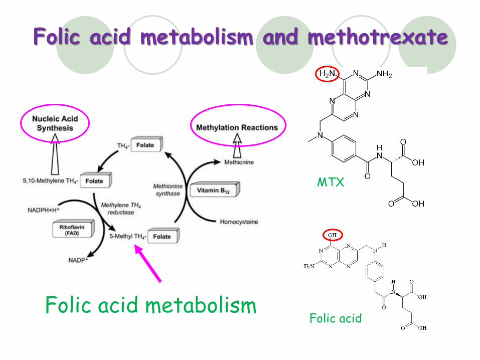
Folic acid metabolism and methotrexate
MTX
Folic acid metabolismFolic acid
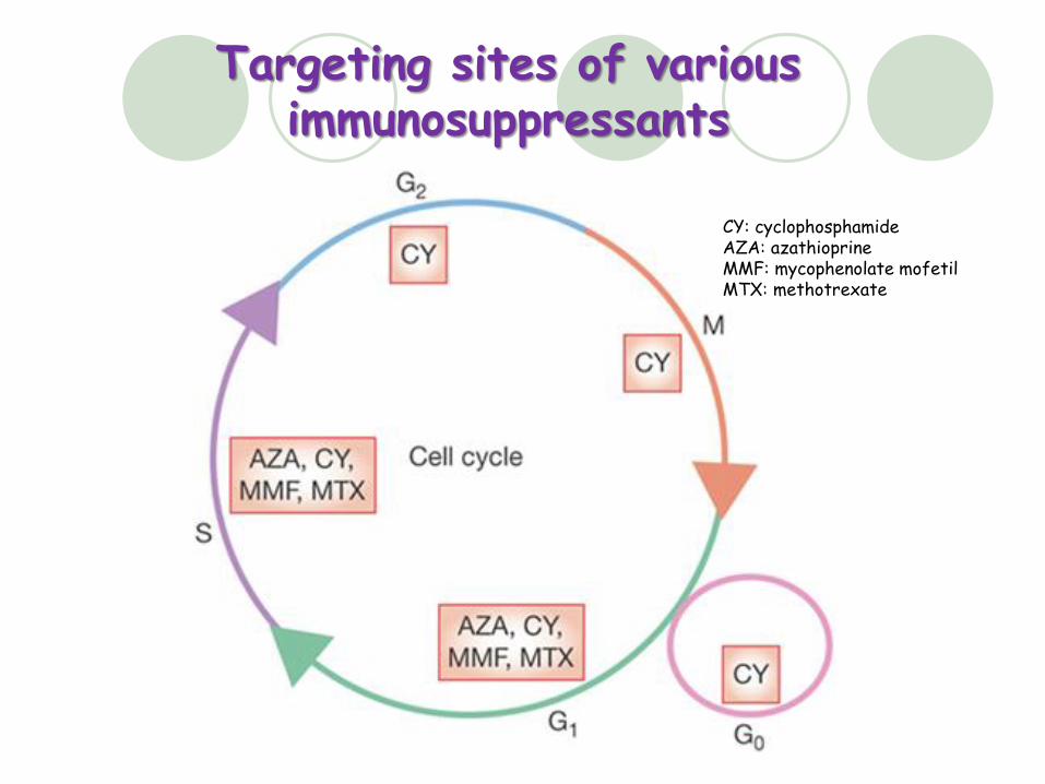
Targeting sites of various immunosuppressants
CY: cyclophosphamideAZA: azathioprineMMF: mycophenolate mofetilMTX: methotrexate
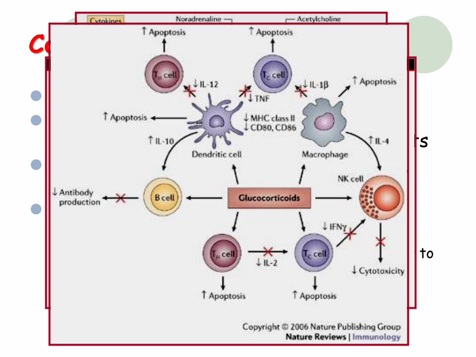
Corticosteroids
Anti-inflammatory and immunosuppressiveAgonists of glucocorticoid receptors, at
higher doses, receptor-independent effects Inhibit T-cell lymphokine (IL-1 and TNF)
production Therapeutic use
Pulse therapy: high-dose i.vi.v bolus methylprednisolone (1 g X 3 D) taper to
maintenance dose after resolution
Oral therapy: middle to lowLocal injection
Mammalian Target of Rapamycin(mTOR)
Alternative term: mechanistic target of rapamycin, FK506 binding protein 12-rapamycin associated protein 1 (FRAP1)
Encoded gene: FRAP1
In PI-3K protein family
A ser/thr PK regulating cell growth, proliferation, motility, & survival, protein synthesis, and transcription
Calcineurin inhibitors
Cyclophilin-binding drugs: • Cyclosporine
• ISA(TX)247
FKBP12-binding drugs: • Tacrolimus
• Modified release tacrolimus
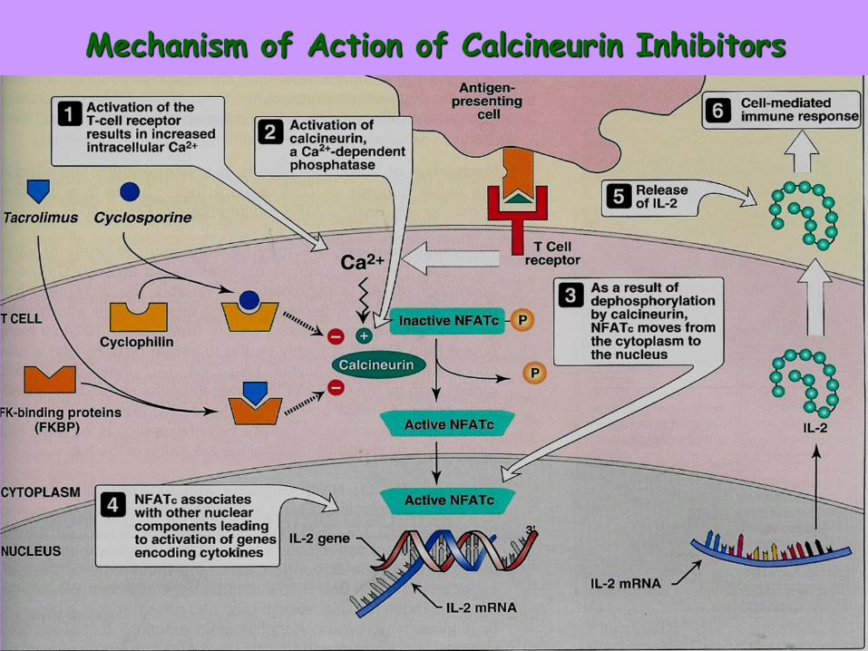
Mechanism of Action of Calcineurin Inhibitors
Cyclosporine A cornerstone of immunosuppression in autoimmune diseases and
transplantation A pro-drug that engages cyclophilin Binding to an intracellular protein of the immunophilin family,
forming a complex that then engages calcineurin Adverse effects
dependent on concentration of the drug nephrotoxicity, hypertension, hyperlipidemia, gingival hyperplasia,
hirsutism, and tremor can also induce the hemolytic–uremic syndrome and post-
transplantation diabetes mellitus. Recent development
Monitoring of the peak cyclosporine levels two hours after administration can better reflect the exact exposure status of the body to the drug
A chemically modified cyclosporine, ISA(TX)247, is under development
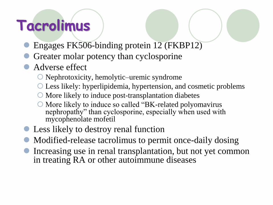
Tacrolimus Engages FK506-binding protein 12 (FKBP12)
Greater molar potency than cyclosporine
Adverse effect Nephrotoxicity, hemolytic–uremic syndrome
Less likely: hyperlipidemia, hypertension, and cosmetic problems
More likely to induce post-transplantation diabetes
More likely to induce so called “BK-related polyomavirus nephropathy” than cyclosporine, especially when used with mycophenolate mofetil
Less likely to destroy renal function
Modified-release tacrolimus to permit once-daily dosing
Increasing use in renal transplantation, but not yet common in treating RA or other autoimmune diseases
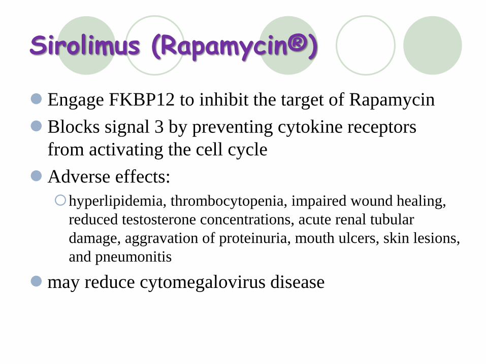
Sirolimus (Rapamycin® )
Engage FKBP12 to inhibit the target of Rapamycin
Blocks signal 3 by preventing cytokine receptors
from activating the cell cycle
Adverse effects:
hyperlipidemia, thrombocytopenia, impaired wound healing,
reduced testosterone concentrations, acute renal tubular
damage, aggravation of proteinuria, mouth ulcers, skin lesions,
and pneumonitis
may reduce cytomegalovirus disease
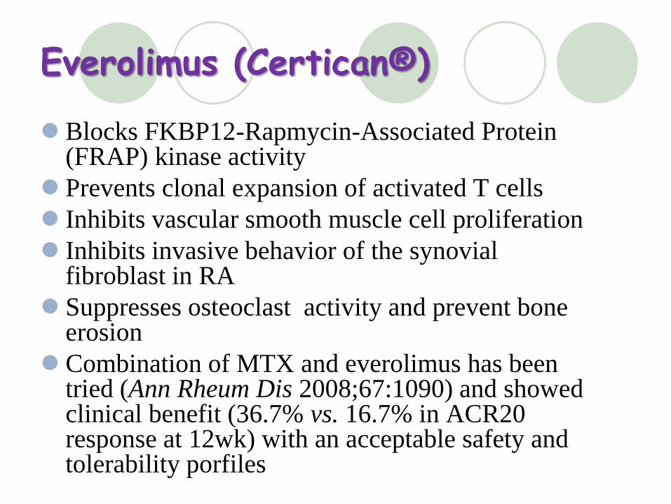
Everolimus (Certican® )
Blocks FKBP12-Rapmycin-Associated Protein (FRAP) kinase activity
Prevents clonal expansion of activated T cells
Inhibits vascular smooth muscle cell proliferation
Inhibits invasive behavior of the synovial fibroblast in RA
Suppresses osteoclast activity and prevent bone erosion
Combination of MTX and everolimus has been tried (Ann Rheum Dis 2008;67:1090) and showed clinical benefit (36.7% vs. 16.7% in ACR20 response at 12wk) with an acceptable safety and tolerability porfiles
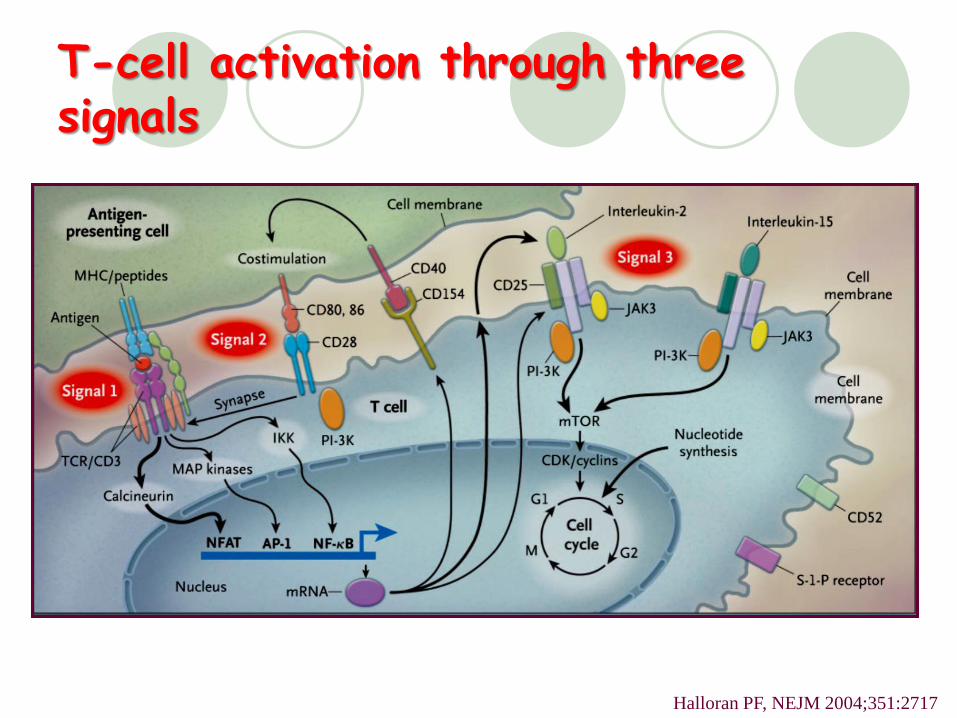
T-cell activation through three signals
Halloran PF, NEJM 2004;351:2717
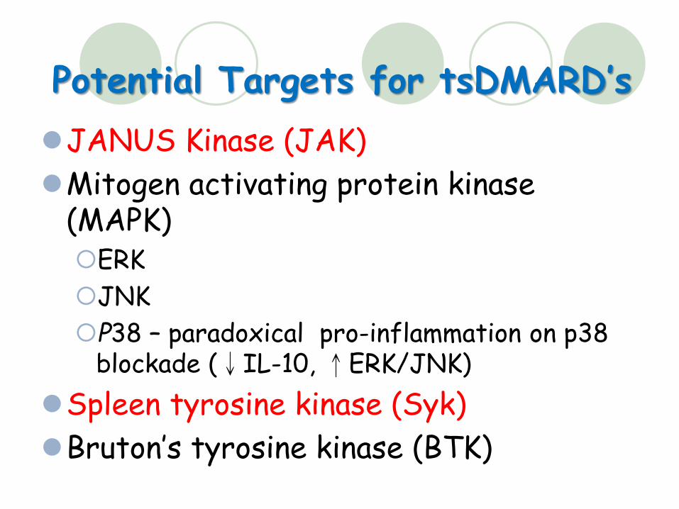
Potential Targets for tsDMARD’s
JANUS Kinase (JAK)
Mitogen activating protein kinase(MAPK)ERK
JNK
P38 – paradoxical pro-inflammation on p38 blockade (↓IL-10, ↑ERK/JNK)
Spleen tyrosine kinase (Syk)
Bruton’s tyrosine kinase (BTK)
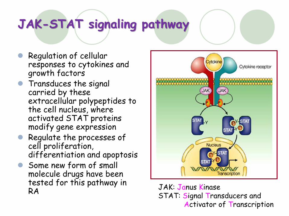
JAK-STAT signaling pathway
Regulation of cellular responses to cytokines and growth factors
Transduces the signal carried by these extracellular polypeptides to the cell nucleus, where activated STAT proteins modify gene expression
Regulate the processes of cell proliferation, differentiation and apoptosis
Some new form of small molecule drugs have been tested for this pathway in RA
PP
JAK: Janus KinaseSTAT: Signal Transducers and
Activator of Transcription
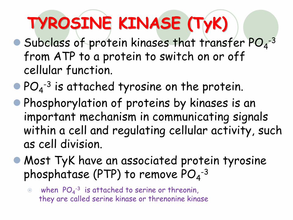
TYROSINE KINASE (TyK) Subclass of protein kinases that transfer PO4
-3
from ATP to a protein to switch on or off cellular function.
PO4-3 is attached tyrosine on the protein.
Phosphorylation of proteins by kinases is an important mechanism in communicating signals within a cell and regulating cellular activity, such as cell division.
Most TyK have an associated protein tyrosine phosphatase (PTP) to remove PO4
-3
when PO4-3 is attached to serine or threonin,
they are called serine kinase or threnonine kinase
Rheumatology 2013;52:1156
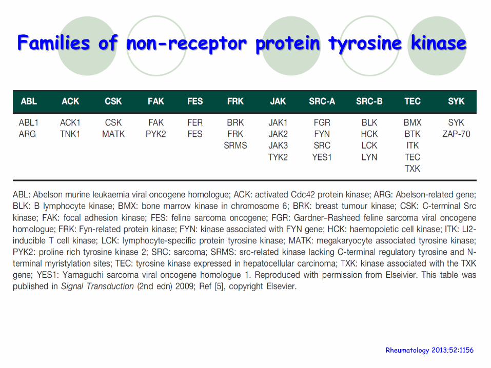
Families of non-receptor protein tyrosine kinase
Janus kinase (JAK)
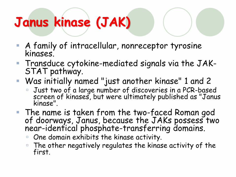
A family of intracellular, nonreceptor tyrosine kinases.
Transduce cytokine-mediated signals via the JAK-STAT pathway.
Was initially named "just another kinase" 1 and 2 Just two of a large number of discoveries in a PCR-based
screen of kinases, but were ultimately published as "Janus kinase".
The name is taken from the two-faced Roman god of doorways, Janus, because the JAKs possess two near-identical phosphate-transferring domains. One domain exhibits the kinase activity. The other negatively regulates the kinase activity of the
first.
The Janus Kinase Family
Janus kinase 1 (JAK1)
Janus kinase 2 (JAK2)
Janus kinase 3 (JAK3)
Tyrosine kinase 2 (TYK2)
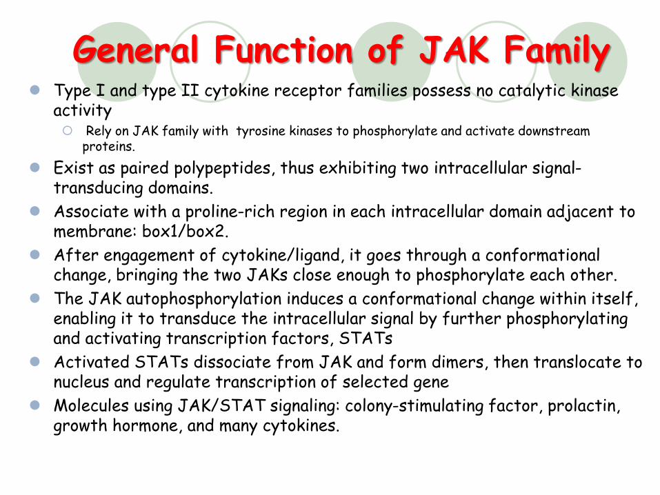
General Function of JAK Family Type I and type II cytokine receptor families possess no catalytic kinase
activity Rely on JAK family with tyrosine kinases to phosphorylate and activate downstream
proteins.
Exist as paired polypeptides, thus exhibiting two intracellular signal-transducing domains.
Associate with a proline-rich region in each intracellular domain adjacent to membrane: box1/box2.
After engagement of cytokine/ligand, it goes through a conformational change, bringing the two JAKs close enough to phosphorylate each other.
The JAK autophosphorylation induces a conformational change within itself, enabling it to transduce the intracellular signal by further phosphorylating and activating transcription factors, STATs
Activated STATs dissociate from JAK and form dimers, then translocate to nucleus and regulate transcription of selected gene
Molecules using JAK/STAT signaling: colony-stimulating factor, prolactin, growth hormone, and many cytokines.
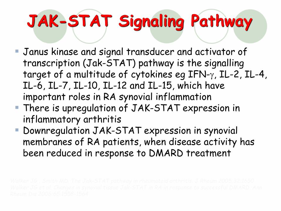
Janus kinase and signal transducer and activator of transcription (Jak-STAT) pathway is the signalling target of a multitude of cytokines eg IFN-g, IL-2, IL-4, IL-6, IL-7, IL-10, IL-12 and IL-15, which have important roles in RA synovial inflammation
There is upregulation of JAK-STAT expression in inflammatory arthritis
Downregulation JAK-STAT expression in synovial membranes of RA patients, when disease activity has been reduced in response to DMARD treatment
JAK-STAT Signaling Pathway
Walker JG , Smith MD. The Jak-STAT pathway in rheumatoid arthritis. J Rheum 2005;32:1650. Walker JG et al. Changes in synovial tissue Jak-STAT in RA in response to successful DMARD. Ann Rheum Dis 2006:65:1558-1564
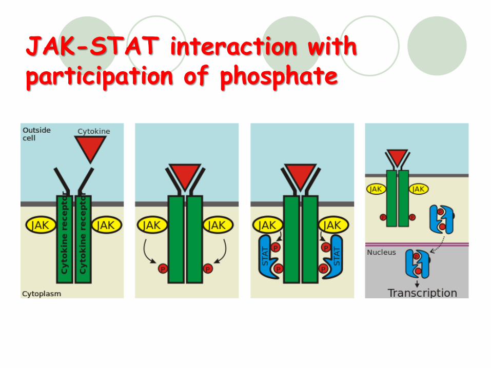
JAK-STAT interaction with participation of phosphate
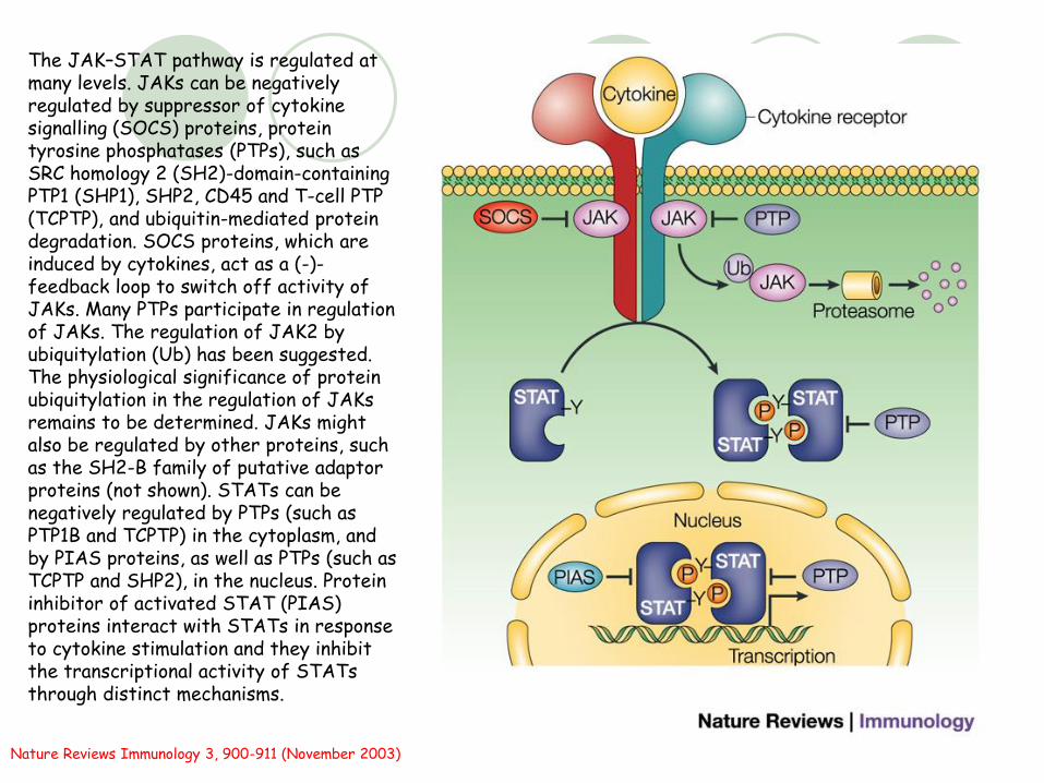
The JAK–STAT pathway is regulated at many levels. JAKs can be negatively regulated by suppressor of cytokine signalling (SOCS) proteins, protein tyrosine phosphatases (PTPs), such as SRC homology 2 (SH2)-domain-containing PTP1 (SHP1), SHP2, CD45 and T-cell PTP (TCPTP), and ubiquitin-mediated protein degradation. SOCS proteins, which are induced by cytokines, act as a (-)-feedback loop to switch off activity of JAKs. Many PTPs participate in regulation of JAKs. The regulation of JAK2 by ubiquitylation (Ub) has been suggested. The physiological significance of protein ubiquitylation in the regulation of JAKs remains to be determined. JAKs might also be regulated by other proteins, such as the SH2-B family of putative adaptor proteins (not shown). STATs can be negatively regulated by PTPs (such as PTP1B and TCPTP) in the cytoplasm, and by PIAS proteins, as well as PTPs (such as TCPTP and SHP2), in the nucleus. Protein inhibitor of activated STAT (PIAS) proteins interact with STATs in response to cytokine stimulation and they inhibit the transcriptional activity of STATs through distinct mechanisms.
Nature Reviews Immunology 3, 900-911 (November 2003)
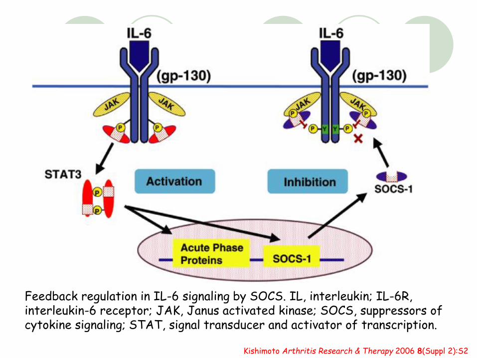
Feedback regulation in IL-6 signaling by SOCS. IL, interleukin; IL-6R, interleukin-6 receptor; JAK, Janus activated kinase; SOCS, suppressors of cytokine signaling; STAT, signal transducer and activator of transcription.
Kishimoto Arthritis Research & Therapy 2006 8(Suppl 2):S2
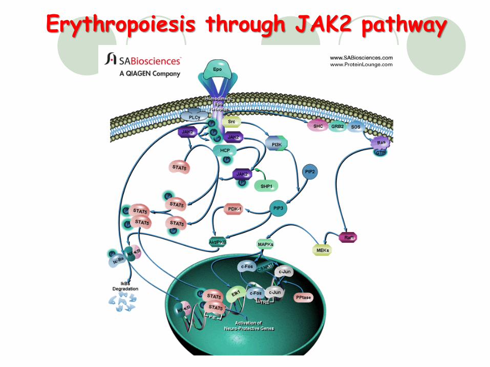
Erythropoiesis through JAK2 pathway
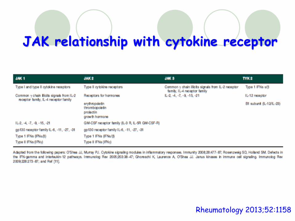
JAK relationship with cytokine receptor
Rheumatology 2013;52:1158
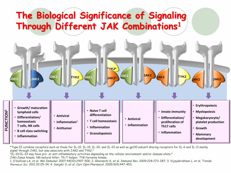
The Biological Significance of Signaling Through Different JAK Combinations1
73
*Type II cytokine receptors such as those for IL-10, IL-19, IL-20, and IL-22 as well as gp130 subunit sharing receptors for IL-6 and IL-11 mainly signal through JAK1, but also associate with JAK2 and TYK2.2†IL-10/IL-22 may have pro- or anti-inflammatory activities depending on the cellular environment and/or disease state.4
JAK=Janus kinase; NK=natural killer; Th=T helper; TYK=tyrosine kinase.1. O’Sullivan LA, et al. Mol Immunol. 2007;44(10):2497-506; 2. Ghoreschi K, et al. Immunol Rev. 2009;228:273-287; 3. Vijayakrishnan L, et al. Trends Pharmacol Sci. 2011;32:25-34; 4. Sanjabi S, et al. Curr Opin Pharmacol. 2009;9(4):447-453.
• Erythropoiesis
• Myelopoiesis
• Megakaryocyte/ platelet production
• Growth
• Mammary development
• Innate immunity
• Differentiation/ proliferation of Th17 cells
• Inflammation
• Antiviral
• Inflammation
• Growth/ maturation lymphoid cells
• Differentiation/ homeostasis T cells, NK cells
• B cell class switching
• Inflammation
• Naive T cell differentiation
• T cell homeostasis
• Inflammation
• Granulopoiesis
• Antiviral
• Inflammation†
• Antitumor
FU
NC
TIO
N3
JAK1 JAK3JAK1 TYK2
TYK2*
JAK2JAK1
JAK1JAK2* JAK2 TYK2
JAK2 JAK2
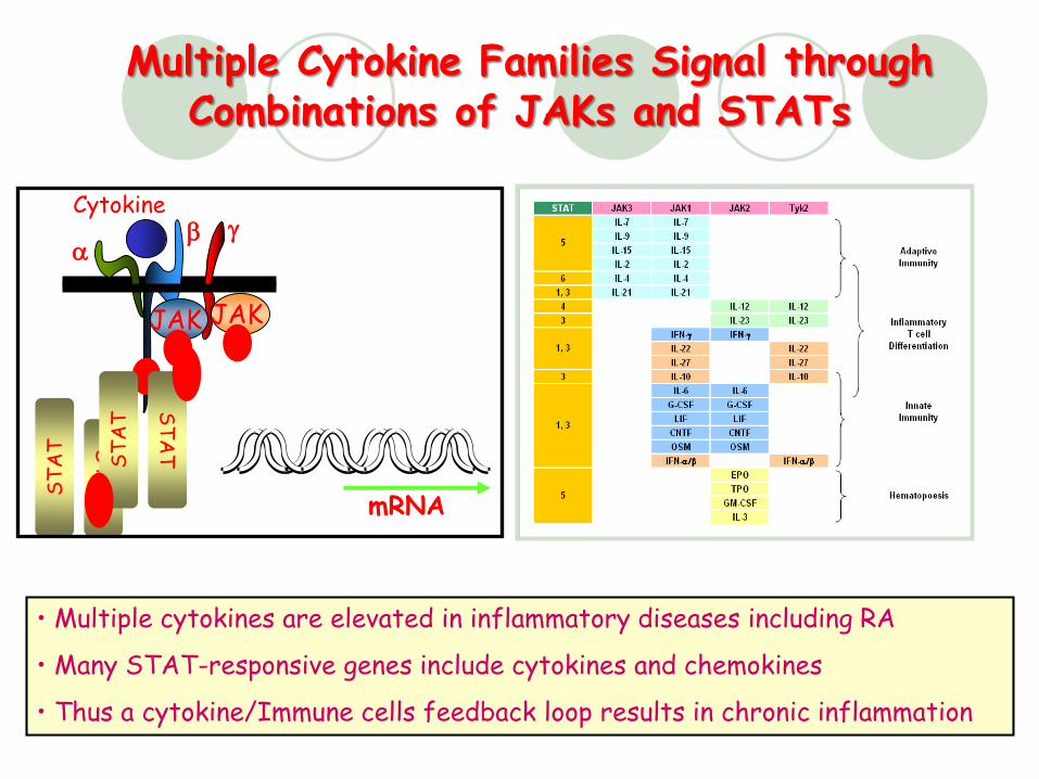
Multiple Cytokine Families Signal through Combinations of JAKs and STATs
JAK JAK
Cytokinegb
mRNA
a
PPP
ST
AT S
TA
T
ST
AT
ST
AT
P
P
• Multiple cytokines are elevated in inflammatory diseases including RA
• Many STAT-responsive genes include cytokines and chemokines
• Thus a cytokine/Immune cells feedback loop results in chronic inflammation
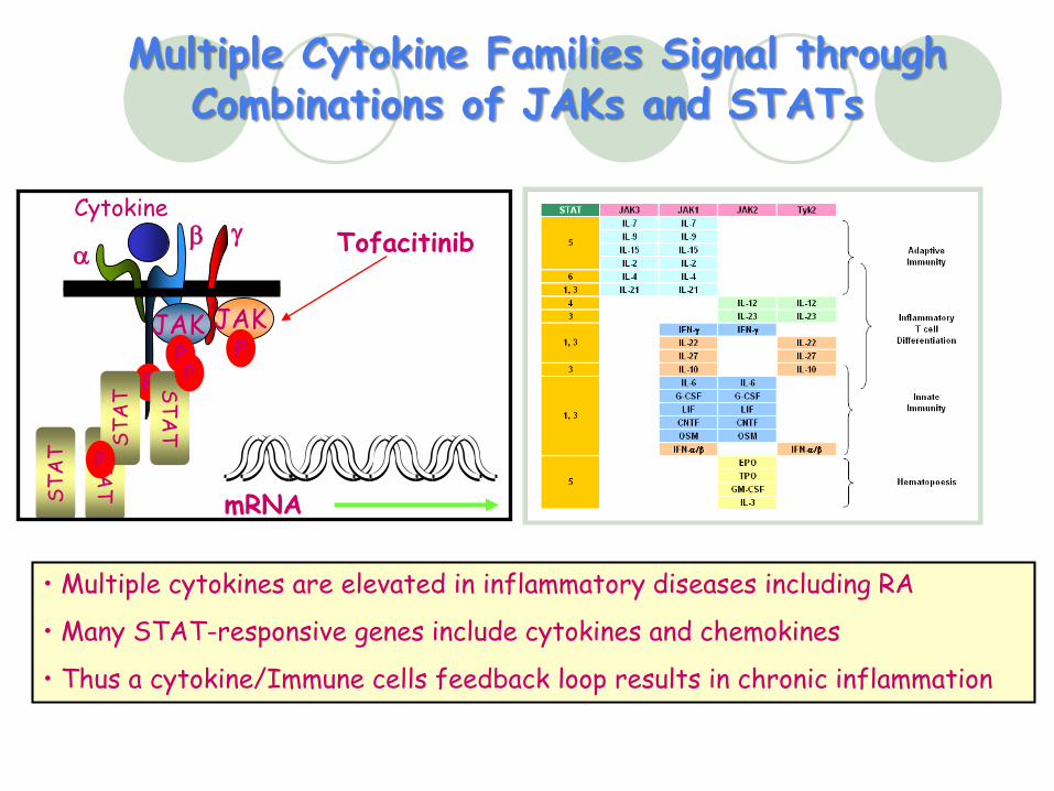
Multiple Cytokine Families Signal through Combinations of JAKs and STATs
JAK JAK
Tofacitinib
Cytokinegb
mRNA
a
PPP
ST
AT
ST
AT
ST
AT
ST
AT
P
P
• Multiple cytokines are elevated in inflammatory diseases including RA
• Many STAT-responsive genes include cytokines and chemokines
• Thus a cytokine/Immune cells feedback loop results in chronic inflammation
76
Tofacitinib
Tofacitinib is a selective inhibitor of Janus Kinases (JAKs)
Specificity at cellular level shows inhibition of JAK 1 and 3
Potent inhibitor of gC cytokine receptor signaling which requires both JAK3 and JAK1
At efficacious exposures in RA disease models, CP-690,550 shows: Reduction in systemic inflammation (cytokines, chemokines, STAT1
responsive genes)
Reduction in inflammatory cell infiltration (Mf, T cells, NK & NKT cells)
Due to potential for JAK/STAT pathway to affect multiple cytokines, efficacy in RA may be achieved without complete inhibition of any given cytokine for the entire dosing interval
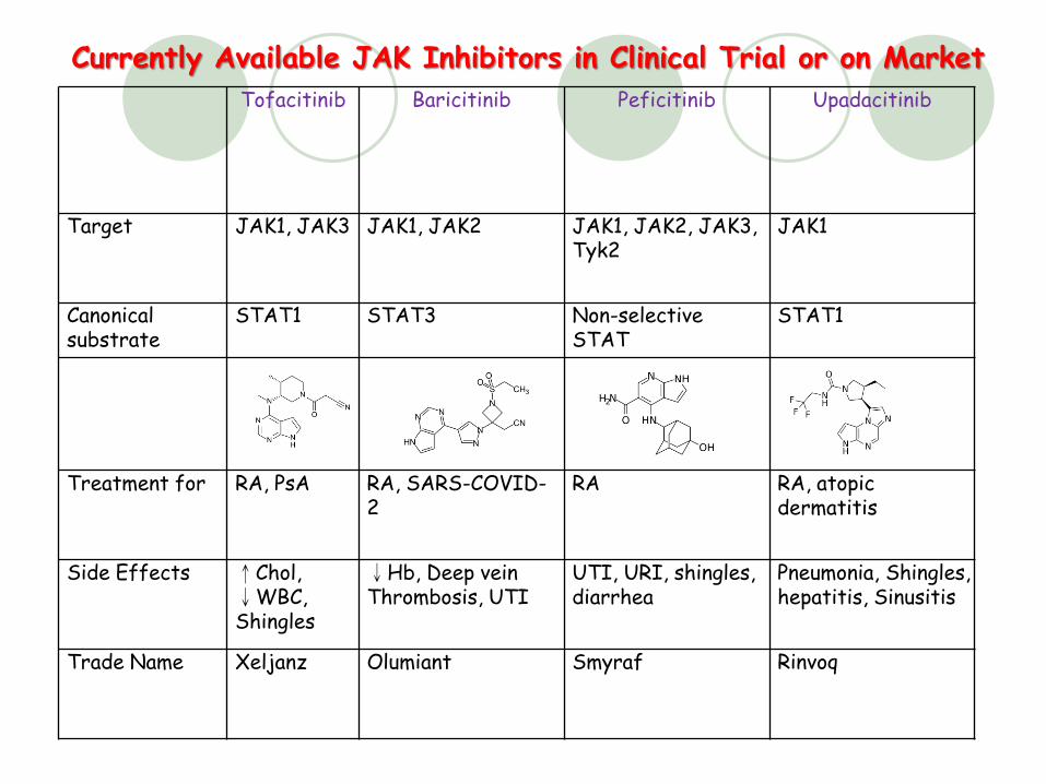
Tofacitinib Baricitinib Peficitinib Upadacitinib
Target JAK1, JAK3 JAK1, JAK2 JAK1, JAK2, JAK3, Tyk2
JAK1
Canonical substrate
STAT1 STAT3 Non-selective STAT
STAT1
Treatment for RA, PsA RA, SARS-COVID-2
RA RA, atopic dermatitis
Side Effects ↑Chol, ↓WBC, Shingles
↓Hb, Deep vein Thrombosis, UTI
UTI, URI, shingles, diarrhea
Pneumonia, Shingles, hepatitis, Sinusitis
Trade Name Xeljanz Olumiant Smyraf Rinvoq
Currently Available JAK Inhibitors in Clinical Trial or on Market
Upadacitinib
Upadacitinib is a second generation Janus kinase inhibitor that is selective for the JAK1 subtype
Reaches highest conc. after 2~4hrs; Not fat soluble; Steady-state 4 days; accumulation is minimal; No first pass effect; 52% bound to plasma proteins; metabolized by CYP3A4 through oxidation to –COOH & glucuronidation(M4).
Spleen Tyrosine Kinase (Syk)
Along with Zap-70, is a member of the Syk family of tyrosine kinases.
A family of non-receptor cytoplasmic tyrosine kinases with a dual SH2 domain separated by a linker domain.
Syk/Zap-70 is expressed in hematopoietic cells (esp. B & T cells, primarily involved in adaptive immunity) ; Syk alone is expressed in a variety of other tissues.
Syk/Zap-70 transmit signals from BCR, TCR and FcgR. Syk transmit signals from a variety of cell surface
receptors including CD79, Fc Receptor, and integrins.
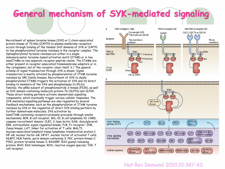
Recruitment of spleen tyrosine kinase (SYK) or ζ-chain-associatedprotein kinase of 70 kDa (ZAP70) to plasma membrane receptors occurs through binding of the tandem SH2 domains of SYK or ZAP70 to two phosphorylated tyrosine residues in the receptor complex. The 2phosphorylated tyrosine residues are either in a single immunoreceptor tyrosine-based activation motif (ITAM) or in two hemITAMs on two separate receptor peptide chains. The ITAMs are either present in receptor-associated transmembrane adaptors or in the cytoplasmic tail of the receptor chain itself. b | The general scheme of signal transduction through SYK is shown. Signal transduction is mostly initiated by phosphorylation of ITAM tyrosine residues by SRC family kinases. Recruitment of SYK to dually phosphorylated ITAMs triggers the activation of SYK and its direct binding to members of the VAV and phospholipase Cγ (PLCγ)families, the p85α subunit of phosphoinositide 3-kinase (PI3K), as well as SH2 domain-containing leukocyte protein 76 (SLP76) and SLP65. These direct binding partners activate downstream signallingcomponents, which eventually trigger various cellular responses. The SYK-mediated signalling pathways are also regulated by several feedback mechanisms, such as the phosphorylation of ITAM tyrosine residues by SYK or the regulation of direct SYK binding partners by further downstream molecules. SYK activation by hemITAM-containing receptors probably proceeds through similar mechanisms. BCR, B cell receptor; BCL-10, B cell lymphoma 10; CARD, caspase-recruitment domain; CLEC, C-type lectin; DAG, diacylglycerol; ERK, extracellular signal-regulated kinase; FcR, Fc receptor; JNK, Janus kinase; LAT, linker for activation of T cells; MALT1, mucosa-associated lymphoid tissue lymphoma translocation protein 1; NF-κB, nuclear factor-κB; NFAT, nuclear factor of activated T cells; NLRP3, NLR family, pyrin domain-containing 3; PKC, protein kinase C; PYK2, protein tyrosine kinase 2; RASGRP, RAS guanyl-releasingprotein; RHO, RAS homologue; ROS, reactive oxygen species; TCR, T cell receptor.
General mechanism of SYK-mediated signaling
Nat Rev Immunol 2010;10:387-43.
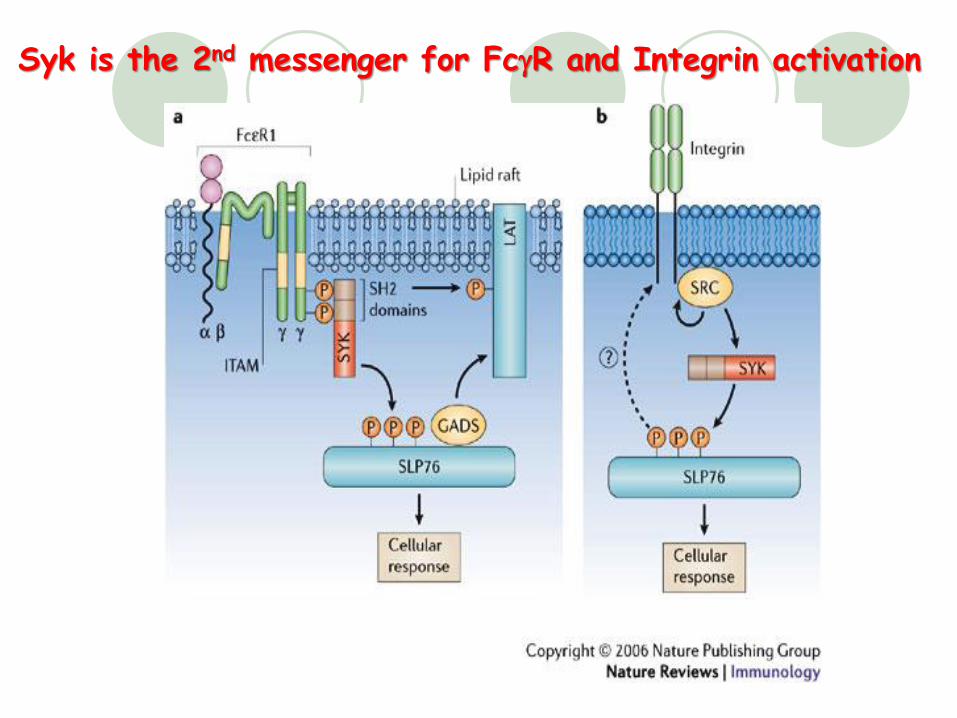
Syk is the 2nd messenger for FcgR and Integrin activation
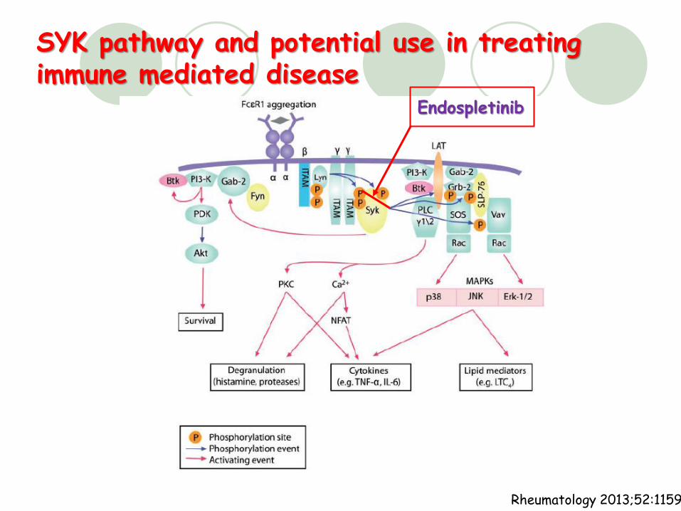
SYK pathway and potential use in treating immune mediated disease
Rheumatology 2013;52:1159
Endospletinib
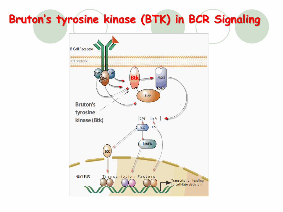
Bruton’s tyrosine kinase (BTK) in BCR Signaling
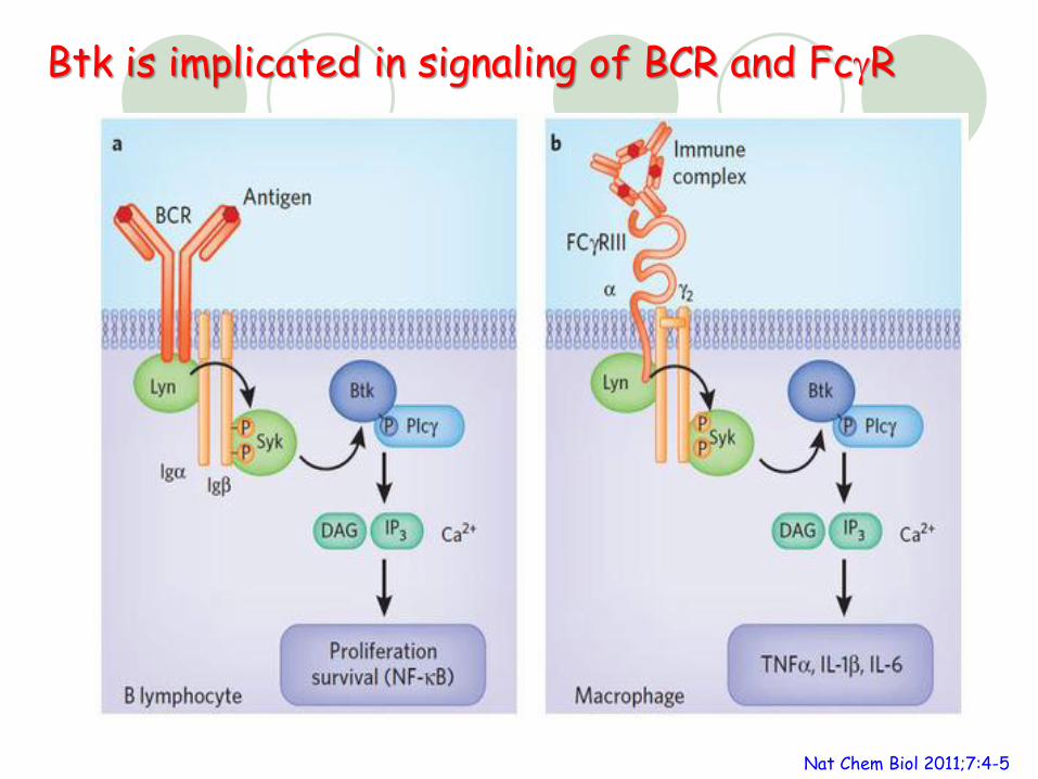
Nat Chem Biol 2011;7:4-5
Btk is implicated in signaling of BCR and FcgR
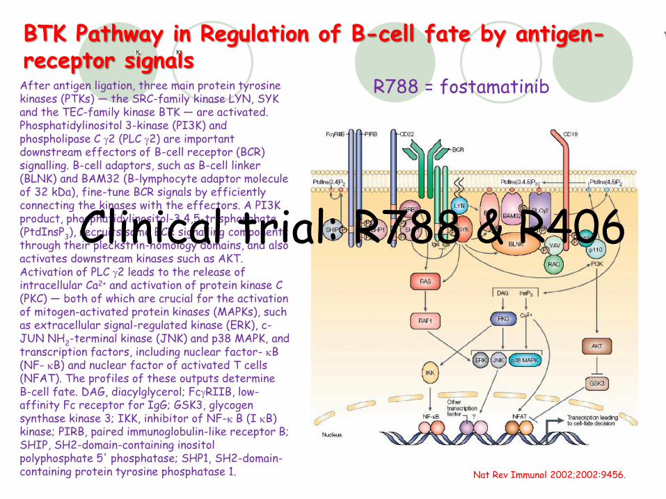
BTK Pathway in Regulation of B-cell fate by antigen-receptor signals
Nat Rev Immunol 2002;2002:9456.
After antigen ligation, three main protein tyrosine kinases (PTKs) — the SRC-family kinase LYN, SYK and the TEC-family kinase BTK — are activated. Phosphatidylinositol 3-kinase (PI3K) and phospholipase C g2 (PLC g2) are important downstream effectors of B-cell receptor (BCR) signalling. B-cell adaptors, such as B-cell linker (BLNK) and BAM32 (B-lymphocyte adaptor molecule of 32 kDa), fine-tune BCR signals by efficiently connecting the kinases with the effectors. A PI3K product, phosphatidylinositol-3,4,5-trisphosphate (PtdInsP3), recruits some BCR signalling components through their pleckstrin-homology domains, and also activates downstream kinases such as AKT. Activation of PLC g2 leads to the release of intracellular Ca2+ and activation of protein kinase C (PKC) — both of which are crucial for the activation of mitogen-activated protein kinases (MAPKs), such as extracellular signal-regulated kinase (ERK), c-JUN NH2-terminal kinase (JNK) and p38 MAPK, and transcription factors, including nuclear factor- kB (NF- kB) and nuclear factor of activated T cells (NFAT). The profiles of these outputs determine B-cell fate. DAG, diacylglycerol; FcgRIIB, low-affinity Fc receptor for IgG; GSK3, glycogen synthase kinase 3; IKK, inhibitor of NF-k B (I kB) kinase; PIRB, paired immunoglobulin-like receptor B; SHIP, SH2-domain-containing inositol polyphosphate 5' phosphatase; SHP1, SH2-domain-containing protein tyrosine phosphatase 1.
Clinical trial: R788 & R406
R788 = fostamatinib
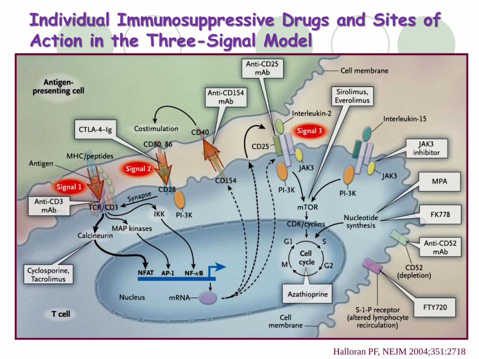
Individual Immunosuppressive Drugs and Sites of Action in the Three-Signal Model
Halloran PF, NEJM 2004;351:2718