Embed Size (px)
Citation preview
Neurotrophins and Their Receptors in Rat Peripheral Trigeminal System during Maxillary Nerve Growth U r m a s Annn/ie,*~ Ulla Pirvola,* J a a n Palgi,*§ Tli la-Riikka Kiema,* Kaia Palm,*~ Max im Moshnyakov,* J u k k a Ylikoski,I a n d Mar t Saarma*§
* Laboratory of Molecular Neurobiology, Institute of Biotechnology, University of Helsinki, Karvaamokuja 3, P.O. Box 45, SF-00014 Helsinki, Finland; ~Department of Pathology, University of Helsinki, ~ 3, p.o. Box 21, SF-00014 Helsinki, Finland; § Department of Molecular Genetics, Institute of Chemical Physics and Biophysics, Estonian Academy of Sciences, Akadeemia tee 23, EE 0026 Tallinn, Estonia; and I Department of Otolaryngology, University of Kuopio, SF-702 11 Kuopio, Finland
Abstract. We examined the expression of the neu- rotrophins (NTFs) and their receptor mRNAs in the rat trigeminal ganglion and the first branchial arch be- fore and at the time of maxillary nerve growth. The maxillary nerve appears first at embryonic day (E)10 and reaches the epithelium of the first branchial arch at El2, as revealed by anti-L1 immunohistochemistry. In situ hybridization demonstrates, that at E10-Ell, neurotrophin-3 (NT-3) mRNA is expressed mainly in the mesenchyme, but neurotrophin-4 (NT-4) mRNA in the epithelium of the first branchial arch. NGF and brain-derived neurotrophic factor (BDNF) mRNAs start to be expressed in the distal part of the first branchial arch shortly before its innervation by the
maxillary nerve. Trigeminal ganglia strongly express the mRNA of trkA at El0 and thereafter. The expres- sion of mRNAs for low-affinity neurotrophin receptor (LANR), trkB, and trkC in trigeminal ganglia is weak at El0, but increases by Ell-E12. NT-3, NT-4, and more prominently BDNF, induce neurite outgrowth from explant cultures of the El0 trigeminal ganglia but no neurites are induced by NGF, despite the expres- sion of trkA. By E12, the neuritogenic potency of NGF also appears. The expression of NT-3 and NT-4 and their receptors in the trigeminal system prior to target field innervation suggests that these NTFs have also other functions than being the target-derived tro- phic factors.
N "EUROTROPHINS ( N T F s ) 1 a re the family of related
proteins with specific effects on developing nervous system. To date, four NTFs have been character-
ized. These are NGF (Levi-Montalcini, 1987), brain- derived neurotrophic factor (BDNT) (Barde et al., 1982; Leibrock et al., 1989), neurotrophin-3 (NT-3) (Ernfors et al., 1990; Holm et al., 1990; Jones and Reichardt, 1990; Kaisho et al., 1990; Maisonpierre et al., 1990; Rosenthal et al., 1990), and neurotrophin-4 (NT-4) (Hallb66k et al., 1991; Ip et al., 1992), also termed neurotrophin-5 (Berke- meier et al., 1991). NTFs are shown to be vitally essential in the development of nervous system at the stage of pro- grammed neuronal death. NTFs, secreted in limiting amounts from relevant peripheral and brain structures are shown to support the survival of sensitive neurons which innervate that particular tissue at the time of programmed neuronal death, thus regulating the number of the innervating neurons (Oppenheim, 1991; Barde, 1989). Such a target-derived neu-
M. Moshnyakov is on leave from Shemakin Institute of Bioorganic Chemis- try, Russian Academy of Sciences, Moscow, Russia.
1. Abbreviations used in this paper: BDNF, brain-derived neurotrophic fac- tor; LANR, low-affinity neurotrophin receptor; NTF, neurotrophin.
rotrophic role is convincingly established for NGF as a pro- totypic molecule for the whole family (Levi-Montalcini, 1987; Barde, 1989). Although the survival-promoting abili- ties of other NTFs are poorly studied in vivo, they are shown to have effects similar to those of NGF on overlapping but also distinct neuronal populations in vitro (Lindsay et al., 1985; Maisonpierre et al., 1990; Holm et al., 1990; Rosen- thai et al., 1990) suggesting the target-derived neurotrophic function for them as well. This suggestion is supported by simultaneous presence of NTF receptor mRNAs in several neuronal populations and the cognate NTFs in the respective innervated tissues during the time of programmed neuronal death (Schechterson and Bothwell, 1992; Ernfors et al., 1992).
To perform their biological effects, the NTFs must bind to appropriate receptors on the surface of the responsive cells. Binding studies have revealed the presence of high- and low- affinity receptors for NGF, BDNF, and NT-3 on chick em- bryonic sensory neurons (SuRer et al., 1979; Rodrfguez- T6bar and Barde, 1988; RDdriguez-T6bar et al., 1990, 1992). The high-affinity receptors are most probably the members of the trk protein tyrosine kinase receptor family: trkA (Martin-Zanca et al., 1989), trkB (Klein et al., 1989), and
© The Rockefeller University Press, 0021-9525/93/0911053/13 $2.00 The Journal of Cell Biology, Volume 122, Number 5, September 1993 1053-1065 1053
on April 9, 2019jcb.rupress.org Downloaded from http://doi.org/10.1083/jcb.122.5.1053Published Online: 1 September, 1993 | Supp Info:
trkC (Lamballe et al., 1991). Binding of cognate NTFs to their corresponding trk receptors ectopically expressed in fibroblasts induces autophosphorylation of the receptors and transduces the signals. Thus, trkA is a functional receptor for NGF (Klein et al., 1991b; Cordon-Cardo et al., 1991; Jing et al., 1992; Ip et al., 1993), trkB can be functionally activated by both BDNF (Klein et al., 1991a; Squinto et al., 1991; Glass et al., 1991; Ip et al., 1993) and NT-4 (Klein et al., 1992; Ip et al., 1993), whereas NT-3 is a ~nonpreferred ~ ligand for trkB (Glass et al., 1991; Klein et al., 1991a; Squinto et al., 1991; Ip et al., 1993). trkC serves as a func- tional receptor for NT-3 (Lamballe et al., 1991; Ip et al., 1993). The low-affinity NGF receptor p75 (Johnson et al., 1986; Radeke et al., 1987) is suggested to be designated as low-affinity neurotrophin receptor (LANR) as it binds NGF, BDNF, NT-3, and NT-4 with similar low-affinity kinetics (Pcxiffguez-T6bar and Barde, 1988; Rodr~guez-T6bar et al., 1990, 1992; Hallb66k et al., 1991). The LANR is known not to be the signal-transducing receptor and its participation in the formation of the high-affinity NTF receptors is con- troversial (Hempstead et al., 1991; Klein et al., 1991/7; Glass et al., 1991; Jing et al., 1992; Ip et al., 1993). trkB (Klein et al., 1990a; Middlemas et al., 1991) and trkC (Tsoulfas et al., 1993) loci yield multiple mRNA species encoding vari- ous receptor isoforms, some of which being catalytic, while some are without the tyrosine kinase domain and thus most probably unable to transduce the signal of the ligand. The truncated trkB transcripts are, however, expressed in several parts of the developing and adult brain of the mouse and rat (Klein et al., 1989, 1990a; Middiemas et al., 1991) suggest- ing that they are important in the processes not consistent with the neurotrophic concept.
In a particular neuronal population, the programed death begins at the time the first neurites reach the target tissue (Rohrer et al., 1988; Davies et al., 1987; Vogel and Davies, 1991). Transcripts of trkA and trkB are also reported in the mouse embryos at the earliest stages when the developing neurons are not yet competent to send out neurites (Martin- Zanca et al., 1990; Klein et al., 1990b) suggesting that NTFs may have the functions in the early embryogenesis other than promoting neuronal survival. For example NGF is shown to direct the course of regenerating neurites of its responsive neurons in vitro (Gundersen and Barrett, 1980), leading to the hypothesis that in vivo it may be a target-derived chemoattractant directing the course of the pioneer neurites during the precise process of neurite pathfinding (Lumsden and Davies, 1983; Placzek et al., 1990). However, the guid- ance of the primary neurites by target-derived gradients of the NTFs is not demonstrated in vivo. Recently, BDNF has been shown to accelerate the maturation of early chick dor- sal root ganglion cells in vitro (Wright et al., 1992). In general, however, the possible roles of NTFs in early processes of neurogenesis, such as proliferation, commit- ment, migration, and maturation of neuronal precursors as well as formation of primary neuritic pathways, are poorly studied.
The trigeminal ganglion is a good model to study the effects of NTFs on early neurogenesis because its develop- ment is morphologically quite well established (Altman and Bayer, 1982; Davies, 1988; Stainier and Gilbert, 1991) but the factors regulating the development are largely unknown. It has been demonstrated that the first neurites of the maxil-
lazy branch of the mouse trigeminal nerve are directed to the organ of innervation-the whisker pad of the first branchial arch-by a soluble agent secreted from the epithelium (Lumsden and Davies, 1983, 1986). The agent named ~igeminal neurotropic factor; is immunochemicaliy differ- ent from NGF, and the expression of NGF is detected in the whisker field at the time when the first neurites contact the cutaneous target (Davies et al., 1987). Correspondingly, the mouse trigeminal ganglion neurons express LANR mRNA at the earliest stages of differentiation, the amount of the transcripts increasing about fivefold when the first axons contact the target field (Davies et al., 1987; Wyatt et al., 1990). The expression of other NTFs and the trk protein tyrosine kinase receptors in the trigeminal system has been studied only at the stages when the neurons have already contacted their targets. Thus, the distal regions of the mouse branchial arches express mRNAs for NGF, BDNF, NT-3, and NT-4 (Davies et al., 1987; Schechterson and Bothwell, 1992; Ernfors et al., 1992; Ib(u3ez et al., 1993), while ex- pression of mRNAs for LANR, trkA, trkB, and trkC have been reported in mouse and rat trigeminal ganglia (Martin- Zanca et al., 1990; Klein et al., 1990b; Wheeler and Both- well, 1992; Schechterson and Bothwell, 1992; Ernfors et al., 1992). However, there is no information available about their expression at the early preinnervation stages of trigeminal ganglion development.
As a first step of identifying the roles of the neurotrophins at the initial stages of development of the trigeminal ganglion neurons, we studied the expression of the mRNAs for all known neurotrophins and their receptors in rat preinnerva- tion trigeminal ganglion and in one of its peripheral targets-the first branchial arch. The ability of the develop- ing trigeminal ganglia to send out neurites in response to NTFs was investigated as well. These studies constitute the basis for further functional studies about the role of the NTFs in the preinnervation embryos.
Materials and Methods
Construction of Neurotrophin Probes The cRNA probes for rat NTFs were synthesized from the cDNA fragments (details of the cloning described in Pirvola et al., 1992) encompassing the following nucleotides: 283-716 of rat preproNGF mRNA (Whittemore et al., 1988), 414-712 of rat preproBDNF mRNA ('13mmusk et al., 1993), 145-331 of rat preproNT-3 mRNA (Maisonpierre et al., 1990), and 295-550 of rat NT-5 mRNA (Berkemeier et al., 1991).
Construction of Neurotrophin Receptor Probes cRNA probes for rat NTF receptors were synthesized from the following regions (details of the cloning described in Ylikoski et al., 1993): nucleo- tides 343-609 corresponding to human trkA (Marfin-Zanca et al., 1989), 562-762 of rat trkC cDNA (Merlio et ai., 1992), and nucleotides 230-535 of rat trkB recognizing catalytic as well as truncated forms of the receptor (Middlemas et al., 1991) (pan-trkB probe). To discriminate the full-length (catalytic) and truncated (non-catalytic) forms of trkB, the probes corre- sponding to nucleotides 1357-1507 of the tyrosine kinase domain of the rat full-length receptor and to nucleotides 1343-1545 of the cytoplasmic do- main of the rat trkB-T1 cDNA (absent in the full-length molecule) (Mid- dlemas et al., 1991) were prepared by polymerase chain reaction and cloned into the PCRII vector (Invitrogen Corporation, San Diego, CA). To detect LANR, a cRNA probe was synthesized from a cDNA fragment correspond- ing to the transmembrane region (nucleotides 853-970) of the rat LANR (Radeke et al., 1987).
The Journal of Cell Biology, Volume 122, 1993 1054
In Situ Hybridizations Sprague-Dawley rats were mated overnight and the day of the vaginal plug was considered as embryonic day (E)0. Paraffin-embedded serial sections of 5 ~,m thickness were cut sagit~ally from whole embryos and collected on 3-aminopropyl ethoxysilane-coated (Sigma Immunochemicals, St. Louis, MO) slides. The pretreatment, hybridization, and posttreatment of the sec- tions were carried out as described (Pirvola et al., 1992).
The 35S-UTP-labeled sense and antisense RNA probes were prepared according to W'flkinson and Green (1990) using an appropriate SP6 or T7 transcription system (Promege Corp., Madison, WI) and used at a final con- centration of 20000 cpm/tA. No hybridization signals above the back- ground levels were detected with sense-strand control probes of all NTFs or NTF receptors. Examples of trkC and NGF sense probe hybridizations are shown (see Fig. 3 i and f ) .
Ganglionic Explant Cultures Trigeminal ganglia were dissected from E10-E12 rat embryos and embed- ded into three-dimensional collagen matrix prepared according to Ebendal (1989). Typically, 3-5 ganglia were cultured in 0.5 ml of matrix in 24-well tissue culture plates (Nunclon Delta, Denmark). The gels were covered by 0.5 ml of Eagle's Basal Medium (GIBCO BRL, Gaithersburg, MD) contain- ing 1% of heat-inactivated horse serum (PAA-Labor- und Forschungsgesell- schaft mbH, Linz, Austria). The collagen gel was prepared into the same medium. Recombinant rat BDNF and NT-3 (a generous gift from Prof. Y.-A. Barde, Max-Planck-Institute for Psychiatry, Munich, Germany) and chromatographically purified mouse BNGF (Iustitute of Physiology, Minsk, Byelornssian Republic) were added to the culture media. All NTFs induced standard neurite outgrowth from E8 chick dorsal root ganglion explants known to respond to all three factors (Maisonpierre et al., 1990) at 5 ng/ml whereas at 1 ng/ml ur below that, virtually no neurites appeared. All three factors were typically applied at 5 ng/ml if not stated otherwise. The explant cultures were incubated at 37°C in the humid atmosphere containing 5% of CO2. The cultures were examined after 24 and 48 h.
Cloning and Expression of Human NT-4 Human gene encoding NT-4 was selected from the human genomic DNA library in EMBL 3 (kindly provided by Dr. R. Allikmets, Institute of Chem- ical Physics and Biophysics, Estonian Academy of Sciences, Tallinn, Esto- nia) using the rat NT-4 cDNA probe. An 840-bp Sau 3AI fragment contain- ing the coding exon of human NT-4 with 5' noncoding flank of 60 bp and
3' noncoding flank of 20 bp, was cloned into BamHI site of pBluescriptSK + (Stratagene, La Jolla, CA) and sequenced. The insert was subeloned into the NotI/XhoI sites of expression vector pMEP4 (Invitrogen Corporation) under inducible metallothionein promoter. COS cells grown to ,,o70% confluence in DME (GIBCO BRL) containing 10% of beat-inactivated FCS (PAA-Labor- und Forschungsgesellschaft mbH, Linz, Austria) were trans- fected with 10 ~tg of the expression plasmid per 6-cm cell culture dish by the Lipofectin Reagent (GIBCO BRL) following the manufacturer's instruc- tions. NT-4 expression was induced by 10/~m ZnC12 in culture medium. The conditioned medium was collected 24 h later from the transfected cells and used in the neurite outgrowth assay on E7-E8 chick dorsal root ganglia and rat trigeminal ganglia as described above. No neurites were obtained by control medium from the transfected but uninduced cells.
Immunohistochemistry Sagittal cryosections were cut from the fixed E10-E12 rat embryos and im- munostained with the affinity-purified rabbit polyclonal antibody to LI cell adhesion molecule (a kind gift from Prof. M. Schachner, ETH, Zurich, Switzerland) in PBS containing 0.25% Triton X-100 and 1% BSA at +4°C overnight followed by FITC-conjugated donkey anti-rabbit IgG secondary antibodies (Jackson ImmunoResearch Laboratories, Inc., West Grove, PA) in the same buffer.
Rat trigeminal ganglia of different ages were cultivated in a thin layer of collagen gel on round coverslips, fixed with 3.5 % paraformaldehyde in PBS and indirect immunofluorescence was performed with anti-neurofilament antibody (a kind gift from Prof. I. Virtanen, Department of Anatomy, University of Helsinki, Finland) in PBS containing 0.25 % Triton X-100 and 1% BSA at +4°C. The specimens were mounted in DABCO (Sigma Im- munochemicals).
Results
Development of Maxillary Nerve The initial stages of the rat maxillary nerve development were studied immunohistochemically using polyclonal anti- bodies to L1 cell adhesion molecule. The first neurites start to grow into the first branchial arch during El0. At Ell , the growing maxillary fibers are in the central region of the first
Figure/. Rat trigeminal gan- # i o n and its peripheral processes at El2. (a) Sagittal section of the rat embryo through the trigeminal gan- glion and the first branchial arch immunostained for L1 cell adhesion molecule. The maxillary nerve has ap- proached the cutaneous target field (arrow). The corre- sponding bright-field micro- graph of the same section is shown (b). oph, ophthalmic nerve; rex, maxillary nerve; rod, mandibular nerve; e, eye; tg, trigeminal ganglion. Bar, 100 pan.
Arun~e et al. Neurotrophins and Their Receptors in Trigeminal System 1055
branchial arch and approach their cutaneous target fields at El2 (Fig. 1). Thus, the rat whisker field innervation begins at the late El2.
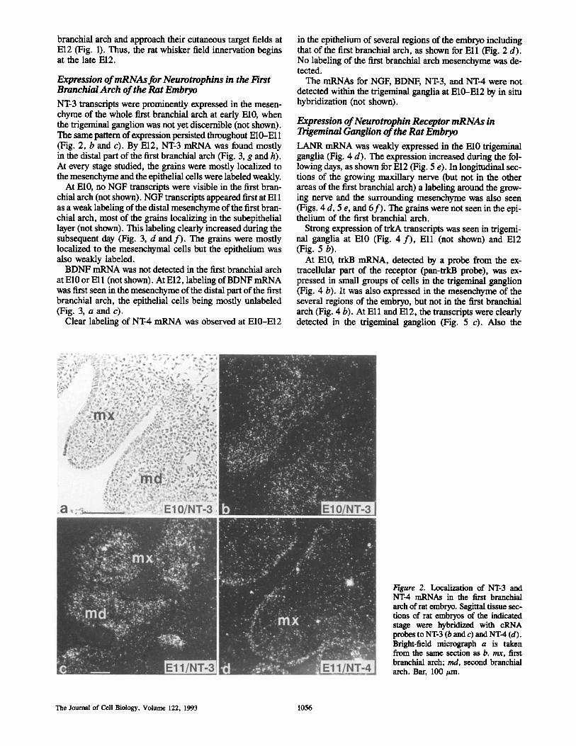
Expression of mRNAs for Neurotrophins in the First Branchial Arch of the Rat Embryo NT-3 transcripts were prominently expressed in the mesen- chyme of the whole first branchial arch at early El0, when the trigeminal ganglion was not yet discernible (not shown). The same pattern of expression persisted throughout E10-E11 (Fig. 2, b and c). By El2, NT-3 mRNA was found mostly in the distal part of the first branchial arch (Fig. 3, g and h). At every stage studied, the grains were mostly localized to the mesenchyme and the epithelial cells were labeled weakly.
At El0, no NGF transcripts were visible in the first bran- chial arch (not shown). NGF transcripts appeared first at Ell as a weak labeling of the distal mesenchyme of the first bran- chial arch, most of the grains localizing in the subepithelial layer(not shown). This labeling clearly increased during the subsequent day (Fig. 3, d and f ) . The grains were mostly localized to the mesencbymal cells but the epithelium was also weakly labeled.
BDNF mRNA was not detected in the first branchial arch at El0 or Ell (not shown). At El2, labeling ofBDNF mRNA was first seen in the mesenchyme of the distal part of the first branchial arch, the epithelial cells being mostly unlabeled (Fig. 3, a and c).
Clear labeling of NT-4 mRNA was observed at E10-E12
in the epithelium of several regions of the embryo including that of the first branchial arch, as shown for Ell (Fig. 2 d). No labeling of the first branchial arch mesenchyme was de- tected.
The mRNAs for NGF, BDNF, NT-3, and NT-4 were not detected within the trigeminal ganglia at E10-E12 by in situ hybridization (not shown).
Expression of Neurotrophin Receptor mRNAs in Trigeminal Ganglion of the Rat Embryo LANR mRNA was weakly expressed in the El0 trigeminal ganglia (Fig. 4 d). The expression increased during the fol- lowing days, as shown for El2 (Fig. 5 e). In longitudinal sec- tions of the growing maxillary nerve (but not in the other areas of the first branchial arch) a labeling around the grow- ing nerve and the surrounding mesenchyme was also seen (Figs. 4 d, 5 e, and 6f ) . The grains were not seen in the epi- thelium of the first branchial arch.
Strong expression of trkA transcripts was seen in trigemi- nal ganglia at El0 (Fig. 4 f ) , Ell (not shown) and El2 (Fig. 5 b).
At El0, trkB mRNA, detected by a probe from the ex- tracellular part of the receptor (pan-trkB probe), was ex- pressed in small groups of cells in the trigeminal ganglion (Fig. 4 b). It was also expressed in the mesenchyme of the several regions of the embryo, but not in the first branchial arch (Fig. 4 b). At Ell and El2, the transcripts were clearly detected in the trigeminal ganglion (Fig. 5 c). Also the
Figure 2. Localization of NT-3 and NT-4 mRNAs in the first branchial arch of rat embryo. Sagittal tissue sec- tions of rat embryos of the indicated stage were hybridized with cRNA probes to NT-3 (b and c) and NT-4 (d). Bright-field micrograph a is taken from the same section as b. rex, first branchial arch; md, second branchial arch. Bar, 100/tin.
The Journal of Cell Biology, Volume 122, 1993 1056
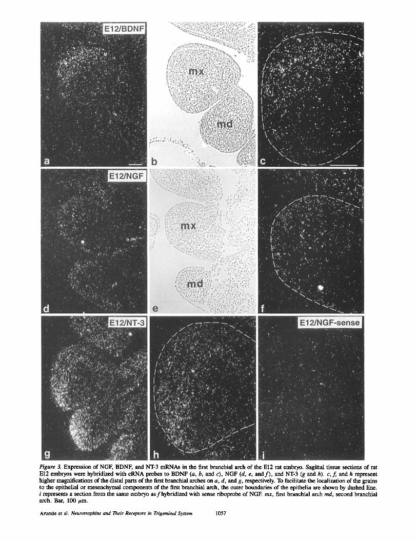
Figure 3. Expression of NGF, BDNF, and NT-3 mRNAs in the first branchial arch of the El2 rat embryo..Sagittal tissue sections of rat El2 embryos were hybridized with cRNA probes to BDNF (a, b, and c), NGF (d, e, and f ) , and NT-3 (g and h). c,f, and h represent higher magnifications of the distal parts of the first branchial arches on a, d, and g, respectively. To facilitate the localization of the grains to the epithelial or mesenchymal components of the first branchial arch, the outer boundaries of the epithelia are shown by dashed line. i represents a section from the same embryo as fhybridized with sense riboprobe of NGE rex, first branchial arch md, second branchial arch. Bar, 100 #m.
Arurn~ et al. Neurotrophins and Their Receptors in Trigeminal System 1057
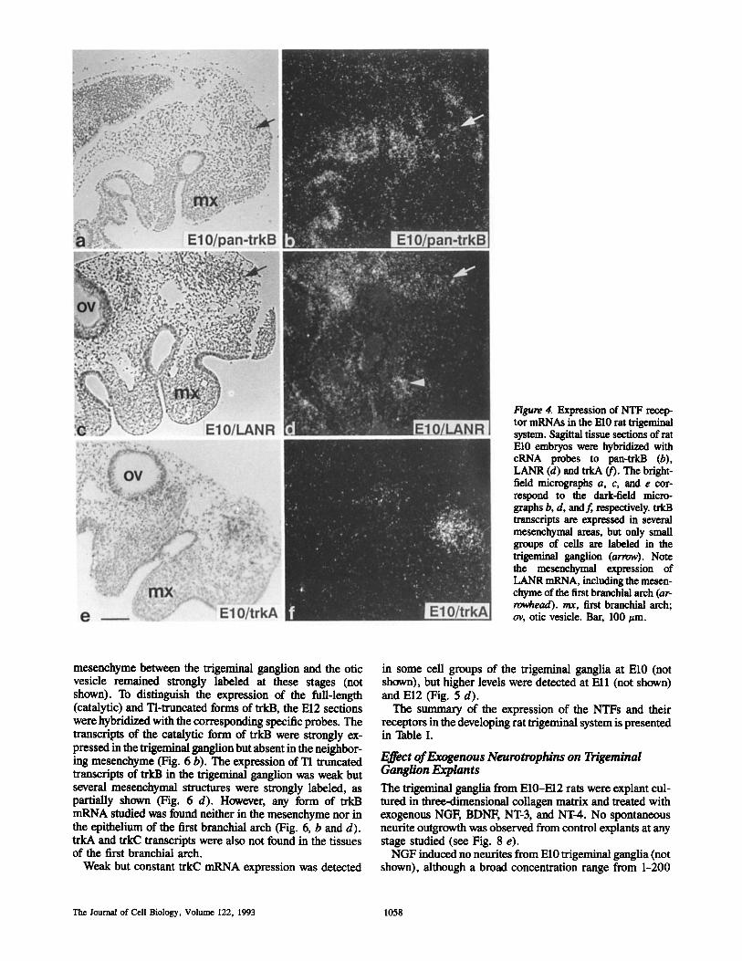
Figure 4. Expression of NTF recep- tor mRNAs in the El0 rat trigeminal system. Sagittal tissue sections of rat El0 embryos were hybridized with cRNA probes to pan-trkB (b), LANR (d) and trkA (f). The bright- field micrographs a, c, and e cor- respond to the dark-field micro- graphs b, d, and f, respectively, trkB transcripts are expressed in several mesenchymal areas, but only small groups of cells are labeled in the trigeminal ganglion (arrow). Note the mesenchymal expression of LANR mRNA, including the mesen- chyme of the first branchial arch (ar- ro~ead), rex, first branchial arch; ov, otic vesicle. Bar, 100 ~tm.
mesenchyme between the trigeminal ganglion and the otic vesicle remained strongly labeled at these stages (not shown). To distinguish the expression of the full-length (catalytic) and Tl-truncated forms of trkB, the El2 sections were hybridized with the corresponding specific probes. The transcripts of the catalytic form of trkB were strongly ex- pressed in the trigeminal ganglion but absent in the neighbor- ing mesenchyme (Fig. 6 b). The expression of TI truncated transcripts of trkB in the trigeminal ganglion was weak but several mesenchymal structures were strongly labeled, as partially shown (Fig. 6 d). However, any form of trkB mRNA studied was found neither in the mesenchyme nor in the epithelium of the first branchial arch (Fig. 6, b and d). trkA and trkC transcripts were also not found in the tissues of the first branchial arch.
Weak but constant trkC mRNA expression was detected
in some cell groups of the trigeminal ganglia at El0 (not shown), but higher levels were detected at Ell (not shown) and El2 (Fig. 5 d).
The summary of the expression of the NTFs and their receptors in the developing rat trigeminal system is presented in Table I.
F_~ct of Exogenous Neurotrophins on Trigeminal Ganglion Explants The trigeminal ganglia from E10-E12 rats were explant cul- tured in three-dimensional collagen matrix and treated with exogenous NGF, BDNF, NT-3, and NT-4. No spontaneous neurite outgrowth was observed from control explants at any stage studied (see Fig. 8 e).
NGF induced no neurites from El0 trigeminal ganglia (not shown), although a broad concentration range from 1-200
The Journal of Cell Biology, Volume 122, 1993 1058
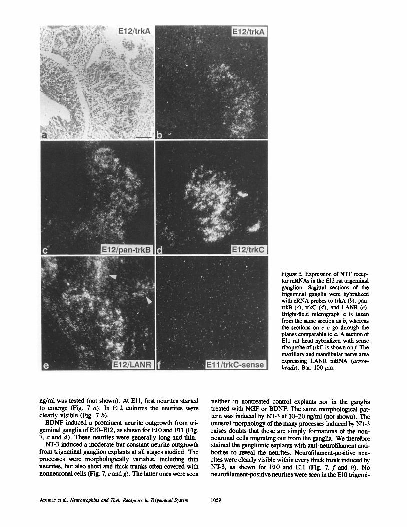
Figure 5. Expression of NTF recep- tor mRNAs in the El2 rat trigeminal ganglion. Sagittal sections of the trigeminal ganglia were hybridized with cRNA probes to trkA (b), pan- trkB (c), trkC (d), and LANR (e). Bright-field micrograph a is taken from the same section as b, whereas the sections on c-e go through the planes comparable to a. A section of Ell rat head hybridized with sense riboprobe of trkC is shown onf. The maxillary and mandibular nerve area expressing LAN-R mRNA (arrow- heads). Bar, 100/~m.
ng/ml was tested (not shown). At Ell, first neurites started to emerge (Fig. 7 a). In E12 cultures the neurites were clearly visible (Fig. 7 b).
BDNF induced a prominent neurite outgrowth from tri- geminal ganglia of E10-E12, as shown for El0 and El l (Fig. 7, c and d). These neurites were generally long and thin.
NT-3 induced a moderate but constant neurite outgrowth from trigeminal ganglion explants at all stages studied. The processes were morphologically variable, including thin neurites, but also short and thick trunks often covered with nonneuronal cells (Fig. 7, e and g). The latter ones were seen
neither in nontreated control explants nor in the ganglia treated with NGF or BDNE The same morphological pat- tern was induced by NT-3 at 10--20 ng/ml (not shown). The unusual morphology of the many processes induced by NT-3 raises doubts that these are simply formations of the non- neuronal cells migrating out from the ganglia. We therefore stained the ganglionic explants with anti-neurofilament anti- bodies to reveal the neurites. Neurotilament-positive neu- rites were clearly visible within every thick trunk induced by NT-3, as shown for El0 and El l (Fig. 7, f and h). No neurofilament-positive neurites were seen in the El0 trigemi-
Arun~e et al. Neurotrophina and Their Receptors in Trigeminal System 1059
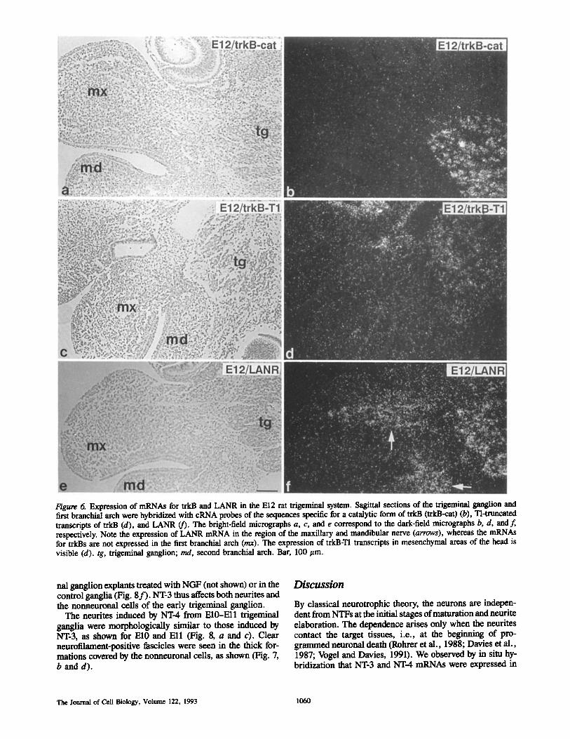
Figure 6. Expression of mRNAs for trkB and LANR in the E12 rat trigeminal system. Sagittal sections of the trigewlnAI ganglion and first branchial arch were hybridized with cRNA probes of the sequences specific for a catalytic form of trkB (trkB-cat) (b), Tl=truncated transcripts of trkB (d), and LANR (f). The bright-field micrographs a, c, and e correspond to the dark-field micrographs b, d, and f, respectively. Note the expression of LANR mRNA in the region of the maxillary and mandibular nerve (arrows), whereas the mRNAs for trkBs are not expressed in the first branchial arch (rex). The expression of trkB-Tl transcripts in mesenchymal areas of the head is visible (d). tg, trigeminal ganglion; rod, second branchial arch. Bar, 100 ~tm.
hal ganglion explants treated with NGF (not shown) or in the control ganglia (Fig. 8 f ) . NT-3 thus affects both neurites and the nonneuronal cells of the early trigeminal ganglion.
The neurites induced by NT-4 from E10-Ell trigeminal ganglia were morphologically similar to those induced by NT-3, as shown for El0 and Ell (Fig. 8, a and c). Clear neurofilament-positive fascicles were seen in the thick for- mations covered by the nonneuronal cells, as shown (Fig. 7, b and d).
Discussion
By classical neurotrophic theory, the neurons are indepen- dent from NTFs at the initial stages of maturation and neurite elaboration. The dependence arises only when the neurites contact the target tissues, i.e., at the beginning of pro- grammed neuronal death (Rohrer et al., 1988; Davies et al., 1987; Vogel and Davies, 1991). We observed by in situ hy- bridization that NT-3 and NT-4 mRNAs were expressed in
The Journal of Cell Biology, Volume 122, 1993 1060
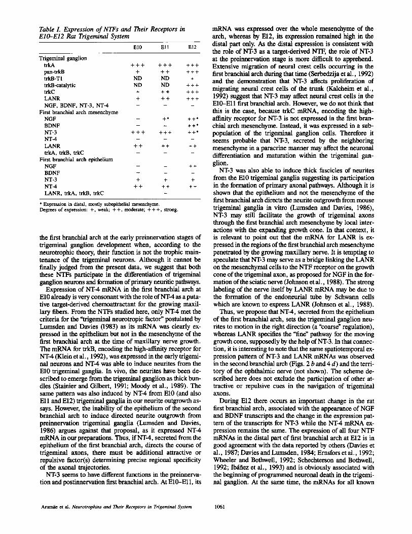
Table L Expression of NTFs and Their Receptors in E10-E12 Rat Trigeminal System
El0 E l l El2
T r i g e m i n a l gang l ion
t r k A + + + + + + + + +
pan- t rkB + + + + + +
t rkB-T1 N D N D +
t rkB-cata ly t ic N D N D + + +
t rkC + + + + + +
L A N R + + + + + +
N G F , B D N F , N T - 3 , N T - 4 - - -
Firs t b ranchia l a r ch m e s e n c h y m e
N G F - + * + + *
B D N F - - + + *
N T - 3 + + + + + + + + *
N T - 4 - - -
L A N R + + + + + +
trkA, trkB, trkC - - - First branchial arch epithelium
NGF - - + + BDNF - - -
N T - 3 + + + NT-4 ++ ++ ++ LANR, trkA, trkB, trkC
* Expression in distal, mostly subepithelial mesenchyme. Degrees of expression: + , weak; + + , moderate; + + + , strong.
the first branchial arch at the early preinnervation stages of trigeminal ganglion development when, according to the neurotrophic theory, their function is not the trophic main- tenance of the trigeminal neurons. Although it cannot be finally judged from the present data, we suggest that both these NTFs participate in the differentiation of trigeminal ganglion neurons and formation of primary neuritic pathways.
Expression of NT-4 mRNA in the first branchial arch at El0 already is very consonant with the role of NT-4 as a puta- tive target-derived chemoattractant for the growing maxil- lary fibers. From the NTFs studied here, only NT-4 met the criteria for the "trigeminal neurotropic factor" postulated by Lumsden and Davies (1983) as its mRNA was dearly ex- pressed in the epithelium but not in the mesenchyme of the first branchial arch at the time of maxillary nerve growth. The mRNA for trkB, encoding the high-affinity receptor for NT-4 (Klein et al., 1992), was expressed in the early trigemi- nal neurons and NT-4 was able to induce neurites from the El0 trigeminal ganglia. In vivo, the neurites have been de- scribed to emerge from the trigeminal ganglion as thick bun- dies (Stainier and Gilbert, 1991; Moody et al., 1989). The same pattern was also induced by NT-4 from El0 (and also Ell and El2) trigeminal ganglia in our neurite outgrowth as- says. However, the inability of the epithelium of the second branchial arch to induce directed neurite outgrowth from preinnervation trigeminal ganglia (Lumsden and Davies, 1986) argues against that proposal, as it expressed NT-4 mRNA in our preparations. Thus, if NT-4, secreted from the epithelium of the first branchial arch, directs the course of trigeminal axons, there must be additional attractive or repulsive factor(s) determining precise regional specificity of the axonal trajectories.
NT-3 seems to have different functions in the preinnerva- tion and postinnervation first branchial arch. At E10--Ell, its
mRNA was expressed over the whole mesenchyme of the arch, whereas by El2, its expression remained high in the distal part only. As the distal expression is consistent with the role of NT-3 as a target-derived NTF, the role of NT-3 at the preirmervation stage is more difficult to apprehend. Extensive migration of neural crest cells occurring in the first branchial arch during that time (Serbedzija et al., 1992) and the demonstration that NT-3 affects proliferation of migrating neural crest cells of the trunk (Kalcheim et al., 1992) suggest that NT-3 may affect neural crest cells in the E10-Ell first branchial arch. However, we do not think that this is the case, because trkC mRNA, encoding the high- affinity receptor for NT-3 is not expressed in the first bran- chial arch mesenchyme. Instead, it was expressed in a sub- population of the trigeminal ganglion cells. Therefore it seems probable that NT-3, secreted by the neighboring mesenchyme in a paracrine manner may affect the neuronal differentiation and maturation within the trigeminal gan- glion.
NT-3 was also able to induce thick fascicles of neurites from the El0 trigeminal ganglia suggesting its participation in the formation of primary axonal pathways. Although it is shown that the epithelium and not the mesenchyme of the first branchial arch directs the neurite outgrowth from mouse trigeminal ganglia in vitro (Lumsden and Davies, 1986), NT-3 may still facilitate the growth of trigeminal axons through the first branchial arch mesenchyme by local inter- actions with the expanding growth cone. In that context, i t
is relevant to point out that the mRNA for LANR is ex- pressed in the regions of the first branchial arch mesenchyme penetrated by the growing maxillary nerve. It is tempting to speculate that NT-3 may serve as a bridge linking the LANR on the mesenchymal cells to the NTF receptor on the growth cone of the trigeminal axon, as proposed for NGF in the for- marion of the sciatic nerve (Johnson et al., 1988). The strong labeling of the nerve itself by LANR mRNA may be due to the formation of the endoneurial tube by Schwann cells which are known to express LANR (Johnson et al., 1988).
Thus, we propose that NT-4, secreted from the epithelium of the first branchial arch, sets the trigeminal ganglion neu- rites to motion in the right direction (a "coarse" regulation), whereas LANR specifies the "fine" pathway for the moving growth cone, supposedly by the help of NT-3. In that connec- tion, it is interesting to note that the same spatiotemporal ex- pression pattern of NT-3 and LANR mRNAs was observed in the second branchial arch (Figs. 2 b and 4 d) and the terri- tory of the ophthalmic nerve (not shown). The scheme de- scribed here does not exclude the participation of other at- tractive or repulsive cues in the navigation of trigeminal axons.
During El2 there occurs an important change in the rat first branchial arch, associated with the appearance of NGF and BDNF transcripts and the change in the expression pat- tern of the transcripts for NT-3 while the NT-4 mRNA ex- pression remains the same. The expression of all four NTF mRNAs in the distal part of first branchial arch at El2 is in good agreement with the data reported by others (Davies et al., 1987; Davies and Lumsden, 1984; Ernsfors et al., 1992; Wheeler and Bothwell, 1992; Schechterson and Bothwell, 1992; ro~tfiez et al., 1993) and is obviously associated with the beginning of programmed neuronal death in the trigemi- nal ganglion. At the same time, the mRNAs for all known
Arumie et al. Neurotrophins and Their Receptors in Trigeminal System 1061
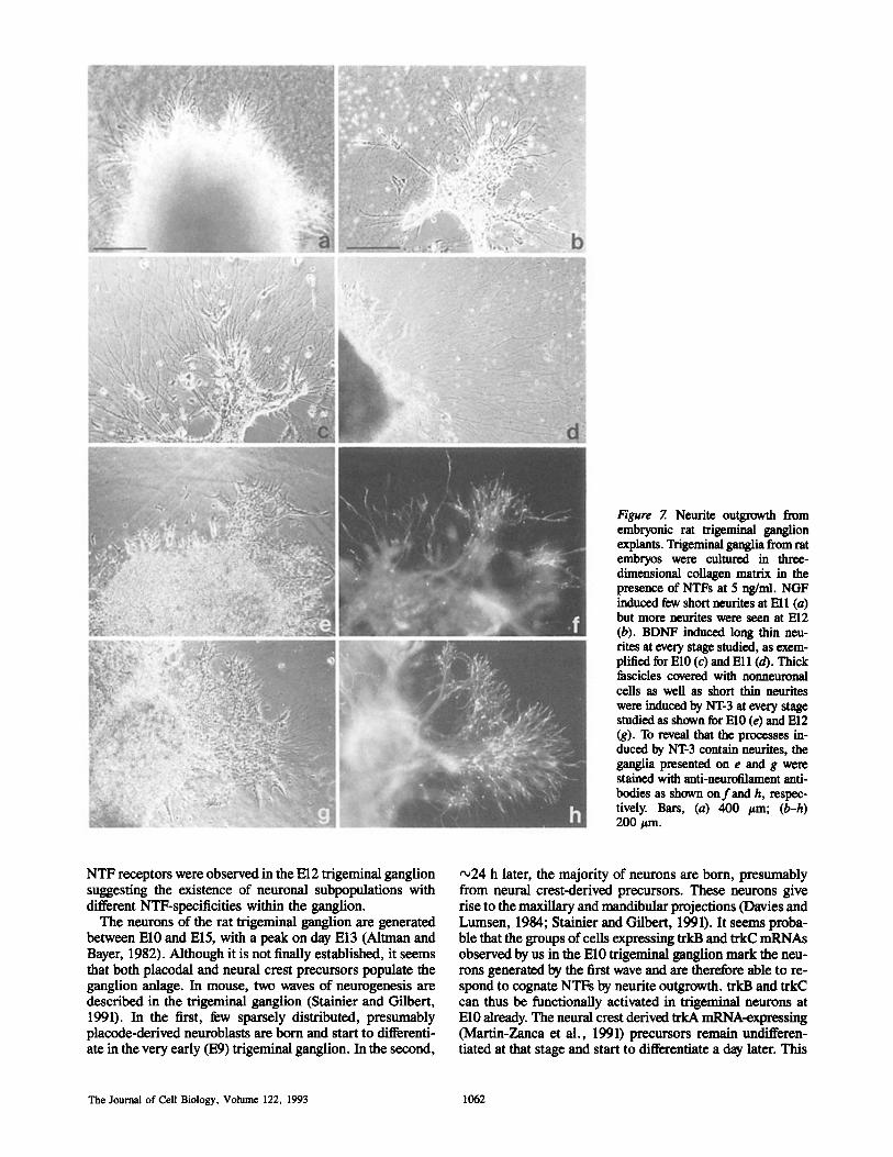
Figure 7. Neurite outgrowth from embryonic rat trigeminal ganglion explants. Trigeminal ganglia from rat embryos were cultured in three- dimensional collagen matrix in the presence of NTFs at 5 ng/ml. NGF induced few short neurites at Ell (a) but more neurites were seen at El2 (b). BDNF induced long thin neu- rites at every stage studied, as exem- plified for El0 (c) and Ell (d). Thick fascicles covered with nonneuronal cells as well as short thin neurites were induced by NT-3 at every stage studied as shown for El0 (e) and El2 (g). To reveal that the processes in- duced by NT-3 contain neurites, the ganglia presented on e and g were stained with anti-neurofilament anti- bodies as shown on f and h, respec- tively. Bars, (a) 400 /~m; (b-h) 200/~m.
NTF receptors were observed in the El2 trigeminal ganglion suggesting the existence of neuronal subpopulations with different NTF-specificities within the ganglion.
The neurons of the rat trigeminal ganglion are generated between El0 and El5, with a peak on day El3 (Almmn and Bayer, 1982). Although it is not finally established, it seems that both placodal and neural crest precursors populate the ganglion anlage. In mouse, two waves of neurogenesis are described in the trigeminal ganglion (Stainier and Gilbert, 1991). In the first, few sparsely distributed, presumably placode-derived neuroblasts are born and start to differenti- ate in the very early (E9) trigeminal ganglion. In the second,
~24 h later, the majority of neurons are born, presumably from neural crest-derived precursors. These neurons give rise to the maxillary and mandibular projections (Davies and Lumsen, 1984; Stainier and Gilbert, 1991). It seems proba- ble that the groups of cells expressing trkB and trkC mRNAs observed by us in the El0 trigeminal ganglion mark the neu- rons generated by the first wave and are therefore able to re- spond to cognate NTFs by neurite outgrowth, trkB and trkC can thus be functionally activated in trigeminal neurons at El0 already. The neural crest derived ~kA mRNA-expressing (Martin-Zanca et al., 1991) precursors remain undifferen- tiated at that stage and start to differentiate a day later. This
The Journal of Cell Biology, Volume 122, 1993 1062
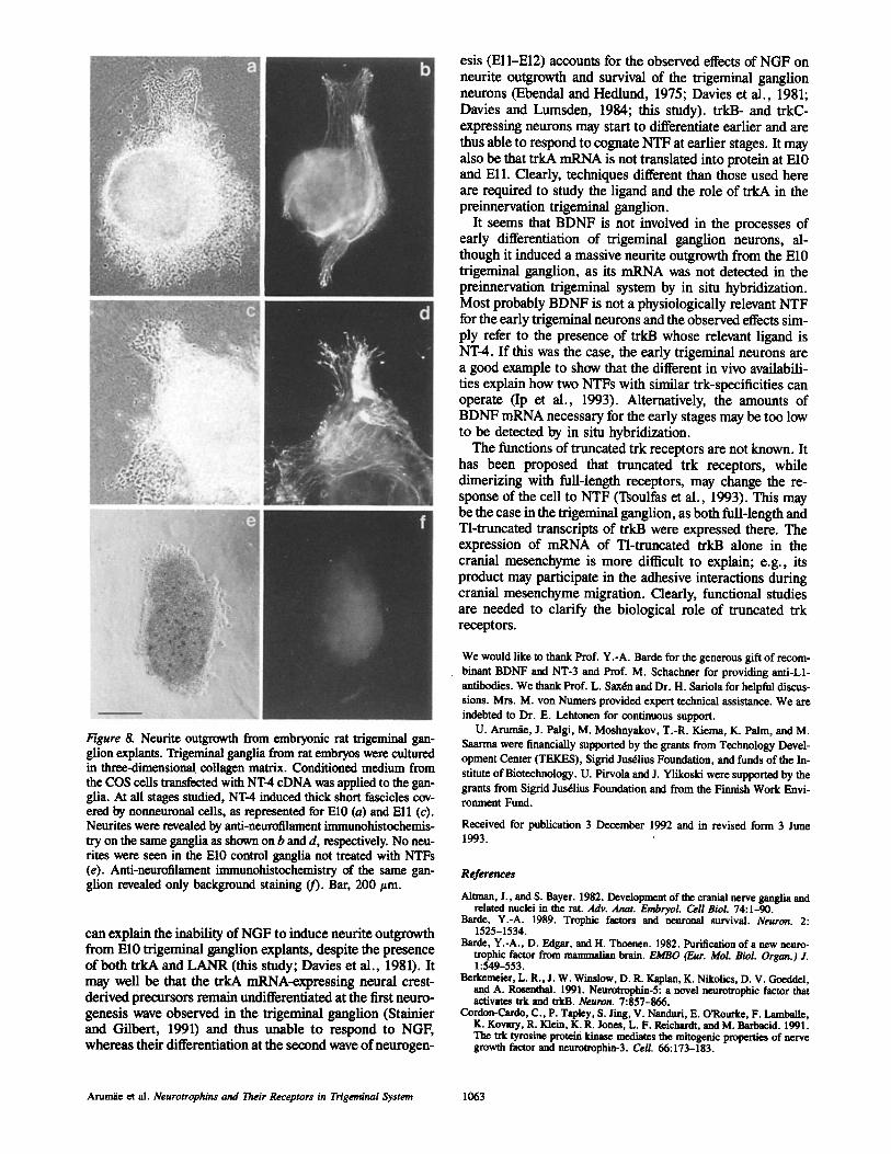
Figure 8. Neurite outgrowth from embryonic rat trigeminal gan- glion explants. Trigeminal ganglia from rat embryos were cultured in three-dimensionaI collagen matrix. Conditioned medium from the COS ceils transfected with NT-4 cDNA was applied to the gan- glia. At all stages studied, NT-4 induced thick short fascicles cov- ered by nonneuronal cells, as represented for El0 (a) and Ell (c). Neurites were revealed by anti-neurolilament immunohistochemis- try on the same ganglia as shown on b and d, respectively. No neu- rites were seen in the El0 control ganglia not treated with NTFs (e). Anti-neurofilament immunohistochemistry of the same gan- glion revealed only background staining (f). Bar, 200 ttm.
can explain the inability of NGF to induce neurite outgrowth from El0 trigeminal ganglion explants, despite the presence of both trkA and LANR (this study; Davies et al., 1981). It may well be that the trkA mRNA-expressing neural crest- derived precursors remain undifferentiated at the first neuro- genesis wave observed in the trigeminal ganglion (Stainier and Gilbert, 1991) and thus unable to respond to NGF, whereas their differentiation at the second wave of neurogen-
esis (Ell-El2) accounts for the observed effects of NGF on neurite outgrowth and survival of the trigeminal ganglion neurons (Ebendal and Hedlund, 1975; Davies et al., 1981; Davies and Lumsden, 1984; this study), trkB- and trkC- expressing neurons may start to differentiate earlier and are thus able to respond to cognate NTF at earlier stages. It may also be that trkA mRNA is not translated into protein at El0 and Ell. Clearly, techniques different than those used here are required to study the ligand and the role of trkA in the preinnervation trigeminal ganglion.
It seems that BDNF is not involved in the processes of early differentiation of trigeminal ganglion neurons, al- though it induced a massive neurite outgrowth from the El0 trigeminal ganglion, as its mRNA was not detected in the preinnervation trigeminal system by in situ hybridization. Most probably BDNF is not a physiologically relevant NTF for the early trigeminal neurons and the observed effects sim- ply refer to the presence of trkB whose relevant ligand is NT-4. If this was the case, the early trigeminal neurons are a good example to show that the different in vivo availabili- ties explain how two NTFs with similar trk-specificities can operate (Ip et al., 1993). Alternatively, the amounts of BDNF mRNA necessary for the early stages may be too low to be detected by in situ hybridization.
The functions of truncated trk receptors are not known. It has been proposed that truncated trk receptors, while dimerizing with full-length receptors, may change the re- sponse of the cell to NTF (Tsoulfas et al., 1993). This may be the case in the trigeminal ganglion, as both full-length and Tl-truncated transcripts of trkB were expressed there. The expression of mRNA of Tl-truncated trkB alone in the cranial mesenchyme is more difficult to explain; e.g., its product may participate in the adhesive interactions during cranial mesencbyme migration. Clearly, functional studies are needed to clarify the biological role of truncated trk receptors.
We would like to thank Prof. Y.-A. Barde for the generous gift of recom- binant BDNF and NT-3 and Prof. M. Schachner for providing anti-L1- antibodies. We thank Prof. L. SaxOn and Dr. H. Sariola for helpful discus- sions. Mrs. M. yon Numers provided expert technical assistance. We are indebted to Dr. E. Lehtonen for continuous support.
U. Arumfie, J. Palgi, M. Moshnyakov, T.-R. Kiema, K. Palm, and M. Saarma were financially supported by the grants from Technology Devel- opment Center (TEKES), Sigrid Jus~lius Foundation, and funds of the In- stitute of Biotechnology. U. Pirvola and J. Ylikoski were supported by the grants from Sigrid Jus61ius Foundation and from the Finnish Work Envi- ronment Fund.
Received for publication 3 December 1992 and in revised form 3 June 1993.
References
Airman, J., and S. Bayer. 1982. Development of the cranial nerve ganglia and related nuclei in the rat. Adv. Anat. Embryol. Cell Biol. 74:1-90.
Barde, Y.-A. 1989. Trophic factors and neuronal survival. Neuron. 2: 1525-1534.
Bal'de, Y.-A., D. Edgar, and H. Thoenen. 1982. Purification of a new neuro- trophic factor from manunalian brain. EMBO (Eur. biol. Biol. Organ.) J. 1:549-553.
Berkemeier, L. R., J. W. Winslow, D. R. Kaplan, K. Nikofics, D. V. Goeddel, and A. Rosenthal. 1991. Neurotrophin-5: a novel neurotrophic factor that activates trk and trkB. Neuron. 7:857-866.
Cordon-Cardo, C., P. Tapley, S. Jing, V. Nanduri, E. O'Rourke, F. Lambane, K. Kovary, R. Klein, K. R. Jones, L. F. Reichardt, andM. Barbacid. 1991. The trk tyrosine protein kinaqe mediates the mitogenic properties of nerve growth factor and neurotrophin-3. Cell. 66:173-183.
A r u n ~ et al. Neurotrophins and Their Receptors in Trigeminal System 1063
Davies, A. M. 1988. The trigeminal system: an advantageous experimental model for studying neuronal development. Development (Comb.). 103 (Suppl.): 175-183.
Davies, A. M., and A. Lumsden. 1984. Relation of target encounter and neu- ronal death to nerve growth factor responsiveness in the developing mouse trigeminal ganglion. J. Comp. Neurol. 223:124-137.
Davies, A. M., A. G. S. Lumsden, H. C. Slavkin, and G. Burnstock. 1981. Influence of nerve growth factor on the embryonic mouse trigeminal gan- glion in culture. Dev. Neurosci. 4:150-156.
Davies, A. M., C. Bandtlow, R. Heumann, S. Korsching, H. Rohrer, and H. Thoenen. 1987. Timing and site of nerve growth factor synthesis in develop- ing skin in relation to innervation and expression of the receptor. Nature (Lond.). 326:353-358.
Ebendal, T. 1989. Use of collagen gels to bicassay nerve growth factor activity. In Nerve Growth Factors. R. A. Rush, editor. John Wiley and Sons Ltd., Chicbester, West Sussex, England. 81-93.
Edendal, T., and K.-O. Hedlund. 1975. Effects of nerve growth factor on the chick embryo trigeminal ganglion in culture. Zoon. 3:33-47.
Emfors, P., C. F. Ib~tfiez, T. Ebendal, L. Olson, and H. Persson. 1990. Molec- ular cloning and neurotrophic activities of a protein with structural similari- ties to nerve growth factor: development and topographical expression in the brain. Proc. Natl. Acad. Sci. USA. 87:5454-5458.
Ernfors, P., J.-P. Merlin, and H. Persson. 1992. Cells expressing mRNA for neurotrophins and their receptors during embryonic rat development. Fur. J. Neurosci. 4:1140-1158.
Glass, D. J., S. H. Nye, P. Hantzopoulos, M. J. Macchi, S. P. Squinto, M. Goldfarb, and G. D. Yancopoulos. 1991. TrkB mediates BDNF/NT-3- dependent survival and proliferation in fibroblasts lacking the low affinity NGF receptor. Cell. 66:405-413.
Gundersen, R. W., and J. N. Barrett. 1980. Characterization of the turning re- sponse of dorsal root nenrites towards nerve growth factor. J. Cell Biol. 87:546-554.
Hallb66k, F., C. F. Ib~h3ez, and H. Persson. 1991. Evolutionary studies of the nerve growth factor family reveal a novel member abundantly expressed in Xenopus ovary. Neuron~ 6:845-858.
Hempstead, B. L., D. Martin-Zanca, D. R. Kaplan, L. F. Parada, and M. V. Chao. 1991. High-affinity NGF binding requires coexpression of the trk proto-oncogene and the low-affinity NGF receptor. Nature (Lond.). 350: 678-683.
Holm, A., J. Leibrock, K. Bailey, and Y.-A. Barde. 1990. Identification and characterization of a novel member of the nerve growth factor/brain-derived neurotrophic factor family. Nature (Lond.). 344:339-341.
Ib~fiez, C. F., P. Ernfors, T. Timmusk, N. Y. Ip, E. Arenas, G. D. Yan- copoulos, and H. Persson. 1993. Neurotrophin-4 is a target derived neuro- trophic factor for neurons of the trigeminal ganglion. Development (Camb.). 117:1345-1353.
Ip, N. Y., C. F. Ib~ez, S. H. Nye, J. McClaln, P. F. Jones, D. R. Gies, L. L. Bolluscio, M. M. Le Beau, R. Espinosa HI, S. P. Squinto, H. Persson, and G. D. Yancopoulos. 1992. Mammalian neurotrophin-4: Structure, chro- mosomal localization, tissue distribution, and receptor specificity. Proc. Natl. Acad. Sci. USA. 89:3060-3064.
Ip, N. Y., T. N. Stitt, P. Tapley, R. Klein, D. J. Glass, J. Fandi, L. A. Greene, M. Barbacid, and G. D. Yancopoulos. 1993. Similarities and differences in the way neurotrophins interact with the trk receptors in neuronal and non- neuronal cells. Neuron. 10:137-149.
Jing, S., P. Tapley, and M. Barbacid. 1992. Nerve growth factor mediates sig- nal transduction through trk homodimer recepturs. Neuron. 9:1067-1079.
Johnson, D., A. Lanahan, C. R. Buck, A. Seghal, C. Morgan, E. Mercer, M. Bothwell, and M. Chao. 1986. Expression and structure of human NGF receptor. Cell. 47:544-554.
Johnson, E. M., M. Taniuchi, and P. S. DiStefano. 1988. Expression and possi- ble function of nerve growth factor receptors on Schwann cells. Trends Neu- rosci. 11:299-304.
Jones, K. J., and L. F. Reichardt. 1990. Molecular cloning of a human gene that is a member of the nerve growth factor family. Proc. Natl. Acad. Sci. USA. 87:8060-8064.
Kaisho, Y., K. Yoshimura, and K. Nakahama. 1990. Cloning and expression of a eDNA encoding a novel human neurotrophic factor. FEBS (Fed. Eur. Biochem. Soc.) Leu. 266:187-191.
Kalcheim, C., C. Carmeli, and A. Rosenthal. 1992. Neurotrophin 3 is a mito- gen for cultured neural crest cells. Proc. Natl. Acad. Sci. USA. 89: 1661-1665.
Klein, R., L. F. Parada, F. Moulier, and M. Barbacid. 1989. TrkB, a novel tyrosine kinase receptor expressed during mouse neural development. EMBO (Eur. Mol. Biol. Organ.)J. 8:3701-3709.
Klein, R., D. Conway, L. F. Parada, and M. Barbacid. 19900. The trkB tyro- sine protein kinase gene codes for a second neurogenic receptor that lacks the catalytic kinase domain. Cell. 61:647-656.
Klein, R., D. Martin-Zanca, M. Barbacid, and L. F. Parada. 1990b. Expression of the tyrosine kinase receptor gene trkB is confined to the murine embryonic and adult nervous system. Development (Can&.). 109:845-850.
Klein, R., V. Nanduri, S. Jing, F. Lamballe, P. Tapley, S. Bryant, C. Cordon- Cardo, K. R. Jones, L. F. Reichardt, and M. Barbacid. 1991a. The trkB tyrosine protein kinase is a receptor for brain-derived neurotrophic factor and neurotrophin-3. Cell. 66:395--403.
Klein, R., S. Jing, V. Nanduri, E. O'Rourke, and M. Barbacid. 1991b. The trk proto-oncogene encodes a receptor for nerve growth factor. Cell. 65: 189-197.
Klein, R., F. Lamballe, S. Bryant, and M. Barbacid. 1992. The trkB tyrosine protein kinase is a receptor for neurotrophin-4. Neuron. 8:947-956.
Lamballe, F., R. Klein, and M. Barbacid. 1991. trkC, a new member of the trk family of tyrosine protein kinases, is a receptor for neurotrophin-3. Cell. 66:967-979.
Leibrock, J., F. Lottspeich, A. Holm, M. Hofer, B. Hengerer, P. Masiakowski, H. Tlmenen, and Y.-A. Barde. 1989. Molecular cloning and expression of brain-derived neurotrophic factor. Nature (Lond.). 341:149-152.
Levi-Montalcini, R. 1987. The nerve growth factor: Thirty-five years later. EMBO (Eur. Mol. Biol. Organ.) J. 6:1145-1154.
Lindsay, R. M., H. Thoenen, and Y.-A. Barde. 1985. Placode and neural crest- derived sensory neurons are responsive at early developmental stages to brain-derived neurotrophic factor. Dev. Biol. 112:319-328.
Lumsden, A. G. S., and A. M. Davies. 1983. Earliest sensory nerve fibers are guided to peripheral targets by attractants other than nerve growth factor. Nature (Lond.). 306:786-788.
Lumsden, A. G. S., and A. M. Davies. 1986. Chemotrophic effect of specific target epithelium in the developing mammalian nervous system. Nature (Lond.). 323:538-539.
Maisonpierre, P. C., L. Belhiscio, S. Squinto, N. Y. Ip, M. E. Furth, R. M. Lindsay, and G. D. Yancopoulos. 1990. Neurotrophin-3: a neurotrophic fac- tor related to NGF and BDNF. Science (Wash. DC). 247:1446-1451.
Martin-Zanca, D., R. Oskam, G. Mitra, T. Copeland, and M. Barbacid. 1989. Molecular and biochemical characterization of the human trk proto- oncogene. Mol. Cell Biol. 9:24-33.
Martin-Zanca, D., M. Barbacid, and L. F. Parada. 1990. Expression of the trk proto-oncogeue is restricted to sensory crania] and spinal ganglia of neural crest origin in mouse development. Genes Dev. 4:683-694.
Merlin, J.-P., P. Ernfors, M. Jaber, and H. Persson. 1992. Molecular cloning of rat trkC and distribution of cells expressing messenger RNAs for members of the trk family in the rat central nervous system. Neurascience. 51: 513-532.
Middiemas, D. S., R. A. Lindberg, and T. Hunter. 1991. trkB, a neural recep- tor protein-tyrosine kinase: evidence for a full length and two truncated receptors. Mol. Cell Biol. 11:143-153.
Moody, S. A., M. S. Quigg, and A. Frankfurter. 1989. Development of the peripheral trigeminal system in the chick revealed by an isotype-specific anti- beta-tubulin monoclonal antibody. J. Comp. Neurol. 279:567-580.
Oppenheim, R. W. 1991. Cell death during development of the nervous system. Annu. Rev. Neurosci. 14:453-501.
Pirvola, U., J. Palgl, J. Ylikoski, E. Lehtonen, U. Arum~e, and M. Saarma. 1992. Brain-derived neurotrophic factor and neurotrophin-3 mRNAs in the peripheral target fields of developing inner ear ganglia. Proc. Natl. Acad. Sc/. USA. 89:9915-9919.
Placzek, M., M. Tessier-Lavigne, T. Jessell, and J. Dodd. 1990. Orientation of commissural axons in vitro to a floor plate-derived chemoattractant. De- velopment (Camb.). 110:19-30.
Radeke, M. J., T. P. Misko, C. Hsu, L. A. Herzenberg, and E. M. Shooter. 1987. Geue transfer and molecular cloning of the rat nerve growth factor receptor. Nature (Lond. ). 325:593-597.
Rodrfguez-Te'bar, A., and Y.-A. Barde. 1988. Binding characteristics of brain- derived neurotrophic factor to its receptors on neurons from the chick em- bryo. J. Neurosci. 8:3337-3342.
Rodrfguez-Te'bar, A., G. Dechant, and Y.-A. Barde. 1990. Binding of brain- derived neurotrophic factor to the nerve growth factor receptor. Neuron. 4:487-492.
Rcxhffguez-T6bar, A., G. Dechant, R. G6tz, and Y.-Z. Barde. 1992. Binding of neurotrophin-3 to its neuronal receptors and interactions with nerve growth factor and brain-derived neurotrophic factor. EMBO (Eur. Mol. Biol. Organ.) J. 11:917-922.
Rohrer, H., R. Heumann, and H. Thoeuen. 1988. The synthesis of nerve growth factor (NGF) in developing skin is independent of innervation. Dev. Biol. 128:240-244.
Rosenthal, A., D. V. Goeddel, T. Nguyen, M. Lewis, A. Shih, G. R. Laramee, K. Nikolics, and J. W. Winslow. 1990. Primary structure and biological ac- tivity of a novel human neurotrophic factor. Neuron. 4:767-773.
Schechterson, L. C., and M. Bothwell. 1992. Novel roles for neurotrophins are suggested by BDNF and NT-3 mRNA expression in developing neurons. Neuron. 9:449-463.
Serbedzija, G. N., M. Bronner-Fraser, and S. E. Fraser. 1992. Vital dye analy- sis of cranial neural crest cell migration in the mouse embryo. Development (Cam/,.). 116:297-307.
Squinto, S. P., T. N. Snitt, T. H. Aldrich, S. Davis, S. M. Bianco, C. Rad- ziejewski, D. J. Glass, P. Masiakowski, M. E. Furth, D. M. Valenzuela, P. S. DiStefano, and G. D. Yancopoulos. 1991. trkB encodes a functional receptor for brain-derived neurotrophic factor and neurotrophin-3 but not nerve growth factor. Cell. 65:885-893.
Stainier, D. Y. R., and W. Gilbert. 1991. Neuronal differentiation and matura- tion in the mouse trigeminal sensory system, in vivo and in vitro. J. Comp. Neurol. 311:300-312.
Sutter, A., R. J. Riopelle, R. M. Harris-Warrick, and E. M. Shooter. 1979. Nerve growth factor receptors. Characterization of two distinct classes of
The Journal of Cell Biology, Volume 122, 1993 1064
binding sites on chick embryo sensory ganglia cells. J. Biol. Chem. 254: 5972-5982.
Timmusk, T., K. Palm, M. Metsis, T. Reintam, V. Paalme, M. Saarma, and H. Persson. 1993. Multiple promoters direct tissue-specific expression of rat BDNF gene. Neuron. 10:475-489.
Tsoulfas, P., D. Soppet, E. Escandon, L. TessaroUo, J.-L. Mendoza-Ramirez, A. Rosenthal, K. Nicolics, and L. F. Parada. 1993. The rat trkC locus en- codes multiple neurogenic receptors that exhibit differential response to neurotrophin-3 in PCI2 cells. Neuron. 10:1-20.
Vogel, K. S., and A. M. Davies. 1991. The duration of neurotrophic factor in- dependence in early sensory neurons is matched to the time course of target field innervation. Neuron. 7:819-830.
Wheeler, E., and M. Bothwell. Spatiotemporal patterns of expression of NGF and the low-afl~ulty NGF receptor in rat embryos suggest functional roles in tissue morphogenesis and myogenesis. 1992. J. Neurosci. 12:930-945.
Whittemore, S. R., P. L. Friedman, D. l.arhamm~, H. Persson, M. Gonzalez- Carvajal, and V. R. Holets. 1988. Rat B-nerve growth factor sequence and site of synthesis in the adult hippocampus. J. Neurosci. Res. 20:403-410.
Wilkinson, D. G., and J. Green. 1990. In Postimplantation Mammalian Em- bryos: A Practical Approach. A. J. Copp and D. L. Cockroft, editors. IRL Press, Oxford. 155-171.
Wright, E. M., K. S. Vogel, and A. M. Davies. 1992. Neurotrophic factors promote the maturation of developing sensory neurons before they become dependent on these factors for survival. Neuron. 9:139-150.
Wyatt, S., E. M. Shooter, and A. M. Davies. 1990. Expression of the NGF receptor gene in sensory neurons and their cutaneous targets prior and during innervation. Neuron. 4:421-427.
Ylikoski, J., U. Pirvola, M. Moshnyakov, J. Palgi, U. Aruraie, and M. Saarma. 1993. Expression patterns of neurotrophin and their receptor mRNAs in the rat inner ear. Hear. Res. 65:69-78.
Arumtie et al. Neurotrophins and Their Receptors in Trigeminal System 1065