Embed Size (px)
Citation preview
NeuroToxicology 53 (2016) 201–214
Full length article
Time and dose dependent effects of oxidative stress induced by cumenehydroperoxide in neuronal excitability of rat motor cortex neurons
R. Pardillo-Díaza, L. Carrascala, M.F. Muñozb, A. Ayalab, P. Nunez-Abadesa,*aDepartment of Physiology, School of Pharmacy, University of Seville, SpainbDepartment of Biochemistry and Molecular Biology, School of Pharmacy, University of Seville, Spain
A R T I C L E I N F O
Article history:Received 30 June 2015Received in revised form 22 January 2016Accepted 9 February 2016Available online 11 February 2016
Keywords:Amyotrophic Lateral SclerosisCumene hydroperoxideLipid peroxidationNeuronal excitabilityPatch clampPyramidal neurons
A B S T R A C T
It has been claimed that oxidative stress and the production of reactive oxygen radicals can contribute toneuron degeneration and might be one factor in the development of different neurological diseases. Inour study, we have attempted to clarify how oxidative damage induces dose dependent changes infunctional membrane properties of neurons by means of whole cell patch clamp techniques in brainslices from young adult rats. Our research demonstrates physiological changes in membrane propertiesof pyramidal motor cortex neurons exposed to 3 concentrations of cumene hydroperoxide (CH; 1, 10 and100 mM) during 30 min. Results show that oxidative stress induced by CH evokes important changes, in aconcentration and time dependent manner, in the neuronal excitability of motor cortex neurons of therat: (i) Low concentration of the drug (1 mM) already blocks inward rectifications (sag) and decreasesaction potential amplitude and gain, a drug concentration which has no effects on other neuronalpopulations, (ii) 10 mM of CH depresses the excitability of pyramidal motor cortex neurons by decreasinginput resistance, amplitude of the action potential, and gain and maximum frequency of the repetitivefiring discharge, and (iii) 100 mM completely blocks the capability to produce repetitive discharge ofaction potentials in all cells. Both larger drug concentrations and/or longer times of exposure to CHnarrow the current working range. This happens because of the increase in the rheobase, and thereduction of the cancelation current. The effects caused by oxidative stress, including those produced bythe level of lipid peroxidation, are practically irreversible and, this, therefore, indicates thatneuroprotective agents should be administered at the first symptoms of alterations to membraneproperties. In fact, the pre-treatment with melatonin, acting as an antioxidant, prevented the lipidperoxidation and the physiological changes induced by CH. Larger cells (as estimated by their cellcapacitance) were also more susceptible to oxidative stress. Our results provide previously unavailableobservations that large size and high sensitivity to oxidative stress (even at low concentrations) makepyramidal neurons of the motor cortex, in particular corticofugal neurons, more susceptible to cell deathwhen compared with other neuronal populations. These results could also shed some light on explainingthe causes behind diseases such as Amyotrophic Lateral Sclerosis.
ã 2016 Elsevier Inc. All rights reserved.
Contents lists available at ScienceDirect
NeuroToxicology
1. Introduction
Oxidative stress is a pathological condition which impliesoverproduction of Reactive Oxygen Species (ROS) under conditionsin which the cell’s antioxidant defense system becomes no longereffective (Heather and Teismann, 2009; Sims-Robinson et al.,2013). ROS are generated as a consequence of an aerobicmetabolism determined by mitochondrial respiration (Hool,2006). To eliminate these ROS, cells develop several protective
* Corresponding author at: Departamento de Fisiología, Facultad de Farmacia,Universidad de Sevilla, C/Profesor García González no. 2, 41012 Sevilla, Spain.
E-mail address: [email protected] (P. Nunez-Abades).
http://dx.doi.org/10.1016/j.neuro.2016.02.0050161-813X/ã 2016 Elsevier Inc. All rights reserved.
mechanisms. Among them, enzymes such as superoxide dismu-tases, catalases and glutathione peroxidases directly transformsome ROS into compounds of lower toxicity through the oxidationof antioxidant metabolites: reduced glutathione, thioredoxin, andascorbic acid (Reynolds et al., 2007; Sha et al., 2013). Low level ofROS participate in cell division and growth regulation, apoptosisregulation, oxidative modifications of biomolecules in extracellu-lar space, protection from pathogen invasion, etc. (Han et al., 2008).High levels of ROS may appear in other situations (such asmitochondrial dysfunction, excitotoxic insult, or inflammation),and cause DNA mutations, ion channel damage, intensification ofthe lipid peroxidation process, and oxidation of proteins and otherbiomolecules (Lambert et al., 2004; Ljubisavljevic, 2014; Pitt et al.,

202 R. Pardillo-Díaz et al. / NeuroToxicology 53 (2016) 201–214
2000) which lead to impairing cell function. They may also affectdifferent transcription factors, growth factors, kinases phospha-tases and cytokines (Arrigo, 1999; Emerit et al., 2004; Tirosh et al.,2000; Valencia and Moran, 2004; Vimard et al., 2011). At the levelof the membrane, ROS alter ATP-sensitive K+ currents, L-type Ca2+
currents (Goldhaber and Liu, 1994; Racay et al., 1997) and delayedrectifier K+ currents (Goldhaber et al., 1989), ions transporters(Kourie, 1998), either through direct oxidation of lipids, or throughalterations of cell membrane proteins and intracellular signalingpathways (Hool, 2006; Zhu et al., 2005). ROS can also affectmembrane properties including variations in cell cytoskeletalarchitecture and membrane stiffness, membrane potential, ionicgradients, action potential duration and amplitude, spontaneousactivity, and excitability (Jovanovic and Jovanovic, 2013; Nakayaet al., 1992; Nani et al., 2010; Pardillo-Díaz et al., 2015; Sinha et al.,2015).
Lipid peroxidation (LPO) is one of the most commonly studiedprocesses of redox cell signalization disorders where free radicalshave a great importance (Ayala et al., 2014). Through differentmechanisms, LPO disrupts membrane barrier function, inactivatesmembrane enzymes, and increases permeability for water,monovalent and divalent ions, and even high molecular weightcompounds (Ferretti and Bacchetti, 2011; Ljubisavljevic, 2014;Nam, 2011). Cumene hydroperoxide (CH) is a stable organicoxidizing agent that is known to penetrate into the innerhydrophobic part of the membrane lipid bilayer, causing extensiveperoxidation of lipids (to a much greater extent than hydrogenperoxide) (Jovanovic and Jovanovic, 2013; van den Berg et al.,1992). CH has also been described to be able to react withaminoacids and proteins with multiple effects, such as oxidation ofside-chains, backbone fragmentation, dimerization/aggregation,unfolding or conformational changes, enzymatic inactivation, andalterations in cellular handling and turnover of proteins, as singletoxygen does (Ayala et al., 2014; Davies, 2003; Gracanin et al., 2009).Consequently, CH has been used to inflict oxidative stress in vitro inneurons (Jovanovic and Jovanovic, 2013; Nakaya et al., 1992; Naniet al., 2010; Pardillo-Díaz et al., 2015). Regarding neuronal celldamage, it is widely believed that oxidative stress has afundamental role in neuronal degeneration and might be onefactor in the development of different diseases, such as Amyo-trophic Lateral Sclerosis (ALS), Parkinson’s, Schizophrenia, Alz-heimer’s (Andersen, 2004; Cabungcal et al., 2014; Cleveland andRothstein, 2001; Reynolds et al., 2007) and ageing (Muller et al.,2007). ALS is a progressive neurodegenerative disease that resultsfrom the death of the upper motor neurons of the motor cortex,including layer pyramidal neurons, that regulate voluntary controlof motor output (Mochizuki et al., 2011). In vitro and in vivo clinicaland preclinical studies show that ALS is characterized by higherlevels of oxidative stress biomarkers and by lower levels ofantioxidant defense biomarkers in the brain and peripheral tissues(reviewed in Niedzielska et al., 2015). Post-mortem studies ontissue samples from ALS patients support the hypothesis of theoxidative damage in proteins, lipids, and DNA (Bogdanov et al.,2000; Ihara et al., 2005; Smith et al., 1998; Tohgi et al., 1999).Indeed, recent positron emission tomography (PET) imaging datain humans have confirmed that oxidative stress is enhanced inmotor cortex in ALS patients compared with controls (Ikawa et al.,2014). Moreover, recent studies have demonstrated extensive earlychanges to the morphology of motor cortex neurons in SOD1 mice(mouse model of ALS based on oxidative stress), which thusconfirms clinically relevant cortical pathophysiology more faith-fully than previously thought (Fogarty et al., 2015; Saba et al.,2015). All this evidence leads to the conclusion that oxidative stressmay be an important factor associated with the development ofneurodegeneration in ALS patients.
In our study, we have attempted to clarify how oxidative stressevokes dose dependent changes in the functional properties ofneurons. In previous studies, we have demonstrated that oxidativestress compromises both neuronal excitability and the capability ofgenerating action potentials (Pardillo-Díaz et al., 2015). In order tounderstand how oxidative stress modifies neuron cell membraneproperties, we have used three concentrations of CH at quantitiesthat do not induce early cell death (Shimura et al., 1985; Vimardet al., 1996, 2011; Vroegop et al., 1995). We have registered theelectrophysiological properties of pyramidal motor cortex neuronsin brain slices by means of whole cell recordings. Our study aims toanswer important questions such as: (i) Does acute oxidative stressinduce functional changes in a dose dependent manner? (ii) In casesuch changes were dose dependent, are some membraneproperties more sensitive to the drug concentration? (iii) Canmembrane properties be recovered to resting values after washoutof the drug? (iv) Can the pre-treatment with antioxidant agents,such as melatonin, prevent the LPO and the physiological changesinduced by CH? (v) Are alterations in membrane propertiesdependent of neuronal size? The answers to these questions couldbe potentially relevant to explain the role oxidative damage mayhave in neurological disease, such as ALS.
2. Experimental procedures
The method has been described in detail in a previouspublication by our lab team (Pardillo-Díaz et al., 2015). Briefly,the present study was carried out in strict accordance with therecommendations of the Guide for the Care and Use of LaboratoryAnimals of the European Community Directive 2003/65 and theSpanish Royal Decree 120/2005. The research protocol wasapproved by the Animal Ethics Committee of the University ofSeville. Wistar rats (20–40 days of age) were deeply anaesthetizedwith chloral hydrate (4%, Panreac). Brains were quickly removedand placed in ice-cold low-calcium Artificial Cerebro-Spinal Fluid(ACSF). Transverse sections (thickness 300 mm) of the primarymotor cortex were cut off on a vibratome (NVLSM1, WPI), placed inan ACSF-filled chamber for 30 min at �37 �C, and then stored at�21 �C. The composition of the ACSF (in mM) was as follows:126 NaCl, 2 KCl, 1.25 Na2HPO4, 26 NaHCO3, 10 glucose, 2 MgCl2, and2CaCl2. For the low-calcium-ACSF solution, the concentrationswere 4 MgCl2mM and 0.1 CaCl2mM. Both ACSF and low-calcium-ACSF solutions were bubbled with 95% O2–5% CO2 (pH 7.4).
2.1. Whole-cell patch-clamp recordings
Slices containing the primary motor cortex were transferred toa recording chamber and superfused at 1–2 ml min�1 withcirculating aerated ACSF warmed to 33� � 1 �C via a feedback-controlled heater (TC 324B; Warner). Neurons were patch-clamped under direct visual control using a Nikon EclipseFN1 microscope equipped with infrared-differential interferencecontrast (IR-DIC) optics, a 40� water immersion objective, and aWAT-902H2 Ultimate Camera. Cortical pyramidal neurons weredistinguished by their typical morphology (Stuart et al., 1993)including a large pyramidal-shaped soma and a prominent apicaldendrite extending vertically toward the pial surface. Patchpipettes were pulled (PC-10, Narishige, Tokyo, Japan) fromborosilicate glass capillaries with filament (inner diameter 0.6,outer diameter 1 mm; Narishige) to a tip resistance of 3–5 MV.Patch pipettes were filled with (in mM): 120 K-gluconate, 10 KCl,10 phosphocreatine disodium salt, 2 MgATP, 0.3 NaGTP, 0.1 EGTA,10HEPES, adjusted to pH 7.3 with KOH. The osmolarity of theintracellular solution was 285 mosmol/kg, adjusted with sucrose.Whole-cell recording configuration was obtained using a micro-manipulator (MP-225, Sutter) and a patch-clamp amplifier

R. Pardillo-Díaz et al. / NeuroToxicology 53 (2016) 201–214 203
(Multiclamp 700B, Axon Instruments, Molecular Devices, Sunny-vale, CA, USA). Giga seals (>1 GV) were always obtained beforerupture of the patch and pipette capacitance was compensated forbefore breaking in, and, in current-clamp mode, the bridge wasperiodically balanced using the auto-adjust feature. Series resis-tance was 20 MV or less during recording. Current-clamprecordings were low-pass Bessel-filtered at 10 kHz; data weredigitized at 2–20 kHz with a Digidata 1550 analog-to-digitalconverter and acquired using the pCLAMP 10 software (MolecularDevices). Data were stored and subsequently prepared for analysiswith the Clampfit 10.4 software (Molecular Devices).
2.2. Drugs and protocols
CH (Sigma–Aldrich, St. Louis, MO, USA) was prepared just priorto experiments from 60 mM stock solutions stored at �80 �C. Toavoid concerns associated with incomplete drug washout orincomplete recovery to control conditions, and, for all protocolsbelow, only one cortical neuron per slice was recorded.
As a preliminary objective, we wanted to know if the effectsdemonstrated for CH at 10 mM in our previous study (Pardillo-Díazet al., 2015) were reversible. In that study, it was found that theapplication of CH (10 mM) produces noticeable effects in someparameters, such as resistance and rheobase, at 5 min and 15 min(Pardillo-Díaz et al., 2015). For this reason, in this work, we havefirst tested the effects of the CH washout 5 min after administra-tion, and we have compared the electrophysiological parametersobtained in current-clamp mode at the beginning, after 5 min CHapplication and 15 min after the washout. We have secondlymeasured the same parameters in a similar experiment in whichwe maintained the CH application for a longer period of 15 minduration.
A second objective of the present study was to compare theeffects of CH at different concentrations: 1 mM (n = 30); 10 mM(n = 30); and 100 mM (n = 30). Data obtained for the 10 mMconcentration are those reported in Pardillo-Díaz et al. (2015)and were recorded in the same experimental run as the datareported here for other concentrations. Each neuron was initiallysuperfused with normal ACSF to study their electrophysiologicalparameters in current-clamp mode under initial condition (time0). After this, the slice would be superfused with ACSF containing 1,10 or 100 mM CH for 30 min, and voltage responses were acquiredat 5, 15 and 30 min after CH application. In order to assess if long-lasting recording (30 min) affects the viability of the cells, we havealso evaluated the electrophysiological properties of 20 neurons inthe absence of the drug, at the same time intervals. Data from thesecells were considered as a control group.
In addition, we wanted to find out the sequential effects thatdifferent concentrations of CH may have on each neuron. In orderto do so, we have applied the 3 different concentrations (1, 10 and100 mM) to the same cell at 3 moments in time. Electrophysiologi-cal parameters were analyzed before drug administration (0 mM),and 10 min after the exposure to each of the 3 concentrations.
To determine the level of oxidative stress induced by CH andwhether it can be prevented by the use of melatonin, we used atechnique to measure hydroperoxide which relies upon the rapidhydroperoxide-mediated oxidation of Fe2+ under acidic conditions.Fe3+ forms a chromophore with xylenol orange which stronglyabsorbs at 560 nm (Jiang et al., 1991). To know the level of LPOinduced by CH, trimmed brain slices (thickness 300 mm) contain-ing primary motor cortex were placed in artificial ACSF with 1,10 and 100 mM of CH and they were each incubated for 5, 15 and30 min. Slices were placed in normal ACSF to stop the chain-reaction evoked by CH and stored in HEPES solution. The sliceswere homogenized during 10 s using a sonicator (Hielscher UP100H). Then, samples were centrifuged at 2400 � g and the
supernatants were collected. Total protein was determined by theLowry method. The protocol for LPO measurements was adaptedfor a microplate reader (Asys UVM 340). 40 mg of protein wasincubated with 90 ml of H2SO4 for 30 min. After addition of 100 mlFOX reagent (0.5 mM ferrous ammonium sulfate, 0.2 mM xylenolorange and 200 mM sorbitol in 25 mM H2SO4) the mixture wasincubated at room temperature for 45 min, protected from light.The formation of ferric ions was detected by measuring theresulting colored complex with xylenol orange. To study if theoxidative stress can be prevented by melatonin the same protocolwas used but brain slices were placed in artificial ACSF with 10 mMof CH and 50 mM of melatonin and they were each incubated for 5,15 and 30 min.
Finally, to investigate if melatonin prevents the physiologicalchanges induced by CH at 10 mM, pyramidal neurons (n = 6) wereinitially superfused with normal ACSF with melatonin (50 mM) tomeasure electrophysiological parameters in current-clamp modefor the initial condition (time 0). After this, each slice wassuperfused with ACSF with melatonin (50 mM) and 10 mM CH for30 min, and voltage responses were recorded during this period.
2.3. Current-clamp recordings and analysis
All pyramidal neurons included in the analysis showed a stableresting membrane potential of �60 mV or less, an action potentialhigher than 80 mV, and repetitive firing in response to depolariza-tion pulses of 1 s. The following parameters were recorded andquantified: (1) cell capacitance. (2) Resting membrane potentialswere measured as the difference between the intracellular andextracellular potentials after withdrawing the recording electrodefrom the cell. (3) Input resistance was determined by passingpositive and negative square current steps (500 ms, 1 Hz; with10 pA increments) and calculated as the slope of the current–voltage plot. When there was evidence of an inward rectification,or sag, the voltage value used for this latter plot was the valueachieved at the peak (for details see Carrascal et al., 2005, 2006).(4) The rheobase was the minimum current injected (square pulseof 100 ms, 1 Hz; 5 pA increments) that generated an actionpotential in 50% of the cases. (5) Voltage depolarization was theincrease in membrane potential required for the cell to reach thespike threshold. To determine spike threshold, the action potentialrecording was differentiated, with the spike onset taken as thevalue of the membrane potential at which the first derivativeexceeded 10 V s�1. Voltage threshold was calculated by addingdepolarization voltage to the resting membrane potential. (6)Spikes were averaged (six sweeps) to measure their character-istics: amplitude and duration (100 ms, 5–20 nA). Amplitude wasthe voltage increment between the resting level and the spikevoltage peak. The value of the duration of the action potential wasdetermined as the width of the spike at its half amplitude (see alsoNunez-Abades et al., 1993). (7) The tonic component of firing wasmeasured from the repetitive discharge evoked by depolarizingsquare current steps (1 s, 0.5 Hz; 10–50 pA increments). Thesteady-state firing frequency was taken as the average numberof spikes during the last 500 ms of the repetitive discharge. Therelationship between the steady-state firing frequency andinjected current was plotted (f–I plot) and the slope (gain)calculated. Maximum frequency was considered as the highestfrequency achieved by the neuron regardless of the currentintensity, and the cancelation current was the intensity current inwhich the neuron would cease to fire.
2.4. Statistical analysis
Results were expressed as mean � standard error of the mean; nrefers to the number of cells. For statistical calculations, we have

204 R. Pardillo-Díaz et al. / NeuroToxicology 53 (2016) 201–214
used SPSS 22.0 (IBM software, Armonk, NY, USA). Data distributionwas first processed with a normality test (Shapiro–Wilk test). Apaired t-test was used to check that the mean values (0 min and30 min) were the same when checking cell viability under whole-cell recording configuration. An analysis of variance (ANOVA) wasused to compare the means of the electrophysiological parametersfor the experiments carried out to check the drug washout effects.Two-way ANOVA was used to analyze the means of the timevariable (0, 5, 15 and 30 min) and for the concentration variable(0 mM, 1 mM, 10 mM, and 100 mM) for each electrophysiologicalparameter. ANOVA was again applied to analyze differences of thesequential application of the drug. If there were significantdifferences, then we used the Bonferroni test to perform pairwisecomparisons between groups. Two-way ANOVA was also used toanalyze the mean values of LPO at 0, 5, 15 and 30 min at the threeconcentrations previously used in this study (1 mM, 10 mM, and100 mM). One factor ANOVA was used to determine if there weresignificant differences between the group treated with CH 10 mMand the group pre-treated with 50 mM melatonin plus 10 mM CH at0, 5, 15 and 30 min. If there were significant differences, Bonferronicorrection was used again to perform a comparison between
Fig. 1. Cumene hydroperoxide (CH) washout effects on membrane potential of pyrami(A) Electrophysiological recording showing that membrane potential and voltage respocondition. (B) CH washout 5 min after drug administration (10 mM). (C) CH washout 15
groups. Two groups of data were considered statistically different ifP � 0.05. The correlation between variables was measured by thePearson’s correlation coefficient (r).
3. Results
We have first evaluated the electrophysiological properties of20 neurons in order to assess whether they had been affected by awhole-cell recording configuration over sessions of at least 30 minduration. As seen in Fig. 1A, and for a representative neuron,membrane potential remained stable at approximately �74 mVthroughout the recording session and the cell responded to acurrent injection (�100 pA) with similar changes in voltage.Membrane resistance was calculated from these voltage changesand resulted in 210, 205, 209 and 210 MV, for 1, 5, 15 and 30 min ofthe recording, respectively (see arrows in Fig. 1A). For all neurons,mean values obtained at the beginning of the recording session andat 5, 15 and 30 min did not show any significant statisticaldifference on membrane potential, input resistance, amplitude ofthe action potential, gain or maximum discharge as seen inFigs. 1,2 and 4 .
dal neurons from the motor cortex.nse to negative current pulses of �100 pA remains stable for 30 min in the control
min after drug administration (10 mM).
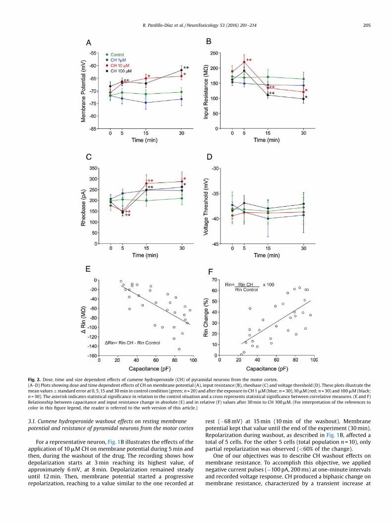
Fig. 2. Dose, time and size dependent effects of cumene hydroperoxide (CH) of pyramidal neurons from the motor cortex.(A–D) Plots showing dose and time dependent effects of CH on membrane potential (A), input resistance (B), rheobase (C) and voltage threshold (D). These plots illustrate themean values � standard error at 0, 5,15 and 30 min in control condition (green; n = 20) and after the exposure to CH 1 mM (blue; n = 30),10 mM (red; n = 30) and 100 mM (black;n = 30). The asterisk indicates statistical significance in relation to the control situation and a cross represents statistical significance between correlative measures. (E and F)Relationship between capacitance and input resistance change in absolute (E) and in relative (F) values after 30 min to CH 100 mM. (For interpretation of the references tocolor in this figure legend, the reader is referred to the web version of this article.)
R. Pardillo-Díaz et al. / NeuroToxicology 53 (2016) 201–214 205
3.1. Cumene hydroperoxide washout effects on resting membranepotential and resistance of pyramidal neurons from the motor cortex
For a representative neuron, Fig. 1B illustrates the effects of theapplication of 10 mM CH on membrane potential during 5 min andthen, during the washout of the drug. The recording shows howdepolarization starts at 3 min reaching its highest value, ofapproximately 6 mV, at 8 min. Depolarization remained steadyuntil 12 min. Then, membrane potential started a progressiverepolarization, reaching to a value similar to the one recorded at
rest (�68 mV) at 15 min (10 min of the washout). Membranepotential kept that value until the end of the experiment (30 min).Repolarization during washout, as described in Fig. 1B, affected atotal of 5 cells. For the other 5 cells (total population n = 10), onlypartial repolarization was observed (<60% of the change).
One of our objectives was to describe CH washout effects onmembrane resistance. To accomplish this objective, we appliednegative current pulses (�100 pA, 200 ms) at one-minute intervalsand recorded voltage response. CH produced a biphasic change onmembrane resistance, characterized by a transient increase at
206 R. Pardillo-Díaz et al. / NeuroToxicology 53 (2016) 201–214
5 min of drug application which then progressively fell undercontrol values. In Fig. 1B, cell resistance shifted from 184 MV, incontrol, to 223 MV at 5 min. Later, it progressively diminished untilreaching 180 MV after recording for 15 min (10 min of washout,indicated by the third arrow in the figure). The same valueremained for a 20 min recording (15 min of washout). At 30 min ofthe recording, the value registered was 181 MV (fourth arrow inthe figure). A total of 6/10 cells presented the same result, while therest showed a membrane resistance with values under the onesregistered at the beginning of the experiment. This could be due toa late effect of CH on resistance, and not necessarily to the washoutitself.
We also looked into the effect of the washout on the firingrepetitive properties. After a CH exposure of 5 min, all the cells(n = 10) in the study presented a decrease in gain (55.2 � 12.6 APs�1 nA�1 vs 36.7 � 15.5 AP s�1 nA�1). After the washout, 4 cellsshowed an improvement in the gain and maximum firingfrequency (<40% of the change). Four neurons did not presentany kind of reversibility to the effects of CH and retained a low gainand low maximum frequency. The remaining 2 cells completelylost their ability to repetitively discharge action potentials duringthe washout. From these experiments, we can conclude that ashort exposure to the drug (5 min) allows for a partial recovery
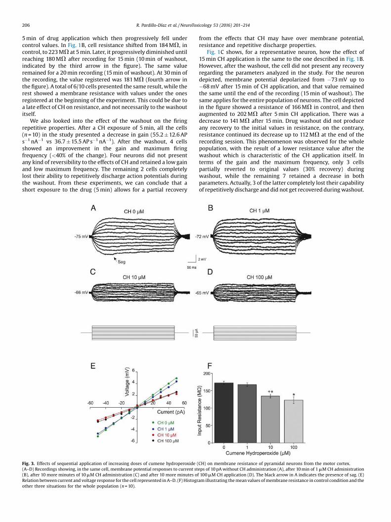
Fig. 3. Effects of sequential application of increasing doses of cumene hydroperoxide ((A–D) Recordings showing, in the same cell, membrane potential responses to current st(B), after 10 more minutes of 10 mM CH administration (C) and after 10 more minutes oRelation between current and voltage response for the cell represented in A–D. (F) Histogrother three situations for the whole population (n = 10).
from the effects that CH may have over membrane potential,resistance and repetitive discharge properties.
Fig. 1C shows, for a representative neuron, how the effect of15 min CH application is the same to the one described in Fig. 1B.However, after the washout, the cell did not present any recoveryregarding the parameters analyzed in the study. For the neurondepicted, membrane potential depolarized from �73 mV up to�68 mV after 15 min of CH application, and that value remainedthe same until the end of the recording (15 min of washout). Thesame applies for the entire population of neurons. The cell depictedin the figure showed a resistance of 166 MV in control, and thenaugmented to 202 MV after 5 min CH application. There was adecrease to 141 MV after 15 min. Drug washout did not produceany recovery to the initial values in resistance, on the contrary,resistance continued its decrease up to 112 MV at the end of therecording session. This phenomenon was observed for the wholepopulation, with the result of a lower resistance value after thewashout which is characteristic of the CH application itself. Interms of the gain and the maximum frequency, only 3 cellspartially reverted to original values (30% recovery) duringwashout, while the remaining 7 retained a decrease in bothparameters. Actually, 3 of the latter completely lost their capabilityof repetitively discharge and did not get recovered during washout.
CH) on membrane resistance of pyramidal neurons from the motor cortex.eps of 10 pA without CH administration (A), after 10 min of 1 mM CH administrationf 100 mM CH application (D). The black arrow in A indicates the presence of sag. (E)am illustrating the mean values of membrane resistance in control condition and the

R. Pardillo-Díaz et al. / NeuroToxicology 53 (2016) 201–214 207
Therefore, continuous exposure to CH (15 min) produced alter-ations to membrane properties that were hardly reversible.
3.2. Dose dependent effects of cumene hydroperoxide on membraneproperties of pyramidal neurons from the motor cortex
Dose response effects of CH were studied during 30 min onmembrane properties, namely membrane potential, input resis-tance, rheobase and threshold voltage. Fig. 2A shows the effects onmembrane potential for control and the 3 concentrations (1, 10 and100 mM). In the cases of control and 1 mM CH, no significantchanges were observed for any of the cells along the 30 minduration of the study. However, for the case of the 10 mM CHconcentration, membrane potential significantly depolarized at
Fig. 4. Dose and time dependent effects of cumene hydroperoxide (CH) on action potent(D), maximum frequency (E) and cancelation current (F) of pyramidal neurons from thThese plots illustrate the mean values � standard error at 0, 5, 15 and 30 min in control con = 30) and 100 mM (black; n = 30). The asterisk indicates statistical significance in relatcorrelative measures. (For interpretation of the references to color in this figure legend
5 min, and even though it reached values slightly more depolarizedat 15 min and 30 min, those differences were not statisticallysignificant when compared with the ones obtained at 5 min. Whenthe concentration of 100 mM CH was applied, depolarization wassignificant at 30 min (�68.6 � 1.3 in initial condition vs�61.9 � 1.4 at 30 min).
Fig. 2B shows the effects on membrane resistance in a pool ofneurons in control condition (n = 20) and each one of the other3 populations of neurons were exposed to one of the 3 drugconcentrations of the study (n = 30 for each one). As seen in thefigure, membrane resistance does not change in control conditionover time neither when 1 mM CH is applied during the 30 min theexperiment lasted. However, input resistance showed a biphasicresponse for the other 2 concentrations, with a significant increase
ial amplitude (A), action potential duration (B), firing repetitive properties (C), gaine motor cortex.ndition (green; n = 20) and after the exposure to CH 1 mM (blue; n = 30), 10 mM (red;ion to the control situation and a cross represents statistical significance between, the reader is referred to the web version of this article.)

208 R. Pardillo-Díaz et al. / NeuroToxicology 53 (2016) 201–214
at 5 min. Subsequently, we observed a significant decrease at15 and 30 min, respectively, when compared with the initial value.More specifically, input resistance augmented at 5 min for the100 mM CH concentration (162.1 �9.5 MV vs 189.2 � 12.4 MV).Later, it diminished at 15 and 30 min (110.6 � 9.8 MV and99.7 � 11.1 MV, respectively). Values at 30 min were slightly lowerthan the ones obtained at 15 min, but there was not a significantdifference between them. The 10 and the 100 mM CH concen-trations had bigger (and significant) effects than 1 mM CH at 5 min,but no differences were found between 10 and 100 mM CH.
To confirm that the 10 and 100 mM concentrations producedsimilar effects on resistance another experiment was carried out(n = 10), in which we analyzed the voltage responses to current
Fig. 5. Effects of sequential application of increasing doses of cumene hydroperoxide ((A–D) Recordings showing, in the same cell, voltage response to depolarizing current puls1 mM CH administration (B), after 10 more minutes of 10 mM CH administration (C) and affiring frequency for the cell represented in A–D. (F) Histogram illustrating the mean valukept their firing properties during 30 min (n = 6).
injections while the neuron was being sequentially exposed toincreasing drug concentrations for 10 min: 0, 1, 10 and 100 mM ofCH. Fig. 3 shows, for a representative neuron, that voltageresponses were similar before drug application and for the1 mM CH concentration, both demonstrating that membraneresistance was not modified in this situation. However, in theabsence of the drug, the neuron (Fig. 3A) presents a voltagerectification of the membrane potential (known as “sag”)approximately at 80 ms, which disappears after 1 mM CH applica-tion (Fig. 3B). The resulting voltage responses obtained when theapplication was of 10 and 100 mM CH were smaller than thoseobtained in control, and were also accompanied by a membranedepolarization from �75 mV to �66 and �65 mV, respectively. The
CH) on firing repetitive properties of pyramidal neurons from the motor cortex.es of 1 s duration and 500 pA intensity without CH administration (A), after 10 min ofter 10 more minutes of 100 mM CH application (D). (E) Relation between current andes of gain in control condition and the other three situations for the population that

R. Pardillo-Díaz et al. / NeuroToxicology 53 (2016) 201–214 209
measure of the membrane resistance for each neuron, before drugapplication and for the 3 drug concentrations, was calculated as theslope that represents the relation between current intensity andvoltage response as depicted in Fig. 3A–D. For the neuron describedin Fig. 3A–D, resistance was 103 MV without CH, 102 MV in 1 mMCH. It dropped to 51 MV in 10 mM CH, and to 50 MV in 100 mM CH,respectively (Fig. 3E). For the whole population (n = 10) resistancewas 170.2 � 6.1 MV in control, 165.3 � 6.7 MV in 1 mM CH. It wentdown again to 135.7 � 5.7 MV in 10 mM CH, and 120.6 � 11.8 MV in100 mM CH, respectively (Fig. 3F). From this data, we conclude thatneurons do not evidence any alteration to membrane potential andresistance in the 1 mM CH concentration in respect to initial values,and that the effects of the 100 mM CH concentration do not varysignificantly from the changes produced by the administration of10 mM CH.
3.3. Dose dependent effects of cumene hydroperoxide on rheobase andvoltage threshold of pyramidal neurons from the motor cortex
The administration of CH triggered up some changes on therheobase of pyramidal neurons from the motor cortex. Fig. 2Cillustrates the effects on the rheobase which depend on the timeand the dose of the CH concentration. No significant change couldbe found on rheobase in control condition and when 1 mM CH wasapplied during 30 min. However, when the concentration appliedwas 10 or 100 mM CH, the rheobase decreased significantly after5 min application in comparison with initial condition (forexample, for 100 mM the decrement was 193.7 � 10.6 pA vs149.6 � 15.7 pA) while it increased significantly if compared withinitial condition after 15 and 30 min application (for 100 mM, theincrease was 249.9 � 41.6 pA and 263.1 �35.3 pA). Values at 30 minwere only slightly larger than those obtained at 15 min, and theydid not differ significantly. Changes in rheobase were parallel tochanges in resistance and, therefore, the voltage thresholdremained unaltered during the 30 min duration for the 3 concen-trations (Fig. 2D). As a conclusion, Ohm’s Law is observable in themembrane behavior of pyramidal neurons, even under the effect ofdifferent concentrations of the drug.
Later on, we analyzed whether the effects of oxidative stressproduced by CH on membrane properties would depend onneuronal size: that is to say, whether larger neurons presented abigger sensitivity to CH or conversely. An estimation of theneuronal size can be inferred from membrane capacitance. Fig. 2Eand F shows the relation between capacitance and resistancechanges of neurons recorded at 100 mM CH, in absolute values(Fig. 2E) and in relative values (Fig. 2F). Data shows a linear relationbetween capacitance and input resistance change when calculatedfrom absolute values (P < 0.001; r > 0.69), and also from relativesvalues (P < 0.001; r > 0.71). Similar results were obtained when10 mM CH was administered (not shown). From these figures, wecan conclude that pyramidal neurons from the motor cortex thathave a larger membrane surface are more likely to sufferalterations in their electrical membrane properties by oxidativestress mediated by CH.
3.4. Dose dependent effects of cumene hydroperoxide on amplitudeand duration of the action potential of pyramidal neurons from themotor cortex
Fig. 4A describes the effects of CH on action potential amplitudein control condition and at the 3 different concentrations. Nodifferences were found in control condition for this parameterwhile the 1 mM CH concentration produced a gradual decrease onaction potential amplitude, which became statistically significantat 30 min. For the 10 and 100 mM concentrations, the meandecrease was more evident, reaching statistical significance at
5 min as compared to the initial condition, and at 30 min incomparison with 15 min. For the 100 mM concentration underinitial condition, the mean amplitude was 120.1 � 2.0, going downto 114.8 � 5.7 mV at 5 min, and even lower to 110.3 � 3.8 mV(15 min) and 91.2 � 3.3 mV (30 min). The highest differencebetween initial and final condition for the measure of theamplitude could be seen when the concentration applied was100 mM. More specifically, for all concentrations, amplitudedifferences were the following: 8.4 � 3.9, 18.8 � 2.3 and28.9 � 5.8 mV, for 1, 10 and 100 mM CH respectively. Differencesof the effects on the action potential were significant between the3 concentrations at 30 min.
In Fig. 4B, we show the effects on the duration of the actionpotential in control condition and for the 3 concentrations. Thisparameter became progressively longer after the 10 and 100 mMCH exposure while remaining unaltered in control condition andafter the 1 mM CH application. However, statistical significancewas already observable at 5 min in comparison with controlcondition, and 30 min in comparison with 15 min condition, forboth 10 mM and 100 mM CH. For example, and for the administra-tion of 100 mM CH, the duration of the action potential was1.50 � 0.11 ms (0 min), 1.82 � 0.13 ms (5 min), 1.87 � 0.28 ms(15 min), and, finally, 2.31 �0.15 ms (30 min).
3.5. Dose dependent effects of cumene hydroperoxide on firingproperties of pyramidal neurons from the motor cortex
The pyramidal neurons in the study exhibited a phasic-tonicdischarge as a response to sustained current depolarizing pulses,not only in control, but also when the concentration applied was1 mM CH (see Fig. 5A and B). Nevertheless, after the administrationof 10 mM CH, 20% of the cells (15 min) and 43% (30 min) lost theirability to discharge action potentials in a repetitive way. This effectwas even greater when the concentration was 100 mM CH affecting40% (5 min), 80% (15 min), and 100% (30 min) of the cells (Fig. 4C).
Fig. 5A–D shows, for a representative neuron (n = 10), the resultsof another experiment in which we analyzed the repetitive firingproperties observed while the neuron was being sequentiallyexposed to each of 3 different drug concentrations for 10 min.Before drug application (0 mM), and for the 1 and 10 mM CHconcentrations, the neuron illustrated in Fig. 5 could dischargealong the whole pulse duration (1 s). However, this ability is lostafter 10 min of being exposed to 100 mM CH, as being depicted inFig. 5D. In this figure, we can observe how the cell ceases todischarge before the pulse is over. Also in Fig. 5A–C, it can beobserved how the firing frequency decreases as the concentrationincreases: 23 AP s�1 in control, 19 AP s�1 for 1 mM CH, and 12 AP s�1
for 10 mM CH. Therefore, this data may suggest that an incrementaldose alters the gain of the discharge. Fig. 5A–D also shows adecrease of amplitude of the action potential in the train, whichbecomes more evident for the 100 mM CH concentration.
We then measured the gain, maximum frequency andcancelation current in those cells that retained their repetitivefiring properties. Fig. 5E represents the relationship between theintensity of the current injected and the firing frequency for thecell depicted in Fig. 5A–D. In this figure, it can be observed how thatrelationship is linear everytime low current intensities wereapplied. However, when intensity increases, accommodationsymptoms are appreciated. Fig. 5E shows how the neuron startsto accommodate at values close to 500 pA for 0 mM CH, 450 pA for1 mM CH, and 400 pA for 10 mM CH. This data indicates that theworking intensity range decreases as the drug concentrationincreases. Besides, when we calculated the gain for the neuronshown in Fig. 5A–E, we could see how this parameter decreasedafter CH application: 53 AP s�1 nA�1 (0 mM CH), 46 AP s�1 nA�1
(1 mM CH), and 26 AP s�1 nA�1 (10 mM CH). This result is applicable

210 R. Pardillo-Díaz et al. / NeuroToxicology 53 (2016) 201–214
to the rest of the population. Fig. 5F shows the mean values of thegain in the different accumulating situations (time and drugconcentration): 40.1 �5.9 AP s�1 nA�1 (0 mM CH), 38.3 � 4.9 AP s�1
nA�1 (1 mM CH), 27.7 � 8.6 AP s�1 nA�1 (10 mM CH) and 15.3 � 4.1AP s�1 nA�1 (100 mM CH). This decrease was statistically significantfor the 10 mM CH and the 100 mM CH concentrations (see Fig. 5F)when compared with initial values and between themselves.
In order to carry out a more detailed analysis of the responseeffects on the gain and maximum frequency, a pool of 30 neuronswere exposed to one of the 3 drug concentrations tested in ourstudy. Fig. 4D illustrates how the gain remained unchanged incontrol condition but progressively decreased after the adminis-tration of 1 mM CH, with statistical significance at 30 min. Thisdecrease in gain was already significant at 15 min for the 10 mM CH,and even earlier (5 min) for the 100 mM CH concentration. For thehighest concentration, the decrease in gain was significant again at15 min when compared with 5 min. By 30 min, all cells had alreadylost their ability to discharge repetitively. Maximum firingfrequency progressively decreased with time, as seen in Fig. 4E,but did not reach significance when 1 mM CH concentration wasadministered. Significance was achieved (5 min) for the 10 and the100 mM CH concentrations. Thus, after 5 min of 100 mM CHexposure, the maximum firing frequency decreased from 27.2 � 3.0AP s�1 to 17.6 � 3.2 AP s�1. The decrease in the maximum frequencywas significantly lower again at 15 min and at 30 min, but only afteradministering 100 mM of CH.
Fig. 6. Prevention by melatonin of lipid peroxidation (LPO) induced by cumene hydrop(A) Graphic showing LPO values, expressed in %, at 0, 5,15 and 30 min after the exposure oLPO values, expressed in %, at 0, 5,15 and 30 min after the exposure of CH 10 mM (n = 6) andthat membrane potential and voltage response to negative current pulses of �100 pA aArrows indicate voltage response at 5, 15 and 30 min. The letter ”a” indicates statisticabetween a concentration and the precedent one for the same period of time, and “c” shmelatonin for the same period of time. (For interpretation of the references to color in
Further evidence of the narrowing of the working intensityrange, after the administration of CH, was observed whenanalyzing the cancelation current (Fig. 4F). This figure illustrateshow the cancelation of repetitive discharge occurs at lowerintensities at doses of 10 and 100 mM CH, but is unaffected incontrol condition and at 1 mM CH. In experiments in which the100 mM CH concentration was tested, the average value for thepopulation in the initial condition was 650.3 � 48.4 pA. Intensityvalues fell progressively to 599.3 � 56.5 pA at 5 min, and503.7 �49.9 pA at 15 min, at which level statistical significancewas reached. After that, it dropped to a value of zero with thecomplete loss of the ability to discharge repetitively for all neuronsat 30 min. Once again, the 10 and 100 mM CH concentrations hadlarger and more significant effects than the 1 mM and amongthemselves with respect to repetitive firing properties (gain,maximum frequency and cancelation current) at 30 min.
3.6. Prevention of lipid peroxidation and membrane excitabilitychanges caused by cumene hydroperoxide by melatonin
Fig. 6A illustrates LPO expressed in percentage as the increasewhen compared with the control situation, as a result of theexposure of the brain slices to 0 (control), 1, 10 and 100 mM of CHduring 0, 5, 15 and 30 min. As seen in Fig. 6A, CH evokes LPO in adose and time dependent manner. Longer time or higher dosecaused a progressive increase in LPO, significant at 5 min at thethree concentrations of CH tested in respect to the control.
eroxide (CH) on pyramidal neurons from the motor cortex.f CH 1 mM (blue; n = 6), 10 mM (red; n = 6) and 100 mM (black; n = 6). (B) Plot showing
CH 10 mM plus melatonin 50 mM (n = 6). (C) Electrophysiological recording showingnd 500 ms remain stable along 30 min exposition to CH 10 mM + 50 mM melatonin.l significance in relation to control situation, “b” represents statistical significanceows statistical significance between 10 mM CH application and 10 mM CH + 50 mM this figure legend, the reader is referred to the web version of this article.)
R. Pardillo-Díaz et al. / NeuroToxicology 53 (2016) 201–214 211
However, the increased lipid peroxide level plateaued in 15 and30 min. Furthermore, it is also observable in the fact that the levelof LPO with CH 1 mM in 30 min is similar to the average value foundafter 1 mM CH exposure in 5 min. Although the increment in LPO isdose dependent, mainly at shorter times, those increments are notproportional to the concentration, i.e., increments in LPO foundwith 100 mM CH are bigger but not statistically different than thosefound with 10 mM CH, but they are statistically different whencompared 10 vs 1 mM CH.
No effect was observed in LPO in cells treated with ACSF with50 mM melatonin when compared with cells treated with ACSFalone. As seen in Fig. 6B, melatonin prevents LPO induced by 10 mMCH. No significant differences in LPO were found between tissuestreated with melatonin alone (control of melatonin) and tissuestreated with melatonin and 10 mM CH at 5,15 or at 30 min althougha moderate increase of LPO was found at 30 min. Irrespective of thetime period applied (5, 15 and 30 min) the differences foundbetween tissues treated with ACSF containing 10 mM of CH andtissues treated with ACSF containing 10 mM of CH and melatonin50 mM, were always significant.
As melatonin prevents the LPO induced by CH, we wondered ifpre-treatment with this antioxidant could act as a neuroprotectorand could prevent the membrane changes induced by 10 mM CH.Fig. 6C illustrates the effects of 50 mM melatonin and 10 mM CHapplication on membrane potential during 30 min. As seen in thisfigure, no changes on membrane potential or input resistance wereproduced for the whole period. The same response was found in4 of the 6 neurons studied and no changes in membrane potential,input resistance or firing properties were found. However, 2 of the6 cells showed gain decrease, around 30 min.
4. Discussion
In the present work, we have studied early changes inmembrane properties of pyramidal motor cortex neurons exposedto 3 concentrations of CH (1,10 and 100 mM). Our data indicate thatCH depresses the excitability of pyramidal motor cortex neuronsdecreasing input resistance, amplitude of the action potential, andgain and maximum frequency of the repetitive firing discharge.These effects were time and dose dependent. Reversibility toalterations produced by CH to membrane properties is inverselyproportional to exposure time: the longer the exposure, the lowerthe reversibility. Among all the membrane properties analyzed, theability to repetitively discharge action potentials was the propertymost affected by the administration of the drug. Our data alsosuggest that large neurons of the motor cortex have moresensitivity to the oxidant. Finally, we have shown that pre-treatment with melatonin, an antioxidant agent, prevents theeffects induced by CH on membrane properties.
The antioxidant benefits of melatonin have been extensivelydescribed in the literature (Reiter et al., 2000; Tan et al., 2007).
4.1. Methodological considerations
Oxidative stress is caused when the production of ROS exceedsthe antioxidant capacity of the tissues (Heather and Teismann,2009; Ljubisavljevic, 2014; Sims-Robinson et al., 2013). In neuronalcell damage, it is believed that this high level of ROS makes a largecontribution to neuron degeneration and might be one factor in thedevelopment of different neurological diseases, such as ALS,Parkinson’s and Alzheimer’s (Andersen, 2004; Reynolds et al.,2007). Our research study has considered pyramidal neurons of thelayer V of the primary motor cortex. Although there are severalpathophysiological mechanisms that trigger motor neuron vul-nerability in ALS, oxidative stress has been described to be animportant factor in the case of pyramidal neurons of the motor
cortex (Cleveland and Rothstein, 2001; Kim et al., 2014). Our resultsdemonstrate that CH is an organic oxidant agent that induceslipidic peroxidation on the cell membrane which, consequently,results in cell alterations (Ayala et al., 2014; Nakaya et al., 1992;Vimard et al., 2011). Indeed, we have demonstrated that the effectsof CH on membrane properties are due to oxidative stress becausemelatonin prevents the electrophysiological effects of CH. In Caiet al. (2009), malondialdehyde, a typical intermediate of lipidperoxidation, gradually damaged hippocampal neurons followinga 3 h exposure at concentrations of 1, 10, 100 and 1,000 mM.According to these authors, malondialdehyde may have biochemi-cal effects on hippocampal neurons that are time and dosedependent. A similar conclusion was reached in works that studiedthe dose response effects of CH toxicity on cultured cells (Shimuraet al., 1985; Vimard et al., 1996, 2011; Vroegop et al., 1995). Thosestudies proved that cell mortality rate is low (less than 5%) whenCH is applied in concentrations under 1000 mM, and withadministration time under 30 min. However, they also reporthow mortality rate reached approximately 70% when administra-tion times are longer (60–180 min) or when drug concentrationsare larger. On the basis of this evidence, we decided to use 3 CHconcentrations (1,10 and 100 mM) and 30 min administration time,which guarantee a low mortality rate, in order to study the doseand time dependent effects of CH. We have analyzed severalelectrophysiological parameters during drug administration withdata being recorded at 5, 15 and 30 min to identify alterations tomembrane properties. Prior to that, we checked that the neurons inthe motor cortex keep their membrane properties stable, underour recording conditions, for a duration of 30 min, as previouslydemonstrated with other pools of neurons (Nani et al., 2010).Therefore, any alteration to the physiological parameters analyzedcan be considered to be the effect of the drug administration to thecells and not to the recording conditions.
4.2. Alteration of membrane properties by oxidative stress
No effects on membrane potential or resistance were observedin our study in control condition and when CH was administered at1 mM. At that concentration, it seems that neurons are able to blockthe oxidative effects on the physiological properties of themembrane, although a significant level of LPO was found. Areason to explain this could be that pyramidal neurons have anefficient scavenging enzyme system that reacts rapidly with CH ata 1 mM concentration, as proposed by Jovanovic and Jovanovic(2013) when attempting to explain the effects that oxidative stresshas on the electrophysiological properties of Retzius neurons of theleech. The values of LPO we found at the 1 mM concentration wereunder 15% of increment in respect to those registered for control.These small values are similar to those found during ageing in thehypothalamus–hypophysis, (Argüelles et al., 2011). It seems thatthose levels of LPO are not high enough to alter membraneproperties. Contrary to the results obtained at the 1 mMconcentration, our exposure of cortical neurons to CH concen-trations of 10 and 100 mM already produced changes in membranepotential, membrane resistance and rheobase as a result of higherlevels of LPO. An unexpected finding was that the 2 concentrationscaused changes of equal magnitude in these parameters. Thisfinding may reflect that values of LPO found when 100 mM wasadministered were only slightly higher, but not significant, thanthose obtained with 10 mM. Our results suggest that oxidativestress affected the channels and/or conductances that determinepassive membrane properties (Frantseva et al., 1998; Nakaya et al.,1992; Nani et al., 2010; Pardillo-Díaz et al., 2015) along a verynarrow concentration range of 1–10 mM, which resembles an “all-or-nothing” response. In any case, conductances that are affectedby oxidative stress would not be those involved in determining the

212 R. Pardillo-Díaz et al. / NeuroToxicology 53 (2016) 201–214
voltage threshold. In our study, regardless of the CH dose used,voltage threshold remained unchanged, as occurs in neurons of thehippocampus exposed to peroxide (Pellmar, 1987).
Exposure of leech neurons to CH (0.25, 1, and 1.5 mM) extendedthe duration of the action potentials in a dose dependent manner(Jovanovic and Jovanovic, 2013). For example, the concentration of0.25 mM did not significantly change the action potential within20 min, but it was significant with 1 mM. Moreover, a higherconcentration (1.5 mM) of CH caused an extreme change in theshape and duration of the action potential. In our study, weobserved that the action potential diminished, while its durationincreased in a progressive way along the time of exposure to CHand in a concentration dependent manner. When the CHconcentration was of 1 mM, the effects on the amplitude of theaction potential were evident at 30 min drug administration.However, with the 10 or 100 mM CH concentrations, effects werealready significant at 5 min, showing the highest alterations inamplitude with the 100 mM CH concentration. It has beenproposed that oxidative stress could be affecting conductancesthat participate in the repolarization of the action potential and theNa+ ionic gradients (Angelova and Muller, 2006; Jovanovic andJovanovic, 2013; Nakaya et al., 1992; Pardillo-Díaz et al., 2015). Thecapability of maintaining repetitive firing of the action potentials isprobably the most dramatic effect produced by CH over theelectrical properties of the membrane. Our results are similar tothose obtained by Jovanovic and Jovanovic (2013) in which CH(1.5 mM, 20 min) produced a complete cancelation of firingproperties in leech neurons. However, they do not coincide withthe study by Nani et al. (2010) in which their H2O2 oxidative model(1 mM, 30 min) caused an increase in the firing of the hypoglossalmotoneurons of rats. This discrepancy might be due to the fact thatH2O2 can only exert the effects achieved by CH at concentrations10 times higher (Vimard et al., 2011). Regardless of which channelsand/or conductances are underlying action potential and repetitivefiring (Pardillo-Díaz et al., 2015), in our study we have demon-strated that they are dramatically affected by oxidative stress in aconcentration dependent manner, being a low concentration of CH(100 mM) high enough to completely block firing discharge.
4.3. Functional considerations
We can conclude that oxidative stress induced by CH evokesimportant changes, in a concentration and time dependentmanner, in the neuronal excitability of rat motor cortex neuronsas demonstrated in modified hippocampal cells (Oh et al., 2012).Low concentration of the drug (1 mM), but only with a longexposure, already blocks the Ih current underlying sag (Robinsonand Siegelbaum, 2003) and may alter conductances involved in theaction potential, and firing properties, at a drug concentrationwhich has no biochemical, physiological or morphological effectsin other neuronal populations (Jovanovic and Jovanovic, 2013;Sinha et al., 2015; Vimard et al., 2011). A concentration of 10 mM ofCH produced changes in membrane potential, membrane resis-tance and rheobase as previously demonstrated by Pardillo-Díazet al. (2015) in a time dependent manner. Finally, 100 mMcompletely blocks the ability to produce repetitive discharge ofaction potentials with long term exposure. Brief exposure (15 minor less) also narrow the current working range because of anincrease in the rheobase and a decrease of the cancelation current.Then, we can conclude that larger concentrations of CH havesimilar consequences as those evoked by smaller concentrations atlonger exposure times.
Present data demonstrate that effects induced by a briefexposure to CH (5 min) are partially reversible, while thoseproduced by a long-term exposure to CH (15 min) are practicallyirreversible. Membrane potential and resistance presented higher
reversibility to the effects of CH. Similarly to our results, H2O2
induced a large depolarizing shift in membrane potential that wasreversible in almost half of the thalamic neurons tested (Frantsevaet al., 1998). Also the change observed in input resistance could bereversed in some cases, but not in frequency depression ofspontaneous postsynaptic currents induced by H2O2 in hypoglossalmotoneurons (Nani et al., 2010). The reversibility of some of theoxidative stress-mediated effects is difficult to explain (Pellmar,1987). Oxygen radicals produced by CH might directly attack ionchannel proteins, or LPO caused by CH might indirectly inhibit ionchannel functions by altering the membrane lipid milieusurrounding the channel protein (Nakaya et al., 1992). Effectsreported to be reversible may be mediated by modified lipid–protein interactions rather than by irreversible protein oxidation(Frantseva et al., 1998). Then, our results may suggest that CHeffects on resting membrane potential and membrane resistanceare, at least in part, a consequence of the observed LPO and, for thatreason, their modifications could be partially reversed with aprompt washout of the drug. This work demonstrates that CHinduces LPO in pyramidal neurons and that it can be prevented bythe use of melatonin. Besides, melatonin also partially or totallyprevents the physiological changes that occur in cortical neuronsunder CH conditions. The antioxidant benefits of melatonin havebeen extensively described. It has been demonstrated thatmelatonin has direct scavenging actions against the free radicalsand related products (Reiter et al., 2000; Tan et al., 2007), actsindirectly by inducing antioxidant enzymes (Reiter et al., 2000),increases the activities of the major antioxidant enzymes (Reiter,2000) and stimulates glutathione synthesis (Barlow-Walden et al.,1995). Melatonin has also been shown to reduce the accumulationof the major products of LPO when membranes are exposed toradical-generating agents (Reiter, 2000) and to preserve, in vivoand in vitro, protein synthesis under oxidative stress condition(Argüelles et al., 2012). Then, we cannot discard in our study thatother mechanisms underlying membrane changes induced by CH,apart from LPO, such as protein oxidation, can also be prevented bymelatonin. However, since 50 mM melatonin has not totallyprevented the effects of long time exposition to CH in some cells,additional trials with melatonin alone (a higher dose) and incombination with other drugs, such as thiol-reducing agents likeN-acetylcysteine or b-mercaptoethanol (Cabungcal et al., 2014;Vimard et al., 2011), or other anti-oxidant substances, such asvitamin C (Chang et al., 2008), are needed to clarify its potentialbenefit. Nevertheless, we propose that neuroprotective agentsshould be administered before membrane properties of neuronsstart to change, given the low reversibility of the effects caused byoxidative stress when this is maintained in time.
In motoneurons, cell capacitance has been used as anestimation for neuronal size (Viana et al., 1994). In other words,larger neurons would present a higher capacitance. Our resultsprovide the first experimental evidence that neurons mostlyaffected by oxidative stress are the largest ones, those with thehighest values of capacitance. Pyramidal neurons of the motorcortex are rather large in comparison with neurons from otherlayers of the motor cortex and from other brain nuclei (Oswaldet al., 2013). Furthermore, regarding pyramidal neurons from themotor cortex, as from other cortical areas, it has been demonstrat-ed that morphological parameters revealed clear differencesbetween large soma, thick shafted, large tufted corticofugalneurons and small soma, slender shafted, small tufted commis-sural neurons (Chagnac-Amitai et al., 1990; Mason and Larkman,1990; Oswald et al., 2013). We propose that corticofugal neuronscould be the most affected by oxidative stress in our study. If weconsidered oxidative stress to be the main cause of neuronal deathin neurodegenerative pathologies (Andersen, 2004; Cabungcalet al., 2014; Cleveland and Rothstein, 2001; Niedzielska et al., 2015;

R. Pardillo-Díaz et al. / NeuroToxicology 53 (2016) 201–214 213
Parakh et al., 2013; Reynolds et al., 2007), our findings (large sizeand high sensitivity to oxidative stress even a low concentration)would serve to explain how pyramidal neurons of the motorcortex, in particular corticofugal neurons, also have the highestdeath rate levels in diseases such as ALS.
Acknowledgements
This work was partially supported by ERDF (European RegionalDevelopment Fund) UNSE10-1E-0949. Mr. Ricardo Pardillo-Diazobtained a research fellowship funded by CACOF. We sincerelythank The Centre of Research Technology and Innovation of theUniversity of Seville (CITIUS) for their technical support, and Ms.Rosa María Andrade García for her technical assistance. Gratitudeto Professor Juan Ramón Lacalle Remigio is expressed for his adviceon statistical analysis.
References
Andersen, J.K., 2004. Oxidative stress in neurodegeneration: cause or consequence?Nat. Med. Suppl. 10, S18–S25.
Angelova, P., Muller, W., 2006. Oxidative modulation of the transient potassiumcurrent IA by intracellular arachidonic acid in rat CA1 pyramidal neurons. Eur. J.Neurosci. 23, 2375–2384.
Argüelles, S., Cano, M., Machado, A., Ayala, A., 2011. Effect of aging and oxidativestress on elongation factor-2 in hypothalamus and hypophysis. Mech. AgeingDev. 132, 55–64.
Argüelles, S., Muñoz, M.F., Cano, M., Machado, A., Ayala, A., 2012. In vitro and in vivoprotection by melatonin against the decline of elongation factor-2 caused bylipid peroxidation: preservation of protein synthesis. J. Pineal Res. 53, 1–10.
Arrigo, A.P., 1999. Gene expression and the thiol redox state. Free Radic. Biol. Med.27, 936–944.
Ayala, A., Munoz, M.F., Arguelles, S., 2014. Lipid peroxidation: production,metabolism, and signaling mechanisms of malondialdehyde and 4-hydroxy-2-nonenal. Oxid. Med. Cell Longev. 360438.
Bogdanov, M., Brown, R.H., Matson, W., Smart, R., Hayden, D., O’Donnell, H., FlintBeal, M., Cudkowicz, M., 2000. Increased oxidative damage to DNA in ALSpatients. Free Radic. Biol. Med. 29, 652–658.
Cai, J., Chen, J., He, H., Yin, Z., Zhu, Z., Yin, D., 2009. Carbonyl stress: malondialdehydeinduces damage on rat hippocampal neurons by disturbance of Ca2+
homeostasis. Cell Biol. Toxicol. 25, 435–445.Barlow-Walden, L.R., Reiter, R.J., Abe, M., Pablos, M., Menendez-Pelaez, A., Chen, L.D.,
Poeggeler, B., 1995. Melatonin stimulates brain glutathione peroxidase activity.Neurochem. Int. 26, 497–502.
Cabungcal, J.H., Counotte, D.S., Lewis, E.M., Tejeda, H.A., Piantadosi, P., Pollock, C.,Calhoon, G.G., Sullivan, E.M., Presgraves, E., Kil, J., Hong, L.E., Cuenod, M., Do, K.Q., O’Donnell, P., 2014. Juvenile antioxidant treatment prevents adult deficits ina developmental model of schizophrenia. Neuron 83, 1073–1084.
Carrascal, L., Nieto-Gonzalez, J.L., Cameron, W.E., Torres, B., Nunez-Abades, P.A.,2005. Changes during the postnatal development in physiological andanatomical characteristics of rat motoneurons studied in vitro. Brain Res. BrainRes. Rev. 49, 377–387.
Carrascal, L., Nieto-Gonzalez, J.L., Nunez-Abades, P., Torres, B., 2006. Temporalsequence of changes in electrophysiological properties of oculomotormotoneurons during postnatal development. Neuroscience 140, 1223–1237.
Chagnac-Amitai, Y., Luhmann, H.J., Prince, D.A., 1990. Burst generating and regularspiking layer 5 pyramidal neurons of rat neocortex have differentmorphological features. J. Comp. Neurol. 296, 598–613.
Chang, H.M., Huang, Y.L., Lan, C.T., Wu, U.I., Hu, M.E., Youn, S.C., 2008. Melatoninpreserves superoxide dismutase activity in hypoglossal motoneurons of adultrats following peripheral nerve injury. J. Pineal Res. 44, 172–180.
Cleveland, D.W., Rothstein, J.D., 2001. From Charcot to Lou Gehrig: decipheringselective motor neuron death in ALS. Nat. Rev. Neurosci. 2, 806–819.
Davies, M.J., 2003. Singlet oxygen-mediated damage to proteins and itsconsequences. Biochem. Biophys. Res. Commun. 305, 761–770.
Emerit, J., Edeas, M., Bricaire, F., 2004. Neurodegenerative diseases and oxidativestress. Biomed. Pharmacother. 58, 39–46.
Ferretti, G., Bacchetti, T., 2011. Peroxidation of lipoproteins in multiple sclerosis. J.Neurol. Sci. 311, 92–97.
Fogarty, M.J., Noakes, P.G., Bellingham, M.C., 2015. Motor cortex layer V pyramidalneurons exhibit dendritic regression, spine loss, and increased synapticexcitation in the presymptomatic hSOD1(G93A) mouse model of amyotrophiclateral sclerosis. J. Neurosci. 35, 643–647.
Frantseva, M.V., Perez Velazquez, J.L., Carlen, P.L., 1998. Changes in membrane andsynaptic properties of thalamocortical circuitry caused by hydrogen peroxide. J.Neurophysiol. 80, 1317–1326.
Goldhaber, J.I., Ji, S., Lamp, S.T., Weiss, J.N.,1989. Effects of exogenous free radicals onelectromechanical function and metabolism in isolated rabbit and guinea pigventricle. Implications for ischemia and reperfusion injury. J. Clin. Invest. 83,1800–1809.
Goldhaber, J.I., Liu, E., 1994. Excitation–contraction coupling in single guinea-pigventricular myocytes exposed to hydrogen peroxide. J. Physiol. 477 (Pt 1), 135–147.
Gracanin, M., Hawkins, C.L., Pattison, D.I., Davies, M.J., 2009. Singlet-oxygen-mediated amino acid and protein oxidation: formation of tryptophan peroxidesand decomposition products. Free Radic. Biol. Med. 47, 92–102.
Han, M.H., Hwang, S.I., Roy, D.B., Lundgren, D.H., Price, J.V., Ousman, S.S., Fernald, G.H., Gerlitz, B., Robinson, W.H., Baranzini, S.E., Grinnell, B.W., Raine, C.S., Sobel, R.A., Han, D.K., Steinman, L., 2008. Proteomic analysis of active multiple sclerosislesions reveals therapeutic targets. Nature 451, 1076–1081.
Heather, L.M., Teismann, P., 2009. Glutathione—a review on its role and significancein Parkinson’s disease. FASEB J. 23, 3263–3272.
Hool, L.C., 2006. Reactive oxygen species in cardiac signalling: from mitochondria toplasma membrane ion channels. Clin. Exp. Pharmacol. Physiol. 33, 146–151.
Ihara, Y., Nobukuni, K., Takata, H., Hayabara, T., 2005. Oxidative stress and metalcontent in blood and cerebrospinal fluid of amyotrophic lateral sclerosispatients with and without a Cu, Znsuperoxide dismutase mutation. Neurol. Res.27, 105–108.
Ikawa, M., Okazawa, H., Tsujikawa, T., Muramatsu, T., Kishitani, T., Kamisawa, T.,Matsunaga, A., Yamamura, O., Mori, T., Hamano, T., Kiyono, Y., Nakamoto, Y.,Yoneda, M., 2014. Increased cerebral oxidative stress in amyotrophic lateralsclerosis: a 62CU-ATSM pet study. Neurology 84, 2033–2039.
Jiang, Z.Y., Woollard, A.C., Wolff, S.P., 1991. Lipid hydroperoxide measurement byoxidation of Fe2þ in the presence of xylenol orange: comparison with the TBAassay and an iodometric method. Lipids 26, 853–856.
Jovanovic, Z., Jovanovic, S., 2013. Comparison of the effects of cumenehydroperoxide and hydrogen peroxide on Retzius nerve cells of the leechHaemopis sanguisuga. Exp. Anim. 62, 9–17.
Kim, C., Lee, H.C., Sung, J.J., 2014. Amyotrophic lateral sclerosis – cell based therapyand novel therapeutic development. Exp. Neurobiol. 23, 207–214.
Kourie, J.I., 1998. Interaction of reactive oxygen species with ion transportmechanisms. Am. J. Physiol. 275, C1–24.
Lambert, A.J., Portero-Otin, M., Pamplona, R., Merry, B.J., 2004. Effect of ageing andcaloric restriction on specific markers of protein oxidative damage andmembrane peroxidizability in rat liver mitochondria. Mech. Ageing Dev. 125,529–538.
Ljubisavljevic, S., 2014. Oxidative stress and neurobiology of demyelination. Mol.Neurobiol. doi:http://dx.doi.org/10.1007/s12035-014-9041-x.
Mason, A., Larkman, A., 1990. Correlations between morphology andelectrophysiology of pyramidal neurons in slices of rat visual cortex. II.Electrophysiology. J. Neurosci. 10, 1415–1428.
Mochizuki, Y., Mizutani, T., Shimizu, T., Kawata, A., 2011. Proportional neuronal lossbetween the primary motor and sensory cortex in amyotrophic lateral sclerosis.Neurosci. Lett. 503, 73–75.
Muller, F.L., Lustgarten, M.S., Jang, Y., Richardson, A., Van Remmen, H., 2007. Trendsin oxidative aging theories. Free Radic. Biol. Med. 43, 477–503.
Nakaya, H., Takeda, Y., Tohse, N., Kanno, M., 1992. Mechanism of the membranedepolarization induced by oxidative stress in guinea-pig ventricular cells. J. Mol.Cell Cardiol. 24, 523–534.
Nam, T.G., 2011. Lipid peroxidation and its toxicological implications. Toxicol. Res.27, 1–6.
Nani, F., Cifra, A., Nistri, A., 2010. Transient oxidative stress evokes early changes inthe functional properties of neonatal rat hypoglossal motoneurons in vitro. Eur.J. Neurosci. 31, 951–966.
Niedzielska, E., Smaga, I., Gawlik, M., Moniczewski, A., Stankowicz, P., Pera, J., Filip,M., 2015. Oxidative stress in neurodegenerative diseases. Mol. Neurobiol. [Epubahead of print].
Nunez-Abades, P.A., Spielmann, J.M., Barrionuevo, G., Cameron, W.E., 1993. In vitroelectrophysiology of developing genioglossal motoneurons in the rat. J.Neurophysiol. 70, 1401–1411.
Oh, J.M., Choi, E.-K., Carp, R.I., Kim, Y.-S., 2012. Oxidative stress impairs autophagicflux in prion protein-deficient hippocampal cells. Autophagy 8 (10), 1448–1461.
Oswald, M.J., Tantirigama, M.L., Sonntag, I., Hughes, S.M., Empson, R.M., 2013.Diversity of layer 5 projection neurons in the mouse motor cortex. Front. Cell.Neurosci. 7, 174.
Parakh, S., Spencer, D.M., Halloran, M.A., Soo, K.Y., Atkin, J.D., 2013. Redox regulationin amyotrophic lateral sclerosis. Oxid. Med. Cell Longev. 40868, 1. doi:http://dx.doi.org/10.1155/2013/408681.
Pardillo-Díaz, R., Carrascal, L., Ayala, A., Nunez-Abades, P., 2015. Oxidative stressinduced by cumene hydroperoxide evokes changes in neuronal excitability ofrat motor cortex neurons. Neuroscience 289, 85–98.
Pellmar, T.C., 1987. Peroxide alters neuronal excitability in the CA1 region of guinea-pig hippocampus in vitro. Neuroscience 23, 447–456.
Pitt, D., Werner, P., Raine, C.S., 2000. Glutamate excitotoxicity in a model of multiplesclerosis. Nat. Med. 6, 67–70.
Racay, P., Kaplan, P., Mezesova, V., Lehotsky, J., 1997. Lipid peroxidation both inhibitsCa(2+)-ATPase and increases Ca2+ permeability of endoplasmic reticulummembrane. Biochem. Mol. Biol. Int. 41, 647–655.
Reiter, R.J., 2000. Melatonin: lowering the high price of free radicals. News Physiol.Sci. 15, 246–250.
Reiter, R.J., Tan, D.X., Osuna, C., Gitto, E., 2000. Actions of melatonin in the reductionof oxidative stress. A review. J. Biomed. Sci. 7, 444–458.
Reynolds, A., Laurie, C., Mosley, R.L., Gendelman, H.E., 2007. Oxidative stress and thepathogenesis of neurodegenerative disorders. Int. Rev. Neurobiol. 82, 297–325.
Robinson, R.B., Siegelbaum, S.A., 2003. Hyperpolarization-activated cation currents:from molecules to physiological function. Annu. Rev. Physiol. 65, 453–480.

214 R. Pardillo-Díaz et al. / NeuroToxicology 53 (2016) 201–214
Saba, L., Viscomi, M.T., Caioli, S., Pignataro, A., Bisicchia, E., Pieri, M., Molinari, M.,Ammassari-Teule, M., Zona, C., 2015. Altered functionality, morphology, andvesicular glutamate transporter expression of cortical motor neurons from apresymptomatic mouse model of Amyotrophic Lateral Sclerosis. Cereb. Cortex[Epub ahead of print].
Sha, W., Martins, A.M., Laubenbacher, R., Mendes, P., Shulaev, V., 2013. The genome-wide early temporal response of Saccharomyces cerevisiae to oxidative stressinduced by cumene hydroperoxide. PLoS One 8, e74939.
Shimura, J., Shimura, F., Hosoya, N., 1985. Functional disability of rat splenocytesprovoked to lipid peroxidation by cumene hydroperoxide. Biochim. Biophys.Acta 845, 43–47.
Sims-Robinson, C., Hur, J., Hayes, J.M., Dauch, J.R., Keller, P.J., Brooks, S.V., Feldman, E.L., 2013. The role of oxidative stress in nervous system aging. PLoS One 8,e68011. doi:http://dx.doi.org/10.1371/journal.pone.0068011.
Sinha, A., Chu, T.T.T., Dao, M., Chandramohanadas, R., 2015. Single-cell evaluation ofred blood cell bio-mechanical and nano-structural alterations upon chemicallyinduced oxidative stress. Sci. Rep. 59768 doi:http://dx.doi.org/10.1038/srep09768.
Smith, R.G., Henry, Y.K., Mattson, M.P., Appel, S.H., 1998. Presence of 4-hydroxynonenal in cerebrospinal fluid of patients with sporadic amyotrophiclateral sclerosis. Ann. Neurol. 44, 696–699.
Stuart, G.J., Dodt, H.U., Sakmann, B., 1993. Patch-clamp recordings from the somaand dendrites of neurons in brain slices using infrared video microscopy.Pflugers Arch. 423, 511–518.
Tan, D.X., Manchester, L.C., Terron, M.P., Flores, L.J., Reiter, R.J., 2007. One molecule,many derivatives: a never-ending interaction of melatonin with reactive oxygenand nitrogen species? J. Pineal Res. 42, 28–42.
Tirosh, O., Sen, C.K., Roy, S., Packer, L., 2000. Cellular and mitochondrial changes inglutamate-induced HT4 neuronal cell death. Neuroscience 97, 531–541.
Tohgi, H., Abe, T., Yamizaki, K., Murata, T., Ishizaki, E., Isobe, C., 1999. Remarkableincrease in cerebrospinal fluid 3-nitrotyrosine in patients with sporadicamyotrophic lateral sclerosis. Ann. Neurol. 46, 129–131.
Valencia, A., Moran, J., 2004. Reactive oxygen species induce different cell deathmechanisms in cultured neurons. Free Radic. Biol. Med. 36, 1112–1125.
van den Berg, J.J., Op den Kamp, J.A., Lubin, B.H., Roelofsen, B., Kuypers, F.A., 1992.Kinetics and site specificity of hydroperoxide-induced oxidative damage in redblood cells. Free Radic. Biol. Med. 12, 487–498.
Viana, F., Bayliss, D.A., Berger, A.J., 1994. Postnatal changes in rat hypoglossalmotoneuron membrane properties. Neuroscience 59, 131–148.
Vimard, F., Nouvelot, A., Duval, D., 1996. Cytotoxic effects of an oxidative stress onneuronal-like pheochromocytoma cells (PC12). Biochem. Pharmacol. 51, 1389–1395.
Vimard, F., Saucet, M., Nicole, O., Feuilloley, M., Duval, D., 2011. Toxicity induced bycumene hydroperoxide in PC12 cells: protective role of thiol donors. J. Biochem.Mol. Toxicol. 25, 205–215.
Vroegop, S.M., Decker, D.E., Buxser, S.E., 1995. Localization of damage induced byreactive oxygen species in cultured cells. Free Radic. Biol. Med. 18, 141–151.
Zhu, D., Tan, K.S., Zhang, X., Sun, A.Y., Sun, G.Y., Lee, J.C., 2005. Hydrogen peroxidealters membrane and cytoskeleton properties and increases intercellularconnections in astrocytes. J. Cell Sci. 118, 3695–3703.


![Campus Oil Limited and Others v. Minister for …Campus Oil Limited and Others v. Minister for Industry and Energy and Others [FN1] (Case 72/83) FN1 Plaintiffs: Campus Oil Ltd., Estuary](https://img.pdfslide.us/doc/110x75/5f23d195a98ce76c0046d218/campus-oil-limited-and-others-v-minister-for-campus-oil-limited-and-others-v-minister.jpg)