Embed Size (px)
Citation preview
http://nnr.sagepub.com/Repair
Neurorehabilitation and Neural
http://nnr.sagepub.com/content/early/2012/07/13/1545968312452631The online version of this article can be found at:
DOI: 10.1177/1545968312452631
published online 13 July 2012Neurorehabil Neural RepairRandolph S. Marshall, Pietro Mazzoni, Laura Lennihan and John W. Krakauer
Tomoko Kitago, Johnny Liang, Vincent S. Huang, Sheila Hayes, Phyllis Simon, Laura Tenteromano, Ronald M. Lazar,Task-Specific Compensation?
Improvement After Constraint-Induced Movement Therapy : Recovery of Normal Motor Control or
Published by:
http://www.sagepublications.com
On behalf of:
American Society of Neurorehabilitation
can be found at:Neurorehabilitation and Neural RepairAdditional services and information for
http://nnr.sagepub.com/cgi/alertsEmail Alerts:
http://nnr.sagepub.com/subscriptionsSubscriptions:
http://www.sagepub.com/journalsReprints.navReprints:
http://www.sagepub.com/journalsPermissions.navPermissions:
What is This?
- Jul 13, 2012OnlineFirst Version of Record >>
at JOHNS HOPKINS UNIV on July 13, 2012nnr.sagepub.comDownloaded from
Neurorehabilitation and Neural RepairXX(X) 1 –11© The Author(s) 2012Reprints and permission: http://www. sagepub.com/journalsPermissions.navDOI: 10.1177/1545968312452631http://nnr.sagepub.com
Introduction
Stroke occurs in approximately 795 000 Americans a year and is the leading cause of adult disability.1 Between 30% and 66% of patients are left with limited arm function 6 months after stroke.2 There is clearly a need for interven-tions that can improve arm function beyond what is regained through spontaneous recovery and conventional rehabilita-tion. Constraint-induced movement therapy (CIMT) has emerged as a promising intervention in subacute3,4 and chronic5,6 stroke. In CIMT, the unaffected arm is restrained for a majority of waking hours while the affected arm under-goes task-based practice.7 The mechanisms underlying the functional improvement seen with CIMT are not well under-stood at either the neural or the behavioral level.8,9 Are patients regaining more normal control of their affected arm after CIMT, or are they learning to use compensatory strate-gies?
The distinction between motor recovery and compensa-tion is often not addressed in rehabilitation studies.10,11 We define motor recovery as the reemergence of movement kinematics similar to those of healthy age-matched con-trols, resulting from a decrease in impairment, whereas compensation involves the use of the unaffected limb or alternative muscle groups on the affected side to accom-plish a task. For example, with hemiparesis patients may
452631 NNRXXX10.1177/1545968312452631Kitago et alNeurorehabilitation and Neural Repair2012© The Author(s) 2010
Reprints and permission: http://www.sagepub.com/journalsPermissions.nav
1 Columbia University College of Physicians and Surgeons, New York, NY, USA
2Helen Hayes Hospital, West Haverstraw, NY, USA3Johns Hopkins University, Baltimore, MD, USA
Corresponding Author:Tomoko Kitago, MD, Neurological Institute, Department of Neurology, Columbia University College of Physicians and Surgeons, 710 West 168th Street, New York, NY 10032, USA Email: [email protected]
Improvement After Constraint-Induced Movement Therapy: Recovery of Normal Motor Control or Task-Specific Compensation?
Tomoko Kitago, MD1, Johnny Liang, MS1, Vincent S. Huang, PhD1, Sheila Hayes2, Phyllis Simon, MS2, Laura Tenteromano2, Ronald M. Lazar, PhD1, Randolph S. Marshall, MD1, Pietro Mazzoni, MD, PhD1, Laura Lennihan, MD1, 2, and John W. Krakauer, MD1, 3
Abstract
Background. Constraint-induced movement therapy (CIMT) has proven effective in increasing functional use of the affected arm in patients with chronic stroke. The mechanism of CIMT is not well understood. Objective. To demonstrate, in a proof-of-concept study, the feasibility of using kinematic measures in conjunction with clinical outcome measures to better understand the mechanism of recovery in chronic stroke patients with mild to moderate motor impairments who undergo CIMT. Methods. A total of 10 patients with chronic stroke were enrolled in a modified CIMT protocol over 2 weeks. Treatment response was assessed with the Action Research Arm Test (ARAT), the Upper-Extremity Fugl-Meyer score (FM-UE), and kinematic analysis of visually guided arm and wrist movements. All assessments were performed twice before the therapeutic intervention and once afterward. Results. There was a clinically meaningful improvement in ARAT from the second pre-CIMT session to the post-CIMT session compared with the change between the 2 pre-CIMT sessions. In contrast, FM-UE and kinematic measures showed no meaningful improvements. Conclusions. Functional improvement in the affected arm after CIMT in patients with chronic stroke appears to be mediated through compensatory strategies rather than a decrease in impairment or return to more normal motor control. We suggest that future large-scale studies of new interventions for neurorehabilitation track performance using kinematic analyses as well as clinical scales.
Keywords
stroke, constraint-induced movement therapy, rehabilitation, kinematics, reaching
at JOHNS HOPKINS UNIV on July 13, 2012nnr.sagepub.comDownloaded from
2 Neurorehabilitation and Neural Repair XX(X)
engage excessive truncal or scapular movement while reaching12,13 or use alternative grasp strategies14 to pick up objects. A concern that has been raised is that emphasis on the use of compensatory strategies to achieve immediate functional gains may be detrimental to long-term motor recovery,13,15,16 although this has not been systematically examined.
Most studies of CIMT have used self-reported question-naires or functional scales that are unable to distinguish between improvements resulting from motor recovery and those attributable to compensation.17 The primary outcome measures in the largest randomized controlled trial of CIMT to date were a patient questionnaire (Motor Activity Log) and a functional arm test (Wolf Motor Function Test) involving timed and strength tasks.4 A few studies have also investigated the effect of CIMT on tests of arm impairment in chronic stroke patients, most commonly using the Upper-Extremity Fugl-Meyer Assessment (FM-UE), with variable results.5,18-22 Mean changes in FM-UE scores in these stud-ies ranged from 3 to 19, with the larger changes occurring with lower-intensity, longer-duration treatment protocols.20,21 Limitations of the FM-UE as an impairment assessment tool include a ceiling effect in patients with mild deficits and its 3-graded scale, which may limit its responsiveness to change.23 Kinematic analysis can provide objective, quantitative, fine-grained measures of arm motor impair-ment after stroke,24 with the ability to detect and quantify even subtle differences in movement patterns, which is crucial for a mechanistic understanding of this increasingly popular therapy.
The few studies that have assessed kinematics before and after CIMT17,18,25,26 have shown reduced reaction time,18 reduced movement duration,17,18,26 increased smoothness,26 reduced path length,18 and reduced trajectory variability.17,26 Massie et al17 found that patients used more shoulder abduc-tion to reach after CIMT, which suggests that some of the functional improvements seen with CIMT may be a result of compensatory strategies rather than reacquisition of normal motor control. These prior kinematic studies charac-terized unrestrained 3-dimensional movements and so did not test patients’ abilities to perform tasks under conditions that do not allow the use of compensatory strategies. Indeed, patients may continue to use compensatory habits despite a degree of neural recovery that would actually allow for a more normal movement pattern if they were to attempt it.27 Therefore, we argue that patients’ level of impairment should be tested on a constrained kinematic task designed to preclude the use of compensation.
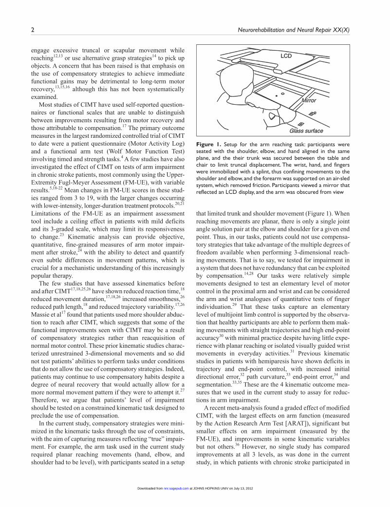
In the current study, compensatory strategies were mini-mized in the kinematic tasks through the use of constraints, with the aim of capturing measures reflecting “true” impair-ment. For example, the arm task used in the current study required planar reaching movements (hand, elbow, and shoulder had to be level), with participants seated in a setup
that limited trunk and shoulder movement (Figure 1). When reaching movements are planar, there is only a single joint angle solution pair at the elbow and shoulder for a given end point. Thus, in our tasks, patients could not use compensa-tory strategies that take advantage of the multiple degrees of freedom available when performing 3-dimensional reach-ing movements. That is to say, we tested for impairment in a system that does not have redundancy that can be exploited by compensation.14,28 Our tasks were relatively simple movements designed to test an elementary level of motor control in the proximal arm and wrist and can be considered the arm and wrist analogues of quantitative tests of finger individuation.29 That these tasks capture an elementary level of multijoint limb control is supported by the observa-tion that healthy participants are able to perform them mak-ing movements with straight trajectories and high end-point accuracy30 with minimal practice despite having little expe-rience with planar reaching or isolated visually guided wrist movements in everyday activities.31 Previous kinematic studies in patients with hemiparesis have shown deficits in trajectory and end-point control, with increased initial directional error,32 path curvature,33 end-point error,34 and segmentation.33,35 These are the 4 kinematic outcome mea-sures that we used in the current study to assay for reduc-tions in arm impairment.
A recent meta-analysis found a graded effect of modified CIMT, with the largest effects on arm function (measured by the Action Research Arm Test [ARAT]), significant but smaller effects on arm impairment (measured by the FM-UE), and improvements in some kinematic variables but not others.36 However, no single study has compared improvements at all 3 levels, as was done in the current study, in which patients with chronic stroke participated in
Figure 1. Setup for the arm reaching task: participants were seated with the shoulder, elbow, and hand aligned in the same plane, and the their trunk was secured between the table and chair to limit truncal displacement. The wrist, hand, and fingers were immobilized with a splint, thus confining movements to the shoulder and elbow, and the forearm was supported on an air-sled system, which removed friction. Participants viewed a mirror that reflected an LCD display, and the arm was obscured from view
at JOHNS HOPKINS UNIV on July 13, 2012nnr.sagepub.comDownloaded from
Kitago et al 3
a modified CIMT program and were evaluated with clinical scales of motor function and impairment as well as with kinematic analysis of arm and wrist movements. We hypothesized here that the main effect of CIMT is to pro-mote functional use of the affected arm primarily through compensatory strategies and not through a reduction in impairment (ie, motor recovery), given the task-oriented nature of the training intervention and findings of a prior study17 of CIMT. Therefore, we predicted that we would see the greatest improvements in the ARAT and more modest, if any, improvements in the FM-UE and in kine-matic variables.
MethodsStudy Participants
A total of 10 patients with chronic stroke were recruited from a local rehabilitation hospital and outpatient neurological practices between May 2005 and February 2007. Participants met the following inclusion criteria: (1) ischemic or hemor-rhagic stroke at least 6 months prior to enrollment, (2) motor deficit involving 1 arm with the ability to perform at least 20° wrist extension and 10° finger extension, (3) significant unaffected hand use for activities of daily living, and (4) ARAT score <50 in the affected arm. Inclusion criteria (2) and (3) are similar to those used in previous studies of CIMT that demonstrated functional motor improvements,4 and we required patients to have a demonstrable deficit on the ARAT to avoid a potential ceiling effect. Patients were excluded if they had only ataxia or sensory symptoms, were unable to give informed consent or were unable to participate in train-ing because of aphasia or cognitive impairment, had signifi-cant pain or musculoskeletal problems that would interfere with intensive training, or were receiving ongoing physical therapy for the arm. All participants signed a written consent form that was approved by the local institutional human research review board.
InterventionAll patients completed 2 weeks of CIMT at home under the supervision of a licensed physical therapist, with a goal of 4 hours a day for 10 consecutive weekdays. This was a modified version of the original CIMT protocol, which consisted of 6 hours of therapy a day for 10 consecutive weekdays.4 Therapy sessions began with active range of motion and/or stretching exercises, then proceeded to task-based activities that were individualized based on the patient’s goals and interests. Activities included folding clothes, eating, writing, throwing a ball, and playing games (eg, chess and Jenga). Task difficulty was adjusted for indi-vidual patients to be sufficiently challenging as determined by the therapist, and shaping techniques were incorporated,
with increasing task difficulty over successive sessions. Verbal feedback was provided to participants during task practice. The time spent in each activity ranged from 1 to 15 minutes, depending on the patient’s level of interest and endurance. The total activity time was recorded for each session by the therapist. Participants were also encouraged to wear the mitt on their unaffected hand outside of therapy and reported their compliance.
Study DesignClinical and kinematic assessments were performed at 3 time points: (1) 3 weeks prior to therapy, (2) 1 week prior to therapy, and (3) within 2 weeks after completion of therapy. Two assessments were performed prior to the CIMT inter-vention to reduce the impact of improvements caused by practice effects, particularly in our kinematic tasks.
Clinical Outcome MeasuresAll clinical assessments were performed by a single physi-cal therapist blinded to the patients’ performance during therapy. The primary clinical outcome measures were the ARAT37 and the FM-UE scale,38 which have been shown to have good reliability, validity, and responsiveness to motor change in patients with chronic stroke.23,39-41
The ARAT is an instrument that tests arm function and consists of 19 items in 4 domains: grasp, grip, pinch, and gross movement. Each domain contains items arranged into hierarchical order of difficulty such that success at the most difficult item of a specific subclass assumes success for all items lower in the hierarchy of the same class. Each item is scored on a 4-point ordinal scale (0 = cannot perform, 1 = per-forms test partially, 2 = completes test but takes abnormally long time or has great difficulty, and 3 = performs test nor-mally), with a maximum score of 57. The ARAT is a functional measure with limited ability to discriminate between “normal” movements and the use of compensatory strategies.10
Arm impairment was assessed using the FM-UE. The FM-UE is scored using a 3-point ordinal scale (0 = cannot perform, 1 = can perform partially, 2 = can perform fully) on 22 items in 4 sections: arm, wrist, hand, and coordination, for a maximum score of 66. This test specifies that all limb subcomponents move in a qualitatively normal manner for a normal performance score to be given. We further subdi-vided the FM-UE into proximal (first 18 items, maximum score 36), wrist (next 5 items, maximum score 10), hand (next 7 items, maximum score 14), and coordination (last 3 items, maximum score 6).
Kinematics: Arm Reaching TaskParticipants moved a cursor by making reaching move-ments over a glass-surface table (Figure 1). Hand position,
at JOHNS HOPKINS UNIV on July 13, 2012nnr.sagepub.comDownloaded from
4 Neurorehabilitation and Neural Repair XX(X)
calibrated to the position of the fingertip, was monitored using a Flock of Birds (Ascension Technology, Burlington, VT) magnetic movement recording system at a frequency of 120 Hz. The experimental computer converted this infor-mation online into hand, elbow, and shoulder positions using custom routines in RealBasic (Real Software, Austin, TX). The computer used real-time hand position informa-tion to control the visual display and to provide visual feedback.
The target set consisted of 8 radially arrayed circles with a 1-cm radius, 45° apart, 8 cm from a center start circle. Each trial began after the participant held the cursor inside the start circle for 750 ms. Participants were instructed to make straight, out-and-back movements with a sharp rever-sal within the target, when they were ready to move after the target appeared. To ensure that movements were made quickly and to ensure consistency between trials and ses-sions, the black cursor froze at its position at 200 ms, and the reversal point was indicated by a white square.42 This time requirement also allowed us to focus on deficits in patients’ feed-forward control by minimizing online correc-tions. Participants were given 1 or 2 practice runs of 88 movements for each arm to become familiar with the task. They completed 2 experimental runs, each comprising 11 cycles of 8 targets presented in random order, for each arm.
Kinematics: Wrist Pointing TaskParticipants sat in a chair with their forearm placed in a rigid splint that maintained the elbow at a 90° angle and the forearm midway between pronation and supination. The participant’s hand was lightly taped in a fist, and a 1.5-cm spherical reflective marker was attached over the index finger’s first interphalangeal joint. The hand was hidden from view. Participants were instructed to make pointing movements through combinations of abduction-adduction and flexion-extension movements around the wrist, so as to point the marker at targets projected onto a vertical com-puter screen. The position of the marker was monitored using a Qualysis ProReflex video camera (model MCU 240; Qualisys, Gothenburg, Sweden) equipped with an infrared strobe coupled to a video digitizer, which recorded the marker’s position in the vertical plane at a frequency of 100 Hz.
The target set for the wrist task consisted of 4 radially arrayed circles, separated by 90°, at a distance, which required a 2-cm movement of the infrared marker. Similarly to the arm reaching task, participants were instructed to make out and back movements, placing both the cursor, which froze after 100 ms, and a white reversal square in the center of the target. They completed 1 familiarization run and 2 experimental runs for each wrist, which consisted of 22 cycles of 4 targets presented in random order.
Analysis of Kinematics
We analyzed hand and wrist position data using custom routines within the IGOR analysis software package (Wavemetrics, Lake Oswego, OR). Position time series were low-pass filtered (Butterworth filter) at 8 Hz for the arm task and 14 Hz for the wrist task and differentiated to yield tangential velocity and acceleration.
The first velocity peak above a threshold (10 cm/s for the arm and 5 cm/s for the wrist) was identified for each trial. These thresholds were chosen to exclude small movements made by some participants who had difficulty stabilizing their hand within the start circle. The start of the movement was defined as either the point at which the velocity crossed 1 cm/s or the first velocity minimum prior to the first veloc-ity peak, whichever was later. The end point of the outward movement was defined as the reversal point—that is, the point where distance from the origin stopped increasing. The movements were further divided into submovements based on their acceleration profiles. The first submovement was defined as the movement up until the first zero crossing of acceleration from negative to positive.43 Each subsequent submovement was defined as the movement between zero crossings of acceleration from negative to positive.
We calculated the following variables for the outward movement: (1) movement time; (2) peak velocity; (3) abso-lute initial directional error, which is the angle between the vector from start position to target and the vector from start position to cursor position at 120 ms after movement start; (4) path curvature, the unsigned area between hand path and the shortest possible path (a straight line joining the start and end points of the movement), divided by the area of a semicircle with the shortest possible path as its diameter; (5) systematic error, the distance between the average end-point position and target center; (6) end-point variance, the determinant of the covariance matrix of the end points cor-rected for the number of observations and averaged across targets; and (7) number of submovements.
The following kinds of trials were excluded: anticipatory movements, movements that did not reach 30% of the distance to the target, movements without reversals, and spatial outliers (in which the movement direction was >90° from the target direction). Anticipatory movements were defined as those with reaction times less than 200 ms, based on the fastest reaction times seen in a similar task when healthy participants were instructed to move “as soon as possible.”44
Statistical AnalysisAll statistical analyses were performed on average group data. One-tailed, paired t tests were performed using Excel (Microsoft, Redmond, WA) on the differences between the
at JOHNS HOPKINS UNIV on July 13, 2012nnr.sagepub.comDownloaded from
Kitago et al 5
change from session 1 to 2 (Δ1) and the change from ses-sion 2 to 3 (Δ2). We chose to compare the changes in values in order to minimize the possible confound of task practice on our measures across sessions and chose 1-tailed tests because our hypotheses were based on improvements in our clinical and kinematic variables.
We performed post hoc power analyses to assess the false-negative rate for each of our comparisons. Meaningful effect size was chosen as 10% of the maximum possible score for the clinical measures.23,41 The study was powered at 0.83 for ARAT, 0.86 for FM total, 0.78 for FM arm, and 0.52 for FM wrist. For the kinematic measures, we used 50% of the patients’ mean deficit (the difference between the patients’ affected side and values obtained for elderly controls in an ongoing study). The magnitude of improve-ment in our kinematic tasks that would be associated with a clinically meaningful reduction in impairment is not known, but we presumed that significant recovery would be accom-panied by a larger percentage improvement in the more sen-sitive45 and responsive46 kinematic measures compared with the clinical scales. For the arm kinematic measures, the study was powered at 0.55 for path curvature, 0.65 for ini-tial directional error, 0.48 for systematic error, 0.45 for end-point variance, and 0.48 for number of submovements. For wrist movements, the study was powered at 0.51 for path curvature, 0.99 for initial directional error, 0.88 for system-atic error, 0.96 for end-point variance, and 0.55 for number of submovements.
ResultsA total of 10 patients with chronic stroke participated in the study, but 1 patient was excluded from the analysis because he did not meet inclusion criteria. Table 1 summarizes patient demographics and stroke location. The mean age of patients was 70 years, and mean time elapsed since stroke was 34.3 months (range = 10-162 months). Of the 9 patients, 7 had ischemic strokes, 5 involved the dominant
hemisphere, and lesions included both cortical and subcor-tical areas. For the study intervention, patients spent on average 2.45 hours practicing activities (range = 1.84-2.89 hours) with the therapist each day. By self-report, they spent an average of 61 min/d (range = 0-251 min/d) wear-ing the mitt outside of therapy. However, 5 of the patients did not wear the mitt at all outside of therapy because of safety concerns. No adverse events were reported during the study.
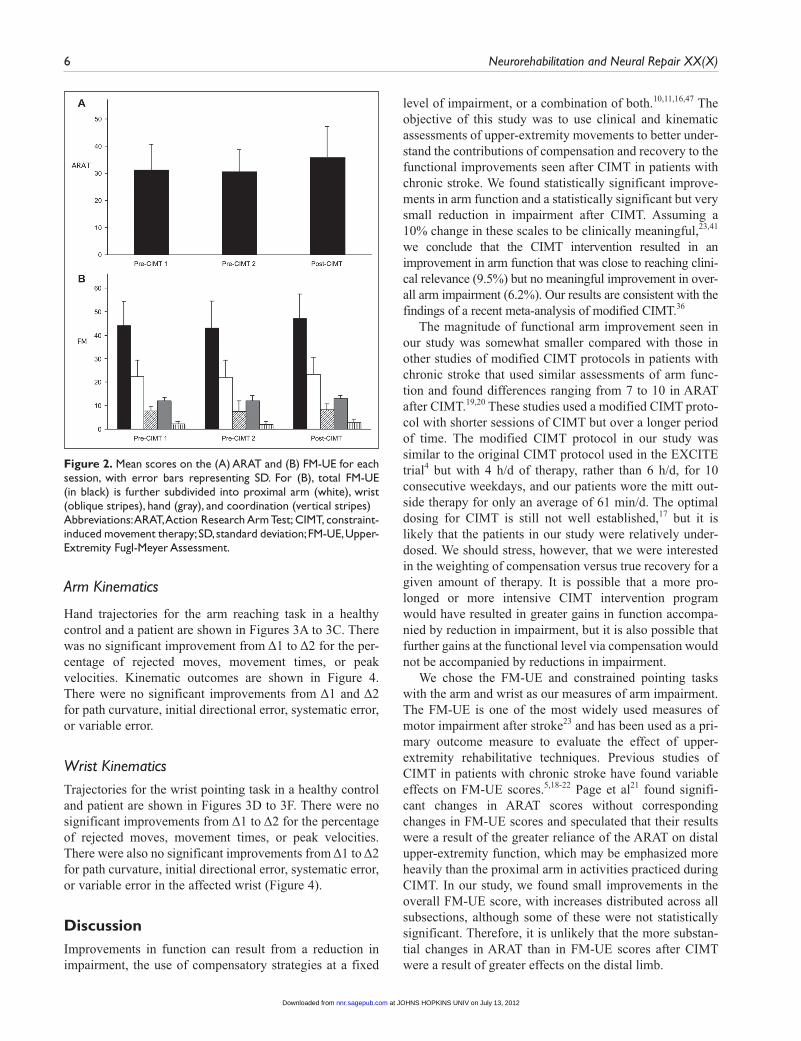
Clinical OutcomesAt baseline, patients were moderately affected, with a mean ARAT score of 31.3 and mean total FM-UE score of 44.3 (Table 2). Clinical outcome measures are summa-rized in Figure 2. Arm function as measured by the ARAT improved significantly more after CIMT than between the 2 pre-CIMT sessions (−0.8 vs 5.4, P = .004). Δ2 was also significantly larger than Δ1 for the total FM-UE (−0.9 vs 4.1, P = .018), FM wrist score (-0.2 vs 0.9, P = .027), and FM coordination score (−0.2 vs 0.9, P = .014) but not for the FM arm (−0.4 vs 1.4, P = .087) or FM hand (−0.1 vs 0.9, P = .148).
Table 1. Patient Demographics and Clinical Characteristics
Patient Sex Age (years) Months Since Stroke Stroke Type Affected Arm Stroke Location
P1 M 66 49 H ND R BGP2 M 57 162 I D L MCA and BGP3 F 77 17 H D L BGP4 M 58 10 I ND R frontalP5 F 77 10 I D L ponsP6 F 81 15 I D L MCA and BGP7 M 83 14 I ND R frontalP8 F 59 22 I ND R ponsP9 M 72 10 I D L MCA and pons
Abbreviations: M, male; F, female; I, ischemic; H, hemorrhagic; D, dominant; ND, nondominant; R, right; L, left; BG, basal ganglia; MCA, middle cerebral artery territory.
Table 2. Patient Baseline Clinical Assessments
Patient ARAT FM-UE Total FM-UE Arm FM-UE Wrist
P1 39 48 23 9P2 44 56 33 9P3 38 48 25 9P4 18 30 13 5P5 32 53 27 10P6 22 36 19 5P7 40 55 30 8P8 23 40 17 9P9 26 33 15 5
Abbreviations: ARAT, Action Research Arm Test; FM-UE, Fugl-Meyer Upper Extremity Assessment.
at JOHNS HOPKINS UNIV on July 13, 2012nnr.sagepub.comDownloaded from
6 Neurorehabilitation and Neural Repair XX(X)
Arm Kinematics
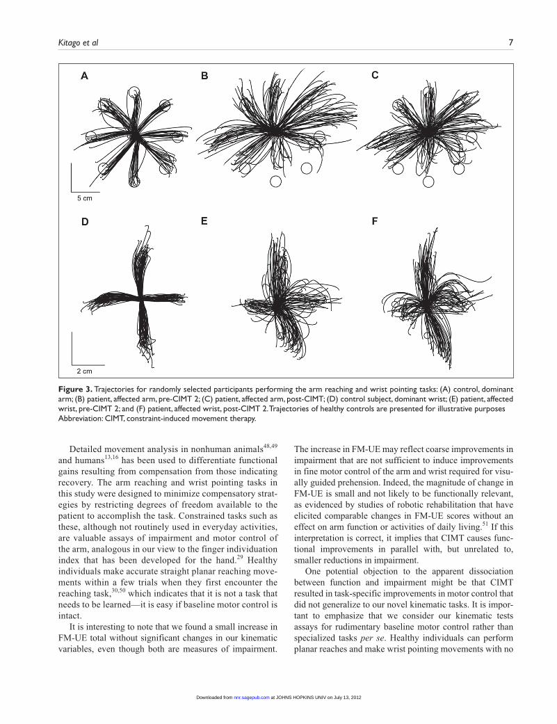
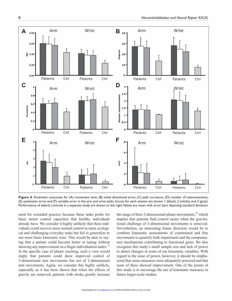
Hand trajectories for the arm reaching task in a healthy control and a patient are shown in Figures 3A to 3C. There was no significant improvement from Δ1 to Δ2 for the per-centage of rejected moves, movement times, or peak velocities. Kinematic outcomes are shown in Figure 4. There were no significant improvements from Δ1 and Δ2 for path curvature, initial directional error, systematic error, or variable error.
Wrist KinematicsTrajectories for the wrist pointing task in a healthy control and patient are shown in Figures 3D to 3F. There were no significant improvements from Δ1 to Δ2 for the percentage of rejected moves, movement times, or peak velocities. There were also no significant improvements from Δ1 to Δ2 for path curvature, initial directional error, systematic error, or variable error in the affected wrist (Figure 4).
DiscussionImprovements in function can result from a reduction in impairment, the use of compensatory strategies at a fixed
level of impairment, or a combination of both.10,11,16,47 The objective of this study was to use clinical and kinematic assessments of upper-extremity movements to better under-stand the contributions of compensation and recovery to the functional improvements seen after CIMT in patients with chronic stroke. We found statistically significant improve-ments in arm function and a statistically significant but very small reduction in impairment after CIMT. Assuming a 10% change in these scales to be clinically meaningful,23,41 we conclude that the CIMT intervention resulted in an improvement in arm function that was close to reaching clini-cal relevance (9.5%) but no meaningful improvement in over-all arm impairment (6.2%). Our results are consistent with the findings of a recent meta-analysis of modified CIMT.36
The magnitude of functional arm improvement seen in our study was somewhat smaller compared with those in other studies of modified CIMT protocols in patients with chronic stroke that used similar assessments of arm func-tion and found differences ranging from 7 to 10 in ARAT after CIMT.19,20 These studies used a modified CIMT proto-col with shorter sessions of CIMT but over a longer period of time. The modified CIMT protocol in our study was similar to the original CIMT protocol used in the EXCITE trial4 but with 4 h/d of therapy, rather than 6 h/d, for 10 consecutive weekdays, and our patients wore the mitt out-side therapy for only an average of 61 min/d. The optimal dosing for CIMT is still not well established,17 but it is likely that the patients in our study were relatively under-dosed. We should stress, however, that we were interested in the weighting of compensation versus true recovery for a given amount of therapy. It is possible that a more pro-longed or more intensive CIMT intervention program would have resulted in greater gains in function accompa-nied by reduction in impairment, but it is also possible that further gains at the functional level via compensation would not be accompanied by reductions in impairment.
We chose the FM-UE and constrained pointing tasks with the arm and wrist as our measures of arm impairment. The FM-UE is one of the most widely used measures of motor impairment after stroke23 and has been used as a pri-mary outcome measure to evaluate the effect of upper-extremity rehabilitative techniques. Previous studies of CIMT in patients with chronic stroke have found variable effects on FM-UE scores.5,18-22 Page et al21 found signifi-cant changes in ARAT scores without corresponding changes in FM-UE scores and speculated that their results were a result of the greater reliance of the ARAT on distal upper-extremity function, which may be emphasized more heavily than the proximal arm in activities practiced during CIMT. In our study, we found small improvements in the overall FM-UE score, with increases distributed across all subsections, although some of these were not statistically significant. Therefore, it is unlikely that the more substan-tial changes in ARAT than in FM-UE scores after CIMT were a result of greater effects on the distal limb.
Figure 2. Mean scores on the (A) ARAT and (B) FM-UE for each session, with error bars representing SD. For (B), total FM-UE (in black) is further subdivided into proximal arm (white), wrist (oblique stripes), hand (gray), and coordination (vertical stripes)Abbreviations: ARAT, Action Research Arm Test; CIMT, constraint-induced movement therapy; SD, standard deviation; FM-UE, Upper-Extremity Fugl-Meyer Assessment.
at JOHNS HOPKINS UNIV on July 13, 2012nnr.sagepub.comDownloaded from
Kitago et al 7
Detailed movement analysis in nonhuman animals48,49 and humans13,16 has been used to differentiate functional gains resulting from compensation from those indicating recovery. The arm reaching and wrist pointing tasks in this study were designed to minimize compensatory strat-egies by restricting degrees of freedom available to the patient to accomplish the task. Constrained tasks such as these, although not routinely used in everyday activities, are valuable assays of impairment and motor control of the arm, analogous in our view to the finger individuation index that has been developed for the hand.29 Healthy individuals make accurate straight planar reaching move-ments within a few trials when they first encounter the reaching task,30,50 which indicates that it is not a task that needs to be learned—it is easy if baseline motor control is intact.
It is interesting to note that we found a small increase in FM-UE total without significant changes in our kinematic variables, even though both are measures of impairment.
The increase in FM-UE may reflect coarse improvements in impairment that are not sufficient to induce improvements in fine motor control of the arm and wrist required for visu-ally guided prehension. Indeed, the magnitude of change in FM-UE is small and not likely to be functionally relevant, as evidenced by studies of robotic rehabilitation that have elicited comparable changes in FM-UE scores without an effect on arm function or activities of daily living.51 If this interpretation is correct, it implies that CIMT causes func-tional improvements in parallel with, but unrelated to, smaller reductions in impairment.
One potential objection to the apparent dissociation between function and impairment might be that CIMT resulted in task-specific improvements in motor control that did not generalize to our novel kinematic tasks. It is impor-tant to emphasize that we consider our kinematic tests assays for rudimentary baseline motor control rather than specialized tasks per se. Healthy individuals can perform planar reaches and make wrist pointing movements with no
Figure 3. Trajectories for randomly selected participants performing the arm reaching and wrist pointing tasks: (A) control, dominant arm; (B) patient, affected arm, pre-CIMT 2; (C) patient, affected arm, post-CIMT; (D) control subject, dominant wrist; (E) patient, affected wrist, pre-CIMT 2; and (F) patient, affected wrist, post-CIMT 2. Trajectories of healthy controls are presented for illustrative purposesAbbreviation: CIMT, constraint-induced movement therapy.
at JOHNS HOPKINS UNIV on July 13, 2012nnr.sagepub.comDownloaded from
8 Neurorehabilitation and Neural Repair XX(X)
need for extended practice because these tasks probe for basic motor control capacities that healthy individuals already have. We consider it highly unlikely that these indi-viduals could recover more normal control in more ecologi-cal and challenging everyday tasks but fail to generalize to our more basic kinematic tests. This would be akin to say-ing that a patient could become better at typing without showing any improvement on a finger individuation index.52 In the specific case of planar reaching, such a view would imply that patients could show improved control of 3-dimensional arm movements but not of 2-dimensional arm movements. Again, we consider this highly unlikely, especially as it has been shown that when the effects of gravity are removed, patients with stroke greatly increase
the range of their 2-dimensional planar movements,53 which implies that patients find control easier when the gravita-tional challenge of 3-dimensional movements is removed. Nevertheless, an interesting future direction would be to combine kinematic assessments of constrained and free movements to quantify both impairment and the compensa-tory mechanisms contributing to functional gains. We also recognize this study’s small sample size and lack of power to detect changes in some of our kinematic variables. With regard to the issue of power, however, it should be empha-sized that some measures were adequately powered and that none of these showed improvement. One of the points of this study is to encourage the use of kinematic measures in future larger-scale studies.
Figure 4. Kinematic outcomes for (A) movement time, (B) initial directional error, (C) path curvature, (D) number of submovements, (E) systematic error, and (F) variable error in the arm and wrist tasks. Scores for each session are shown: 1 (black), 2 (white), and 3 (gray). Performance of elderly controls in a separate study are shown to the right. Values are mean with error bars depicting standard deviation
at JOHNS HOPKINS UNIV on July 13, 2012nnr.sagepub.comDownloaded from
Kitago et al 9
Our interpretation of the current data is that CIMT induces task-specific compensatory strategies through prac-tice in the chronic stage of stroke despite an invariant impairment level. CIMT could have a qualitatively different effect if applied very early after stroke if larger reductions in impairment than expected from spontaneous recovery occurred.54 Then, patients would not resort to compensatory strategies. Although early CIMT did not show a benefit in 1 trial, impairment was not assessed.55
Our study and others demonstrate the feasibility of using kinematic analysis to obtain more detailed, objective measures of movement patterns and motor performance in studies of novel neurorehabilitative interventions.56-58 Analysis at the kinematic level will provide us with the ability to differentiate between functional gains achieved through compensation versus those achieved through true recovery of motor control.45 At the current time, conven-tional therapy has largely focused on teaching compensa-tory strategies, with a goal of achieving early functional independence. As mentioned above, early focus on com-pensation may limit long-term recovery.13,14,16 Even in the acute stroke period, patients may learn not to use or explore more normal movements with the affected limb because of habitual reliance on compensatory strategies and thus miss out on a time window of plasticity within which true recovery could be maximized11. Detailed kinematic assess-ments of the kind used here could contribute to a better understanding of the interactions between spontaneous recovery, the development of compensatory behaviors, and rehabilitation and their relative contributions to long-term outcome.16,59
Declaration of Conflicting Interests
The author(s) declared no potential conflicts of interest with respect to the research, authorship, and/or publication of this article.
Funding
The author(s) disclosed receipt of the following financial support for the research, authorship, and/or publication of this article: The work was supported by the following grants: National Institutes of Health Grant Number K02-NS048099 and the Suellen Jones Saunders Memorial Fund.
References
1. Lloyd-Jones D, Adams RJ, Brown TM, et al. Heart disease and stroke statistics: 2010 update. Circulation. 2010;121:e46-e215.
2. Kwakkel G, Kollen BJ, van der Grond J, Prevo AJ. Probability of regaining dexterity in the flaccid upper limb: impact of severity of paresis and time since onset in acute stroke. Stroke. 2003;34:2181-2186.
3. Wu CY, Chuang LL, Lin KC, Chen HC, Tsay PK. Randomized trial of distributed constraint-induced therapy versus bilateral
arm training for the rehabilitation of upper-limb motor con-trol and function after stroke. Neurorehabil Neural Repair. 2011;25:130-139.
4. Boake C, Noser EA, Ro T, et al. Constraint-induced movement therapy during early stroke rehabilitation. Neurorehabil Neural Repair. 2007;21:14-24.
5. Bonifer NM, Anderson KM, Arciniegas DB. Constraint-induced movement therapy after stroke: efficacy for patients with minimal upper-extremity motor ability. Arch Phys Med Rehabil. 2005;86:1867-1873.
6. Wolf SL, Winstein CJ, Miller JP, et al. Effect of constraint-induced movement therapy on upper extremity function 3 to 9 months after stroke: the EXCITE randomized clinical trial. JAMA. 2006;296:2095-2104.
7. Mark VW, Taub E. Constraint-induced movement therapy for chronic stroke hemiparesis and other disabilities. Restor Neurol Neurosci. 2004;22(3-5):317-336.
8. Sawaki L, Butler AJ, Leng X, et al. Constraint-induced move-ment therapy results in increased motor map area in subjects 3 to 9 months after stroke. Neurorehabil Neural Repair. 2008;22: 505-513.
9. Alberts JL, Butler AJ, Wolf SL. The effects of constraint-induced therapy on precision grip: a preliminary study. Neu-rorehabil Neural Repair. 2004;18:250-258.
10. Levin MF, Kleim JA, Wolf SL. What do motor “recovery” and “compensation” mean in patients following stroke? Neurore-habil Neural Repair. 2009;23:313-319.
11. Cirstea MC, Levin MF. Compensatory strategies for reaching in stroke. Brain. 2000;123(pt 5):940-953.
12. Roby-Brami A, Feydy A, Combeaud M, Biryukova EV, Bussel B, Levin MF. Motor compensation and recovery for reaching in stroke patients. Acta Neurol Scand. 2003;107: 369-381.
13. Raghavan P, Santello M, Gordon AM, Krakauer JW. Compen-satory motor control after stroke: an alternative joint strategy for object-dependent shaping of hand posture. J Neurophysiol. 2010;103:3034-3043.
14. Wu CY, Chen YA, Chen HC, Lin KC, Yeh IL. Pilot trial of distributed constraint-induced therapy with trunk restraint to improve poststroke reach to grasp and trunk kinematics. Neurorehabil Neural Repair. 2012;26:247-255.
15. Lum PS, Mulroy S, Amdur RL, Requejo P, Prilutsky BI, Dromerick AW. Gains in upper extremity function after stroke via recovery or compensation: potential differential effects on amount of real-world limb use. Top Stroke Rehabil. 2009;16:237-253.
16. Massie C, Malcolm MP, Greene D, Thaut M. The effects of constraint-induced therapy on kinematic outcomes and com-pensatory movement patterns: an exploratory study. Arch Phys Med Rehabil. 2009;90:571-579.
17. Wu CY, Chen CL, Tang SF, Lin KC, Huang YY. Kinematic and clinical analyses of upper-extremity movements after constraint-induced movement therapy in patients with stroke: a randomized controlled trial. Arch Phys Med Rehabil. 2007;88:964-970.
at JOHNS HOPKINS UNIV on July 13, 2012nnr.sagepub.comDownloaded from
10 Neurorehabilitation and Neural Repair XX(X)
18. Szaflarski JP, Page SJ, Kissela BM, Lee JH, Levine P, Strakowski SM. Cortical reorganization following modified constraint-induced movement therapy: a study of 4 patients with chronic stroke. Arch Phys Med Rehabil. 2006;87:1052-1058.
19. Page SJ, Sisto S, Levine P, McGrath RE. Efficacy of modi-fied constraint-induced movement therapy in chronic stroke: a single-blinded randomized controlled trial. Arch Phys Med Rehabil. 2004;85:14-18.
20. Page SJ, Levine P, Leonard A, Szaflarski JP, Kissela BM. Modified constraint-induced therapy in chronic stroke: results of a single-blinded randomized controlled trial. Phys Ther. 2008;88:333-340.
21. Lin KC, Chang YF, Wu CY, Chen YA. Effects of constraint-induced therapy versus bilateral arm training on motor performance, daily functions, and quality of life in stroke survivors. Neurorehabil Neural Repair. 2009;23:441-448.
22. Gladstone DJ, Danells CJ, Black SE. The Fugl-Meyer assess-ment of motor recovery after stroke: a critical review of its measurement properties. Neurorehabil Neural Repair. 2002; 16:232-240.
23. Subramanian SK, Yamanaka J, Chilingaryan G, Levin MF. Validity of movement pattern kinematics as measures of arm motor impairment poststroke. Stroke. 2010;41:2303-2308.
24. Wu CY, Lin KC, Chen HC, Chen IH, Hong WH. Effects of modified constraint-induced movement therapy on move-ment kinematics and daily function in patients with stroke: a kinematic study of motor control mechanisms. Neurorehabil Neural Repair. 2007;21:460-466.
25. Caimmi M, Carda S, Giovanzana C, et al. Using kinematic analysis to evaluate constraint-induced movement therapy in chronic stroke patients. Neurorehabil Neural Repair. 2008; 22:31-39.
26. Huang VS, Krakauer JW. Robotic neurorehabilitation: a com-putational motor learning perspective. J Neuroeng Rehabil. 2009;6:5.
27. Diedrichsen J, Shadmehr R, Ivry RB. The coordination of movement: optimal feedback control and beyond. Trends Cogn Sci. 2010;14:31-39.
28. Lang CE, Schieber MH. Reduced muscle selectivity dur-ing individuated finger movements in humans after damage to the motor cortex or corticospinal tract. J Neurophysiol. 2004;91:1722-1733.
29. Gordon J, Ghilardi MF, Ghez C. Accuracy of planar reaching movements: I. Independence of direction and extent variability. Exp Brain Res. 1994;99:97-111.
30. Howard IS, Ingram JN, Kording KP, Wolpert DM. Statistics of natural movements are reflected in motor errors. J Neurophysiol. 2009;102:1902-1910.
31. Beer RF, Dewald JP, Rymer WZ. Deficits in the coordination of multijoint arm movements in patients with hemiparesis: evidence for disturbed control of limb dynamics. Exp Brain Res. 2000;131:305-319.
32. Levin MF. Interjoint coordination during pointing movements is disrupted in spastic hemiparesis. Brain. 1996;119:281-293.
33. Lang CE, Wagner JM, Edwards DF, Sahrmann SA, Dromerick AW. Recovery of grasp versus reach in people with hemipare-sis poststroke. Neurorehabil Neural Repair. 2006;20:444-454.
34. Dipietro L, Krebs HI, Fasoli SE, Volpe BT, Hogan N. Submove-ment changes characterize generalization of motor recovery after stroke. Cortex. 2009;45:318-324.
35. Shi YX, Tian JH, Yang KH, Zhao Y. Modified constraint-induced movement therapy versus traditional rehabilitation in patients with upper-extremity dysfunction after stroke: a systematic review and meta-analysis. Arch Phys Med Rehabil. 2011;92: 972-982.
36. Lyle RC. A performance test for assessment of upper limb function in physical rehabilitation treatment and research. Int J Rehabil Res. 1981;4:483-492.
37. Fugl-Meyer AR, Jaasko L, Leyman I, Olsson S, Steglind S. The post-stroke hemiplegic patient: 1. A method for evaluation of physical performance. Scand J Rehabil Med. 1975;7:13-31.
38. Hsieh YW, Wu CY, Lin KC, Chang YF, Chen CL, Liu JS. Responsiveness and validity of three outcome measures of motor function after stroke rehabilitation. Stroke. 2009;40: 1386-1391.
39. Lang CE, Wagner JM, Dromerick AW, Edwards DF. Measure-ment of upper-extremity function early after stroke: properties of the action research arm test. Arch Phys Med Rehabil. 2006; 87:1605-1610.
40. Yoxbatiran N, Der-Yeghiaian L, Cramer SC. A standard-ized approach to performing the Action Research Arm Test. Neurorehabil Neural Repair. 2008;22:78-90.
41. Mazzoni P, Krakauer JW. An implicit plan overrides an explicit strategy during visuomotor adaptation. J Neurosci. 2006;26:3642-3645.
42. Meyer DE, Abrams RA, Kornblum S, Wright CE, Smith JE. Optimality in human motor performance: ideal control of rapid aimed movements. Psychol Rev. 1988;95:340-370.
43. Ghilardi MF, Moisello C, Silvestri G, Ghez C, Krakauer JW. Learning of a sequential motor skill comprises explicit and implicit components that consolidate differently. J Neuro-physiol. 2009;101:2218-2229.
44. Coderre AM, Zeid AA, Dukelow SP, et al. Assessment of upper-limb sensorimotor function of subacute stroke patients using visually guided reaching. Neurorehabil Neural Repair. 2010;24:528-541.
45. Platz T, Prass K, Denzler P, Bock S, Mauritz KH. Testing a motor performance series and a kinematic motion analysis as measures of performance in high-functioning stroke patients: reliability, validity, and responsiveness to therapeutic inter-vention. Arch Phys Med Rehabil. 1999;80:270-277.
46. Murphy TH, Corbett D. Plasticity during stroke recovery: from synapse to behaviour. Nat Rev Neurosci. 2009;10: 861-872.
at JOHNS HOPKINS UNIV on July 13, 2012nnr.sagepub.comDownloaded from
Kitago et al 11
47. Whishaw IQ, Pellis SM, Gorny B, Kolb B, Tetzlaff W. Prox-imal and distal impairments in rat forelimb use in reaching follow unilateral pyramidal tract lesions. Behav Brain Res. 1993;56:59-76.
48. Moon SK, Alaverdashvili M, Cross AR, Whishaw IQ. Both compensation and recovery of skilled reaching following small photothrombotic stroke to motor cortex in the rat. Exp Neurol. 2009;218:145-153.
49. Morasso P. Spatial control of arm movements. Exp Brain Res. 1981;42:223-227.
50. Prange GB, Jannink MJ, Groothuis-Oudshoorn CG, Hermens HJ, Ijzerman MJ. Systematic review of the effect of robot-aided therapy on recovery of the hemiparetic arm after stroke. J Rehabil Res Dev. 2006;43:171-184.
51. Lang CE, Schieber MH. Differential impairment of individu-ated finger movements in humans after damage to the motor cortex or the corticospinal tract. J Neurophysiol. 2003;90: 1160-1170.
52. Beer RF, Dewald JP, Dawson ML, Rymer WZ. Target- dependent differences between free and constrained arm movements in chronic hemiparesis. Exp Brain Res. 2004;156: 458-470.
53. Prabhakaran S, Zarahn E, Riley C, et al. Inter-individual variability in the capacity for motor recovery after ischemic stroke. Neurorehabil Neural Repair. 2008;22:64-71.
54. Dromerick AW, Lang CE, Birkenmeier RL, et al. Very Early Constraint-Induced Movement during Stroke Rehabilita-tion (VECTORS): a single-center RCT. Neurology. 2009;73: 195-201.
55. Alt Murphy M, Willen C, Sunnerhagen KS. Kinematic vari-ables quantifying upper-extremity performance after stroke during reaching and drinking from a glass. Neurorehabil Neural Repair. 2011;25:71-80.
56. Bosecker C, Dipietro L, Volpe B, Krebs HI. Kinematic robot-based evaluation scales and clinical counterparts to measure upper limb motor performance in patients with chronic stroke. Neurorehabil Neural Repair. 2010;24:62-69.
57. Verheyden G, van Duijnhoven HJ, Burnett M, et al. Kine-matic analysis of head, trunk and pelvis movement when people early after stroke reach sideways. Neurorehabil Neural Repair. 2011;25:656-663.
58. Kwakkel G, Kollen BJ, Krebs HI. Effects of robot-assisted therapy on upper limb recovery after stroke: a systematic review. Neurorehabil Neural Repair. 2008;22:111-121.
at JOHNS HOPKINS UNIV on July 13, 2012nnr.sagepub.comDownloaded from