Embed Size (px)
Citation preview
NeuroradiologyNeuroradiology
Natasha Wehrli, MS4Natasha Wehrli, MS4
University of Pennsylvania School of MedicineUniversity of Pennsylvania School of Medicine
Q: What is radiology?Q: What is radiology?
Answer (from Wikipedia):Answer (from Wikipedia): Radiology is the branch of medical science dealing with the medical use of radiation devices and other forms of energy for the purpose of obtaining visual information as part of medical imaging. Interventional radiology is the performance of medical procedures with the help of medical imaging.
My answer: the coolest field of medicine ever!
What is a neuroradiologist?What is a neuroradiologist?
A neuroradiologist is a radiologist who specializes in the use of x-rays and other scanning devices for the diagnosis and treatment of diseases of the nervous system.
A neuroradiologist must be concerned with the clinical imaging, therapy, and basic science of the central and peripheral nervous system, including but not limited to the brain, spine, head and neck.
Goals/ObjectivesGoals/Objectives
Review basic anatomic landmarks in the Review basic anatomic landmarks in the brainbrain
Learn the three different orientations Learn the three different orientations neuroradiologists use to view images of neuroradiologists use to view images of the brainthe brain
Be able to distinguish between a CT scan, Be able to distinguish between a CT scan, T1-weighted MR image, and a T2-T1-weighted MR image, and a T2-weighted MR image of the brainweighted MR image of the brain

OrientationOrientation
QuickTime™ and aTIFF (Uncompressed) decompressor
are needed to see this picture.
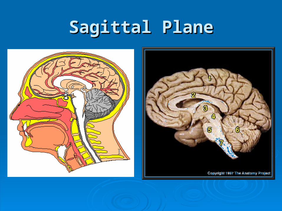
Sagittal PlaneSagittal Plane

Coronal PlaneCoronal Plane
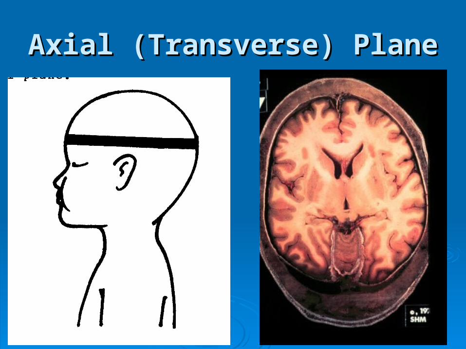
Axial (Transverse) PlaneAxial (Transverse) Plane

What is a CT (or CAT) scan?What is a CT (or CAT) scan?
CT stands for CT stands for “computed “computed tomography” - this is tomography” - this is a complex machine a complex machine that uses x-rays to that uses x-rays to create three-create three-dimensional images dimensional images of the bodyof the body
What is bright/dark on CT?What is bright/dark on CT?
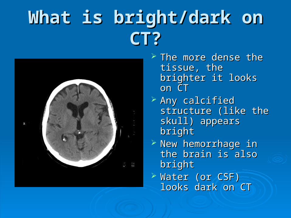
The more dense the The more dense the tissue, the brighter it tissue, the brighter it looks on CTlooks on CT
Any calcified structure Any calcified structure (like the skull) (like the skull) appears bright appears bright
New hemorrhage in New hemorrhage in the brain is also brightthe brain is also bright
Water (or CSF) looks Water (or CSF) looks dark on CTdark on CT
What is MR?What is MR?
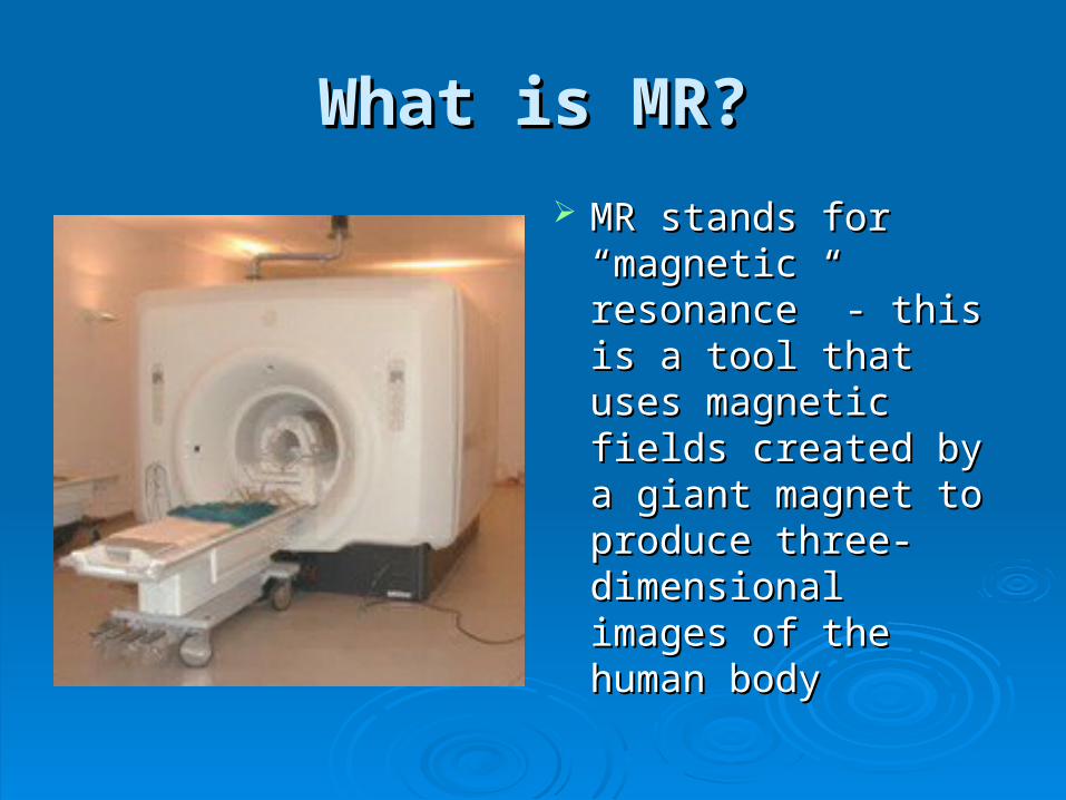
MR stands for MR stands for “magnetic resonance” “magnetic resonance” - this is a tool that - this is a tool that uses magnetic fields uses magnetic fields created by a giant created by a giant magnet to produce magnet to produce three-dimensional three-dimensional images of the human images of the human bodybody

The two most common types The two most common types of MR images are…of MR images are…
T1-weighted MR imagesT1-weighted MR images- - useful to look at normal useful to look at normal anatomyanatomy of the brain of the brain
T2-weight MR imagesT2-weight MR images - - useful to look at abnormal useful to look at abnormal processes (or processes (or pathologypathology) ) in the brainin the brain

What is bright/dark on T1?What is bright/dark on T1?
Fat is brightFat is bright White matter (inner White matter (inner
part of brain) is part of brain) is brighter than gray brighter than gray matter (cortex or matter (cortex or outer part of the outer part of the brain)brain)
Water (CSF) is darkWater (CSF) is dark
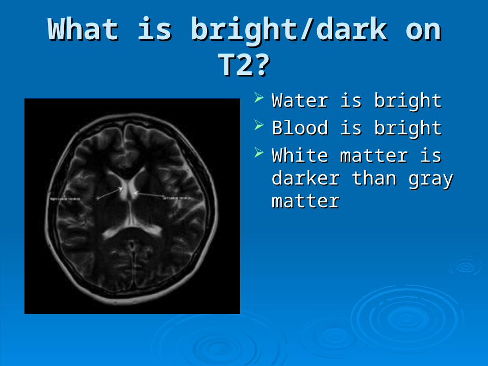
What is bright/dark on T2?What is bright/dark on T2?
Water is brightWater is bright Blood is brightBlood is bright White matter is darker White matter is darker
than gray matterthan gray matter

Other Studies - AngiogramOther Studies - Angiogram
Some neurologists Some neurologists just want to look at just want to look at blood vessels in the blood vessels in the brainbrain
They can inject a They can inject a contrast agent and contrast agent and then use x-rays to see then use x-rays to see the blood flowing the blood flowing inside the arteryinside the artery
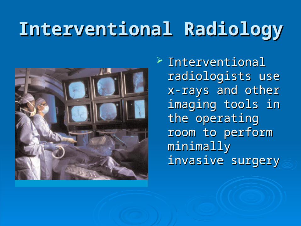
Interventional RadiologyInterventional Radiology
Interventional Interventional radiologists use x-radiologists use x-rays and other rays and other imaging tools in the imaging tools in the operating room to operating room to perform minimally perform minimally invasive surgeryinvasive surgery
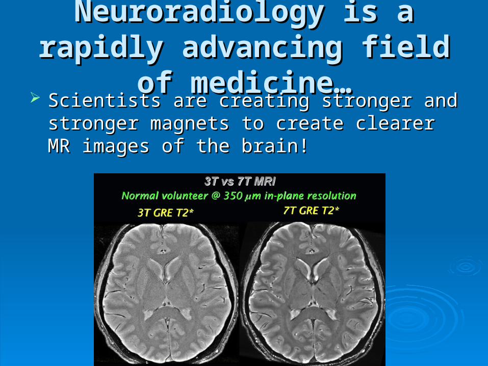
Neuroradiology is a rapidly Neuroradiology is a rapidly advancing field of medicine…advancing field of medicine…
Scientists are creating stronger and stronger Scientists are creating stronger and stronger magnets to create clearer MR images of the magnets to create clearer MR images of the brain!brain!
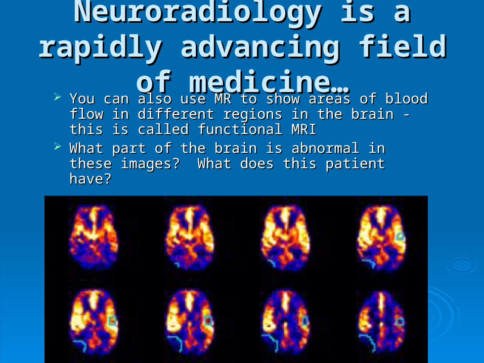
Neuroradiology is a rapidly Neuroradiology is a rapidly advancing field of medicine…advancing field of medicine…
You can also use MR to show areas of blood flow You can also use MR to show areas of blood flow in different regions in the brain - this is called in different regions in the brain - this is called functional MRIfunctional MRI
What part of the brain is abnormal in these What part of the brain is abnormal in these images? What does this patient have?images? What does this patient have?
NeuroradiologyNeuroradiology
At the end of today, you should be able to At the end of today, you should be able to identify the 3 different orientations of the brain identify the 3 different orientations of the brain as well as a few important structures in the brainas well as a few important structures in the brain
You should also attempt to distinguish CT, T1-You should also attempt to distinguish CT, T1-weighted MR and T2-weighted MR imagesweighted MR and T2-weighted MR imagesConsider a career in neuroradiology! It’s a very Consider a career in neuroradiology! It’s a very rewarding, high-tech specialty with a lot of fun rewarding, high-tech specialty with a lot of fun problem-solving. Neuroradiologists also play an problem-solving. Neuroradiologists also play an important role in diagnosing diseases and important role in diagnosing diseases and helping patients get the most appropriate helping patients get the most appropriate medical care.medical care.

Thanks!Thanks!
References/ResourcesReferences/Resources
http://www.images.http://www.images.googlegoogle.com.com
http://www.http://www.wikipediawikipedia.com.com