Embed Size (px)
Citation preview
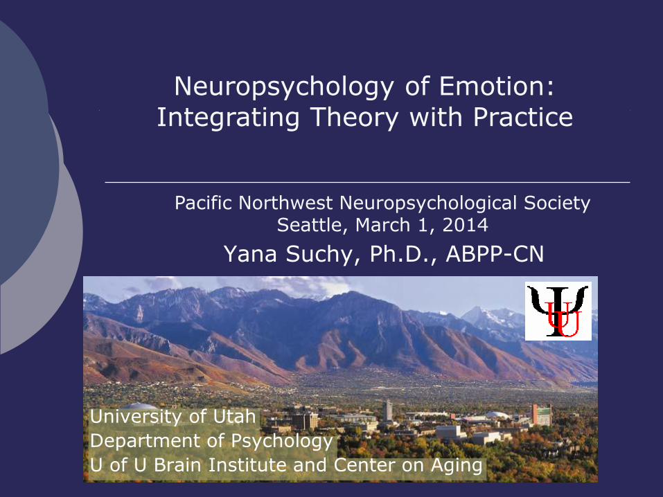
Pacific Northwest Neuropsychological Society Seattle, March 1, 2014
Neuropsychology of Emotion: Integrating Theory with Practice
Yana Suchy, Ph.D., ABPP-CN
University of Utah
Department of Psychology
U of U Brain Institute and Center on Aging
Learning objectives
(1) To describe five primary components of emotional processing and their neuroanatomic substrates
(2) To be aware of clinical populations that exhibit deficits in emotional processing
(3) To understand how neurocognitive abilities and test performance are affected by strengths and weaknesses in individual
components of emotional processing
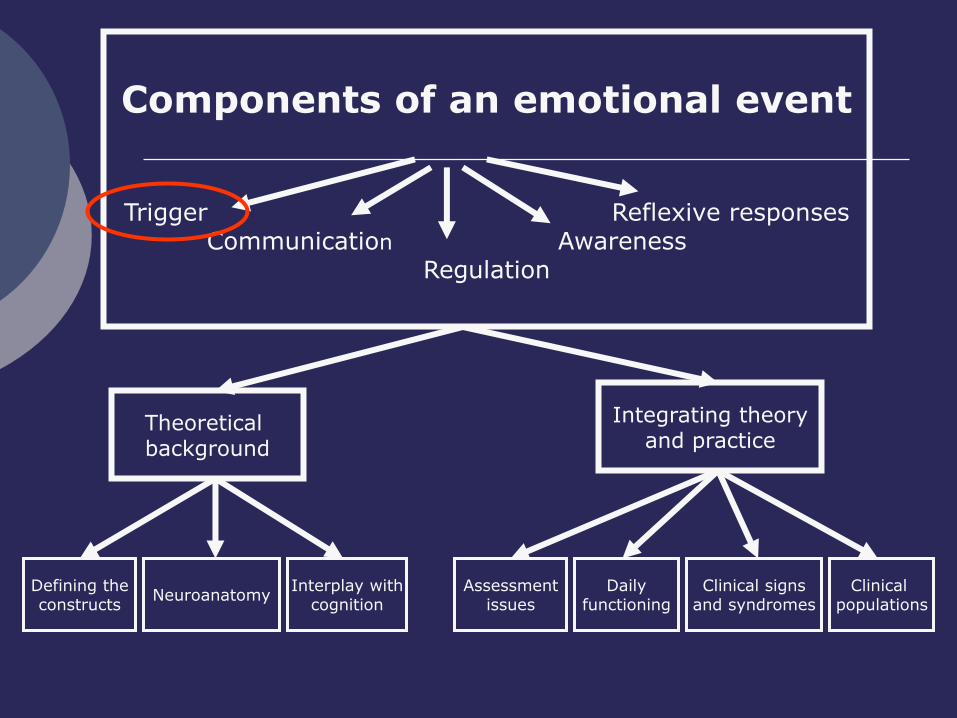
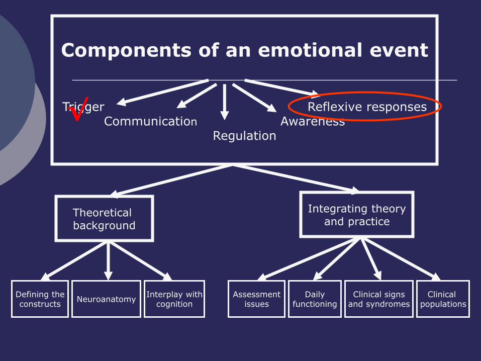
Components of an emotional event
Trigger Reflexive responses Communication Awareness
Regulation
Theoretical background
Integrating theory and practice
Defining the constructs
Neuroanatomy Interplay with
cognition Assessment
issues Clinical signs
and syndromes Clinical
populations Daily
functioning
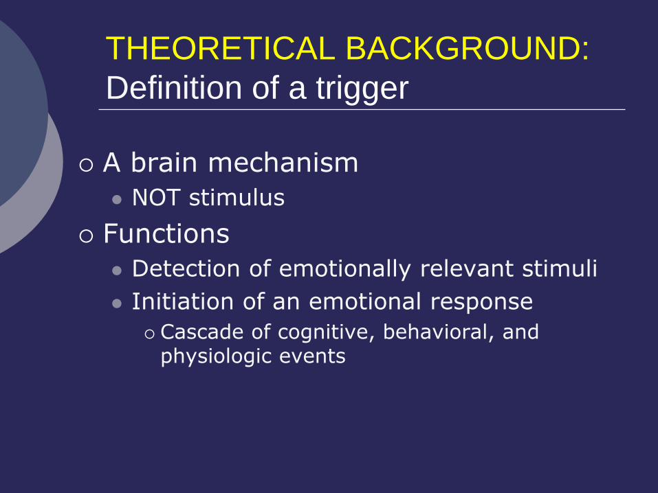
THEORETICAL BACKGROUND:
Definition of a trigger
A brain mechanism
NOT stimulus
Functions
Detection of emotionally relevant stimuli
Initiation of an emotional response
Cascade of cognitive, behavioral, and physiologic events
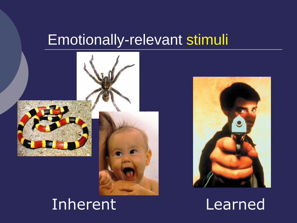
Emotionally-relevant stimuli
Inherent Learned
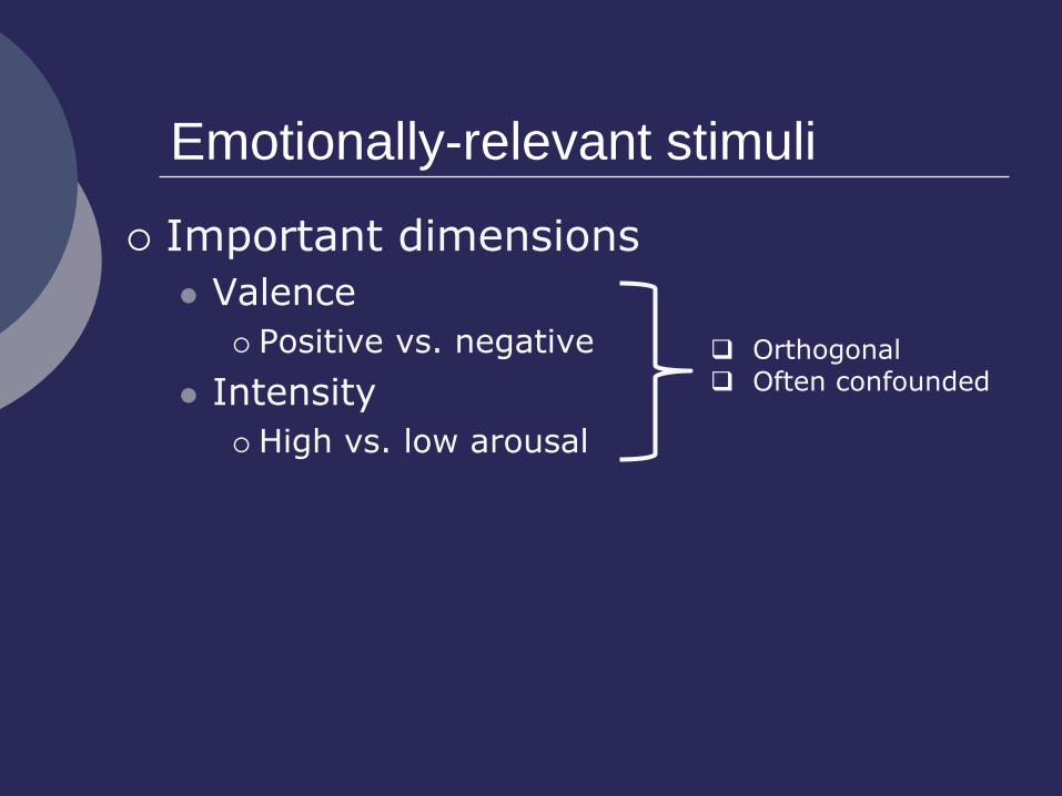
Emotionally-relevant stimuli
Important dimensions
Valence
Positive vs. negative
Intensity
High vs. low arousal
Orthogonal Often confounded
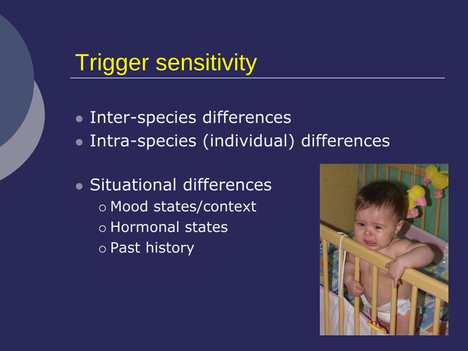
Trigger sensitivity
Inter-species differences
Intra-species (individual) differences
Situational differences
Mood states/context
Hormonal states
Past history
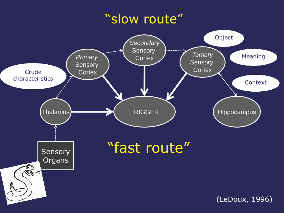
Thalamus
Sensory Organs
Primary
Sensory
Cortex
Hippocampus
Secondary
Sensory
Cortex Tertiary
Sensory
Cortex
TRIGGER
Crude characteristics
Object
Meaning
Context
“fast route”
“slow route”
(LeDoux, 1996)
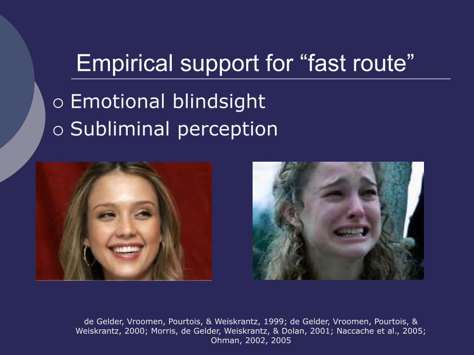
Empirical support for “fast route”
Emotional blindsight
Subliminal perception
de Gelder, Vroomen, Pourtois, & Weiskrantz, 1999; de Gelder, Vroomen, Pourtois, & Weiskrantz, 2000; Morris, de Gelder, Weiskrantz, & Dolan, 2001; Naccache et al., 2005;
Ohman, 2002, 2005
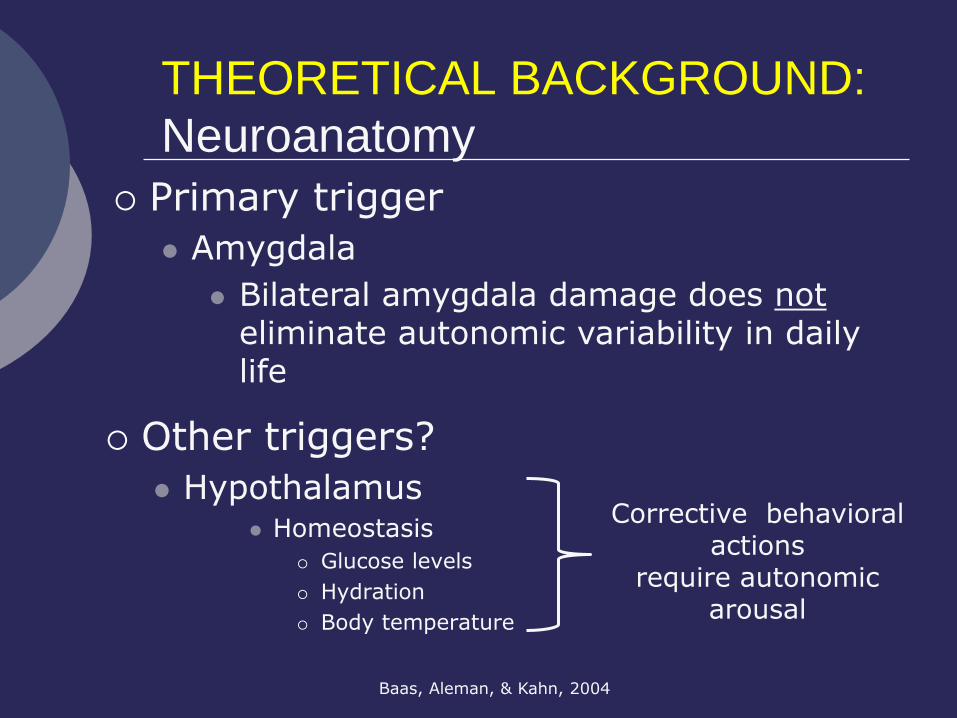
THEORETICAL BACKGROUND:
Neuroanatomy
Other triggers?
Hypothalamus Homeostasis
Glucose levels
Hydration
Body temperature
Baas, Aleman, & Kahn, 2004
Corrective behavioral actions
require autonomic arousal
Primary trigger
Amygdala
Bilateral amygdala damage does not eliminate autonomic variability in daily life
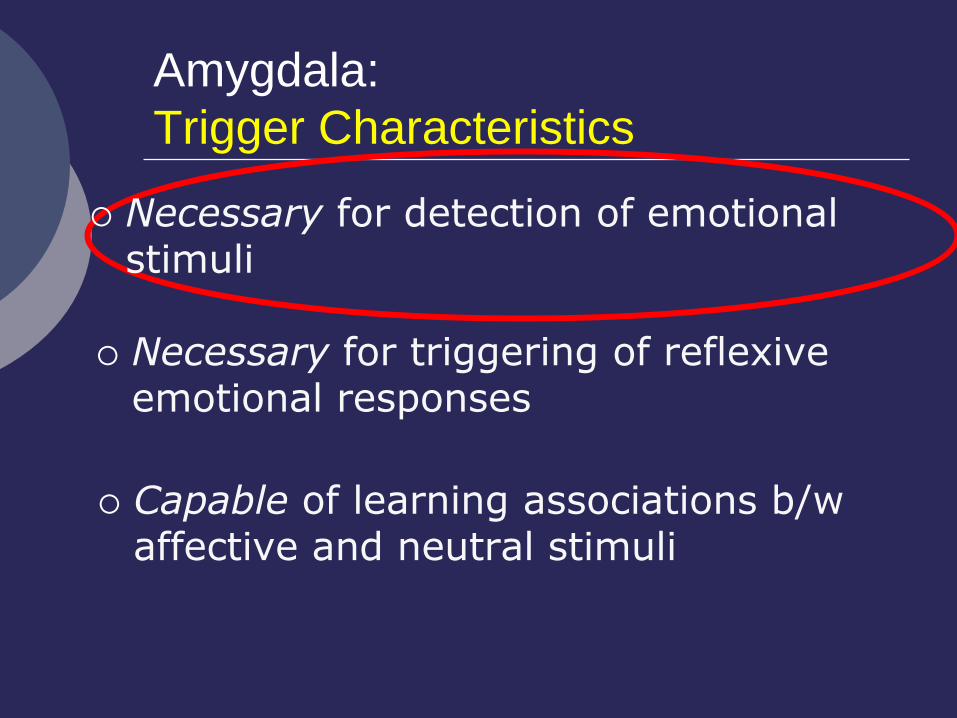
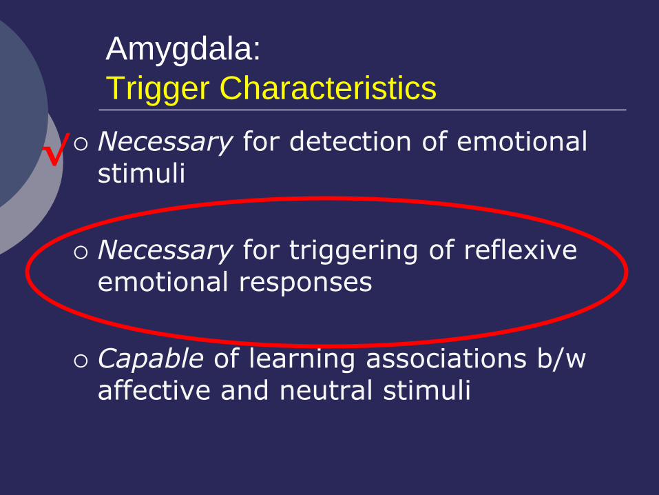
Amygdala:
Trigger Characteristics
Capable of learning associations b/w affective and neutral stimuli
Necessary for detection of emotional stimuli
Necessary for triggering of reflexive emotional responses
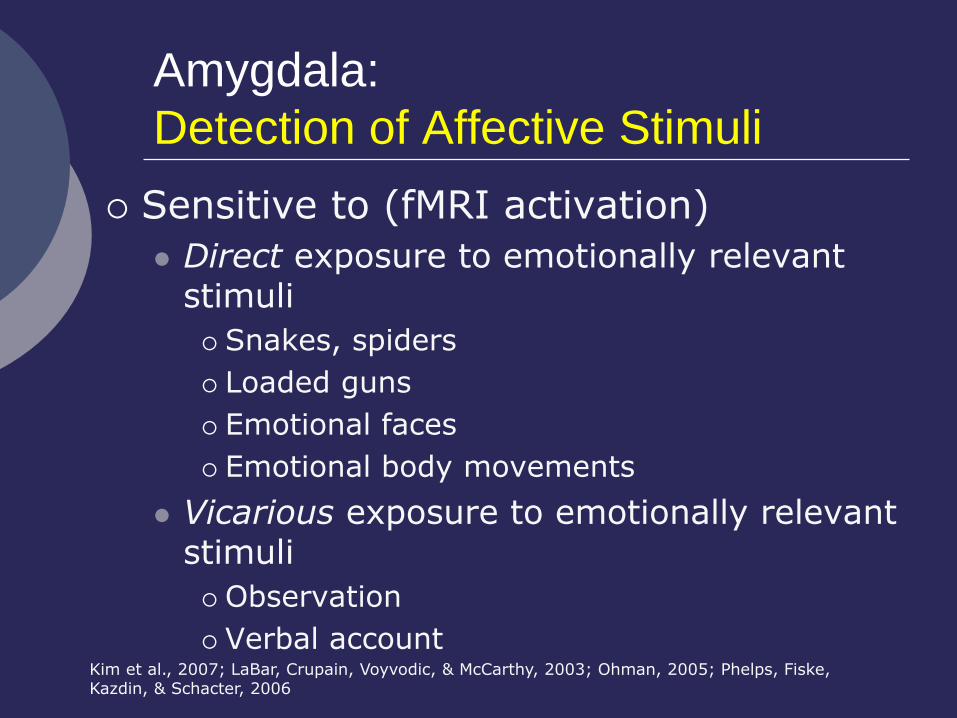
Amygdala:
Detection of Affective Stimuli
Sensitive to (fMRI activation)
Direct exposure to emotionally relevant stimuli
Snakes, spiders
Loaded guns
Emotional faces
Emotional body movements
Vicarious exposure to emotionally relevant stimuli
Observation
Verbal account
Kim et al., 2007; LaBar, Crupain, Voyvodic, & McCarthy, 2003; Ohman, 2005; Phelps, Fiske, Kazdin, & Schacter, 2006
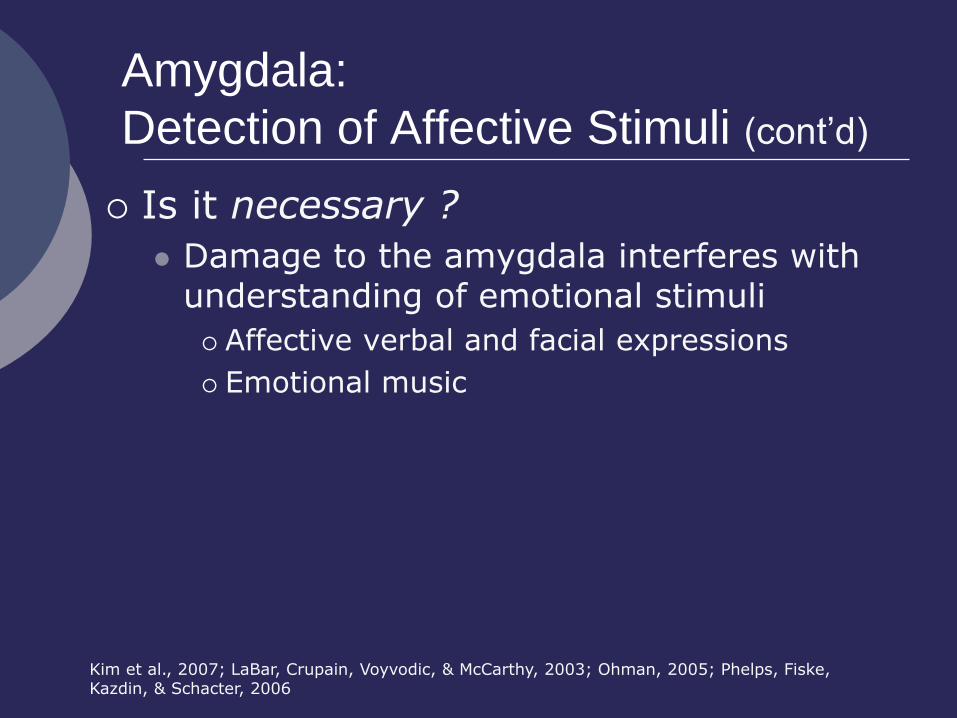
Amygdala:
Detection of Affective Stimuli (cont’d)
Is it necessary ?
Damage to the amygdala interferes with understanding of emotional stimuli
Affective verbal and facial expressions
Emotional music
Kim et al., 2007; LaBar, Crupain, Voyvodic, & McCarthy, 2003; Ohman, 2005; Phelps, Fiske, Kazdin, & Schacter, 2006
Amygdala:
Trigger Characteristics
Necessary for detection of emotional stimuli
Necessary for triggering of reflexive emotional responses
Capable of learning associations b/w affective and neutral stimuli
√
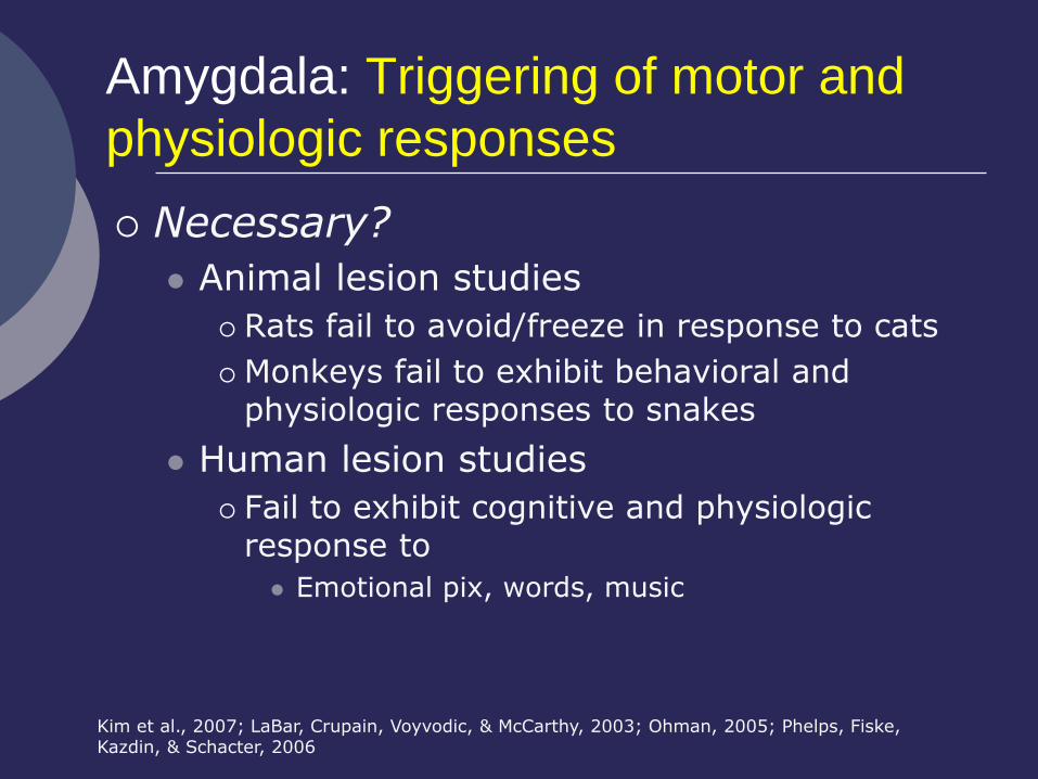
Amygdala: Triggering of motor and
physiologic responses
Necessary?
Animal lesion studies
Rats fail to avoid/freeze in response to cats
Monkeys fail to exhibit behavioral and physiologic responses to snakes
Human lesion studies
Fail to exhibit cognitive and physiologic response to
Emotional pix, words, music
Kim et al., 2007; LaBar, Crupain, Voyvodic, & McCarthy, 2003; Ohman, 2005; Phelps, Fiske, Kazdin, & Schacter, 2006
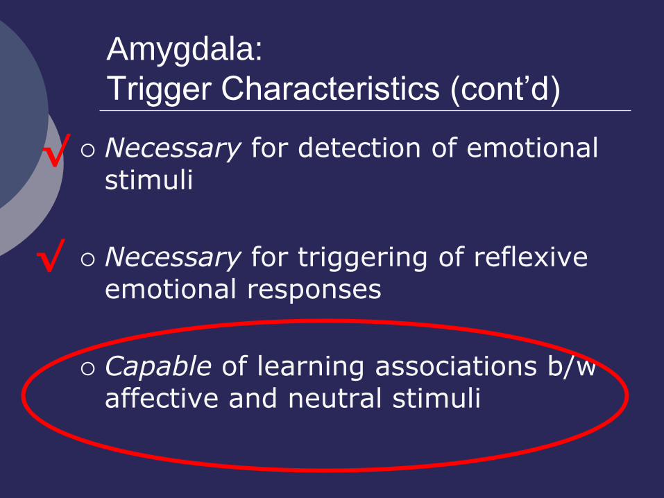
Amygdala:
Trigger Characteristics (cont’d)
Necessary for detection of emotional stimuli
Necessary for triggering of reflexive emotional responses
Capable of learning associations b/w affective and neutral stimuli
√
√
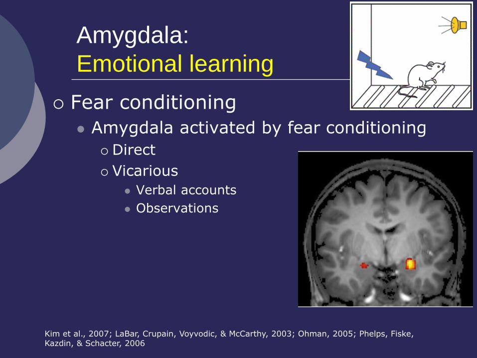
Amygdala:
Emotional learning
Fear conditioning
Amygdala activated by fear conditioning
Direct
Vicarious
Verbal accounts
Observations
Kim et al., 2007; LaBar, Crupain, Voyvodic, & McCarthy, 2003; Ohman, 2005; Phelps, Fiske, Kazdin, & Schacter, 2006
Emotional learning:
Other structures
Hippocampus
Orbitofrontal cortex
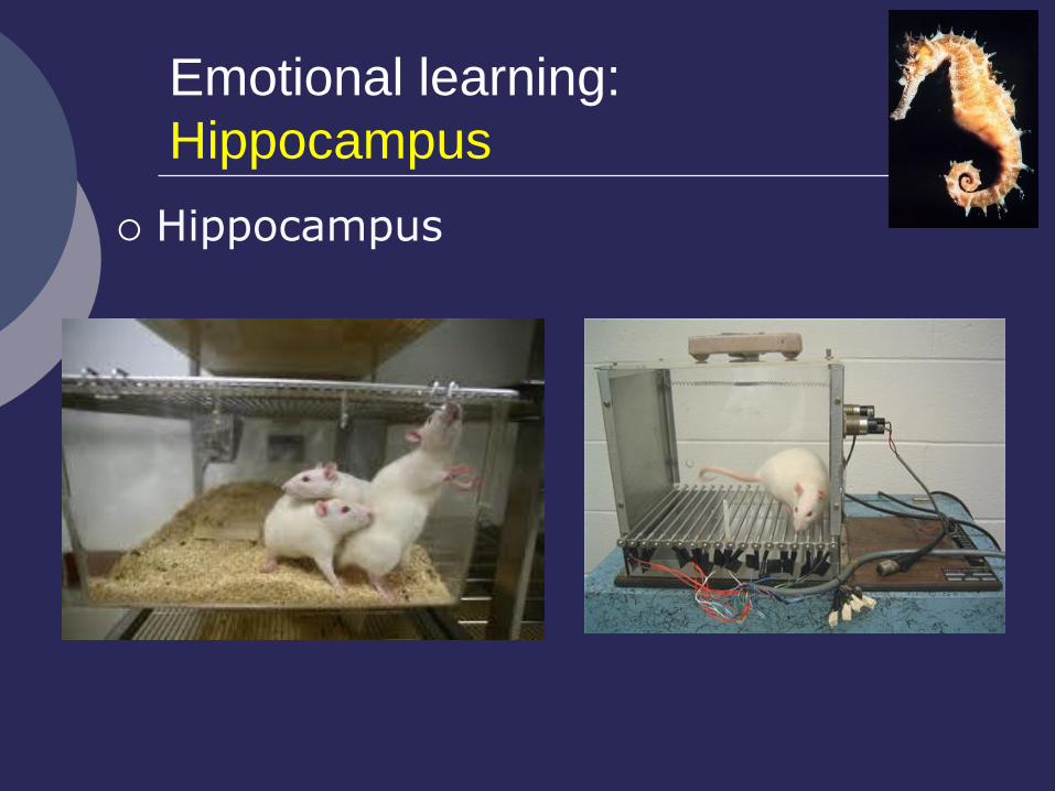
Emotional learning:
Hippocampus
Hippocampus
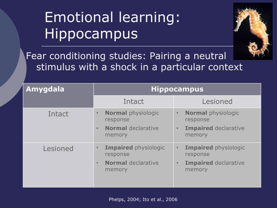
Emotional learning: Hippocampus
Phelps, 2004; Ito et al., 2006
Fear conditioning studies: Pairing a neutral stimulus with a shock in a particular context
Amygdala
Hippocampus
Intact Lesioned
Intact • Normal physiologic response
• Normal declarative memory
• Normal physiologic response
• Impaired declarative memory
Lesioned • Impaired physiologic response
• Normal declarative memory
• Impaired physiologic response
• Impaired declarative memory
Emotional learning:
Orbitofrontal cortex
Encoding associations between emotional and sensory information (all sensory modalities)
Rapid updating of contingencies as they change
BUT:
Amygdala may be necessary for learning to take place
Rolls, 2004
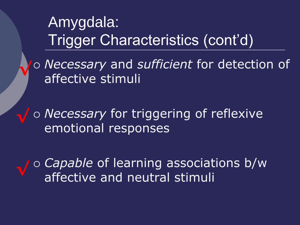
Amygdala:
Trigger Characteristics (cont’d)
Necessary and sufficient for detection of affective stimuli
Necessary for triggering of reflexive emotional responses
Capable of learning associations b/w affective and neutral stimuli
√
√
√
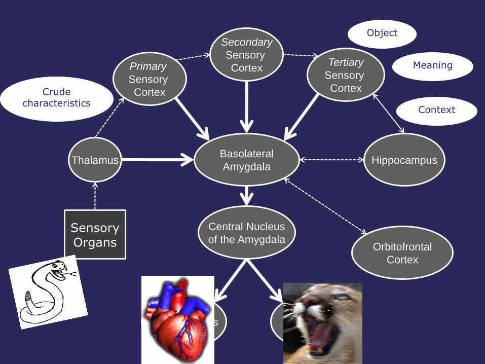
Thalamus
Sensory Organs
Primary
Sensory
Cortex
Hippocampus
Secondary
Sensory
Cortex Tertiary
Sensory
Cortex
Central Nucleus
of the Amygdala
Basolateral
Amygdala
Hypothalamus Brain Stem
Nuclei
Crude characteristics
Object
Meaning
Context
Orbitofrontal
Cortex
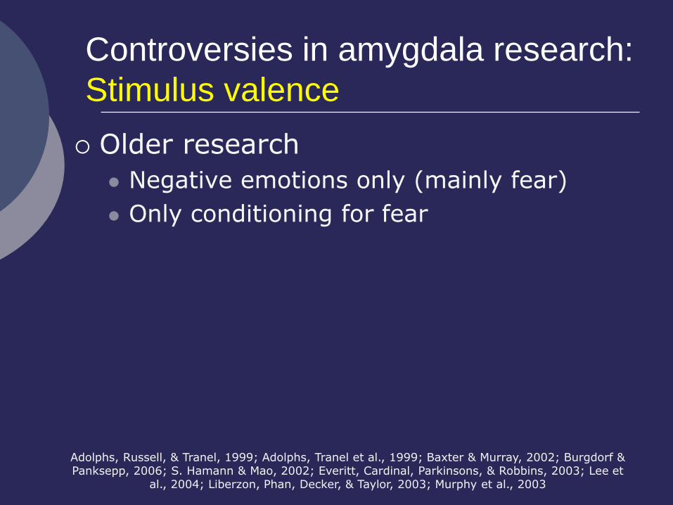
Controversies in amygdala research:
Stimulus valence
Older research
Negative emotions only (mainly fear)
Only conditioning for fear
Adolphs, Russell, & Tranel, 1999; Adolphs, Tranel et al., 1999; Baxter & Murray, 2002; Burgdorf & Panksepp, 2006; S. Hamann & Mao, 2002; Everitt, Cardinal, Parkinsons, & Robbins, 2003; Lee et
al., 2004; Liberzon, Phan, Decker, & Taylor, 2003; Murphy et al., 2003
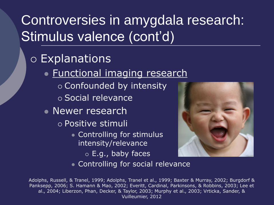
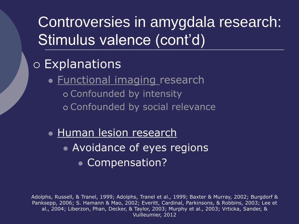
Controversies in amygdala research:
Stimulus valence (cont’d)
Explanations
Functional imaging research
Confounded by intensity
Social relevance
Newer research
Positive stimuli
Controlling for stimulus intensity/relevance
E.g., baby faces
Controlling for social relevance
Adolphs, Russell, & Tranel, 1999; Adolphs, Tranel et al., 1999; Baxter & Murray, 2002; Burgdorf & Panksepp, 2006; S. Hamann & Mao, 2002; Everitt, Cardinal, Parkinsons, & Robbins, 2003; Lee et
al., 2004; Liberzon, Phan, Decker, & Taylor, 2003; Murphy et al., 2003; Vrticka, Sander, & Vuilleumier, 2012
Controversies in amygdala research:
Stimulus valence (cont’d)
Explanations
Functional imaging research
Confounded by intensity
Confounded by social relevance
Adolphs, Russell, & Tranel, 1999; Adolphs, Tranel et al., 1999; Baxter & Murray, 2002; Burgdorf & Panksepp, 2006; S. Hamann & Mao, 2002; Everitt, Cardinal, Parkinsons, & Robbins, 2003; Lee et
al., 2004; Liberzon, Phan, Decker, & Taylor, 2003; Murphy et al., 2003; Vrticka, Sander, & Vuilleumier, 2012
Human lesion research
Avoidance of eyes regions
Compensation?
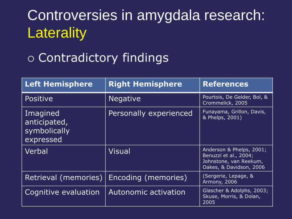
Left Hemisphere Right Hemisphere References
Positive Negative Pourtois, De Gelder, Bol, & Crommelick, 2005
Imagined anticipated, symbolically expressed
Personally experienced Funayama, Grillon, Davis, & Phelps, 2001)
Verbal Visual Anderson & Phelps, 2001; Benuzzi et al., 2004; Johnstone, van Reekum, Oakes, & Davidson, 2006
Retrieval (memories) Encoding (memories) (Sergerie, Lepage, & Armony, 2006
Cognitive evaluation Autonomic activation Glascher & Adolphs, 2003; Skuse, Morris, & Dolan, 2005
Contradictory findings
Controversies in amygdala research:
Laterality
THEORETICAL BACKGROUND:
Interplay with cognition
Attention
Memory
Judgment
“Emotional” decision making
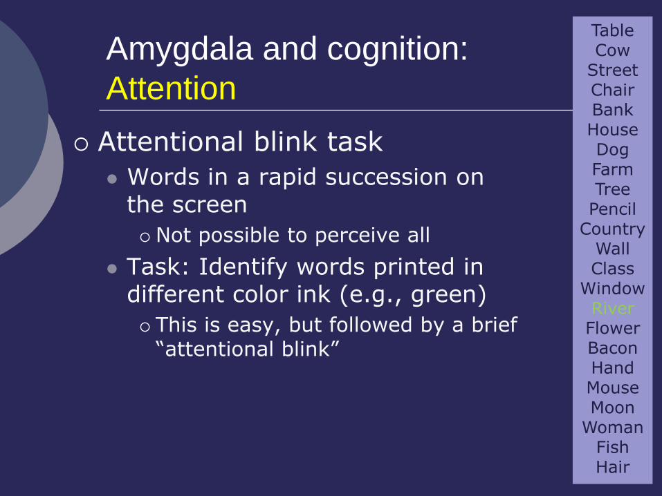
Amygdala and cognition:
Attention
Attentional blink task
Words in a rapid succession on the screen
Not possible to perceive all
Task: Identify words printed in different color ink (e.g., green)
This is easy, but followed by a brief “attentional blink”
Table Cow
Street Chair Bank House Dog Farm Tree
Pencil Country
Wall Class
Window River
Flower Bacon Hand Mouse Moon
Woman Fish Hair
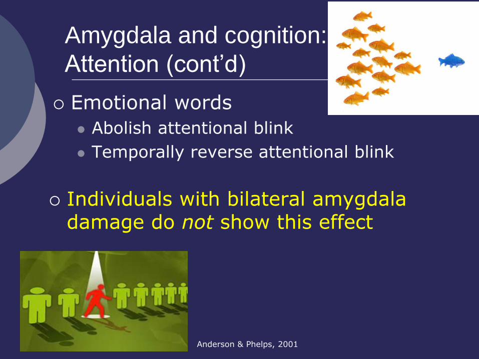
Amygdala and cognition:
Attention (cont’d)
Emotional words
Abolish attentional blink
Temporally reverse attentional blink
Anderson & Phelps, 2001
Individuals with bilateral amygdala damage do not show this effect
Amygdala and cognition:
Memory
Amygdala facilitates declarative memory
List of words OR series of photos
Some emotional, some neutral
Recognition memory better for emotional stimuli
Buchanan, Denburg, Tranel, & Adolphs, 2001; Phelps et al., 1998
Individuals with bilateral amygdala damage do not show this effect
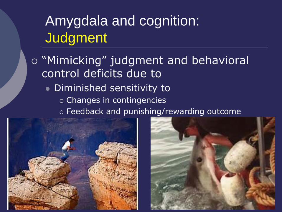
Amygdala and cognition:
Judgment
“Mimicking” judgment and behavioral control deficits due to
Diminished sensitivity to
Changes in contingencies
Feedback and punishing/rewarding outcome
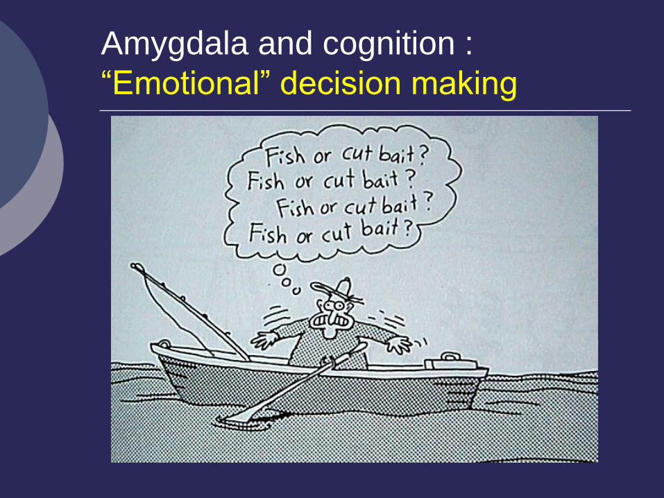
Amygdala and cognition :
“Emotional” decision making
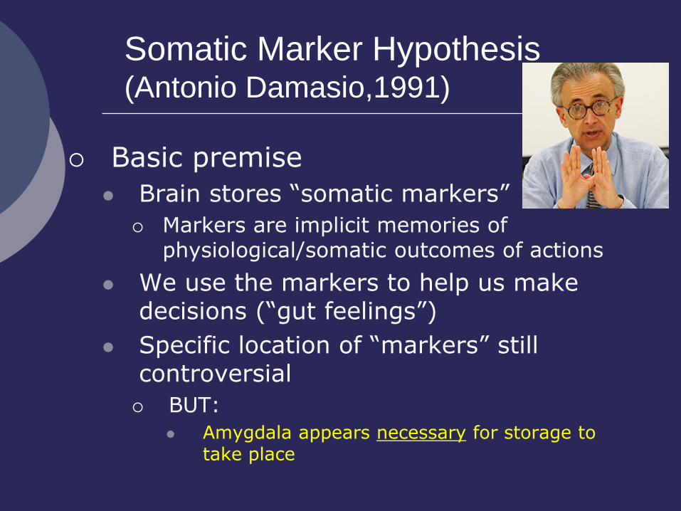
Somatic Marker Hypothesis (Antonio Damasio,1991)
Basic premise
Brain stores “somatic markers”
Markers are implicit memories of physiological/somatic outcomes of actions
We use the markers to help us make decisions (“gut feelings”)
Specific location of “markers” still controversial
BUT:
Amygdala appears necessary for storage to take place
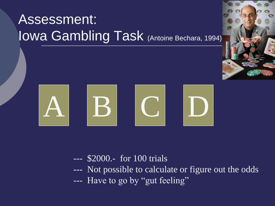
Assessment:
Iowa Gambling Task (Antoine Bechara, 1994)
A B C D
--- $2000.- for 100 trials
--- Not possible to calculate or figure out the odds
--- Have to go by “gut feeling”
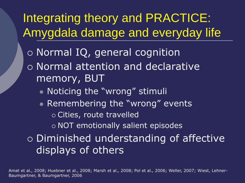
Integrating theory and PRACTICE:
Amygdala damage and everyday life
Normal IQ, general cognition
Normal attention and declarative memory, BUT
Noticing the “wrong” stimuli
Remembering the “wrong” events
Cities, route travelled
NOT emotionally salient episodes
Diminished understanding of affective displays of others
Amat et al., 2008; Huebner et al., 2008; Marsh et al., 2008; Pol et al., 2006; Weller, 2007; Wiest, Lehner-Baumgartner, & Baumgartner, 2006
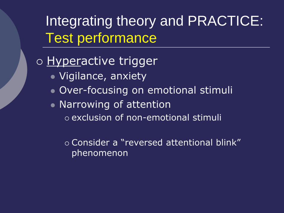
Integrating theory and PRACTICE:
Test performance
Hyperactive trigger
Vigilance, anxiety
Over-focusing on emotional stimuli
Narrowing of attention
exclusion of non-emotional stimuli
Consider a “reversed attentional blink” phenomenon
Integrating theory and PRACTICE:
Test performance (cont’d)
Hypoactive trigger
Failure to benefit from facilitation conferred by amygdala onto emotional stimuli in test material
Anna Thompson
Reading comprehension
Crouse, 2005; Suchy et al, 2009
Integrating theory and PRACTICE: Clinical Syndromes
Human Kluver and Bucy syndrome
Capgras syndrome
Heinrich Klüver Paul Bucy
(1897-1979) (1904-1992)
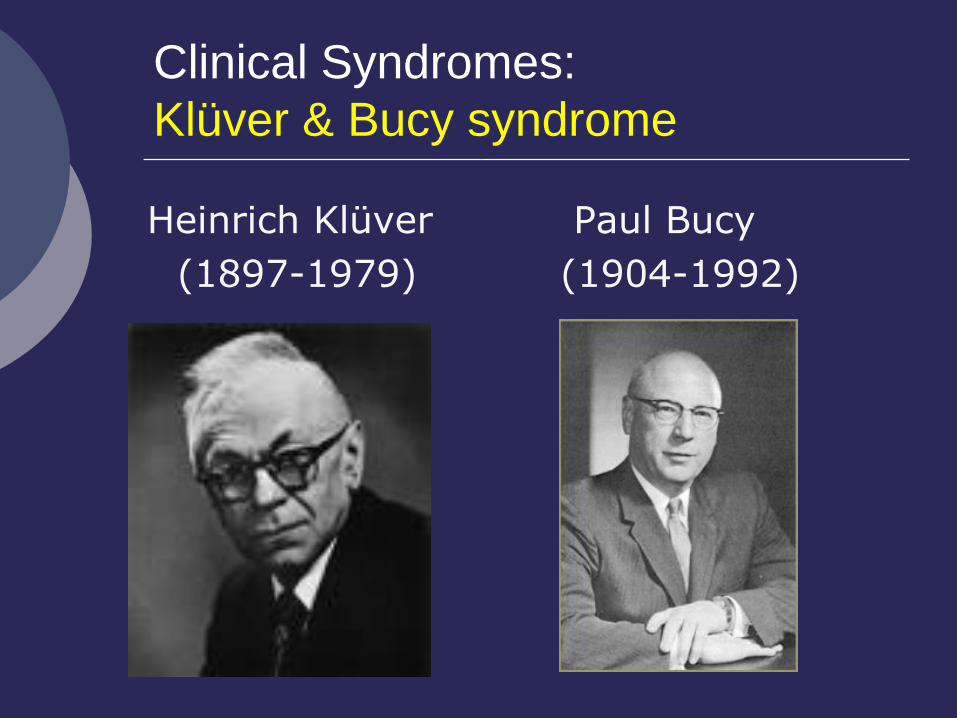
Clinical Syndromes:
Klüver & Bucy syndrome
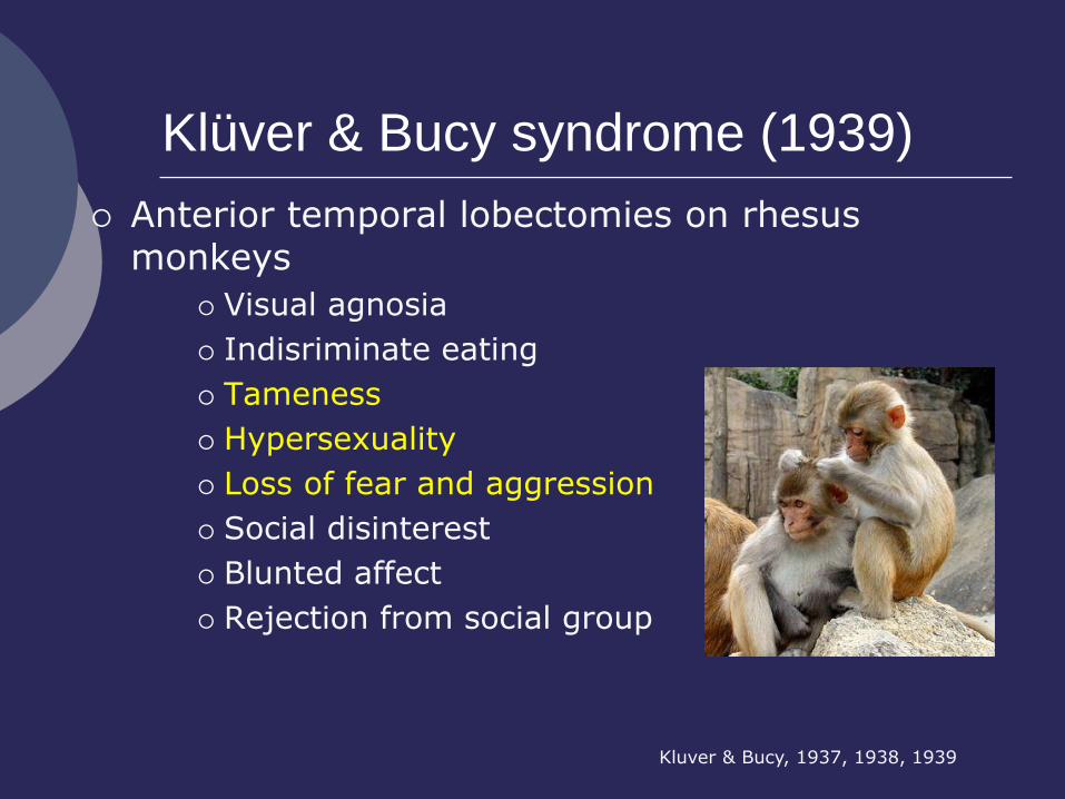
Klüver & Bucy syndrome (1939)
Anterior temporal lobectomies on rhesus monkeys
Visual agnosia
Indisriminate eating
Tameness
Hypersexuality
Loss of fear and aggression
Social disinterest
Blunted affect
Rejection from social group
Kluver & Bucy, 1937, 1938, 1939
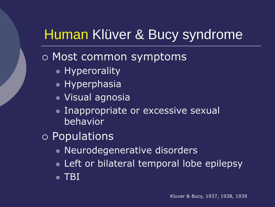
Human Klüver & Bucy syndrome
Most common symptoms
Hyperorality
Hyperphasia
Visual agnosia
Inappropriate or excessive sexual behavior
Populations
Neurodegenerative disorders
Left or bilateral temporal lobe epilepsy
TBI
Kluver & Bucy, 1937, 1938, 1939
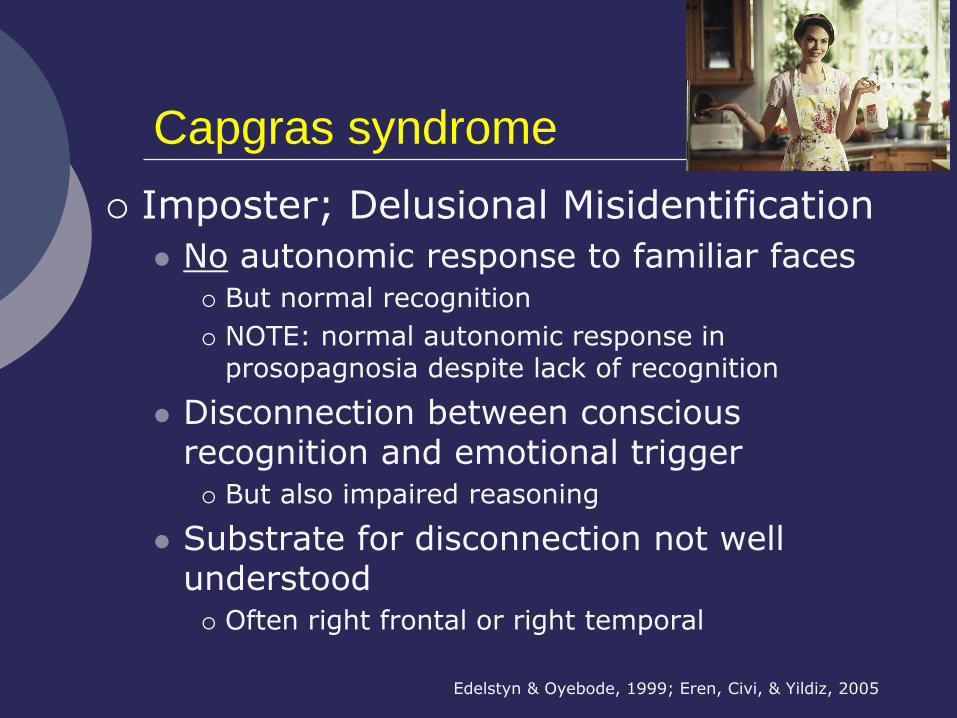
Capgras syndrome
Imposter; Delusional Misidentification
No autonomic response to familiar faces But normal recognition
NOTE: normal autonomic response in prosopagnosia despite lack of recognition
Disconnection between conscious recognition and emotional trigger But also impaired reasoning
Substrate for disconnection not well understood Often right frontal or right temporal
Edelstyn & Oyebode, 1999; Eren, Civi, & Yildiz, 2005
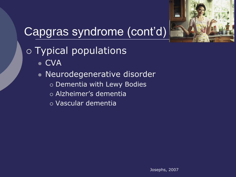
Capgras syndrome (cont’d)
Typical populations
CVA
Neurodegenerative disorder
Dementia with Lewy Bodies
Alzheimer’s dementia
Vascular dementia
Josephs, 2007
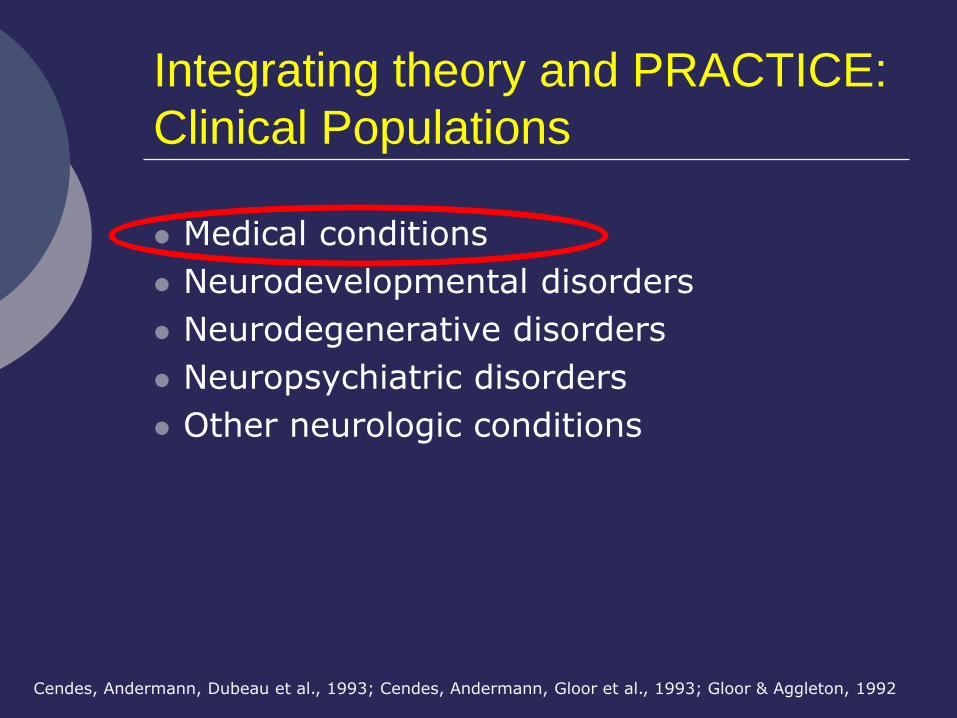
Integrating theory and PRACTICE:
Clinical Populations
Medical conditions
Neurodevelopmental disorders
Neurodegenerative disorders
Neuropsychiatric disorders
Other neurologic conditions
Cendes, Andermann, Dubeau et al., 1993; Cendes, Andermann, Gloor et al., 1993; Gloor & Aggleton, 1992
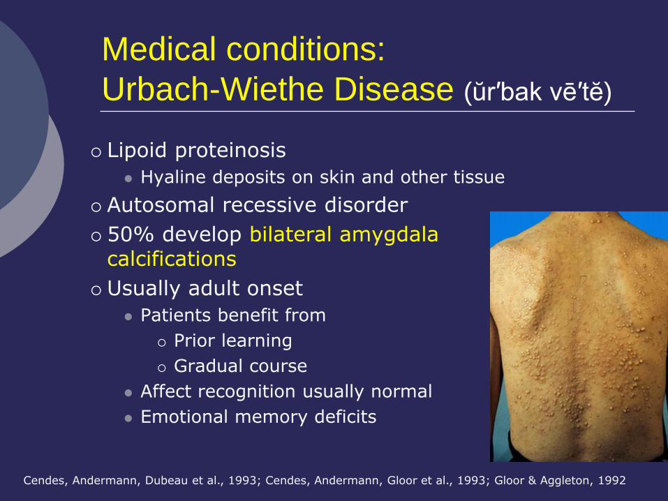
Medical conditions:
Urbach-Wiethe Disease (ŭr′bak vē′tĕ)
Lipoid proteinosis
Hyaline deposits on skin and other tissue
Autosomal recessive disorder
50% develop bilateral amygdala calcifications
Usually adult onset
Patients benefit from
Prior learning
Gradual course
Affect recognition usually normal
Emotional memory deficits
Cendes, Andermann, Dubeau et al., 1993; Cendes, Andermann, Gloor et al., 1993; Gloor & Aggleton, 1992
Neurodevelopmental disorders:
Autism
Amygdala abnormalities demonstrated via
Neuroimaging
Impaired processing of facial affect
BUT:
Normal facilitation by non-social emotional stimuli
Normal fear potentiation and startle
Bernier, Dawson, Panagiotides, & Webb, 2005; Boelte & Poustka, 2003; South, Ozonoff, Suchy, et al., 2008
Neurodevelopmental disorders:
Turner syndrome
Chromosomal disorder (monosomy X)
Physical characteristics
Short stature, webbed neck, gonadal dysfunction
Cognitive weaknesses
Visual spatial and executive
Affective abnormalities
Poor facial affect recognition
Social/interpersonal difficulties
Structural and functional amyg abnormalities
Brown et al., 2002; Kesler et al, 2004; Skuse et al., 2005
Neurodevelopmental disorders:
Fragile X syndrome
Most common genetic cause of MR
Multiple cognitive and emotional abnormalities
Abnormal gaze & avoidance of eye contact
Increased hippocampal and amygdalar volume
Increased activation in hippo and amyg in response to eye contact
Inconsistency re facial affect recognition
Watson et al, 2008; Dalton et al, 2008; Hagan et al., 2008
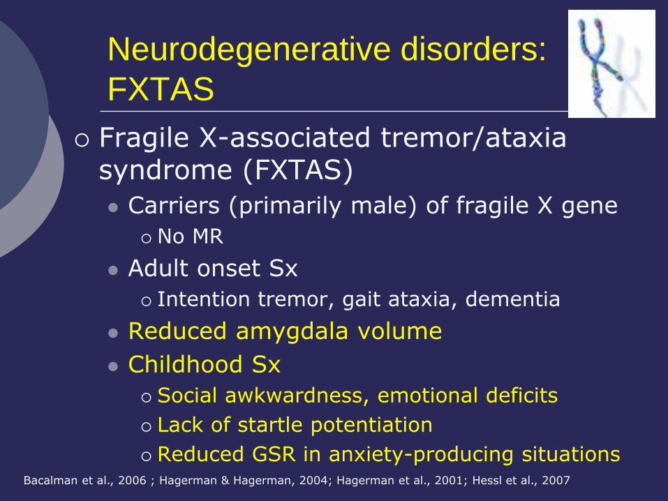
Neurodegenerative disorders:
FXTAS
Fragile X-associated tremor/ataxia syndrome (FXTAS)
Carriers (primarily male) of fragile X gene
No MR
Adult onset Sx
Intention tremor, gait ataxia, dementia
Reduced amygdala volume
Childhood Sx
Social awkwardness, emotional deficits
Lack of startle potentiation
Reduced GSR in anxiety-producing situations
Bacalman et al., 2006 ; Hagerman & Hagerman, 2004; Hagerman et al., 2001; Hessl et al., 2007
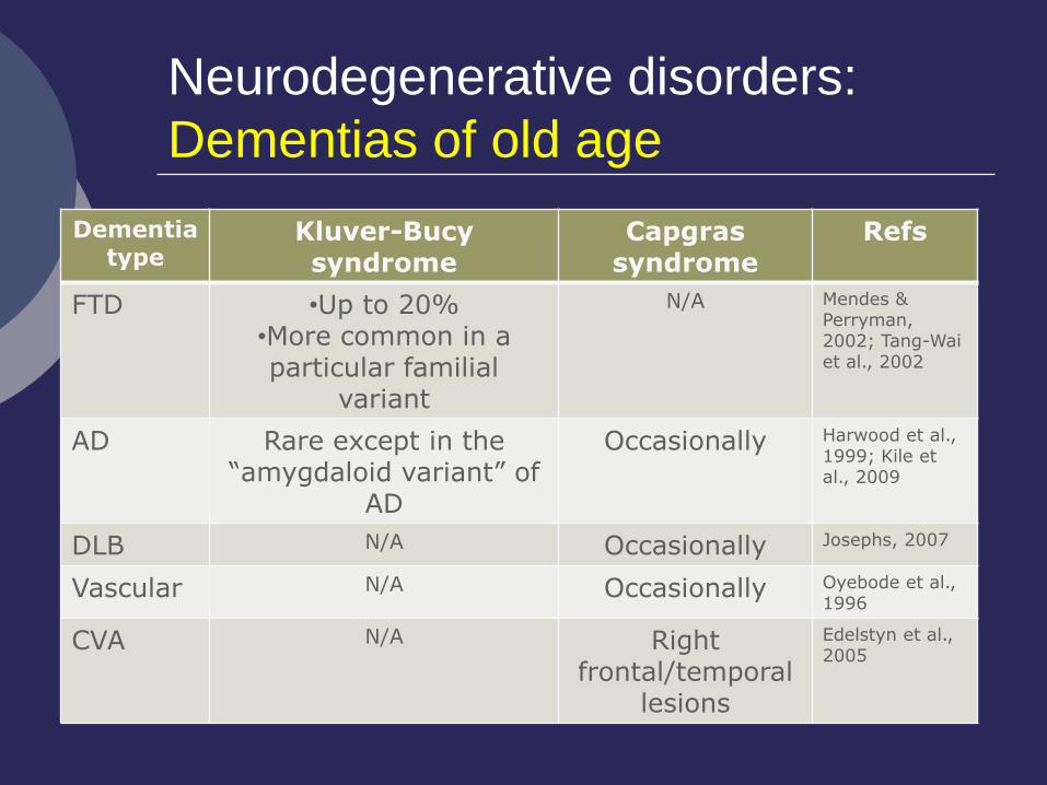
Neurodegenerative disorders:
Dementias of old age
Dementia type
Kluver-Bucy syndrome
Capgras syndrome
Refs
FTD •Up to 20% •More common in a particular familial
variant
N/A Mendes & Perryman, 2002; Tang-Wai et al., 2002
AD Rare except in the “amygdaloid variant” of
AD
Occasionally
Harwood et al., 1999; Kile et al., 2009
DLB N/A Occasionally Josephs, 2007
Vascular N/A Occasionally Oyebode et al., 1996
CVA N/A Right frontal/temporal
lesions
Edelstyn et al., 2005
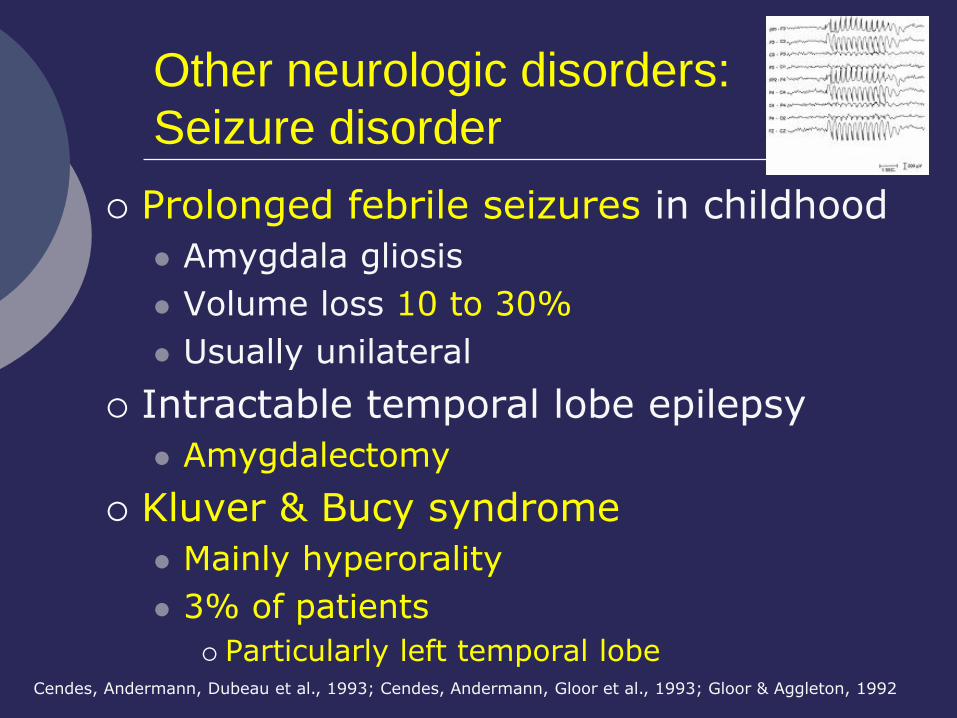
Other neurologic disorders:
Seizure disorder
Prolonged febrile seizures in childhood
Amygdala gliosis
Volume loss 10 to 30%
Usually unilateral
Intractable temporal lobe epilepsy
Amygdalectomy
Kluver & Bucy syndrome
Mainly hyperorality
3% of patients
Particularly left temporal lobe
Cendes, Andermann, Dubeau et al., 1993; Cendes, Andermann, Gloor et al., 1993; Gloor & Aggleton, 1992
Neuropsychiatric disorders:
Psychopathy
Diagnostic criteria (Hare PCL-R)
Antisocial behavior
Lack of long-term goals
Failure to achieve adult life-style
Shallow affect and callousness
Sensation seeking
Hare 1991, 1996
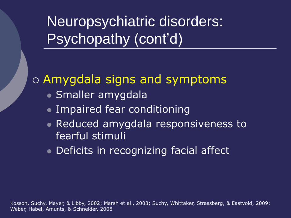
Neuropsychiatric disorders:
Psychopathy (cont’d)
Amygdala signs and symptoms
Smaller amygdala
Impaired fear conditioning
Reduced amygdala responsiveness to fearful stimuli
Deficits in recognizing facial affect
Kosson, Suchy, Mayer, & Libby, 2002; Marsh et al., 2008; Suchy, Whittaker, Strassberg, & Eastvold, 2009; Weber, Habel, Amunts, & Schneider, 2008
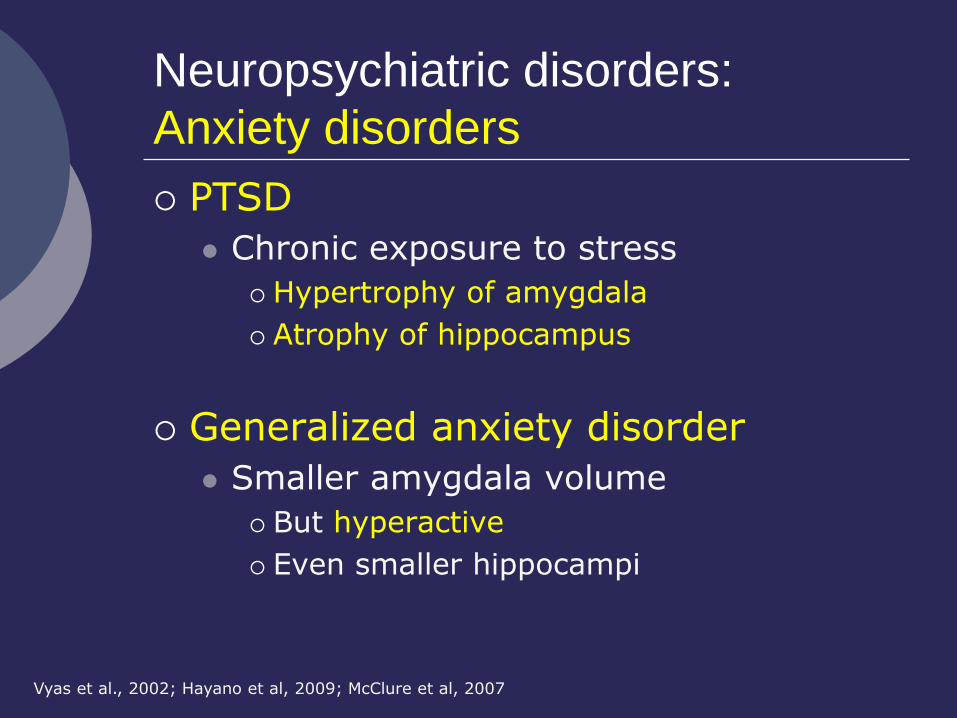
Neuropsychiatric disorders:
Anxiety disorders
PTSD
Chronic exposure to stress
Hypertrophy of amygdala
Atrophy of hippocampus
Generalized anxiety disorder
Smaller amygdala volume
But hyperactive
Even smaller hippocampi
Vyas et al., 2002; Hayano et al, 2009; McClure et al, 2007
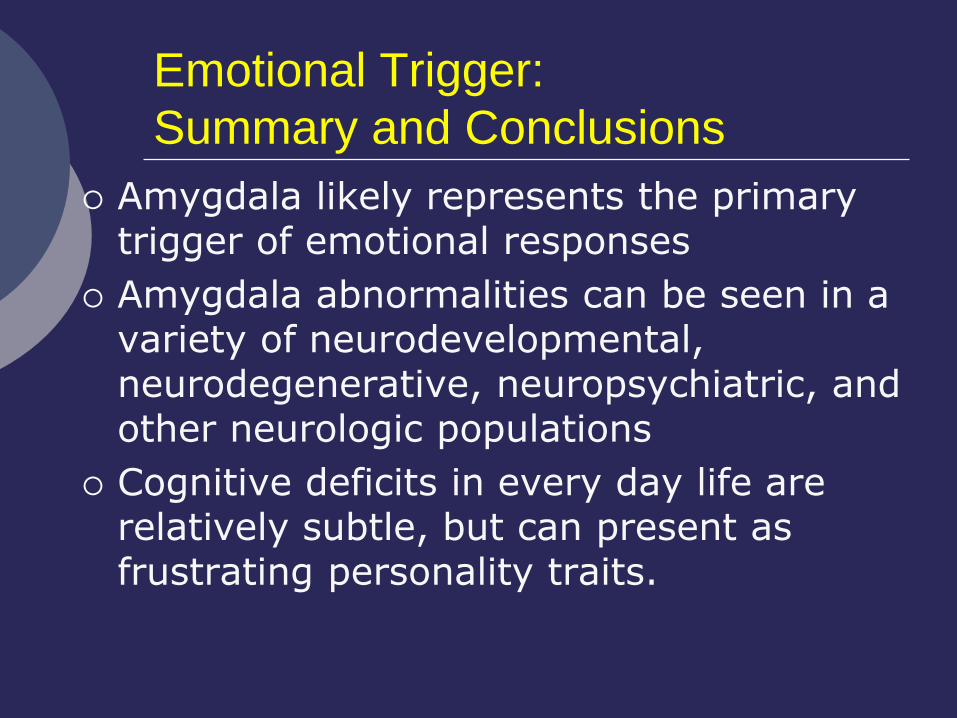
Emotional Trigger:
Summary and Conclusions
Amygdala likely represents the primary trigger of emotional responses
Amygdala abnormalities can be seen in a variety of neurodevelopmental, neurodegenerative, neuropsychiatric, and other neurologic populations
Cognitive deficits in every day life are relatively subtle, but can present as frustrating personality traits.
Components of an emotional event
Trigger Reflexive responses Communication Awareness
Regulation
Theoretical background
Integrating theory and practice
Defining the constructs
Neuroanatomy Interplay with
cognition Assessment
issues Clinical signs
and syndromes Clinical
populations Daily
functioning
√
THEORETICAL BACKGROUND:
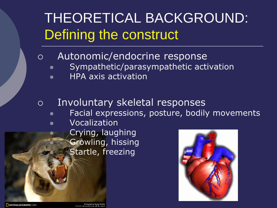
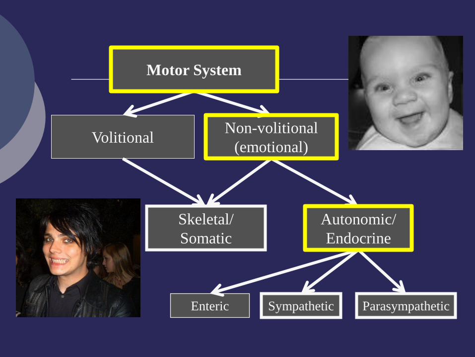
Defining the construct
Autonomic/endocrine response Sympathetic/parasympathetic activation HPA axis activation
Involuntary skeletal responses Facial expressions, posture, bodily movements Vocalization Crying, laughing Growling, hissing Startle, freezing
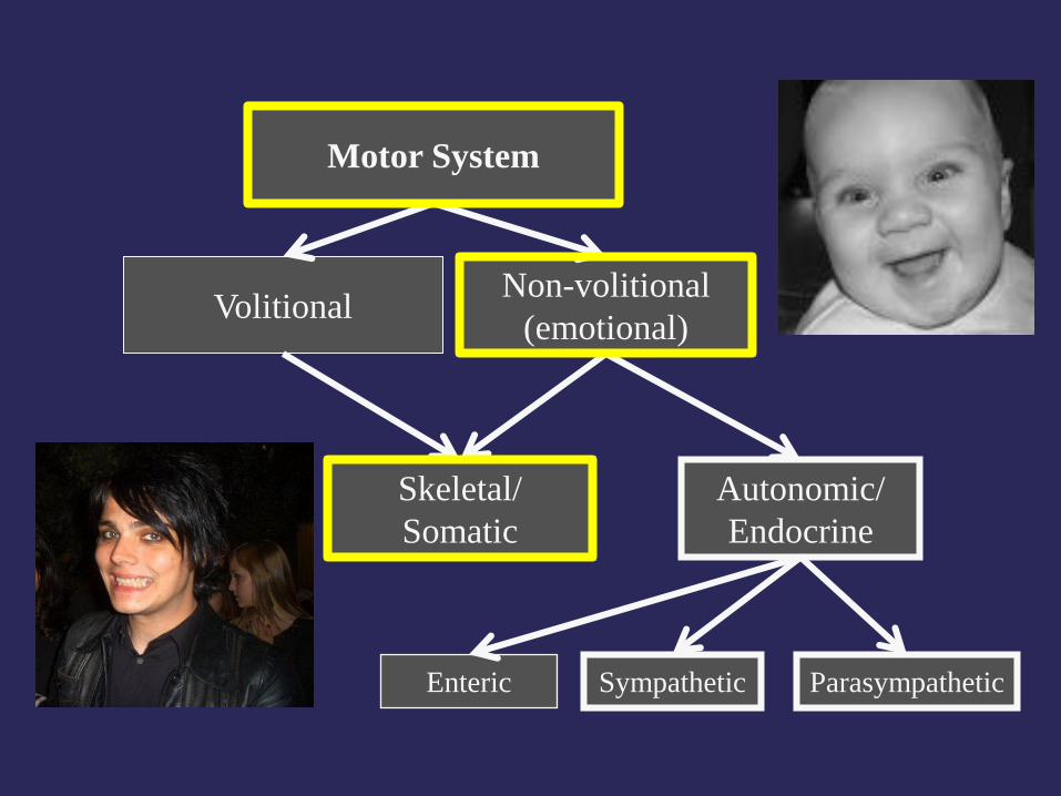
Motor System
Volitional Non-volitional
(emotional)
Skeletal/
Somatic
Autonomic/
Endocrine
Sympathetic
Parasympathetic
Enteric
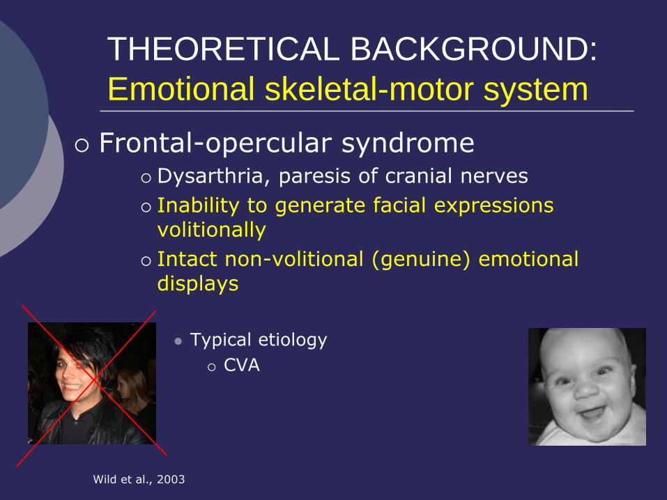
THEORETICAL BACKGROUND:
Emotional skeletal-motor system
Frontal-opercular syndrome Dysarthria, paresis of cranial nerves
Inability to generate facial expressions volitionally
Intact non-volitional (genuine) emotional displays
Typical etiology
CVA
Wild et al., 2003
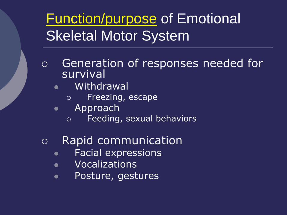
Function/purpose of Emotional
Skeletal Motor System
Generation of responses needed for survival
Withdrawal Freezing, escape
Approach Feeding, sexual behaviors
Rapid communication Facial expressions Vocalizations Posture, gestures
Motor System
Volitional Non-volitional
(emotional)
Skeletal/
Somatic
Autonomic/
Endocrine
Sympathetic
Parasympathetic
Enteric
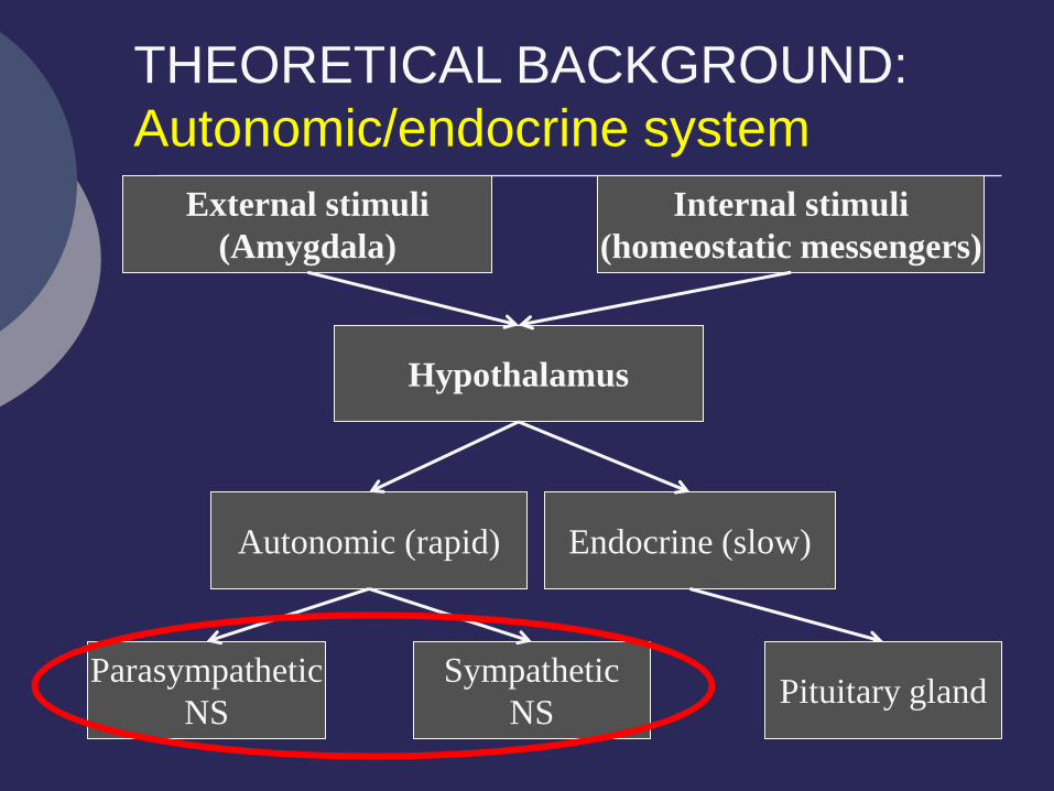
Hypothalamus
Autonomic (rapid) Endocrine (slow)
Sympathetic
NS Pituitary gland
Parasympathetic
NS
THEORETICAL BACKGROUND:
Autonomic/endocrine system
External stimuli
(Amygdala)
Internal stimuli
(homeostatic messengers)
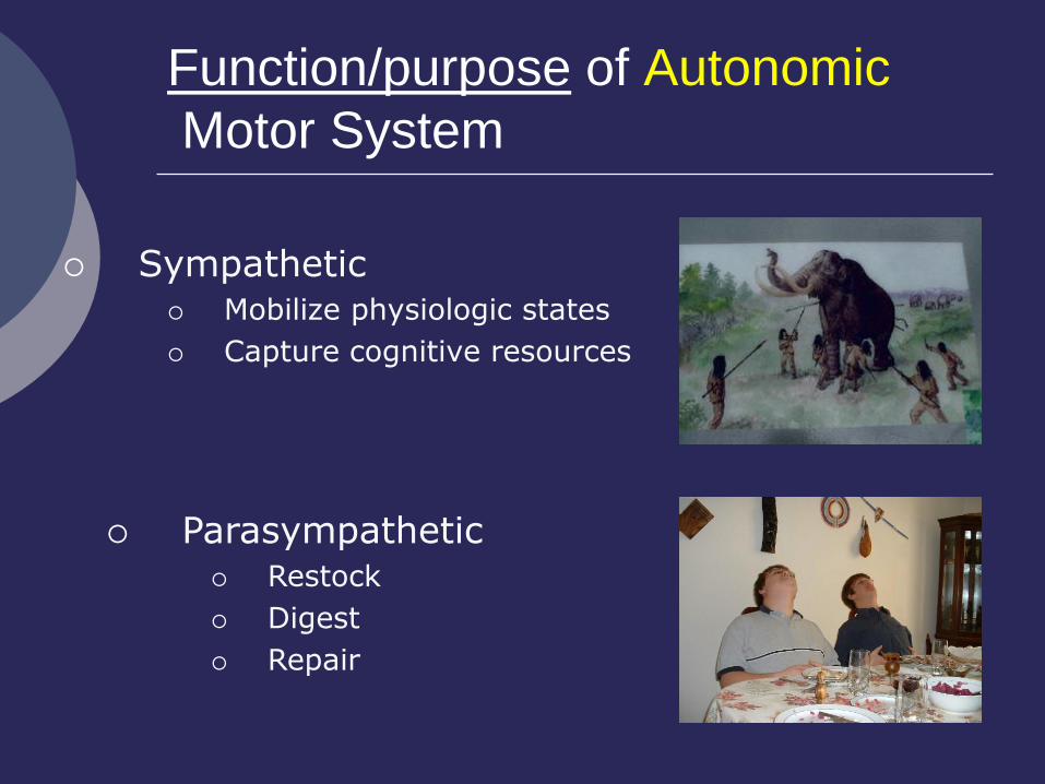
Function/purpose of Autonomic
Motor System
Sympathetic Mobilize physiologic states
Capture cognitive resources
Parasympathetic Restock
Digest
Repair
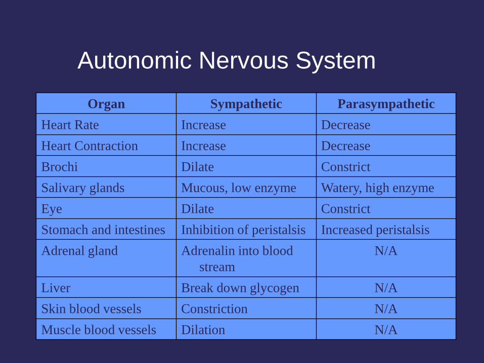
Autonomic Nervous System
Organ Sympathetic Parasympathetic
Heart Rate Increase Decrease
Heart Contraction Increase Decrease
Brochi Dilate Constrict
Salivary glands Mucous, low enzyme Watery, high enzyme
Eye Dilate Constrict
Stomach and intestines Inhibition of peristalsis Increased peristalsis
Adrenal gland Adrenalin into blood
stream
N/A
Liver Break down glycogen N/A
Skin blood vessels Constriction N/A
Muscle blood vessels Dilation N/A
Sympathetic NS
Parasympathetic NS
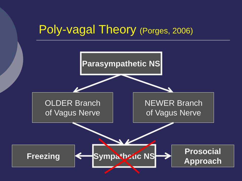
Poly-vagal Theory (Porges, 2006)
OLDER Branch
of Vagus Nerve
NEWER Branch
of Vagus Nerve
Freezing Prosocial
Approach
THEORETICAL BACKGROUND:
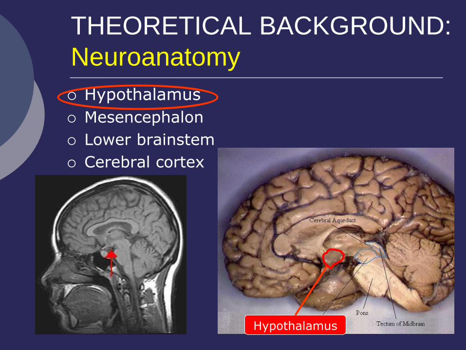
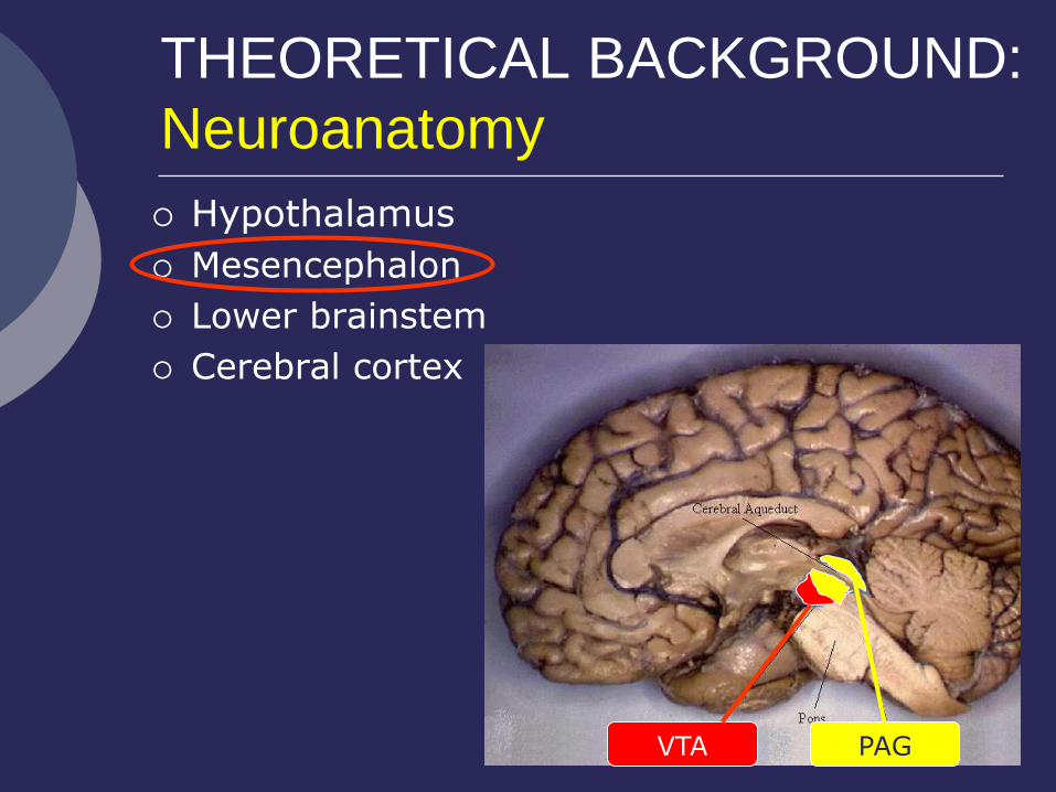
Neuroanatomy
Hypothalamus
Mesencephalon
Lower brainstem
Cerebral cortex
Hypothalamus
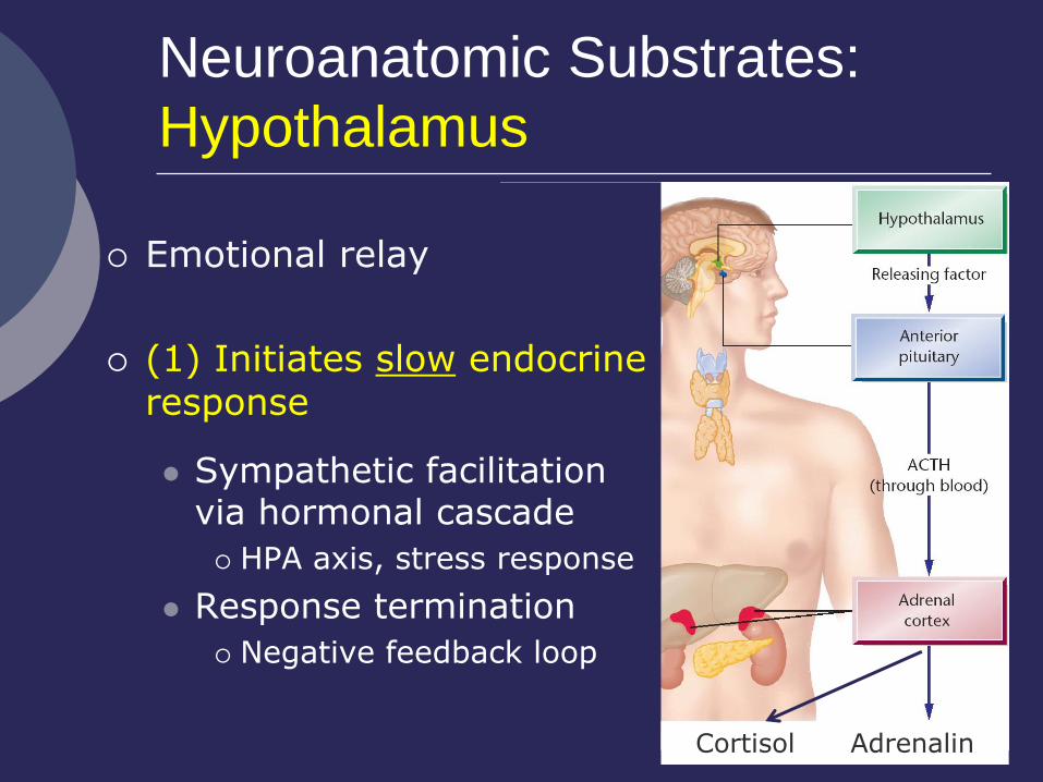
Neuroanatomic Substrates:
Hypothalamus
Emotional relay
(1) Initiates slow endocrine response
Cortisol Adrenalin
Sympathetic facilitation via hormonal cascade
HPA axis, stress response
Response termination
Negative feedback loop
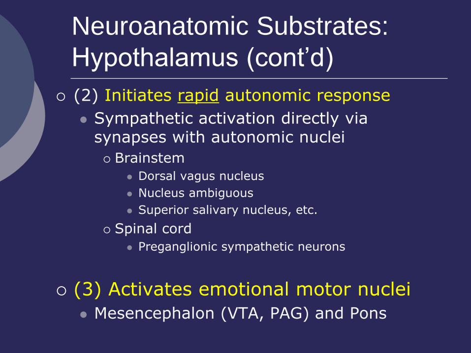
Neuroanatomic Substrates:
Hypothalamus (cont’d)
(2) Initiates rapid autonomic response
Sympathetic activation directly via synapses with autonomic nuclei
Brainstem
Dorsal vagus nucleus
Nucleus ambiguous
Superior salivary nucleus, etc.
Spinal cord
Preganglionic sympathetic neurons
(3) Activates emotional motor nuclei
Mesencephalon (VTA, PAG) and Pons
THEORETICAL BACKGROUND:
Neuroanatomy
Hypothalamus
Mesencephalon
Lower brainstem
Cerebral cortex
VTA PAG
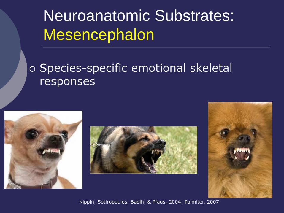
Neuroanatomic Substrates:
Mesencephalon
Species-specific emotional skeletal responses
Kippin, Sotiropoulos, Badih, & Pfaus, 2004; Palmiter, 2007
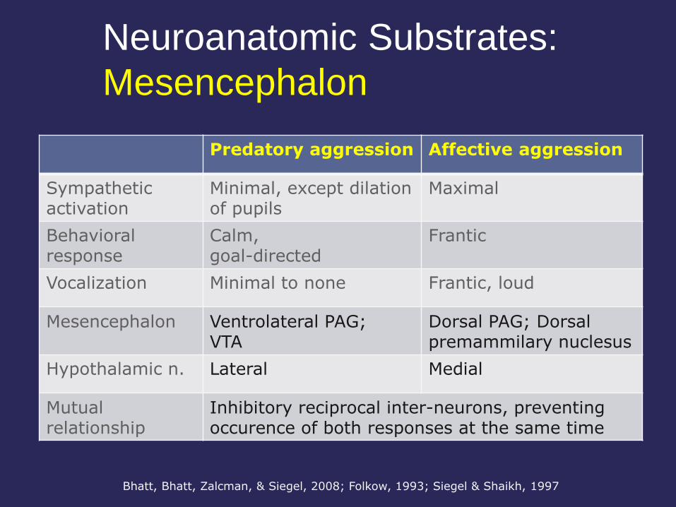
Neuroanatomic Substrates:
Mesencephalon
Bhatt, Bhatt, Zalcman, & Siegel, 2008; Folkow, 1993; Siegel & Shaikh, 1997
Predatory aggression Affective aggression
Sympathetic activation
Minimal, except dilation of pupils
Maximal
Behavioral response
Calm, goal-directed
Frantic
Vocalization Minimal to none Frantic, loud
Mesencephalon Ventrolateral PAG; VTA
Dorsal PAG; Dorsal premammilary nuclesus
Hypothalamic n. Lateral Medial
Mutual relationship
Inhibitory reciprocal inter-neurons, preventing occurence of both responses at the same time
Neuroanatomic Substrates:
Mesencephalon (cont’d)
Appetitive behaviors
Eating, drinking, mating
VTA
Hypothalamic nuclei
Other structures medial forebrain bundle, NAc, striatum/ventral
pallidum, ventral prefrontal cortex, cerebellum, anterior cingulate cortex, olfactory bulb, temporal cortex, area postrema
Kippin, Sotiropoulos, Badih, & Pfaus, 2004; Palmiter, 2007
Neuroanatomic Substrates:
Mesencephalon (cont’d)
Further autonomic control
Projections to lower brain stem
Species-specific emotional skeletal responses
THEORETICAL BACKGROUND:
Neuroanatomy
Hypothalamus
Mesencephalon
Lower brainstem
Cerebral cortex
Pons Medulla
Cerebellum
Neuroanatomic Substrates:
Lower brainstem
Medulla oblongata Nucleus ambiguous, salivary nucleus, dorsal
motor nucleus
Efferents to vital organs and glands
Cerebellum Direct reciprocal projections w/ hypothalamus
Pons Crying, laughing
Ascending projections to the cerebral cortex
RAS
Schmahmann, 2001; Wild, Rodden, Grodd, & Ruch, 2003; Zhu, Yung, Chow, Chan, & Wang, 2006
THEORETICAL BACKGROUND:
Neuroanatomy
Hypothalamus
Mesencephalon
Lower brainstem
Cerebral cortex
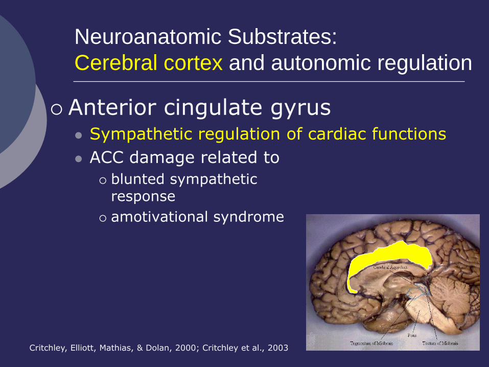
Neuroanatomic Substrates:
Cerebral cortex and autonomic regulation
Anterior cingulate gyrus Sympathetic regulation of cardiac functions
ACC damage related to
blunted sympathetic response
amotivational syndrome
Critchley, Elliott, Mathias, & Dolan, 2000; Critchley et al., 2003
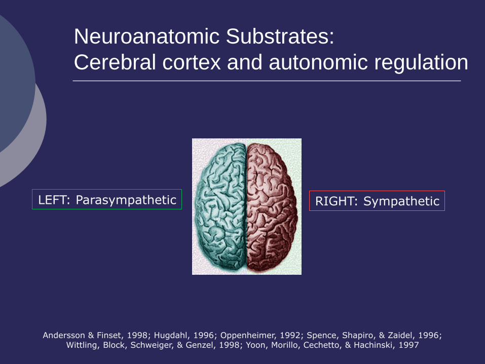
Neuroanatomic Substrates:
Cerebral cortex and autonomic regulation
Andersson & Finset, 1998; Hugdahl, 1996; Oppenheimer, 1992; Spence, Shapiro, & Zaidel, 1996; Wittling, Block, Schweiger, & Genzel, 1998; Yoon, Morillo, Cechetto, & Hachinski, 1997
LEFT: Parasympathetic RIGHT: Sympathetic
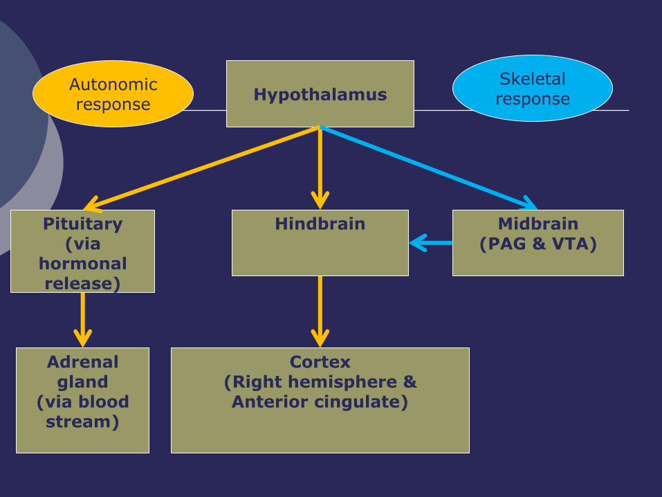
Hypothalamus
Pituitary (via
hormonal release)
Hindbrain
Autonomic response
Skeletal response
Midbrain (PAG & VTA)
Cortex (Right hemisphere & Anterior cingulate)
Adrenal gland
(via blood stream)
Integrating theory and PRACTICE
Test performance
Clinical syndromes
Emotional skeletal motor dysfunction
Autonomic/endocrine dysfunction
Assessment
Clinical populations
Integrating theory and PRACTICE:
Test performance
Autonomic hypo-activation affects performances on measures of
Attention
Speed and accuracy on CPT tasks
Psychomotor speed
Executive abilities
Goal-setting facilitates autonomic activation, which in turn facilitates better executive performance
Barry, Clarke, McCarthy, Selikowitz, & Rushby, 2005; Gellatly & Meyer, 1992; Melis & van Boxtel, 2007
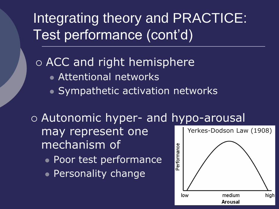
Integrating theory and PRACTICE:
Test performance (cont’d)
ACC and right hemisphere
Attentional networks
Sympathetic activation networks
Autonomic hyper- and hypo-arousal may represent one mechanism of
Poor test performance
Personality change
Yerkes-Dodson Law (1908)
Sometimes I think you only married me
because I lived next door…
Integrating theory and PRACTICE:
Clinical syndromes
Skeletal motor dysfunction Pseudobulbar affect
Gelastic seizure
Frontal opercular syndrome
Facial emotional paresis
Autonomic/endocrine dysfunction Autonomic failure
Autonomic (“visceral”) auras
PAID
Post-traumatic hypo-pituitarism
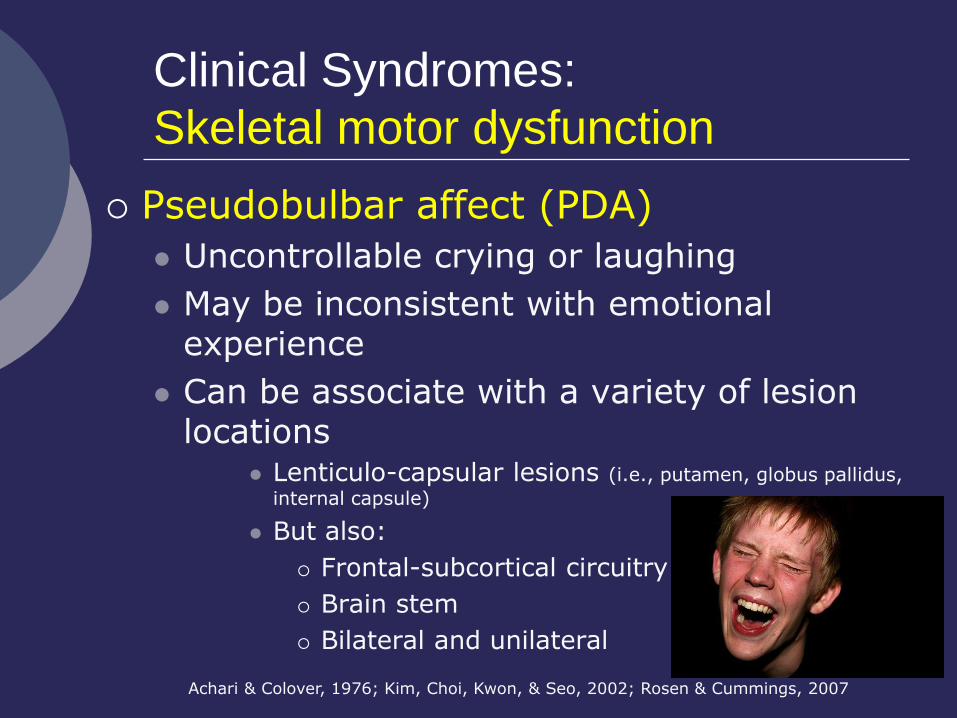
Clinical Syndromes:
Skeletal motor dysfunction
Pseudobulbar affect (PDA)
Uncontrollable crying or laughing
May be inconsistent with emotional experience
Can be associate with a variety of lesion locations
Lenticulo-capsular lesions (i.e., putamen, globus pallidus,
internal capsule)
But also:
Frontal-subcortical circuitry
Brain stem
Bilateral and unilateral
Achari & Colover, 1976; Kim, Choi, Kwon, & Seo, 2002; Rosen & Cummings, 2007
Clinical Syndromes:
Skeletal motor dysfunction (cont’d)
Gelastic seizures
Brief episodes of laughter (30 ss or less)
Occasionally longer, status epilepticus
Associated with many types of seizures
Partial
Generalized
Petit mal/ absence
Difficult to distinguish from natural laughter
Daly & Mulder, 1957; Glassman, Dryer, & McCartney, 1986; Loiseau, Cohadon, & Cohadon, 1971
Clinical Syndromes:
Skeletal motor dysfunction (cont’d)
Frontal Opercular Syndrome (Foix-Chavany-
Marie Syndrome)
Dysarthria
Paresis of cranial nerves
Involuntary facial expressions intact
Volitional facial displays impaired
Facial emotional paresis
Involuntary facial expressions impaired
Volitional facial displays intact
Daly & Mulder, 1957; Glassman, Dryer, & McCartney, 1986; Loiseau, Cohadon, & Cohadon, 1971
Clinical Syndromes:
Autonomic dysfunction
Autonomic (“visceral”) auras
Associated with temporal lobe epilepsy
Epigastric or abdominal signs most common
Rarely
Nausea, vomiting
Cardiovascular, papillary, genital, urinary, pillomotor
“As if” emotions
Fogarasi, Janszky, & Tuxhorn, 2006
Clinical Syndromes:
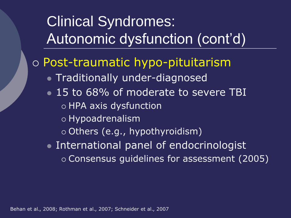
Autonomic dysfunction (cont’d)
Post-traumatic hypo-pituitarism
Traditionally under-diagnosed
15 to 68% of moderate to severe TBI
HPA axis dysfunction
Hypoadrenalism
Others (e.g., hypothyroidism)
International panel of endocrinologist
Consensus guidelines for assessment (2005)
Behan et al., 2008; Rothman et al., 2007; Schneider et al., 2007
Clinical Syndromes:
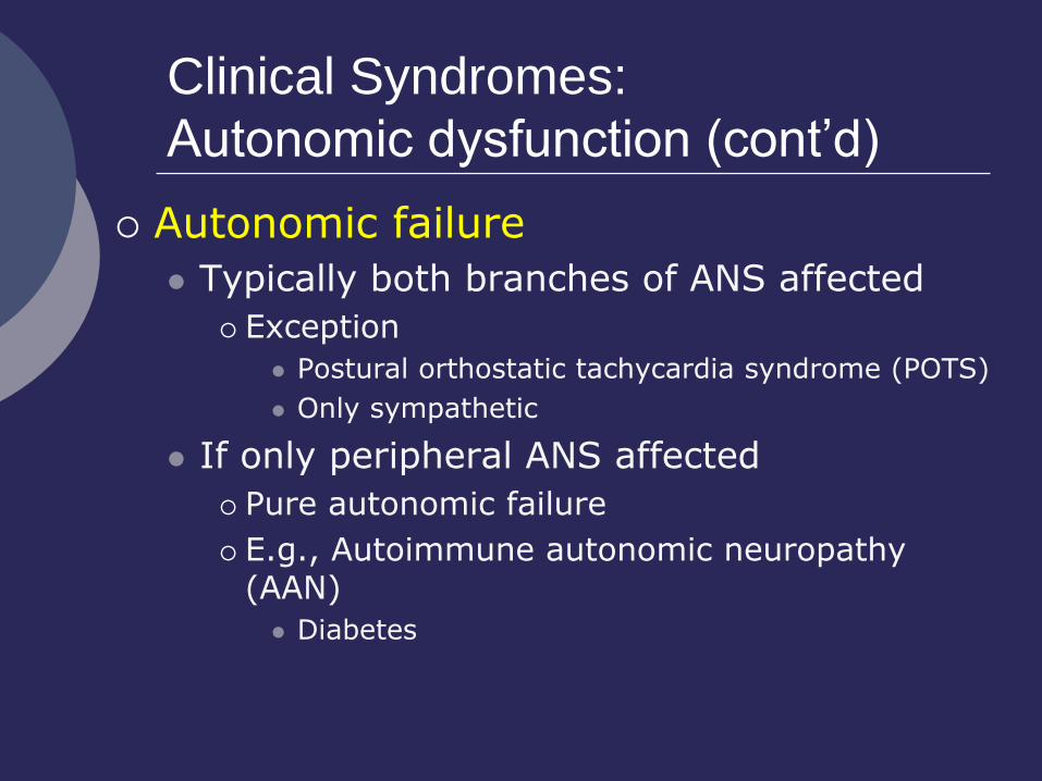
Autonomic dysfunction (cont’d)
Autonomic failure
Typically both branches of ANS affected
Exception
Postural orthostatic tachycardia syndrome (POTS)
Only sympathetic
If only peripheral ANS affected
Pure autonomic failure
E.g., Autoimmune autonomic neuropathy (AAN)
Diabetes
Clinical Syndromes:
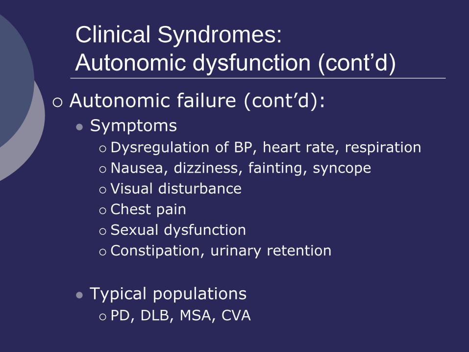
Autonomic dysfunction (cont’d)
Autonomic failure (cont’d):
Symptoms
Dysregulation of BP, heart rate, respiration
Nausea, dizziness, fainting, syncope
Visual disturbance
Chest pain
Sexual dysfunction
Constipation, urinary retention
Typical populations
PD, DLB, MSA, CVA
Integrating theory and PRACTICE:
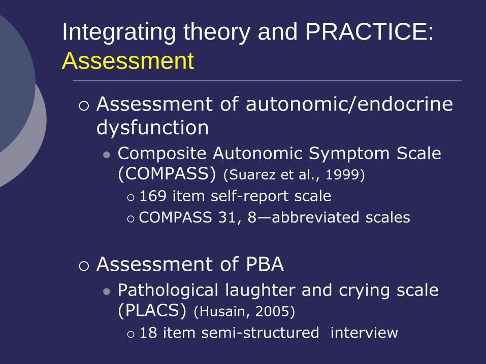
Assessment
Assessment of autonomic/endocrine dysfunction
Composite Autonomic Symptom Scale (COMPASS) (Suarez et al., 1999)
169 item self-report scale
COMPASS 31, 8—abbreviated scales
Assessment of PBA
Pathological laughter and crying scale (PLACS) (Husain, 2005)
18 item semi-structured interview
Integrating theory and PRACTICE:
Clinical Populations
Hypothalamic Hamartoma (HH)
Rare, benign tumor
Begins to develop in the first trimester of gestation
Sx
Gelastic seizures (onset in infancy)
Early childhood—often unnoticed
Pharmacologically intractable
New laser Tx/surgery available
Cognition varies
Behavior problems, aggression
Prigatano et al., 2008; Striano et al., 2005; Quigg & Barbaro, 2008
Integrating theory and PRACTICE:
Clinical Populations (cont’d)
ADHD
Autonomic hypoactivation
Dysfunction of ACC and right hemisphere
Biofeedback training to increase autonomic arousal improves performance on CPT
Crowell et al, 2006; Casey et al., 1997; Colla et al., 2008
Integrating theory and PRACTICE:
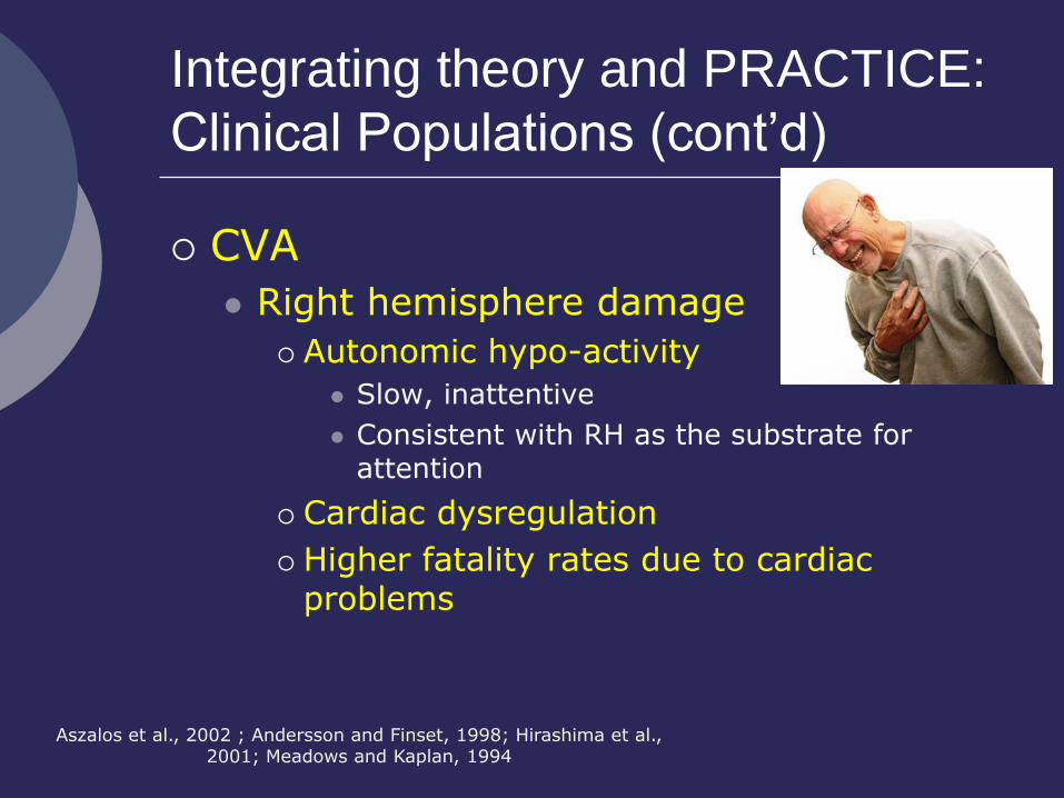
Clinical Populations (cont’d)
CVA
Right hemisphere damage
Autonomic hypo-activity
Slow, inattentive
Consistent with RH as the substrate for attention
Cardiac dysregulation
Higher fatality rates due to cardiac problems
Aszalos et al., 2002 ; Andersson and Finset, 1998; Hirashima et al., 2001; Meadows and Kaplan, 1994
Integrating theory and PRACTICE:
Clinical Populations (cont’d)
CVA (cont’d)
Hypothalamus
Endocrine and autonomic disruptions
Lower brainstem
Autonomic disruption
Lenticulo-capsular region (i.e., putamen, globus
pallidus, internal capsule)
Pseudobulbar affect
Aszalos et al., 2002 ; Andersson and Finset, 1998; Celik at al., 2004; Hirashima et al., 2001; Meadows and Kaplan, 1994 ; Weddell, 1994
Integrating theory and PRACTICE:
Clinical Populations (cont’d)
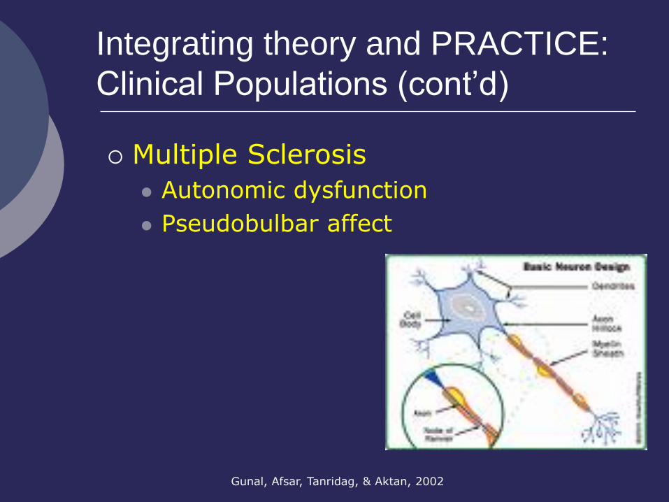
Multiple Sclerosis
Autonomic dysfunction
Pseudobulbar affect
Gunal, Afsar, Tanridag, & Aktan, 2002
Integrating theory and PRACTICE:
Clinical Populations (cont’d)
Dettmers et al., 1993; Brooks et al., 2004
Amyotrophic Lateral Sclerosis
Gradual degeneration of upper motor neurons
Pseudobulbar affect common
Mild autonomic dysregulation
Integrating theory and PRACTICE:
Clinical Populations (cont’d)
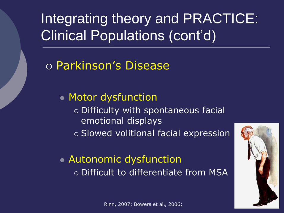
Parkinson’s Disease
Motor dysfunction
Difficulty with spontaneous facial emotional displays
Slowed volitional facial expression
Autonomic dysfunction
Difficult to differentiate from MSA
Rinn, 2007; Bowers et al., 2006;
Integrating theory and PRACTICE:
Clinical Populations (cont’d)
Multiple System Atrophy
Umbrella term for Striatonigral degeneration
Shy Dragger syndrome
Olivopontocerebellar atrophy
Progressive degeneration
Basal ganglia, Pons, Medulla oblongata, Autonomic neurons in the brain stem and spinal cord
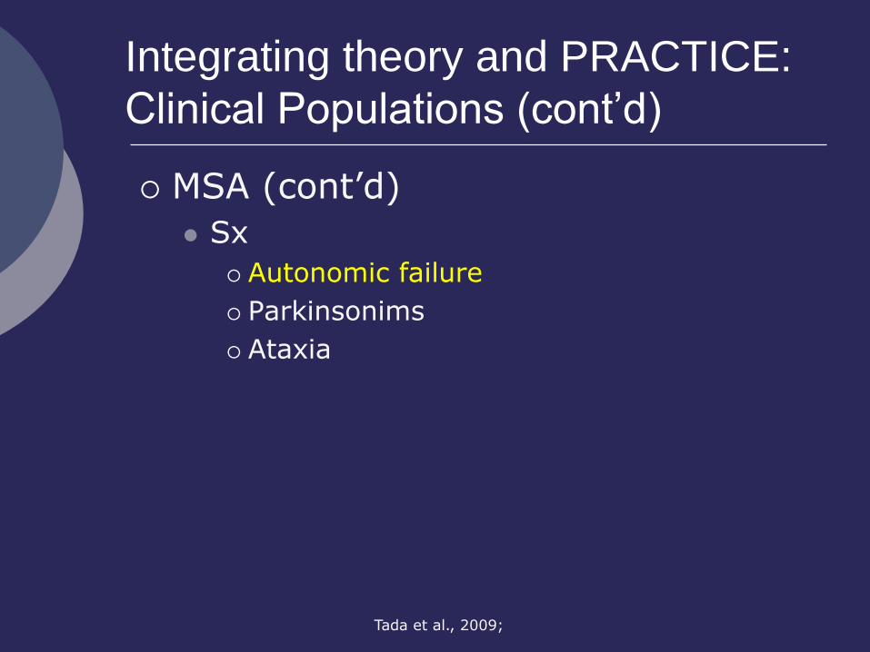
Integrating theory and PRACTICE:
Clinical Populations (cont’d)
MSA (cont’d)
Sx
Autonomic failure
Parkinsonims
Ataxia
Tada et al., 2009;
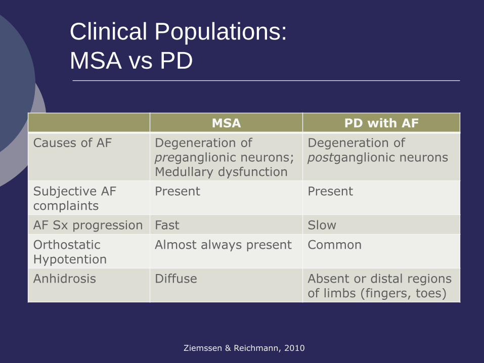
Clinical Populations:
MSA vs PD
Ziemssen & Reichmann, 2010
MSA PD with AF
Causes of AF Degeneration of preganglionic neurons; Medullary dysfunction
Degeneration of postganglionic neurons
Subjective AF complaints
Present Present
AF Sx progression Fast Slow
Orthostatic Hypotention
Almost always present Common
Anhidrosis Diffuse Absent or distal regions of limbs (fingers, toes)
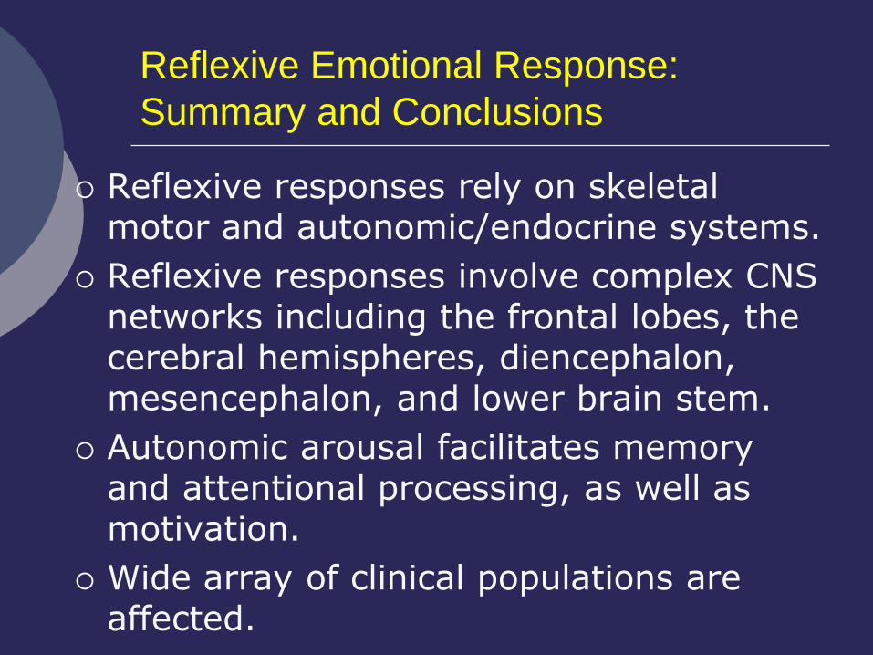
Reflexive Emotional Response:
Summary and Conclusions
Reflexive responses rely on skeletal motor and autonomic/endocrine systems.
Reflexive responses involve complex CNS networks including the frontal lobes, the cerebral hemispheres, diencephalon, mesencephalon, and lower brain stem.
Autonomic arousal facilitates memory and attentional processing, as well as motivation.
Wide array of clinical populations are affected.
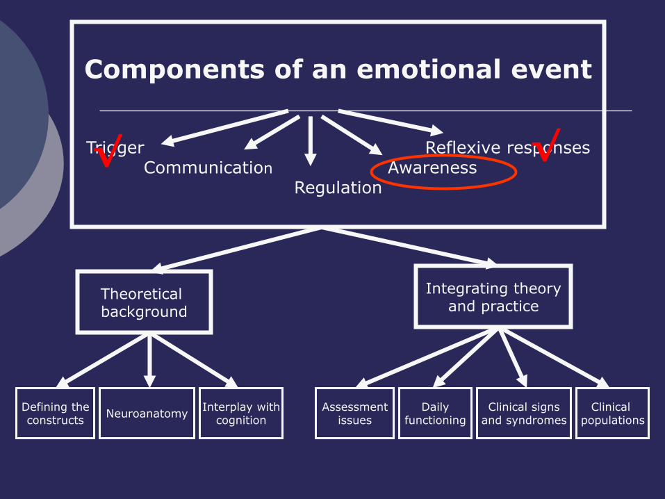
Components of an emotional event
Trigger Reflexive responses Communication Awareness
Regulation
Theoretical background
Integrating theory and practice
Defining the constructs
Neuroanatomy Interplay with
cognition Assessment
issues Clinical signs
and syndromes Clinical
populations Daily
functioning
√ √
THEORETICAL BACKGROUND:
Components of awareness
Interoceptive awareness
Ability to detect own physiologic reactions
Correlates with intensity of experience
Pure autonomic failure
Deficits in subjective feeling states
Critchley, Wiens, Rotshtein, Ohman, & Dolan, 2004; Heims, Critchley, Dolan, Mathias, & Cipolotti, 2004; Pollatos, Kirsch, & Schandry, 2005; Pollatos, Schandry, Auer, & Kaufmann, 2007
THEORETICAL BACKGROUND:
Components of awareness (cont’d)
Emotional (feeling) awareness
Includes the ability to
Feel
Understand
Discuss
Dissociable from interoceptive awareness
THEORETICAL BACKGROUND:
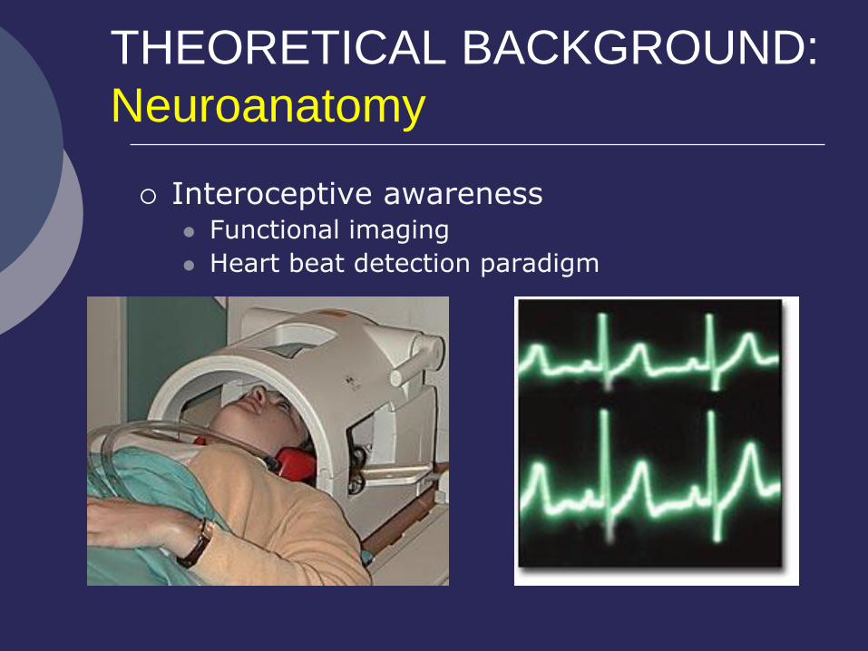
Neuroanatomy
Interoceptive awareness Functional imaging
Heart beat detection paradigm
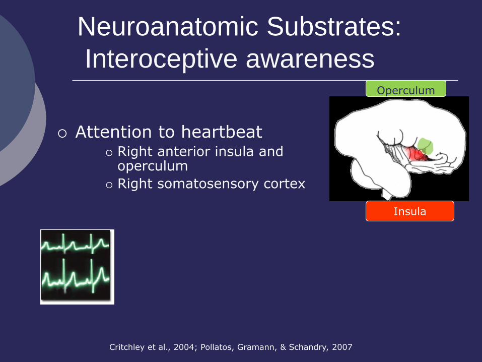
Neuroanatomic Substrates:
Interoceptive awareness
Attention to heartbeat Right anterior insula and
operculum
Right somatosensory cortex
Critchley et al., 2004; Pollatos, Gramann, & Schandry, 2007
Operculum
Insula
Neuroanatomic Substrates:
Interoceptive awareness (cont’d)
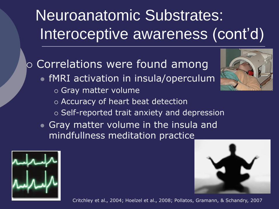
Correlations were found among
fMRI activation in insula/operculum
Gray matter volume
Accuracy of heart beat detection
Self-reported trait anxiety and depression
Gray matter volume in the insula and mindfullness meditation practice
Critchley et al., 2004; Hoelzel et al., 2008; Pollatos, Gramann, & Schandry, 2007
Neuroanatomic Substrates:
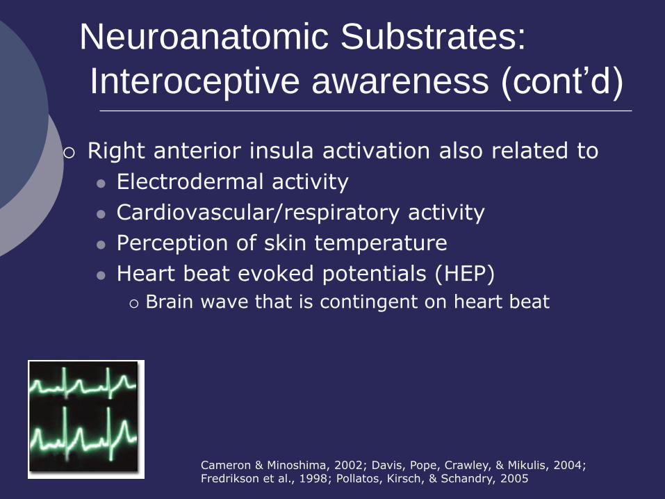
Interoceptive awareness (cont’d)
Right anterior insula activation also related to
Electrodermal activity
Cardiovascular/respiratory activity
Perception of skin temperature
Heart beat evoked potentials (HEP)
Brain wave that is contingent on heart beat
Cameron & Minoshima, 2002; Davis, Pope, Crawley, & Mikulis, 2004; Fredrikson et al., 1998; Pollatos, Kirsch, & Schandry, 2005
THEORETICAL BACKGROUND:
Neuroanatomy
Emotional (feeling) Awareness
Functional imaging in normals
Wide-spread activation
Method dependent and emotion-specific
Common networks
Thalamus
Hypothalamus
Midbrain
Medial PFC
Anterior, mid, and posterior cingulate
Orbitofrontal
Berthoz, Blair, Le Clec'h, & Martinot, 2002; Gerrards-Hesse, Spies, & Hesse, 1994; Reiman et al., 1997; Weiss, Salloum, & Schneider, 1999; Lane, et al., 1997; Reiman et al., 1997
THEORETICAL BACKGROUND:
Neuroanatomy (cont’d)
Emotional Awareness (cont’d)
Functional imaging in alexithymics
Greater activation in the anterior insula (bilateral)
Decreased activation in
Posterior and anterior cingulate gyrus
DLPFC
Pons and cerebellum
Slower inter-hemispheric transfer
Karlsson, Naaanen, & Stenman, 2008; Mantani, Okamoto, Shirao, Okada, & Yamawaki, 2005; Moriguchi et al., 2007; Moriguchi et al., 2007; Richter et al., 2006
Integrating Theory and PRACTICE
Clinical syndromes
Alexithymia
Clinical populations
Assessment
Alexithymia:
Definition
Inability to
Consciously experience
Identify
Describe
Taylor, 1984; Warnes, 1986
emotions
physiologic response to emotional stimuli
Normal ability to
Exhibit
Be aware of
Interoceptive
Awareness
Feeling Awareness
Anxiety
Low High Impaired
Low
High High
Low Impaired
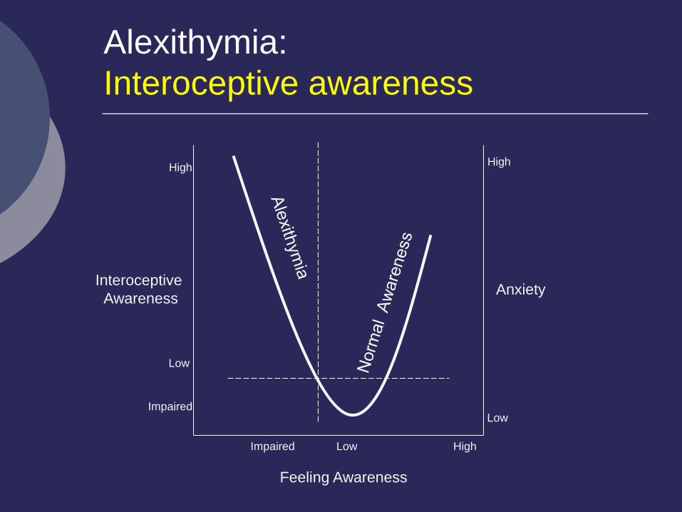
Alexithymia:
Interoceptive awareness

Alexithymia:
Processing Deficits
Usage of emotional words to describe emotional situations
Matching emotional stimuli with emotional self-report
Identifying emotional expressions of others
Understanding seriousness of emotional situations
Empathy
Guttman & Laporte, 2002; Lane, 1996; Luminet, Rime, Bagby, & Taylor, 2004; Mann, Wise, Trinidad, & Kohanski, 1994; Moriguchi et al., 2007; Parker, Prkachin, & Prkachin, 2005; Vanman, Dawson, & Brennan, 1998
Alexithymia:
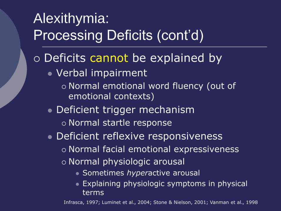
Processing Deficits (cont’d)
Deficits cannot be explained by
Verbal impairment
Normal emotional word fluency (out of emotional contexts)
Deficient trigger mechanism
Normal startle response
Deficient reflexive responsiveness
Normal facial emotional expressiveness
Normal physiologic arousal
Sometimes hyperactive arousal
Explaining physiologic symptoms in physical terms
Infrasca, 1997; Luminet et al., 2004; Stone & Nielson, 2001; Vanman et al., 1998
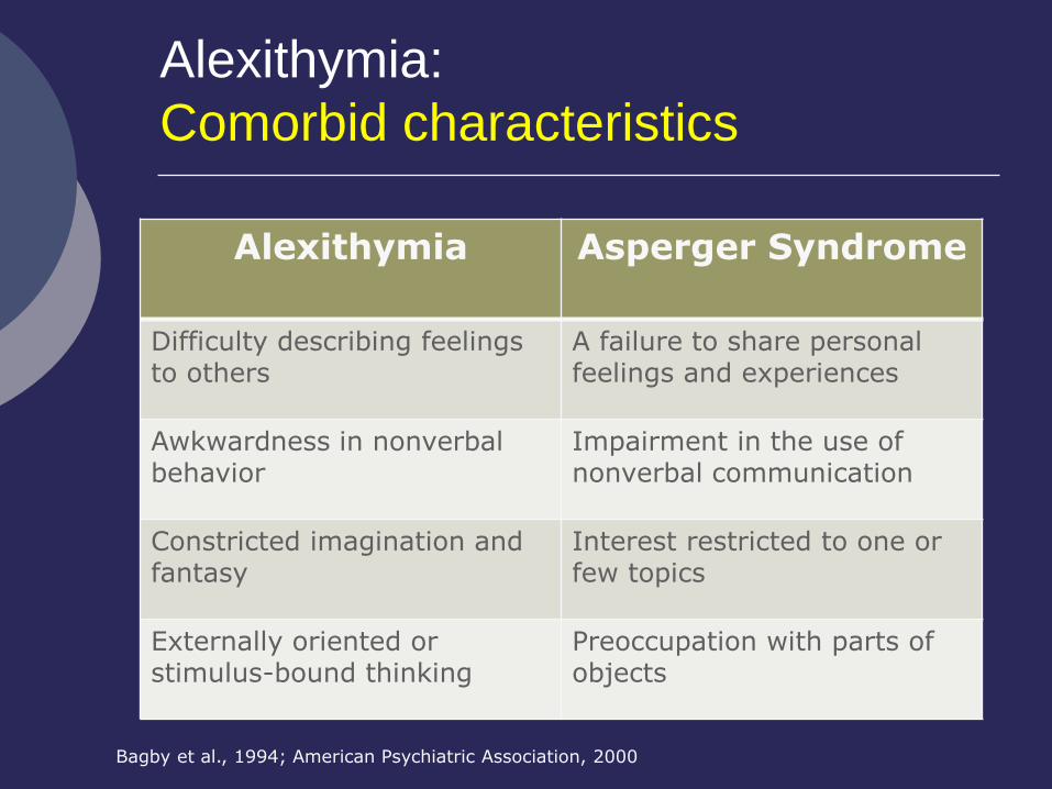
Alexithymia:
Comorbid characteristics
Alexithymia Asperger Syndrome
Difficulty describing feelings to others
A failure to share personal feelings and experiences
Awkwardness in nonverbal behavior
Impairment in the use of nonverbal communication
Constricted imagination and fantasy
Interest restricted to one or few topics
Externally oriented or stimulus-bound thinking
Preoccupation with parts of objects
Bagby et al., 1994; American Psychiatric Association, 2000
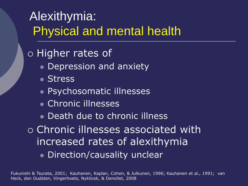
Alexithymia:
Physical and mental health
Higher rates of
Depression and anxiety
Stress
Psychosomatic illnesses
Chronic illnesses
Death due to chronic illness
Chronic illnesses associated with increased rates of alexithymia
Direction/causality unclear
Fukunishi & Tsurata, 2001; Kauhanen, Kaplan, Cohen, & Julkunen, 1996; Kauhanen et al., 1991; van Heck, den Oudsten, Vingerhoets, Nyklicek, & Denollet, 2008
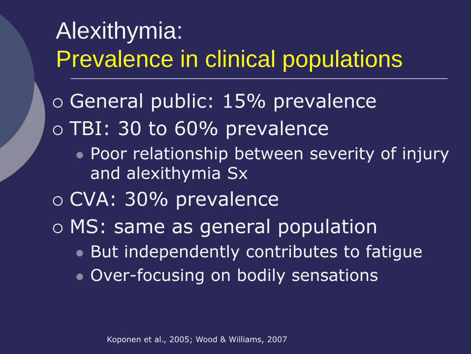
Alexithymia:
Prevalence in clinical populations
General public: 15% prevalence
TBI: 30 to 60% prevalence
Poor relationship between severity of injury and alexithymia Sx
CVA: 30% prevalence
MS: same as general population
But independently contributes to fatigue
Over-focusing on bodily sensations
Koponen et al., 2005; Wood & Williams, 2007
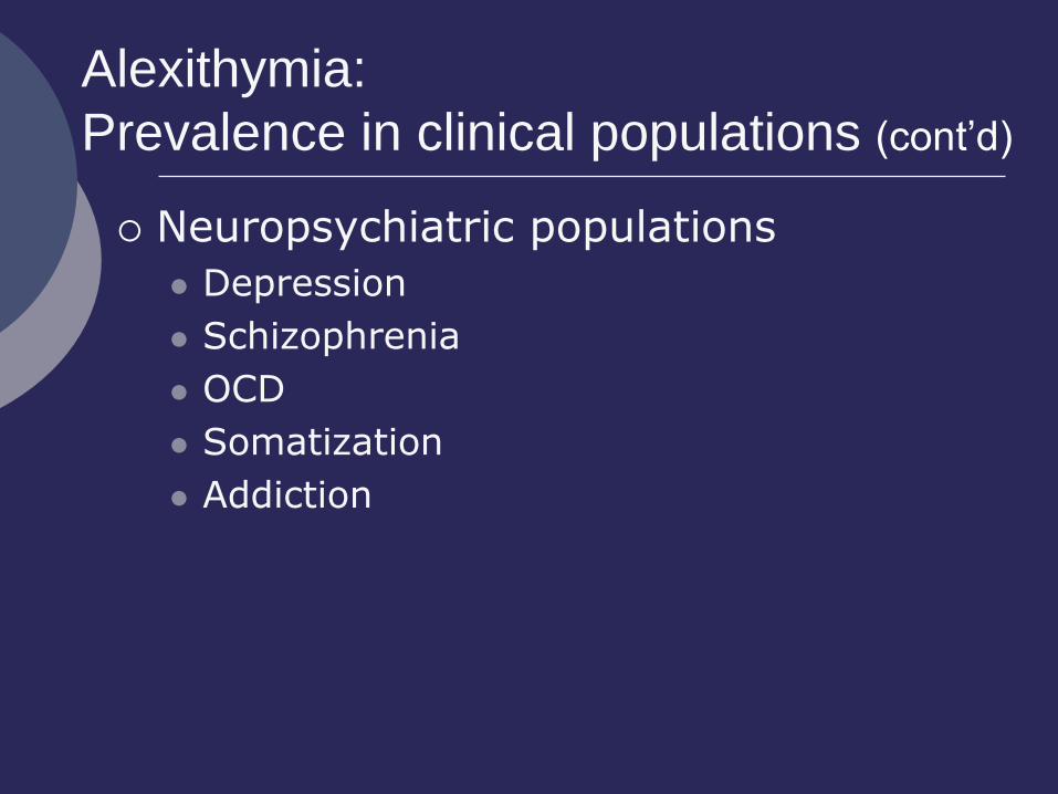
Alexithymia:
Prevalence in clinical populations (cont’d)
Neuropsychiatric populations
Depression
Schizophrenia
OCD
Somatization
Addiction
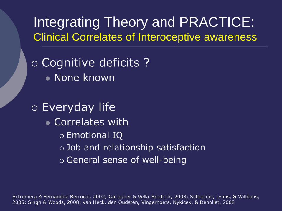
Integrating Theory and PRACTICE: Clinical Correlates of Interoceptive awareness
Cognitive deficits ?
None known
Everyday life
Correlates with
Emotional IQ
Job and relationship satisfaction
General sense of well-being
Extremera & Fernandez-Berrocal, 2002; Gallagher & Vella-Brodrick, 2008; Schneider, Lyons, & Williams, 2005; Singh & Woods, 2008; van Heck, den Oudsten, Vingerhoets, Nykicek, & Denollet, 2008
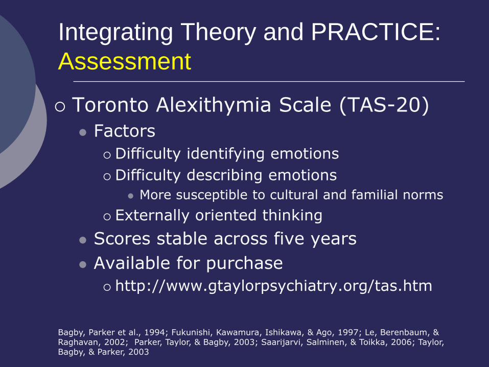
Integrating Theory and PRACTICE:
Assessment
Toronto Alexithymia Scale (TAS-20)
Factors
Difficulty identifying emotions
Difficulty describing emotions
More susceptible to cultural and familial norms
Externally oriented thinking
Scores stable across five years
Available for purchase
http://www.gtaylorpsychiatry.org/tas.htm
Bagby, Parker et al., 1994; Fukunishi, Kawamura, Ishikawa, & Ago, 1997; Le, Berenbaum, & Raghavan, 2002; Parker, Taylor, & Bagby, 2003; Saarijarvi, Salminen, & Toikka, 2006; Taylor, Bagby, & Parker, 2003
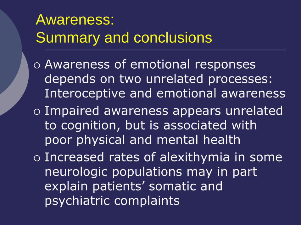
Awareness:
Summary and conclusions
Awareness of emotional responses depends on two unrelated processes: Interoceptive and emotional awareness
Impaired awareness appears unrelated to cognition, but is associated with poor physical and mental health
Increased rates of alexithymia in some neurologic populations may in part explain patients’ somatic and psychiatric complaints

Components of an emotional event
Trigger Reflexive responses Communication Awareness
Regulation
Theoretical background
Integrating theory and practice
Defining the constructs
Neuroanatomy Interplay with
cognition Assessment
issues Clinical signs
and syndromes Clinical
populations Daily
functioning
√ √
√
THEORETICAL BACKGROUND:
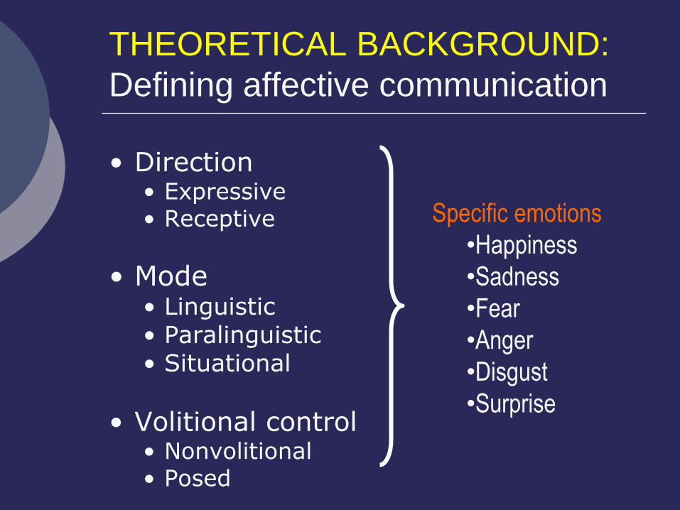
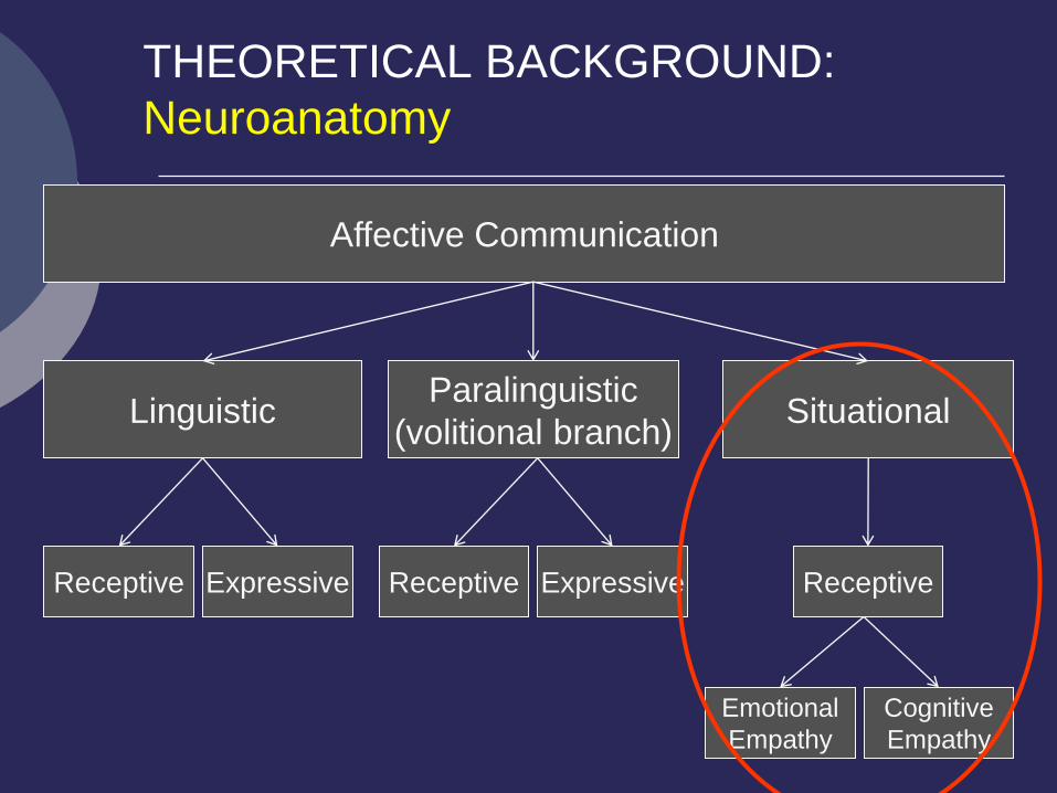
Defining affective communication
• Direction • Expressive • Receptive
• Mode • Linguistic • Paralinguistic • Situational
• Volitional control • Nonvolitional • Posed
Specific emotions
•Happiness
•Sadness
•Fear
•Anger
•Disgust
•Surprise

THEORETICAL BACKGROUND:
Volitional vs. non-volitional comm.
Dependant variables
Type of communication References
Volitional Non-volitional
Physiologic arousal
Absent or minimal Present Boiten et al., 1996
Emotional experience
Absent or minimal Present Boiten et al., 1996
Facial symmetry
Less symmetric More symmetric Ekman et al, 1981
Ease of recognition
Easier More difficult Gosselin & Kirouac, 1995
Facial muscles Somewhat different muscle groups used in each
Boiten et al., 1996
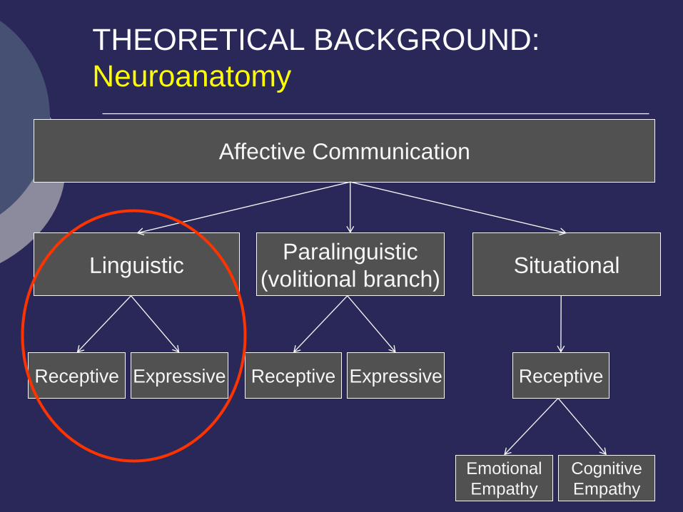
Linguistic Paralinguistic
(volitional branch)
Receptive
Affective Communication
Situational
Expressive Receptive Expressive
Emotional
Empathy
Cognitive
Empathy
Receptive
THEORETICAL BACKGROUND:
Neuroanatomy
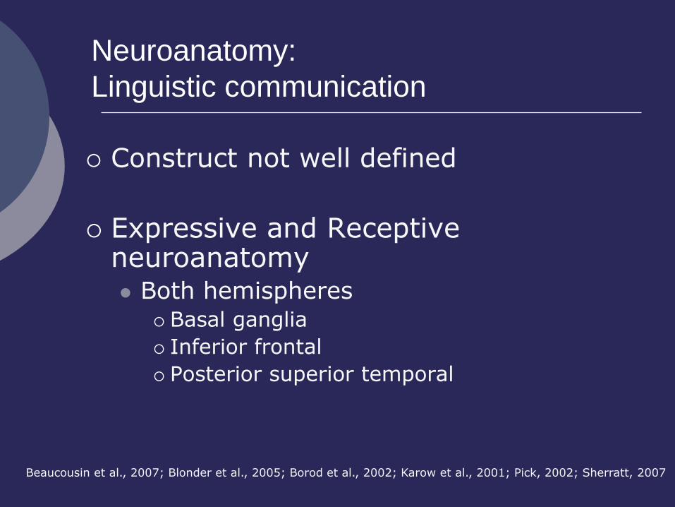
Neuroanatomy:
Linguistic communication
Construct not well defined
Expressive and Receptive neuroanatomy Both hemispheres
Basal ganglia
Inferior frontal
Posterior superior temporal
Beaucousin et al., 2007; Blonder et al., 2005; Borod et al., 2002; Karow et al., 2001; Pick, 2002; Sherratt, 2007
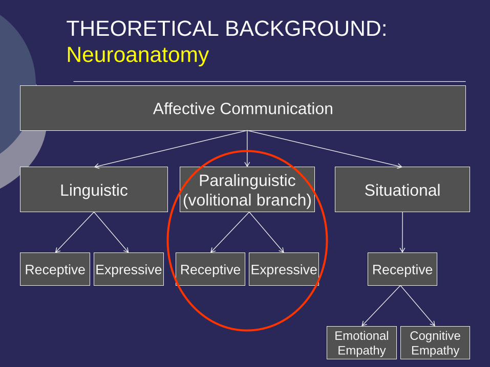
Linguistic Paralinguistic
(volitional branch)
Receptive
Affective Communication
Situational
Expressive Receptive Expressive
Emotional
Empathy
Cognitive
Empathy
Receptive
THEORETICAL BACKGROUND:
Neuroanatomy
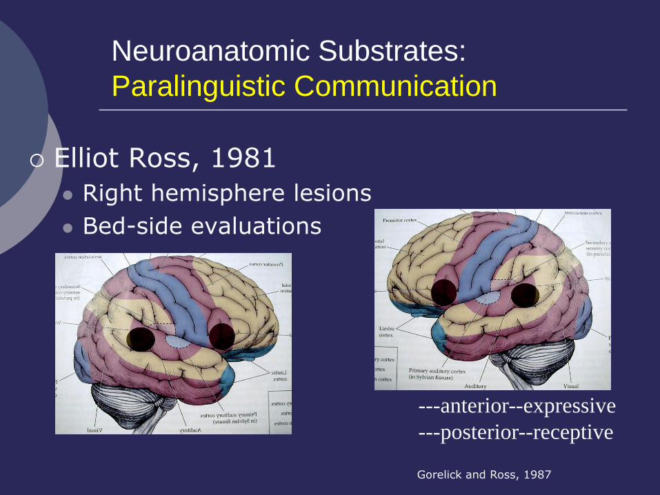
Neuroanatomic Substrates:
Paralinguistic Communication
Elliot Ross, 1981
Right hemisphere lesions
Bed-side evaluations
---anterior--expressive
---posterior--receptive
Gorelick and Ross, 1987
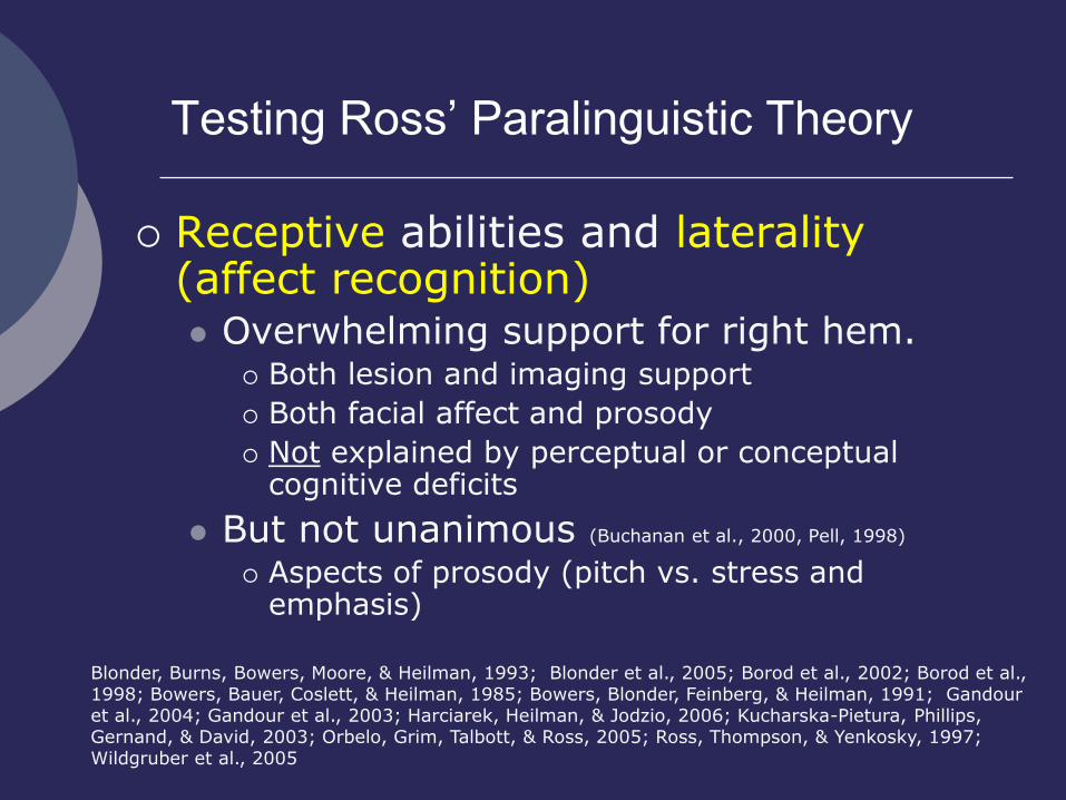
Testing Ross’ Paralinguistic Theory
Receptive abilities and laterality (affect recognition) Overwhelming support for right hem.
Both lesion and imaging support
Both facial affect and prosody
Not explained by perceptual or conceptual cognitive deficits
But not unanimous (Buchanan et al., 2000, Pell, 1998)
Aspects of prosody (pitch vs. stress and emphasis)
Blonder, Burns, Bowers, Moore, & Heilman, 1993; Blonder et al., 2005; Borod et al., 2002; Borod et al., 1998; Bowers, Bauer, Coslett, & Heilman, 1985; Bowers, Blonder, Feinberg, & Heilman, 1991; Gandour et al., 2004; Gandour et al., 2003; Harciarek, Heilman, & Jodzio, 2006; Kucharska-Pietura, Phillips, Gernand, & David, 2003; Orbelo, Grim, Talbott, & Ross, 2005; Ross, Thompson, & Yenkosky, 1997; Wildgruber et al., 2005
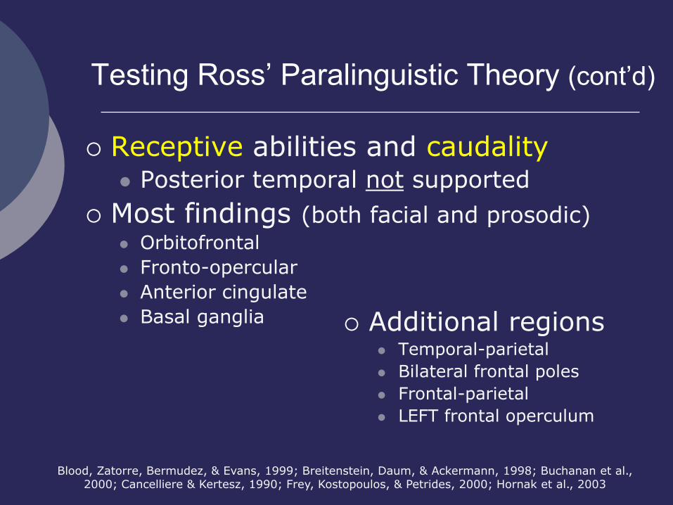
Testing Ross’ Paralinguistic Theory (cont’d)
Receptive abilities and caudality Posterior temporal not supported
Most findings (both facial and prosodic) Orbitofrontal
Fronto-opercular
Anterior cingulate
Basal ganglia
Blood, Zatorre, Bermudez, & Evans, 1999; Breitenstein, Daum, & Ackermann, 1998; Buchanan et al., 2000; Cancelliere & Kertesz, 1990; Frey, Kostopoulos, & Petrides, 2000; Hornak et al., 2003
Additional regions Temporal-parietal
Bilateral frontal poles
Frontal-parietal
LEFT frontal operculum
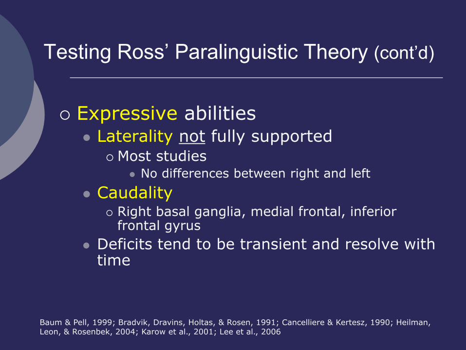
Testing Ross’ Paralinguistic Theory (cont’d)
Expressive abilities Laterality not fully supported
Most studies No differences between right and left
Caudality Right basal ganglia, medial frontal, inferior
frontal gyrus
Deficits tend to be transient and resolve with time
Baum & Pell, 1999; Bradvik, Dravins, Holtas, & Rosen, 1991; Cancelliere & Kertesz, 1990; Heilman, Leon, & Rosenbek, 2004; Karow et al., 2001; Lee et al., 2006
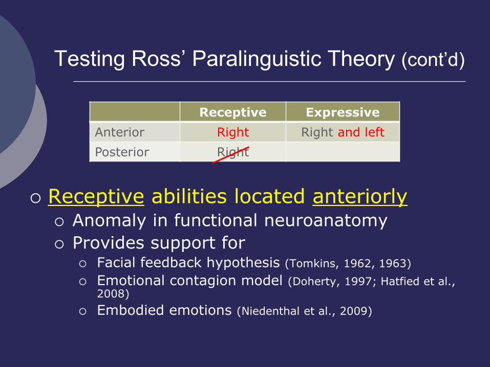
Testing Ross’ Paralinguistic Theory (cont’d)
Receptive Expressive
Anterior Right
Posterior Right
Receptive Expressive
Anterior Right Right and left
Posterior Right
Receptive abilities located anteriorly Anomaly in functional neuroanatomy
Provides support for Facial feedback hypothesis (Tomkins, 1962, 1963)
Emotional contagion model (Doherty, 1997; Hatfied et al., 2008)
Embodied emotions (Niedenthal et al., 2009)
Linguistic Paralinguistic
(volitional branch)
Receptive
Affective Communication
Situational
Expressive Receptive Expressive
Emotional
Empathy
Cognitive
Empathy
Receptive
THEORETICAL BACKGROUND:
Neuroanatomy
Neuroanatomic Substrates:
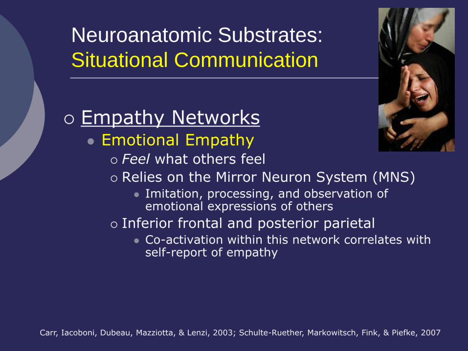
Situational Communication
Empathy Networks Emotional Empathy
Feel what others feel
Relies on the Mirror Neuron System (MNS) Imitation, processing, and observation of
emotional expressions of others
Inferior frontal and posterior parietal Co-activation within this network correlates with
self-report of empathy
Carr, Iacoboni, Dubeau, Mazziotta, & Lenzi, 2003; Schulte-Ruether, Markowitsch, Fink, & Piefke, 2007
Neuroanatomic Substrates:
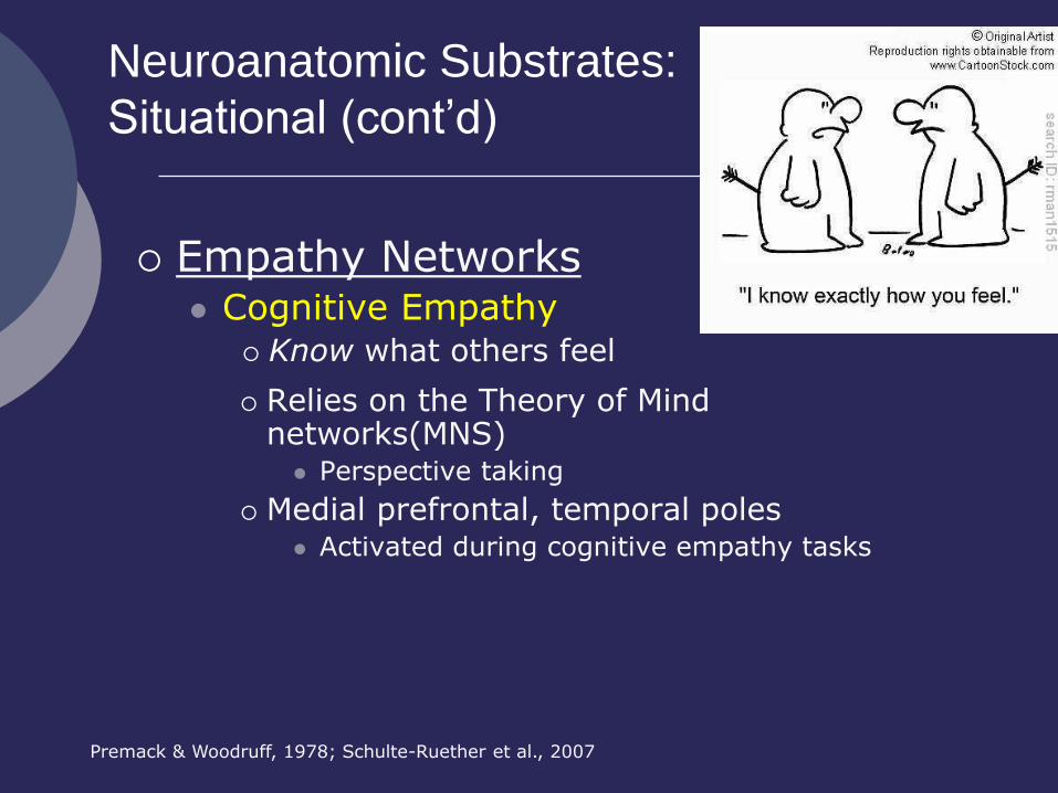
Situational (cont’d)
Empathy Networks Cognitive Empathy
Know what others feel
Premack & Woodruff, 1978; Schulte-Ruether et al., 2007
Relies on the Theory of Mind networks(MNS) Perspective taking
Medial prefrontal, temporal poles Activated during cognitive empathy tasks
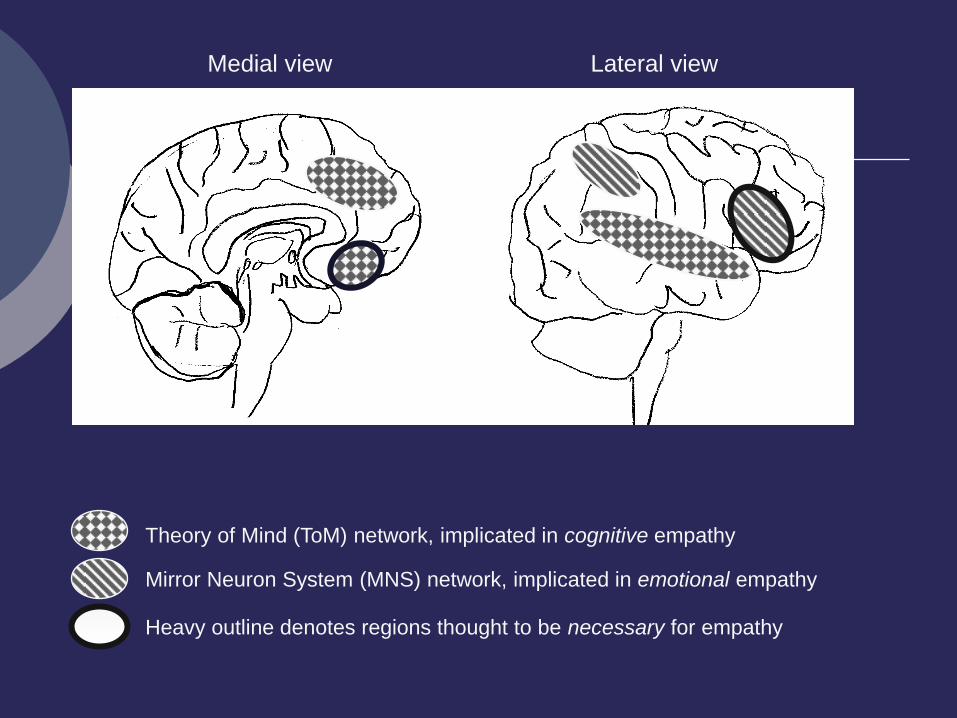
Theory of Mind (ToM) network, implicated in cognitive empathy
Mirror Neuron System (MNS) network, implicated in emotional empathy
Heavy outline denotes regions thought to be necessary for empathy
Medial view Lateral view
Test performance
Populations
Neurodevelopmental
Neuropsychiatric
Neurodegenerative
Other neurologic
Integrating Theory and PRACTICE
Integrating theory and PRACTICE:
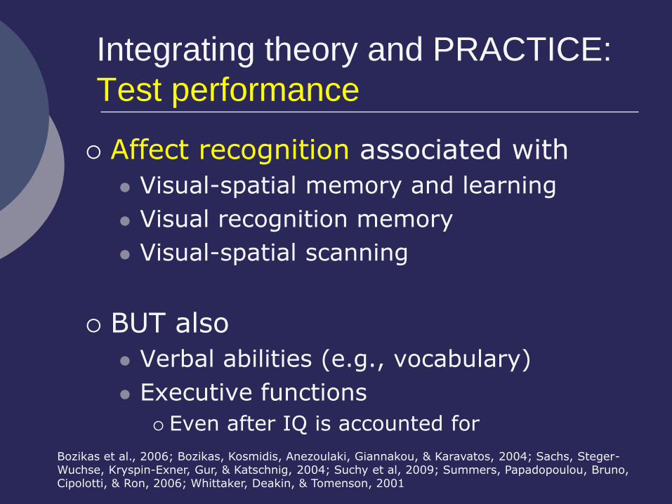
Test performance
Affect recognition associated with
Visual-spatial memory and learning
Visual recognition memory
Visual-spatial scanning
BUT also
Verbal abilities (e.g., vocabulary)
Executive functions
Even after IQ is accounted for
Bozikas et al., 2006; Bozikas, Kosmidis, Anezoulaki, Giannakou, & Karavatos, 2004; Sachs, Steger-Wuchse, Kryspin-Exner, Gur, & Katschnig, 2004; Suchy et al, 2009; Summers, Papadopoulou, Bruno, Cipolotti, & Ron, 2006; Whittaker, Deakin, & Tomenson, 2001
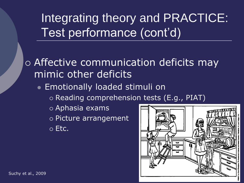
Integrating theory and PRACTICE:
Test performance (cont’d)
Affective communication deficits may mimic other deficits
Emotionally loaded stimuli on
Reading comprehension tests (E.g., PIAT)
Aphasia exams
Picture arrangement
Etc.
Suchy et al., 2009
Integrating theory and PRACTICE:
Test performance (cont’d)
Cognitive empathy associated with
Cognitive Flexibility
Shamay-Tsoory, Tomer, Goldsher, Berger, & Aharon-Peretz, 2004
Integrating theory and PRACTICE:
Clinical populations
Interpretive considerations
Most research examined only facial affect
Most populations exhibit deficits in recognizing some, but not all, emotions
Many studies do not examine individual emotions
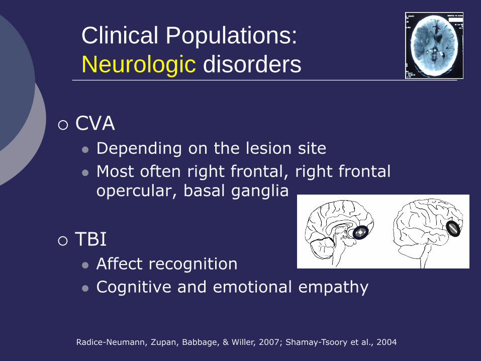
Clinical Populations:
Neurologic disorders
CVA
Depending on the lesion site
Most often right frontal, right frontal opercular, basal ganglia
TBI
Affect recognition
Cognitive and emotional empathy
Radice-Neumann, Zupan, Babbage, & Willer, 2007; Shamay-Tsoory et al., 2004
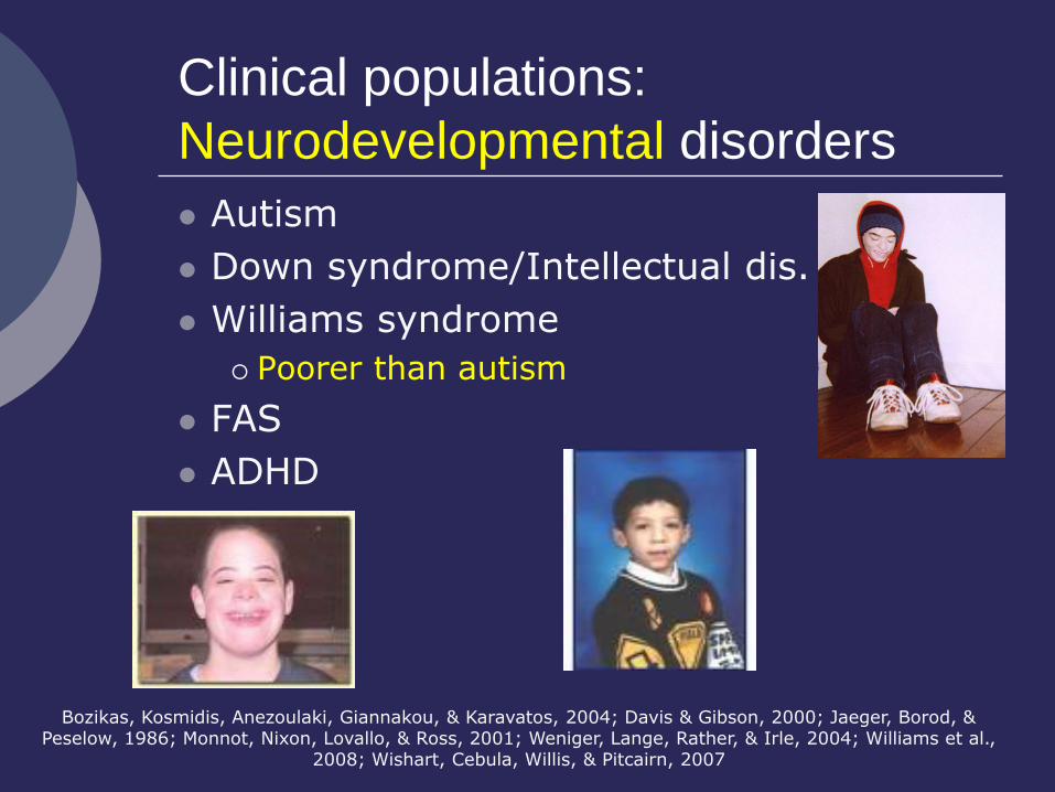
Clinical populations:
Neurodevelopmental disorders
Autism
Down syndrome/Intellectual dis.
Williams syndrome
Poorer than autism
FAS
ADHD
Bozikas, Kosmidis, Anezoulaki, Giannakou, & Karavatos, 2004; Davis & Gibson, 2000; Jaeger, Borod, &
Peselow, 1986; Monnot, Nixon, Lovallo, & Ross, 2001; Weniger, Lange, Rather, & Irle, 2004; Williams et al., 2008; Wishart, Cebula, Willis, & Pitcairn, 2007
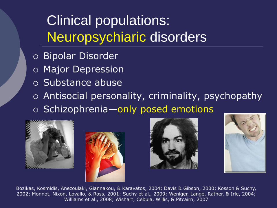
Clinical populations:
Neuropsychiaric disorders
Bipolar Disorder
Major Depression
Substance abuse
Antisocial personality, criminality, psychopathy
Schizophrenia—only posed emotions
Bozikas, Kosmidis, Anezoulaki, Giannakou, & Karavatos, 2004; Davis & Gibson, 2000; Kosson & Suchy, 2002; Monnot, Nixon, Lovallo, & Ross, 2001; Suchy et al., 2009; Weniger, Lange, Rather, & Irle, 2004;
Williams et al., 2008; Wishart, Cebula, Willis, & Pitcairn, 2007
Clinical Populations:
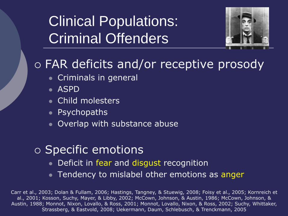
Criminal Offenders
FAR deficits and/or receptive prosody Criminals in general
ASPD
Child molesters
Psychopaths
Overlap with substance abuse
Specific emotions Deficit in fear and disgust recognition
Tendency to mislabel other emotions as anger
Carr et al., 2003; Dolan & Fullam, 2006; Hastings, Tangney, & Stuewig, 2008; Foisy et al., 2005; Kornreich et al., 2001; Kosson, Suchy, Mayer, & Libby, 2002; McCown, Johnson, & Austin, 1986; McCown, Johnson, &
Austin, 1988; Monnot, Nixon, Lovallo, & Ross, 2001; Monnot, Lovallo, Nixon, & Ross, 2002; Suchy, Whittaker, Strassberg, & Eastvold, 2008; Uekermann, Daum, Schlebusch, & Trenckmann, 2005
Clinical populations:
Neurodegenerative disorders
AD, ALS, FTD, HD, PD
Unique profiles with respect to
Type of emotional communication deficits
Specific emotions affected
Cognitive or psychiatric correlates of deficits
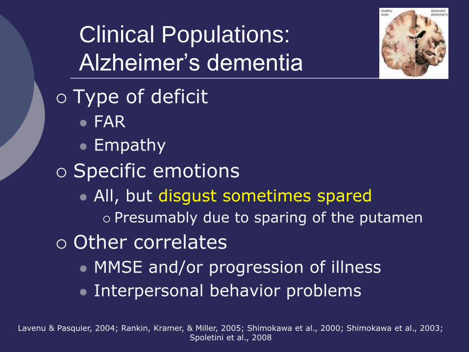
Clinical Populations:
Alzheimer’s dementia
Type of deficit
FAR
Empathy
Specific emotions
All, but disgust sometimes spared
Presumably due to sparing of the putamen
Other correlates
MMSE and/or progression of illness
Interpersonal behavior problems
Lavenu & Pasquier, 2004; Rankin, Kramer, & Miller, 2005; Shimokawa et al., 2000; Shimokawa et al., 2003; Spoletini et al., 2008
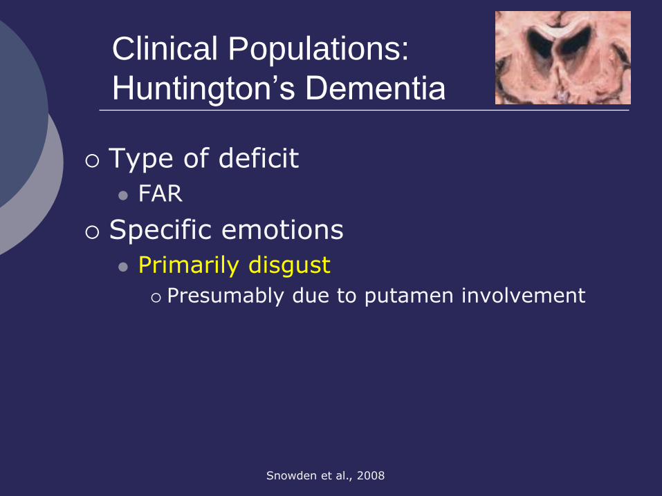
Clinical Populations:
Huntington’s Dementia
Type of deficit
FAR
Specific emotions
Primarily disgust
Presumably due to putamen involvement
Snowden et al., 2008
Clinical Populations:
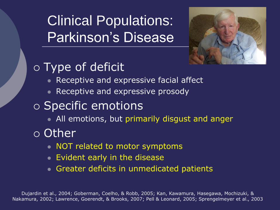
Parkinson’s Disease
Type of deficit Receptive and expressive facial affect
Receptive and expressive prosody
Specific emotions All emotions, but primarily disgust and anger
Other NOT related to motor symptoms
Evident early in the disease
Greater deficits in unmedicated patients
Dujardin et al., 2004; Goberman, Coelho, & Robb, 2005; Kan, Kawamura, Hasegawa, Mochizuki, & Nakamura, 2002; Lawrence, Goerendt, & Brooks, 2007; Pell & Leonard, 2005; Sprengelmeyer et al., 2003
Clinical Populations:
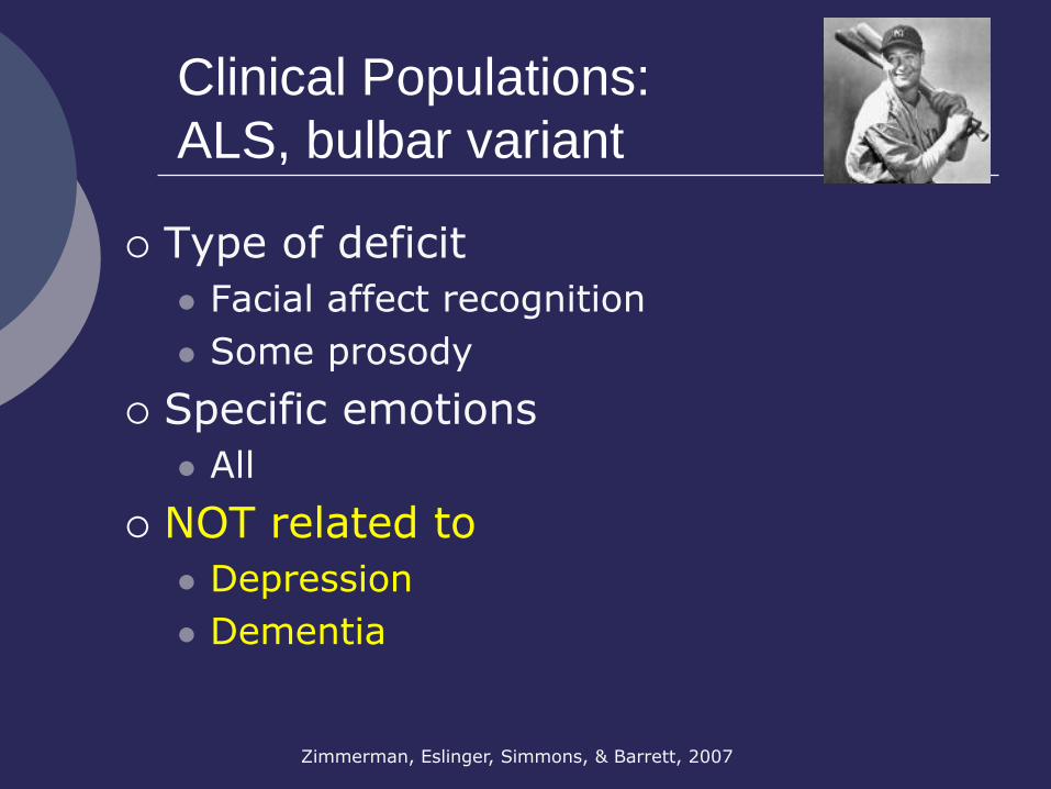
ALS, bulbar variant
Type of deficit
Facial affect recognition
Some prosody
Specific emotions
All
NOT related to
Depression
Dementia
Zimmerman, Eslinger, Simmons, & Barrett, 2007
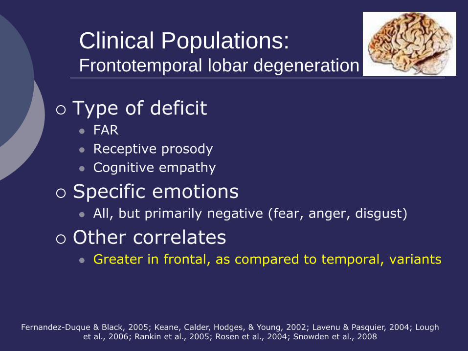
Clinical Populations: Frontotemporal lobar degeneration
Type of deficit FAR
Receptive prosody
Cognitive empathy
Specific emotions All, but primarily negative (fear, anger, disgust)
Other correlates Greater in frontal, as compared to temporal, variants
Fernandez-Duque & Black, 2005; Keane, Calder, Hodges, & Young, 2002; Lavenu & Pasquier, 2004; Lough et al., 2006; Rankin et al., 2005; Rosen et al., 2004; Snowden et al., 2008
Integrating theory and PRACTICE:
Assessment
WAIS-IV: Advanced Clinical Solutions
Social Cognition Test
Facial Expressions
Social Interactions
Prosody
The Awareness of Social Inference Test (TASIT)
Emotion Evaluation
Social Inference
Wechsler, 2008; McDonald et al., 2002
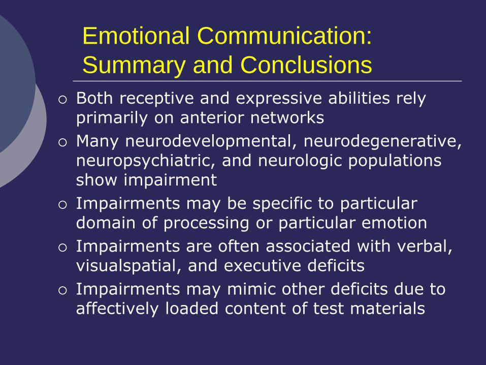
Emotional Communication:
Summary and Conclusions
Both receptive and expressive abilities rely primarily on anterior networks
Many neurodevelopmental, neurodegenerative, neuropsychiatric, and neurologic populations show impairment
Impairments may be specific to particular domain of processing or particular emotion
Impairments are often associated with verbal, visualspatial, and executive deficits
Impairments may mimic other deficits due to affectively loaded content of test materials
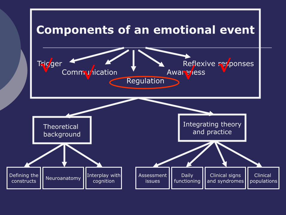
Components of an emotional event
Trigger Reflexive responses Communication Awareness
Regulation
Theoretical background
Integrating theory and practice
Defining the constructs
Neuroanatomy Interplay with
cognition Assessment
issues Clinical signs
and syndromes Clinical
populations Daily
functioning
√ √ √
√
THEORETICAL BACKGROUND:
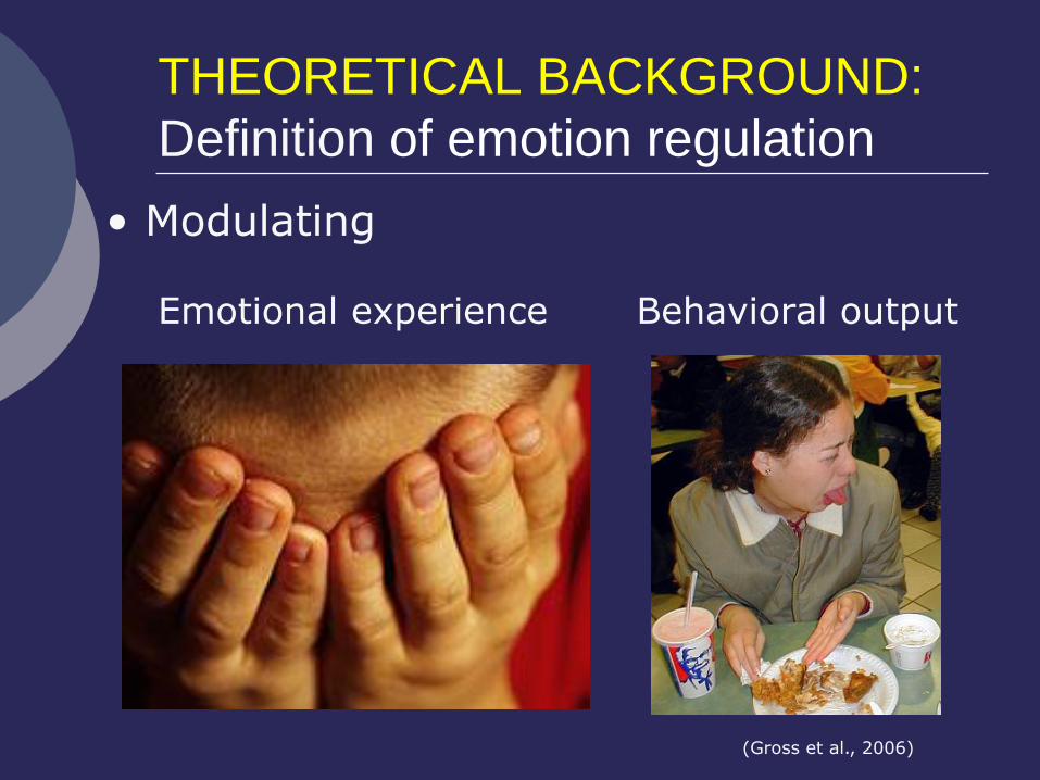
Definition of emotion regulation
• Modulating Emotional experience Behavioral output
(Gross et al., 2006)
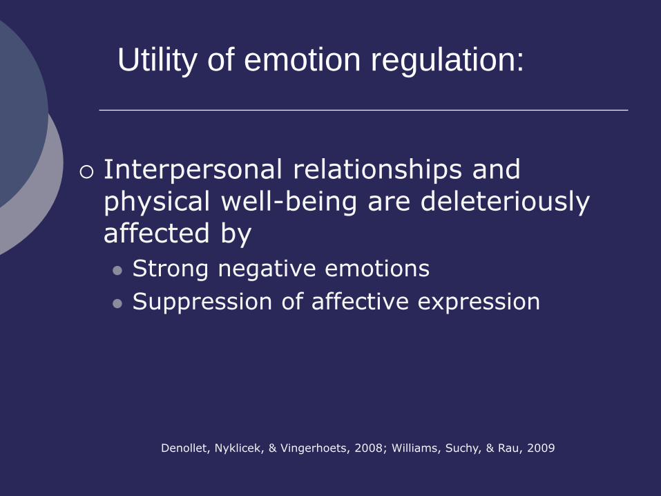
Utility of emotion regulation:
Interpersonal relationships and physical well-being are deleteriously affected by
Strong negative emotions
Suppression of affective expression
Denollet, Nyklicek, & Vingerhoets, 2008; Williams, Suchy, & Rau, 2009
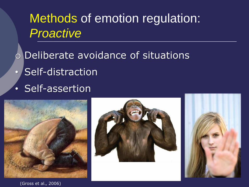
Methods of emotion regulation:
Proactive
(Gross et al., 2006)
Deliberate avoidance of situations
LA-LA-LA-LA
• Self-distraction
• Self-assertion
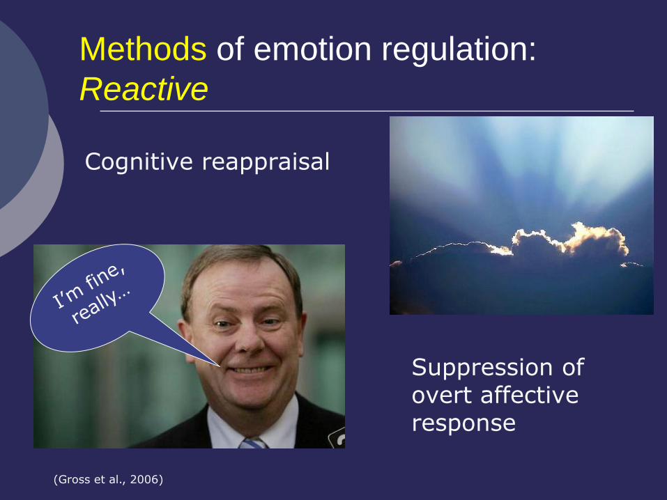
Methods of emotion regulation:
Reactive
Cognitive reappraisal
(Gross et al., 2006)
Suppression of overt affective response

THEORETICAL BACKGROUND:
Neuroanatomy
Methodology
Lesion studies
Functional imaging
View films or photos
Suppress or exaggerate
Feelings
Facial expressions
Neuroanatomic substrates:
Functional imaging
Common networks
Anterior cingulate gyrus
Dorsolateral prefrontal cortex
Ventral and ventromedial prefrontal cortex
Abler, Hofer, & Viviani, 2008; Ochsner & Gross, 2008; Ohira et al., 2006
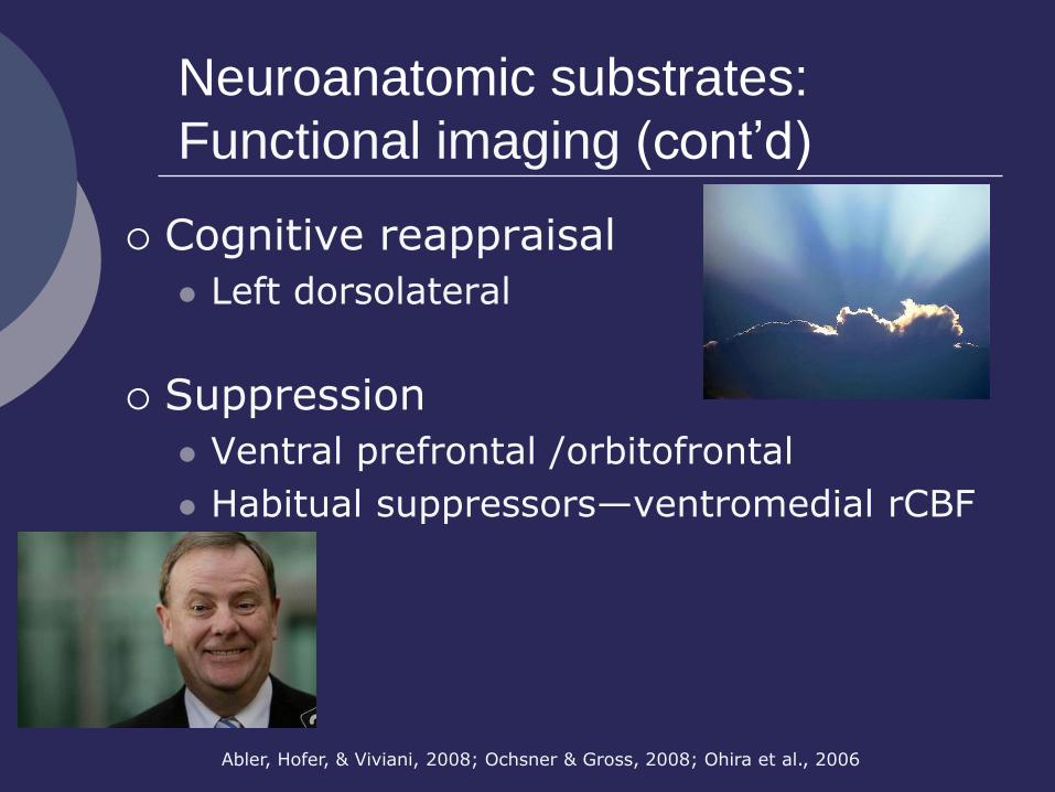
Neuroanatomic substrates:
Functional imaging (cont’d)
Cognitive reappraisal
Left dorsolateral
Abler, Hofer, & Viviani, 2008; Ochsner & Gross, 2008; Ohira et al., 2006
Suppression
Ventral prefrontal /orbitofrontal
Habitual suppressors—ventromedial rCBF
Neuroanatomic substrates:
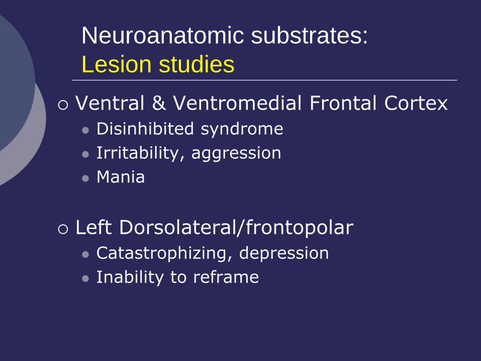
Lesion studies
Ventral & Ventromedial Frontal Cortex
Disinhibited syndrome
Irritability, aggression
Mania
Left Dorsolateral/frontopolar
Catastrophizing, depression
Inability to reframe
THEORETICAL BACKGROUND:
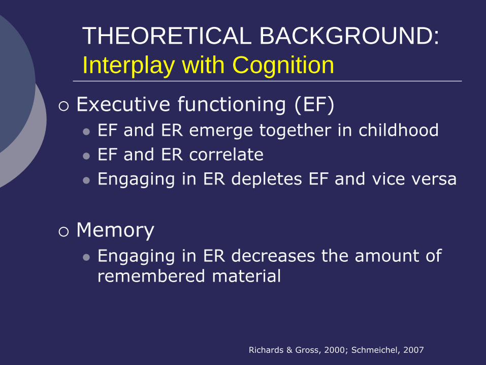
Interplay with Cognition
Executive functioning (EF)
EF and ER emerge together in childhood
EF and ER correlate
Engaging in ER depletes EF and vice versa
Memory
Engaging in ER decreases the amount of remembered material
Richards & Gross, 2000; Schmeichel, 2007
Integrating Theory and PRACTICE
Syndromes
Populations
Assessment
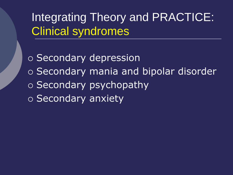
Integrating Theory and PRACTICE:
Clinical syndromes
Secondary depression
Secondary mania and bipolar disorder
Secondary psychopathy
Secondary anxiety
Secondary depression
Similar to endogenous depression
Generally responds to pharmacotherapy and CBT
Left frontal lesions
Severity correlates with distance from frontal pole
Populations
CVA, TBI, dementia, epilepsy, MS
Narashima et al., 2003; Vataja et al., 2004
Secondary mania/bipolar disorder
Right hemisphere lesions
Ventral/anterior temporal for mania
Basal ganglia and thalamus for bipolar
Mood disorder vs. disinhibition
Responds to traditional treatments
Populations
TBI, CVA, brain tumors, dementia, epilepsy, HIV infection
Narashima et al., 2003; Vataja et al., 2004
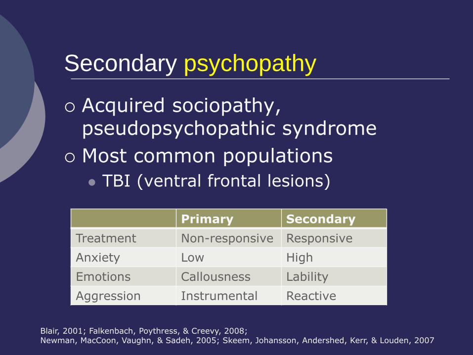
Acquired sociopathy, pseudopsychopathic syndrome
Most common populations
TBI (ventral frontal lesions)
Secondary psychopathy
Blair, 2001; Falkenbach, Poythress, & Creevy, 2008; Newman, MacCoon, Vaughn, & Sadeh, 2005; Skeem, Johansson, Andershed, Kerr, & Louden, 2007
Primary Secondary
Treatment Non-responsive Responsive
Anxiety Low High
Emotions Callousness Lability
Aggression Instrumental Reactive
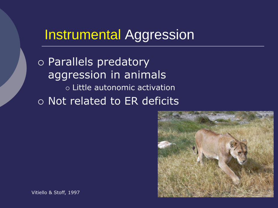
Instrumental Aggression
Parallels predatory aggression in animals
Little autonomic activation
Not related to ER deficits
Vitiello & Stoff, 1997
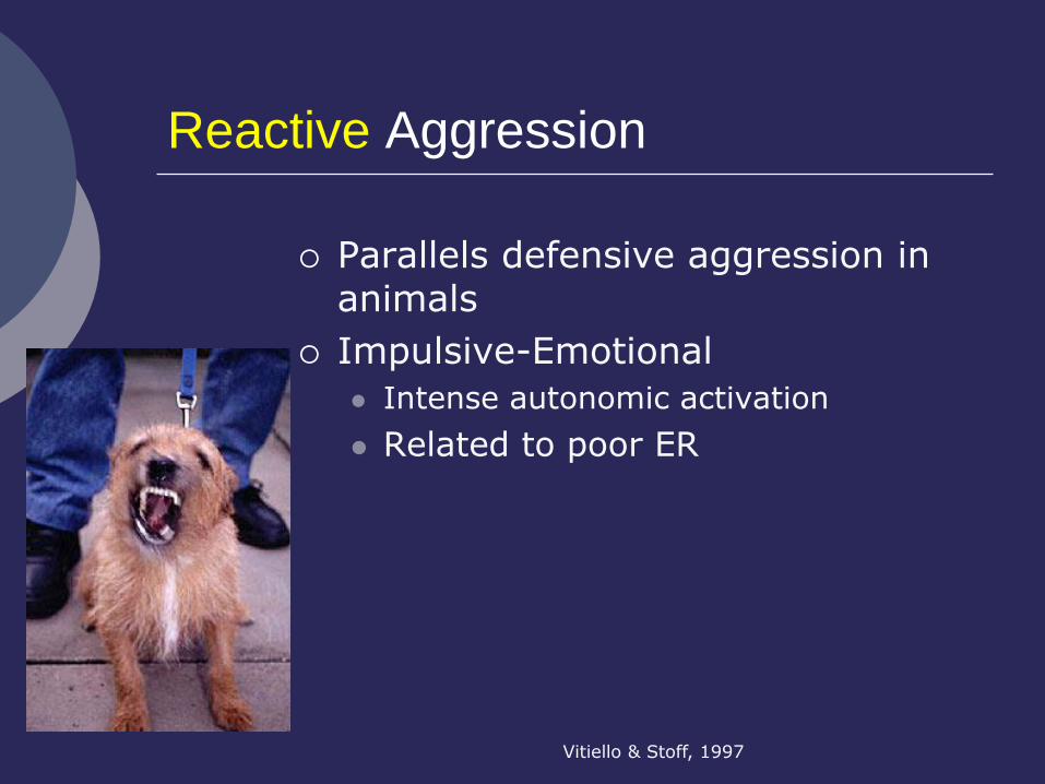
Reactive Aggression
Parallels defensive aggression in animals
Impulsive-Emotional
Intense autonomic activation
Related to poor ER
Vitiello & Stoff, 1997
Secondary anxiety
All types of anxiety reported
Lesion location
Inconsistencies in the literature
Possibly ventromedial and orbitofrontal
Populations
TBI and CVA
Hiott & Labbate, 2002; Moore, Terryberry-Spohr, & Hope, 2006; Williams & Evans, 2003
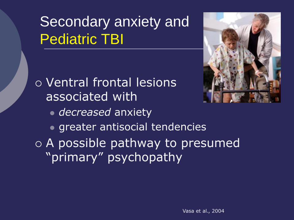
Secondary anxiety and
Pediatric TBI
Ventral frontal lesions associated with
decreased anxiety
greater antisocial tendencies
A possible pathway to presumed “primary” psychopathy
Vasa et al., 2004
Integrating Theory and PRACTICE:
Clinical populations
Dementias
TBI
CVA
Epilepsy
MS
Populations:
Dementias
Depression most common across all dementias (50%)
Dementia-specific ER problems:
FTD
Mostly disinhibition and lability
AD
Verbal and physical aggression
PD and VD
Mostly depression
Engelborghs et al., 2005; Lind, Edman, Sjogren, Wallin, & Karlsson, 2002; Ritchie & Lovestone, 2002

Populations:
Traumatic Brain Injury (TBI)
Most common symptoms of ER dysfunction
Depression
Anxiety
Irritability/aggression
Social inappropriateness
American Psychiatric Association, 1994; Fann et al., 2004
Populations:
TBI (cont’d)
Etiology of ER deficits
Neurogenic vs. psychogenic
Exacerbation of premorbid psychopathology?
Litigation
Ruff, 2005
Populations:
TBI (cont’d)
Evidence of neurogenic ER deficits
Greater frequency of new onset mood/anxiety disorders than expected in general population
Sagduyu, 2002; Schwartz et al., 2003
Populations:
TBI (cont’d)
Evidence of psychogenic ER deficits
Greater rate of premorbid psychopathology and life stress among the “miserable minority”
For review of the relevant issues, see Ruff (2005)
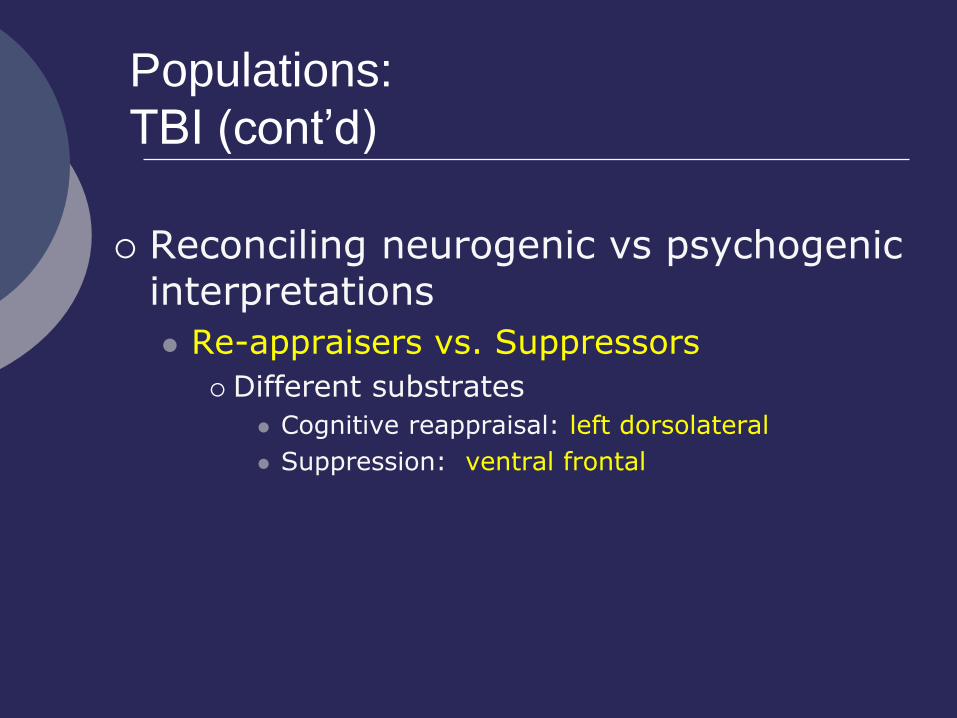
Populations:
TBI (cont’d)
Reconciling neurogenic vs psychogenic interpretations
Re-appraisers vs. Suppressors
Different substrates
Cognitive reappraisal: left dorsolateral
Suppression: ventral frontal
Individual Differences
Dorsolateral
Prefrontal
Cortex
Orbitofrontal
Cortex
Preferential
Re-appraiser
Preferential
Suppressor
Re-appraisal of
Situation
EMOTIONAL EVENT
Appropriate
Behavioral
Control
Decrease in
Emotional
Arousal
Suppression
of Behavior
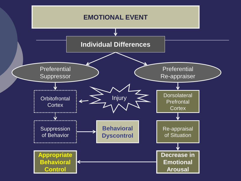
Figure 9.1
Individual Differences
Dorsolateral
Prefrontal
Cortex
Orbitofrontal
Cortex
Preferential
Re-appraiser
Preferential
Suppressor
Re-appraisal
of Situation
Behavioral
Dyscontrol
EMOTIONAL EVENT
Appropriate
Behavioral
Control
Decrease in
Emotional
Arousal
Injury
Suppression
of Behavior
Populations:
TBI (cont’d)
Reconciling neurogenic vs psychogenic interpretations
Re-appraisers vs. Suppressors
Different substrates
Cognitive reappraisal: left dorsolateral
Suppression: ventral frontal
Different success in coping
Cognitive reappraisers: healthy, adjusted
Suppressors: stressed, interpersonal problems
Populations:
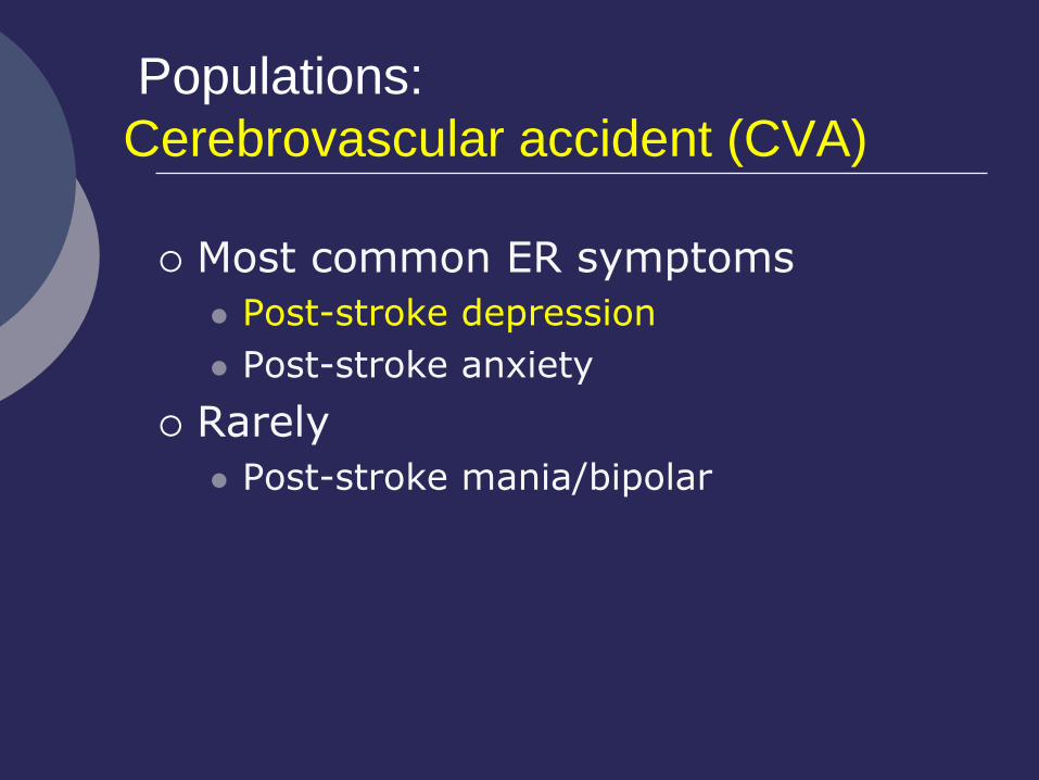
Cerebrovascular accident (CVA)
Most common ER symptoms
Post-stroke depression
Post-stroke anxiety
Rarely
Post-stroke mania/bipolar
CVA:
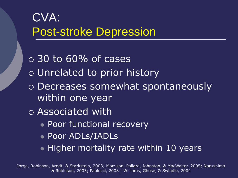
Post-stroke Depression
30 to 60% of cases
Unrelated to prior history
Decreases somewhat spontaneously within one year
Associated with
Poor functional recovery
Poor ADLs/IADLs
Higher mortality rate within 10 years
Jorge, Robinson, Arndt, & Starkstein, 2003; Morrison, Pollard, Johnston, & MacWalter, 2005; Narushima & Robinson, 2003; Paolucci, 2008 ; Williams, Ghose, & Swindle, 2004
CVA:
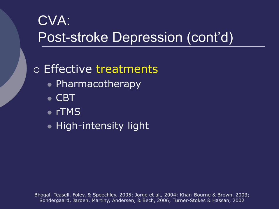
Post-stroke Depression (cont’d)
Effective treatments
Pharmacotherapy
CBT
rTMS
High-intensity light
Bhogal, Teasell, Foley, & Speechley, 2005; Jorge et al., 2004; Khan-Bourne & Brown, 2003; Sondergaard, Jarden, Martiny, Andersen, & Bech, 2006; Turner-Stokes & Hassan, 2002
CVA:
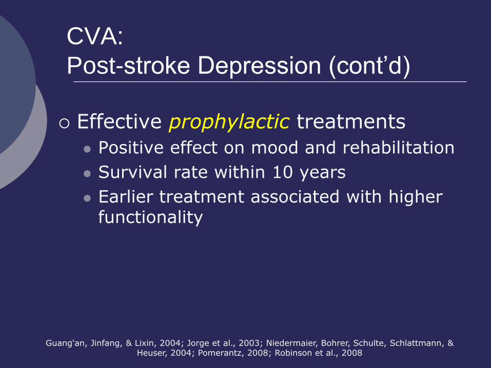
Post-stroke Depression (cont’d)
Effective prophylactic treatments
Positive effect on mood and rehabilitation
Survival rate within 10 years
Earlier treatment associated with higher functionality
Guang'an, Jinfang, & Lixin, 2004; Jorge et al., 2003; Niedermaier, Bohrer, Schulte, Schlattmann, & Heuser, 2004; Pomerantz, 2008; Robinson et al., 2008
CVA:
Post-stroke Anxiety
Often comorbid with depression
When alone, associated with right frontal lesions
Robinson, 1997
CVA:
Post-stroke Mania
Rare (<1%)
Associated with family history of mood disorder
Goyal et al., 2006; Robinson, 1997; Robinson et al., 1988
Integrating Theory and PRACTICE:
Assessment
Emotion regulation depletes EF resources (and vice versa)
Depletion may last for many hours
Consider
Stereoptype threat
Grieving
Anxiety
“bad day”
Emotion Regulation:
Summary and Conclusions
Different ER styles are associated with different neuroanatomic substrates and different health outcomes
Frontal lobe lesions are the primary cause of ER deficits in neurologic populations
Different ER styles may explain the premorbid psychopathology among the TBI “miserable minority”
Prophylactic treatment of post-stroke depression may have both short-term and long-term benefits
GENERAL Conclusions
Examination of emotional processing at the level of five primary domains proves useful for the study of
functional neuroanatomy
clinically relevant deficits in cognition/test performance
clinically relevant issues related to mental and physical health
Efforts should be taken to enhance assessment of emotional processing