Embed Size (px)
Citation preview
Neuropsychology Review pp588-nerv-378447 September 20, 2002 12:3 Style file version March 18, 1999
NERV, Vol. 12, No. 3, September 2002 (C© 2002)
Neuropsychological Functioning in Chronic Lyme Disease
Holly James Westervelt1,2,4 and Robert J. McCaffrey3
Lyme disease is currently the most common vector-borne illness in the United States. The disease ismultisystemic, and chronic disease, in particular, may be associated with neuropsychological deficits.However, to date, only a few empirical studies exist, which examine the neuropsychological sequelaeassociated with chronic Lyme disease. A review of the literature shows that the deficits observedin adults with chronic Lyme disease are generally consistent with the deficits that can be seen inprocesses with primarily frontal systems involvement. These observations are generally consistentwith neuroradiologic findings. The clinical presentation in chronic Lyme disease and the nature of theneuropsychological deficits are discussed, as are several central issues in understanding neuropsy-chological functioning in chronic Lyme disease, such as the impact of chronic illness, response totreatment, and the relationship between neuropsychological performance and depression, fatigue, andneurological indicators of disease.
KEY WORDS: Lyme disease; neuroborreliosis; neuropsychological functioning; chronic illness; fatigue.
Lyme disease was first recognized as a cause of arthri-tis in 1975 (Steere et al., 1977), and it is now the mostcommon vector-borne disease in the United States (Whiteet al., 1991). Lyme disease is multisystemic, with der-matologic, arthritic, ophthalmologic, cardiac, neurologic,and psychiatric manifestations (Burgdorfer, 1991). Thechronic phase of the illness may also be associated withneuropsychological deficits, though these are among themost poorly understood and poorly defined manifesta-tions of Lyme disease. The initial sections of this reviewdiscuss the epidemiology, transmission, and clinical pre-sentation of Lyme disease, including a brief overview ofthe psychiatric findings, differential diagnosis, pathogen-esis, and EEG/neuroradiologic findings. These sectionsare followed by a review of the adult neuropsychologi-cal literature, with a detailed description of the publishedcontrolled studies and discussion highlighting some of the
1Department of Psychiatry and Human Behavior, Brown MedicalSchool, Providence, Rhode Island.
2Department of Psychiatry, Rhode Island Hospital, Providence, RhodeIsland.
3Department of Psychology, University at Albany, State University ofNew York, Albany, New York.
4To whom correspondence should be addressed at Physician’s OfficeBuilding 430, Rhode Island Hospital, 110 Lockwood Street, Provi-dence, Rhode Island 02903. E-mail:
central issues in understanding neuropsychological func-tioning in chronic Lyme disease, such as the impact ofchronic illness, the effects of medical treatment, and therelationship between neuropsychological functioning anddepression, neurologic status, and fatigue. Given that neu-ropsychologically, Lyme disease may present differentlyin children than in adults, the effects of Lyme disease inchildren are not presented in this review; those interestedin neuropsychological functioning in children can refer tothe following papers: Adams et al., 1994, 1999; Bloomet al., 1998. Psychlit and Medline searches for the timeperiod of January 1975 to April 2001 identified studies inthe current review.
EPIDEMIOLOGY
The identification of epidemic arthritis in Lyme, Con-necticut, as a tick-borne illness was first made in the UnitedStates in 1975 (Steere et al., 1977), with the earliest doc-umented case of Lyme disease in the United States oc-curring in 1962 (Steere et al., 1986). In Europe, how-ever, a tick-borne disease much like Lyme disease, Garin–Bujadoux–Bannwarth’s syndrome, has been recognizedfor over 100 years. In the United States, national surveil-lance of Lyme disease began in 1982, though national
153
1040-7308/02/0900-0153C© 2002 Plenum Publishing Corporation
Neuropsychology Review pp588-nerv-378447 September 20, 2002 12:3 Style file version March 18, 1999
154 Westervelt and McCaffrey
mandatory reporting has been in effect only since 1991(Centers for Disease Control [CDC], 1991). In 1995, therewere 11,700 reported cases of Lyme disease across 43states, with foci in the Northeast from Massachusetts toMaryland (greatest in New York), the Midwest inMinnesota and Wisconsin, and the West in California andOregon (CDC, 1995). At that time, the incidence was 4.49per 100,000 (CDC, 1995), although in areas of focal epi-demic, attack rates have been reported to reach up to 60%,as was found among residents living in the area surround-ing a nature preserve in a coastal Massachusetts commu-nity (Lastavica et al., 1989).
Steere et al. (1986) examined the risk factors for in-fection in a study of 160 inhabitants of an endemic area,Great Island, Massachusetts. The male-to-female infec-tion rate was found to be 1.2:1, with the rate of infectionincreasing with age (this is in contrast with a CDC reportthat found a peak at ages 25–44). Steere et al. found thataffected and unaffected individuals didnot differ signifi-cantly in terms of pet ownership or outdoor activity level.The risk of infection is seasonal, with peaks in the monthsof June and October, corresponding to the peak feedingtimes of ixodid ticks.
TRANSMISSION
In 1982, the spirocheteBorrelia burgdorferi wasidentified as the causative agent of Lyme disease(Burgdorfer et al., 1982). The spirochete is transmittedby the bite of an infected nymphal or adult female ixodestick belonging to theIxodes ricinuscomplex, includingI. dammini(also calledI. scapularis, which is also a vec-tor for the agents of human granulocytic ehrlichiosis andbabesiosis) in the Northeastern and Midwestern UnitedStates,I. pacificusin the Western United States,I. ricinusin Europe, andI. persulcatusin Asia (Steere, 1989). Ticksof this kind feed once during each of their three stagesof their 2-year life cycle, with the larval ticks typicallyfeeding during late summer; the nymphal ticks, during thefollowing spring and early summer; and the adult ticks,during that autumn. The preferred host for both the larvaland nymphal ticks is the white-footed mouse, which is tol-erant to infection, with humans serving as incidental hosts.After a larval tick feeds on an infected host, the spirochetesremain within the midgut of the tick until the tick attachesitself to a new host the following year. The spirochetesthen migrate to the tick’s salivary glands and are injectedinto the new host when the tick feeds. Ticks must remainattached to the host for 12–48 hr for transmission to occur(Piesman, Mather, et al., 1987; Piesman, Maupin et al.,1987).
PATHOGENESIS AND NEUROPATHOLOGICALFINDINGS
The pathogenesis of this disease, particularly thepersistent symptoms, is not yet fully understood. Steere(1997) hypothesized that following an incubation periodof 3–32 days after the spirochete is injected into the skinor bloodstream of the host, the organism migrates out-ward in the skin, resulting in a characteristic skin lesion,or is blood-borne to organs such as the brain, liver, spleen,or to other skin sites. The organism has been shown toinvade the central nervous system (CNS) within the firstfew weeks of infection (Garcia-Monco et al., 1990), andonce in the CNS, the spirochetes may remain latent forextended periods of time, only to cause illness monthsto years after infection (Coyle, 1992). In later disease, thefate of the organism and cause of later symptoms is unclear(Steere et al., 1983). Coyle (1992) has proposed three pos-sible mechanisms to explain later neurologic infection: (1)disease could result from persistent infection; (2) diseasecould result from a spirochete-triggered specific immuneresponse; or (3) disease could result from a spirochete-triggered nonspecific inflammatory process. Coyle notesthat support exists for all three mechanisms, and it is pos-sible that different manifestations of the disease have dif-ferent underlying processes. Rowe (2000) describes thepossible mechanisms underlying chronic disease in sim-ilar terms, stating that persisting disease may result fromcontinued presence of the spirochetes, or from the hostimmune response against the organism or against tissueautoantigens. Indeed, the possibility of a triggered autoim-mune response in Lyme disease is being considered (e.g.,Sigal and Williams, 1997), though this issue continues tobe debated (Rowe, 2000).
Particularly, given that the disease tends not to belethal and the causal agent was identified less than 20years ago, neuropathological findings in neuroborreliosishave not been well described. Several case reports indi-cate hypoxic–ischemic damage caused by occlusive vas-cular changes, with perivascular leukocyte infiltration andmeningitis (Miklossy et al., 1990; Oksi et al., 1993). Inaddition, Kobayashi et al. (1997) describe an autopsiedcase that showed only mild occlusive vascular changes,and more prominently, spongiform changes, neuronal cellloss, and microglial activation involving the cerebral cor-tex, thalamus, superior colliculus, dentate nucleus, inferiorolivary nucleus, and spinal cord.
CLINICAL PRESENTATION
Lyme disease has often been described as occur-ring in three stages: acute local, early/acute disseminated,
Neuropsychology Review pp588-nerv-378447 September 20, 2002 12:3 Style file version March 18, 1999
Neuropsychological Functioning in Chronic Lyme Disease 155
and late/chronic disseminated. The distinction of sepa-rate phases, however, is likely artificial, as the symptomsfrom each “stage” can overlap, and patients may remainasymptomatic during one or more stages. Currently, Lymedisease is more often categorized as simply either “acute”or “chronic,” with the label of “chronic” disease gener-ally referring to the persistence of symptoms despite stan-dard antibiotic therapy. Nonetheless, for the purpose ofillustrating the varying clinical presentations and typicalcourse, the symptoms will be described in the traditionallyidentified phases.
Acute Local
The most characteristic manifestation of early in-fection is the annular skin lesion, erythema chronicummigrans (now referred to as simply erythema migrans[EM]), which occurs in 60–80% of patients with Lymedisease (Coyle, 1992). The skin lesion is often describedas a pathognomonic indicator of Lyme disease, thoughthis has become one of many debated diagnostic issues(Melski, 1999). In addition to the rash, a flu-like illnessis often present in the early stage of infection (Fallon andNields, 1994; Pachner, 1986; Steere, 1989; Steere et al.,1984,1985). Flu-like symptoms tend to include headache,stiff neck, lethargy, irritability, fatigue, malaise, sorethroat, fever, myalgias, arthralgias, chills, lymphadenopa-thy, or abdominal pain (Pachner, 1986; Pachner and Steere,1985; Reik et al., 1986; Steere et al., 1983). In a focalepidemic in coastal Massachusetts, 91% of infected pa-tients experienced the typical early symptoms of EM orflu-like illness (Lastavica et al., 1989). However, Steereet al. (1983) found that with the exception of fatigue andlethargy, which were often constant, the early signs andsymptoms of Lyme disease are frequently intermittent andvariable during a period of several weeks. Among a subsetof patients, the symptoms associated with later phases ofthe disease may be the first manifestations of infection.
Early/Acute Disseminated
In approximately 15–20% of patients, the disease dis-seminates to other organ systems (Pachner et al., 1989).Symptoms indicative of dissemination tend to developapproximately 1 month following initial infection andtypically include neurologic abnormalities, or less likely,cardiac dysfunction (Pachner and Steere, 1985). In mostcases, the symptoms resolve within months (Pachneret al., 1989).
The classic neurologic triad of symptoms at this stageincludes meningitis, cranial neuritis, and radiculoneuritis,
though these symptoms can occur alone or in any com-bination (Halperin, 1999; Pachner and Steere, 1985). Inaddition to the peripheral nervous system abnormali-ties, encephalitic symptoms consisting of sleep distur-bances, difficulty concentrating, poor memory, irritability,and emotional lability may also be present (Pachner andSteere, 1985).
Late/Chronic Disseminated
Chronic abnormalities consist primarily of arthri-tis, mild to severe persistent encephalitic/encephalopathicsymptoms, polyneuropathy, and profound fatigue (Fallonet al., 1992). Polyneuropathies can include spinal or radic-ular pain, paresthesias, sensory loss, and lower motor neu-ron weakness (Fallon et al., 1992). Chronic neurologic ab-normalities may emerge from 1 month up to 14 years afterinitial infection (Logigian et al., 1990).
Encephalitic symptoms may be particularly commonin long-standing Lyme borreliosis (Halperin et al., 1989).These symptoms, which include the neuropsychologicalsymptoms to be more fully defined in the following sec-tions, typically involve complaints of difficulty with con-centration and memory (Fallon et al., 1992; Halperin et al.,1989). Other common complaints include word-findingproblems, sleep disturbances, photophobia, auditory hy-peracusis, dyslexic-like errors when writing or speaking,spatial disorientation, extreme irritability, and mood la-bility (Fallon et al., 1992; Logigian et al., 1990). In thisphase of the disease, intrathecal production of specificantibodies toB. burgdorferi may occur and is sugges-tive of active CNS disease (Halperin et al., 1989). How-ever, these symptoms may also occur in patients with nocerebrospinal fluid (CSF) findings or other evidence ofCNS invasion (Fallon et al., 1997). Often in this phase,the distinction is made between “chronic Lyme disease”(generally referring to persistent symptoms with serologicor intrathecal evidence of ongoing infection despite stan-dard treatment), which may include Lyme neuroborre-liosis (neurologicsymptoms with objective evidence ofCNS infection), and “post-Lyme syndrome” (persistentsymptoms despite standard treatmentwithout objectiveevidence of continued infection). However, it is not yetknown if these are separate syndromes, and some au-thors use these terms interchangeably. Halperin (2000)suggests that when encephalopathy occurs in the absenceof objective CNS dysfunction, patients typically have ac-tive inflammatory disease evident elsewhere. He specu-lates that Lyme encephalopathy is most likely a metabolicencephalopathy, possibly mediated by inflammatorycytokines.

Neuropsychology Review pp588-nerv-378447 September 20, 2002 12:3 Style file version March 18, 1999
156 Westervelt and McCaffrey
Atypical Neurologic Presentations
Although rare, case reports also indicate that lateLyme disease may be associated with dementia, whichcan be severe enough to require total care (Ackermannet al., 1988; MacDonald and Miranda, 1987; Reik et al.,1986), delirium (Caliendo et al., 1995), demyelinatingdisorders mimicking multiple sclerosis (MS; Oksi et al.,1996; Pachner et al., 1989; Reik et al., 1985), seizure dis-orders (Oksi et al., 1996; Reik et al., 1986), hemiparesis(Oksi et al., 1996; Uldry et al., 1987; Veenendaal-Hilberset al., 1988), Tullio phenomenon (i.e., vertigo and nystag-mus in response to loud sounds; Nields and Kveton, 1991),intracranial arteritis (Midgard and Hofstad, 1987), apha-sia (Broderick et al., 1987; Kohler et al., 1988), apraxia(Broderick et al., 1987), ataxia (Ackermann et al., 1988;Kohler et al., 1988; Pachner et al., 1989; Weder et al.,1987), vasculitis (Oksi et al., 1996; Schmutzhard et al.,1988), recurrent transient ischemic attacks (Kohler et al.,1988), and strokes (Kohler et al., 1988; Uldry et al., 1987;Weder et al., 1987). It is not surprising that Lyme dis-ease has been dubbed “the new great imitator” (Pachner,1989), as its tremendous variability in symptom presenta-tion is reminiscent of syphilis, another spirochetal disease.Others, however, have speculated that as we learn moreabout Lyme disease, we may discover that misdiagnosisaccounts for what has been supposed to be the proteannature of the disease (Finkel and Halperin, 1992).
Differential Diagnosis
The CDC’s national surveillance case definition forLyme disease includes the following diagnostic crite-ria: (1) EM of at least 5 cm in diameter or (2) labora-tory confirmation of exposure toB. burgdorferi and atleast one systemic manifestation. Systemic manifestationmust be either be musculoskeletal (arthritis), neurologic(lymphocytic meningitis, cranial neuritis, radiculopathy,encephalomyelitis with intrathecal antibody production),or cardiac (second or third degree atrioventricular con-duction delays). Laboratory confirmation requires the iso-lation of B burgdorferi, the demonstration of diagnosticlevel of B. burgdorferiimmunoglobulin M or G (IgM orIgG) antibodies in serum or CSF, or a rising specific an-tibody titer on serum samples taken from acutely ill andconvalescent patients (CDC, 1991).
Many clinicians and scientists feel that these crite-ria are too strict (Coyle, 1992; Fallon et al., 1992; Fallonand Nields, 1994). The limits and variability of serologi-cal tests (Bakken et al., 1992; Schwartz et al., 1989), thepossibility of seronegative Lyme (Liegner, 1993), and thelack of matured antibody response in patients who are
tested within the first few weeks of infection or who maynot have received complete treatment early in the courseof the disease (Halperin, 2000) create further diagnos-tic challenges. Appropriate epidemiologic data, includingthe presence of proper vectors and the spirochete in theenvironment, in-depth histories, complete physical exam-inations and diagnostic studies, and objective responsesto therapy (Finkel and Halperin, 1992) may be helpful indiagnosis, particularly in ambiguous cases.
However, regardless of the diagnostic criteria used,diagnosis in some cases can be difficult, given the varietyof symptom presentations. Consideration of differentialdiagnoses varies depending upon the phase and symp-toms of the disease. Diagnostic uncertainty occurs mostoften in the disseminated phases of the disease, particu-larly when the symptoms of late Lyme disease are not pre-ceded by the characteristic skin lesion (Pachner, 1989).The diseases with which it is most commonly confusedare MS (Coyle, 1992; Fallon et al., 1992; Pachner andSteere, 1984; Steere, 1989), fibromyalgia (Fallon et al.,1992; Steere, 1989; Steere et al., 1993), and chronic fa-tigue syndrome (CFS; Fallon et al., 1992; Steere, 1989;Steere et al., 1993). Given that Lyme disease may involvethe presence of oligoclonal bands in the CSF, hyperin-tense white matter lesions on T2-weighted images, andcross-reactivity between myelin basic protein and a bor-relia protein, the ability to differentiate between Lymedisease and MS may be especially difficult in certain pa-tients (Karussis et al., 1999). Some authors are also care-ful to note that the presence of CFS or fibromyalgia doesnot necessarily exclude the possibility that these symp-toms (e.g., fatigue, myalgia, arthralgia, headache, sleepdisturbance) are reflective of Lyme disease, particularlygiven the hypothesis that CFS and fibromyalgia may betriggered by infectious agents (Fallon et al., 1998). De-pending on the presenting symptoms, other rule-out di-agnoses can include rheumatoid arthritis (Fallon et al.,1992), Reiter’s syndrome (Steere et al., 1984), late stagesyphilis (Fallon et al., 1992; Pachner and Steere, 1984),Guillian–Barre syndrome (Fallon et al., 1992; Pachnerand Steere, 1984), acquired immune deficiency syndrome(Fallon et al., 1992), systemic lupus (Fallon et al., 1992),amyotrophic lateral sclerosis (Steere, 1989), brain tu-mor (Pachner, 1989), other dementias (Pachner, 1989;Steere, 1989), and psychiatric illnesses (Fallon et al., 1992;Pachner, 1989).
Depression and Other Psychiatric Findings
Psychiatric symptoms are most typically reportedduring the chronic phase of the illness, though these

Neuropsychology Review pp588-nerv-378447 September 20, 2002 12:3 Style file version March 18, 1999
Neuropsychological Functioning in Chronic Lyme Disease 157
symptoms are less frequently reported than are arthri-tis and encephalitis. With the exception of depression,psychiatric disturbances have been described primarily incase studies. The psychiatric disorders and symptoms de-scribed anecdotally in the literature include obsessive–compulsive disorder (Fallon et al., 1995; Fallon andNields, 1994), panic (Fallon et al., 1993, 1995; Fallonand Nields, 1994), agoraphobia (Fallon and Nields, 1994),paranoia (Fallon et al., 1995), thought disorders (Roelckeet al., 1992), delusions (Fallon et al., 1995, Roelckeet al., 1992), hallucinations (Fallon et al., 1995; Fallon andNields, 1994), anorexia nervosa (Pachner et al., 1989), vi-olent outbursts (Fallon et al., 1995; Pachner et al., 1989),and mania (Fallon et al., 1993, 1995). In many of thesecases, psychiatric symptoms reportedly occurred in pa-tients with no prior histories of psychiatric disturbance andresponded well to antibiotic therapy (Fallon and Nields,1994).
Depression appears to be one of the most commonlyreported and studied psychiatric problems in Lyme dis-ease. Fallon and Nields (1994) reviewed nine studies thatevaluated the incidence of depression among patients withLyme disease. Problems with irritability, mood lability,or depression were reported in seven of the nine studies,with the frequency of these symptoms ranging from 26%to 66% of the samples. As was observed in many of theother cases mentioned above, Fallon and Nields found thatdepression was common among patients who had no priorhistory of a depressive episode.
Fallon et al. (1993) note that, as is true for otherCNS disorders, it is difficult to determine whether thepsychiatric symptoms, particularly depression, are directneuropsychiatric consequences of CNS infection or oc-cur secondary to another process. In support of the formertheory, they report that improvement in psychiatric symp-toms can be seen with antibiotic therapy, and the onsetof psychiatric symptoms does not always coincide withworsening physical symptoms. Contrary to these obser-vations, Finkel and Halperin (1992) report that antibiotictherapy has little effect on the depressive symptoms inpatients with Lyme disease.
EEG AND NEURORADIOLOGIC FINDINGS
Aside from positive CSF findings, other evidence forCNS infection comes from EEG and neuroimaging stud-ies. EEG is often normal, except in cases of encephalitis.For example, Pachner and Steere (1985) found EEG abnor-malities in 9 of 11 patients with encephalitis, but in only 4of 27 Lyme patients without encephalitic symptoms (e.g.,sleep disturbance, difficulty concentrating, poor memory,
irritability, and mood lability). The EEG abnormalitiestended to include mild slowing, often with excess sharpwave activity. Broderick et al. (1987) also described aLyme patient with aphasia and apraxia, who showed per-sistent slow wave activity over the entire left hemisphere,that was maximal in the left central temporo-parietal re-gion.
Neuroimaging studies are also often unremark-able, even in some patients with debilitating neuro-logic/neuropsychologic deficits (Coyle, 1992). When ab-normalities on magnetic resonance imaging (MRI) arepresent, findings tend to include lesions in the periventric-ular white matter (Halperin et al., 1988, 1989; Kohler et al.,1988; Logigian et al., 1990; Pachner et al., 1989). In somecases, the lesions have been found to be reversible fol-lowing antibiotic treatment (Halperin et al., 1989; Oksi etal., 1996). Other neuroimaging findings include enlargedventricles (Kohler et al., 1988; Oksi et al., 1996), corti-cal atrophy (Caliendo et al., 1995; Finkel and Halperin,1992; Oksi et al., 1996), and, more rarely, involvementof the basal ganglia (Finkel and Halperin, 1992). Single-photon-emission computed tomography (SPECT) is morelikely to show abnormalities, which typically include mul-tifocal areas of decreased perfusion in both the cortex andsubcortical white matter (Fallon et al., 1997). In a reviewof several recent studies, Fallon et al. (1997) note thatSPECT images tend to show a heterogeneous pattern thatis somewhat nonspecific and may be similar to abnor-malities seen in patients with Creutzfeldt–Jakob disease,cerebral vasculitis, and CFS.
Logigian et al. (1997) compared SPECT findingsin 22 patients with Lyme disease (13 with Lyme en-cephalopathy, 9 with probable Lyme encephalopathy) to26 healthy controls. They observed a multifocal pattern ofcerebral hypoperfusion affecting both the cortex and deepstructures of the brain in all 13 patients with objective ev-idence of Lyme encephalopathy. Perfusion defects weremost prominent in the subcortical frontotemporal whitematter and basal ganglia, as well as in the frontal cortexand cingulate gyrus. This pattern was also apparent in 5 ofthe 9 patients with probable Lyme encephalopathy (56%),as well as in 2 of 26 normal controls (15%). Perfusion de-fects in the Lyme encephalopathy group were greater thanin the probable encephalopathy group, which in turn, weregreater than in the control group. Similar to findings ofreversible lesions on MRI following treatment (Halperinet al., 1989; Oksi et al., 1996), Logigian et al. (1997)found that after a 1-month course of intravenous antibi-otic treatment, all 13 patients with Lyme encephalopathyshowed improvement in cerebral perfusion on follow-upSPECT, with perfusion in these patients appearing similarto that of the probable encephalopathy group. Eleven of the

Neuropsychology Review pp588-nerv-378447 September 20, 2002 12:3 Style file version March 18, 1999
158 Westervelt and McCaffrey
13 patients also reported subjective improvement in mem-ory and other neuropsychiatric symptoms. Thus, perfusiondefects appear to be a seemingly pervasive difficulty inLyme encephalopathy that is at least partially reversiblewith antibiotic therapy.
The etiology of these defects, however, is not en-tirely clear. Fallon et al. (1997) state that hypoperfusiondefects may result from any process that alters the radio-tracer distribution, including vascular delivery to neurons,transport of the tracer into the cells, or retention of the ra-diotracer in the cells. Problems may, therefore, result fromcellular dysfunction (e.g., due to direct infection of neu-rons), metabolic disturbance (e.g., indirect effects of neu-rotoxic immunomodulators), or vascular problems (e.g.,vasculitis).
NEUROPSYCHOLOGICAL FUNCTIONING
As stated in preceding sections, when present, neu-ropsychological dysfunction tends to occur along withother encephalitic symptoms in the chronic phase of thedisease. As such, the relatively sparse literature on theneuropsychological sequelae of Lyme disease almost ex-clusively involves study of patients with late disseminateddisease. It is unclear how often patients in this phasepresent with these symptoms. Ackermann et al. (1984)found that approximately 20% of those with neurologicalmanifestations of the European tick-borne disease, Garin–Bujadoux–Bannwarth’s syndrome, complain of diffi-culty with memory, concentration, or behavioral changes(Ackermann et al., 1984). Similarly, Shadick et al. (1994)found that 31% of randomly selected patients with priorhistories of Lyme disease showed persistent neuropsycho-logical impairment at a mean of 6 years after infection.The essential details of 11 controlled neuropsychologicalstudies reported in the literature are outlined in Table 1(presented in chronological order, as in the text). Omit-ted from this table and discussion is a study conducted byShadick et al. (1999), given that the specifics of the neu-rocognitive examination were not included in their report.
Review of Controlled Studies
The first controlled study of neuropsychologicalfunctioning in Lyme disease reported in the literatureexamined 15 Lyme patients with cognitive complaints,which persisted beyond antibiotic therapy (Krupp et al.,1991). Compared with 10 healthy controls, the Lymegroup performed significantly worse on a measure of ver-bal fluency (Controlled Oral Word Association [COWA])and several measures of memory (Wechsler MemoryScale-Revised [WMS-R] Logical Memory, WMS-R Ver-
bal Paired Associates, and the Selective Reminding Test[SRT]). The Lyme group also had significantly higherscores on a measure of depression. However, the differ-ences between groups on the neuropsychological mea-sures remained significant after statistically controlling fordepression, suggesting that depression did not solely ac-count for these differences. Krupp et al. also compared theLyme group’s performance to normative data, identifyingthe frequency of mild (defined as three test scores fallingat least one standard deviation [SD] below the normativemean), moderate (four test scores≤1SD), and severe (fiveor more test scores≤1 SD or three or more test scores≤2 SD) impairment. Of the 15 Lyme patients, 9 weredescribed as “impaired” (1 mild, 6 moderate, 2 severe).Eight of the 15 Lyme patients also underwent neuroimag-ing via MRI. Two of these patients, both of whom weredescribed as cognitively impaired, had scans that revealedone or more small areas of increased signal intensity in thesubcortical white matter on T2-weighted images, thoughoverall, Krupp et al. stated that MRI findings were notpredictably related to neurocognitive findings. Similarly,antibody levels in serum and CSF (the latter presumablyreflective of CNS infection) had no relationship to neu-ropsychological functioning. In contrast, fatigue severitywas inversely correlated with memory performance. De-pression was also significantly correlated with neuropsy-chological performance, though this relationship was notin the expected direction, in that greater depression wasassociated with better memory performance. However, in5 of the 6 Lyme patientswithoutcognitive dysfunction, thedepression ratings were among the highest in the group,suggesting that in these patients, psychological factorslikely contributed to their perception of neuropsycholog-ical dysfunction (one of the criteria for inclusion in thestudy).
Kaplan et al. (1992) examined the impact of psy-chological factors, particularly depression, anxiety, andsomatic concerns, on memory functioning in 20 patientswith Lyme encephalopathy (defined as complaints of dis-turbances in memory, mood, or sleep). Nineteen of the20 patients had abnormalities on at least one neurologi-cal test, including 13 with abnormal CSF studies and 2with abnormal MRI scans. In an attempt to control forthe affective and physical symptoms of the disease, thesepatients were compared with 11 depressed patients withcognitive complaints determined to be due to depressionand 11 patients with fibromyalgia. Kaplan et al. found thatthe Lyme patients performed significantly worse on theWMS Visual Reproduction and Associate Learning sub-tests, as well as on the recall trials of the California VerbalLearning Test (CVLT). Delayed recall on the CVLT, inparticular, was sensitive in identifying group membership

Neuropsychology Review pp588-nerv-378447 September 20, 2002 12:3 Style file version March 18, 1999
Neuropsychological Functioning in Chronic Lyme Disease 159
Table I. Essential Details of the 11 Controlled Studies in the Literature
Study N Lyme inclusion criteria Neuropsychological tests Behavioral measures
Krupp et al., 1991 15 patients with Lyme10 healthy hospital personnel
controls matched for age andeducation
1. History of priorBburgdorferi infection basedon physical findings(dermatologic,rheumatologic, or ofperipheral nervous system)and positive serumantibody titers toBburgdorferi
2. Complaints of persistentcognitive dysfunction thatfollowed clinical signs ofLyme borreliosis and didnot respond to initialantibiotic treatment
3. History of normalneurobehavioralfunctioning before theonset of Lyme borreliosis
Premorbid IQWAIS-R
InformationVocabulary
Attention/InformationProcessing
WAIS-RDigit SpanDigit Symbol
Trail Making TestPart A
Executive FunctioningTrail Making Test
Part BWAIS-R
SimilaritiesCOWA*Booklet Category TestVisuospatial SkillsWAIS-R
Block DesignObject Assembly
MemorySelective Reminding Test,
6 trialsTotal recall*long-term retrieval*long-term storage*delayed recall*consistent retrieval*recognition
WMS-RLogical Memory,
immediate recall*Paired Associates*
CES-D*Fatigue Severity Scale (NR)
Kaplan et al.,1992
20 patients with Lyme (L), asubset of patients fromLogigian, et al. 1990 study
11 control patients withdepression (D)
11 control patients withfibromyalgia (F)
(C): Combined controls
1. Disturbances in memory,mood, or sleep
2. Well recognizedmanifestations of Lyme
Premorbid IQWAIS-R or ShipleyAttention/Information
ProcessingWMS
Digits forwardDigits backward
MemoryCVLT
Trial 5* (L < F,D)Immediate recall*
(L < C)Delayed recall* (L < F)Recognition
WMSLogical MemoryVisual Reproductions*
(L < C)Associate Learning*
(L < D)Rey–Osterrieth Figure
Immediate recallDelayed recall
Beck DepressionInventoryMMPIHypochondriasis*
(F > L,D)Depression* (D > L)Hysteria* (F > L)Psychopathic Deviate*
(F,D > L)Masculinity/FemininityParanoia* (D > L)Psychasthenia* (D > L)Schizophrenia* (D > L)HypomaniaSocial Introversion

Neuropsychology Review pp588-nerv-378447 September 20, 2002 12:3 Style file version March 18, 1999
160 Westervelt and McCaffrey
Table II. (Continued)
Study N Lyme inclusion criteria Neuropsychological tests Behavioral measures
Shadick et al., 1994 38 patients with Lyme43 healthy controls
Met CDC criteria Premorbid IQShipley AbstractionAttention/Information
Processing:Trail Making Test
Part AExecutive FunctioningTrail Making Test
Part BStroop interferenceMotor Skills:Perdue Pegboard
Dominant hand*Nondominant hand
MemoryCVLT
Trial 5Short delay recallLong delay recall*
WMSPaired AssociatesVisual Reproductions
None
Benke et al., 1995 20 patients with Lyme20 non-brain damaged
neurological patient controlsmatched for age and education
1. Classical triad ofmeningitis, cranialneuritis, andradiculoneuritis
2. Intrathecalproduction of IgGof IgM antibodiesduring the acutephase of the disease
Attention/InformationProcessing
Letter cancellation taskChoice reaction time
Reaction timeAccuracy*
Executive FunctioningMental flexibility test
Reaction timeAccuracy*
COWA*Ravens ProgressiveMatricesLanguageRapid SyllableRepetition*Visuospatial SkillsWAIS
Block DesignMemoryGerman CVLT (MGT)
Trials 1–5*Short delay free recall*Short delay cued recall*Long delay free recall*Long delay cued recall*Recognition*
None

Neuropsychology Review pp588-nerv-378447 September 20, 2002 12:3 Style file version March 18, 1999
Neuropsychological Functioning in Chronic Lyme Disease 161
Table II. (Continued)
Study N Lyme inclusion criteria Neuropsychological tests Behavioral measures
Bujak et al., 1996 23 patients with post-Lymesyndrome
23 patients fully recovered fromLyme
1. Met CDC criteria2. Completed full
course of antibiotictherapy
Attention/InformationProcessing
WMS-RAttention/concentration
Index*Trail Making Test
Part A
Beck DepressionInventory*MMPI
Hypochondriasis*DepressionHysteria*PsychopathicDeviateMasculinity/Femininity
Executive FunctioningPart B
MemoryCVLTWMS-R
Verbal Memory IndexVisual Memory IndexGeneral Memory Index
ParanoiaPsychastheniaSchizophreniaHypomaniaSocial Introversion
SCL–90-RSomatization*Obsessive*Interpersonal sensitivityDepression*Anxiety*Hostility*Phobic anxietyParanoid ideationPsychoticGeneral Severity*Positive Symptom DistressPositive Symptoms (total)*
Ravdin et al., 1996 21 patients with Lyme (L)21 patients with osteomyelitis (O)21 healthy controls (HC)
Met CDC criteria Premorbid IQNational Adult Reading
TestMemoryCVLT
Trials 1–5Long delay free recall*
(HC > L,O)DiscriminabilitySerial positionClustering strategy
Beck DepressionInventory
Cognitive IndexFatigue Severity Scale*
(L > O > HC)Self-Rating Scale of Memory
Functions* (L > O, HC)
Gaudino et al., 1997 38 patients with post-Lymesyndrome (PLS)
25 patient with chronic fatiguesyndrome (CFS)
56 healthy controls (C)
1. Met CDC criteriaor had historieshighly suggestiveof Lyme disease
2. Seropositivity toBburgdorferi

Neuropsychology Review pp588-nerv-378447 September 20, 2002 12:3 Style file version March 18, 1999
162 Westervelt and McCaffrey
Table II. (Continued)
Study N Lyme inclusion criteria Neuropsychological tests Behavioral measures
3. Persistent severefatigue for 6 ormore monthsfollowing antibiotictherapy
Premorbid IQWAIS-R
InformationVocabulary
Attention/InformationProcessing
WAIS-RDigit Symbol* (CFS < C)Digit Span* (L < C)
Executive FunctioningTrail Making Test
Part B* (L < C)COWA* (L < C)Motor SkillsFinger Tapping Test
dominant hand* (L < C)MemorySelective Reminding Test
sum of recall* (L < C)WMS-R
Logical MemoryBenton Visual Retention
Test
CES-D* (L, CFS > C)SCIDFatigue Severity Scale*
(L, CFS > C)
Pollina, Sliwinskiet al., 1999
16 patients with histories ofLyme disease
15 healthy age and educationmatched controls
1. Met CDC criteria2. Persistent
symptoms offatigue or cognitivecomplaints despiteadequate antibiotictherapy
Premorbid IQShipley Vocabulary TestAttention/Information
ProcessingComputerized RT TasksMatching (motor speed)Alphabet–Arithmetic
(cognitive speed)*MemorySelective Reminding Test
Long-term StorageConsistent long-term
retrieval*Delayed recall*
CES-D*Fatigue Severity Scale(NR)
Pollina, Elkins et al.,1999
25 patients with Lyme disease23 community age- and
education-matched controls(compared only on thecomputerized RT tasks)
1. History of infectionbased on objectivefindings of EMrash, migratoryarthritis, cranialneuropathy, andpositive serumantibody titers forBburgdorferi
2. Persistence offatigue or cognitiveproblems that didnot resolve withantibiotic therapy atleast 2 monthsbefore study
Premorbid IQShipley VocabularyAttention/Information
ProcessingComputerized RT TasksMatching (motor speed)Alphabet Arithmetic
(cognitive speed)*MemorySelective Reminding Test
CES-D*
Neuropsychology Review pp588-nerv-378447 September 20, 2002 12:3 Style file version March 18, 1999
Neuropsychological Functioning in Chronic Lyme Disease 163
Table II. (Continued)
Study N Lyme inclusion criteria Neuropsychological tests Behavioral measures
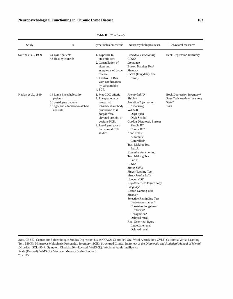
Svetina et al., 1999 44 Lyme patients43 Healthy controls
1. Exposure toendemic area
2. Constellation ofsigns andsymptoms of Lymedisease
3. Positive ELISAwith confirmationby Western blot
4. PCR
Executive FunctioningCOWALanguageBoston Naming Test*MemoryCVLT (long delay free
recall)
Beck Depression Inventory
Kaplan et al., 1999 14 Lyme Encephalopathypatients
18 post-Lyme patients15 age- and education-matched
controls
1. Met CDC criteria2. Encephalopathy
group hadintrathecal antibodyproduction toB.burgdorferi,elevated protein, orpositive PCR.
3. Post-Lyme grouphad normal CSFstudies
Premorbid IQShipleyAttention/Information
ProcessingWAIS-R
Digit SpanDigit Symbol
Gordon Diagnostic SystemSimple RTChoice RT*
2 and 7 TestAutomaticControlled*
Trail Making TestPart A
Executive FunctioningTrail Making Test
Part B
Beck Depression Inventory*State Trait Anxiety InventoryState*Trait
COWAMotor SkillsFinger Tapping TestVisuo-Spatial SkillsHooper VOTRey–Osterrieth Figure copyLanguageBoston Naming TestMemorySelective Reminding Test
Long-term storage*Consistent long-term
retrieval*Recognition*Delayed recall
Rey–Osterrieth figureImmediate recallDelayed recall
Note.CES-D: Centers for Epidemiologic Studies Depression Scale; COWA: Controlled Oral Word Association; CVLT: California Verbal LearningTest; MMPI: Minnesota Multiphasic Personality Inventory; SCID: Structured Clinical Interview of theDiagnostic and Statistical Manual of MentalDisorders; SCL–90-R: Symptom Checklist90—Revised; WAIS-(R): Wechsler Adult IntelligenceScale (Revised); WMS (R): Wechsler Memory Scale-(Revised).*p< .05.

Neuropsychology Review pp588-nerv-378447 September 20, 2002 12:3 Style file version March 18, 1999
164 Westervelt and McCaffrey
in a discriminant function analysis. Psychopathology wasalso measured using the Minnesota Multiphasic Personal-ity Inventory (MMPI) and the Beck Depression Inventory(BDI). On the MMPI, the Lyme group had lower scoreson the scales most sensitive to depression and anxiety rel-ative to the depressed group, and fewer somatic concernsthan the fibromyalgia group. In contrast with MMPI find-ings, there was no significant difference among groupson the BDI. Depression, as measured by the MMPI, hada significant negative correlation only with the AssociateLearning subtest of the WMS in the Lyme group, and notwith any of the other memory measures. When the Lymegroup was divided in two groups based on their MMPIscale 1 (Hypochondriasis) score, there was no difference inmemory performance between the high- and low-scoringgroups. When divided based on the MMPI scale 2 (Depres-sion) score, only immediate recall on the CVLT yieldedsignificant differences between groups (with the groupwith greater depression showing worse memory perfor-mance). Thus, Kaplan et al. concluded that the memoryimpairment observed in Lyme encephalopathy cannot beexplained solely by affective symptoms, despite the com-mon finding that Lyme patients endorse more depressivesymptoms than do normal controls.
Shadick et al. (1994) examined the prevalence of per-sistent symptoms in 38 randomly selected patients fromthe community with histories of Lyme disease. Comparedwith 43 healthy controls, overall, the Lyme group per-formed significantly poorer on the delayed recall trial ofthe CVLT, as well as on the Purdue Pegboard (dominanthand), a measure of fine motor coordination. Twelve ofthe 38 patients with Lyme disease scored more than twostandard deviations below the mean on two or more mem-ory tests, whereas only 5 of the 43 control participantsshowed such poor performances. For unclear reasons,however, only 4 of the Lyme patients were described asshowing “neurocognitive sequelae.” Two of those patientswere diagnosed with mild encephalopathy, 1 with neu-roborreliosis, and 1 with progressive supranuclear palsy.The latter two participants had positive Lyme titers. Theonly significant risk factor for persistent neurocognitiveimpairment was the length of infection before treatment.Unfortunately, although the authors also measured theprevalence of a number of physical sequelae, as well asserum antibody reactivity, they did not examine the rela-tionship between these variables and neurocognitive func-tioning in the group as a whole.
A European study compared cognitive functioningin 20 patients with long-standing Lyme disease (mean of52 months post acute infection) to 20 non-brain-damagedneurologic controls (Benke et al., 1995). Nineteen ofthe 20 participants with Lyme disease showed CSF evi-
dence of lymphocytic pleocytosis during acute illness. Re-sults of neuropsychological testing showed that the Lymegroup performed significantly worse on both the recalland recognition indices of the German CVLT, verbal flu-ency, rapid syllable repetition, and the accuracy scores ofmental flexibility and choice reaction time tasks. Memorywas frequently affected, with 50% of the patients in theLyme group performing at or below an unspecified cutoffscore based on the control group’s performance. Amongthese patients, 6 showed only memory deficits, whereasthe remaining 4 demonstrated global neuropsychologicalimpairment. The severity of neurological deficits at the on-set of the illness was significantly correlated with perfor-mance on only the mental flexibility task, suggesting thatthe degree of neurological impairment was not a good indi-cator of overall neuropsychological performance. Lengthof time, since the diagnosis was made, was not corre-lated with any of the neuropsychological variables. Over-all, Benke et al. state that this study demonstrates thatcognitive deficits may persist in Lyme disease for years,even in patients with only minor residual neurologicalsymptoms.
Bujak et al. (1996) compared neuropsychologicaland psychological functioning in 23 patients with “post-Lyme syndrome” (defined as persistent arthralgia, fatigue,and subjective memory loss despite presumably adequateantibiotic therapy) to 23 patients who had prior histo-ries of Lyme disease but no residual symptoms. All pa-tients showed antibody reactivity during the initial phaseof the disease, and at the time of the current examina-tion, 48% of both groups continued to be seropositiveby immunoblotting or ELISA. Two of the post-Lyme pa-tients additionally showed persistent neurologic involve-ment (Bell’s palsy). Results of the neuropsychologicalevaluation revealed that both groups had poorer verbalthan visual memory, though the post-Lyme group per-formed significantly worse than the recovered group ononly the attention/concentration subscale of the WMS-R. The post-Lyme group also obtained higher scores onthe somatization, obsessive–compulsive disorder, depres-sion, anxiety, hostility, and summary scales of the Symp-tom Checklist-90-Revised (SCL-90-R), though all scoreswere within normal limits. In addition, higher scores wereobtained in the post-Lyme group on the BDI, as well asscales 1 (Hypochondriasis) and 3 (Hysteria) of the MMPI,though, again, these scores were within the normal range.There was no correlation between fatigue or arthralgia (asmeasured by a visual analog scale) and performance onthe WMS-R, BDI, and MMPI, nor was there any signif-icant relationship between the BDI and WMS-R scores.The presence of both fatigue and arthralgia during the ini-tial presentation of the disease was the best predictor of

Neuropsychology Review pp588-nerv-378447 September 20, 2002 12:3 Style file version March 18, 1999
Neuropsychological Functioning in Chronic Lyme Disease 165
subsequent development of post-Lyme syndrome (30% ofthe post-Lyme group met criteria for fibromyalgia, 13%for chronic fatigue, and 44% had symptoms resemblingthese conditions but failed to meet criteria). The presenceof fatigue duringinitial illness increased the likelihoodof post-Lyme syndrome by nearly 50-fold, and the pres-ence of arthralgia by nearly 32-fold. Used together, thiscombination of symptoms correctly identified 21 and 23patients in the post-Lyme and recovered groups, respec-tively. The authors concluded that patients with post-Lymesyndrome differ from those who have recovered from theillness on a number of clinical features, though the objec-tive neuropsychological deficits were fairly limited. Be-cause many of the patients with persistent symptoms hadundergone multiple courses of antibiotic therapy withoutprolonged benefit in their chronic symptoms, Bujak et al.state thatactiveborrelial infection in the brain is an un-likely cause of post-Lyme syndrome. Rather, they con-sider the possibility that these patients had disseminatedborreliosis with subclinical CNS involvement during theirLyme disease infection, with this CNS disruption result-ing in localized brain dysfunction and subsequent residualdifficulties (i.e., subjective memory loss and poor concen-tration). They note that the specific factors (e.g., geneticor personality variables) that predispose patients to post-Lyme syndrome, however, remain undefined.
Ravdin et al. (1996) examined memory functioningin 21 patients with Lyme disease compared with both apatient control group consisting of 21 individuals with os-teomyelitis and 21 healthy controls. The purpose of thestudy was to determine the extent to which memory dys-function was present above and beyond what may be foundin other patient groups as a result of systemic illness andcoexisting emotional and physical complaints. They alsoexamined the relationship betweenself-reportof mem-ory dysfunction andobjectivememory impairment, thelatter measured by CVLT performance. Fifteen of the 21patients in the Lyme group had neurologic complaints,though none had CSF evidence of Lyme disease, and poly-merase chain reaction (PCR) was negative in all patients.Sixteen of the Lyme patients also had MRI data, with 3of these patients showing “abnormal” scans (multifocalbright signals). Results of this study showed no signifi-cant differences among groups on a measure of the non-physiologic aspects of depression (the BDI minus itemsmeasuring physical symptoms of depression). In contrast,both patient groups were significantly more fatigued thanthe healthy control group, with the Lyme patients describ-ing greater fatigue than the osteomyelitis group. Similar tofindings from previous studies, fatigue was significantlybut inversely correlated with memory performance in theLyme group. When controlling for the effects of fatigue,
measures of memory functioning revealed significant dif-ferences between the healthy controls and both patientgroups on recall indices. There was no significant differ-ence between the Lyme and osteomyelitis groups, thoughcloser analysis revealed that 38–48% of the patients in theLyme group performed at the borderline to impaired levelon total learning and long delay free recall (defined asTscores <40), whereas only 19–24% of the patients withosteomyelitis performed at or below this level. In contrastwith performance on recall indices, there was no signifi-cant difference on the recognition trial, suggesting that thepoorer performance in the patient groups was due primar-ily to retrieval deficits. The Lyme group was more likelyto endorse memory dysfunction on a self-report measurethan the other two groups, though there was no significantrelationship between subjective ratings of daily memorycapabilities and objective memory performance.Subjec-tiveratings of memory impairment, however, were signif-icantly correlated with both depression ratings and fatiguein the Lyme group.
Gaudino et al. (1997) further examined the relation-ship between physical and psychiatric symptomatologyand cognitive complaints by comparing neuropsycholog-ical performance in 38 patient with post-Lyme syndrome(defined as severe fatigue, malaise, and cognitive com-plaints that persist 6 months or more after completion ofadequate antibiotic therapy) to 25 patients with CFS and56 healthy controls. Post-Lyme syndrome and CFS sharea number of symptoms, including, most obviously, pro-found and debilitating fatigue, as well as a number ofaffective symptoms. Both illnesses, however, are hetero-geneous with respect to the nature and severity of the clin-ical symptoms, as well as the presence of neuropsychiatricdisturbance. Results showed that both CFS and post-Lymepatients described more fatigue and depression than didthe healthy controls, though the two patient groups did notdiffer from each other on these measures. The relationshipbetween depression and fatigue and the neuropsycholog-ical variables was not examined. On neuropsychologicalmeasures, the CFS group performed more poorly than didcontrols on only the Digit Symbol subtest of the WechslerAdult Intelligence Scale-Revised (WAIS-R). In contrast,the Lyme group performed worse than the control groupon measures of attention (Digit Span subtest of the WAIS-R), verbal learning (SRT), verbal fluency (COWA), andfine motor speed (Finger Tapping Test [FTT], dominanthand). When patients were further subgrouped accordingto premorbid psychiatric histories, Gaudino et al. foundthat post-Lyme patients with no premorbid psychiatrichistories performed significantly worse than those withprior psychiatric histories on most cognitive tests, thoughthese differences were statistically significant only on the

Neuropsychology Review pp588-nerv-378447 September 20, 2002 12:3 Style file version March 18, 1999
166 Westervelt and McCaffrey
SRT and Digit Span subtest of the WAIS-R. Given that9 of the 10 post-Lyme patients with premorbid psychi-atric histories also had psychiatric disorders at the timeof testing (whereas none of the patients without premor-bid psychiatric histories had concurrent psychiatric dis-orders), the authors concluded that psychological factorsmay have contributed to the perception of cognitive lossin the patients with psychiatric disturbances. In contrast,among those patients who lacked histories of psychiatricdifficulties, their subjective impressions of loss were con-firmed on objective testing. In these instances, the objec-tive neuropsychological deficits were attributed to a mildencephalopathic process resulting from Lyme disease.
Pollina, Sliwinski, et al. (1999) examined cognitiveprocessing speed on a measure with minimal peripheralnerve/muscle functioning demands in a group of 16 pa-tients (with histories of well-documented Lyme disease)who had completed standard antibiotic treatment at least2 months before evaluation. Twelve of the 16 Lyme pa-tients had CSF data available: 3 had intrathecal antibodysynthesis, 2 had mildly elevated CSF protein, and noneshowed pleocytosis. In addition, “several” patients under-went MRI imaging, with nonspecific white matter lesionsobserved “occasionally.” Compared with 15 age- andeducation-matched healthy control participants, the Lymepatients performed significantly poorer on a computer-ized measure of cognitive processing speed (Alphabet–Arithmetic task), though the groups did not differ on a sim-ple perceptual/motor speed-matching task. Closer analysisof these findings suggested that the deficit on the speededprocessing task was most likely due to slowness in initi-ating the cognitive processes involved in the task ratherthan repetitively performing the task. Performance on themeasure of cognitive speed did not correlate with mem-ory performance on the SRT (also poorer in the Lymegroup), suggesting that these deficits were independent ofone another. The Lyme group also reported significantlymore depressive symptoms than did the healthy controls,though when depression was added to the analyses as a co-variate, differences on the cognitive measures remained.In addition, fatigue was related to only one of the SRTvariables, suggesting that noncognitive variables such asfatigue and depression did not have a significant impacton neuropsychological performance.
Pollina, Elkins, et al. (1999) extended their previ-ous work suggesting that cognitive slowing is process-specific in Lyme patients by examining 25 patients withwell-recognized manifestations of Lyme disease which,despite treatment, were followed by unresolved symp-toms of fatigue and/or cognitive problems. Twenty-threecommunity age- and education-matched volunteers werealso examined. All participants underwent examination
with the computerized measure of processing speed men-tioned previously (Alphabet Arithmetic). The Lyme pa-tients were also administered standard neuropsychologi-cal tests (e.g., measures of word-reading, fund of knowl-edge, expressive vocabulary, attentional capacity, psy-chomotor speed, speed of information processing, visuo-construction, learning/memory, and depression). Age,level of depression, and estimated premorbid intellectualability were used as covariates. Pollina and colleaguesfound that the groups did not differ on the experimentaltask in terms of accuracy, nor did they differ in terms ofperceptual motor speed and incrementing speed. However,the Lyme patients showed slower speed of initiation rela-tive to the healthy controls. In the Lyme patients, the au-thors found no relationship between initiation speed andperformance on specific cognitive domains (test scoreswere combined to create domains of premorbid IQ, short-term memory, and long-term memory). Initiation speedwas found to correlate with performance on part A of theTrail Making Test (TMT) and Digit Symbol, but not withpart B of the TMT or the Paced Auditory Serial AdditionTask (PASAT). The authors note that the Lyme patientsreported more depressive symptoms than did the controls,though there was no significant correlation between theirdepression scores and initiation speed. In conclusion, Pol-lina, Elkins, et al. (1999) state that this study supportstheir hypothesis that Lyme patients experience cognitiveslowing that is independent of age, premorbid abilities,and degree of depression, and that may be specific to theamount and type of information processing required bya given task. Their finding that initiation speed did notcorrelate with other cognitive domains argues against apattern of global impairment, though the Lyme patients’performances were not compared with control data on thestandard neuropsychological measures.
Svetina et al. (1999) examined confrontation namingin a group of 44 patients with confirmed Lyme diseaseand 43 healthy, serologic-negative controls. Ninety-onepercent of the Lyme patients were vaguely described ashaving progressed to the “second stage.” Svetina et al.note that word-finding difficulty is a frequent complaintof many patients with Lyme disease, though this aspectof language had not been examined previously. Svetina etal. also measured phonemic and semantic verbal fluency,memory, and depression. In their sample, 55% of the pa-tients with Lyme disease complained of word-finding dif-ficulties, in contrast with 14% of the controls. Results offormal testing revealed significantly lower Boston NamingTest (BNT) scores in the Lyme group relative to controls,though they found no difference between groups on theverbal fluency measures or on the other measures given.When the BNT scores were categorized according to

Neuropsychology Review pp588-nerv-378447 September 20, 2002 12:3 Style file version March 18, 1999
Neuropsychological Functioning in Chronic Lyme Disease 167
impairment (defined as a score of less than 2SD belowthe mean of the control group; i.e., a cutoff of 51), 36%of the Lyme patients fell within this range, compared withonly 9% of the control participants. There was no asso-ciation between actual test performance on the BNT andself-report of naming difficulties. A significant correla-tion between naming and scores on the CVLT was found,suggesting a relationship between naming and memoryretrieval; there was no significant correlation between theBNT and verbal fluency. When the groups were separatedbased on level of depression, there were no significant dif-ferences among groups on the BNT. In sum, the authorsconclude that there appears to be a subtle naming deficit inLyme disease that may be a result of a generalized impair-ment in retrieval, though in this group there was no sig-nificant difference in memory performance between theLyme disease and control groups.
Lastly, Kaplan et al. (1999) investigated the relation-ship between objective evidence of CNS infection andneuropsychological performance. The study groups con-sisted of 32 patients who met CDC criteria for Lyme dis-ease, including 14 patients with evidence of CNS infec-tion (i.e., one or more signs of abnormal CSF: intrathecalantibody production toB. burgdorferi, elevated protein,or a positive PCR forB. burgdorferiDNA; “Lyme en-cephalopathy” group) and 18 patients without evidenceof CNS infection (“post-Lyme” group). Fifteen age- andeducation-matched community and hospital volunteerswithout histories of Lyme disease were also studied. Mostof the Lyme patients (11 of 14 Lyme encephalopathypatients and 13 of 18 post-Lyme patients) complainedof memory problems, though none of the controls re-ported similar concerns. Results showed that the post-Lyme group had slower reaction times on the Gordon Di-agnostic System (GDS), a vigilance task, than those of thecontrol group (there was no significant difference betweenthe other groups), and there was a nonsignificant trend inthe post-Lyme group toward difficulty with selective at-tention. The Lyme encephalopathy group performed sig-nificantly worse in aspects of verbal learning (SRT) thanthe other two groups, which did not differ. On mood mea-sures, both Lyme groups had higher depression scores thandid the control group, with the post-Lyme patients report-ing more depressive and state anxiety symptoms than didthe Lyme encephalopathy or control group. Examinationof the relationship between mood measures and neuropsy-chological performance revealed that higher levels of stateanxiety were associated with poorer performance on theselective attention task in the whole sample and in the con-trol group. In the post-Lyme group, higher state anxietyscores were also associated with slower reaction time onthe GDS, but not with any other measure. The depression
measure (BDI) did not correlate with neuropsychologicalperformance on any of the attention or memory measuresin any of the groups. Kaplan et al. make note of the find-ing that the post-Lyme group performed worse than did theother groups on measures of attention and psychomotorspeed, and that anxiety was correlated with slower reac-tion time on the vigilance task. They speculate that anxietymay have affected performance in this group by narrowingthe focus of attention beyond an adaptive level. Kaplan etal. state that, in contrast, the Lyme encephalopathy groupappears to have a neurologic basis associated with theirdeficit (i.e., memory loss).
DISCUSSION OF THENEUROPSYCHOLOGICAL LITERATURE
Summary of Cognitive Findings
Overall, the results of the neuropsychological studiesin Lyme disease rather consistently demonstrate deficitson memory measures, with most difficulty with free re-call on list-learning tasks (Benke et al., 1995; Gaudino etal., 1997; Kaplan et al., 1992, 1999; Krupp et al., 1991;Pollina, Sliwinski, et al., 1999; Ravdin et al., 1996;Shadick et al., 1994). Exceptions include the studies byBujak et al. (1996) and Svetina et al. (1999). The lackof significant findings on the CVLT in the Svetina etal. study is somewhat surprising. The Bujak et al. study,however, did not utilize a healthy control group; rather,they compared patients with post-Lyme syndrome to pa-tients who had apparently fully recovered from Lyme dis-ease. The possibility that subtle deficits were present in“recovered” Lyme patients should be considered, how-ever, particularly given that at the time of evaluation,48% ofbothgroups continued to be seropositive (of note,seropositivity is an indicator of past or present infec-tion and not necessarily active disease). It is also pos-sible that the discrepant findings reflect differences in themeasures used, rather than the participants studied. Bu-jak et al. used the WMS-R, whereas the majority of theother studies report deficits on the CVLT or SRT, bothverbal list-learning tasks. In other studies using subtestsof the WMS-R such as Logical Memory (often in ad-dition to the CVLT or SRT), findings have been moreinconsistent, perhaps reflecting the greater demand forthe use of active organization/mnemonic strategies andthe requirements of sustained effort on the list-learningtasks (Kaplan and Jones-Woodward, 1997). In addition,Tremont et al. (2000) have found that verbal list-learningtasks such as the CVLT have a stronger relationshipwith measures of executive functioning than do other

Neuropsychology Review pp588-nerv-378447 September 20, 2002 12:3 Style file version March 18, 1999
168 Westervelt and McCaffrey
verbal memory tasks (such as story recall). This finding issupported by functional imaging studies that have shownthat tasks requiring active encoding and retrieval strategiesinvolve substantial frontal systems activation (Fletcher etal., 1995; Grasby et al., 1993), whereas story recall tasks(“consolidation tasks”) may be more purely reliant onthe integrity of the hippocampus and other mesial tem-poral structures (Tremont et al., 2000). Thus, it is possiblethat the observed deficits on verbal list-learning tasks inchronic Lyme patients may reflect the additional impactof frontal systems dysfunction on learning and memoryon these tasks. Consistent with this hypothesis (and incontrast with the impairments observed in performanceon recall indices of verbal list-learning tasks), recognitionperformance in the Lyme groups has generally been foundto be equivalent to that of the controls. A brief report byMasur et al. (1992) describes similar findings.
Often, deficits have also been found on the COWA(Benke et al., 1995; Gaudino et al., 1997; Krupp et al.,1991), a word-list generation task often described as ameasure of verbal fluency and an indicator of frontal sys-tems functioning, though this is not an entirely consis-tent finding (cf. Kaplan et al., 1999; Svetina et al., 1999).Other traditional “executive” tasks requiring set shifting,abstraction, concept formation, and response inhibition(e.g., part B of the TMT, Booklet Category Test, WAIS-RSimilarities, Raven’s Progressive Matrices, Stroop), how-ever, have generally failed to distinguish patients withLyme disease from controls, though other areas of func-tioning often considered as involving the frontal systemshave shown impairment. These include deficits in men-tal flexibility (Benke et al., 1995), choice reaction time(Kaplan et al., 1999), selective attention (Kaplan et al.,1999), upper extremity fine motor dexterity (Gaudino etal., 1997; Shadick et al., 1994), and speed of informationprocessing/initiation (Pollina, Elkins, et al., 1999; Pollina,Sliwinski, et al. 1999). Interestingly, Pollina, Sliwinski, etal. (1999) noted that the speed of information process-ing deficits observed in their group of chronic Lyme pa-tients was not significantly related to performance on theSRT, suggesting that slowed processing cannot accountfor the deficits seen on this learning task. Language andoral/motor deficits have also been described, including im-pairments in rapid syllable repetition (Benke et al., 1995;Gasse et al., 1992), and, in one of two studies measuringthe construct, confrontation naming (Svetina et al., 1999).Measures of brief attention (Benke et al., 1995; Bujak etal., 1996; Gaudino et al., 1997; Kaplan et al., 1992, 1999;Krupp et al., 1991) show more inconsistent results, thoughto some extent, the apparent inconsistencies may reflectdifferences in the measures used. Lastly, performance bythe groups with Lyme disease on measures of general in-
telligence (Benke et al., 1995; Kaplan et al., 1992; Kruppet al., 1991; Ravdin et al., 1996) has consistently beencomparable to that of the control groups.
Thus, the neuropsychological deficits observed inLyme disease appear to predominantly reflect frontal sys-tems dysfunction. These findings are consistent with pos-itive results from neuroimaging studies in Lyme disease,though it should be noted that this appears to be a typi-cal pattern of performance in Lyme disease regardless ofwhether imaging shows abnormalities. The nature of thedeficits is, therefore, similar in many respects to the kindof deficits seen in other infectious disease processes in-volving primarily subcortical/frontal systems functioningsuch as HIV infection, as well as in autoimmune disordersand fatiguing illnesses involving subcortical white matter(e.g., MS, systemic lupus). The autoimmune disorders, inparticular, may provide good conceptual models for Lymedisease, given the high overlap in physical, psychiatric,and neuropsychologic symptoms.
Limitations of the Neuropsychological Literature
There are several limitations of the current literaturethat merit elaboration. First, the expected degree of cog-nitive impairment in Lyme disease is difficult to predictfrom the current literature. Most studies describe relativelymild deficits, though reliance on group statistics may ob-scure individual findings within a relatively heterogeneousgroup. This limitation speaks not only to challenges inidentifying the degree of impairment expected, but also,to some extent, in the specific nature of the impairmentswhich can be anticipated given the variability in symptompresentation in these patients. In an attempt to addressthe limits of group statistics, some authors have identifiedthe level of impairment in each patient studied. For ex-ample, as described in greater detail in the review of thecontrolled studies, Krupp et al. (1991) divided the Lymegroup by level of impairment and found that 9 of the 15patients studied were impaired by their criteria. Most ofthose 9 patients were described as “moderately” impaired,though clearly there was substantial variability within thegroup. Several other researchers have commented on thepercentage of patients within the group who perform be-low a specified cutoff for impairment on a given measure(e.g., Svetina et al., 1999), though variability in the extentof impairment is often not reported. Preliminary data fromour own study examining the relationship between cogni-tive functions and psychological factors such as depres-sion, anxiety, fatigue, and tolerance for mental effort in 17patients with Lyme disease (including 12 with multipletick-borne diseases) and 17 healthy controls (Westervelt

Neuropsychology Review pp588-nerv-378447 September 20, 2002 12:3 Style file version March 18, 1999
Neuropsychological Functioning in Chronic Lyme Disease 169
and McCaffrey, 1999) provide evidence of the variabil-ity in symptom severity and presentation. For example,in our sample, the Lyme group’s performance was sig-nificantly worse than that of the control group’s on thePASAT. On this measure, 10 of 17 patients (59%) in theLyme group exhibited impairment (a score≥1 SD be-low the mean of the control group). This included 1 pa-tient with mild impairment (1–1.4SD below the controlmean), 2 with moderate impairment (1.5–1.9SD belowthe control mean), and 7 with severe impairment (≥2 SDbelow the control mean). The Lyme group also performedsignificantly worse than did the control group on phone-mic verbal fluency (FAS), including 9 patients with im-paired performance. In contrast with the prior example,however, most patients showed only mild impairment onthis measure (N= 7), only 2 showed moderate impair-ment, and none were severely impaired. Even on tasksfor which no significant group differences were observed,the Lyme patients were often at least twice as likely toshow impairments, though patients who were impairedon one measure may not have been the same patients whowere impaired on another, arguing against the presenceof global impairment in the subset of impaired patients.Thus, given the variability in group findings from onestudy to another, closer attention to the presentation ofindividual patients within each study may be beneficial inunderstanding the range of intensity of symptom severityand variety of impairments which can be observed in thisdisease. This suggests that, in contrast with other neu-ropsychological disorders such as Alzheimer’s disease,there may not be a “classic” neuropsychological patternin Lyme disease, though some impairments certainly ap-pear more often than others. Lastly, the possibility thatthe variability in symptom presentation and severity isperhaps in part a reflection of different underlying mecha-nisms (e.g., persisting CNS infection vs. an inflammatoryresponse or immune reaction) is worth considering. Thisraises the possibility that the groups studied are less ho-mogeneous than assumed, again making generalizationsdifficult.
Other limitations in the current neuropsychologicalliterature on Lyme disease include the use of very smallsamples. Apparently, the effect sizes are robust enough toyield significant findings in many instances regardless ofthe small samples, though low statistical power makes thelikelihood of discovering more modest deficits unlikely.In addition, given the small size of the samples, outliersin either direction may have a substantial impact on groupstatistics. This also limits the extent to which findings canbe generalized, though, as previously mentioned, the abil-ity to make global statements in this group is difficult,given the variability of patient presentations.
Impact of Chronic Illness
It is noteworthy that several of the controlled stud-ies used patient control groups, including groups withdepression (Kaplan et al., 1992), fibromyalgia (Kaplanet al., 1992), neurologic dysfunction without brain dam-age (Benke et al., 1995), osteomyelitis (Ravdin et al.,1996), and CFS (Gaudino et al., 1997). Except for com-parison with osteomyelitis patients, in each study provid-ing direct comparisons, the group with Lyme disease per-formed poorer than the other patient groups on severalmeasures of neuropsychological functioning. In additionto these controlled studies, in a brief report, Gasse et al.(1992) described the neuropsychological functioning in15 patients who had been treated for Lyme disease andhad recoveredneurologically, compared with both healthycontrols and patients who had suffered from viral meningi-tis/encephalitis. The results showed impaired verbal learn-ing in both patient groups, though the participants withhistories of Lyme disease had more extensive findings, in-cluding deficits on word-list generation and syllable rep-etition. Measures of psychomotor speed, sustained atten-tion, visuo-spatial ability, and other aspects of executivefunctioning were reportedly normal. Taken together, thesestudies indicate that the deficits observed in the groupswith Lyme disease cannot be solely attributed to the pres-ence of a chronic illness.
Relationship with Depression
As noted in previous sections, depression is a fairlyfrequently reported problem in chronic Lyme disease.Given the known cognitive effects of depression, several ofthe controlled neuropsychological studies of Lyme diseasehave considered the possible impact of depression on neu-ropsychological functioning. The nature of the observedrelationship, however, has been somewhat surprising. Twoof the nine controlled studies examining the relationshipbetween depression and cognition found significantposi-tivecorrelations between depression and memory perfor-mance, indicating that an increase in depression scoreswas associated withbettermemory functioning (Gaudinoet al., 1997; Krupp et al., 1991). In contrast, Bujak et al.(1996), Kaplan et al. (1999), Pollina, Elkins, et al. (1999);Pollina, Sliwinski, et al. (1999), Ravdin et al. (1996), andSvetina et al. (1999) found no relationship between objec-tive memory performance and depression, though Ravdinet al. (1996) noted that depression was significantly corre-lated withsubjective perceptionof cognitive loss. Finally,Kaplan et al. (1992) found anegativecorrelation, thoughwith only one measure of memory (WMS-R Associate
Neuropsychology Review pp588-nerv-378447 September 20, 2002 12:3 Style file version March 18, 1999
170 Westervelt and McCaffrey
Learning). It should also be noted that although Kaplan etal. (1999) found no relationship between depression andneuropsychological functioning, they did find some rela-tionship between state anxiety and slower reaction timein a post-Lyme group, though there was no relationshipbetween anxiety and neuropsychological performance inthe Lyme encephalopathy group.
In two recent studies, the relationship between moodstates and neuropsychological performance has been moredirectly addressed. Barr et al. (1999) examined 55 patientsat the time of their initial diagnosis with Lyme disease.All patients were administered the Self-Ratings of Mem-ory Questionnaire, the BDI, and the CVLT. On the self-report memory measure, the group complained most ofan inability to recall names, hold information in mem-ory, and recall what one was doing, as well as a lack ofconfidence that information will be retained. They didnot describe a general sense of confusion or difficultywith remote memory. Patients with depression (as definedby a BDI score of greater than 10) endorsed relativelymore items suggesting memory complaints than did non-depressed patients, though there was no distinguishablepattern to the items that were endorsed. The majority ofthe patients rated their memory as worse than before. Itshould be noted that most of the scores on the BDI werebelow the level associated with depression. Specifically,when the groups were divided according to BDI scores,63.6% reported minimal depression (BDI scores of 0–9),25% of the group was within the mild range (BDI scoresof 10–16), and only 10% was within the moderate to se-vere range (BDI scores of 17–30). Significant correlationswere found between the self-report memory questionnaireand the BDI scores (p= –.57), in that patients endorsingmore depressive symptoms also reported having poorermemory functioning. In contrast, there was no signifi-cant relationship between BDI scores and performance onthe CVLT, or between CVLT performance and measuresof subjective memory loss. Furthermore, on the CVLT,the three depression groups did not differ in their perfor-mances on total learning and delayed recall, and the meanscores for all groups fell within the average range com-pared with normative standards. Individual patients whosememory scores were lower than one standard deviation be-low the mean of the normative data showed no tendencyto underreport symptoms of memory disturbance, argu-ing against anosognosia in patients with weaker memoryperformance.
Elkins et al. (1999) characterized the psychologicalstatus of a group of 30 post-Lyme patients (all with docu-mented neurologic symptoms at the time of initial diagno-sis) and examined the relationship between reported symp-toms and neuropsychological performance. They screened
for the presence of current and lifetimeDMS-IVAxis I di-agnoses, as well as examining positive and negative moodstates by using the Positive and Negative Affect Sched-ule (PANAS). The authors note that positive and negativeaffect have been useful in distinguishing affective statesassociated with psychiatric disorders. Specifically, theystate that depressive disorders are characterized by lowpositive affect and heightened negative affect, whereasanxiety disorders are associated with heightened negativeaffect alone. They also note that previous studies haveshown that CFS is characterized by low positive affect,and in these patients, low positive affect has been relatedto cognitive and motor slowing. Elkins et al. also exam-ined cognitive performance in their post-Lyme group byusing a variety of neuropsychological measures (WAIS-RVocabulary, Block Design, Digit Span, and Digit Symbol,Wide Range Achievement Test-Revised Reading, WMS-R Logical Memory story A or B, SRT, PASAT, and TMT).Results showed that 37% of the participants met currentcriteria for aDSM-IVAxis I disorder, and 50% met life-time criteria for an Axis I disorder, though only 20% metcriteria for a disorder before the onset of their Lyme dis-ease. Elkins et al. state that although the percentage ofparticipants meeting the lifetime criteria for an Axis I dis-order is comparable with that in the general population, itmay be noteworthy that the incidence of an Axis I disordernearly doubled following the onset of Lyme disease in thisgroup. On the PANAS, the group had significantly lowerlevels of positive affect compared with published norms,but comparable negative affect levels. As discussed, thispattern differs from that typically seen in major depres-sion, and the authors speculate that low positive affect inthis group may reflect the impact of fatigue on pleasantand active engagement with one’s environment. Positiveand negative affect were not significantly correlated, norwere they associated with the presence of a current psy-chiatric disorder. In contrast, low levels of positive affectcorresponded to subjective illness severity, but not withobjective indicators of the course and nature of the diseaseprocess itself (e.g., duration of illness, the presence of neu-rologic symptoms at the time of diagnosis, or antibiotictreatment history). The cognitive measures were clusteredinto four areas of functioning: (1) general intelligence, (2)attention, (3) verbal learning, and (4) psychomotor speed.Compared with normative data, the group’s performancewas not impaired (i.e., it was not more than one standarddeviation below the normative mean), though the PASATand SRT approached this level (–0.88 to –0.99SDbelowthe mean). In multiple regression analyses, none of thepsychological measures significantly predicted cognitiveperformance in any of the four domains. Although lim-ited by lack of a comparison group, this study provides

Neuropsychology Review pp588-nerv-378447 September 20, 2002 12:3 Style file version March 18, 1999
Neuropsychological Functioning in Chronic Lyme Disease 171
interesting data by demonstrating not only a lack of as-sociation between current depressive symptoms and neu-ropsychological functioning as other studies have, but alsoa lack of association between other mood states and thepresence of current and life-time Axis I disorders.
Kaplan and Jones-Woodward (1997) point out thatalthough the literature has demonstrated that depressionratings are often higher in patients with Lyme disease rela-tive to controls, the average depression scores in the Lymegroups are often below the suggested cutoff for clinicallysignificant depression. In addition, as noted earlier in thissection, the one study directly comparing patients withLyme disease and healthy depressed controls found thatthe Lyme group performed significantly worse on neu-ropsychological measures than did the depressed controls.Therefore, the general lack of significant negative correla-tions between depression scores and neuropsychologicalfunctioning in these patients may not be surprising. How-ever, Kaplan and Jones-Woodward note that the stress andanxiety associated with chronic illness may be a factorin perceivedmemory disturbance, even though these pa-tients may not beclinically depressed. The possibility thatthe presence of depression-like symptoms may better re-flect fatigue or other physical symptoms of the diseaseis also worth considering, particularly given that the onestudy that examined only the “cognitive” (i.e., nonphysi-cal) aspects of depression found no difference between theLyme patients and controls. Thus, overall, depression ordepression-like symptoms donotappear to be causally re-lated to objective neuropsychological impairment in Lymedisease, though they may be related to the perception ofcognitive loss.
The importance of identifying neuropsychologicalimpairments as beingcausally relatedto other symptomssuch as depression versus beingindependent symptoms,which may often occur together as part of the same un-derlying pathology, is not just semantic. Rather, it mayhave important implications. First, although seldom dis-cussed in the literature, there tends to be some debateamong clinicians regarding the etiology of chronic Lymedisease/post-Lyme syndrome, particularly the neuropsy-chological deficits/complaints. Bujak et al. (1996) hintat this debate by indicating that it is unknown whetherpersonality factors may predispose patients to post-Lymesyndrome. This statement suggests that some may viewchronic Lyme/post-Lyme syndrome as a primarily psy-chiatric disturbance, or at least as involving a strong psy-chiatric component. This perception may greatly impactthe treating clinician’s view of the patient, as well as theirwillingness to consider the need for further treatment ortype of treatment to be recommended. Although psychi-atric conditions should certainly be addressed, this should
not occur at the exclusion of consideration of other appro-priate medical treatments or cognitive remediation, whenindicated.
Relationship with Neurologic Indicators of Disease
The relationship between neurocognitive function-ing and neurologic status is unclear. In a brief report,Kaplan et al. (1995) first attempted to elucidate this re-lationship by comparing memory functioning on the SRTin healthy controls (n= 11) to Lyme patients with (n= 16)and without (n= 21) increased CSF protein and/or in-trathecal antibody production. They found that the pa-tients with abnormal CSF scored significantly worse onthe SRT. They interpreted these findings as suggestingthat memory loss is caused by CNS infection, as evi-denced by CSF abnormalities. Similarly (as described ingreater detail in a previous section), in a full report directlyinvestigating this issue, Kaplan et al. (1999) comparedthe neuropsychological performance in chronic Lyme pa-tients with and without objective evidence of persistentCNS infection. They found that only the group with ob-jective encephalopathic symptoms demonstrated memoryimpairment. The “post-Lyme” group (those without ob-jective evidence of CNS dysfunction) performed worseon a measure of reaction time on a vigilance task, thoughthe authors speculated that this deficit was likely due toincreased anxiety in this group. In contrast, however, Pol-lina, Sliwinski, et al. (1999) refer to preliminary data com-paring cognitive processing speed and memory in Lymepatients with and without CNS evidence of infection,which showed no significant group difference. Further-more, in other studies discussed in prior sections, statisti-cally significant differences in cognitive functioning werefound in Lyme patients relative to healthy controls, evenwhen none of the patients in the Lyme sample showedCSF evidence of persisting disease (e.g., Ravdin et al.,1996).
Although not the primary focus of most of the neu-ropsychological studies reported in the literature, severalinvestigators have attempted more peripherally to addressthis issue by including MRI or CSF findings when avail-able. For example, Krupp et al. (1991) note that amongtheir sample of 15 Lyme patients, 8 underwent neuroimag-ing via MRI. Two of these patients, both of whom weredescribed as cognitively impaired, had scans that revealedone or more small areas of increased signal intensity in thesubcortical white matter on T2-weighted images, thoughoverall, Krupp et al. stated that MRI findings were notpredictably related to neurocognitive findings. Similarly,antibody levels in serum and CSF (the latter presumably

Neuropsychology Review pp588-nerv-378447 September 20, 2002 12:3 Style file version March 18, 1999
172 Westervelt and McCaffrey
reflective of CNS infection) had no relationship to neu-ropsychological functioning. Logigian et al. (1990) de-scribe neuropsychological functioning in 27 Lyme pa-tients with chronic neurologic abnormalities, including18 participants with CSF evidence of Lyme disease. Theyfound that only half of the patients with abnormal CSFfindings showed objective memory impairment accordingto published normative data. In contrast, among the 19 pa-tients who underwent MRI imaging, 5 had abnormal scansconsisting of numerous small areas of increased T2-signalintensity primarily in the peripheral white matter, and all5 of these patients showed evidence of objective memoryimpairment.
In sum, the relationship between neurologic evidenceof activeCNS involvement, particularly via CSF findings,and neuropsychological functioning remains unclear. Todate, the one controlled study examining this issue sug-gests that patients with evidence of active CNS infectionare more likely to show cognitive impairment than thosewithout (Kaplan et al., 1999). However, other evidencedemonstrates that although positive neurologic findingsare often associated with neuropsychological dysfunction,negative imaging or CSF studies are not necessarily in-dicative of normal neuropsychological functioning. It ispossible that among cognitively impaired patients with-out evidence of active CNS infection, other factors accountfor the observed neuropsychological impairment (e.g., in-flammatory or immune reactions; prior invasion of theCNS resulting in irreversible damage that can be objec-tively observed on neuropsychological measures and, insome instances, neuroimaging studies).
Relationship to Fatigue
Fatigue appears to be a common symptom in chronicLyme disease. In fact, a recent study by Treib et al. (2000)suggests that fatigue may be the sole manifestation of theillness. The relationship between fatigue and neuropsy-chological symptoms, however, has remained an unclearand debated issue.
Three of the four controlled studies measuring fa-tigue found a significant negative correlation betweenfatigue and memory performance (Krupp et al., 1991;Pollina, Sliwinski, et al., 1999; Ravdin et al., 1996), andthe fourth did not examine the relationship (Gaudino et al.,1997). However, whether a causal relationship exits is un-clear. Pollina, Sliwinski, et al. (1999) suggest that “someof the memory deficits are the result of noncognitive fac-tors such as fatigue” (p. 77). Kaplan and Jones-Woodward(1997) also state that “tasks requiring sustained effort, likethe CVLT, may be more vulnerable to the effects of fa-
tigue, and Lyme patients do report greater fatigue thancontrols” (p. 34). Presumably, the assumption is that theamount of effort Lyme patients are able to exert is lim-ited by the extent of their fatigue, and that this reducedability to sustain effort results in poorer performance onsome neuropsychological measures. Given that the tasksat which chronic Lyme patients often fail tend to be par-ticularly effortful, this hypothesis is reasonable. How-ever, given the finding that Lyme patients appear to showgreater neuropsychological impairment than do patientswith CFS (Gaudino et al., 1997), it is plausible that thesesymptoms often occur together without being causallyrelated. That is, fatigue and cognitive impairment mayrepresent independent symptoms of the same underlyingprocess that co-vary in intensity as a function of diseaseseverity.
Preliminary data from our laboratory (describedbriefly above) support the latter hypothesis (Westerveltand McCaffrey, 1999). We examined the relationshipbetween performance on neuropsychological measures(CVLT, PASAT, FTT, grooved pegboard, COWA, andWord Attack subtest from the Woodcock–Johnson Psy-choeducational Battery—Revised) and fatigue (FatigueSeverity Scale), depression (BDI), anxiety (State TraitAnxiety Inventory), and tolerance for mental effort (Men-tal Effort Tolerance Questionnaire; Dornic et al., 1991).We hypothesized that if fatigue were causally related toneuropsychological performance, tolerance for mental ef-fort would be the mediating variable. Although the Lymepatients performed more poorly than did the controls on anumber of neuropsychological measures (PASAT, FAS,and number of perseverative responses on the CVLT)and also endorsed more depression, anxiety, fatigue, andintolerance for mental effort, there were no statisticallysignificant relationships between any of the psycholog-ical and neurocognitive measures in the Lyme group.In addition, there was no significant relationship be-tween fatigue and tolerance for mental effort in the Lymegroup.
It should be noted that although our study results callinto question the assumption of a direct causal relationshipbetween fatigue and neuropsychological performance, theimpact of chronic fatigue on the ability to persistently pro-vide cognitive effort is likely more complicated than ourstudy suggests and needs to be further considered. Fa-tigued patients in clinical practice often note that theyfeel able to “rise to the occasion” during the course ofa testing session, though they often complain of feelingmentally depleted following the exam and state that theywould be unable to provide that level of effort on a consis-tent basis. In an elegant recent study, Krupp and Elkins(2000) investigated the impact of prolonged cognitive

Neuropsychology Review pp588-nerv-378447 September 20, 2002 12:3 Style file version March 18, 1999
Neuropsychological Functioning in Chronic Lyme Disease 173
exertion on neuropsychological test performance in an-other fatiguing illness—MS. In their study, Krupp andElkins examined the impact of cognitive fatigue by admin-istering a neuropsychological battery twice—once beforeand once after the administration of a continuous cog-nitively effortful task (the Alphabetic–Arithmetic Test).Alternate forms were used when possible to minimizethe effects of prior exposure. Results showed that theMS patients (N= 45) and controls (N= 14) did not dif-fer in their neuropsychological performance at baseline.However, following the continuous mental exertion task,the MS patients’ performance declined on measures ofverbal list-learning (SRT), visual memory (10/36 Spa-tial Recall Test), and executive functioning (Tower ofHanoi), whereas the control patients’ performance im-proved. Changes in scores showed statistically significantdeclines in the MS group on aspects of the SRT and Towerof Hanoi, with statistically significant improvement onthese measures in the control group. Performance betweenthe groups following the exertion task was significantlydifferent on the SRT, visual memory task, and COWA.Krupp and Elkins also examined performance during themental exertion task and found that there were no differ-ences between groups on the first half of the task, thoughthe MS group had slower reaction times during the sec-ond half. Of note, measures of state mood and fatigue wereadministered throughout the exam. Although both groupsdescribed experiencing increasing cognitive and physicalfatigue throughout the testing session, only the MS groupdemonstrated evidence of objective fatigue.
These findings provide an excellent illustration ofsome of the complexities in studying the relationship be-tween fatigue and cognitive performance. In the exam-ple above, the fatigued and control groups did not differsignificantly at baseline, suggesting that in at least somefatigued patients, the detrimental impact of cognitive ex-ertion may not be readily seen on initial exam. These find-ings also hint at the possibility that deficits that may beapparentbeforefatigued patients are asked to provide con-tinuous, sustained effort are less likely a reflection of theirfatigue than other factors. Furthermore, this study sug-gests that the kind of tasks that are most likely to inducemore widespread cognitive problems due to fatigue maybe rather specific—that is, tasks requiring prolonged andconstant effort. Lastly, the extent of decline due to fatiguein this study needs to be underscored. It is notable thatnot only the fatigued patients performed worse than didthe controls following the continuous effort task, but alsothat their performance deteriorated. This suggests that thedegree of fatigue was significant enough to offset any ben-efits of prior exposure to the testing materials presentedearlier in the same-day session.
Treatment Effects
In most patients, early treatment with oral antibioticscan successfully shorten the course of early symptomsand reduce the likelihood of later complications (Athreyaand Rose, 1996; Reik et al., 1986; Steere, 2001). Typi-cal agents for treating acute early disease include doxy-cycline and amoxicillin (Halperin, 1999), though otherantibiotics may also be effective. In approximately 14%of patients, typically in those with more severe disease,symptoms may initially worsen with the advent of antibi-otic treatment before improvement is seen, a phenomenonsimilar to the Jarisch–Herxheimer reaction that can oc-cur in the antibiotic treatment of syphilis (Burrascano,1989). Patients who do not respond to oral antibiotics orthose with symptoms of later disease may respond to in-travenous antibiotic therapy (Fallon et al., 1995; Finkeland Halperin, 1992). Ceftriaxone is commonly used intra-venously to treat chronic, particularly neurologic, symp-toms (Halperin, 1999). It remains controversial whetherprolonged antibiotic treatment is beneficial in previouslytreated patients with persisting symptoms. A recent ran-domized trial suggests that additional 90-day treatment ofintravenous and oral antibiotics does not improve symp-toms more than placebo (Klempner et al., 2001). Thosewho are not treated at all until later in the disease may bemore likely to develop chronic disorders, including neu-ropsychological impairment, than those treated early inthe disease (Fallon et al., 1995).
The reversibility of neuropsychological deficits fol-lowing treatment has been evaluated in two studies.Halperin et al. (1988) examined 17 patients with pre-sumably previously untreated, late Lyme disease (meanduration of disease= 32.6 months) complaining of poormemory or other cognitive difficulties. Of these patients,4 presented with other neurologic involvement, and 1 hadabnormal CSF findings. All patients were tested beforeantibiotic treatment and retested 5–28 weeks followingtreatment. Significant improvement was found on tests ofverbal and visual recall, a test of problem solving (Book-let Category Test), and measures described by the authorsas indicators of psychomotor speed (WAIS-R Block De-sign and Purdue Pegboard). Although Halperin et al. at-tempted to control for practice effects across assessmentsby using alternate forms with the CVLT and WMS, with-out the comparison of a control group, the relative contri-butions of practice/test sophistication and improvementsin cognitive functioning due to treatment effects are al-most impossible to tease apart (McCaffrey et al., 2000;McCaffrey and Westervelt, 1995). The possible impactof practice effects/test sophistication may be of particularconcern in the current study, as all of the measures that

Neuropsychology Review pp588-nerv-378447 September 20, 2002 12:3 Style file version March 18, 1999
174 Westervelt and McCaffrey
showed improvement are among those considered mostlikely to be affected by practice [i.e., memory tasks, taskswith a single, easily conceptualized response, and taskswith a speeded component (Lezak, 1995)].
Pass et al. (1987) describe 8 patients with Lyme dis-ease who were also tested with a battery of neuropsycho-logical instruments both before and after antibiotic treat-ment. Pass et al. similarly found significant improvementon most measures of cognitive functioning after treatment.Tests measuring memory, attention, and problem solvingwere most improved. As with the Halperin et al. (1989)study, however, no control group was included, and therelative contributions of practice and recovery cannot bedetermined. However, given that brain lesions detected onMRI and perfusion defects as seen on SPECT have beenfound to be reversible (Halperin et al., 1989; Logigianet al., 1997), the possibility that cognitive deficits mayalso reverse or at least improve seems plausible, thoughcontrolled studies are required to document the extent ofthe recovery. As suggested in previous sections, however,the etiology of chronic deficits is unclear. If, as many re-searchers suggest, persisting symptoms in patients whohave been previously adequately treated are not reflectiveof chronic, active infection, then the underlying cause ofthese symptoms must be determined if appropriate treat-ments are to be identified.
SUMMARY AND FUTURE DIRECTIONS
Over the past decade, the nature and extent of the neu-ropsychological deficits in Lyme disease have just begunto be elucidated in controlled studies. With some consis-tency, patients with chronic Lyme disease have demon-strated generally mild deficits in some aspects of verbalmemory (particularly list-learning), verbal fluency, upperextremity fine motor speed/coordination and oral agility,and speed of information processing, though the extentand nature of impairments is somewhat variable. However,overall the nature of these deficits is generally consistentwith what can be seen in other disorders with primarilyfrontal systems involvement, including many autoimmunedisorders. Indeed, the autoimmune disorders, particularlythose involving fatigue, may provide a good conceptualframework within which to consider the assessment ofcognitive functioning in chronic Lyme disease, given thehigh degree of symptom overlap among these disorders(e.g., fatigue and other physical concerns, cognitive com-plaints, psychiatric findings, and, in some cases, relaps-ing and remitting course). Some investigators have foundcorrelations between neuropsychological functioning andfindings on neuroimaging studies. However, the nature of
the relationship between direct indicators of active CNSinfection via CSF analysis and neuropsychological func-tioning remains elusive. Although there is some evidenceto suggest that patients with evidence of active CNS in-fection are more likely to show cognitive impairmentthan those without, patients without evidence of active in-fection can nevertheless demonstrate neuropsychologicaldeficits.
In addition to demonstrated neuropsychologicaldeficits, chronic Lyme patients have also often shown af-fective symptoms and fatigue. Although there appears tobe little relationship between objective neuropsycholog-ical functioning and affective symptoms such as depres-sion, depression may be related to the perception of cog-nitive loss. In contrast, most studies examining the rela-tionship between fatigue and neurocognitive functioninghave found a significant negative correlation. The relation-ship between fatigue and cognitive functioning, however,is likely complex, and it is unclear whether a causal re-lationship exists, or if these are independent symptomsof the same underlying disease process. Lastly, there havebeen no controlled empirical studies examining the impactof antibiotic treatment on neuropsychological functioning,though two case series suggest that these deficits may be atleast partially reversible with appropriate antibiotic ther-apy. The possible benefits of behavioral treatments suchas cognitive remediation have not yet been empirically ex-plored. However, the possibility that patients with chronicLyme disease may benefit from cognitive rehabilitationmerits consideration for treatment options, as well as for atopic of future scientific investigation. Further research de-signed to more fully understand the relationship betweenneuropsychological functioning and neurologic indicatorsof disease and fatigue is clearly needed, as are controlledpharmacologic and behavioral treatment studies. Further-more, Lyme patients often report that their cognitive symp-toms relapse and remit, at times in conjunction with addi-tional antibiotic treatment. Therefore, longitudinal studiesmay be helpful not only in determining the extent to whichneuropsychological impairments respond to treatment, butalso in identifying factors relating to relapse and remis-sion. Important considerations in treatment studies alsoinclude the uncertain etiology of persisting impairments.It has been speculated that the heterogeneity in symptompresentation in patients with persisting impairment mayreflect different underlying etiologies. Clearly, an under-standing of the cause of these symptoms will be criticalto both clarifying why symptoms persist in some patientswith Lyme disease, as well as, and more importantly, iden-tifying how to treat these symptoms.
Lastly, newer problems, including the increasingnumber of patients with multiple tick diseases (in

Neuropsychology Review pp588-nerv-378447 September 20, 2002 12:3 Style file version March 18, 1999
Neuropsychological Functioning in Chronic Lyme Disease 175
particular, babesiosis and human granulocytic ehrlichio-sis [HGE]) have surfaced. Some investigators suggest thatthe rate of coinfection with HGE in patients with Lymedisease may be as high as one in five in some endemicareas (De Martino et al., 2001). To date, however, therehave been few published studies examining the impact ofmultiple tick diseases on overall functioning, and thosewhich exist are inconsistent (Wang et al., 2000). Further-more, there have been no published studies examiningneuropsychological sequelae in either babesiosis or HGEalone, or in combination with Lyme disease.
ACKNOWLEDGMENTS
We gratefully acknowledge Drs. Geoffrey Tremontand Robert Stern for their valuable comments on thispaper. In addition, we also thank Drs. Drew Anderson,Richard Seegal, and Jerid Fisher for their input on earlyportions of this manuscript.
REFERENCES
Ackermann, R., Horstrup, P., and Schmidt, R. (1984). Tick-bornemenigopolyneuritis (Garin-Bujadoux, Bannwarth).Yale J. Biol.Med.57: 485–490.
Ackermann, R., Rehse-Kupper, B., Gollmer, E., and Schmidt, R. (1988).Chronic manifestations of erythema migrans borreliosis.Ann. N. Y.Acad. Sci.539:16–23.
Adams, W. V., Rose, C. D., Eppes, S. C., and Klein, J. D. (1994). Cog-nitive effects of Lyme disease in children.Pediatrics94: 185–189.
Adams, W. V., Rose, C. D., Eppes, S. C., and Klein, J. D. (1999). Long-term cognitive effects of Lyme disease in children.Appl. Neuropsy-chol.6: 39–45.
Athreya, B. H., and Rose, C. D. (1996). Lyme disease.Curr. Probl.Pediatr.26: 189–207.
Bakken, L. L., Case, K. L., Callister, S. M., Bourdeau, N. J., and Schell,R. F. (1992). Performance of 45 laboratories participating in a pro-ficiency testing program for Lyme disease serology.JAMA 268:891–895.
Barr, W. B., Rastogi, R., Ravdin, L., and Hilton, E. (1999). Relationsamong indexes of memory disturbance and depression in patientswith Lyme disease.Appl. Neuropsychol.6: 12–18.
Benke, Th., Gasse, Th., Hittmair-Delazer, M., and Schmutzard,E. (1995). Lyme encephalopathy: long-term neuropsychologicaldeficits years after acute neuroborreliosis.Acta Neurol. Scand.91:353–357.
Bloom, B. J., Wyckoff, P. M., Meissner, C., and Steere, A. C. (1998).Neurocognitive abnormalities in children after classic manifesta-tions of Lyme disease.Pediatr. Infect. Dis. J.17: 189–196.
Broderick, J. P., Sandok, B. A., and Mertz, L. D. (1987). Focal encephali-tis in a young woman 6 years after the onset of Lyme disease: Ter-tiary Lyme disease?Mayo Clin. Proc.62: 313–316.
Bujak, D. I., Weinstein, A., and Dornbush, R. L. (1996). Clinical andneurocognitive features of the post Lyme syndrome.J. Rheumatol.23: 1392–1397.
Burgdorfer, W. (1991). Lyme borreliosis: tenTen years after the discoveryof the etiologic agent,Borrelia burgdorferi. Infection19: 257–262.
Burgdorfer, W., Barbour, A. G., Hayes, S. F., Benach, J. L., Grunwaldt,E., and Davis, J. P. (1982). Lyme disease: A tick borne spirochete?Science216:1317–1319.
Burrascano, J. (1989). Late-stage Lyme disease: Treatment options andguidelines.Intern. Med.10: 102–107.
Caliendo, M. V., Kushon, D. J., and Helz, J. W. (1995). Delirium andLyme disease.Psychosomatics36: 69–74.
Centers for Disease Control (1991). Lyme disease surveillance: UnitedStates, 1989–1990.MMWR40: 417–421.
Centers for Disease Control (1995). Summary of notifiable diseases,United States, 1995.MMWR44: 6, 10–11, 41, 73–74.
Coyle, P. K. (1992). Neurologic Lyme disease.Semin. Neurol.12: 200–208.
De Martino, S. J., Carlyon, J. A., and Fikrig, E. (2001). Coinfection withBorrelia burgdorferiand the agent of human granulocytic ehrili-chiosis.N. Engl. J. Med.345:150–151.
Dornic, S., Ekehammer, B., and Laaksonen, T. (1991). Tolerance formental effort: Self-ratings related to perception, performance, andpersonality.Person. Individ. Diff.12: 313–319.
Elkins, L. E., Pollina, D. A., Scheffer, S. R., and Krupp, L. B. (1999). Psy-chological states and neuropsychological performances in chronicLyme disease.Appl. Neuropsychol.6: 19–26.
Fallon, B. A., Das, S., Plutchok, J. J., Tager, F., Liegner, K., and Van Heer-tum, R. (1997). Functional brain imaging and neuropsychologicaltesting in Lyme disease.Clin. Infect. Dis.25(Suppl 1): S57–63.
Fallon, B. A., Kochevar, J. M., Gaito, A., and Nields, J. A. (1998). Theunderdiagnosis of neuropsychiatric Lyme disease in children andadults.Psychiatr. Clin. North Am.21: 693–703.
Fallon, B. A., and Nields, J. A. (1994). Lyme disease: A neuropsychiatricillness.Am. J. Psychiatry151:1571–1583.
Fallon, B. A., Nields, J. A., Burrascano, J. J., Liegner, K., DelBene, D.,and Liebowitz, M. R. (1992). The neuropsychiatric manifestationsof Lyme borreliosis.Psychiatr. Q.63: 95–115.
Fallon, B. A., Nields, J. A., Parsons, B., Liebowitz, M. R., and Klein, D.F. (1993). Psychiatric manifestations of Lyme borreliosis.J. Clin.Psychiatry54: 263–268.
Fallon, B. A., Schwartzberg, M., Bransfield, R., Zimmerman, B., Scotti,A., Weber, A., et al. (1995). Late-stage neuropsychiatric Lyme bor-reliosis. Differential diagnosis and treatment.Psychosomatics36:295–300.
Finkel, M. J., and Halperin, J. J. (1992). Nervous system Lymeborreliosis—revisited.Arch. Neurol.49: 102–107.
Fletcher, P. C., Frith, C. D., Grasby, P. M., Shallice, T., Frackowiak, R.S., and Dolan, R. J. (1995). Brain systems for encoding and retrievalof auditory-verbal memory. An in vivo study in humans.Brain118:401–416.
Garcia-Monoco, J. C., Villar, B. F., Alen, J. C., and Benach, J. L. (1990).Borrelia burgdorferi in the central nervous system: Experimentaland clinical evidence for early invasion.J. Infect. Dis.161: 1187–1193.
Gasse, Th., Hittmar, M., Schmutzhard, E., and Benke, Th. (1992). Neu-ropsychology of late Lyme disease.J. Clin. Exp. Neuropsychol.14:384.
Gaudino, E. A., Coyle, P. K., and Krupp, L. B. (1997). Post-Lyme syn-drome and chronic fatigue syndrome. Neuropsychiatric similaritiesand differences.Arch. Neurol.54: 1372–1376.
Grasby, P. M., Frith, C. D., Friston, K. J., Bench, C., Frackowiak, R.S., and Dolan, R. J. (1993). Functional mapping of brain areasimplicated in auditory-verbal memory function.Brain 116:1–20.
Halperin, J. J. (1999). Neuroborreliosis (Nervous system Lyme disease).Curr. Treat. Options Neurol.1: 139–145.
Halperin, J. J. (2000). Nervous system Lyme disease.Infect. Med.17:556–560.
Halperin, J. J., Luft, B. J., Anand, A. K., Roque, C. T., Alvarez, O.,Volkman, D. J., et al. (1989). Lyme neuroborreliosis: centralCentralnervous system manifestations.Neurology39: 753–759.
Halperin, J. J., Pass, H. L., Anand, A. K., Luft, B. J., Volkman, D. J.,and Dattwyler, R. J. (1988). Nervous system abnormalities in Lymedisease.Ann. N. Y. Acad. Sci.539:24–34.
Kaplan, R. F., Jones, L. C., Meadows, M. E., Logigian, E., and Steere, A.C. (1995). CNS infection and memory disturbance in Lyme disease.J. Int. Neuropsychol. Soc.1: 347.

Neuropsychology Review pp588-nerv-378447 September 20, 2002 12:3 Style file version March 18, 1999
176 Westervelt and McCaffrey
Kaplan, R. F., and Jones-Woodward, L. (1997). Lyme encephalopa-thy: A neuropsychological perspective.Semin. Neurol.17: 31–37.
Kaplan, R. F., Jones-Woodward, L., Workman, K., Steere, A. C.,Logigian, E. L., and Meadows, M. E. (1999). Neuropsychologicaldeficits in Lyme disease patients with and without other evidenceof central nervous system pathology.Appl. Neuropsychol.6: 3–11.
Kaplan, R. F., Meadows, M. E., Vincent, L. C., Logigian, E. L., andSteere, A. C. (1992). Memory impairment and depression in patientswith Lyme encephalopathy: Comparison with fibromyalgia andnonpsychotically depressed patients.Neurology42: 1263–1267.
Karussis, D., Weiner, H. L., and Abramsky, O. (1999). Multiple sclerosisvs Lyme disease: A case presentation to a discussant and a reviewof the literature.Mult. Scler.5: 395–402.
Klempner, M. S., Hu, L. T., Evans, J., Schmid, C. H., Johnson, G. M.,Trevino, R. P., et al. (2001). Two controlled trials of antibiotic treat-ment in patients with persistent symptoms and a history of Lymedisease.N. Engl. J. Med.345:85–92.
Kobayaski, K., Mizukoshi, C., Aoki, T., Muramori, F., Hayashi, M.,Miyazu, K., et al. (1997).Borrelia burgdorferi-seropositive chronicencephalomyelopathy: Lyme neurroborreliosis? An autopsied re-port.Dement. Geriatr. Cogn. Disord.8: 384–390.
Kohler, V. J., Kern, U., Kasper, J., Rhese-Kupper, B., and Thoden, U.(1988). Chronic central nervous system involvement in Lyme bor-reliosis.Neurology38: 863–867.
Krupp, L. B., and Elkins, L. E. (2000). Fatigue and declines in cognitivefunctioning in multiple sclerosis.Neurology55: 934–939.
Krupp, L. B., Masur, D., Schwartz, J., Coyle, P. K., Langenbach, L. J.,Fernquist, S. K., et al. (1991). Cognitive functioning in late Lymeborreliosis.Arch. Neurol.48: 1125–1129.
Lastavica, C. C., Wilson, M. L., Berardi, V. P., Spielman, A., andDeblinger, R. D. (1989). Rapid emergence of a focal epidemic ofLyme disease in coastal Massachusetts.N. Engl. J. Med.320:133–137.
Lezak, M. D. (1995).Neuropsychological Assessment(3rd Ed.), OxfordUniversity Press, New York.
Liegner, K. B. (1993). Lyme disease: The sensible pursuit of answers.J.Clin. Microbiol. 31: 1961–1963.
Logigian, E. L., Johnson, K. A., Kijewski, M. F., Kaplan, R. F., Becker,J. A., Jones, K. J., et al. (1997). Reversible cerebral hypoperfusionin Lyme encephalopathy.Neurology49: 1661–1670.
Logigian, E. L., Kaplan, R. F., and Steere, A. C. (1990). Chronic neu-rologic manifestations of Lyme disease: Encephalopathy, polyneu-ropathy, and leukoencephalitis.N. Engl. J. Med.323:1438–1444.
MacDonald, A. B., and Miranda, J. M. (1987). Concurrent neocoritcalborreliosis and Alzheimer’s disease.Hum. Pathol.18: 759–761.
Masur, D., Krupp, L., Coyle, P., Halperin, J., Jandorf, L., and Schwartz,J. (1992). Persistent memory deficits in Lyme disease.J. Clin. Exp.Neuropsychol.14: 58–59.
McCaffrey, R. J., Duff, K., and Westervelt, H. J. (2000).Practitioner’sGuide to Evaluating Change with Neuropsychological AssessmentInstruments, Plenum Press, New York.
McCaffrey, R. J., and Westervelt, H. J. (1995). Issues associated withrepeated neuropsychological assessments.Neuropsychol. Rev.5:203–221.
Melski, J. W. (1999). Language, logic, and Lyme disease.Arch. Derma-tol. 135:1398–1400.
Midgard, R., and Hofstad, H. (1987). Unusual manifestations of nervoussystemBorrelia burgdorferiinfection.Arch. Neurol.44: 781–783.
Miklossy, J., Kuntzer, T., Bogousslavsky, J., Regli, F., and Janzer, R.C. (1990). Meningovascular form of neuroborreliosis: Similaritiesbetween neuropathologic findings in a case of Lyme disease andthose occurring in tertiary neurosyphilis.Acta Neuropathol. (Berl.)80: 568–572.
Nields, J. A., and Kveton, J. F. (1991). Tullio phenomenon and seroneg-ative Lyme borreliosis.Lancet338:128–129.
Oksi, J., Kalimo, H., Marttila, R. J., Marjamaki, M., Sonninen, P.,Nikoskelainen, J., et al. (1996). Inflammatory brain changes in
Lyme borreliosis. A report on three patients and review of the lit-erature.Brain 119:2143–2154.
Oksi, J., Viljanen, M. K., Kalimo, H., Peltonen, R., Martila, R., Salomaa,P. L., et al. (1993). Fetal encephalitis caused by concomitant infec-tion with tick-borne encephalitis virus andBorrelia burgdorferi.Clin. Infect. Dis.16: 392–396.
Pachner, A. R. (1986). Spirochetal diseases of the CNS.Neurol. Clin.4:207–222.
Pachner, A. R. (1989). Neurologic manifestations of Lyme disease,the new “great imitator.”Rev. Infect. Dis.11(Suppl. 6): s1482–s1486.
Pachner, A. R., Duray, P., and Steere, A. C. (1989). Central nervoussystem manifestations of Lyme disease.Arch. Neurol.46: 790–795.
Pachner, A. R., and Steere, A. C. (1984). Neurological findings of Lymedisease.Yale J. Biol. Med.57: 481–483.
Pachner, A. R., and Steere, A. C. (1985). The triad of neurologic mani-festations of Lyme disease: Meningitis, cranial neuritis, and radicu-loneuritis.Neurology35: 47–53.
Pass, H. L., Dattwyler, R., Ollo, C., and Mitchel, M. (1987). Nseuropsy-chological measures of a reversible dementia in Lyme disease.J.Clin. Exp. Neuropsychol.9: 75.
Piesman, J., Mather, T. N., Sinsky, R. J., and Spielman, A. (1987). Du-ration of tick attachment andBorrelia burgdorferitransmission.J.Clin. Microbiol. 25: 557–558.
Piesman, J., Maupin, G. O., Campos, E. G., and Happ, C. M. (1991).Duration of adult female ixodes dammini attachment and transmis-sion ofBorrelia burgdorferiwith description of needle aspirationisolation method.J. Infect. Dis.163:895–897.
Pollina, D. A., Elkins, L. E., Squires, N. K., Scheffer, S. R., and Krupp,L. B. (1999). Does process-specific slowing account for cognitivedeficits in Lyme disease?Appl. Neuropsychol.6: 27–32.
Pollina, D. A., Sliwinski, M., Squires, N. K., and Krupp, L. B. (1999).Cognitive processing speed in Lyme disease.Neuropsychiatry Neu-ropsychol. Behav. Neurol.12: 72–78.
Ravdin, L. D., Hilton, E., Primeau, M., Clements, C., and Barr, W. B.(1996). Memory functioning in Lyme borreliosis.J. Clin. Psychia-try 57: 282–286.
Reik, L., Burgdorfer, W., and Donaldson, J. O. (1986). Neurologic ab-normalities in Lyme disease without erythema chronicum migrans.Am. J. Med.81: 923–927.
Reik, L., Smith, L., Kahn, A., and Nelson, W. (1985). Demyelinatingencephalopathy in Lyme disease.Neurology35: 267–269.
Roelcke, U., Barnett, W., Wilder-Smith, E., Sigmund, D., and Hacke, W.(1992). Untreated neuroborreliosis: Bannwarth’s syndrome evolv-ing into acute schizophrenia-like psychosis.J. Neurol.239: 129–131.
Rowe, P. M. (2000). Chronic Lyme disease: The debate goes on.Lancet355:1436.
Schmutzhard, E., Pohl, P., Stockhammer, G., Kleedorfer, B., and Stanek,G. (1988). Unusual neurological manifestations of second-stageLyme borreliosis.Ann. N. Y. Acad. Sci.539:495–496.
Schwartz, B. S., Goldstein, M. D., Ribeiro, J. M. C., Schulze, T. L., andShahied, S. I. (1989). Antibody testing in Lyme disease.JAMA262:3431–3434.
Shadick, N. A., Phillips, C. B., Sangha, O., Logigian, E. L., Kaplan,R. F., Wright, E. A., et al. (1999). Musculoskeletal and neurologicoutcomes in patients with previously treated Lyme disease.Ann.Intern. Med.131:919–926.
Shadick, N. A., Phillips, C. B., Logigian, E. L., Steere, A. C., Kaplan, R.F., Berardi, V. P., et al. (1994). The long-term clinical outcome ofLyme disease. A population-based retrospective cohort study.Ann.Intern. Med.121:560–567.
Sigal, L. H., and Williams, S. (1997). A monoclonal antibody toBorreliaburgdorferiflagellin modifies neuroblastoma cell neuritogenesis invitro: A possible role for autoimmunity in the neuropathy of Lymedisease.Infect. Immun.65: 1722–1728.
Steere, A. C. (1989). Lyme disease.N. Engl. J. Med.321:586–594.
Neuropsychology Review pp588-nerv-378447 September 20, 2002 12:3 Style file version March 18, 1999
Neuropsychological Functioning in Chronic Lyme Disease 177
Steere, A. C. (1997). Diagnosis and treatment of Lyme arthritis.Med.Clin. North Am.81: 179–194.
Steere, A. C. (2001). Lyme disease.N. Engl. J. Med.345:115–125.Steere, A. C., Bartenhagen, N. H., Craft, J. E., Hutchinson, G. J.,
Newman, J. H., Rahn, D. W., et al. (1983). The early clinical man-ifestations of Lyme disease.Ann. Intern. Med.99: 76–82.
Steere, A. C., Green, J., Schoen, R. T., Taylor, E., Hutchinson, G. J.,Rahn, D. W., et al. (1985). Successful parenteral penicillin therapyof established Lyme arthritis.N. Engl. J. Med.312:869–874.
Steere, A. C., Malawista, S. E., Bartenhagen, N. H., Spieler, P. N.,Newman, J. H., Rahn, D. W., et al. (1984). The clinical spec-trum and treatment of Lyme disease.Yale J. Biol. Med.57: 453–461.
Steere, A. C., Malawista, S. E., Snydman, D. R., Shope, R. E., Andiman,W. A., Ross, M. A., et al. (1977). Lyme arthritis: An epidemic ofoligoarticular arthritis in children and adults in three Connecticutcommunities.Rheumatism20: 7–17.\
Steere, A. C., Taylor, E., McHugh, G. L., and Logigian, E. L. (1993).The overdiagnosis of Lyme disease.JAMA269:1812–1816.
Steere, A. C., Taylor, E., Wilson, M. L., Levine, J. F., and Spielman, A.(1986). Longitudinal assessment of the clinical and epidemiologicalfeatures of Lyme disease in a defined population.J. Infect. Dis.154:295–300.
Svetina, C., Barr, W. B., Rastogi, R., and Hilton, E. (1999). The neu-ropsychological examination of naming in Lyme borreliosis.Appl.Neuropsychol.6: 33–38.
Treib, J., Grauer, M. T., Haass, A., Langenbach, J., Holzer, G., andWoessner, R. (2000). Chronic fatigue syndrome in patients withLyme borreliosis.Eur. Neurol.43: 107–109.
Tremont, G., Halpert, S., Javorsky, D., and Stern, R. A. (2000). Differ-ential impact of executive dysfunction on verbal list learning andstory recall.Clin. Neuropsychol.14: 295–302.
Uldry, P. A., Regli, R., and Bobousslovsky, J. (1987). Cerebral angiopa-thy and recurrent strokes followingBorrelia burgdorferiinfection.J. Neurol. Neurosurg. Psychiatry50: 1703–1704.
Veendendall-Hilbers, J. A., Perquin, W. V. M., Hoogland, P. H., andDoornbos, L. (1988). Basal meningo-vasculitis and occlusion ofthe basilar artery in two cases ofBorrelia burgdorferi infection.Neurology38: 1317–1319.
Wang, T. J., Liang, M. H., Sangha, O., Phillips, C. B., Lew, R. A., Wright,E. A., et al. (2000). Coexposure toBorrelia burgdorferiand babesiamicroti does not worsen the long-term outcome of Lyme disease.Clin. Infect. Dis.31: 1149–1154.
Weder, B., Wiedersheim, P., Matter, L., Steck, A., and Otto, F. (1987).Chronic progressive neurological involvement inBorrelia burgdor-feri infection.J. Neurol.234:40–43.
Westervelt, H. J., and McCaffrey, R. J. (1999). Neuropsychological func-tioning in chronic Lyme disease: The role of mental effort [Ab-stract].Arch. Clin. Neuropsychol.14: 650–651.
White, D. J., Chang, H. G., Benach, J. L., Bosler, E. M., Meldrum, S. C.,Means, R. G., et al. (1991). The geographical spread and temporalincrease of the Lyme disease epidemic.JAMA266:1230–1236.