Embed Size (px)
Citation preview

Nfi
NGT
FKN
Befi
Ola
Mwma(m(3
Rdtisb
ICafivacustbe
CrGd
1
europsychological decline after catheter ablation of atrialbrillation
iko Schwarz, PhD,*† Malte Kuniss, MD,‡ Max Nedelmann, MD,*† Manfred Kaps, MD,*eorg Bachmann, MD,§ Thomas Neumann, MD,‡ Heinz-Friedrich Pitschner, MD,‡
ibo Gerriets, MD*†
rom the *Department of Neurology, Justus-Liebig University Giessen, and †Experimental Neurology Research Group,erckhoff-Clinic, ‡Department of Cardiology, Kerckhoff-Clinic, and §Department of Radiology, Kerckhoff-Clinic, Bad
auheim, Germany.cmP
Cajb
Ks
AccMRrt
(
ACKGROUND Cerebral embolic events represent recognized sideffects after catheter ablation in the treatment of recurrent atrialbrillation (AF).
BJECTIVE The study was performed to analyze the neuropsycho-ogical outcome and to detect new embolic ischemic brain lesionsfter therapeutic left atrial catheter ablation of AF.
ETHODS We enrolled 23 patients with recurrent AF who under-ent elective circumferential pulmonary vein isolation. The pri-ary endpoint was the neuropsychological outcome 3 monthsfter intervention in contrast to the results of non-AF controlsn � 23) without ablation and in covariance of baseline perfor-ance. Cerebral diffusion-weighted magnetic resonance imaging
DWI) was performed in 21 AF patients at baseline, 2–4 days, andmonths after intervention.
ESULTS In 3/21 patients (14.3%), new ischemic lesions wereetected on DWI shortly after intervention. In one patient, aerritorial middle cerebral artery infarct occurred with severe clin-cal symptoms. The other two patients represented clinically silentmall lesions. In contrast to the control group and in covariance of
dr
erabmdcudlctcose. (Received April 12, 2010; accepted July 20, 2010.)
547-5271/$ -see front matter © 2010 Heart Rhythm Society. All rights reserved
hological outcome in verbal memory (one of five cognitive do-ains) with an effect size of d � 0.93[t (.05; 42) � �3.53;�.001; false discovery rate (FDR)crit�.01].
ONCLUSION Adverse neuropsychological changes after lefttrial catheter ablation are verifiable in verbal memory and, con-oined with ischemic brain lesions on DWI, might represent cere-ral side effects of this procedure.
EYWORDS Catheter ablation; Atrial fibrillation; Cognition; Diffu-ion weighted imaging; Stroke
BBREVIATIONS AF � atrial fibrillation; ANCOVA � analysis ofovariance; DWI � diffusion-weighted imaging; FDR � false dis-overy rate; FOV � field of view; MES � microembolic signal;RI � magnetic resonance imaging; PVI � pulmonary vein isolation;F � radiofrequency; SD � standard deviation; SEM � standard er-or of mean; TEE � transesophageal echocardiography; TIA �ransient ischemic attack; TSE � turbo spin-echo
Heart Rhythm 2010;7:1761–1767) © 2010 Heart Rhythm Society. All
aseline performance, the ablation group showed worse neuropsy- rights reserved.ntroductionatheter ablation is increasingly used and recommended assecond-line therapy to treat recurrent symptomatic atrial
brillation (AF) in patients who remain refractory to con-entional antiarrhythmic treatment.1 The technique involvesn electrical isolation of the pulmonary veins achieved byircumferential ablation. Transmural lesions are producedsing radiofrequency (RF) energy to destroy myocardialleeves extending into the pulmonary veins where AF ishought to originate.2 New ablation techniques like cryoa-lation, using a liquid refrigerant that flows through a cath-ter to destroy selective tissue locations by freezing, were
Drs. Pitschner and Kuniss were members of the advisory boards ofryoCath before the company was sold to Medtronic. Address reprintequests and correspondence: Prof. Dr. Tibo Gerriets, Am Steg 14, 35392iessen, Germany. E-mail address: [email protected].
esigned in the past years and showed reasonable successates.3
Stroke, transient ischemic attacks (TIA), and othermbolic events due to thrombogenicity of the procedureepresent recognized side effects during and after lefttrial catheter ablation.4,5 The incidence of stroke variesetween less than 1% and 7%.6 According to a largeulticenter study, ischemic brain lesion (and myocardial
amage) was the third most frequent cause of death, afterardiac tamponade— including irreversible pump fail-re—and atrioesophageal fistulas.7 Thrombus formationuring and after catheter ablation might result from plate-et and coagulation system activation either directly at theatheter material or at the site of endothelial applica-ion.8,9 Cerebral complications, supposedly caused byardiogenic embolism, were thus reported as intra-, peri-,r postprocedural strokes/TIA with manifest neurological
ymptoms.. doi:10.1016/j.hrthm.2010.07.035

i(dptglmrecg
MTaiipAacopfcr(wttMiug—Mtcfi
bfic
smacfiwf
t
sw(teagmpvLc
CgdupcrFsd
swwmc
(Acstptsa23arice
utvmmc
d
1762 Heart Rhythm, Vol 7, No 12, December 2010
Transcranial Doppler monitoring of basal cerebral arter-es indicates that several thousand microembolic signalsMES) can be detected within the basal cerebral arteriesuring the ablation process.10 The microembolic load ofatients undergoing AF ablation is therefore comparable tohose subjected to major cardiac surgery (i.e., bypass sur-ery or valve replacement). Since cardiac surgery can causeong-lasting neuropsychological changes11–13 and cerebral
icroembolization has been shown to play an importantole in this context, the present study was conducted tovaluate whether left atrial ablation might lead to neuropsy-hological deficits similar to those after major cardiac sur-ery.
aterials and methodswenty-six consecutive AF patients undergoing elective lefttrial catheter ablation by circumferential pulmonary veinsolation (PVI) were originally included in the study. Atnclusion, a baseline neuropsychological assessment waserformed. Inclusion criteria were recurrent symptomaticF refractory to antiarrhythmic treatment, age �18 years,
nd German language. Exclusion criteria were previouserebral brain damages (e.g., after stroke, traumatic injury)r presence of severe neurological/psychiatric disorders,revious ablation procedures or ablation procedure per-ormed under general anesthesia, previous surgeries underardiopulmonary bypass, and contraindication for magneticesonance imaging (MRI) (e.g., metal, claustrophobia). Two7.7%) of 26 AF patients fulfilled exclusion criteria andere accordingly excluded from further analysis. One pa-
ient (3.8%) refused the ablation procedure shortly beforereatment. Twenty-three patients, who completed either
RI or cognitive assessment or both at all time points, werencluded in further analysis. Furthermore, 23 healthy vol-nteers—who were comparable regarding age, education,ender, and global medical characteristics (except for AF)were enrolled with the same exclusion criteria (except forRI contraindication) and completed cognitive examina-
ion at baseline and 3 months later. Volunteers were re-ruited mainly from staff, community facilities, and homesor the aged. All patients and healthy control clients signednformed consent.
Transesophageal echocardiography (TEE) was performedefore the ablation procedure in patients who were not suf-ciently anticoagulated or in those patients for whom anti-oagulation was not documented adequately.
Procedures were performed in a fasting state under con-cious sedation and analgesia with appropriate doses ofidazolam and piritramid. After transseptal puncture, hep-
rin was administered and infused to maintain an activatedlotting time of �300 seconds. Measurements were per-ormed every 30 minutes routinely. In addition, continuousnfusions with heparinized saline by a rate of 180 mL/hourere connected to the transseptal sheaths to avoid clot
ormation or air embolism.The left atrium was accessed via the transseptal route from
he right femoral vein. In RF ablation, a steerable 8-Fr trans- b
eptal sheath (Agilis, St. Jude Medical, Eschborn, Germany)as used in addition to a second, nonsteerable 8-Fr sheath
SL1, St. Jude Medical). The RF-based circumferential isola-ion of pulmonary veins was performed using a 4-mm irrigat-d-tip catheter (Celsius Thermo-Cool, Biosense Webster, Di-mond Bar, California, USA). The cutoff temperature of theenerator was 42°C, and energy delivery was limited to aaximum of 35 W. Successful ablation was defined as com-
lete elimination of all fragmented signals at the pulmonaryein ostium. Mapping was performed using either a 10-polarasso catheter (Biosense Webster) or a high-density mappingatheter (MESH, Bard Inc., Karlsruhe, Germany).
In cryoballoon ablation, a steerable 12-Fr sheath (FlexCath,ryoCath Medtronic, Meerbusch, Germany) was used touide the cryoballoon catheter. As in RF ablation proce-ures, a second nonsteerable 8-Fr transseptal sheath wassed to guide the mapping catheter. Cryocatheter ablation ofulmonary veins was performed using a 23 or 28 mm cryoballoonatheter (ArcticFront, CryoCath Medtronic). If necessary, aesidual gap was closed using a large-tip cryocatheterreezorMAX. Mapping of the PVs was performed by auitable Lasso catheter. The cryoballoon ablation proce-ure is described in detail elsewhere.3
Complete isolation was verified as a reduction of allignals �0.2 mV. In sinus rhythm, exit block from the veinas confirmed by pacing at the location of bipolar signalsithin the pulmonary vein ostium in all patients. A 20-inute observation period was used after the isolation to
heck for possible recurrence of conduction.During the ablation procedure, the majority of patients
81%) were in sinus rhythm and four patients (19%) were inF. For postablation management, intravenous heparin was
ontinued to achieve a partial thromboplastin time of 50–70econds. Between the postinterventional days 2 and 5, a 24-o 48-hour Holter electrocardiogram was obtained in everyatient. Before hospital discharge, all patients underwentransthoracic echocardiography to exclude pericardial effu-ion. Oral anticoagulation with coumadin was started 1 dayfter PVI, targeting an international normalized ratio of.0–3.0 for at least 3 months. During a blanking period ofmonths, antiarrhythmic treatment was allowed even in
symptomatic patients to facilitate maintenance of sinushythm. Antiarrhythmic drugs were stopped after 3 monthsn the majority of patients. Antiarrhythmic drug therapy wasontinued only in patients with ongoing highly symptomaticpisodes of AF beyond 3 months after intervention.
After discharge from the hospital, patients were sched-led for quarterly follow-up visits. Seven-day Holter elec-rocardiogram recordings were obtained at each follow-upisit. At 3 months of follow-up, neuropsychological assess-ent was repeated and compared with the baseline assess-ent. Overall, 30 patients receive AF ablations in this
linical center weekly, with a success rate of 72%.Cerebral MRI was performed at baseline (1 week to 1
ay before procedure), and 2–4 days and 3 months posta-
lation, using a 1.5T device (SONATA; Siemens, Erlangen,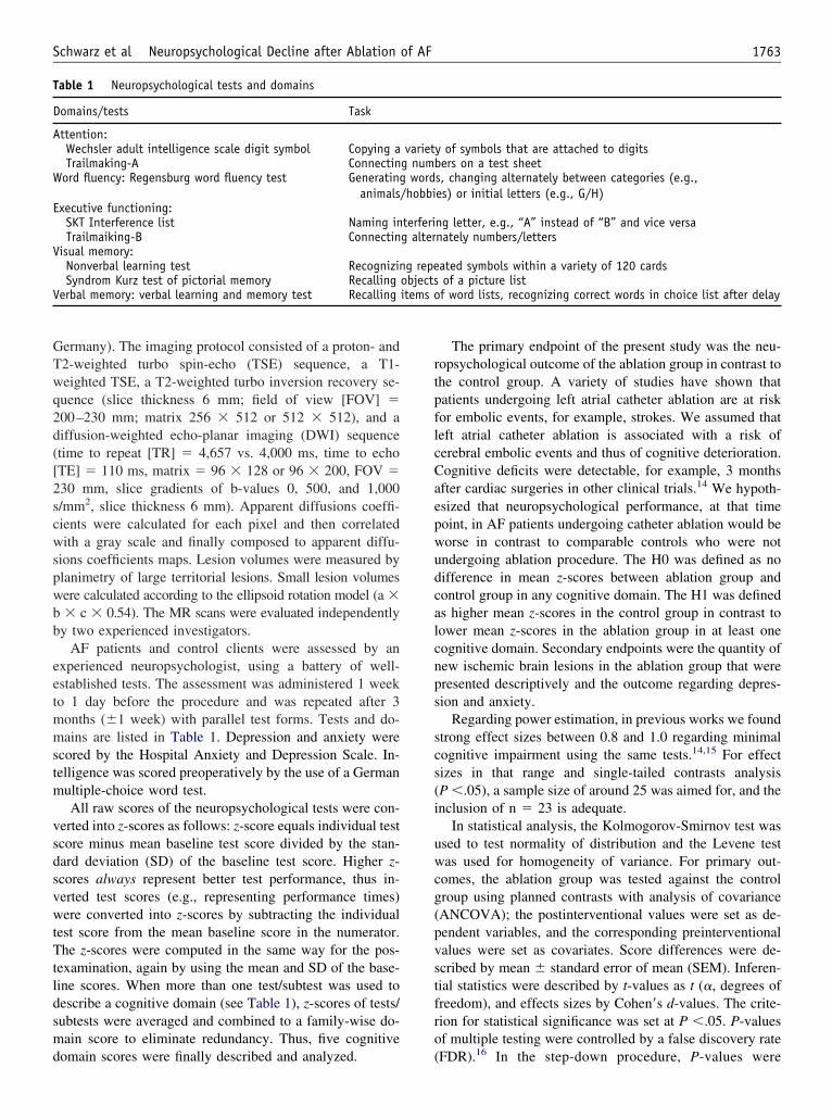
GTwq2d([2scwspwbb
eetmmstm
vsdsvwtTtldsmd
rtpflcCaepwudcalcnps
scs(i
uwcg(pvstfro
T
D
A
W
E
V
V items o
1763Schwarz et al Neuropsychological Decline after Ablation of AF
ermany). The imaging protocol consisted of a proton- and2-weighted turbo spin-echo (TSE) sequence, a T1-eighted TSE, a T2-weighted turbo inversion recovery se-uence (slice thickness 6 mm; field of view [FOV] �00–230 mm; matrix 256 � 512 or 512 � 512), and aiffusion-weighted echo-planar imaging (DWI) sequencetime to repeat [TR] � 4,657 vs. 4,000 ms, time to echoTE] � 110 ms, matrix � 96 � 128 or 96 � 200, FOV �30 mm, slice gradients of b-values 0, 500, and 1,000/mm2, slice thickness 6 mm). Apparent diffusions coeffi-ients were calculated for each pixel and then correlatedith a gray scale and finally composed to apparent diffu-
ions coefficients maps. Lesion volumes were measured bylanimetry of large territorial lesions. Small lesion volumesere calculated according to the ellipsoid rotation model (a �� c � 0.54). The MR scans were evaluated independently
y two experienced investigators.AF patients and control clients were assessed by an
xperienced neuropsychologist, using a battery of well-stablished tests. The assessment was administered 1 weeko 1 day before the procedure and was repeated after 3onths (�1 week) with parallel test forms. Tests and do-ains are listed in Table 1. Depression and anxiety were
cored by the Hospital Anxiety and Depression Scale. In-elligence was scored preoperatively by the use of a Germanultiple-choice word test.All raw scores of the neuropsychological tests were con-
erted into z-scores as follows: z-score equals individual testcore minus mean baseline test score divided by the stan-ard deviation (SD) of the baseline test score. Higher z-cores always represent better test performance, thus in-erted test scores (e.g., representing performance times)ere converted into z-scores by subtracting the individual
est score from the mean baseline score in the numerator.he z-scores were computed in the same way for the pos-
examination, again by using the mean and SD of the base-ine scores. When more than one test/subtest was used toescribe a cognitive domain (see Table 1), z-scores of tests/ubtests were averaged and combined to a family-wise do-ain score to eliminate redundancy. Thus, five cognitive
able 1 Neuropsychological tests and domains
omains/tests Task
ttention:Wechsler adult intelligence scale digit symbol Copying aTrailmaking-A Connectin
ord fluency: Regensburg word fluency test Generatinganimals
xecutive functioning:SKT Interference list Naming inTrailmaiking-B Connectin
isual memory:Nonverbal learning test RecognizinSyndrom Kurz test of pictorial memory Recalling
erbal memory: verbal learning and memory test Recalling
omain scores were finally described and analyzed. (
The primary endpoint of the present study was the neu-opsychological outcome of the ablation group in contrast tohe control group. A variety of studies have shown thatatients undergoing left atrial catheter ablation are at riskor embolic events, for example, strokes. We assumed thateft atrial catheter ablation is associated with a risk oferebral embolic events and thus of cognitive deterioration.ognitive deficits were detectable, for example, 3 monthsfter cardiac surgeries in other clinical trials.14 We hypoth-sized that neuropsychological performance, at that timeoint, in AF patients undergoing catheter ablation would beorse in contrast to comparable controls who were notndergoing ablation procedure. The H0 was defined as noifference in mean z-scores between ablation group andontrol group in any cognitive domain. The H1 was defineds higher mean z-scores in the control group in contrast toower mean z-scores in the ablation group in at least oneognitive domain. Secondary endpoints were the quantity ofew ischemic brain lesions in the ablation group that wereresented descriptively and the outcome regarding depres-ion and anxiety.
Regarding power estimation, in previous works we foundtrong effect sizes between 0.8 and 1.0 regarding minimalognitive impairment using the same tests.14,15 For effectizes in that range and single-tailed contrasts analysisP �.05), a sample size of around 25 was aimed for, and thenclusion of n � 23 is adequate.
In statistical analysis, the Kolmogorov-Smirnov test wassed to test normality of distribution and the Levene testas used for homogeneity of variance. For primary out-
omes, the ablation group was tested against the controlroup using planned contrasts with analysis of covarianceANCOVA); the postinterventional values were set as de-endent variables, and the corresponding preinterventionalalues were set as covariates. Score differences were de-cribed by mean � standard error of mean (SEM). Inferen-ial statistics were described by t-values as t (�, degrees ofreedom), and effects sizes by Cohen=s d-values. The crite-ion for statistical significance was set at P �.05. P-valuesf multiple testing were controlled by a false discovery rate
y of symbols that are attached to digitsers on a test sheet
s, changing alternately between categories (e.g.,es) or initial letters (e.g., G/H)
ng letter, e.g., “A” instead of “B” and vice versanately numbers/letters
ated symbols within a variety of 120 cardsof a picture listf word lists, recognizing correct words in choice list after delay
varietg numb
word/hobbi
terferig alter
g repeobjects
FDR).16 In the step-down procedure, P-values were

r
j
v
at�wn
RBaci
Tcnp1
(MdOat
pai(i
wspi
tSgw
T
AFEDAHAR
CR
T
P
P
H
Fil
1764 Heart Rhythm, Vol 7, No 12, December 2010
anked in ascending order, and null hypothesis was re-
ected if: p�i� �i
m�, where m was the number of P-
alues and i was the rank of the P-value.Regarding other group differences (e.g., baseline char-
cteristics), continuous variables were evaluated by simple-tests. Categorical variables were analyzed using Pearson=s-test. Baseline variables and other medical characteristicsere described by mean � SD, and categorical data byumber of cases and percentages.
esultsaseline characteristics of the ablation and control groupsre presented in Table 2. The groups did not differ statisti-ally (P �.05) regarding age, gender, education, and med-cal characteristics (besides recurrent AF).
Procedural and cardiac characteristics are presented inable 3. RF ablation was performed in 13 patients (56.5%),ryoablation in nine patients (39.1%), and combined tech-iques of ablation in one patient (4.3%). Mean � SD totalrocedure time was 240.3 � 78.8 minutes, ranging from65 to 510 minutes.
MRI at all time points was obtained in 21 patients91.3%). Two patients (8.7%) refused a postprocedural
RI. In 3/21 patients (14.3%), new ischemic lesions wereetected on DWI within the first week after the ablation.ne out of 21 patients (4.8%) suffered a major stroke,
ffecting large portions of the left middle cerebral arteryerritory, including the basal ganglia (Figure 1). The patient
able 2 Baseline and medical characteristics
Ablation group Control group
ge, mean � SD 54.2 � 9.2 58.5 � 5.4emale gender 10 (43.5) 13 (56.5)ducation, mean � SD, years 10.4 � 2.1 10.8 � 2.6iabetes 1 (4.3) 1 (4.3)rterial hypertension 8 (34.8) 10 (43.5)ypercholesterinemia 6 (26.1) 5 (21.7)dipositas 2 (8.7) 2 (8.7)educed left ventricularejection �60%
3 (13.0) 0 (0)
oronary artery disease 1 (4.3) 0 (0)ecurrent AF 23 (100) 0 (0)
Note: Data in parentheses are percents.
able 3 Intra- and postprocedural characteristics
Range, minimum/maximum
rocedure (%):Cryo 9 (39.1) —RF ablation 13 (56.5) —Combined 1 (4.3) —
rocedure time, mean �SD, minutes
240.3 � 78.8 165/510
eparin amount, mean � 24,950 � 30,003 13,000/120,000
2SD, IU
resented symptoms of contralateral hemiplegia and severephasia. In two (9.5%) out of 21 patients, clinically silentschemic lesions of 4 and 5 mm in diameter were detectedFigure 2). Three months after the procedure no additionalschemic lesions were detected in DWI or T2W MRI.
Pre- and postprocedural neuropsychological examinationas obtained in 21 patients (91.3%). The patient who had
uffered a major stroke was not assessable at follow-up. Oneatient (4.3%) refused a second neuropsychological exam-nation.
Baseline neuropsychological performance was similar inhe ablation and in the control group (Table 4). Only mean �EM scores in word fluency differed between the ablationroup (�0.50 � 0.20) and the control group (0.39 � 0.19),herein the controls showed better z-scores [t (.05; 42) �
igure 1 Territorial stroke after catheter ablation. A: DWI: ischemicnfarct in the region of the left artery cerebral media. B: MR angiography:eft middle cerebral artery occlusion.
.89; P � .006].

ri
sc0da�
latMw(wf
eAgta
DTcnt
csiwasrsssMnmrn
Fep
T
D
AWEVVDA
1765Schwarz et al Neuropsychological Decline after Ablation of AF
Results at 3 months are presented in Table 5. As a mainesult, the ablation group declined from their baseline leveln the verbal memory domain, whereas the control group
igure 2 DWI: two patients displayed small and clinically silent isch-mic lesions (A: subcortical; B: cortical), detected 2 days after the ablationrocedure.
able 4 Mean z-scores � SEM of neuropsychological domains a
Ablation group
omain Baseline
ttention/concentration �0.17 � 0.25ord fluency �0.50 � 0.20xecutive functioning �0.23 � 0.18isual memory 0.12 � 0.19erbal memory �0.18 � 0.20epression 4.24 � 3.55nxiety 7.95 � 3.98
Note: Higher z-scores always represent better performance. Higher raw scores
howed practice effects (Table 5, Figure 3). The statisticalontrast with a mean difference of �0.69 in z-scores (SEM �.19) between the groups displayed a strong effect size of� 0.93 in the ANCOVA [t (.05; 42) � �3.53; P �.001],
nd the P-level fit the alpha-correction criteria (FDRcrit
.01). Overall, 56.5% of ablation patients deteriorated (ateast mildly) from their baseline values in verbal memory,s compared with 17.4% of controls. The deterioration inhe ablation group ranged from �0.2 to �1.2 in z-scores.
ild dropdown (�0.4 in z-scores) was seen in one patientith evidence of microlesion but not in the second patient
�0.5 in z-scores) with evidence of microlesion. The patientith severe media infarct was excluded from cognitive
ollow-up assessment.Regarding depression and anxiety, no baseline differ-
nces between the groups were detected in t-tests (P �.05).t 3 months (and in covariance of baseline scores), noroup differences were found, thus the ablation group andhe control group did not differ regarding depression andnxiety (Table 5).
iscussionhe present study was conducted to determine neurologicalomplications and—for the first time—to detect possibleeuropsychological changes after catheter ablation in thereatment of AF.
Ischemic stroke is a rare but serious complication ofatheter ablation.3–5 Comparable to previously publishederies, one patient (4.3%) suffered a large territorial stroken our cohort. Furthermore, we detected two patients (9.5%)ith subclinical ischemic lesions on DWI, comparable to
nother catheter ablation study.9 The occurrence of strokes,ubclinical ischemic lesions, and TIA suggests an increasedisk for cerebral embolism during the procedure. This as-umption is corroborated by transcranial Doppler ultrasoundtudies for the detection of periprocedural microembolicignals. Sauren et al10 found 3,908 � 2,816 (mean � SD)ES within the basal cerebral arteries during the percuta-
eous ablation process—a number that is comparable to theicroembolic load during heart surgery using extracorpo-
eal circulation.17,18 Although the composition of MES can-ot be determined beyond a doubt by means of ultrasound,19
an raw scores � SD of depression/anxiety
Control group
ths Baseline 3 months
� 0.23 0.15 � 0.23 0.19 � 0.20� 0.22 0.39 � 0.19 0.17 � 0.21� 0.16 0.21 � 0.17 0.24 � 0.15� 0.20 �0.10 � 0.17 �0.29 � 0.18� 0.17 �0.23 � 0.19 0.36 � 0.16� 3.10 3.35 � 2.31 3.26 � 2.43� 3.92 5.96 � 2.90 5.30 � 2.79
nd me
3 mon
�0.05�0.20
0.07�0.03�0.30
3.106.14
represent more symptoms of depression/anxiety.

at
rbscmee
nimcmgtitaKsa
mptefdnnctemhhomsinwst
vliDbtstcrb
loatc
ta
T
D
AWEVVDA
b
Farp
1766 Heart Rhythm, Vol 7, No 12, December 2010
mixture of gaseous and solid microemboli can be assumedo be the cause of the high-intensity Doppler signals.14
The magnitude of cerebral microemboli and the occur-ence of strokes and TIAs suggest that neurological hazardseyond territorial stroke—probably similar to major cardiacurgery—can be expected during the catheter ablation pro-ess. During and after catheter ablation, thrombus formationight result from platelet and coagulation system activation
ither directly at the catheter material or at the site ofndothelial application.8,9
The present data support this risk apprehension. Besidesew ischemic lesions on DWI, we detected adverse changesn one cognitive domain. In “verbal memory functions,” theean performance of the ablation group declined, while the
ontrols showed strong practice effects. Practice effectsean learning, and the absence of learning in the ablation
roup refers probably to a cognitive deficit.20,21 However,his effect was seen in only one out of five domains. Theres variety of possible explanations for the lack of effects inhe other domains. One possible explanation is methodicalnd could refer to a specific sensitivity of the particular test.ilminster et al22 found the same test (in an English ver-
ion) to be the only measure in large variety of tests that wasble to detect cognitive decline after cardiac surgeries, com-
able 5 Planned contrasts with ANCOVA between ablation and
omain Degrees of freedom Mean
ttention/concentration 40 �0.0ord fluency 41 0.3xecutive functioning 42 0.0isual memory 41 0.1erbal memory 42 �0.6epression 42 �0.6nxiety 42 �0.5
aIf P �FDR, H1 was accepted.Effect size d � 0.93.
igure 3 Verbal memory functioning before and 3 months after theblation (mean z-scores). The ablation group (blue continuous line) dete-iorates, whereas the control group (red dashed line) improves owing to
lractice effects.
enting, “prompted to pick the most discriminating neuro-sychological test post hoc, the Rey memory test would behe obvious choice” (p. 1,873). Another explanation, how-ver, could be a special vulnerability of the specific brainunction (memory) after subtle cerebral damages after car-iac interventions. It is interesting that the pattern of cog-itive changes in the present study closely resembles theeuropsychological findings after major heart surgery. Re-ently, Gerriets et al14 described substantial changes, par-icularly in memory functions after bypass surgery. A vari-ty of other studies indicated that in particular this domainight be prone to minimal cerebral injury in the context of
eart surgeries.22,23 It is possible that minimal ischemia orypoxia rapidly impairs circuits of hippocampus/temporalr frontal lobe structures that are associated with declarativeemory functions due to reduced blood flow and/or oxygen
upply. Regarding cardiac surgery, however, a variety ofnterventional strategies were shown to be useful in mitigatingeurological and neuropsychological side effects, especiallyhen reducing microembolic events.14,24,25 These findings
uggest that microembolism seems to be an important aspect ofhe underlying pathophysiology.
Interestingly, in the current study, the decline of scores inerbal memory functions, as compared with the baselineevel, occurred in the majority of ablation patients, but onlyn one out of the two cases with evidence of microlesions.ecline was thus not explainable by evidence of microem-olic lesion as detected on MRI. However, it is possible thathese cases represent only the “tip of the iceberg” and thatmall lesions are often beyond the MRI resolution. Alterna-ively, decline in cognitive functions is multifactorial and notorrelated to focal lesions only. We described similar resultsecently in samples of patients undergoing coronary arteryypass grafting (CABG).14,15
The present paper identifies a hitherto unknown neuro-ogical side effect of catheter ablation. Although the mem-ry deficits described here are not severe and appear not toffect the activities of daily living in a significant manner,his adverse effect needs to be taken into account in theonsideration of the pros and cons for PVI.
There are certain limitations to the current study. Al-hough the analysis was powerful enough to detect inter-ction effects between ablation and control groups, a
l group of neuropsychological domains and depression/anxiety
nce SEM t P P �FDR?a
0.22 �0.18 .856 H00.23 1.53 .133 H00.18 0.28 .780 H00.23 0.54 .593 H00.19 b�3.53 �.001 H10.65 �1.06 .296 H00.72 �0.78 .442 H0
contro
differe
4552996
arger sample size would be an overall advantage. More-
ohtdtHcttssptop
R
1
1
1
1
1
1
1
1
1
1
2
2
2
2
2
1767Schwarz et al Neuropsychological Decline after Ablation of AF
ver, the absence of follow-up MRI in two patients is aandicap for the estimation of the true lesion rate and ofhe impact of embolic events on the cognitive outcome. Aisadvantage is the choice of healthy volunteers as con-rols instead of AF patients without catheter ablation.owever, owing to heterogeneity of treatment and un-
ertainty in regard to which treatment (ablation, medica-ion) will be chosen within the time frame of follow-up,he inclusion of such a control group has not been pos-ible in our center. Further studies with larger sampleizes, longer observation time, and a control group ofatients with AF who are not subjected to catheter abla-ion are required to determine an overall risk-benefit analysisf catheter ablation with respect to stroke incidence, cardiacerformance, neuropsychology, and quality of life.
eferences1. Fuster V, Ryden LE, Cannom DS, et al. ACC/AHA/ESC 2006 guidelines for the
management of patients with atrial fibrillation—executive summary: a report ofthe American College of Cardiology/American Heart Association Task Force onPractice Guidelines and the European Society of Cardiology Committee forPractice Guidelines (Writing Committee to Revise the 2001 Guidelines for theManagement of Patients With Atrial Fibrillation). J Am Coll Cardiol 2006;48:854–906.
2. Haissaguerre M, Jais P, Shah DC, et al. Spontaneous initiation of atrial fibril-lation by ectopic beats originating in the pulmonary veins. N Engl J Med1998;339:659–666.
3. Neumann T, Vogt J, Schumacher B, et al. Circumferential pulmonary veinisolation with the cryoballoon technique results from a prospective 3-centerstudy. J Am Coll Cardiol 2008;52:273–278.
4. Marrouche NF, Dresing T, Cole C, et al. Circular mapping and ablation of thepulmonary vein for treatment of atrial fibrillation: impact of different cathetertechnologies. J Am Coll Cardiol 2002;40:464–474.
5. Cappato R, Calkins H, Chen SA, et al. Worldwide survey on the methods,efficacy, and safety of catheter ablation for human atrial fibrillation. Circulation2005;111:1100–1105.
6. Rodgers M, McKenna C, Palmer S, et al. Curative catheter ablation in atrialfibrillation and typical atrial flutter: systematic review and economic evaluation.Health Technol Assess 2008;12:iii–iv, xi–xiii, 1–198.
7. Cappato R, Calkins H, Chen SA, et al. Prevalence and causes of fataloutcome in catheter ablation of atrial fibrillation. J Am Coll Cardiol 2009;53:1798 –1803.
8. Lee DS, Dorian P, Downar E, et al. Thrombogenicity of radiofrequency2
ablation procedures: what factors influence thrombin generation? Europace2001;3:195–200.
9. Lickfett L, Hackenbroch M, Lewalter T, et al. Cerebral diffusion-weightedmagnetic resonance imaging: a tool to monitor the thrombogenicity of left atrialcatheter ablation. J Cardiovasc Electrophysiol 2006;17:1–7.
0. Sauren LD, Van Bell Y, DeRoy L, et al. Transcranial measurement of cerebralmicroembolic signals during endocardial pulmonary vein isolation: comparisonof three different ablation techniques. J Cardiovasc Electrophysiol 2009;20:2201–2207.
1. Gao L, Taha R, Gauvin D, Othmen LB, Wang Y, Blaise G. Postoperativecognitive dysfunction after cardiac surgery. Chest 2005;128:3664–3670.
2. Newman MF. Open heart surgery and cognitive decline. Cleve Clin J Med2007;74(Suppl 1):S52–55.
3. Selnes OA, McKhann GM, Borowicz LM Jr., Grega MA. Cognitive and neu-robehavioral dysfunction after cardiac bypass procedures. Neurol Clin 2006;24:133–145.
4. Gerriets T, Schwarz N, Sammer G, et al. Protecting the brain from gaseous andsolid micro-emboli during coronary artery bypass grafting: a randomized con-trolled trial. Eur Heart J 2010;31:360–368.
5. Gerriets T, Schwarz N, Bachmann G, et al. Evaluation of methods to predictearly long-term neurobehavioral outcome after coronary artery bypass grafting.Am J Cardiol 2010;105:1095–1101.
6. Benjamini Y, Hochberg Y. Controlling the false discovery rate: a practical andpowerful approach to multiple testing. J R Stat Soc 1995;57:289–300.
7. Schoenburg M, Kraus B, Muehling A, et al. The dynamic air bubble trap reducescerebral microembolism during cardiopulmonary bypass. J Thorac CardiovascSurg 2003;126:1455–1460.
8. Borger MA, Peniston CM, Weisel RD, Vasiliou M, Green RE, Feindel CM.Neuropsychologic impairment after coronary bypass surgery: effect of gaseousmicroemboli during perfusionist interventions. J Thorac Cardiovasc Surg 2001;121:743–749.
9. Schoenburg M, Baer J, Schwarz N, et al. EmboDop: insufficient automaticmicroemboli identification. Stroke 2006;37:342–343.
0. Keith JR, Puente AE. Deficiencies in the detection of cognitive deficits. Neu-ropsychology 2002;16:434–439.
1. Keith JR, Puente AE, Malcolmson KL, Tartt S, Coleman AE, Marks HF Jr.Assessing postoperative cognitive change after cardiopulmonary bypass surgery.Neuropsychology 2002;16:411–421.
2. Kilminster S, Treasure T, McMillan T, Holt DW. Neuropsychological changeand S-100 protein release in 130 unselected patients undergoing cardiac surgery.Stroke 1999;30:1869–1874.
3. Knipp SC, Matatko N, Wilhelm H, et al. Evaluation of brain injury aftercoronary artery bypass grafting. A prospective study using neuropsychologicalassessment and diffusion-weighted magnetic resonance imaging. Eur J Cardio-thorac Surg 2004;25:791–800.
4. Pugsley W, Klinger L, Paschalis C, Treasure T, Harrison M, Newman S. Theimpact of microemboli during cardiopulmonary bypass on neuropsychologicalfunctioning. Stroke 1994;25:1393–1399.
5. Newman SP, Harrison MJ. Coronary-artery bypass surgery and the brain: per-sisting concerns. Lancet Neurol 2002;1:119–125.