Embed Size (px)
Citation preview
Santosh Kumar Panda et al, J. Global Trends Pharm Sci, 2017; 8(4): 4455 - 4466
4455
NEUROPROTECTIVE ACTIVITY OF METHANOLIC EXTRACT OF ALPINEA
GALANGA AGAINST EXCITOTOXICITY PRODUCED BY PTZ-INDUCED
KINDLING MODEL IN MICE
Santosh Kumar Panda*1, A. Venkateshwar Reddy2.
1Research Scholar, Singhania University, Pacheri Bari, Jhunjhunu, Rajasthan-333515.
2 Anwarul Uloom College of Pharmacy, New Mallepally, Hyderabad-500001.
*Corresponding Author E-mail: [email protected].
ARTICLE INFO ABSTRACT
Key Words
Antiepileptogenic;
Neuroprotective;
Oxidative stress;
MEAG; Diazepam;
PTZ; kindled;
Seizure scores.
Alpinea galanga has been used for years together for its medicinal
properties in Chinese medicine system. The use of antiepileptic drugs
(AEDs) having neuroprotective action and its possible role in disease
modification (i.e., antiepileptogenesis) is gaining interest day by day.
The present study aimed to investigate the antiepileptogenic activity of
methanolic extract of Alpinea galanga rhizomes (MEAG) and
subsequently its neuroprotective action against excitotoxicity and
reactive oxygen species (ROS) generation in the brain of
pentylenetetrazole (PTZ) –induced kindled mice. MEAG was studied for
its suppressive action on convulsion and seizure scores
(antiepileptogenic effect) and protection of neurons against PTZ-induced
oxidative stress injury (neuroprotective effect) when given during
kindling acquisition as a pretreatment before every PTZ injection. A
major antiepileptic drug Diazepam was also tested for comparison.
MEAG and Diazepam showed antiepileptogenic activity as they caused
reduction in development of seizure scores and reduced the sensitivity of
kindled mice to the convulsive and lethal effects of PTZ and MEAG was
found to be superior to Diazepam in preventing seizure scores. MEAG
and Diazepam significantly decreased oxidative stress injury in the mice
brain as compared to PTZ-kindled group. MEAG showed more
neuroprotective action as compared to Diazepam as it enhanced the
antioxidant enzyme levels in the mice brain. The results obtained from
the study support the hypothesis that neuroprotective action of MEAG
not only correlate with its ability to inhibit ROS formation but also for
its ability to suppress seizure generation.
INTRODUCTION
A drug with improved anticonvulsant and
anti-epileptogenic efficacy is need of the
hour as currently available antiepileptic
drugs failed to give adequate protection
against epileptic seizures in one-third of
epilepsy patients as epilepsy still remains
one of the major neurological disorders for
which safer drugs are awaited. [1]. During
seizures prolonged over excitation of
neurons leads to neuronal injury and even
An Elsevier Indexed Journal ISSN-2230-7346
Journal of Global Trends in Pharmaceutical Sciences
Santosh Kumar Panda et al, J. Global Trends Pharm Sci, 2017; 8(4): 4455 - 4466
4456
death due to some poorly understood
underlying biochemical mechanism. The
currently available antiepileptic drugs also
failed to control progressive epileptogenic
consequences like neurodegeneration[2].
Accumulating research evidence supports
the hypothesis that oxidative stress during
experimental epilepsy leads to abnormal
structure alteration of membrane lipids,
cellular proteins, DNA and RNA [3] [4].
Molecular oxygen originating from
different sources like mitochondria by
different biochemical processess gives rise
to reactive oxygen species (ROS).
Superoxide dismutase (SOD), an
intracellular antioxidant enzyme rapidly
and specifically reduces O2- to hydrogen
peroxide (H2O2). Another antioxidant
enzyme, Glutathione peroxidase (GPx) acts
on H2O2 to detoxify it to water [5]. Catalase,
the most common antioxidant enzyme
found in living organism also catalyzes the
decomposition of H2O2 to water and
oxygen [6]. It is the most common enzyme
protecting the cells and neurons from
oxidative damage by ROS. One catalase
molecule can convert millions of H2O2 to
water and oxygen every second. The
oxidant-antioxidant system works in
equilibrium in normal conditions.
However an inbalance between the
systems causes tissue and neuronal
damage by oxidative stress. ROS results in
lipid peroxidation by attacking membrane
lipids. Lipid peroxidation end product
Malondialdehyde (MDA) thus serves as an
index for tissue damage by lipid
peroxidation. Nitric oxide (NO) role in
cellular protection is contraindicatory as
sometimes it acts as an oxidant and
sometimes as a scavenger of O2-[7] [8].
Oxidative injury to brain is the major
pathway of neuronal damage in many
acute and chronic neuronal disorders like
epilepsy, Parkinson’s disease and
Alzheimer’s disease[9][10]. Therefore, the
current trend is to look for a novel
antiepileptic drug having antioxidant
property. The novel drug should give
protection against epilepsy, prevent
neurodegeneration by ROS and have
neuroprotection activity. This will not only
give a alteration to the treatment of
epilepsy but will also support the
hypothesis on free radicals crucial role on
the pathogenesis of brain damage by
neurotoxins[11].
Alpinea galanga , also called as
jujube or Indian date is a commom plant in
India belonging to the family
Zingiberaceae. Many investigations on its
pharmacological activity justify its
traditional therapeutic value. It has CNS
depressant activity as it is used in many
Ayurvedic preparations. Also it is reported
to have antioxidant property as it is a rich
source of flavonoids[12]. Therefore, the
present study was undertaken to evaluate
the neuroprotective activity of methanolic
extract of Alpinea galanga (MEAG)
rhizomes on experimentaly induced PTZ-
kindled model in mice and also to evaluate
whether administration of MEAG gives
protection against PTZ induced seizures in
mice. Also, by the end of the study we will
be able to compare Diazepam with MEAG
for its antiepileptic and neuroprotective
activity as former is a major antiepileptic
drug, biochemically and clinically.
Materials and Methods
Plant
The Alpinea galanga rhizomes were
purchased from YUCCA Enterprises,
MUMBAI. Then it was authenticated in
Botanical Survey of India, Deccan
Regional Centre, Hyderabad, Government
of India. The plant material collected was
shade dried to retain its important
phytoconstituents and then subjected to
size reduction and powder preparation for
further extraction process.
Animals
Swiss Albino mice of either sex weighing
between 25 to 30 g were procured from
Sainath Agencies, Hyderabad. Animals
were housed at an ambient temperature of
Santosh Kumar Panda et al, J. Global Trends Pharm Sci, 2017; 8(4): 4455 - 4466
4457
25±1ºC and 12hr/12hr light dark cycle in
polypropylene cages. Animals were
acclimatized to lab condition for 7 days
before starting of the experiment. Animals
were fed with standard diet and water ad
libitum. All animal studies were performed
in accordance to guideline of CPCSEA
and Institutional Animal Ethical
Committee (IAEC) of Anwarul Uloom
College of Pharmacy, Hyderabad
(CPCSEA Reg. No. 1534/PO/Re/S/2011
/CPCSEA) and permission letter Ref.
No. IAEC/AUCOP/2016/02.
Preparation of Plant Extract
The rhizomes were dried in shade and
coarsely powdered. The powder was
passed through sieve no. 16.
Approximately 200 g of powder was
extracted in 60% methanol using a soxhlet
apparatus. The extract mass was weighed
in a digital balance and the % yield was
calculated. The extract was kept in a
beaker covered with alluminium foil
labeled and kept in freeze for future use.
The methanol extracts of Alpinea galanga
(MEAG) rhizomes were subjected to the
following investigations: Preliminary
phytochemical screening, Determination of
maximum tolerated dose in animals,
Antiepileptogenic and Neuroprotective
activity.
Preliminary phytochemical screening of
extracts
The extracts were subjected to different
chemical tests to detect the chemical
constituents present in them. 0.5 gm of
extract was dissolved in 5 ml of distilled
water and filtered. The filtrate was used to
determine the presence of various
phytoconstituents[13][14].
Determination of Maximum Tolerated
Dose
The literature survey showed that
the rhizomes of Alpinea galanga and their
various extract were used of several
studies. Hence, on the basis of available
information on the plant, limit test for
crude methanolic extract of rhizomes of
Alpinea galanga (MEAG) was conducted
at the highest starting dose level i.e. 2000
mg/ kg of body weight. Three female mice
were fasted over night with free access to
water. Each animal received single dose of
MEAG (2000 mg/kg, orally). After
administration of the extract, animals were
observed for any mortality, morbidity,
body weight and signs of toxicity for 14
days (OECD guidelines 423, 2000, for
testing of chemicals).
Procedure for induction of kindled
seizures
The experiment was performed according
to the method described by Erakovic[15].
Swiss Albino mice were grouped into 6,
each consisting of 6 animals, weighing 25-
30 g each. The first three groups served as
non-kindling groups and received normal
saline (2 ml/kg) (orally), diazepam (2
mg/kg) (orally) and plant extract (600
mg/kg of MEAG) (orally) respectively on
1,3,5,8,10,12, 15,17,19,22,24 and 26 days
of treatment. The next three groups served
as kindling groups and received PTZ (35
mg/kg) (s.c.) 30 minutes after treatment.
On the 26 th day a PTZ dose of 75 mg/kg
s.c. were given to the three kindled groups.
Kindling was produced by a total of 11
treatments with PTZ (35 mg/kg) (s.c.) on
every second day (Monday, Wednesday
and Friday). Animals were observed for 30
minutes after the last drug administration
and for an additional 30 minutes for
lethality before returning to the home cage.
Seizure intensity was evaluated using the
following modified scale.
0. No response.
1. Ear and facial twitching.
2. Convulsive waves axially through
the body.
3. Myoclonic body jerks.
4. Generalized clonic convulsions,
turns over into side position.
Santosh Kumar Panda et al, J. Global Trends Pharm Sci, 2017; 8(4): 4455 - 4466
4458
5. Generalized convulsions with tonic
extension episode and status
epilepticus.
6. Mortality.
The animals were considered to be
kindled after having received 11 PTZ
injections and having reached at least three
consecutive stage 4 or 5 seizures. After
completion of the study the animals were
sacrificed by cervical dislocation and brain
will be removed for evaluation of
neuroprotective activity.
Sample preparation and biochemical
estimation
Brain Homogenate
Animals were sacrificed by cervical
dislocation; brains were removed quickly
and frozen for biochemical analysis.
Brains were homogenized in ten volumes
of ice cold Tris-HCl buffer (50 mM, pH
7.4). The homogenate sample was taken
for estimation of lipid peroxidation
(MDA), nitric oxide (NO). The rest of the
homogenate were centrifuged at 1500 rpm
for 15 minutes at 4 ºC and the supernatant
thus obtained were used for the estimation
of SOD and Catalase[16]. Estimation of
Glutathione peroxidase (GPx) was done
seperately following the procedure given
by Paglia[17].
Total Protein
The phenolic group of tyrosine and
tryptophan residues (amino acids) in a
protein will produce a blue purple colour
complex, with maximum absorption in the
region of 660 nm wavelength, with Folin-
Ciocalteau reagent which consists of
sodium tungstate molybdate and
phosphate. Different dilutions of BSA
solutions were prepared by mixing stock
BSA solutions (5 mg/ml) and water in a
test tube and the absorbance was measured
at 660 nm to get a standard calibration
curve. The concentration of unknown
sample was determined from standard
graph.18].
Malondialdehyde (MDA)
MDA reacts with thiobarbituric acid
(TBA) to generate a colored product,
which can be measured spectro
photometrically. 1 ml of sample was added
to 1 ml of 40% TCA followed by addition
of 2 ml of 0.67% TBA. The mixture was
kept 10 minutes in a boiling water bath,
cooled immediately in ice-cold water bath,
centrifuged at 6,000 rpm for 30 seconds
and absorbance of supernatant was
recorded at 530 nm. Then MDA was
calculated based on molar extinction
coefficient (offered by 1M solution) i.e.
(1.54 x 10⁵ M-1cm-1) and the formula
A=KCT [19].
Superoxide Dismutase (SOD)
This method was based on the ability of
superoxide dismutase to inhibit the auto
oxidation of pyrogallol. For Control 2.9 ml
of Tris buffer, 0.1 ml of pyrogallol
solution was added and mixed, and reading
was taken at 420 nm exactly after 1 minute
30 seconds and 3 minutes 30 seconds. For
Sample 2.8 ml of Tris buffer, 0.1 ml of
sample solution was added and mixed, and
the reaction was started by adding 0.1 ml
of pyrogallol solution. Reading was taken
same as control and calculation was done
using the formula. Units of SOD/3 ml of
assay mixture = (A-B) x 100/A x 50,
where A and B are initial and final
absorbance reading of control and
sample[20].
Catalase
The decomposition of H2O2 can be
followed directly by decrease in
absorbance at 240 nm. The difference in
absorbance (A) per unit time is a measure
of catalase activity. For sample 1 ml of the
homogenized tissue was mixed with 2 ml
of the H2O2 solution and mixed well, and
for blank 1 ml of the phosphate buffer was

Santosh Kumar Panda et al, J. Global Trends Pharm Sci, 2017; 8(4): 4455 - 4466
4459
mixed with 2 ml of H2O2 solution. The
reaction was started by addition of H2O2
and the change in absorption was noted
from 0 to 60 seconds. Catalytic activity
will be calculated by using the formula Z =
ΔA x V x 1000/€ x d x T[21].
Glutathione Peroxidase (GPx)
Glutathione peroxidase activity will be
measured by NADPH using a coupled
reaction system consisting of reduced
glutathione, glutathione reductase and
hydrogen peroxide. The brain tissue was
homogenized in 5 volumes of phosphate
buffer and centrifuged at 13,000 rpm for
10 minutes. The supernatent was
considered as the sample. In a tube 600 µl
of potassium phosphate buffer, 300 µl of
glutathione reduced and 300 µl of NADPH
was added. 300 µl of the sample was
added to the tube. 900 µl of distilled water
was added to make the volume 2700 µl.
The reaction was started by adding 300 µl
of 12 mM H2O2. Absorbance was
measured at 340 nm for 3 minutes.
Glutathione peroxidase activity will be
calculated by using the formula U = ΔA x
V x 1000/€ x d x T [22].
Nitric Oxide (NO)
This assay was based on the enzymatic
conversion of nitrate to nitrate reductase.
The reaction was followed by a
spectrophotometric detection of nitrite as
an azo dye product of the Griess reaction,
which absorbs light at 540 nm. 100 µL of
Griess reagent, 300 µL of the nitrite
containing sample and 2.6 µL of deionized
water will be mixed in a
spectrophotometer cuvette. The mixture
was incubated for 30 minutes at room
temperature and absorbance was noted.
The concentration was determined from a
graph [23].
Statistical Analysis
The results will be expressed as
Mean±SEM. Statistical analysis of the
values observed in all the experimental
methods will be performed by ANOVA
followed by Dunnett’s multiple
comparison test. For statistical analysis
Microsoft Office Excel and Graphpad
Prism version 6.0 will be used. * p < 0.05
will be considered as statistically
significant.
Results
Phytochemical Examination of Extracts
Preliminary phytochemical analysis of
methanolic extract of Alpinea galanga
rhizomes showed presence of flavonoids,
steroids, terpenoids, tannins,
carbohydrates, proteins, fixed oils and
fats.
Maximum Tolerated Dose (MTD)
The animal does not show any signs of
toxicity even after 7 days of drug
administration and no mortality were
observed. Based on the observation 2000
mg/kg of the extract was found to be safe
and it is taken as the maximum tolerated
does. Three test doses 200 mg/kg, 400
mg/kg and 600 mg/kg were selected
arbitrarily based on the maximum tolerated
does (MTD) calculation.
Effect of Diazepam and MEAG on
induction of kindling by PTZ
All mice in the entire groups survived
without any complications at the end of the
kindling period. In the PTZk group,
repeated administration of a subconvulsant
dose of PTZ (35 mg/kg, s.c.) on every
second day (for 24 days, 11 injections)
resulted in increasing convulsive activity
leading to generalized clonic-tonic seizure
score of more than 5. Administration of
Diazepam at the dose of 2 mg/kg and
MEAG 600 mg/kg respectively did not
modify the course of kindling induced by
PTZ. However, treatment with MEAG
suppressed the kindled seizure as seen in
the decreases in mean seizure scores on the
Santosh Kumar Panda et al, J. Global Trends Pharm Sci, 2017; 8(4): 4455 - 4466
4460
study days (Fig.1). On the test day (PTZ,
75 mg/kg s.c.), PTZ-kindled mice
developed a classical pattern of limbic type
motor seizures with a mean seizure score 6
(Table 1). The convulsive response
consisted of first twitch, short-lasting
episodes of clonic seizures, and then
continuous clonic seizures with wild
running ending with falling of the animal
and tonic seizures. Pretreatment with
Diazepam and MEAG did not prevent the
development of seizure. Diazepam and
MEAG pretreated PTZ kindled animals
produced a seizure intensity which was
less than the saline-treated PTZ kindled
mice. Apart from this, the Diazepam and
MEAG PTZk group showed a higher
latent period as compared to the saline
treated PTZk group (p=<0.0001). On the
test day (day 26), after the treatment of
PTZ 75 mg/kg s.c. all the 6 animals in
the saline treated PTZk died while 2 out
of 6 animals survived in the Diazepam
PTZk group. All the 6 animals survived
in the MEAG PTZk group.
Kindling was induced by a total of 11
treatments with 35 mg/kg PTZ s.c. on
every second days (Monday, Wednesday,
and Friday). Diazepam pretreatment did
not a l t e r the course of kindling induced
by PTZ. There was no statistically
difference for mean seizure scores between
the PTZk groups and Diazepam PTZk group.
However, treatment with MEAG
suppressed the kindled seizure
significantly, as none of the animals could
achieve a score of 4 with 11 injections of
PTZ. At the end of the study the
Diazepam and MEAG PTZk group
attained a mean seizure score of 5 and 3.5
respectively while saline treated group
attained a score of 6.
Biochemical estimation of oxidant/anti-
oxidant stress markers
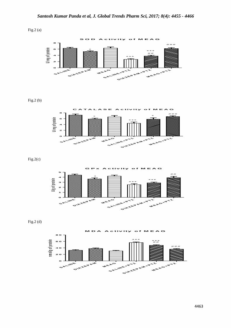
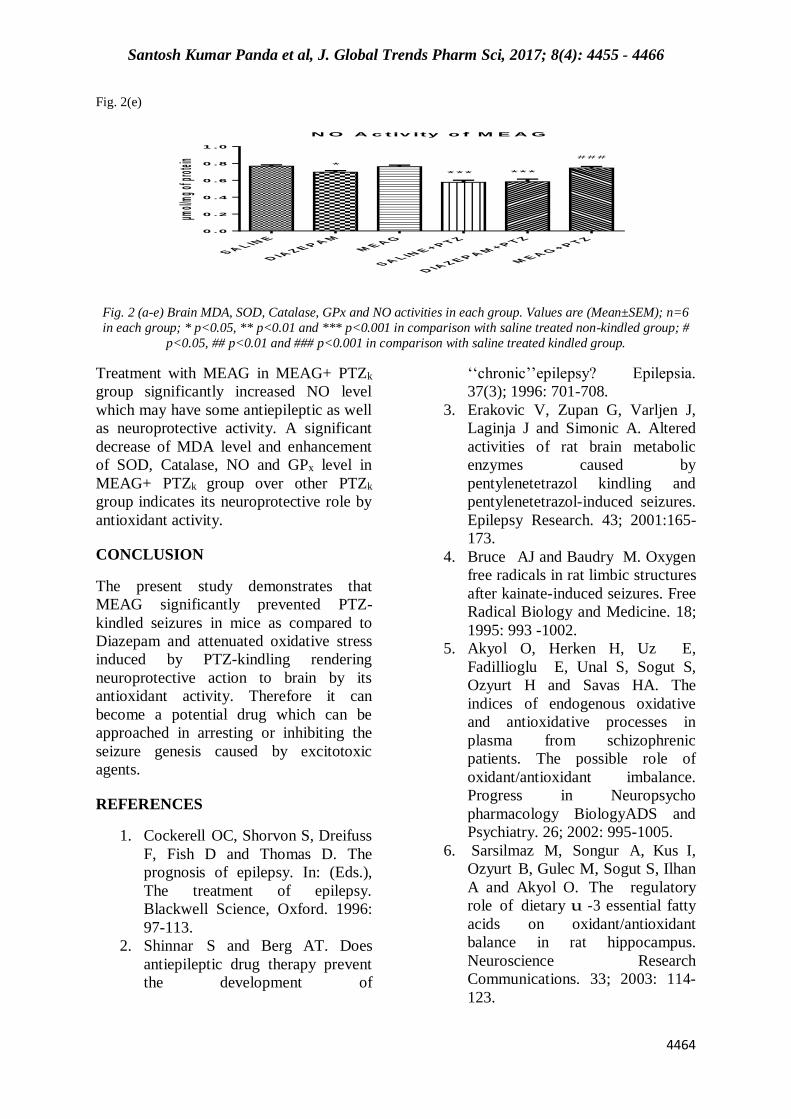
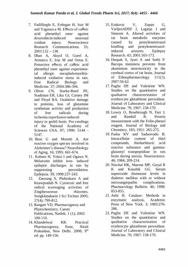
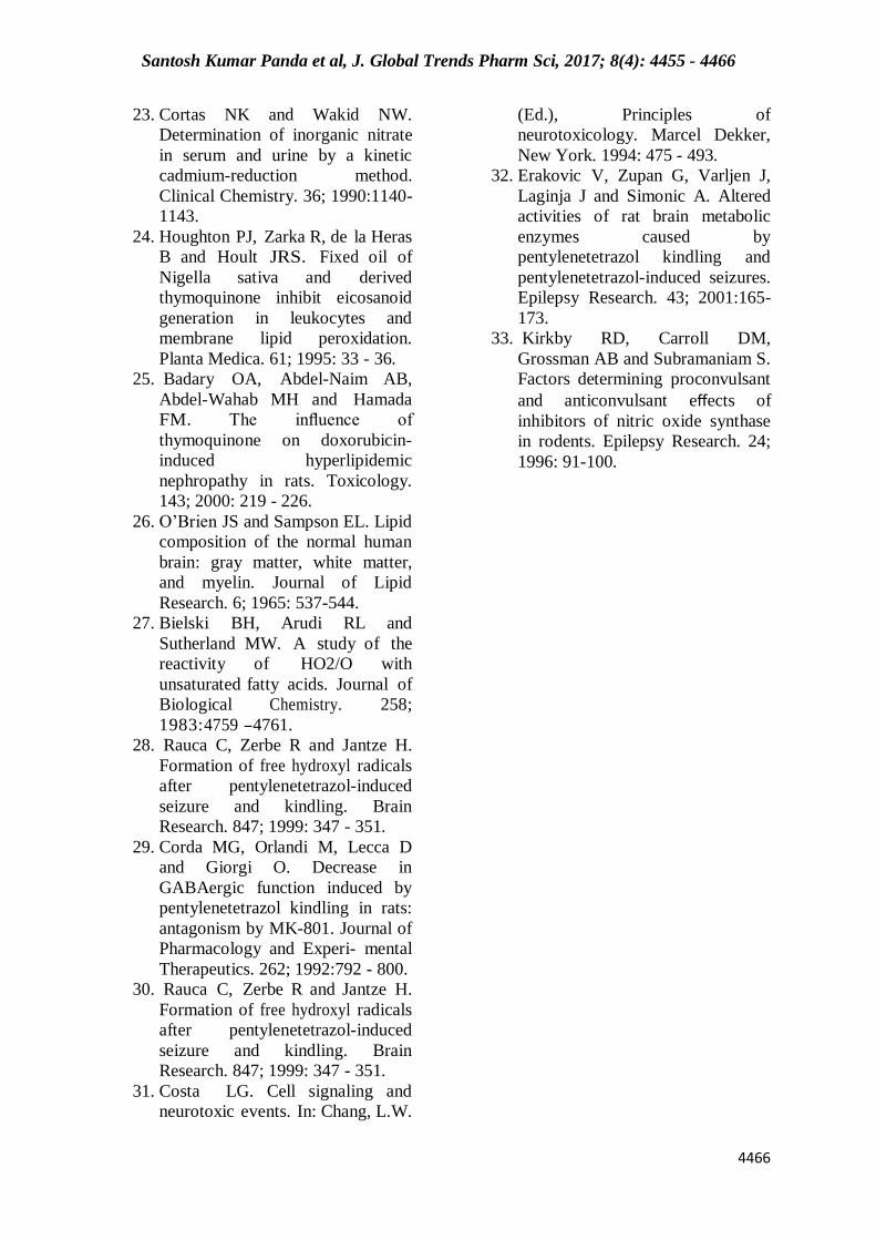
Fig.2- a to e shows the brain levels of
oxidant/antioxidant stress markers in the
kindled and non-kindled mice. Superoxide
dismutase activity was significantly
decreased in the brain tissue in the PTZk
group in comparison with other groups.
PTZ kindling-induced decrement in the
SOD activity was prevented by MEAG
(p=<0.0001). . The PTZ induced kindling
stress also decreased the Catalase activity
in the brain of mice. Pretreatment with
MEAG significantly brought the Catalase
level back to normal (p=<0.0001) as
compared to the saline treated PTZk
group. There was a significantly lower
level of GSH-Px in the PTZk group as
compared with the other groups.
M E A G -treated PTZk groups showed significantly higher levels of GSH-Px
compared with the PTZk group
(p=0.0013). PTZ kindling produced a
significant increase in the brain tissue
MDA c o n t e n t , an index for lipid
peroxidation, when compared with other
groups. PTZk-induced increment in MDA
content of the tissue was significantly
preventd by MEAG treatments
(p=<0.0001). Therefore, Diazepam and
MEAG treated PTZk groups showed
significantly lower levels of MDA as
compared with the PTZk group. The tissue
MDA contents in MEAG group remained
near the control value. PTZ kindling
produced a significant decrease in the NO
activity in brain tissue when compared
with other groups. MEAG administration
caused a significant increase in the NO
levels in comparison with the PTZk group
(p=0.0003).
DISCUSSION
The results from the present study
demonstrated that the MEAG clearly have
antiepileptic property against the
development of seizures in PTZ-kindled
mice model. MEAG also showed
significant effect in controlling clonic-
tonic seizures and lethality in PTZ-kindled
mice model. Moreover, the experimental
results demonstrated that the MEAG was
very much effective as a neuroprotective
agent against PTZ-kindled seizures by
means of its antioxidant actions in
controlling the free radicals generated due
Santosh Kumar Panda et al, J. Global Trends Pharm Sci, 2017; 8(4): 4455 - 4466
4461
to excitotoxicity[24]. In recent years, plants
are investigated extensively for their
medicinal properties in the light of
scientific developments due to their potent
pharmacological activities, low toxicity
and economic viability. MEAG is a very
effective free radical scavenger showing
potent antioxidant activity and protecting
the neurons against free radical damage.
Therefore, MEAG can be used in diseases
in which free radicals are involved, eg.
Anoxia, ischaemia of heart and brain,
arteriosclerosis, cancer and rheumatism
[25]. An imbalance between higher levels
of cellular reactive oxygen species (ROS)
(eg. O2-, ˙OH, NO˙ and ONOO-) and
cellular antioxidant defense results in
oxidative stress. Different defense
mechanisms exist in the brain like
enzymatic (Catalase, SOD,GPx), non-
enzymatic (Glutathione) and dietary
(Vitamin A, E, C, β-carotene, quinones
and flavonoids) to scavenge the reactive
oxygen species and act as antioxidants.
The vulnerability of brain tissue to ROS is
very high because blood perfusion and
aerobic metabolism is very high in brain
and it has a relatively poor enzymatic
antioxidant defense [26]. Brain is also very
rich in lipids that are highly susceptible to
oxidative damage and the adult damaged
neuronal DNA cannot be effectively
repaired since there is no DNA replication
[27]. PTZ-kindling model, one of the most
important rodent experimental epilepsy
model shows oxidative stress in the central
nervous system [28]. The PTZ-kindled
model is characterized by an increased
susceptibility to seizures after a single or
repeated subconvulsive dose of PTZ. PTZ
acts by blocking the chloride ionophore
complex of the GABA-A receptor. Single
or reapeated dose administration of PTZ
leads to decrease in GABAergic function [29].
It also modifies the density and
sensitivity of different glutamate receptors
subtypes in the brain regions [30]. PTZ also
stimulates a variety of biochemical
processess in the brain including activation
of membrane phospholipases, nucleases
and proteases. Marked alterations in
metabolism of the membrane
phospholipids results in the liberation of
free fatty acids (FFAs), free radicals,
diacylglycerols and lipid peroxides [31]. We
have selected the PTZ kindling model
instead of single dose PTZ model basing
on the study results. Different study had
shown the difference between single dose
PTZ model and PTZ kindling model on the
changes of free fatty acid contents in
different parts of brain [32]. Their finding
suggested that PTZ induced kindling
distinctively impaired antioxidant defence
in mice brain which a single dose of PTZ
failed to do the same. For example a single
dose of PTZ did not alter any SOD activity
in frontal cortex, but PTZ kindling caused
a marked decrease in SOD activity. The
present study supports the hypothesis that
PTZ induced seizures actively enhances
oxidative stress in brain tissue. The
primary and most important antioxidant
enzyme SOD, rapidly scavenges the
superoxide anion radicals to hydrogen
peroxide which is less toxic than
superoxide anion. In our study there was
significant lower level of SOD in PTZk
mice. Treatment with MEAG significantly
increased the SOD level in MEAG+ PTZk
mice, indicating neuroprotective and
antioxidant activity. Catalase is an enzyme
which converts the byproduct H2O2
formed by scavenging of superoxide anion
into less toxic water and oxygen. In our
experiment it is observed that the catalase
level in PTZk group is significantly low.
The treated group MEAG+ PTZk showed
significant increase in catalase level
indicating antioxidant and neuroprotective
activity. Glutathione peroxidase, an
endogenous antioxidant reacts with the
free radicals and prevents the generation of
hydroxyl radicals, the most toxic form of
free radicals. During this defensive
process, reduced glutathione is converted
to its oxidised form by GPx. The decreased
GPx activity in PTZk mice in our study
indicates increased generation of free

Santosh Kumar Panda et al, J. Global Trends Pharm Sci, 2017; 8(4): 4455 - 4466
4462
radicals in kindled model and its depletion
during the process of combating oxidative
stress. Treatment with MEAG in MEAG+
PTZk group significantly increases GPx
level and thus indicates antioxidant and
neuroprotective activity. The elevated
level of MDA, a marker of lipid
peroxidation indicates a increased
generation of free radicals in PTZk mice.
Treatement with plant extract, i.e. MEAG
significantly decreased the level of MDA
in PTZk mice. Therefore, the significantly
lower levels of MDA in MEAG+ PTZk
mice as in comparision with PTZk mice
indicate attenuation of lipid peroxidation
and free radical generation. In our present
study, NO level was reduced after kindling
seizures in PTZk group. The role of NO in
the pathophysiology of seizures remains
unclear and debatable. Experiments using
nonselective NOS inhibitors yielded
conflicting results. Therefore, authers have
proposed both pro and anticonvulsant roles
of NO [33]. More research is required for an
adequate conclusion on the role of NO.
Fig.1- Effects of Diazepam and MEAG pretreatment on the development of PTZ-kindled
seizures
P T Z -IN D U C E D K IN D L IN G A C T IV IT Y O F M E Z J
N o . o f D a y s
Mea
n Se
izur
e Sc
ores
0 1 0 2 0 3 0
0
2
4
6
8
S A L IN E + P T Z
D IA Z E P A M + P T Z
M E A G + P T Z
Table-:1- Comparision of the convulsive and lethal effects of PTZ (75mg/kg) (s.c.) in
kindled mice on day 26.
Groups Dose (mg/kg) Latent period in
seconds
Median
seizure score
Lethality
Control
Saline(2ml/kg)(p.o.)
+
PTZ(75mg/kg)(s.c.)
50.33±12.06
6.0±0.1
6/6
Standard
Diazepam(2mg/kg)(p.o.)
+
PTZ(75mg/kg)(s.c.)
416.2±35.57***
(p=<0.0001)
5.0±0.2**
(p=0.0012)
2/6*
(p=0.0101)
Test
MEAG(600mg/kg)(p.o.)
+
PTZ(75mg/kg)(s.c.)
591.8±43.62***,#
(*p=<0.0001)
(#p=0.0109)
3.5±0.2***,###
(*p=<0.0001)
(#p=0.0003)
0/6***
(*p=<0.0001)
(#p=0.1440)
Values are (Mean±SEM); n=6 in each group;* p<0.05, ** p< 0.01 and *** p<0.001 in comparison with
control group; # p<0.05, ### p<0.001 in comparison with standard group.
Santosh Kumar Panda et al, J. Global Trends Pharm Sci, 2017; 8(4): 4455 - 4466
4463
Fig.2 (a)
S O D A c tiv ity o f M E A G
U/ mg
of pr
otein
S A L IN E
D IA Z E P A MM E A G
S A L IN E + P T Z
D IA Z E P A M + P T Z
M E A G + P T Z0
2
4
6
8 ###
*
***
##
***
Fig.2 (b)
C A T A L A S E A c tiv ity o f M E A G
U/ mg
of pr
otein
S A L IN E
D IA Z E P A MM E A G
S A L IN E + P T Z
D IA Z E P A M + P T Z
M E A G + P T Z0
2
4
6
8
*
***
* ###
Fig.2(c)
G P x A c tiv ity o f M E A G
U/g of
prote
in
S A L IN E
D IA Z E P A MM E A G
S A L IN E + P T Z
D IA Z E P A M + P T Z
M E A G + P T Z0
1
2
3
4
5
*** ***
*
##
Fig.2 (d)
M D A A c tiv ity o f M E A G
nmol/
g of p
rotein
S A L IN E
D IA Z E P A MM E A G
S A L IN E + P T Z
D IA Z E P A M + P T Z
M E A G + P T Z0
2 0
4 0
6 0
8 0
*** ##
###
***
Santosh Kumar Panda et al, J. Global Trends Pharm Sci, 2017; 8(4): 4455 - 4466
4464
Fig. 2(e)
N O A c tiv ity o f M E A G
µmol/
mg of
prote
in
S A L IN E
D IA Z E P A MM E A G
S A L IN E + P T Z
D IA Z E P A M + P T Z
M E A G + P T Z0 .0
0 .2
0 .4
0 .6
0 .8
1 .0
*
*** ***
###
Fig. 2 (a-e) Brain MDA, SOD, Catalase, GPx and NO activities in each group. Values are (Mean±SEM); n=6
in each group; * p<0.05, ** p<0.01 and *** p<0.001 in comparison with saline treated non-kindled group; #
p<0.05, ## p<0.01 and ### p<0.001 in comparison with saline treated kindled group.
Treatment with MEAG in MEAG+ PTZk
group significantly increased NO level
which may have some antiepileptic as well
as neuroprotective activity. A significant
decrease of MDA level and enhancement
of SOD, Catalase, NO and GPx level in
MEAG+ PTZk group over other PTZk
group indicates its neuroprotective role by
antioxidant activity.
CONCLUSION
The present study demonstrates that
MEAG significantly prevented PTZ-
kindled seizures in mice as compared to
Diazepam and attenuated oxidative stress
induced by PTZ-kindling rendering
neuroprotective action to brain by its
antioxidant activity. Therefore it can
become a potential drug which can be
approached in arresting or inhibiting the
seizure genesis caused by excitotoxic
agents.
REFERENCES
1. Cockerell OC, Shorvon S, Dreifuss
F, Fish D and Thomas D. The
prognosis of epilepsy. In: (Eds.),
The treatment of epilepsy.
Blackwell Science, Oxford. 1996:
97-113.
2. Shinnar S and Berg AT. Does
antiepileptic drug therapy prevent
the development of
‘‘chronic’’epilepsy? Epilepsia.
37(3); 1996: 701-708.
3. Erakovic V, Zupan G, Varljen J,
Laginja J and Simonic A. Altered
activities of rat brain metabolic
enzymes caused by
pentylenetetrazol kindling and
pentylenetetrazol-induced seizures.
Epilepsy Research. 43; 2001:165-
173.
4. Bruce AJ and Baudry M. Oxygen
free radicals in rat limbic structures
after kainate-induced seizures. Free
Radical Biology and Medicine. 18;
1995: 993 -1002.
5. Akyol O, Herken H, Uz E,
Fadillioglu E, Unal S, Sogut S,
Ozyurt H and Savas HA. The
indices of endogenous oxidative
and antioxidative processes in
plasma from schizophrenic
patients. The possible role of
oxidant/antioxidant imbalance.
Progress in Neuropsycho
pharmacology BiologyADS and
Psychiatry. 26; 2002: 995-1005.
6. Sarsilmaz M, Songur A, Kus I,
Ozyurt B, Gulec M, Sogut S, Ilhan
A and Akyol O. The regulatory
role of dietary u -3 essential fatty
acids on oxidant/antioxidant
balance in rat hippocampus.
Neuroscience Research
Communications. 33; 2003: 114-
123.
Santosh Kumar Panda et al, J. Global Trends Pharm Sci, 2017; 8(4): 4455 - 4466
4465
7. Fadillioglu E, Erdogan H, Iraz M
and Yagmurca M. Effects of caffeic
acid phenethyl ester against
doxorubicin-induced neuronal
oxidant injury. Neuroscience
Research Communications. 33;
2003:132 - 138.
8. Ilhan A, Akyol O, Gurel A,
Armutcu F, Iraz M and Oztas E.
Protective effects of caffeic acid
phenethyl ester against experimen-
tal allergic encephalomyelitis-
induced oxidative stress in rats.
Free Radical Biology and
Medicine. 37; 2004:386-394.
9. Oliver CN, Starke-Reed PE,
Stadtman ER, Liku GJ, Carney JM
and Floyd RA. Oxidative damage
to proteins, loss of glutamine
synthetase activity and production
of free radicals during
ischemia:reperfusion-induced
injury to gerbil brain. Pro-ceedings
of the National Academy of
Sciences USA. 87; 1990: 5144 -
5147.
10. Beni G and Moretti A. Are
reactive oxygen species involved in
Alzheimer’s disease? Neurobiology
of Aging. 16; 1995: 661-674.
11. Kabuto H, Yokoi I and Ogawa N.
Melatonin inhibit iron- induced
epileptic discharges in rats by
suppressing peroxidation.
Epilepsia. 39; 1998:237-243.
12. Zaeoung S, Plubrukarn A and
Keawpradub N. Cytotoxic and free
radical scavenging activities of
Zingiberaceous rhizomes.
Songklanakarin J Sci Techno 2005;
27(4); 799-812.
13. Rangari VD. Pharmacognosy and
Phytochemistry. Career
Publications, Nashik. 1 (1); 2002:
100-110.
14. Khandelwal KR. Practical
Pharmacognosy, Pune, Nirali
Prakashan, New Delhi. 2008; 9th
ed: pp. 149-156.
15. Erakovic V, Zupan G,
VarljenADSF J, Laginja J and
Simonic A. Altered activities of
rat brain metabolic enzymes
caused by pentylenetetrazol
kindling and pentylenetetrazol-
induced seizures. Epilepsy
Research. 43; 2001:165-173.
16. Deepak S, Jyoti A and Sethi P.
Bacopa monniera prevents from
aluminium neurotoxicity in the
cerebral cortex of rat brain. Journal
of Ethnopharmacology. 111(1);
2007:56-62.
17. Paglia DE and Valentine WN.
Studies on the quantitative and
qualitative characterization of
erythrocyte glutathione peroxidase.
Journal of Laboratory and Clinical
Medicine. 70; 1967: 158-170.
18. Lowry O, Rosebrough N, Farr L
and Randall R. Protein
measurement with the Folin-phenol
reagent. Journal of Biology and
Chemistry. 183; 1951: 265-275.
19. Pasha KV and Sadasivadu B.
Intracellular content of thiol
compounds, thiobarbituric acid
reactive substance and gamma-
glutamyl transpeptidase in rats
brain during anoxia. Neuroscience.
46; 1984; 209-214.
20. Nischal HK, Sharma MP, Goyal R
K and Kaushik GG. Serum
superoxide dismutase levels in
diabetes mellitus with or without
microangiopathic complications.
Pharmacology Bulletin. 46; 1998:
853-855.
21. Aebi H. Catalase: Methods in
enzymatic analysis, Academic
Press of New York. 3; 1983:276-
286.
22. Paglia DE and Valentine WN.
Studies on the quantitative and
qualitative characterization of
erythrocyte glutathione peroxidase.
Journal of Laboratory and Clinical
Medicine. 70; 1967: 158-170.
Santosh Kumar Panda et al, J. Global Trends Pharm Sci, 2017; 8(4): 4455 - 4466
4466
23. Cortas NK and Wakid NW.
Determination of inorganic nitrate
in serum and urine by a kinetic
cadmium-reduction method.
Clinical Chemistry. 36; 1990:1140-
1143.
24. Houghton PJ, Zarka R, de la Heras
B and Hoult JRS. Fixed oil of
Nigella sativa and derived
thymoquinone inhibit eicosanoid
generation in leukocytes and
membrane lipid peroxidation.
Planta Medica. 61; 1995: 33 - 36.
25. Badary OA, Abdel-Naim AB,
Abdel-Wahab MH and Hamada
FM. The influence of
thymoquinone on doxorubicin-
induced hyperlipidemic
nephropathy in rats. Toxicology.
143; 2000: 219 - 226.
26. O’Brien JS and Sampson EL. Lipid
composition of the normal human
brain: gray matter, white matter,
and myelin. Journal of Lipid
Research. 6; 1965: 537-544.
27. Bielski BH, Arudi RL and
Sutherland MW. A study of the
reactivity of HO2/O with
unsaturated fatty acids. Journal of
Biological Chemistry. 258;
1983:4759 -4761.
28. Rauca C, Zerbe R and Jantze H.
Formation of free hydroxyl radicals
after pentylenetetrazol-induced
seizure and kindling. Brain
Research. 847; 1999: 347 - 351.
29. Corda MG, Orlandi M, Lecca D
and Giorgi O. Decrease in
GABAergic function induced by
pentylenetetrazol kindling in rats:
antagonism by MK-801. Journal of
Pharmacology and Experi- mental
Therapeutics. 262; 1992:792 - 800.
30. Rauca C, Zerbe R and Jantze H.
Formation of free hydroxyl radicals
after pentylenetetrazol-induced
seizure and kindling. Brain
Research. 847; 1999: 347 - 351.
31. Costa LG. Cell signaling and
neurotoxic events. In: Chang, L.W.
(Ed.), Principles of
neurotoxicology. Marcel Dekker,
New York. 1994: 475 - 493.
32. Erakovic V, Zupan G, Varljen J,
Laginja J and Simonic A. Altered
activities of rat brain metabolic
enzymes caused by
pentylenetetrazol kindling and
pentylenetetrazol-induced seizures.
Epilepsy Research. 43; 2001:165-
173.
33. Kirkby RD, Carroll DM,
Grossman AB and Subramaniam S.
Factors determining proconvulsant
and anticonvulsant effects of
inhibitors of nitric oxide synthase
in rodents. Epilepsy Research. 24;
1996: 91-100.