Embed Size (px)
Citation preview
14
Neuroprotection in Animal Models of Global Cerebral Ischemia
Miguel Cervantes1, Ignacio González-Burgos2, Graciela Letechipía-Vallejo1, María Esther Olvera-Cortés3 and Gabriela Moralí4
1Laboratorio de Neurociencias. Facultad de Ciencias Médicas y Biológicas “Dr. Ignacio Chávez”, Universidad Michoacana de San Nicolás de Hidalgo,
Morelia, Michoacán 2Laboratorio de Psicobiología, División de Neurociencias,
Centro de Investigación, Biomédica de Occidente Instituto Mexicano del Seguro Social, Guadalajara, Jalisco
3Laboratorio de Neurofisiología Experimental, Centro de Investigación Biomédica de Michoacán,
Instituto Mexicano del Seguro Social, Morelia, Michoacán 4Unidad de Investigación Médica en Farmacología,
UMAE Hospital de Especialidades, CMN S XXI, Instituto Mexicano del Seguro Social, México, D.F.
Mexico
1. Introduction
The present chapter deals with some of the main lines of experimental research on global
cerebral ischemia, through which a substantial knowledge has been generated, that has
contributed in an important measure both to the understanding of the mechanisms of
cerebral damage induced by ischemia, and of the subsequent post-ischemic
neuroregenerative and cerebral plastic processes taking place in the remaining or newly
differentiated neurons. Thus, data obtained from experimental designs in animal models of
global cerebral ischemia, on key molecular and cellular events triggered by this condition,
have provided a substantial background from which neuroprotection can be rationally
approached, in order to develop strategies aimed to antagonize, to interrupt, or to slow the
sequence of injurious biochemical and molecular events that would result in irreversible
ischemic injury; as well as to promote brain repair and plasticity processes which can favor
functional preservation or recovery after global cerebral ischemia.
Transient global cerebral ischemia, which can mainly occur during cardiac arrest and
cardiopulmonary resuscitation, but also during asphyxiation, hypotensive shock, or
extracorporeal circulation, is a pathophysiological condition that is associated with great
morbidity and requires intensive medical treatment (Madl & Holzer, 2004). In certain
clinical situations (surgical repair of the thoracic aorta, complex congenital heart lesions, and
also during implantable cardiac defibrillator testing in patients with drug-resistant
www.intechopen.com
Advances in the Preclinical Study of Ischemic Stroke
306
ventricular fibrillation) the possible occurrence of transient global cerebral ischemia, and
some neuroprotective procedures, can be anticipated (Hogue et al., 2008); however, this is
not the case of cardiac arrest.
Cardiopulmonary arrest remains as one among the most frequent causes of death and
disability around the world. Despite quick emergency responses and better techniques of
defibrillation, the chances of survival following cardiac arrest are still poor, between 20-50%
of patients in whom cardiopulmonary resuscitation is attempted. A complex
pathophysiological condition is elicited by cardiac arrest, since it results in whole-body
ischemia which compromises systemic circulatory homeostasis and cerebral, pulmonary,
renal, and cardiac functions. In the course of cardiac arrest, global cerebral blood flow is
severely impaired with the consequent risk of ischemic damage of brain cells, which
magnitude seems to be associated with the cumulative time staying in cardiac arrest. Thus,
most deaths (60%) during the post-resuscitation period have been attributed to extensive
brain injury and neuronal damage that develops as a consequence of alteration of cell
processes triggered by cerebral ischemia and reperfusion, during and after cardiac arrest. In
addition, it is known that transient interruption or reduction of blood flow in the whole
brain, are main causes of permanent brain damage and functional disruptions in human
beings, and near around a half of surviving patients show permanent impairment of
cognitive functions, such as learning and memory, attention, and executive functioning, and
only a small proportion (less than 10%) of those survivors are able to reassume their former
usual life styles (Geocardin et al., 2008; Grubb et al., 2000; Krause et al, 1986; Schneider et al.,
2009). Thus, development of effective cytoprotective therapies that may be common to the
organs more sensitive to cardiac arrest, such as heart or brain, could result in improvement
of survival and better outcome following this whole ischemic episode (Karanjia &
Geocardin, 2011).
Experimental protocols aimed to gain relevant information regarding those pathophysiological phenomena leading to cerebral damage elicited by ischemia have included, since long time, the use of animal models of cerebral ischemia, in order to support better diagnostic, prophylactic and clinical-therapeutic procedures for ischemic cerebrovascular diseases in human beings (Ginsberg & Busto, 1989; Gupta & Briyal, 2004; Hartman et al., 2005, Hossmann, 2008; Traystman, 2003). Thus, biochemical, electrophysiological, histological, and behavioral parameters of ischemic brain damage have been included in experimental designs to evaluate the efficacy and safety of pharmacological and non pharmacological neuroprotective procedures against brain injury resulting from the significant reduction of blood supply to the whole brain, in several animal models of global cerebral ischemia. Even though a great number of pharmacological agents have proven to exert effective neuroprotective actions against cellular events leading to ischemic brain injury in experimental models of global cerebral ischemia, unfortunately they have not had enough clinical relevance to date. On the other hand, after evaluation of its effectiveness as a neuroprotective strategy in animal models of global cerebral ischemia, hypothermia has been tested in clinical trials in patients having suffered cardiac arrest, the most frequent cause of global cerebral ischemia in human beings (Castren et al., 2009; Geocardin et al., 2008; Greer, 2006; Inamasu et al., 2010; Knapp et al., 2011; Seder & Jarrah, 2008,). It seems that new and better strategies to translate preclinical data supporting the potential clinical usefulness of neuroprotective drugs to clinical trials, must be developed.
www.intechopen.com
Neuroprotection in Animal Models of Global Cerebral Ischemia
307
2. Animal models of global cerebral ischemia
Animal models of global cerebral ischemia allow studying, at different levels of biological organization of the central nervous system, the development and temporal course of those processes that may result in irreversible ischemic neuronal damage, as well as in the subsequent cell repair and plasticity underlying either permanent cerebral functional impairment or recovery as a result of intrinsic brain mechanisms or neuroprotective procedures. Thus, animal-related factors (species, strain, age, sex, co-morbidities), animal-model-related factors (choice of ischemic model, anesthetic procedures, duration of ischemia, reperfusion, survival, possibility of monitoring of physiological parameters), selective vulnerability of specific neuron types in several brain structures, outcome assessment (histopathological, biochemical, functional, parameters of brain injury in specific cerebral structures), short- or long-term experimental design, pharmacological characteristics of the presumptive neuroprotective agent itself, timing and dose-response of neuroprotective drug administration with reference to starting and ending of the ischemic episode, may account for the relevance of results from these investigations. Models of cerebral ischemia have been also developed in in vitro models, in particular brain tissue slices and neuronal cultures, allowing to study in detail the cellular phenomena leading either to neuronal damage or to neural recovery and plasticity after ischemia (Benítez-King, 2006; Goldberg & Choi, 1993; Kasai et al., 2003; Whittingham et al., 1984). Several conditions have to be fulfilled by animal models of global cerebral ischemia in order to become appropriate counterparts of these pathophysiological conditions in human beings, as well as to yield reliable and valid results in supporting clinical therapeutic approaches. Thus, it could be expected that in animal models of global cerebral ischemia the ischemic episode can be induced in a constant and reproducible manner: low variation for the extent, temporal course, and magnitude of the resulting ischemic brain injury under specific experimental conditions, including duration of the ischemic episode; easy control of possible deviations of important physiological variables, feasible neurological, neuropathological, and functional evaluations; lack of influence of anesthetic drugs and surgical procedures on the mechanisms of brain injury, brain recovery and/or neuroprotection; short-, intermediate- and long-term follow up of the outcome; and economical, easily available experimental animals of those species better accepted by public animal welfare concerns to be used in experimental protocols of cerebral ischemia and neuroprotection.
2.1 Main animal models of global cerebral ischemia
Models of global cerebral ischemia have been performed in both large (monkeys, sheep, dogs, pigs, cats, rabbits) and small animals (gerbils, rats, mice). Among these, both advantages and disadvantages can be recognized according to several practical aspects: main objectives of the model; monitoring procedures to be used; nature, number and timing of simultaneous parameters to be recorded in order to evaluate the ischemic brain injury and recovery; degree of similarity of structural and functional characteristics of brains of experimental animals to those of the human brain; and updated ethical outlines for the use of experimental animals in research protocols. Since the whole brain is exposed to transient ischemia and reperfusion as a result of cardiac arrest and the subsequent cardiorespiratory resuscitation to allow survival in human beings, animal models of global cerebral ischemia have been designed attempting to totally or
www.intechopen.com
Advances in the Preclinical Study of Ischemic Stroke
308
partially mimic the consequences of this clinical condition on the brain (Ginsberg & Busto, 1989; Gupta & Briyal, 2004; Mc Bean & Kelly, 1998; Traystman, 2003), which are the main cause of neuronal injury to selective vulnerable brain regions, and neurological or cognitive impairment, in human beings. Cardiac arrest (induced by injection of KCl, electric shock, thoracic compression, asphyxia,
and mechanical obstruction of the ascending aorta) followed by cardiopulmonary
resuscitation (by artificial ventilation, closed chest massage and electrical defibrillation),
both in large experimental animals (formerly a common model, but nowadays rarely used)
and also in rodents, has been a technique to produce global cerebral ischemia in an attempt
to closely resemble the clinical situation of cardiac arrest, including complete ischemia and
reperfusion in renal, splachnic and other peripheral organs. This technique seemed to be an
excellent model of global cerebral ischemia, but it is expensive when large experimental
animals are used, and intensive care (cardiopulmonary support under unconsciousness,
control of blood pressure, pH, body fluids, and temperature) must be provided to the
animals, especially during the first 24-48 h after the cardiac arrest. Complete acute global
cerebral ischemia during cardiac arrest (8-20 min) and a variable period of incomplete
cerebral ischemia during reperfusion, even after a successful cardiopulmonary resuscitation,
as well as damage in those brain structures most vulnerable to ischemia, can be expected
from this model (Berkowitz, et al., 1991; Bleyaert et al., 1978; Dave et al., 2004; Hossmann,
2008; Katz et al., 1995; Kofler et al., 2004; Radovsky et al., 1995; Safar et al., 1976; Todd et al.,
1982). In particular, models of global cerebral ischemia in mice are currently of interest
because of the availability of transgenic and knock-out strains for identification of cellular
pathways of ischemic damage, and for neuroprotection studies.
Several other animal models of global cerebral ischemia have been designed in cats, monkeys, gerbils, mice, and rats, in order to circumscribe to the brain those harmful effects of the reduced blood flow that follows a cardiac arrest, avoiding affecting other vital organs in a whole body ischemia condition, as can be expected from animal models of cardiac arrest (Ginsberg & Busto, 1989). Decapitation in small animals has been used as a model of global cerebral ischemia, only
allowing the study of the immediate alterations of some biochemical and metabolic
parameters elicited by ischemia in the brain contained into the head (Abe et al., 1983; Ikeda
et al., 1986; Lowry et al., 1964; Yoshida et al., 1985).
A neck tourniquet or a neck cuff, whether they include or not arterial hypotension, have also been used to produce global cerebral ischemia in rats, cats, dogs, or monkeys. However, these techniques lead to variable ischemic outcomes since the produced ischemia may not be complete because of a remaining cerebral blood flow through the vertebral arteries, as well as complications due to vagal compression and venous congestion (Chopp et al., 1987, 1988; Grenell 1946; Nemoto et al., 1977; Sheller et al., 1992; Siemkowits & Gjedde, 1980; Siemkowitz & Hansen, 1978). Reduction of cerebral blood flow near to zero has been accomplished in cats and monkeys, by occlusion of the innominate and left subclavian arteries near the aortic arch, and pharmacologically induced hypotension (below 80 mm Hg), without involvement of other organs in the ischemic phenomena. However, these experimental animals require intensive care procedures to their survival, and studies of long-term recovery are difficult to achieve (Bodsch et al., 1986; Clavier et al., 1994; Hossmann, 1971; Hossmann & Grose Ophoff, 1986; Zimmerman & Hossmann, 1975).
www.intechopen.com
Neuroprotection in Animal Models of Global Cerebral Ischemia
309
Gerbils usually lack of a common posterior communicating artery connecting the carotid and vertebro-basilar arterial system. Thus, the bilateral common carotid artery occlusion results in a reduction of global cerebral blood flow near to zero and injury of the most vulnerable brain structures (hippocampal CA1 pyramidal neurons after 5 min of ischemia) in most animals (Kirino, 1982). This model of forebrain global cerebral ischemia may fail in some animals in which a complete Willis circle persists, and the high susceptibility of gerbils to seizures may influence the ischemic outcome. The four-vessel occlusion (4-VO) and the two-vessel occlusion with hypotension (2-VO) models in rats became, nowadays, the most widely used animal models that simulate the reduction of blood flow, as it would occur by effect of cardiac arrest, on the forebrain. The 4-VO model (Ginsberg & Busto, 1989; Pulsinelli & Brierley 1979; Pulsinelli & Buchan 1988; Pulsinelli & Duffy 1983; Pulsinelli et al., 1982) provides a method of reversible forebrain ischemia in awake, freely moving rats (but also in anesthetized rats). In a first step of the model procedures, vertebral arteries are permanently occluded and 24 or 48 hours later, the ischemia is produced through transient (10 – 20 min) occlusion of the common carotid arteries under light inhaled anesthesia so that the ischemic episode occurs while the animal is unanesthetized. Loss of the righting reflex, and unconsciousness persisting for at least 20 min after the onset of reperfusion have to occur for each animal to be included in the study. In this way, a reduction in cerebral blood flow to less than 5% of control values, which is followed by hyperemia during 5 to 15 min after reperfusion, and subsequent hypoperfusion lasting for 24 hr result in main ischemic neuronal damage in hippocampus, neocortex and striatum, along hours to days after ischemia, its magnitude relating to the duration of the ischemia. The effects of this insult are, however, quite variable between rat strains, as well as between those individuals surviving (survival rate, 50-75%) after having fulfilled the criteria required to be included in the experimental groups. Similar consequences in selectively vulnerable neurons in specific brain structures result from the 2-VO model of forebrain ischemia, in which bilateral common carotid artery occlusion and systemic hypotension (blood withdrawal and subsequent return with or without pharmacological procedures, leading to arterial blood pressure below 50 mm Hg) are combined to provoke reversible forebrain ischemia (Eklof & Siesjo 1972a, 1972b; Smith et al., 1984a, 1984b). Mouse models of global cerebral ischemia have been developed through bilateral common carotid occlusion and controlled pulmonary ventilation (Traystman, 2003). It is known that animal models of global cerebral ischemia require adequate control of certain variables, such as careful control of animal’s temperature and blood glucose concentration, in order to achieve consistent pathophysiological effects and brain injury (Colbourne & Corbett, 1994; Lipton, 1999; Siemkowicz, 1981; Siemkowicz.& Gjedde 1980). Hyperthermia and hyperglycemia increase brain injury, while hypothermia results in neuroprotection by itself.
3. Cellular mechanisms of neuronal injury, neuronal repair and plasticity
Models of global cerebral ischemia in experimental animals, as well as in vitro models, in particular brain tissue slices and neuronal cultures, have allowed to study in detail the cellular phenomena leading either to neuronal damage, or to neural repair and plasticity after ischemia. From these studies it has been known that mechanisms of cellular damage, repair and plasticity may be the same, in general, both if reduction of blood flow to the brain tissue results from occlusion of one of the main cerebral arteries as would occur in focal
www.intechopen.com
Advances in the Preclinical Study of Ischemic Stroke
310
ischemia, and if it is the result of reduction of blood flow to the whole brain as it would occur after a cardiorrespiratory arrest.
3.1 Cellular mechanisms of neuronal injury
Interruption of blood flow and hence, of glucose and oxygen supply to the brain, results in an immediate severe energy failure in terms of ATP depletion that leads to alterations of the cell membrane ionic gradients and a severe breakdown in cellular homeostasis. Several mechanisms of neuronal damage are triggered and evolve both in cascade and as parallel pathways (Gwag et al, 2002; Lakhan et al, 2009; Lipton, 1999; Mehta et al, 2007; Schneider et al, 2009; Sugawara et al, 2004; Warner et al., 2004). In particular, a massive accumulation of intracellular calcium and sodium occurs because of failure of their energy-dependent efflux processes, and anoxic depolarization. This further leads to accumulation of lactate and hydrogen ions, and as a consequence, to decreased pH. As a result of anoxic depolarization, excitatory aminoacids such as glutamate and aspartate
are released, activating ligand-gated calcium and sodium channels with a further influx of
these ions into the cells. Calcium is also released from intracellular pools, and its excessive,
unregulated intracellular overload causes direct Ca2+-dependent activation of lipases,
proteases, and endonucleases leading to breakdown of structural and functional proteins,
and damage to cytoskeleton and macromolecules including nucleic acids. A result of these
phenomena is, among others, cell membrane lipoperoxidation.
Excessive intracellular calcium activate abnormal cell processes promoting functional
derangements of mitochondria and an increased production of free radicals, exceeding the
neuronal antioxidant reserves, and imposing risks to the structural and functional integrity
of neuronal cells. The brain is highly susceptible to oxidative damage as a consequence of its
high lipid and metal content, as well as other biochemical characteristics (Margaill et al.,
2005; Reiter et al., 2005; Warner el al., 2004). Reperfusion and reoxygenation of the ischemic
tissue, which must be reestablished within minutes in an effort to prevent severe
neurological damage and favor survival of individuals, also may provide chemical
substrates for further increasing cellular alterations, neuronal death and neurological
deficits (Margaill et al., 2005).
Free radicals also contribute to the breakdown of the blood-brain barrier and brain edema.
Reactive oxygen and nitrogen species including superoxide, hydroxyl free radical, and
peroxylnitrite anion are also important mediators of inflammatory tissue damage, of
activation and secretion of inflammatory cytokines such as tumor necrosis factor α,
interleukin-1, and interleukin-6, and of expression of cyclo-oxigenase (COX)-2, and
inducible nitric oxide synthase generating nitric oxide that also contributes to neuronal
damage. These changes favor inflammatory reactions soon after cerebral
ischemia/reperfusion (Barone & Feuerstein, 1999; Lakhan et al, 2009; Lipton, 1999; Mehta et
al, 2007).
Calcium overload may additionally lead to mitochondrial damage and trigger an apoptotic
cascade. The pro-apoptotic cascade involves nuclear factor κB- and p53-dependent
pathways, changes in the Bcl-2 to Bax ratio, opening of the mitochondrial transition pore,
release of cytochrome c, and activation of caspases (Chan, 2001; Chinopoulos & Adam-Vizi,
2006). In addition, caspase-independent pathways may also contribute to neuronal
apoptosis.
www.intechopen.com
Neuroprotection in Animal Models of Global Cerebral Ischemia
311
Several gene families such as immediate early genes, heat-shock proteins, and inflammation-and apoptosis-related genes, are known to be differentially expressed during cerebral ischemia, and some neuropathologic processes triggered by ischemia seem to be mediated in part by alterations of molecular transcriptional and translational activities (Mehta et al, 2007). Activation of DNA fragmentation enzymes and energy-consuming DNA repair enzymes, finally lead to DNA breakdown, interruption of protein synthesis, and cell death (Iadecola & Alexander, 2001; Leker & Shohami, 2002). In addition to the above mentioned cellular processes of ischemic damage, brain ischemia/reperfusion may also trigger cellular mechanisms for neuronal repair, and functional recovery through neuronal plasticity involving remaining neurons in vulnerable damaged or undamaged brain structures (Barone & Feuerstein, 1999; Bendel et al., 2005; Crepel et al., 2003; Hurtado et al., 2006; Jourdain et al., 2002; Ruan et al., 2006). The different ischemia/reperfusion induced cellular mechanisms leading either to brain injury and neuronal death, or to neuronal repair, as well as plasticity and brain functional recovery, may occur in a sequential or simultaneous manner. Their latencies and temporal course, from minutes to weeks, are important references in attempting to establish their differential relevance in those critical periods for neuronal damage and death, as well as the “window of opportunity” for specific neuroprotective procedures (Barone & Feuerstein, 1999; Lipton, 1999; Leker & Shohami, 2002; Pulsinelli et al., 1997).
3.2 Differential neuronal vulnerability in animal models of global cerebral ischemia
Brain injury is expected to occur when cerebral blood flow is reduced to less than 10-20% of
the normal value; the greater the reduction and/or longer lasting, the worst damage. Under
these conditions, damage to specific brain structures due to immediate or delayed death of
highly vulnerable neuronal groups, including the pyramidal neurons of the CA1 subfield of
the hippocampus, and to a lesser degree those in layers 3 and 5 of the cerebral cortex, the
Purkinje cells of the cerebellum, and spiny neurons in the striatum, take place after global
cerebral ischemia (Ginsberg & Busto, 1989; Pulsinelli, 1985). Experimental models of global
cerebral ischemia have allowed to know some neuronal characteristics that seem to account
for selective vulnerability to ischemia, including a high density of excitatory glutamatergic
synapses; low antioxidant enzyme reserves; high content of transition metals; increased
expression of pro-apoptotic Bax protein; thus leading to differential susceptibility of some
cell processes (Ca2+ homeostasis, oxidative-antioxidative balance, functional mitochondrial
stability) to become out of physiological control under ischemia (Arai et al., 2001; Araki et
al., 1989; Chen et al., 1996; Lipton 1999; Schmidt-Kastner et al., 2001; Sugawara et al., 1999).
Brain injury after global ischemia/reperfusion is finally evidenced by neuronal death,
affecting the neuronal population, circuit connectivity and functioning in specific brain
structures involved in the neural integration of cognitive brain functions and behavior.
3.3 Cellular mechanisms of neuronal plasticity and repair
Cellular mechanisms of neuronal repair and plasticity have been observed to occur in vulnerable brain structures in which damage or death of neurons resulted from a sequence of pathophysiological phenomena triggered by global cerebral ischemia and the subsequent reperfusion. Thus, structural and functional characteristics of those neuronal components of circuits in the hippocampus and prefrontal cortex, which are identified, among others, as
www.intechopen.com
Advances in the Preclinical Study of Ischemic Stroke
312
highly vulnerable to ischemia, and their correlation with the integration of specific cerebral functions (mainly cognitive functions) after global cerebral ischemia, have been analyzed. In this sense, short- and long-term structural alterations have been shown to occur in the remaining pyramidal neurons of the hippocampus after ischemia; thus, axonal degeneration as well as reduction of dendritic length and arborizations, of number and shape of dendritic spines, and of number of synapses, are usually related to impairment of cognitive functions and recognized as degenerative changes. By contrast, cytoarchitectural adjustments such as axonal and dendritic sprouting, increase of number of dendritic spines and synapses, changes in the relative proportion of spine types, are interpreted as compensatory plastic responses of surviving neurons. They contribute to neuronal circuit remodeling and functional recovery, and have been correlated with preservation of cognitive functions after the ischemic insult, even in absence of neuroprotective procedures (Briones et al., 2006; Jourdain et al., 2002; Mudrick & Baimbridge, 1989; Neigh et al., 2004; Onodera et al., 1990; Ruan et al., 2006; Skibo & Nikonenko, 2010; Sorra & Harris, 2000). In addition, neurogenesis and integration of newly differentiated neurons into neuronal circuits in the Ammon’s horn may contribute to recovery of hippocampal-dependent cognitive functions (Bendel et al., 2005; Bernabeu & Sharp, 2000). Similarly, reductions of dendritic length, arborization, and dendritic spine density have also
been described, among various cytoarchitectural adjustments, in sensorymotor cortex
pyramidal neurons following global cerebral ischemia (Akulinin et al., 1997, 1998, 2004).
These cytoarchitectural alterations could be influenced by the extent of neuronal remaining
connections; thus, either reduction or increase of afferent connections may result in changes
in dendritic arborizations and spine density (Fiala et al., 2002; Johansson & Belinchenko,
2002). It has been emphasized the functional relevance of neuronal connections from the
hippocampus to the prefrontal cortex for synaptogenesis and neuronal plasticity accounting
for learning and memory (González-Burgos, 2009; Laroche et al., 2000). Thus, a permanent
deafferentation of pyramidal neurons at cortical layer V after the extensive reduction of
pyramidal neuron population of the CA1 subfield of the Ammon’s horn as expected to occur
after global ischemia (Letechipía-Vallejo et al., 2007), may lead to changes in neuronal
activity, which may in turn affect the cytoarchitectural characteristics of pyramidal
prefrontal cortex neurons (García-Chávez et al., 2008; Wellman & Sengelaub, 1991).
These dendritic restructuring (Neigh el al., 2004; Ruan el al., 2006) and reactive synaptogenesis (Briones et al., 2005; Crepel et al., 2003; Jourdain et al., 2002, Kovalenko et al., 2006) among other phenomena including the activation of a variety of potential growth-promoting processes (Arvidsson et al., 2001; Gobbo & O´Mara, 2004; Schmidt-Kastner et al., 2001), that occur in neurons surviving to the ischemic insult in vulnerable brain structures, seem to be a part of mechanisms of adaptive changes, probably accounting for neuronal conditions favoring synaptic plasticity and functional recovery. In fact, a long-term progressive continuous plastic reorganization of the dendritic tree and dendritic spines, initially altered by acute global cerebral ischemia, has been shown to occur in pyramidal neurons at layers 3 and 5 of the sensorymotor cortex of the rat (Akulinin et al., 1997, 1998, 2004). Thus, preservation or recovery of hippocampal- and pre-frontal cortex- dependent functions
after global cerebral ischemia, may involve long-term cytoarchitectural modifications in
those remaining hippocampal CA1 and prefronto-cortical (layers 3 and 5) pyramidal
neurons, since their morpho-functional organization is critical for normal learning and
memory performance (Block, 1999; McDonald & White, 1993; McNamara & Skelton, 1993;
Olsen et al., 1994; Olvera-Cortés et al., 2002; Silva et al., 1998), on the basis of the major role
www.intechopen.com
Neuroprotection in Animal Models of Global Cerebral Ischemia
313
played by the CA1 region for the output of information flowing through the hippocampus,
via the tri-synaptic circuit (Herreras et al., 1987). It is well known that the prefrontal cortex is
directly involved in the organization of sequenced motor actions during working-memory
performance (Fuster, 1999; I. Lee & Kesner, 2003), and that hippocampal projections supply
of spatial information to the prefrontal cortex allowing suitability of motor responses in the
spatial context (Jung et al., 1998). These phenomena may be altered not only by gross lesions
of the prefrontal cortex, but fine alterations of its neuronal circuits may also result in
impairment of the spatial working memory (Fritts et al., 1998; Lambe et al., 2000; I. Lee &
Kesner, 2003; Olvera-Cortés et al., 2001; Taylor et al., 2003). Experimental data have shown
that variations in cognitive behavioral performance are related to plastic changes in
dendritic spines (Pérez-Vega et al., 2000). In addition, excitatory information flows mostly
through dendritic spines-mediated synaptic contacts (Gray, 1959), which are highly
sensitive to electrical stimulation and yet to mnemonic activity-related electrical phenomena
(Harris, 1999; Hartman et al., 2005; Onodera et al., 1990).
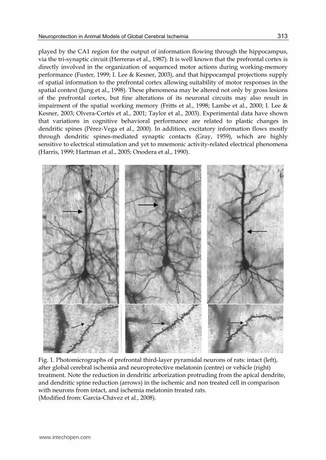
Fig. 1. Photomicrographs of prefrontal third-layer pyramidal neurons of rats: intact (left), after global cerebral ischemia and neuroprotective melatonin (centre) or vehicle (right) treatment. Note the reduction in dendritic arborization protruding from the apical dendrite, and dendritic spine reduction (arrows) in the ischemic and non treated cell in comparison with neurons from intact, and ischemia melatonin treated rats. (Modified from: García-Chávez et al., 2008).
www.intechopen.com
Advances in the Preclinical Study of Ischemic Stroke
314
Since long-term preservation of the neuronal substrate in cerebral vulnerable structures underlying functional recovery after cerebral ischemia has been considered to be a major end point of neuroprotective strategies (STAIR, 1999) it can be expected that experimental designs for neuroprotection studies may lead to reliable interpretations of the efficiency of neuroprotective agents, in view of the proven capability of intrinsic cerebral mechanisms to promote , by themselves, neuronal repair and plasticity after ischemia. Some neuronal proteins that are involved in structural and functional aspects of synaptic connectivity and neuronal circuits remodeling have been evaluated as parameters of ischemic damage and neuroprotection. In this sense, synaptophysin has been shown to be reduced in the frontal motor and temporal cortex of human beings that have been survived for 1 week to 1 year after a cardiac arrest (Akulinin et al., 1998). Besides, a reduction of synaptophysin 2, Munc-18-interacting proteins, 1-3 days after global cerebral ischemia in mice has been related to delayed neuronal death (Nishimura et al., 2000). On the other hand it has been proposed that progesterone-induced increase (3-35 days after ischemia) in the expression of synaptophysin and growth-associated protein 43, and the effects of venlafaxine preventing the decrease of synaptophysin, in the rat hippocampus are evidences of the neuroprotective effects of these drugs (Fang et al., 2010; Zhao et al., 2011).
4. Approaches to neuroprotection in animal models of global cerebral ischemia
The experimental approach to neuroprotection aimed to influence, through pharmacological and non pharmacological procedures, those early and late neural phenomena accounting either for brain damage or for neuronal repair, plasticity and functional recovery after global cerebral ischemia and reperfusion, has resulted in a considerable amount of reliable information along the last 40 years. Different strategies of neuroprotection attempting to prevent, reduce, or stop the progress of the ischemic brain damage have been assayed in animal models of global cerebral ischemia, under the premise of an opposition relationship between the mechanism(s) of action of the presumptive neuroprotective drugs or non pharmacological procedures, and the pathophysiological mechanisms of brain damage, which has been maintained as targets of neuroprotective strategies. Neuroprotection studies in animal models of global cerebral ischemia have maintained the main objective of support proposals of pharmacological and non-pharmacological neuroprotective procedures to be incorporated as a matter for clinical trials aimed to a better management of human beings exposed to global cerebral ischemia, frequently as a consequence of a cardiorespiratory arrest. Translation of knowledge about neuroprotection obtained from models in experimental animals, to clinical practice has not been successful. This situation has been also observed in the case of focal cerebral ischemia, leading to consensus meetings (Fisher et al., 2009; STAIR, 1999) attempting to establish the better conditions for preclinical studies of neuroprotection as to give reliable results to be applied in clinical conditions. If opinion of these consensuses may be recognized as applicable to preclinical studies of global cerebral ischemia, it is apparent that some factors must be taken in account for designing and carrying of the respective experimental protocols. Thus, studies in animal models of global cerebral ischemia should give information on effective neuroprotective doses in the case of drugs being tested; hence, dose-response relationships should be investigated. Routes of drug administration and pharmacokinetic characteristics
www.intechopen.com

Neuroprotection in Animal Models of Global Cerebral Ischemia
315
should also be taken in account as to be compatible with their potential use in human beings. The time window of opportunity for the effective neuroprotective treatment is an important factor to be considered in preclinical models that may predict the timing of neuroprotective procedures in clinical situations with reference to the onset of global cerebral ischemia and subsequent reperfusion. The initial hypothesis that opportunity window for neuroprotective procedures would be limited to a short period after the ischemic episode has been changed in view of experimental evidence. Thus, different drugs or neuroprotective procedures having predominant mechanisms of action against specific cellular processes of ischemia damage occurring lately within the pathophysiological cascade, may allow to neuroprotection even when administered hours or days after ischemia. Besides, the opportunity time window may be further extended when it is expected that neuroprotective procedures act through promotion of cellular processes of neuronal repair and plasticity. In view of the multiple pathophysiological processes occurring both in sequence and
simultaneously after ischemia and reperfusion, it is considered as an advantage for
presumptive neuroprotective drugs to have multiple cellular or molecular mechanisms of
action, as occurring with some originally endogenous compounds, namely melatonin,
estradiol and progesterone (El-Abhar et al, 2002; Hurn et al, 1995; Jover-Mengual et al, 2010;
Lebesgue et al, 2009; Reiter et al, 2005; Wang et al, 2008). By contrast, most synthetic drugs
only have one mechanism of action accounting for neuroprotection. Attempting to
counteract several mechanisms of ischemic brain injury would require the simultaneous
administration of several drugs (Hicks et al, 1999; Matsumoto et al, 1993; Pazos et al, 1999;
del Pilar Fernández et al, 1998; Sánchez-Casado et al, 2007; Zapater et al, 1997) (Table 1).
Recommendations arisen from these consensuses of opinion have also highlighted the
importance of long-term studies to identify whether functional preservation or recovery
may be attributed to effects of the neuroprotective procedures, and/or to intrinsic
mechanisms of plasticity and repair triggered by ischemia per se. Reliable parameters of
long-term structural and functional outcome may allow to evaluate the final result of the
neuroprotective procedures on cerebral structures vulnerable to ischemia. Thus, evaluation
of neuronal population, cytoarchitectonic characteristics, and connectivity of the neural
circuits in these vulnerable structures, as well as different aspects of cognitive functions
depending on them should be included as a part of experimental designs of
neuroprotection. It has been described that the neuronal population of remaining neurons in CA1 at survival times of 2-3 weeks may be less than that evaluated 3-4 months after the ischemic episode, suggesting that, without exogenous intervention, CA1 neurons may have been repopulate, became integrated to the hippocampal neuronal circuits, and contribute to functional recovery (Bendel, et al 2005; von Euler et al., 2006, Hartman et al, 2005, Nakatomi et al., 2002). Obviously, the potential repopulation complicates the interpretation of learning and memory studies after global cerebral ischemia, because short-term studies may not give an adequate end point of the cognitive alteration after global cerebral ischemia, which seems to require a long-term follow up. Experimental designs to evaluate the potential of neuroprotective drugs or hypothermia may have not met all requirements set by these consensuses in a single study, but integration of results of the many experimental studies may give enough information as to support proposals for their clinical usefulness.
www.intechopen.com
Advances in the Preclinical Study of Ischemic Stroke
316
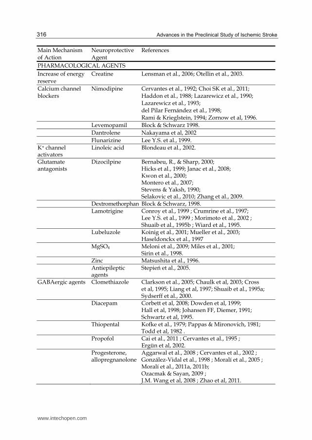
Main Mechanism of Action
Neuroprotective Agent
References
PHARMACOLOGICAL AGENTS
Increase of energy reserve
Creatine Lensman et al., 2006; Otellin et al., 2003.
Calcium channel blockers
Nimodipine Cervantes et al., 1992; Choi SK et al., 2011; Haddon et al., 1988; Lazarewicz et al., 1990; Lazarewicz et al., 1993; del Pilar Fernández et al., 1998; Rami & Krieglstein, 1994; Zornow et al, 1996.
Levemopamil Block & Schwarz 1998.
Dantrolene Nakayama et al, 2002
Flunarizine Lee Y.S. et al., 1999. K+ channel activators
Linoleic acid Blondeau et al., 2002.
Glutamate antagonists
Dizocilpine Bernabeu, R., & Sharp, 2000; Hicks et al., 1999; Janac et al., 2008; Kwon et al., 2000; Montero et al., 2007; Stevens & Yaksh, 1990; Selakovic et al., 2010; Zhang et al., 2009.
Dextromethorphan Block & Schwarz, 1998. Lamotrigine Conroy et al., 1999 ; Crumrine et al., 1997;
Lee Y.S. et al., 1999 ; Morimoto et al., 2002 ; Shuaib et al., 1995b ; Wiard et al., 1995.
Lubeluzole Koinig et al., 2001; Mueller et al., 2003; Haseldonckx et al., 1997
MgSO4 Meloni et al., 2009; Miles et al., 2001; Sirin et al., 1998.
Zinc Matsushita et al., 1996. Antiepileptic
agentsStepień et al., 2005.
GABAergic agents Clomethiazole Clarkson et al., 2005; Chaulk et al, 2003; Cross et al, 1995; Liang et al, 1997; Shuaib et al., 1995a; Sydserff et al., 2000.
Diacepam Corbett et al, 2008; Dowden et al, 1999; Hall et al, 1998; Johansen FF, Diemer, 1991; Schwartz et al, 1995.
Thiopental Kofke et al., 1979; Pappas & Mironovich, 1981; Todd et al, 1982 .
Propofol Cai et al., 2011 ; Cervantes et al., 1995 ; Ergün et al, 2002.
Progesterone, allopregnanolone
Aggarwal et al., 2008 ; Cervantes et al., 2002 ; González-Vidal et al., 1998 ; Moralí et al., 2005 ; Moralí et al., 2011a, 2011b; Ozacmak & Sayan, 2009 ; J.M. Wang et al, 2008 ; Zhao et al, 2011.
www.intechopen.com
Neuroprotection in Animal Models of Global Cerebral Ischemia
317
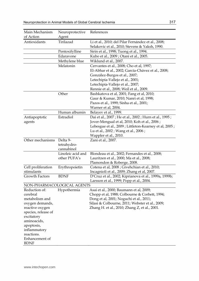
Main Mechanism of Action
Neuroprotective Agent
References
Antioxidants Tirilazad Li et al., 2010; del Pilar Fernández et al., 2008; Selakovic et al., 2010; Stevens & Yaksh, 1990.
Pentoxifylline Sirin et al., 1998; Tuong et al., 1994.
Edaravone Kubo et al., 2009 ; Otani et al., 2005.
Methylene blue Wiklund et al., 2007.
Melatonin Cervantes et al., 2008; Cho et al, 1997; El-Abhar et al., 2002; García-Chávez et al., 2008; González-Burgos et al., 2007; Letechipía-Vallejo et al., 2001; Letechipía-Vallejo et al., 2007; Rennie et al., 2008; Weil et al., 2009.
Other Bashkatova et al, 2001; Fang et al, 2010; Gaur & Kumar, 2010; Nanri et al, 1998; Pazos et al., 1999; Sinha et al., 2001; Warner et al, 2004.
Human albumin Belayev et al., 1999.
Antiapoptotic agents
Estradiol Dai et al., 2007 ; He et al., 2002 ; Hurn et al., 1995 ; Jover-Mengual et al, 2010; Koh et al., 2006 ; Lebesgue et al., 2009 ; Littleton-Kearney et al, 2005 ; Lu et al., 2002 ; Wang et al., 2006 ; Wappler et al., 2010.
Other mechanisms Delta 9-tetrahydro-cannabinol
Zani et al., 2007.
Linoleic acid and other PUFA’s
Blondeau et al., 2002; Fernandes et al., 2008; Lauritzen et al., 2000; Ma et al., 2008; Plamondon & Roberge, 2008.
Cell proliferation stimulants
Erythropoietin Cotena et al, 2008 ; Givehchian et al., 2010; Incagnioli et al., 2009; Zhang et al, 2007.
Growth Factors BDNF D'Cruz et al., 2002; Kiprianova et al., 1999a, 1999b; Larsson et al., 1999; Popp et al., 2004.
NON-PHARMACOLOGICAL AGENTSReduction of: cerebral metabolism and oxygen demands, reactive oxygen species, release of excitatory aminoacids, apoptosis, inflammatory reactions. Enhancement of BDNF
Hypothermia Asai et al., 2000; Baumann et al, 2009; Chopp et al, 1988; Colbourne & Corbett, 1994; Dong et al, 2001; Noguchi et al., 2011; Silasi & Colbourne, 2011; Webster et al., 2009; Zhang H. et al., 2010; Zhang Z, et al., 2001.
www.intechopen.com
Advances in the Preclinical Study of Ischemic Stroke
318
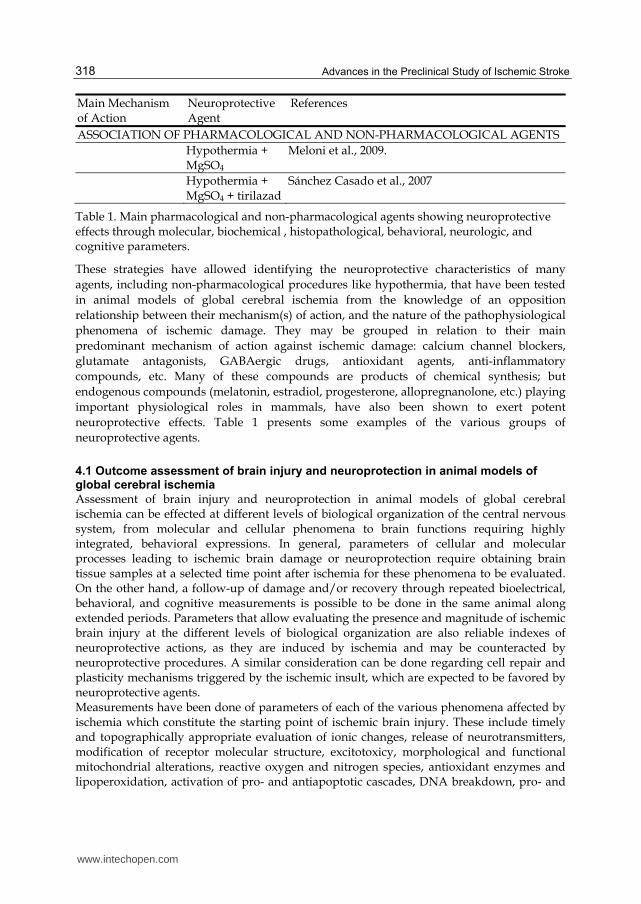
Main Mechanism of Action
Neuroprotective Agent
References
ASSOCIATION OF PHARMACOLOGICAL AND NON-PHARMACOLOGICAL AGENTS
Hypothermia + MgSO4
Meloni et al., 2009.
Hypothermia + MgSO4 + tirilazad
Sánchez Casado et al., 2007
Table 1. Main pharmacological and non-pharmacological agents showing neuroprotective effects through molecular, biochemical , histopathological, behavioral, neurologic, and cognitive parameters.
These strategies have allowed identifying the neuroprotective characteristics of many
agents, including non-pharmacological procedures like hypothermia, that have been tested
in animal models of global cerebral ischemia from the knowledge of an opposition
relationship between their mechanism(s) of action, and the nature of the pathophysiological
phenomena of ischemic damage. They may be grouped in relation to their main
predominant mechanism of action against ischemic damage: calcium channel blockers,
glutamate antagonists, GABAergic drugs, antioxidant agents, anti-inflammatory
compounds, etc. Many of these compounds are products of chemical synthesis; but
endogenous compounds (melatonin, estradiol, progesterone, allopregnanolone, etc.) playing
important physiological roles in mammals, have also been shown to exert potent
neuroprotective effects. Table 1 presents some examples of the various groups of
neuroprotective agents.
4.1 Outcome assessment of brain injury and neuroprotection in animal models of global cerebral ischemia
Assessment of brain injury and neuroprotection in animal models of global cerebral ischemia can be effected at different levels of biological organization of the central nervous system, from molecular and cellular phenomena to brain functions requiring highly integrated, behavioral expressions. In general, parameters of cellular and molecular processes leading to ischemic brain damage or neuroprotection require obtaining brain tissue samples at a selected time point after ischemia for these phenomena to be evaluated. On the other hand, a follow-up of damage and/or recovery through repeated bioelectrical, behavioral, and cognitive measurements is possible to be done in the same animal along extended periods. Parameters that allow evaluating the presence and magnitude of ischemic brain injury at the different levels of biological organization are also reliable indexes of neuroprotective actions, as they are induced by ischemia and may be counteracted by neuroprotective procedures. A similar consideration can be done regarding cell repair and plasticity mechanisms triggered by the ischemic insult, which are expected to be favored by neuroprotective agents. Measurements have been done of parameters of each of the various phenomena affected by ischemia which constitute the starting point of ischemic brain injury. These include timely and topographically appropriate evaluation of ionic changes, release of neurotransmitters, modification of receptor molecular structure, excitotoxicy, morphological and functional mitochondrial alterations, reactive oxygen and nitrogen species, antioxidant enzymes and lipoperoxidation, activation of pro- and antiapoptotic cascades, DNA breakdown, pro- and
www.intechopen.com
Neuroprotection in Animal Models of Global Cerebral Ischemia
319
anti-inflammatory processes, among others (Lakhan et al, 2009; Lipton, 1999; Mehta et al, 2007; Schneider et al, 2009). Neurological, behavioral, electrophysiological and histopathological correlates of the outcome after global cerebral ischemia being end points of cellular processes triggered by ischemia, give information about ischemic brain injury and neuroprotection.
4.1.1 Neurological assessment
Global cerebral ischemia usually does not result in long lasting focal neurological deficits in rats. Thus neurological deficit scores resulting from sensorimotor tests assessing motor-sensory functions in rats, including placement reactions, righting and flexion reflexes, equilibrium, spontaneous motility, among others may be altered shortly after (24 h) global cerebral ischemia, but they appear recovered 7 days after ischemia. These transient neurological deficits have been interpreted as functional alteration of hippocampus and striatum; though correlation between neurological deficit scores and ischemic neuronal damage in these structures, not always were found (Block, 1999; Hartman et al., 2005; Kofler et al, 2004).
4.1.2 Mood and behavioral assessment
Elevated, four (two open and two closed) arms plus maze, and open field tests have been
used, among other to evaluate anxiety after global cerebral ischemia especially in rodents.
Thus scores of latency to enter to open arms, the number of open and closed arms entries
and rears are taken as parameters of anxiety in the elevated plus maze, while in the open
field (circular arena 80 cm in diameter, three concentric rings and lines radiating from the
center) tests, the number of segments entered with all the four paws, the number of rears,
and the number of faecal boli are indexes of anxiety (Nelson et al., 1997).
4.1.3 Cognitive functions assessment
Since the clinical consequences of cardiac arrest, as the main cause of global cerebral ischemia, have been consistently described as long-term alterations of cognitive functions, it can be expected that similar cognitive deficits may be elicited by global cerebral ischemia in experimental animals. In fact, the most vulnerable neurons to ischemia are located in brain structures involved in cognitive processes (Ginsberg & Busto, 1989; Gionet et al., 1991; Pulsinelli, 1985); thus, evaluation of cognitive functions mainly dependent on hippocampus, striatum and prefrontal cortex, and its electrophysiological and morphological correlates may be reliable parameters of brain injury and neuroprotection after global cerebral ischemia. The magnitude and type of cognitive deficits in experimental animals submitted to global cerebral ischemia may vary considerably depending on the animal model, the survival times of testing, and the specific behavioral tests that could have been used. Among these procedures to evaluate cognitive functions, the Morris water maze, the eight-arms radial Olton maze, and the T maze, have been widely used in assessing learning and memory in both 2VO and 4VO models in rats, and its correlation with neuronal loss (Block, 1999; Hartmann et al., 2005; Olsen et al., 1994; Volpe et al., 1984), and functional and morphological characteristics of the neural substrate underlying cognitive functions in brain structures vulnerable to ischemia. Novel object recognition tests have been shown to be a reliable index of cognitive functions since rats or mice normally spend more time exploring novel objects, whereas animals with recognition memory deficits will explore novel and
www.intechopen.com
Advances in the Preclinical Study of Ischemic Stroke
320
familiar objects equally (Hartman et al., 2005). Cognitive functions have also been assessed in rodents through conditioned avoidance tasks (Block, 1999; Kofler et al., 2004; Langdon et al., 2008). Several paradigms in the Morris water maze and in the eight-arms radial Olton maze, that have been used in most of neuroprotection studies in which cognitive functions are assessed, have proven to be useful for testing hippocampal, striatum and prefrontal cortex functioning as end points of brain damage or neuroprotection after global cerebral ischemia (Morris, 1984; Olton et al, 1982). Hippocampal functioning has been evaluated in rats and mice through some behavioral paradigms that require the integrity of this brain structure and related structures in the temporal lobe (Barnes, 1979; Morris et al., 1982, 1990), in order to configure cognitive spatial representations, i.e., a cognitive spatial map (Cassels, 1998; Jarrad, 1993; McDonald and White, 1994; 1995; Moser et al, 1993). Thus parameters of spatial learning training to locate a hidden platform, (escape latency: time spent by the animal to reach the platform; swimming path length: distance swam until reaching the platform; searching strategy: pattern of the swimming path towards the platform) and probe trial to evaluate retention of spatial learning (time spent, or the distance traveled by the animal in each of the four quadrants of the maze; number of crossings over the former platform location) in the Morris water maze including extra maze spatial clues, have been used in testing the morpho-functional state of the hippocampus (Dalm et al 2000; D’Hooge & De Deyn, 2001; Eichenbaum et al, 1990; Morris, 1984; Myhrer, 2003).. Under these training conditions and since there are no intra maze clues to guide the animal’s behavior, it is assumed that, to achieve the goal, the animal has to build the cognitive map and thus, a hippocampal processing of information occurs (Gallagher and Pelleymounter, 1988, O’Keffe & Nadel 1978). For this reason, studies of neuroprotection use the spatial learning in the Morris water maze paradigm, as a reliable index of the hippocampal functioning. However, in addition to place learning, spatial navigation in the water maze may occur through at least, two additional strategies not depending on the hippocampus but on the striatum: signal learning and egocentric learning (Brandeis et al 1989; Gallagher & Pelleymounter, 1988; O’Keefe & Nadel 1978). Signal learning is displayed when the animal reaches a visible platform, or a visible stimulus indicating (signaling) the location of the platform within the maze. Learning of the association between the stimulus and the response is established and depends on the functioning of the striatum (McDonald & White, 1994). The egocentric learning occurs when the animal develops stereotyped motor patterns to locate the invisible platform on the basis of the proprioceptive information provided by its own movement. It is also an ability that depends on the memory system to which the striatum belongs (McDonald & White, 1994; McDonald & White 1995; Oliveira et al., 1997). Results obtained when evaluating both adult and aged male rats, show that some adult rats may use either place, hippocampal dependent allocentric, or striatum-dependent, egocentric strategies; on the other hand, aged rats use egocentric, as their main swimming strategy to solve the task (Dalm et al., 2000; Olvera-Cortés et al, 2011). Thus, deficits in the performance of this task may indicate an alteration of any of these two abilities, place and egocentric learning, so that different parameters should be evaluated to assess the mechanism underlying the observed deficit (D’Hooge & De Deyn, 2001). A qualitative analysis of the swimming paths both during the training period and the probe trial may allow a better determining of the strategy used by the rat in solving the task in the water maze.
www.intechopen.com
Neuroprotection in Animal Models of Global Cerebral Ischemia
321
Spatial working memory can be evaluated by using the 8-arms Olton radial maze (Myhrer,
2003; Olton, 1983, 1987; Olton et al., 1982; Shibata et al., 2007). For a daily standard
evaluation all eight arms are baited and the rat is allowed to collect food from each arm; the
number of errors, defined as a re-entry into an arm that had already been visited, is
recorded in order to evaluate withholding and updating of information about each arm
visited and rewarding obtained. An alternative maze configuration in which only some of
the eight arms are baited allows to evaluate reference memory besides working memory
through recording of the number of reference memory errors (number of entries into
unbaited arms) and working memory errors (re-entry into an already visited arm).
Performance in the Olton maze requires an adequate functioning of hippocampal-
prefrontocortical neuronal circuits, and is a reliable parameter of morpho-functional
integrity of these brain structures after ischemia and neuroprotection (Cassel et al., 1998;
Fritts et al., 1998; Izaki et al., 2008; Kolb, 1990, Kolb et al 1982; Laroche et al., 2000; Olton et
al., 1982; Seamans et al., 1995; Winocur, 1982). An aquatic version of the 8-arm radial maze
has also been described (Kolb et al, 1982), and used to correlate hippocampal pyramidal
neurons damage and working memory performance (Nelson et al. 1997).
4.1.4 Histopathological assessment
Neuronal population of different neuron types in brain vulnerable structures has been
considered as a reliable parameter of ischemia brain damage and neuroprotection. Thus,
pyramidal neuron population in the Ammon´s horn of the hippocampus and in the
neocortex (Bleayert et al, 1978; Colbourne & Corbett, 1994; García-Chávez et al., 2008;
Hartman et al, 2005; Johansen & Diemer, 1991; Kirino, 1982; Letechipía-Vallejo et al., 2007;
Moralí et al., 2011b; Pulsinelli, 1985; Schmidt-Kastner & Freund, 1991; Shuaib et al, 1995), or
different neuron types in other brain vulnerable structures (Block & Schwartz, 1998;
Cervantes et al., 2002), have been evaluated through the number and proportion of
surviving neurons. However, most of these studies deal with histopathological assessment
of the hippocampus, the highest vulnerable brain region to global cerebral ischemia. Usually
four separate counts of surviving neurons in selected areas of the Ammon´s horn are
obtained from each of five coronal sections of the hippocampus per rat, stained with cresyl
violet for a total of 20 counts per animal, under the different experimental conditions
(Hartman et al., 2005). Similar procedures are followed for neuronal counting in other brain
structures vulnerable to ischemia.
Immunohistochemical staining techniques have been also used in animal models of global cerebral ischemia and neuroprotection in order to identify specific proteins or fluorescent DNA labels that may selectively mark cells undergoing an acute necrotic or apoptotic process, as well as the activation of specific cellular processes involved in neuronal damage or repair and survival. Immunohistochemical marks (c-fos/c-jun, heat shock proteins, Bcl-2/Bax immunoreactivity, among others) allow to identify neuron types and neuroanatomical regions where ischemia-induced phenomena take place. Besides, immunohistochemical markers of glial fibrillary acidic protein (GFAP) as well as microglia cell surface components lead to identification of reactive gliosis in the hippocampus, as a consequence of global cerebral ischemia and ischemic neuronal death, which elicited activation of microglial cells and interleukine 1 release that may trigger an astrocyte reaction mainly located in the stratum lacunosum-moleculare, stratum moleculare, and hilus, and
www.intechopen.com
Advances in the Preclinical Study of Ischemic Stroke
322
persisting for weeks after ischemia (Buffo et al., 2010; Choi JS et al, 2008; Mori et al, 2008; Morioka et al., 1991, 1992; Nikonenko et al., 2009; Petito & Halaby, 1993). The efficacy of neuroprotective agents can also be determined on the basis of the success in preventing the occurrence of necrosis, apoptosis, heat shock expression, gliosis, etc., as indicated by the immunohistochemical biomarkers (Scallet, 1995). Different parameters of the glial reaction elicited by global cerebral ischemia have been used as indexes of brain damage or neuroprotection (Cervantes et al., 2002; de Yebra et al., 2006; Duan et al., 2011; Korzhevskii et al., 2005; Piao et al., 2002; Soltys et al., 2003). Neuronal cytoarchitecture and fine structure parameters of synaptic connectivity have also been used for histopathological assessment after brain damage and neuroprotection (Briones et al., 2006; García-Chávez et al., 2008; González-Burgos et al., 2007; Johansson & Belichenko, 2002; Kovalenko et al., 2006, Moralí et al., 2011a; Nikonenko et al., 2009; Ruan et al., 2006).
4.2 Therapeutic opportunity window in animal models of global cerebral ischemia
In any case, recognition of a “therapeutic opportunity window” or “therapeutic time window” in relation to the timing of the ischemic episode, the temporal course of the mechanisms of brain damage and/or repair, and the exerting of actions of presumptive pharmacological or non pharmacological neuroprotective agents, has been a relevant aspect in the approach to neuroprotection in experimental models of global cerebral ischemia (Pulsinelli et al., 1997; Barone & Feuerstein, 1999). In these, the beginning and the extent of this therapeutic window can be expected to be different according to the actions of neuroprotective procedures against immediate or late cellular mechanisms of brain damage, or in favor of later long-lasting cerebral processes of repair and plasticity. Thus optimal neuroprotective effectiveness may require a schedule of drug administration in which drug actions are coincident with the therapeutic opportunity window, that have to be established for different drugs according to their specific mechanisms of action and pharmacokinetic characteristics. In this sense, counteracting of immediate cell mechanisms of neuronal damage may require the administration of neuroprotective drugs before the ischemic episode, though its administration has to be continued afterwards for variable periods. By contrast, drug-promoting repair or plasticity processes admit the starting of neuroprotective treatment hours or days after ischemia. Accordingly, designs of neuroprotective studies in experimental animals in supporting
proposals of neuroprotection for patients exposed to global cerebral ischemia due to
cardiorespiratory arrest, should take in account that this clinical condition usually occurs
unexpectedly, and requires cardiorespiratory resuscitation maneuvers; thus neuroprotection
procedures have to be installed soon, but after the ischemic episode. Experimental designs
of neuroprotection studies assessing neuroprotective procedures against late neuronal
damage processes or promoting neuronal repair and plasticity, favoring functional
preservation and recovery, may lead supporting to a wideness of the therapeutic
opportunity window, for neuroprotection in human beings.
4.2.1 Prophylactic neuroprotection
Transient global cerebral ischemia can occur during certain clinical situations which can either be anticipated, occur during intraoperative emergencies, or even induced, like extracorporeal circulation for cardiac surgery. Under these conditions, prophylactic neuroprotection as that provided by intraoperative hypothermia and pharmacological
www.intechopen.com
Neuroprotection in Animal Models of Global Cerebral Ischemia
323
neuroprotection are possible alternatives to prevent or reduce the risk of ischemic neuronal damage (Savitz & Fisher, 2007; Weigl et al, 2005). This has stimulated designing of experimental studies on prophylactic neuroprotection to assess the effectiveness of several agents and their clinical potential. Some neuroprotective agents have proven to be more effective when applied before the ischemic insult than when given later in time, in particular those agents affecting the early cellular phenomena induced by ischemia, such as calcium channel blockers, GABAergic and anti-excitotoxic agents, as well as antioxidant drugs (Weigl et al, 2005). Pharmacological treatments (antihypertensive, antidiabetic, antithrombotic, antiatherogenic drugs) effective in modifying in the long term the risk for cardiac arrest or cardiac infarct which may result in global cerebral ischemia or in severe hypoperfusion have also been proposed as prophylactic neuroprotection procedures (Savitz & Fisher, 2007).
5. Conclusion
Though an increasing number of drugs have proven to be effective neuroprotective agents in experimental models of global cerebral ischemia, data supporting proposals for their clinical use have not been enough to influence clinical management and outcome of patients exposed to global cerebral ischemia in clinical trials. However, after its evaluation in animal models of global cerebral ischemia, special interest has been paid to carry out clinical trials with a non-pharmacological procedure, hypothermia, as a part of the intensive care of patients after a cardiorespiratory arrest. Nevertheless, the wide perspectives to gain information on neuroprotection through experimental designs including animal models of global cerebral ischemia are maintained to date, despite the tendency to preferentially conduct studies on rodents; in particular if differences between experimental animals and human beings are taken into account, and attention is paid to reproduce those components mainly accounting for brain damage after global cerebral ischemia.
6. Acknowledgement
Partially supported by Instituto Mexicano del Seguro Social, MEXICO (2006/1A/I/029; FIS/IMSS/PROT/196).
7. References
Abe, K.; Yoshida, S.; Watson, B.D.; Busto, R.; Kogure, K. & Ginsberg, M.D. (1983). Alpha-Tocopherol and Ubiquinones in Rat Brain Subjected to Decapitation Ischemia. Brain Research,Vol.273, No.1, (August 1983), pp. 166-169, ISSN 0006-8993
Aggarwal, R.; Medhi, B.; Pathak, A.; Dhawan, V. & Chakrabarti, A. (2008). Neuroprotective Effect of Progesterone on Acute Phase Changes Induced by Partial Global Cerebral Ischaemia in Mice. Journal of Pharmacy and Pharmacology,Vol.60, No.6, (May 2008), pp. 731-737, ISSN 0022-3573
Akulinin, V.A.; Belichenko, P.V. & Dahlstrom, A. (1998). Quantitative Analysis of Synaptophysin Immunoreactivity in Human Neocortex after Cardiac Arrest: Confocal Laser Scanning Microscopy Study. Resuscitation,Vol.39, No.3, (March 1999), pp. 207-213, ISSN 0300-9572
www.intechopen.com
Advances in the Preclinical Study of Ischemic Stroke
324
Akulinin, V.A.; Semchenko, V.V.; Stepanov, S.S. & Belichenko, P.V. (2004). Structural Changes in the Dendritic Spines of Pyramidal Neurons in Layer III of the Sensorimotor Cortex of the Rat Cerebral Cortex in the Late Post-Ischemic Period. Neuroscience and Behavioral Physiology,Vol.34, No.3, (May 2004), pp. 221-227, ISSN 0097-0549
Akulinin, V.A.; Stepanov, S.S.; Semchenko, V.V. & Belichenko, P.V. (1997). Dendritic Changes of the Pyramidal Neurons in Layer V of Sensory-motor Cortex of the Rat Brain During the Postresuscitation Period. Resuscitation,Vol.35, No.2, (October 1997), pp. 157-164, ISSN 0300-9572
Arai, K.; Ikegaya, Y.; Nakatani, Y.; Kudo, I.; Nishiyama, N. & Matsuki, N. (2001). Phospholipase A2 Mediates Ischemic Injury in the Hippocampus: a Regional Difference of Neuronal Vulnerability. European Journal of Neuroscience, Vol.13, No.12, (June 2001), pp. 2319-2323, ISSN 1460-9568
Araki, T.; Kato, H. & Kogure, K. (1989). Selective Neuronal Vulnerability Following Transient Cerebral Ischemia in the Gerbil: Distribution and Time Course. Acta Neurologica Scandinavica,Vol.80, No.6, (December 1989), pp. 548-553, ISSN 0001-6314
Asai, S.; Zhao, H.; Kohno, T.; Takahashi, Y.; Nagata, T. & Ishikawa, K. (2000). Quantitative Evaluation of Extracellular Glutamate Concentration in Postischemic Glutamate Re-uptake, Dependent on Brain Temperature, in the Rat Following Severe Global Brain Ischemia. Brain Research, Vol.864, No.1, (May 2000), pp. 60-68, ISSN 0006-8993
Arvidsson, A.; Kokaia, Z.; Airaksinen, M.S.; Saarma, M. & Lindvall, O. (2001). Stroke Induces Widespread Changes of Gene Expression for Glial Cell Line-derived Neurotrophic Factor Family Receptors in the Adult Rat Brain. Neuroscience,Vol.106, No.1, (September 2001), pp. 27-41, ISSN 0306-4522
Barnes, C.A. (1979). Memory Deficits Associated With Senescence: A Neurophysiological and Behavioral Study in the Rat. Journal of Comparative & Physiological Psychology,Vol.93, No.1, (February 1979), pp. 74-104, ISSN 0021-9940
Barone, F.C. & Feuerstein, G.Z. (1999). Inflammatory Mediators and Stroke: New Opportunities for Novel Therapeutics. Journal of Cerebral Blood Flow & Metabolism,Vol.19, No.8, (August 1999), pp. 819-834, ISSN 0271-678X
Bashkatova, V.G.; Koshelev, V.B.; Fadyukova, O.E.; Alexeev, A.A.; Vanin, A.F.; Rayevsky, K.S.; Ashmarin, I.P. & Armstrong, D.M. (2001). Novel Synthetic Analogue of ACTH 4-10 (Semax) but not Glycine Prevents the Enhanced Nitric Oxide Generation in Cerebral Cortex of Rats with Incomplete Global Ischemia. Brain Research,Vol.894, No.1, (March 2001), pp. 145-149, ISSN 0006-8993
Baumann, E.; Preston, E.; Slinn, J. & Stanimirovic, D. (2009). Post-ischemic Hypothermia Attenuates Loss of the Vascular Basement Membrane Proteins, Agrin and SPARC, and the Blood-brain Barrier Disruption After Global Cerebral Ischemia. Brain Research, Vol.1269, No.1, (May 2009), pp. 185-97, ISSN 0006-8993
Belayev, L.; Saul, I.; Huh, P.W.; Finotti, N.; Zhao, W.; Busto, R. & Ginsberg, M.D. (1999). Neuroprotective Effect of High-dose Albumin Therapy Against Global Ischemic Brain Injury in Rats. Brain Research, Vol.845, No.1, (October n 1999), pp. 107-111, ISSN 0006-8993
Bendel, O.; Bueters, T.; von Euler, M.; Ove Ogren, S.; Sandin, J. & von Euler, G. (2005). Reappearance of Hippocampal CA1 Neurons after Ischemia is Associated with
www.intechopen.com
Neuroprotection in Animal Models of Global Cerebral Ischemia
325
Recovery of Learning and Memory. Journal of Cerebral Blood Flow and Metabolism, Vol.25, No.12, (December 2005), pp. 1586-1595, ISSN 0271-678X
Benítez-King, G. (2006). Melatonin as a Cytoskeletal Modulator: Implications for Cell Physiology and Disease. Journal of Pineal Research,Vol.40, No.1, (November 2005), pp. 1-9, ISSN 0742-3098
Berkowitz, I.D.; Gervais, H.; Schleien, C.L.; Koehler, R.C.; Dean, J.M. & Traystman, R.J. (1991). Epinephrine Dosage Effects on Cerebral and Myocardial Blood Flow in an Infant Swine Model of Cardiopulmonary Resuscitation. Anesthesiology,Vol.75, No.6, (December 1991), pp. 1041-1050, ISSN 0003-3022
Bernabeu, R. & Sharp, F.R. (2000). NMDA and AMPA/Kainate Glutamate Receptors Modulate Dentate Neurogenesis and CA3 Synapsin-I in Normal and Ischemic Hippocampus. Journal of Cerebral Blood Flow & Metabolism,Vol.20, No.12, (December 2000), pp. 1669-1680, ISSN 0271-678X
Bleyaert, A.L.; Nemoto, E.M.; Safar, P.; Stezoski, S.M.; Mickell, J.J.; Moossy, J. & Rao, G.R. (1978). Thiopental Amelioration of Brain Damage After Global Ischemia in Monkeys. Anesthesiology,Vol.49, No.6, (December 1978), pp. 390-398, ISSN 0003-3022
Block, F. (1999). Global Ischemia and Behavioural Deficits. Progress in Neurobiology,Vol.58, No.3, (May 1999), pp. 279-295, ISSN 0301-0082
Block, F. & Schwarz, M. (1998). Global Ischemic Neuronal Damage Relates to Behavioural Deficits: A Pharmacological Approach. Neuroscience,Vol.82, No.3, (March 1998), pp. 791-803, ISSN 0306-4522
Blondeau, N.; Widmann, C.; Lazdunski, M. & Heurteaux, C. (2002). Polyunsaturated Fatty Acids Induce Ischemic and Epileptic Tolerance. Neuroscience,Vol.109, No.2, (January 2002), pp. 231-241, ISSN 0306-4522
Bodsch, W.; Barbier, A.; Oehmichen, M.; Grosse Ophoff, B. & Hossmann, K.A. (1986). Recovery of Monkey Brain After Prolonged Ischemia. II. Protein Synthesis and Morphological Alterations. Journal of Cerebral Blood Flow & Metabolism,Vol.6, No.1, (February 1986), pp. 22-33, ISSN 0271-678X
Bortolotto, Z.A.; Collett, V.J.; Conquet, F.; Jia, Z.; van der Putten, H. & Collingridge, G.L. (2005). The Regulation of Hippocampal LTP by the Molecular Switch, a Form of Metaplasticity, Requires mGlu5 Receptors. Neuropharmacology,Vol.49, Suppl 1, (July 2005), pp. 13-25, ISSN 0028-3908
Briones, T.L.; Suh, E.; Jozsa, L.; Rogozinska, M.; Woods, J. & Wadowska, M. (2005). Changes in Number of Synapses and Mitochondria in Presynaptic Terminals in the Dentate Gyrus Following Cerebral Ischemia and Rehabilitation Training. Brain Research,Vol.1033, No.1, (February 2005), pp. 51-57, ISSN 0006-8993
Briones, T.L.; Suh, E.; Jozsa, L. & Woods, J. (2006). Behaviorally Induced Synaptogenesis and Dendritic Growth in the Hippocampal Region Following Transient Global Cerebral Ischemia are Accompanied by Improvement in Spatial Learning. Experimental Neurology,Vol.198, No.2, (February 2006), pp. 530-538, ISSN 0014-4886
Buffo, A.; Rolando, C. & Ceruti, S. (2010). Astrocytes in the Damaged Brain: Molecular and Cellular Insights into their Reactive Response and Healing Potential. Biochemical Pharmacology, Vol.79, No.2, (January 2010), pp. 77-89, ISSN 0006-2952
Cai, J.; Hu, Y.; Li, W.; Li, L.; Li, S.; Zhang, M. & Li, Q. (2011). The Neuroprotective Effect of Propofol against Brain Ischemia is Mediated by the Glutamatergic Signaling
www.intechopen.com
Advances in the Preclinical Study of Ischemic Stroke
326
Pathway in Rats. Neurochemical Research, Vol.36, No.10, (October 2011), pp. 1724-1731, ISSN 0364-3190
Cassel, J.C.; Cassel, S.; Galani, R.; Kelche, C.; Will, B. & Jarrard, L. (1998). Fimbria-Fornix vs Selective Hippocampal Lesions in Rats: Effects on Locomotor Activity and Spatial Learning and Memory. Neurobiology of Learning and Memory,Vol.69, No.1, (June 1998), pp. 22-45, ISSN 1074-7427
Castren, M.; Silfvast, T.; Rubertsson, S.; Niskanen, M.; Valsson, F.; Wanscher, M. & Sunde, K. (2009). Scandinavian Clinical Practice Guidelines for Therapeutic Hypothermia and Post-resuscitation Care After Cardiac Arrest. Acta Anaesthesiologica Scandinavica,Vol.53, No.3, (February 2009), pp. 280-288, ISSN 1399-6576
Cervantes, M.; Chávez-Carrillo, I. & Antonio-Ocampo, A. (1992). Effects of Nimodipine on Multiunit Activity of Several Brain Structures Following Acute Global Cerebral Ischemia-anoxia in Cats. Boletín de Estudios Médicos y Biológicos Vol.40, No.1-4, (January-December 1992), pp. 21-30, ISSN 0067-9666
Cervantes, M.; González-Vidal, M.D.; Ruelas, R.; Escobar, A. & Moralí, G. (2002). Neuroprotective Effects of Progesterone on Damage Elicited by Acute Global Cerebral Ischemia in Neurons of the Caudate Nucleus. Archives of Medical Research,Vol.33, No.1, (February 2002), pp. 6-14, ISSN 0188-4409
Cervantes, M.; Moralí, G. & Letechipía-Vallejo, G. (2008). Melatonin and Ischemia-reperfusion Injury of the Brain. Journal of Pineal Research,Vol.45, No.1, (January 2008), pp. 1-7, ISSN 1600-079X
Cervantes, M.; Ruelas, R.; Chávez-Carrillo, I.; Contreras-Gómez, A. & Antonio-Ocampo, A. (1995). Effects of Propofol on Alterations of Multineuronal Activity of Limbic and Mesencephalic Structures and Neurological Deficit Elicited by Acute Global Cerebral Ischemia. Archives of Medical Research,Vol.26, No.4, (January 1995), pp. 385-395, ISSN 0188-4409
Chan, P.H. (2001), Reactive Oxygen Radicals in Signalling and Damage in the Ischemic Brain. Journal of Cerebral Blood Flow & Metabolism Vol. 21, No. 1, (January 2001), pp. 2-14, ISSN 0271-678X
Chaulk, D.; Wells, J.; Evans, S.; Jackson, D. & Corbett, D. (2003). Long-term Effects of Clomethiazole in a Model of Global Ischemia. Experimental Neurology,Vol.182, No.2, (August 2003), pp. 476-482, ISSN 0014-4886
Chen, J.; Zhu, R.L.; Nakayama, M.; Kawaguchi, K.; Jin, K.; Stetler, R.A.; Simon, R.P. & Graham, S.H. (1996). Expression of the Apoptosis-effector Gene, Bax, is Up-regulated in Vulnerable Hippocampal CA1 Neurons Following Global Ischemia. Journal of Neurochemistry,Vol.67, No.1, (July 1996), pp. 64-71, ISSN 0022-3042
Chinopoulos, C. & Adam-Vizi, V. (2006). Calcium, mitochondria and Oxidative Stress in Neuronal Pathology. Novel Aspects of an Enduring Theme. FEBS Journal, Vol. 273, No. 3, (February 2006), pp. 433-450, ISSN
Cho, S.; Joh, T.H.; Baik, H.H.; Dibinis, C. & Volpe, B.T. (1997). Melatonin Administration Protects CA1 Hippocampal Neurons After Transient Forebrain Ischemia in Rats. Brain Research,Vol.755, No.2, (May 1997), pp. 335-338, ISSN 0006-8993
Choi, S.K.; Lee, G.J.; Choi, S.; Kim, Y.J.; Park, H.K. & Park, B.J. (2011). Neuroprotective Effects by Nimodipine Treatment in the Experimental Global Ischemic Rat Model: Real Time Estimation of Glutamate. Journal of Korean Neurosurgical Society Vol.49, No.1, (April 2011), pp. 1-7, ISSN 1598-7876
www.intechopen.com
Neuroprotection in Animal Models of Global Cerebral Ischemia
327
Choi, J.S.; Shin, Y.J.; Cha, J.H.; Kim, H.Y.; Choi, J.Y.; Chun, M.H. & Lee, M.Y. (2008). Induction of Suppressor of Cytokine Signaling-3 in Astrocytes of the Rat Hippocampus Following Transient Forebrain Ischemia. Neuroscience Letters, Vol.441, No.3, (August 2008), pp. 323-327, ISSN 0304-3940
Chopp, M.; Frinak, S.; Walton, D.R.; Smith, M.B. & Welch, K.M. (1987). Intracellular Acidosis During and After Cerebral Ischemia: in Vivo Nuclear Magnetic Resonance Study of Hyperglycemia in Cats. Stroke,Vol.18, No.5, (September 1987), pp. 919-923, ISSN 0039-2499
Chopp, M.; Welch, K.M.; Tidwell, C.D.; Knight, R. & Helpern, J.A. (1988). Effect of Mild Hyperthermia on Recovery of Metabolic Function After Global Cerebral Ischemia in Cats. Stroke,Vol.19, No.12, (December 1988), pp. 1521-1525, ISSN 0039-2499
Clarkson, A.N.; Liu, H.; Rahman, R.; Jackson, D.M.; Appleton, I. & Kerr, D.S. (2005). Clomethiazole: Mechanisms Underlying Lasting Neuroprotection Following Hypoxia-ischemia. FASEB Journal,Vol.19, No.8, (April 2005), pp. 1036-1038, ISSN 1530-6860
Clavier, N.; Kirsch, J. R.; Hurn, P.D. & Traystman, R.J. (1994). Effect of Postischemic Hypoperfusion on Vasodilatory Mechanisms in Cats. American Journal of Physiology,Vol.267, No.5 Pt 2, (November 1994), pp. H2012-2018, ISSN 0002-9513
Colbourne, F. & Corbett, D. (1994). Delayed and Prolonged Post-ischemic Hypothermia is Neuroprotective in the Gerbil. Brain Research,Vol.654, No.2, (August 1994), pp. 265-272, ISNN 0006-8993
Conroy, B.P.; Black, D.; Lin, C.Y.; Jenkins, L.W.; Crumrine, R.C.; DeWitt, D.S. & Johnston, W.E. (1999). Lamotrigine Attenuates Cortical Glutamate Release during Global Cerebral Ischemia in Pigs on Cardiopulmonary Bypass. Anesthesiology,Vol.90, No.3, (March 1999), pp. 844-854, ISSN 0003-3022
Corbett, D.; Larsen, J. & Langdon, K.D. (2008) Diazepam Delays the Death of Hippocampal CA1 Neurons Following Global Ischemia. Experimental Neurology, Vol.214, No.2, (December 2008), pp. 309-14, ISSN
Cotena, S.; Piazza, O. & Tufano, R. (2008). The Use of Erythtropoietin in Cerebral Diseases. Panminerva Medica,Vol.50, No.2, (July 2008), pp. 185-192, ISSN 0031-0808
Crepel, V.; Epsztein, J. & Ben-Ari, Y. (2003). Ischemia Induces Short- and Long-Term Remodeling of Synaptic Activity in the Hippocampus. Journal of Cellular and Molecular Medicine,Vol.7, No.4, (February 2004), pp. 401-407, ISSN 1582-1838
Cross, A.J.; Jones, J.A.; Snares, M.; Jostell, K.G.; Bredberg, U. & Green, A.R. (1995). The Protective Action of Chlormethiazole against Ischaemia-induced Neurodegeneration in Gerbils when Infused at Doses Having Little Sedative or Anticonvulsant Activity. British Journal of Pharmacology Vol.114, No.8, (April 1995), pp. 1625-1630, ISSN 0007-1188
Crumrine, R.C.; Bergstrand, K.; Cooper, A.T.; Faison, W.L. & Cooper, B.R. (1997). Lamotrigine Protects Hippocampal CA1 Neurons from Ischemic Damage after Cardiac Arrest. Stroke,Vol.28, No.11, (November 1997), pp. 2230-2236, ISSN 0039-2499
D'Cruz, B.J.; Fertig, K.C.; Filiano, A.J.; Hicks, S.D.; DeFranco, D.B. & Callaway, C.W. (2002). Hypothermic Reperfusion After Cardiac Arrest Augments Brain-Derived Neurotrophic Factor Activation. Journal of Cerebral Blood Flow & Metabolism, (July 2002), Vol.22, No.7, pp. 843-851, ISSN 0271-678X
www.intechopen.com
Advances in the Preclinical Study of Ischemic Stroke
328
D'Hooge, R. & De Deyn, P.P. (2001). Applications of the Morris Water Maze in the Study of Learning and Memory. Brain Research Brain Research Reviews,Vol.36, No.1, (August 2001), pp. 60-90, ISSN 0006-8993
Dai, X.; Chen, L. & Sokabe, M. (2007). Neurosteroid Estradiol Rescues Ischemia-induced Deficit in the Long-term Potentiation of Rat Hippocampal CA1 Neurons. Neuropharmacology,Vol.52, No.4, (January 2007), pp. 1124-1138, ISSN 0028-3908
Dalm, S.; Grootendorst, J.; de Kloet, E.R. & Oitzl, M.S. (2000). Quantification of Swim Patterns in the Morris Water Maze. Behavior Research Methods, Instruments, & Computers,Vol.32, No.1, (April 2000), pp. 134-139, ISSN 0743-3808
Dave, K.R.; Raval, A.P.; Prado, R.; Katz, L.M.; Sick, T.J.; Ginsberg, M.D.; Busto, R. & Perez-Pinzon, M.A. (2004). Mild Cardiopulmonary Arrest Promotes Synaptic Dysfunction in Rat Hippocampus. Brain Research,Vol.1024, No.1-2, (September 2004), pp. 89-96, ISSN 0006-8993
De Yebra, L.; Malpesa, Y.; Ursu, G.; Pugliese, M.; Lievéns, J.C.; Goff, L.K. & Mahy, N. (2006). Dissociation Between Hippocampal Neuronal Loss, Astroglial and Microglial Reactivity after Pharmacologically Induced Reverse Glutamate Transport. Neurochemistry International, Vol.49, No.7, (December 2006), pp. 691-697, ISSN 0197-0186
Dong, H.; Moody-Corbett, F.; Colbourne, F.; Pittman, Q. & Corbett, D. (2001). Electrophysiological Properties of CA1 Neurons Protected by Postischemic Hypothermia in Gerbils. Stroke, Vol.32, No.3, (March 2001), pp. 788-95, ISSN 0039-2499
Dowden, J.; Reid, C.; Dooley, P. & Corbett, D. (1999). Diazepam-Induced Neuroprotection: Dissociating the Effects of Hypothermia Following Global Ischemia. Brain Research, Vol.829, No.1-2, (May 1999), pp. 1-6, ISSN 0006-8993
Duan, Y.L.; Wang, S.Y.; Zeng, Q.W.; Su, D.S.; Li, W.; Wang, X.R. & Zhao, Z. (2011). Astroglial Reaction to Delta Opioid Peptide [D-Ala2, D-Leu5] Enkephalin Confers Neuroprotection Against Global Ischemia in the Adult Rat Hippocampus. Neuroscience, Vol.192, (September 2011), pp. 81-90, ISSN 0306-4522
Eichenbaum, H.; Stewart, C. & Morris, R. G. (1990). Hippocampal Representation in Place Learning. Journal of Neuroscience, Vol.10, No.11, (November 1990), pp. 3531-3542, ISSN 0270-6474
Eklof, B. & Siesjo, B.K. (1972a). The Effect of Bilateral Carotid Artery Ligation Upon Acid-base Parameters and Substrate Levels in the Rat Brain. Acta Physiologica Scandinavica,Vol.86, No.4, (December 1972), pp. 528-538, ISSN 0001-6772
Eklof, B. & Siesjo, B.K. (1972b). The Effect of Bilateral Carotid Artery Ligation Upon the Blood Flow and the Energy State of the Rat Brain. Acta Physiologica Scandinavica,Vol.86, No.2, (October 1972), pp. 155-165, ISSN 0001-6772
El-Abhar, H.S.; Shaalan, M.; Barakat, M. & El-Denshary, E.S. (2002). Effect of Melatonin and Nifedipine on some Antioxidant Enzymes and Different Energy Fuels in the Blood and Brain of Global Ischemic Rats. Journal of Pineal Research,Vol.33, No.2, (August 2002), pp. 87-94, ISSN 0742-3098
Ergün, R.; Akdemir, G.; Sen, S.; Taşçi, A. & Ergüngör. F. (2002). Neuroprotective Effects of Propofol Following Global Cerebral Ischemia in Rats. Neurosurgical Review, Vol.25, No.1-2, (March 2002), pp. 95-98, ISSN 0344-5607
www.intechopen.com
Neuroprotection in Animal Models of Global Cerebral Ischemia
329
von Euler, M.; Bendel, O.; Bueters, T.; Sandin, J. & von Euler, G. (2006).Profound but Transient Deficits in Learning and memory after Global Cerebral Ischemia Using a Novel Water Maze Test. Behavioral Brain research, Vol.166, No.2, (January 2006), pp. 204-210, ISSN 0166-4328
Fang, S.; Yan, B.; Wang, D.; Bi, X.; Zhang, Y.; He, J.; Xu, H.; Yang, Y.; Kong, J.; Wu, J. & Li, X.M. (2010). Chronic Effects of Venlafaxine on Synaptophysin and Neuronal Cell Adhesion Molecule in the Hippocampus of Cerebral Ischemic Mice. Biochemistry and Cell Biology Vol.88, No.4, (July 2010), pp. 655-663, ISSN 1208-6002
Fernandes, J.S.; Mori, M.A.; Ekuni, R.; Oliveira, R.M. & Milani, H. (2008). Long-term Treatment with Fish Oil Prevents Memory Impairments but not Hippocampal Damage in Rats Subjected to Transient, Global Cerebral Ischemia. Nutrition Research Vol.28, No.11, (December 2008), pp. 798-808, 1879-0739
Fiala, J.C.; Spacek, J. & Harris, K.M. (2002). Dendritic Spine Pathology: Cause or Consequence of Neurological Disorders? Brain Research Reviews,Vol.39, No.1, (June 2002), pp. 29-54, ISSN 0165-0173
Fisher, M.; Feuerstein, G.; Howells, D.W.; Hurn, P.D.; Kent, T.A.; Savitz, S.I. & Lo, E.H. (2009). Update of the Stroke Therapy Academic Industry Roundtable Preclinical Recommendations. Stroke,Vol.40, No.6, (February 2009), pp. 2244-2250, ISSN 0039-2499
Fritts, M.E.; Asbury, E.T.; Horton, J.E. & Isaac, W.L. (1998). Medial Prefrontal Lesion Deficits Involving or Sparing the Prelimbic Area in the Rat. Physiology & Behavior,Vol.64, No.3, (September 1998), pp. 373-380, ISSN 0031-9384
Fuster, J.M. (1997). The Prefrontal Cortex. Anatomy, Physiology, and Neuropsychology of the Frontal Lobe, Lippincott-Raven, ISBN 978-0881674668, New York, USA
Fuster, J.M. (1999). Synopsis of Function and Dysfunction of the Frontal Lobe. Acta Psychiatrica Scandinavica, Vol.395, Supplementum, (May 1999), pp. 51-57, ISSN 0065-159
Gallagher, M. & Pelleymounter, M.A. (1988). Spatial Learning Deficits in Old Rats: a Model for Memory Decline in the Aged. Neurobiology of Aging,Vol.9, No.5-6, (September 1988), pp. 549-556, ISSN 0197-4580
García-Chávez, D.; González-Burgos, I.; Letechipía-Vallejo, G.; López-Loeza, E.; Moralí, G. & Cervantes, M. (2008). Long-term Evaluation of Cytoarchitectonic Characteristics of Prefrontal Cortex Pyramidal Neurons, Following Global Cerebral Ischemia and Neuroprotective Melatonin Treatment, in Rats. Neuroscience Letters,Vol.448, No.1, (October 2008), pp. 148-152, ISSN 0304-3940
Gaur, V. & Kumar, A. (2010). Protective Effect of Desipramine, Venlafaxine and Trazodone against Experimental Animal Model of Transient Global Ischemia: Possible Involvement of NO-cGMP Pathway. Brain Research,Vol.1353, (July 2010), pp. 204-212, ISSN 0006-8993
Geocadin, R.G.; Koenig, M.A.; Jia, X.; Stevens, R.D. & Peberdy, M.A. (2008). Management of Brain Injury After Resuscitation from Cardiac Arrest. Neurologic Clinics,Vol.26, No.2, (June 2008), pp. 487-506, ix, ISSN 0733-8619
Ginsberg, M.D. & Busto, R. (1989). Rodent Models of Cerebral Ischemia. Stroke,Vol.20, No.12, (December 1989), pp. 1627-1642, ISSN 0039-2499
Gionet, T.X.; Thomas, J.D.; Warner, D.S.; Goodlett, C.R.; Wasserman, E.A. & West, J.R. (1991). Forebrain Ischemia Induces Selective Behavioral Impairments Associated
www.intechopen.com
Advances in the Preclinical Study of Ischemic Stroke
330
with Hippocampal Injury in Rats. Stroke,Vol.22, No.8, (August 1991), pp. 1040-1047, ISSN 0039-2499
Givehchian, M.; Beschorner, R.; Ehmann, C.; Frauenlob, L.; Morgalla, M.; Hashemi, B.; Ziemer, G. & Scheule, A.M. (2010). Neuroprotective Effects of Erythropoietin During Deep Hypothermic Circulatory Arrest. European Journal of Cardio-thoracic Surgery,Vol.37, No.3, (September 2009), pp. 662-668, ISSN 1873-734X
Gobbo, O.L. & O'Mara, S.M. (2004). Impact of Enriched-environment Housing on Brain-derived Neurotrophic Factor and on Cognitive Performance After a Transient Global Ischemia. Behavioral Brain Research,Vol.152, No.2, (June 2004), pp. 231-241, ISSN 0166-4328
Goldberg, M.P. & Choi, D.W. (1993). Combined Oxygen and Glucose Deprivation in Cortical Cell Culture: Calcium-dependent and Calcium-independent Mechanisms of Neuronal Injury. Journal of Neuroscience,Vol.13, No.8, (August 1993), pp. 3510-3524, ISSN 0270-6474
González-Burgos, I. (2009). Dendritic Spines Plasticity and Learning/Memory Processes: Theory, Evidence and Prospectives, In: Dendritic Spines. Biochemistry, Modelling and Properties. L.R. Baylon, (Ed.), pp. 163-186. Nova Science Publishers, Inc, ISBN 978-1607414605, New York, USA
González-Burgos, I.; Letechipía-Vallejo, G.; López-Loeza, E.; Moralí, G. & Cervantes, M. (2007). Long-term Study of Dendritic Spines from Hippocampal CA1 Pyramidal Cells, After Neuroprotective Melatonin Treatment Following Global Cerebral Ischemia in Rats. Neuroscience Letters,Vol.423, No.2, (August 2007), pp. 162-166, ISSN 0304-3940
González-Vidal, M.D.; Cervera-Gaviria, M.; Ruelas, R.; Escobar, A.; Moralí, G. & Cervantes, M. (1998). Progesterone: Protective Effects on the Cat Hippocampal Neuronal Damage Due to Acute Global Cerebral Ischemia. Archives of Medical Research,Vol.29, No.2, (July 1998), pp. 117-124, ISSN 0188-4409
Gray, E.G. (1959). Electron Microscopy of Synaptic Contacts on Dendrite Spines of the Cerebral Cortex. Nature,Vol.183, No.4675, (June 1959), pp. 1592-1593, ISSN 0028-0836
Greer, D.M. (2006). Hypothermia for Cardiac Arrest. Current Neurology and Neuroscience Reports,Vol.6, No.6, (November 2006), pp. 518-524, ISSN 1528-4042
Grenell, R.G. (1946). Central Nervous System Resistance; the Effects of Temporary Arrest of Cerebral Circulation for Periods of Two to Ten Minutes. Journal of Neuropathology & Experimental Neurology,Vol.5, (April 1946), pp. 131-154, ISSN 0022-3069
Grubb, N.R.; Fox, K.A.; Smith, K.; Best, J.; Blane, A.; Ebmeier, K.P.; Glabus, M.F. & O'Carroll, R.E. (2000). Memory Impairment in Out-of-Hospital Cardiac Arrest Survivors is Associated with Global Reduction in Brain Volume, not Focal Hippocampal Injury. Stroke,Vol.31, No.7, (July 2000), pp. 1509-1514, ISSN 0039-2499
Gupta, Y.K. & Briyal, S. (2004). Animal Models of Cerebral Ischemia for Evaluation of Drugs. Indian Journal of Physiology and Pharmacology,Vol.48, No.4, (May 2005), pp. 379-394, ISSN 0019-5499
Gwag, B.J.; Won, S.J. & Kim, D.Y. (2002). Excitotoxicity, Oxidative Stress, and Apoptosis in Ischemic Neuronal Death, In: New Concepts in Cerebral Ischemia. Methods and New Frontiers In Neuroscience, R.C.S. Lin, (Ed.), 79-112, CRC Press, ISBN 0-8493-0119-X, Boca Raton, USA
www.intechopen.com
Neuroprotection in Animal Models of Global Cerebral Ischemia
331
Haddon, W.S.; Prough, D.S.; Kong, D. & Petrozza, P. (1988). Effects of Nimodipine on the Production of Thromboxane A2 Following Total Global Cerebral Ischemia. Journal of Neurosurgery,Vol.69, No.3, (September 1988), pp. 416-420, ISSN 0022-3085
Hall, E.D.; Fleck, T.J. & Oostveen, J.A. (1998). Comparative Neuroprotective Properties of the Benzodiazepine Receptor Full Agonist Diazepam and the Partial Agonist PNU-101017 in the Gerbil Forebrain Ischemia Model. Brain Research, Vol.798, No.1-2, (July 1998), pp. 325-329, ISSN 0006-8993
Harris, K.M. (1999). Calcium From Internal Stores Modifies Dendritic Spine Shape. Proceedings of the National Academy of Sciences of the United States of America,Vol.96, No.22, (October 1999), pp. 12213-12215, ISSN 0027-8424
Hartman, R.E.; Lee, J.M.; Zipfel, G.J. & Wozniak, D.F. (2005). Characterizing Learning Deficits and Hippocampal Neuron Loss Following Transient Global Cerebral Ischemia in Rats. Brain Research,Vol.1043, No.1-2, (May 2005), pp. 48-56, ISSN 0006-8993
Haseldonckx, M.; Van Reempts, J.; Van de Ven, M.; Wouters, L. & Borgers, M. (1997). Protection with Lubeluzole Against Delayed Ischemic Brain Damage in Rats. A Quantitative Histopathologic Study. Stroke,Vol.28, No.2, (February 1997), pp. 428-432, ISSN 0039-2499
He, Z.; He, Y.J.; Day, A.L. & Simpkins, J.W. (2002). Proestrus Levels of Estradiol During Transient Global Cerebral Ischemia Improves the Histological Outcome of the Hippocampal CA1 Region: Perfusion-dependent and-independent Mechanisms. Journal of the Neurological Sciences,Vol.193, No.2, (January 2002), pp. 79-87, ISSN 0022-510X
Herreras, O.; Solis, J.M.; Martin del Rio, R. & Lerma, J. (1987). Characteristics of CA1 Activation Through the Hippocampal Trisynaptic Pathway in the Unanaesthetized Rat. Brain Research,Vol.413, No.1, (June 1987), pp. 75-86, ISSN 0006-8993
Hicks, C.A.; Ward, M.A.; Swettenham, J.B. & O'Neill, M.J. (1999). Synergistic Neuroprotective Effects by Combining an NMDA or AMPA Receptor Antagonist with Nitric Oxide Synthase Inhibitors in Global Cerebral Ischaemia. European Journal of Pharmacology,Vol.381, No.2-3, (November 1999), pp. 113-119, ISSN 0014-2999
Hogue, C.W.; Gottesman, R.F. & Stearns, J. (2008). Mechanisms of Cerebral Injury from Cardiac Surgery. Critical Care Clinics,Vol.24, No.1, (February 2008), pp. 83-98, viii-ix, ISSN 0749-0704
Hossmann, K.A. (1971). Cortical Steady Potential, Impedance and Excitability Changes During and After Total Ischemia of Cat Brain. Experimental Neurology,Vol.32, No.2, (August 1971), pp. 163-175, ISSN 0014-4886
Hossmann, K.A. (2008). Cerebral Ischemia: Models, Methods and Outcomes. Neuropharmacology,Vol.55, No.3, (January 2008), pp. 257-270, ISSN 0028-3908
Hossmann, K.A. & Grosse Ophoff, B. (1986). Recovery of Monkey Brain After Prolonged Ischemia. I. Electrophysiology and Brain Electrolytes. Journal of Cerebral Blood Flow & Metabolism,Vol.6, No.1, (February 1986), pp. 15-21, ISSN 0271-678X
Hurn, P.D.; Littleton-Kearney, M.T.; Kirsch, J.R.; Dharmarajan, A.M. & Traystman, R.J. (1995). Postischemic Cerebral Blood Flow Recovery in the Female: Effect of 17 Beta-Estradiol. Journal of Cerebral Blood Flow & Metabolism,Vol.15, No.4, (July 1995), pp. 666-672, ISSN 0271-678X
www.intechopen.com
Advances in the Preclinical Study of Ischemic Stroke
332
Hurtado, O.; Pradillo, J.M.; Alonso-Escolano, D.; Lorenzo, P.; Sobrino, T.; Castillo, J.; Lizasoain, I. & Moro M.A. (2006). Neurorepair versus Neuroprotection in Stroke. Cerebrovascular Diseases, Vol. 21, (Suppl. 2), pp. 54-63, ISSN 1015-9770
Iadecola, C. & Alexander, M. (2001) Cerebral Ischemia and Inflammation. Current Opinion in Neurology, Vol. 14, No.1 , (February 2001), pp. 89-94, ISSN 0959-4388
Ikeda, M.; Yoshida, S.; Busto, R.; Santiso, M. & Ginsberg, M.D. (1986). Polyphosphoinositides as a Probable Source of Brain Free Fatty Acids Accumulated at the Onset of Ischemia. Journal of Neurochemistry,Vol.47, No.1, (July 1986), pp. 123-132, ISSN 0022-3042
Inamasu, J.; Nakatsukasa, M.; Suzuki, M. & Miyatake, S. (2010). Therapeutic Hypothermia for Out-of-Hospital Cardiac Arrest: An Update for Neurosurgeons. World Neurosurgery, Vol. 74, No. 1, (July 2010), pp. 120-128, ISSN 1878-8750
Incagnoli, P.; Ramond, A.; Joyeux-Faure, M.; Pepin, J.L.; Levy, P. & Ribuot, C. (2009). Erythropoietin Improved Initial Resuscitation and Increased Survival After Cardiac Arrest in Rats. Resuscitation,Vol.80, No.6, (May 2009), pp. 696-700, ISSN 1873-1570
Izaki, Y.; Takita, M. & Akema, T. (2008). Specific Role of the Posterior Dorsal Hippocampus-Prefrontal Cortex in Short-Term Working Memory. European Journal of Neuroscience,Vol.27, No.11, (June 2008), pp. 3029-3034, ISSN 1460-9568
Janac, B.; Selakovic, V. & Radenovic, L. (2008). Temporal Patterns of Motor Behavioural Improvements by MK-801 in Mongolian Gerbils Submitted to Different Duration of Global Cerebral Ischemia. Behavioural Brain Research,Vol.194, No.1, (July 2008), pp. 72-78, ISSN 0166-4328
Jarrard, L.E. (1993). On the Role of the Hippocampus in Learning and Memory in the Rat. Behavioral and Neural Biology,Vol.60, No.1, (July 1993), pp. 9-26, ISSN 0163-1047
Johansen, F.F. & Diemer, N.H. (1991). Enhancement of GABA Neurotransmission After Cerebral Ischemia in the Rat Reduces Loss of Hippocampal CA1 Pyramidal Cells. Acta Neurologica Scandinavica, Vol.84, No.1, (July 1991), pp. 1-6. ISSN
Johansson, B.B. & Belichenko, P.V. (2002). Neuronal Plasticity and Dendritic Spines: Effect of Environmental Enrichment on Intact and Postischemic Rat Brain. Journal of Cerebral Blood Flow & Metabolism,Vol.22, No.1, (January 2002), pp. 89-96, ISSN 0271-678X
Jourdain, P.; Nikonenko, I.; Alberi, S. & Muller, D. (2002). Remodeling of Hippocampal Synaptic Networks by a Brief Anoxia-hypoglycemia. Journal of Neuroscience,Vol.22, No.8, (April 2002), pp. 3108-3116, ISSN 1529-2401
Jover-Mengual, T.; Miyawaki, T.; Latuszek, A.; Alborch, E.; Zukin, R.S. & Etgen, A.M. (2010). Acute Estradiol Protects CA1 Neurons from Ischemia-induced Apoptotic Cell Death Via the PI3K/Akt Pathway. Brain Research,Vol.1321, (February 2010), pp. 1-12, ISSN 1872-6240
Jung, M.W.; Qin, Y.; McNaughton, B.L. & Barnes, C.A. (1998). Firing Characteristics of Deep Layer Neurons in Prefrontal Cortex in Rats Performing Spatial Working Memory Tasks. Cerebral Cortex,Vol.8, No.5, (August 1998), pp. 437-450, ISSN 1047-3211
Karanjia, N. & Geocadin, R.G. (2011). Post-cardiac Arrest Syndrome: Update on Brain Injury Management and Prognostication. Current Treatment Options in Neurology,Vol.13, No.2, (January 2011), pp. 191-203, ISSN 1534-3138
Kasai, H.; Matsuzaki, M.; Noguchi, J.; Yasumatsu, N. & Nakahara, H. (2003). Structure-Stability-Function Relationships of Dendritic Spines. Trends in Neurosciences,Vol.26, No.7, (July 2003), pp. 360-368, ISSN 0166-2236
www.intechopen.com
Neuroprotection in Animal Models of Global Cerebral Ischemia
333
Katz, L.; Ebmeyer, U.; Safar, P.; Radovsky, A. & Neumar, R. (1995). Outcome Model of Asphyxial Cardiac Arrest in Rats. Journal of Cerebral Blood Flow & Metabolism,Vol.15, No.6, (November 1995), pp. 1032-1039, ISSN 0271-678X
Kiprianova, I.; Freiman, T.M.; Desiderato, S.; Schwab, S.; Galmbacher, R.; Gillardon, F. & Spranger, M. (1999a). Brain-Derived Neurotrophic Factor Prevents Neuronal Death and Glial Activation After Global Ischemia in the Rat. Journal of Neuroscience Research, Vol.56, No.1, (April 1999), pp. 21-27, ISSN 0270-6474
Kiprianova, I.; Sandkühler, J.; Schwab, S.; Hoyer, S. & Spranger, M. (1999b). Brain-Derived Neurotrophic Factor Improves Long-Term Potentiation and Cognitive Functions After Transient Forebrain Ischemia in the Rat. Experimentsal Neurology, (October 1999), Vol.159, No.2, pp. 511-519, ISSN
Kirino, T. (1982). Delayed Neuronal Death in the Gerbil Hippocampus Following Ischemia. Brain Research, Vol.239, No.1, (May 1982), pp. 57-69, ISSN 0006-8993
Knapp, J.; Heinzmann, A.; Schneider, A.; Padosch, S.A.; Böttiger, B.W.; Teschendorf, P. & Popp, E. (2011). Hypothermia and Neuroprotection by Sulfide After Cardiac Arrest and Cardiopulmonary Resuscitation. Resuscitation, Vol.82, No.8, (August 2011) pp. 1076-1080, ISSN
Kofke, W.A.: Nemoto, E.M.; Hossmann, K.A.; Taylor, F.; Kessler, P.D. & Stezoski, S.W. (1979). Brain Blood Flow and Metabolism after Global Ischemia and Post-Insult Thiopental Therapy in Monkeys. Stroke, (September-October 1979), Vol.10, No.5, pp. 554-560, ISSN 0039-2499
Kofler, J.; Hattori, K.; Sawada, M.; DeVries, A.C.; Martin, L.J.; Hurn, P.D. & Traystman, R.J. (2004). Histopathological and Behavioral Characterization of a Novel Model of Cardiac Arrest and Cardiopulmonary Resuscitation in Mice. Journal of Neuroscience Methods,Vol.136, No.1, (May 2004), pp. 33-44, ISSN 0165-0270
Koh, P.O.; Cho, G.J. & Choi, W.S. (2006). 17beta-Estradiol Pretreatment Prevents the Global Ischemic Injury-Induced Decrease of Akt Activation and Bad Phosphorylation in Gerbils. Journal of Veterinary Medical Science,Vol.68, No.10, (November 2006), pp. 1019-1022, ISSN 0916-7250
Koinig, H.; Vornik, V.; Rueda, C. & Zornow, M. H. (2001). Lubeluzole Inhibits Accumulation of Extracellular Glutamate in the Hippocampus During Transient Global Cerebral Ischemia. Brain Research, Vol.898, No.2, (April 2001), pp. 297-302, ISSN 0006-8993
Kolb, B. (1990) In: The Cerebral Cortex of the Rat. B. Kolb & R.C. Tees, (Eds.), 437-458, MIT Press, ISBN: 978-026-2610-64-3, Cambridge, Massachussetts, USA
Kolb, B.; Pittman, K.; Sutherland, R.J. & Whishaw, I.Q. (1982). Dissociation of the Contributions of the Prefrontal Cortex and Dorsomedial Thalamic Nucleus to Spatially Guided Behavior in the Rat. Behavioural Brain Research, Vol.6, No.4, (December 1982), pp. 365-378, ISSN 0166-4328
Kolb, B.; Teskey, G.C. & Gibb, R. (2010). Factors Influencing Cerebral Plasticity in the Normal and Injured Brain. Frontiers in Human Neuroscience, Vol.4, (November 2010), pp. 1-12, ISSN 1662-5161
Korzhevskii, D.E.; Otellin, V.A.; Grigor’ev, I.P.; Kostkin, V.B.; Polenov, S.A.; Lentsman, M.V. & Balestrino, M. (2005). Structural Organization of Astrocytes in the Rat Hippocampus in the Post-ischemic Period. Neuroscience & Behavioral Physiology, Vol.35, No.4, (May 2005), pp. 389-392, ISSN 0097-0549
www.intechopen.com
Advances in the Preclinical Study of Ischemic Stroke
334
Kovalenko, T.; Osadchenko, I.; Nikonenko, A.; Lushnikova, I.; Voronin, K.; Nikonenko, I.; Muller, D. & Skibo, G. (2006). Ischemia-Induced Modifications in Hippocampal CA1 Stratum Radiatum Excitatory Synapses. Hippocampus, Vol.16, No.10, (August 2006), pp. 814-825, ISSN 1050-9631
Krause, G.S.; Kumar, K.; White, B.C.; Aust, S.D. & Wiegenstein, J.G. (1986). Ischemia, Resuscitation, and Reperfusion: Mechanisms of Tissue Injury and Prospects for Protection. American Heart Journal, Vol.111, No.4, (April 1986), pp. 768-780, ISSN 0002-8703
Kubo, K.; Nakao, S.; Jomura, S.; Sakamoto, S.; Miyamoto, E.; Xu, Y.; Tomimoto, H.; Inada, T. & Shingu, K. (2009). Edaravone, a Free Radical Scavenger, Mitigates Both Gray and White Matter Damages After Global Cerebral Ischemia in Rats. Brain Research, Vol. 1279, No. 3, (July 2009), pp. 139-46, ISSN ISSN 0006-8993
Kwon, Y.B.; Yang, I.S.; Kang, K.S.; Han, H.J.; Lee, Y.S. & Lee, J.H. (2000). Effects of Dizocilpine Pretreatment on Parvalbumin Immunoreactivity and Fos Expression after Cerebral Ischemia in the Hippocampus of the Mongolian Gerbil. Journal of Veterinary Medical Science,Vol.62, No.2, (March 2000), pp. 141-146, ISSN 0916-7250
Lakhan, S.E.; Kirchgessner, A. & Hofer, M. (2009). Inflammatory Mechanisms in Ischemic Stroke: Therapeutic Approaches. Journal of Translational Medicine,Vol.7, (November 2009), pp. 97, ISSN 1479-5876
Lambe, E.K.; Goldman-Rakic, P.S. & Aghajanian, G.K. (2000). Serotonin Induces EPSCs Preferentially in Layer V Pyramidal Neurons of the Frontal Cortex in the Rat. Cerebral Cortex,Vol.10, No.10, (September 2000), pp. 974-980, ISSN 1047-3211
Langdon, K.D.; Granter-Button, S. & Corbett, D. (2008). Persistent Behavioral Impairments and Neuroinflammation Following Global Ischemia in the Rat. European Journal of Neuroscience,Vol.28, No.11, (November 2008), pp. 2310-2318, ISSN 1460-9568
Laroche, S.; Davis, S. & Jay, T. M. (2000). Plasticity at Hippocampal to Prefrontal Cortex Synapses: Dual Roles in Working Memory and Consolidation. Hippocampus,Vol.10, No.4, (September 2000), pp. 438-446, ISSN 1050-9631
Larsson, E.; Nanobashvili, A.; Kokaia, Z & Lindvall, O. (1999) Evidence for Neuroprotective Effects of Endogenous Brain-Derived Neurotrophic Factor After Global Forebrain Ischemia in Rats. Journal of Cerebral Blood Flow & Metab, Vol.19, No.11, (November 1999), pp. 1220-1228, ISSN
Lauritzen, I.; Blondeau, N.; Heurteaux, C.; Widmann, C.; Romey, G. & Lazdunski, M. (2000). Polyunsaturated Fatty Acids are Potent Neuroprotectors. EMBO Journal,Vol.19, No.8, (April 2000), pp. 1784-1793, ISSN 0261-4189
Lazarewicz, J.W.; Pluta, R.; Puka, M. & Salinska, E. (1990). Diverse Mechanisms of Neuronal Protection by Nimodipine in Experimental Rabbit Brain Ischemia. Stroke, Vol.21, (12 Supplement), (December 1990), pp. IV108-110, ISSN 0039-2499
Lazarewicz, J.W.; Pluta, R.; Puka, M. & Salinska, E. (1993). Local Nimodipine Application Improves Early Functional Recovery in the Rabbit Hippocampus After 15-min Global Cerebral Ischemia. Acta Neurobiologiae Experimentalis Vol.53, No.4, (January 1993), pp. 499-510, ISSN 0065-1400
Lebesgue, D.; Chevaleyre, V.; Zukin, R.S. & Etgen, A.M. (2009). Estradiol Rescues Neurons from Global Ischemia-Induced Cell Death: Multiple Cellular Pathways of Neuroprotection. Steroids, Vol.74, No.7, (May 2009), pp. 555-561, ISSN 0039-128X
www.intechopen.com
Neuroprotection in Animal Models of Global Cerebral Ischemia
335
Lee, I. & Kesner, R. P. (2003). Time-dependent Relationship between the Dorsal Hippocampus and the Prefrontal Cortex in Spatial Memory. Journal of Neuroscience, Vol.23, No.4, (February 2003), pp. 1517-1523, ISSN 1529-2401
Lee, Y.S.; Yoon, B.W. & Roh, J.K. (1999). Neuroprotective Effects of Lamotrigine Enhanced by Flunarizine in Gerbil Global Ischemia. Neuroscience Letters, Vol.265, No.3, (May 1999), pp. 215-217, ISSN 0304-3940
Leker, R.R. & Shohami, E. (2002). Cerebral Ischemia and Trauma – Different Ethiologies yet Similar Mechanisms: Neuroprotective Opportunities. Brain Research Brain Research Reviews, Vol. 39, No. 1, (January 2002), pp. 55-73, ISSN 0165-0173
Lensman, M.; Korzhevskii, D.E.; Mourovets, V.O.; Kostkin, V.B.; Izvarina, N.; Perasso L.; Gandolfo C.; Otellin, V.A.; Polenov, S.A. & Balestrino, M. (2006). Intracerebroventricular Administration of Creatine Protects against Damage by Global Cerebral Ischemia in Rat. Brain Research. Vol. 1114, No. 1, (October 2006), pp. 187-194, ISSN 0006-8993
Letechipía-Vallejo, G.; González-Burgos, I. & Cervantes, M. (2001). Neuroprotective Effect of Melatonin on Brain Damage Induced by Acute Global Cerebral Ischemia in Cats. Archives of Medical Research, Vol.32, No.3, (June 2001), pp. 186-192, ISSN 0188-4409
Letechipía-Vallejo, G.; López-Loeza, E.; Espinoza-González, V.; González-Burgos, I.; Olvera-Cortés, M. E.; Moralí, G. & Cervantes, M. (2007). Long-term Morphological and Functional Evaluation of the Neuroprotective Effects of Post-ischemic Treatment with Melatonin in Rats. Journal of Pineal Research, Vol.42, No.2, (February 2007), pp. 138-146, ISSN 0742-3098
Li, R.C.; Guo, S.Z.; Lee, S.K. & Gozal, D. (2010). Neuroglobin Protects Neurons Against Oxidative Stress in Global Ischemia. Journal of Cerebral Blood Flow & Metabolism, Vol.30, No.11, (June 2010), pp. 1874-1882, ISSN 1559-7016
Liang, S.P.; Kanthan, R.; Shuaib, A., & Wishart, T. (1997). Effects of Clomethiazole on Radial-arm Maze Performance Following Global Forebrain Ischemia in Gerbils. Brain Research, Vol.751, No.2, (March 1997), pp. 189-195, ISSN 0006-8993
Lipton, P. (1999). Ischemic Cell Death in Brain Neurons. Physiology Reviews, Vol.79, No.4, (October 1999), pp. 1431-1568, ISSN 0031-9333
Littleton-Kearney, M.T.; Gaines, J.M.; Callahan, K.P.; Murphy, S.J. & Hurn, P.D. (2005). Effects of Estrogen on Platelet Reactivity after Transient Forebrain Ischemia in Rats. Biological Research for Nursing, Vol.7, No.2, (November 2005), pp. 135-145, ISSN 1099-8004
Lowry, O.H.; Passonneau, J.V.; Hasselberger, F.X. & Schulz, D.W. (1964). Effect of Ischemia on Known Substrates and Cofactors of the Glycolytic Pathway in Brain. Journal of Biological Chemistry, Vol.239, (January 1964), pp. 18-30, ISSN 0021-9258
Lu, A.; Ran, R.Q.; Clark, J.; Reilly, M.; Nee, A. & Sharp, F.R. (2002). 17-beta-Estradiol Induces Heat Shock Proteins in Brain Arteries and Potentiates Ischemic Heat Shock Protein Induction in Glia and Neurons. Journal of Cerebral Blood Flow & Metabolism, Vol.22, No.2, (February 2002), pp. 183-195, ISSN 0271-678X
Ma, D.; Lu, L.; Boneva, N.B.; Warashina, S.; Kaplamadzhiev, D.B.; Mori, Y.; Nakaya, M.A.; Kikuchi, M.; Tonchev, A.B.,; Okano, H. & Yamashima, T. (2008). Expression of Free Fatty Acid Receptor GPR40 in the Neurogenic Niche of Adult Monkey Hippocampus. Hippocampus, Vol.18, No.3, (December 2007), pp. 326-333, ISSN 1098-1063
www.intechopen.com
Advances in the Preclinical Study of Ischemic Stroke
336
Madl, C. & Holzer, M. (2004). Brain Function after Resuscitation from Cardiac Arrest. Current Opinion in Critical Care, Vol.10, No.3, (May 2004), pp. 213-217, ISSN 1070-5295
Margaill, I.; Plotkine, M. & Lerouet, D. (2005). Antioxidant Strategies in the Treatment of Stroke. Free Radical Biology & Medicine, Vol. 39, No.4 , (August 2005), pp. 429-443, ISSN 0891-5849
Matsumoto, M.; Scheller, M.S.; Zornow, M.H. & Strnat, M.A. (1993). Effect of S-emopamil, Nimodipine, and Mild Hypothermia on Hippocampal Glutamate Concentrations after Repeated Cerebral Ischemia in Rabbits. Stroke, Vol.24, No.8, (August 1993), pp. 1228-1234, ISSN 0039-2499
Matsushita, K.; Kitagawa, K.; Matsuyama, T.; Ohtsuki, T.; Taguchi, A.; Mandai, K.; Mabuchi, T.; Yagita, Y.; Yanagihara, T. & Matsumoto, M. (1996). Effect of Systemic Zinc Administration on Delayed Neuronal Death in the Gerbil Hippocampus. Brain Research, Vol.743, No.1-2, (December 1996), pp. 362-365, ISSN 0006-8993
McBean, D.E. & Kelly, P.A. (1998). Rodent Models of Global Cerebral Ischemia: a Comparison of Two-vessel Occlusion and Four-vessel Occlusion. General Pharmacology, Vol.30, No.4, (April 1998), pp. 431-434, ISSN 0306-3623
McDonald, R.J. & White, N.M. (1993). A Triple Dissociation of Memory Systems: Hippocampus, Amygdala, and Dorsal Striatum. Behavior Neuroscience, Vol.107, No.1, (February 1993), pp. 3-22, ISSN 0735-7044
McDonald, R.J. & White, N.M. (1994). Parallel Information Processing in the Water Maze: Evidence for Independent Memory Systems Involving Dorsal Striatum and Hippocampus. Behavioral and Neural Biology, Vol.61, No.3, (May 1994), pp. 260-270, ISSN 0163-1047
McDonald, R.J. & White, N.M. (1995). Hippocampal and Nonhippocampal Contributions to Place Learning in Rats. Behavioral Neuroscience, Vol.109, No.4, (August 1995), pp. 579-593, ISSN 0735-7044
McNamara, R.K. & Skelton, R.W. (1993). The Neuropharmacological and Neurochemical Basis of Place Learning in the Morris Water Maze. Brain Research Brain Research Reviews, Vol.18, No.1, (January 1993), pp. 33-49, ISSN 0165-0173
Mehta, S.L.; Manhas, N. & Raghubir, R. (2007). Molecular Targets in Cerebral Ischemia for Developing Novel Therapeutics. Brain Research Reviews, Vol.54, No.1, (January 2007), pp. 34-66, ISSN 0165-0173
Meloni, B.P.; Campbell, K.; Zhu, H. & Knuckey, N.W. (2009). In Search of Clinical Neuroprotection after Brain Ischemia: the Case for Mild Hypothermia (35 Degrees C) and Magnesium. Stroke, Vol.40, No.6, (April 2009), pp. 2236-2240, ISSN 0039-2499
Miles, A.N.; Majda, B.T.; Meloni, B.P. & Knuckey, N.W. (2001). Postischemic Intravenous Administration of Magnesium Sulfate Inhibits Hippocampal CA1 Neuronal Death after Transient Global Ischemia in Rats. Neurosurgery, Vol.49, No.6, (February 2002), pp. 1443-1450, ISSN 0148-396X
Montero, M.; Nielsen, M.; Ronn, L.C.; Moller, A.; Noraberg, J. & Zimmer, J. (2007). Neuroprotective Effects of the AMPA Antagonist PNQX in Oxygen-glucose Deprivation in Mouse Hippocampal Slice Cultures and Global Cerebral Ischemia in Gerbils. Brain Research, Vol.1177, (September 2007), pp. 124-135, ISSN 0006-8993
www.intechopen.com
Neuroprotection in Animal Models of Global Cerebral Ischemia
337
Moralí, G.; Letechipía-Vallejo, G.; López-Loeza, E.; Montes, P.; Hernández-Morales, L. & Cervantes, M. (2005). Post-ischemic Administration of Progesterone in Rats Exerts Neuroprotective Effects on the Hippocampus. Neuroscience Letters, Vol.382, No.3, (May 2005), pp. 286-290, ISSN 0304-3940
Moralí, G.; Montes, P.; González-Burgos, I.; Velázquez-Zamora, D.A. & Cervantes, M. (2011a). Cytoarchitectural Characteristics of Hippocampal CA1 Pyramidal Neurons of Rats, Four Months After Global Cerebral Ischemia and Progesterone Treatment. Restorative Neurology and Neuroscience, (September 2011), ISSN 0922-6028 [Epub ahead of print]
Moralí, G.; Montes, P.; Hernández-Morales, L.; Monfil, T.; Espinosa-García, C. & Cervantes, M. (2011b). Neuroprotective Effects of Progesterone and Allopregnanolone on Long-term Cognitive Outcome after Global Cerebral Ischemia. Restorative Neurology and Neuroscience, Vol.29, No.1, (February 2011), pp. 1-15, ISSN 0922-6028
Mori, T.; Tan, J.; Arendash, G.W.; Koyama, N.; Nojima, Y. & Town, T. (2008). Overexpression of Human S100B Exacerbates Brain Damage and Periinfarct Gliosis after Permanent Focal Ischemia. Stroke, Vol.39, No.7, (July 2008), pp. 2114-2121, ISSN 0039-2499
Morimoto, Y.; Kwon, J. Y.; Deyo, D. J. & Zornow, M. H. (2002). Effects of Lamotrigine on Conditioned Learning after Global Cerebral Ischemia in Rabbits. Journal of Anesthesia,Vol.16, No.4, (October 2003), pp. 349-353, ISSN 0913-8668
Morioka, T.; Kalehua, A.N. & Streit, W.J. (1991). The Microglial Reaction in the Rat Dorsal Hippocampus Following Transient Forebrain Ischemia. Journal of Cerebral Blood Flow & Metabolism, Vol.11, No.6, (November 1991), pp. 966-973, ISSN 0271-678X
Morioka, T.; Kalehua, A.N. & Streit, W.J. (1992). Progressive Expression of Immunomolecules on Microglial Cells in Rat Dorsal Hippocampus Following Transient Forebrain Ischemia. Acta Neuropathologica (Berlin), Vol.83, No.2, (February 1992), pp. 149-157, ISSN 0001-6322
Morris, R. (1984). Development of a Water-maze Procedure for Studying Spatial Learning in the Rat. Journal of Neuroscience Methods, Vol.11, No.1, (May 1984), pp. 47-60, ISSN 0165-0270
Morris, R.G.; Garrud, P.; Rawlins, J.N. & O'Keefe, J. (1982). Place Navigation Impaired in Rats with Hippocampal Lesions. Nature, Vol.297, No.5868, (June 1982), pp. 681-683, ISSN 0028-0836
Morris, R.G.; Schenk, F.; Tweedie, F. & Jarrard, L E. (1990). Ibotenate Lesions of Hippocampus and/or Subiculum: Dissociating Components of Allocentric Spatial Learning. European Journal of Neuroscience,Vol.2, No.12, (January 1990), pp. 1016-1028, ISSN 1460-9568
Moser, E.; Moser, M.B. & Andersen, P. (1993). Spatial Learning Impairment Parallels the Magnitude of Dorsal Hippocampal Lesions, but is Hardly Present Following Ventral Lesions. Journal of Neuroscience, Vol.13, No.9, (September 1993), pp. 3916-3925, ISSN 0270-6474
Mudrick, L.A. & Baimbridge, K.G. (1989). Long-term Structural Changes in the Rat Hippocampal Formation Following Cerebral Ischemia. Brain Research, Vol.493, No.1, (July 1989), pp. 179-184, ISSN 0006-8993
www.intechopen.com
Advances in the Preclinical Study of Ischemic Stroke
338
Mueller, R.N.; Deyo, D.J.,; Brantley, D.R.; Disterhoft, J.F. & Zornow, M.H. (2003). Lubeluzole and Conditioned Learning after Cerebral Ischemia. Experimental Brain Research,Vol.152, No.3, (August 2003), pp. 329-334, ISSN 0014-4819
Myhrer, T. (2003). Neurotransmitter Systems Involved in Learning and Memory in the Rat: a Meta-analysis Based on Studies of Four Behavioral Tasks. Brain Research Brain Research Reviews, Vol.41, No.2-3, (March 2003), pp. 268-287, ISSN 0165-0173
Nakatomi, H.; Kuriu, T.; Okabe, S.; Yamamoto, S.; Hatano, O.; Kawahara, N.; Tamura, A.; Kirino, T. & Nakafuku, M. (2002). Regeneration of Hippocampal Pyramidal Neurons after Ischemic Brain Injury by Recruitment of Endogenous Neural Progenitors. Cell, Vol.110, No.4, (August 2002), pp. 429-441, ISSN 0092-8674
Nakayama, R.; Yano, T.; Ushijima, K.; Abe, E. & Terasaki, H. (2002). Effects of Dantrolene on Extracellular Glutamate Concentration and Neuronal Death in the Rat Hippocampal CA1 Region Subjected to Transient Ischemia. Anesthesiology, Vol.96, No.3, (March 2002), pp. 705-710, ISSN 0003-3022
Nanri, K.; Montecot, C.; Springhetti, V.; Seylaz, J. & Pinard, E. (1998). The Selective Inhibitor of Neuronal Nitric Oxide Synthase, 7-nitroindazole, Reduces the Delayed Neuronal Damage Due to Forebrain Ischemia in Rats. Stroke, Vol.29, No.6, (June 1998), pp. 1248-1253; discussion 1253-1244, ISSN 0039-2499
Neigh, G.N.; Glasper, E.R.; Kofler, J.; Traystman, R.J.; Mervis, R.F.; Bachstetter, A. & DeVries, A.C. (2004). Cardiac Arrest with Cardiopulmonary Resuscitation Reduces Dendritic Spine Density in CA1 Pyramidal Cells and Selectively Alters Acquisition of Spatial Memory. European Journal of Neuroscience, Vol.20, No.7, (September 2004), pp. 1865-1872, ISSN 0953-816X
Nelson, A.; Lebessi, A.; Sowinski, P. & Hodges, H. (1997). Comparison of Effects of Global Cerebral Ischaemia on Spatial Learning in the Standard and Radial Water Maze: Relationship of Hippocampal Damage to Performance. Behavioral Brain Research, Vol.85, No.1, (April 1997), pp. 93-115, ISSN 0166-4328
Nemoto, E.M.; Bleyaert, A.L.; Stezoski, S.W.; Moossy, J.; Rao, G.R. & Safar, P. (1977). Global Brain Ischemia: a Reproducible Monkey Model. Stroke, Vol.8, No.5, (September 1977), pp. 558-564, ISSN 0039-2499
Nikonenko, A.G.; Radenovic, L.; Andjus, P.R. & Skibo, G.G. (2009). Structural Features of Ischemic Damage in the Hippocampus. The Anatomical Record, Vol.292, No.12, (December 2009), pp. 1914-1921, ISSN 1932-8486
Nishimura, H.; Matsuyama, T.; Obata, K.; Nakajima, Y.; Kitano, H.; Sugita, M. & Okamoto, M. (2000). Changes in Mint1, a Novel Synaptic Protein, after Transient Global Ischemia in Mouse Hippocampus. Journal of Cerebral Blood Flow & Metabolism, Vol.20, No.10, (November 2000), pp. 1437-1445, ISSN 0271-678X
Noguchi, K.; Matsumoto, N.; Shiozaki, T.; Tasaki, O.; Ogura, H.; Kuwagata, Y.; Sugimoto, H. & Seiyama A. (2011). Effects of Timing and Duration of Hypothermia on Survival in an Experimental Gerbil Model of Global Ischaemia. Resuscitation, Vol.82, No.4, (April 2011), pp. 481-486, ISSN 0300-9572
O'Keefe, J. & Nadel, L. (1978). The Hippocampus as a Cognitive Map, Oxford University Press, ISBN 0-19-857206-9, New York, USA
Oliveira, M.G.; Bueno, O.F.; Pomarico, A.C. & Gugliano, E.B. (1997). Strategies Used by Hippocampal- and Caudate-Putamen-Lesioned Rats in a Learning Task.
www.intechopen.com
Neuroprotection in Animal Models of Global Cerebral Ischemia
339
Neurobiology of Learning and Memory, Vol.68, No.1, (July 1997), pp. 32-41, ISSN 1074-7427
Olsen, G.M.; Scheel-Kruger, J.; Moller, A. & Jensen, L.H. (1994). Relation of Spatial Learning of Rats in the Morris Water Maze Task to the Number of Viable CA1 Neurons Following Four-vessel Occlusion. Behavioral Neuroscience,Vol.108, No.4, (August 1994), pp. 681-690, ISSN 0735-7044
Olton, D.S. (1983). Memory Functions and the Hippocampus, In: Neurobiology of the Hippocampus, W. Seifer, (Ed.), 335-373, Academic Press, ISBN 0126348804, New York, USA
Olton, D.S. (1987). The Radial Arm Maze as a Tool in Behavioral Pharmacology. Physiology & Behavior, Vol. 40, No. 6, (December 1987), pp. 793-797, ISSN 0031-9384
Olton, D.S.; Walker, J.A. & Wolf, W.A. (1982). A Disconnection Analysis of Hippocampal Function. Brain Research, Vol.233, No.2, (February 1982), pp. 241-253, ISSN 0006-8993
Olvera-Cortés, E.; Barajas-Pérez, M.; Morales-Villagrán, A. & González-Burgos, I. (2001). Cerebral Serotonin Depletion Induces Egocentric Learning Improvement in Developing Rats. Neuroscience Letters, Vol.313, No.1-2, (October 2001), pp. 29-32, ISSN 0304-3940
Olvera-Cortés, E.; Cervantes, M. & González-Burgos, I. (2002). Place-learning, but not Cue-learning Training, Modifies the Hippocampal Theta Rhythm in Rats. Brain Research Bulletin, Vol.58, No.3, (July 2002), pp. 261-270, ISSN 0361-9230.
Olvera-Cortés, M.E.; García-Alcántar, I.; Gutiérrez-Guzmán, B.E.; Hernández-Pérez, J.J.; López-Vázquez, M.A. & Cervantes, M. (2012). Differential Learning-Related Changes in Theta Activity During Place Learning in Young and Old Rats. Behavioural Brain Research, Behavioural Brain Research, Vol.226, No.2, (January 2012), pp. 555-562, ISSN 0166-4328 (In Press)
Onodera, H.; Aoki, H.; Yae, T. & Kogure, K. (1990). Post-ischemic Synaptic Plasticity in the Rat Hippocampus After Long-term Survival: Histochemical and Autoradiographic Study. Neuroscience, Vol.38, No.1, (January 1990), pp. 125-136, ISSN 0306-4522
Otani, H.; Togashi, H.; Jesmin, S.; Sakuma, I.; Yamaguchi, T.; Matsumoto, M.; Kakehata, H. & Yoshioka, M. (2005). Temporal Effects of Edaravone, a Free Radical Scavenger, on Transient Ischemia-induced Neuronal Dysfunction in the Rat Hippocampus. European Journal of Pharmacology, Vol. 512, No. 2-3, (April 2005), pp. 129-37, ISSN 0014-2999
Otellin, V.A.; Korzhevskii, D.E.; Kostkin, V.B.; Balestrino, M.; Lensman, M.V. & Polenov, S.A. (2003). The Neuroprotective Effect of Creatine in Rats with Cerebral Ischemia. Doklady Biological Sciences, Vol. 390, No. 3, (May-Jun 2003), pp. 197-199, ISSN 0012-4966
Ozacmak, V.H. & Sayan, H. (2009). The Effects of 17beta Estradiol, 17alpha Estradiol and Progesterone on Oxidative Stress Biomarkers in Ovariectomized Female Rat Brain Subjected to Global Cerebral Ischemia. Physiological Research, Vol.58, No.6, (December 2008), pp. 909-912, ISSN 0862-8408
Pappas, T.N. & Mironovich, R.O. (1981). Barbiturate-Induced Comma to Protect Against Cerebral Ischemia and Increased Intracranial Pressure. American Journal of Hospital Pharmacy, (April 1981), Vol.38, No.4, pp. 494-498, ISSN 0002-9289
www.intechopen.com
Advances in the Preclinical Study of Ischemic Stroke
340
Pazos, A.J.; Green, E.J.; Busto, R.; McCabe, P.M.; Baena, R.C.; Ginsberg, M.D.; Globus, M.Y.; Schneiderman, N. & Dietrich, W.D. (1999). Effects of Combined Postischemic Hypothermia and Delayed N-tert-butyl-alpha-pheylnitrone (PBN) Administration on Histopathological and Behavioral Deficits Associated with Transient Global Ischemia in Rats. Brain Research, Vol.846, No.2, (November 1999), pp. 186-195, ISSN 0006-8993
Pérez-Vega, M.I.; Feria-Velasco, A. & González-Burgos, I. (2000). Prefrontocortical Serotonin Depletion Results in Plastic Changes of Prefrontocortical Pyramidal Neurons, Underlying a Greater Efficiency of Short-term Memory. Brain Research Bulletin, Vol.53, No.3, (December 2000), pp. 291-300, ISSN 0361-9230
Petito, C.K. & Halaby, I.A. (1993). Relationship Between Ischemia and Ischemic Neuronal Necrosis to Astrocyte Expression of Glial Fibrillary Acidic Protein. International Journal of Developmental Neuroscience, Vol.11, No.2, (April 1993), pp. 239-47, ISSN 0736-5748
Piao, C.S.; Che, Y.; Han, P.L. & Lee, J.K. (2002). Delayed and Differential Induction of p38 MAPK Isoforms in Microglia and Astrocytes in the Brain after Transient Global Ischemia. Brain Research. Molecular Brain Research, Vol.107, No.2, (November 2002), pp. 137-144, ISSN 0169-328X
del Pilar Fernández, M., Meizoso, M.J., Lodeiro, M.J. & Belmonte, A. (1998). Effect of Desmethyl Tirilazad, Dizocilpine Maleate and Nimodipine on Brain Nitric Oxide Synthase Activity and Cyclic Guanosine Monophosphate During Cerebral Ischemia in Rats. Pharmacology,Vol.57, No.4, (September 1998), pp. 174-179, ISSN 0031-7012
Plamondon, H. & Roberge, M.C. (2008). Dietary PUFA Supplements Reduce Memory Deficits But not CA1 Ischemic Injury in Rats. Physiology & Behavior,Vol.95, No.3, (August 2008), pp. 492-500, ISSN 0031-9384
Popp, E.; Padosch, S.A.; Vogel, P.; Schäbitz, W.R.; Schwab, S. & Böttiger, B.W. (2004). Effects of Intracerebroventricular Application of Brain-Derived Neurotrophic Factor on Cerebral Recovery After Cardiac Arrest in Rats. Critical Care Medicine, (September 2004), Vol.32, (9 Suppl), pp. S359-65, ISSN 0090-3493
Pulsinelli, W.A. (1985). Selective Neuronal Vulnerability: Morphological and Molecular Characteristics. Progress in Brain Research, Vol.63, (January 1985), pp. 29-37, ISSN 0079-6123
Pulsinelli, W.A. & Brierley, J.B. (1979). A New Model of Bilateral Hemispheric Ischemia in the Unanesthetized Rat. Stroke, Vol.10, No.3, (May 1979), pp. 267-272, ISSN 0039-2499
Pulsinelli, W.A. & Buchan, A.M. (1988). The Four-vessel Occlusion Rat Model: Method for Complete Occlusion of Vertebral Arteries and Control of Collateral Circulation. Stroke, Vol.19, No.7, (July 1988), pp. 913-914, ISSN 0039-2499
Pulsinelli, W.A. & Duffy, T.E. (1983). Regional Energy Balance in Rat Brain after Transient Forebrain Ischemia. Journal of Neurochemistry, Vol.40, No.5, (May 1983), pp. 1500-1503, ISSN 0022-3042
Pulsinelli, W.A.; Jacewicz, M.; Levy, D.E.; Petito, C.K. & Plum, F. (1997). Ischemic Brain Injury and the Therapeutic Window. Annals of the New York Academy of Sciences, Vol.835, (June 1998), pp. 187-193, ISSN 0077-8923
www.intechopen.com
Neuroprotection in Animal Models of Global Cerebral Ischemia
341
Pulsinelli, W.A.; Levy, D.E. & Duffy, T.E. (1982). Regional Cerebral Blood Flow and Glucose Metabolism Following Transient Forebrain Ischemia. Annals of Neurology, Vol.11, No.5, (May 1982), pp. 499-502, ISSN 0364-5134
Radovsky, A.; Safar, P.; Sterz, F.; Leonov, Y.; Reich, H. & Kuboyama, K. (1995). Regional Prevalence and Distribution of Ischemic Neurons in Dog Brains 96 Hours After Cardiac Arrest of 0 to 20 Minutes. Stroke, Vol.26, No.11, (November 1995), pp. 2127-2133; discussion 2133-2124, ISSN 0039-2499
Rami, A. & Krieglstein, J. (1994). Neuronal Protective Effects of Calcium Antagonists in Cerebral Ischemia. Life Sciences, Vol.55, No.25-26, (January 1994), pp. 2105-2113, ISSN 0024-3205
Reiter, R.J.; Tan, D.X.; Leon, J.; Kilic, U. & Kilic, E. (2005). When Melatonin Gets on your Nerves: Its Beneficial Actions in Experimental Models of Stroke. Experimental Biology & Medicine (Maywood), Vol.230, No.2, (February 2005), pp. 104-117 ISSN 1535-3702
Rennie, K.; de Butte, M.; Frechette, M. & Pappas, B.A. (2008). Chronic and Acute Melatonin Effects in Gerbil Global Forebrain Ischemia: Long-term Neural and Behavioral Outcome. Journal of Pineal Research, Vol.44, No.2, (February 2008), pp. 149-156, ISSN 1600-079X
Roberge, M.C.; Hotte-Bernard, J.; Messier, C. & Plamondon, H. (2008). Food Restriction Attenuates Ischemia-induced Spatial Learning and Memory Deficits Despite Extensive CA1 Ischemic Injury. Behavioral Brain Research, Vol.187, No.1, (October 2007), pp. 123-132, ISSN 0166-4328
Robinson, T.E. & Kolb, B. (2004). Structural Plasticity Associated with Exposure to Drugs of Abuse. Neuropharmacology, Vol.47, Suppl 1, (October 2004), pp. 33-46, ISSN 0028-3908
Ruan, Y.W.; Zou, B.; Fan, Y.; Li, Y.; Lin, N.; Zeng, Y.S.; Gao, T.M.; Yao, Z. & Xu, Z.C. (2006). Dendritic Plasticity of CA1 Pyramidal Neurons After Transient Global Ischemia. Neuroscience, Vol.140, No.1, (March 2006), pp. 191-201, ISSN 0306-4522
Safar, P.; Stezoski, W. & Nemoto, E.M. (1976). Amelioration of Brain Damage after 12 Minutes' Cardiac Arrest in Dogs. Archives of Neurology, Vol.33, No.2, (February 1976), pp. 91-95, ISSN 0003-9942
Sánchez-Casado, M.; Sánchez-Ledesma, M.J.; Goncalves-Estella, J.M.; Abad-Hernández, M.M.; García-March, G. & Broseta-Rodrigo, J. (2007). Effect of Combination Therapy with Hypothermia, Magnesium and Tirilazad in an Experimental Model of Diffuse Cerebral Ischemia]. Medicina Intensiva, Vol.31, No.3, (April 2007), pp. 113-119, ISSN 0210-5691
Savitz, S.I. & Fisher, M. (2007). Prophylactic Neuroprotection. Current Drug Targets, Vol.8, No.7, (July 2007), pp. 846-849, ISSN 1389-4501
Scallet, A.C. (1995). Quantitative Histological Evaluation of Neuroprotective Compounds. Annals of the New York Academy of Sciences, Vol.765, (September 1995), pp. 47-61, ISSN 0077-8923, Book: ISBN 0-89766-946-0
Scheller, M.S.; Grafe, M.R.; Zornow, M.H. & Fleischer, J.E. (1992). Effects of Ischemia Duration on Neurological Outcome, CA1 Histopathology, and Nonmatching to Sample Learning in Monkeys. Stroke,Vol.23, No.10, (October 1992), pp. 1471-1476; discussion 1477-1478, ISSN 0039-2499
www.intechopen.com
Advances in the Preclinical Study of Ischemic Stroke
342
Schmidt-Kastner, R. & Freund, T.F. (1991). Selective Vulnerability of the Hippocampus in Brain Ischemia. Neuroscience, Vol.40, No.3, (March 1991), pp. 599-636, ISSN 0306-4522
Schmidt-Kastner, R.; Truettner, J.; Lin, B.; Zhao, W.; Saul, J.; Busto, R. & Ginsberg, M.D. (2001). Transient Changes in Brain-Derived Neurotrophic Factor (BDNF) mRNA Expression in Hippocampus During Moderate Ischemia Induced by Chronic Bilateral Common Carotid Artery Occlussion in the Rat. Molecular Brain Research, Vol. 92, No.1-2, (August 2001), pp. 157-166, ISSN 0169-328X
Schneider, A.; Bottiger, B.W. & Popp, E. (2009). Cerebral Resuscitation after Cardiocirculatory Arrest. Anesthesia and Analgesia, Vol.108, No.3, (February 2009), pp. 971-979, ISSN 1526-7598
Schwartz, R.D.; Yu, X.; Katzman, M.R.; Hayden-Hixson, D.M. & Perry, J.M. (1995). Diazepam, Given Postischemia, Protects Selectively Vulnerable Neurons in the Rat Hippocampus and Striatum. Journal of Neuroscience, Vol.15, (1 Pt 2), (January 1995), pp. 529-39, ISSN 0270-6474
Seamans, J.K.; Floresco, S.B. & Phillips, A.G. (1995). Functional Differences Between the Prelimbic and Anterior Cingulate Regions of the Rat Prefrontal Cortex. Behavioral Neuroscience, Vol.109, No.6, (December 1995), pp. 1063-1073, ISSN 0735-7044
Seder, D.B. & Jarrah, S. (2008). Therapeutic Hypothermia for Cardiac Arrest: a Practical Approach. Current Neurology and Neuroscience Reports, Vol.8, No.6, (October 2008), pp. 508-517, ISSN 1534-6293
Selakovic, V.; Janac, B. & Radenovic, L. (2010). MK-801 Effect on Regional Cerebral Oxidative Stress Rate Induced by Different Duration of Global Ischemia in Gerbils. Molecular and Cellular Biochemistry, Vol.342, No.1-2, (April 2010), pp. 35-50, ISSN 1573-4919
Shibata, M.; Yamasaki, N.; Miyakawa, T.; Kalaria, R.N.; Fujita, Y.; Ohtani, R.; Ihara, M.; Takahashi, R. & Tomimoto, H. (2007). Selective Impairment of Working Memory in a Mouse Model of Chronic Cerebral Hypoperfusion. Stroke, Vol.38, No.10, (September 2007), pp. 2826-2832, ISSN 0039-2499
Shuaib, A.; Ijaz, S. & Kanthan, R. (1995a). Clomethiazole Protects the Brain in Transient Forebrain Ischemia When Used up to 4 h After the Insult. Neuroscience Letters, Vol. 197, No. 2, (September 1995), pp. 109-12, ISSN 0304-3940
Shuaib, A.; Mahmood, R.H.; Wishart, T.; Kanthan, R.; Murabit, M.A.; Ijaz, S.; Miyashita, H. & Howlett, W. (1995b). Neuroprotective Effects of Lamotrigine in Global Ischemia in Gerbils. A Histological, in Vivo Microdialysis and Behavioral Study. Brain Research, Vol.702, No.1-2, (December 1995), pp. 199-206, ISSN 0006-8993
Siemkowicz, E. (1981). Hyperglycemia in the Reperfusion Period Hampers Recovery from Cerebral Ischemia. Acta Neurologica Scandinavica, Vol.64, No.3, (September 1981), pp. 207-216, ISSN 0001-6314
Siemkowicz, E. & Gjedde, A. (1980). Post-ischemic Coma in Rat: Effect of Different Pre-ischemic Blood Glucose Levels on Cerebral Metabolic Recovery after Ischemia. Acta Neurologica Scandinavica,Vol.110, No.3, (November 1980), pp. 225-232, ISSN 0001-6772
Siemkowicz, E. & Hansen, A.J. (1978). Clinical Restitution Following Cerebral Ischemia in Hypo-, Normo- and Hyperglycemic Rats. Acta Neurologica Scandinavica,Vol.58, No.1, (July 1978), pp. 1-8, ISSN 0001-6314
www.intechopen.com
Neuroprotection in Animal Models of Global Cerebral Ischemia
343
Silasi, G. & Colbourne, F. (2011). Therapeutic Hypothermia Influences Cell Genesis and Survival in the Rat Hippocampus Following Global Ischemia. Journal of Cerebral Blood Flow and Metab, Vol.31, No.8, (August 2011), pp. 1725-35, ISSN
Silva, A.J.; Giese, K.P.; Fedorov, N.B.; Frankland, P.W. & Kogan, J.H. (1998). Molecular, Cellular, and Neuroanatomical Substrates of Place Learning. Neurobiology of Learning and Memory, Vol.70, No.1-2, (October 1998), pp. 44-61, ISSN 1074-7427
Sinha, J.; Das, N. & Basu, M. K. (2001). Liposomal Antioxidants in Combating Ischemia-reperfusion Injury in Rat Brain. Biomedicine and Pharmacotherapy, Vol.55, No.5, (June 2001), pp. 264-271, ISSN 0753-3322
Sirin, B.H.; Coskun, E.; Yilik, L.; Ortac, R.; Sirin, H. & Tetik, C. (1998). Neuroprotective Effects of Preischemia Subcutaneous Magnesium Sulfate in Transient Cerebral Ischemia. European Journal of Cardio-thoracic Surgery,Vol.14, No.1, (September 1998), pp. 82-88, ISSN 1010-7940
Sirin, B.H.; Yilik, L.; Coskun, E.; Ortac, R. & Sirin, H. (1998). Pentoxifylline Reduces Injury of the Brain in Transient Ischaemia. Acta Cardiologica, Vol.53, No.2, (July 1998), pp. 89-95, ISSN 0001-5385
Skibo, G.G. & Nikonenko, A.G. (2010). Brain Plasticity after Ischemic Episode. Vitamins & Hormones, Vol.82, (May 2010), pp. 107-127, ISSN 0083-6729
Smith, M.L.; Auer, R.N. & Siesjo, B.K. (1984a). The Density and Distribution of Ischemic Brain Injury in the Rat Following 2-10 min of Forebrain Ischemia. Acta Neuropathologica,Vol.64, No.4, (January 1984), pp. 319-332, ISSN 0001-6322
Smith, M.L.; Bendek, G.; Dahlgren, N.; Rosen, I.; Wieloch, T. & Siesjo, B.K. (1984b). Models for Studying Long-term Recovery Following Forebrain Ischemia in the Rat. 2. A 2-Vessel Occlusion Model. Acta Physiologica Scandinavica,Vol.69, No.6, (June 1984), pp. 385-401, ISSN 0001-6314
Soltyz, Z.; Janeczko, K.; Orzylowska-Sliwinska, O.; Zaremba, M.; Januszewski, S. & Aderfeld-Nowak, B. (2003). Morphological Ttransformation of Cells Immunopositive to GFAP, TrkA or p75 in CA1 Hippocampal Area Following Transient Global Ischemia in the Rat. A Quantitative Study. Brain Research, Vol.987, No.2, (October 2003), pp. 186-193, ISSN 0006-8993
Sorra, K.E. & Harris, K.M. (2000). Overview on the Structure, Composition, Function, Development, and Plasticity of Hippocampal Dendritic Spines. Hippocampus, Vol.10, No.5, (November 2000), pp. 501-511, ISSN 1050-9631
STAIR. (1999). Recommendations for Standards Regarding Preclinical Neuroprotective and Restorative Drug Development. Stroke, Vol.30, No.12, (December 1999), pp. 2752-2758, ISSN 0039-2499
Stepień, K.; Tomaszewski, M. & Czuczwar, S.J. (2005). Profile of Anticonvulsant Activity and Neuroprotective Effects of Novel and Potential Antiepileptic Drugs -- An Update. Pharmacological Reports,Vol.57, No.6, (December 2005), pp. 719-733, ISSN 1734-1140
Stevens, M.K. & Yaksh, T.L. (1990). Systematic Studies on the Effects of the NMDA Receptor Antagonist MK-801 on Cerebral Blood Flow and Responsivity, EEG, and Blood-brain Barrier Following Complete Reversible Cerebral Ischemia. Journal of Cerebral Blood Flow & Metabolism, Vol.10, No.1, (January 1990), pp. 77-88, ISSN 0271-678X
Sugawara, T.; Fujimura, M.; Morita-Fujimura, Y.; Kawase, M. & Chan, P.H. (1999). Mitochondrial Release of Cytochrome c Corresponds to the Selective Vulnerability
www.intechopen.com
Advances in the Preclinical Study of Ischemic Stroke
344
of Hippocampal CA1 Neurons in Rats After Transient Global Cerebral Ischemia. Journal of Neuroscience, Vol. 19, No. 22, (November 1999), pp. RC39 1-6. ISSN 1529-2401
Sugawara, T.; Fujimura, M.; Noshita, N.; Kim, G.W.; Saito, A.; Hayashi, T.; Narasimhan, P.; Maier, C.M. & Chan, P.H. (2004). Neuronal Death/Survival Signaling Pathways in Cerebral Ischemia. NeuroRx, Vol.1, No.1, (February 2005), pp. 17-25, ISSN 1545-5343
Sydserff, S.G.; Cross, A.J.; Murray, T.K.; Jones, J.A. & Green, A.R. (2000). Clomethiazole is Neuroprotective in Models of Global and Focal Cerebral Ischemia when Infused at Doses Producing Clinically Relevant Plasma Concentrations. Brain Research, Vol.862, No.1-2, (May 2000), pp. 59-62, ISSN 0006-8993
Tarelo-Acuna, L.; Olvera-Cortés, E. & González-Burgos, I. (2000). Prenatal and Postnatal Exposure to Ethanol Induces Changes in the Shape of the Dendritic Spines from Hippocampal CA1 Pyramidal Neurons of the Rat. Neuroscience Letters, Vol.286, No.1, (May 2000), pp. 13-16, ISSN 0304-3940
Taylor, C.L.; Latimer, M.P. & Winn, P. (2003). Impaired Delayed Spatial Win-shift Behaviour on the Eight Arm Radial Maze Following Excitotoxic Lesions of the Medial Prefrontal Cortex in the Rat. Behavioral Brain Research, Vol.147, No.1-2, (December 2003), pp. 107-114, ISSN 0166-4328
Todd, M.M.; Chadwick, H.S.; Shapiro, H.M.; Dunlop, B.J.; Marshall, L.F. & Dueck, R. (1982). The Neurologic Effects of Thiopental Therapy Following Experimental Cardiac Arrest in Cats. Anesthesiology, Vol.57, No.2, (August 1982), pp. 76-86, ISSN 0003-3022
Toung, T.J.; Kirsch, J.R.; Maruki, Y. & Traystman, R.J. (1994). Effects of Pentoxifylline on Cerebral Blood Flow, Metabolism, and Evoked Response after Total Cerebral Ischemia in Dogs. Critical Care Medicine, Vol.22, No.2, (February 1994), pp. 273-281, ISSN 0090-3493
Traystman, R.J. (2003). Animal Models of Focal and Global Cerebral Ischemia. Institute for Laboratory Animal Research, Vol.44, No.2, (March 2003), pp. 85-95, ISSN 1084-2020
Volpe, B.T.; Pulsinelli, W.A.; Tribuna, J. & Davis, H.P. (1984). Behavioral Performance of Rats Following Transient Forebrain Ischemia. Stroke, Vol.15, No.3, (May 1984), pp. 558-562, ISSN 0039-2499
Wang, J.M.; Liu, L.; Irwin, R.W.; Chen, S. & Diaz-Brinton, R. (2008). Regenerative Potential of Allopregnanolone. Brain Research Reviews, Vol.57, No.2, (March 2008), pp. 398-409, ISSN 0165-0173
Wang, R.; Zhang, Q.G.; Han, D.; Xu, J.; Lu, Q. & Zhang, G. Y. (2006). Inhibition of MLK3-MKK4/7-JNK1/2 Pathway by Akt1 in Exogenous Estrogen-induced Neuroprotection against Transient Global Cerebral Ischemia by a Non-genomic Mechanism in Male Rats. Journal of Neurochemistry, Vol.99, No.6, (October 2006), pp. 1543-1554, ISSN 0022-3042
Wappler, E.A.; Felszeghy, K.; Szilagyi, G.; Gal, A.; Skopal, J.; Mehra, R.D.; Nyakas, C. & Nagy, Z. (2010). Neuroprotective Effects of Estrogen Treatment on Ischemia-induced Behavioural Deficits in Ovariectomized Gerbils at Different Ages. Behavioral Brain Research, Vol.209, No.1, (January 2010), pp. 42-48, ISSN 1872-7549
Warner, D.S.; Sheng, H. & Batinie-Haberle, I. (2004). Oxidants, Antioxidants and the Ischemic Brain. Journal of Experimental Biology, Vol. 207, Pt.18, (August 2004), pp. 3221-3231, ISSN 0022-0949
www.intechopen.com
Neuroprotection in Animal Models of Global Cerebral Ischemia
345
Webster, C.M.; Kelly, S.; Koike, M.A.; Chock, V.Y.; Giffard, R.G. & Yenari, M.A. (2009). Inflammation and NFkappaB Activation is Decreased by Hypothermia Following Global Cerebral Ischemia. Neurobiology of Disease, Vol.33, No.2, (February 2009), pp. 301-312, ISSN 0969-9961
Weigl, M.; Tenze, G.; Steinlechner, B.; Skhirtladze, K.; Reining, G.; Bernardo, M.; Pedicelli, E. & Dworschak, M. (2005). A Systematic Review of Currently Available Pharmacological Neuroprotective Agents as a Sole Intervention before Anticipated or Induced Cardiac Arrest. Resuscitation, Vol.65, No.1, (April 2005), pp. 21-39, ISSN 0300-9572
Weil, Z.M.; Karelina, K.; Su, A.J.; Barker, J.M.; Norman, G.J.; Zhang, N.; Devries, A.C. & Nelson, R.J. (2009). Time-of-day Determines Neuronal Damage and Mortality after Cardiac Arrest. Neurobiology of Disease, Vol.36, No.2, (August 2009), pp. 352-360, ISSN 0969-9961
Wellman, C.L. & Sengelaub, D.R. (1991). Cortical Neuroanatomical Correlates of Behavioral Deficits Produced by Lesion of the Basal Forebrain in Rats. Behavioral and Neural Biology, Vol.56, No.1, (July 1991), pp. 1-24, ISSN 0163-1047
Whittingham, T.S.; Lust, W.D. & Passonneau, J.V. (1984). An in Vitro Model of Ischemia: Metabolic and Electrical Alterations in the Hippocampal Slice. Journal of Neuroscience, Vol.4, No.3, (March 1984), pp. 793-802, ISSN 0270-6474
Wiard, R.P.; Dickerson, M.C.; Beek, O.; Norton, R. & Cooper, B.R. (1995). Neuroprotective Properties of the Novel Antiepileptic Lamotrigine in a Gerbil Model of Global Cerebral Ischemia. Stroke, Vol.26, No.3, (March 1995), pp. 466-472, ISSN 0039-2499
Wiklund, L.; Basu, S.; Miclescu, A.; Wiklund, P.; Ronquist, G. & Sharma, H.S. (2007). Neuro- and Cardioprotective Effects of Blockade of Nitric Oxide Action by Administration of Methylene Blue. Annals of the New York Academy of Sciences, Vol.1122, (December 2007), pp. 231-244, ISSN 0077-8923
Winocur, G. (1982). Radial-arm-maze Behavior by Rats with Dorsal Hippocampal Lesions: Effect of Cuing. Journal of Comparative and Physiological Psychology, Vol.96, No.2, (April 1982), pp. 155-169, ISSN 0021-9940
Yoshida, S.; Busto, R.; Watson, B.D.; Santiso, M. & Ginsberg, M.D. (1985). Postischemic Cerebral Lipid Peroxidation in Vitro: Modification by Dietary Vitamin E. Journal of Neurochemistry,Vol.44, No.5, (May 1985), pp. 1593-1601, ISSN 0022-3042
Zani, A.; Braida, D.; Capurro, V. & Sala, M. (2007). Delta9-tetrahydrocannabinol (THC) and AM 404 Protect against Cerebral Ischaemia in Gerbils through a Mechanism Involving Cannabinoid and Opioid Receptors. British Journal of Pharmacology Vol.152, No.8, (October 2007), pp. 1301-1311, ISSN 0007-1188
Zapater, P.; Moreno, J. & Horga, J.F. (1997). Neuroprotection by the Novel Calcium Antagonist PCA50938, Nimodipine and Flunarizine, in Gerbil Global Brain Ischemia. Brain Research,Vol.772, No.1-2, (December 1997), pp. 57-62, ISSN 0006-8993
Zhang, Z.; Sobel, R.A.; Cheng, D.; Steinberg, G.K. & Yenari, M.A. (2001). Mild Hypothermia Increases Bcl-2 Protein Expression Following Global Cerebral Ischemia. Brain Research Molecular Brain Research, Vol.95, No.1-2, (November 2001), pp. 75-85, ISSN
Zhang, F.; Wang, S.; Cao, G.; Gao, Y. & Chen, J. (2007). Signal Transducers and Activators of Transcription 5 Contributes to Erythropoietin-mediated Neuroprotection Against
www.intechopen.com
Advances in the Preclinical Study of Ischemic Stroke
346
Hippocampal Neuronal Death after Transient Global Cerebral Ischemia. Neurobiology of Disease, Vol.25, No.1, (September 2006), pp. 45-53, ISSN 0969-9961
Zhang, Q.G.; Wang, R.M.; Han, D.; Yang, L.C.; Li, J. & Brann, D.W. (2009). Preconditioning Neuroprotection in Gobal Cerebral Ischemia Involves NMDA Receptor-mediated ERK-JNK3 Crosstalk. Neuroscience Research, Vol.63, No.3, (April 2009), pp. 205-212, ISSN 0168-0102
Zhang, H.; Xu, G.; Zhang, J.; Murong, S.; Mei, Y. & Tong E. (2010). Mild Hypothermia Reduces Ischemic Neuron Death via Altering the Expression of p53 and bcl-2. Neurological Research, Vol.32, No.4, (May 2010), pp. 384-9, ISSN 0161-6412
Zhao, Y.; Wang, J.; Liu, C.; Jiang, C.; Zhao, C. & Zhu, Z. (2011). Progesterone Influences Postischemic Synaptogenesis in the CA1 Region of the Hippocampus in Rats. Synapse, Vol.65, No.9, (February 2011), pp. 880-891, ISSN 1098-2396
Zimmermann, V. & Hossmann, K.A. (1975). Resuscitation of the Monkey Brain after One Hour's Complete Ischemia. II. Brain Water and Electrolytes. Brain Research, Vol.85, No.1, (February 1975), pp. 1-11, ISSN 0006-8993
Zornow, M.H. & Prough, D.S. (1996). Neuroprotective Properties of Calcium-channel Blockers. New Horizons, Vol.4, No.1, (February 1996), pp. 107-114, ISSN 1063-7389
www.intechopen.com
Advances in the Preclinical Study of Ischemic StrokeEdited by Dr. Maurizio Balestrino
ISBN 978-953-51-0290-8Hard cover, 530 pagesPublisher InTechPublished online 16, March, 2012Published in print edition March, 2012
InTech EuropeUniversity Campus STeP Ri Slavka Krautzeka 83/A 51000 Rijeka, Croatia Phone: +385 (51) 770 447 Fax: +385 (51) 686 166www.intechopen.com
InTech ChinaUnit 405, Office Block, Hotel Equatorial Shanghai No.65, Yan An Road (West), Shanghai, 200040, China
Phone: +86-21-62489820 Fax: +86-21-62489821
This book reports innovations in the preclinical study of stroke, including - novel tools and findings in animalmodels of stroke, - novel biochemical mechanisms through which ischemic damage may be both generatedand limited, - novel pathways to neuroprotection. Although hypothermia has been so far the sole"neuroprotection" treatment that has survived the translation from preclinical to clinical studies, progress inboth preclinical studies and in the design of clinical trials will hopefully provide more and better treatments forischemic stroke. This book aims at providing the preclinical scientist with innovative knowledge and tools toinvestigate novel mechanisms of, and treatments for, ischemic brain damage.
How to referenceIn order to correctly reference this scholarly work, feel free to copy and paste the following:
Miguel Cervantes, Ignacio González-Burgos, Graciela Letechipía-Vallejo, María Esther Olvera-Cortés andGabriela Moralí (2012). Neuroprotection in Animal Models of Global Cerebral Ischemia, Advances in thePreclinical Study of Ischemic Stroke, Dr. Maurizio Balestrino (Ed.), ISBN: 978-953-51-0290-8, InTech,Available from: http://www.intechopen.com/books/advances-in-the-preclinical-study-of-ischemic-stroke/experimental-approach-of-neuroprotection-in-animal-models-of-cerebral-ischemia

© 2012 The Author(s). Licensee IntechOpen. This is an open access articledistributed under the terms of the Creative Commons Attribution 3.0License, which permits unrestricted use, distribution, and reproduction inany medium, provided the original work is properly cited.