Embed Size (px)
Citation preview

Neuropathophysiology of Mult iple Sclerosis

Table of Contents
• Introduction and Objectives• Chapter 1: Structure and Function of the Central Nervous System• Chapter 2: Immune System• Chapter 3: What Is Multiple Sclerosis?• Chapter 4: Immunopathogenesis of Multiple Sclerosis• Chapter 5: Clinical Pathology of Multiple Sclerosis
2

This information is for your background educational purposes only.
3

Introduction and Objectives
Introduction This module is the first of several modules that explores the various facets of multiple sclerosis (MS). This module provides learners with an educational tool that focuses on the neuropathophysiology of MS. Basic scientific concepts will be reviewed, which include an overview of the central nervous system (CNS) and the immune system. In addition, the pathology, immunology, and mechanisms of MS will be reviewed.
Objectives• Describe the structure and function of the CNS• Distinguish between innate and adaptive immunity• Identify the potential causes and mechanisms of MS• Understand the immunology and clinical pathology of MS
4
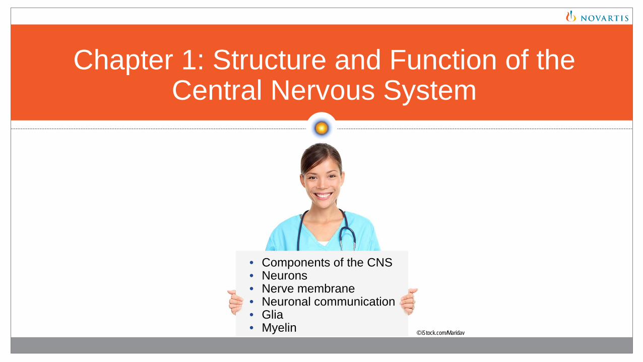
Chapter 1: Structure and Function of the Central Nervous System
• Components of the CNS• Neurons• Nerve membrane• Neuronal communication• Glia• Myelin © iStock.com/Maridav

Components of the Nervous System
• The nervous system is divided into central and peripheral components
• The central nervous system, or CNS, consists of the brain and spinal cord
• The peripheral nervous system (PNS) includes sensory neurons that link sensory receptors, on or near the surface of the body, with relevant processing circuits in the CNS
Components of the Nervous System© iStock.com/ikuvshinov
Purves D, et al, eds. Studying the nervous system. In: Neuroscience. 5th ed. Sunderland, MA: Sinauer Associates, Inc; 2012:1-21.
6
Central Nervous System
Peripheral Nervous System
Brain
Spinal Cord
Cranial Nerves
Spinal Nerves

Brain and Spinal Cord
• The structure of the brain includes the cerebral hemispheres, diencephalon, cerebellum, and brainstem2
• The brainstem contains nerves that send and receive signals between the forebrain and the spinal cord3
• The interior of the spinal cord consists of gray matter that is surrounded by white matter2,3
• Sensory information first travels through the spinal nerves to the spinal cord via the dorsal roots2
• Motor commands can then leave the spinal cord via the ventral roots2
Internal Anatomy of the Spinal Cord4Central Nervous System1
© iStock.com/Dr_MicrobeSpinal cord
Cerebrum
Cerebellum
Brainstem:MidbrainPonsMedulla oblongata
1. Tortora GJ, Derrickson B. The brain and cranial nerves. In: Roesch B, et al, eds. Principles of Anatomy and Physiology. 12th ed. Hoboken, NJ: John Wiley & Sons, Inc; 2009:495-545.2. Purves D, et al, eds. Studying the nervous system. Neuroscience. 5th ed. Sunderland, MA: Sinauer Associates, Inc; 2012:1-21.3. Purves D, et al, eds. Lower motor neuron circuits and motor control. Neuroscience. 5th ed. Sunderland, MA: Sinauer Associates, Inc; 2012:353-374.4. Tortora GJ, Derrickson B. The spinal cord and spinal nerves. In: Roesch B, et al, eds. Principles of Anatomy and Physiology. 12th ed. Hoboken, NJ: John Wiley & Sons, Inc; 2009:460-494.
7
White matter
Spinal nerve
Gray matter
Vertebra
© iStock.com/7activestudio
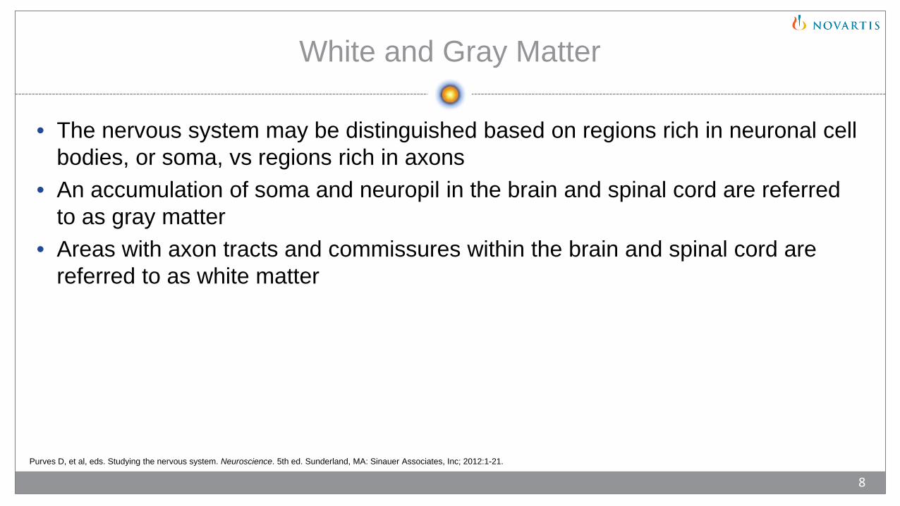
White and Gray Matter
• The nervous system may be distinguished based on regions rich in neuronal cell bodies, or soma, vs regions rich in axons
• An accumulation of soma and neuropil in the brain and spinal cord are referred to as gray matter
• Areas with axon tracts and commissures within the brain and spinal cord are referred to as white matter
Purves D, et al, eds. Studying the nervous system. Neuroscience. 5th ed. Sunderland, MA: Sinauer Associates, Inc; 2012:1-21.
8
Cell Body (Soma)
1. Purves D, et al, eds. Studying the nervous system. Neuroscience. 5th ed. Sunderland, MA: Sinauer Associates, Inc; 2012:1-21.2. Tortora GJ, Derrickson B. Nervous tissue. In: Roesch B, et al, eds. Principles of Anatomy and Physiology. 12th ed. Hoboken, NJ: John Wiley & Sons, Inc; 2009:415-459.
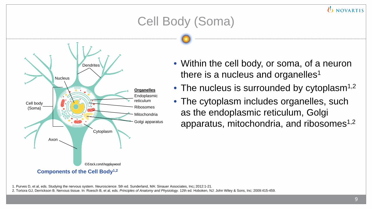
• Within the cell body, or soma, of a neuron there is a nucleus and organelles1
• The nucleus is surrounded by cytoplasm1,2
• The cytoplasm includes organelles, such as the endoplasmic reticulum, Golgi apparatus, mitochondria, and ribosomes1,2
9
© iStock.com/shopplaywood
Components of the Cell Body1,2
Cell body (Soma)
Axon
OrganellesEndoplasmicreticulumRibosomes
Mitochondria
Golgi apparatus
Dendrites
Nucleus
Cytoplasm
Dendrites and Axons
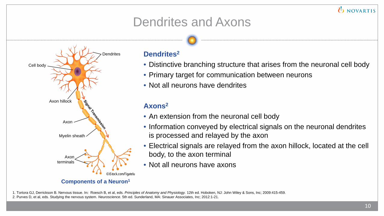
Dendrites2
• Distinctive branching structure that arises from the neuronal cell body• Primary target for communication between neurons• Not all neurons have dendrites
Axons2
• An extension from the neuronal cell body• Information conveyed by electrical signals on the neuronal dendrites
is processed and relayed by the axon• Electrical signals are relayed from the axon hillock, located at the cell
body, to the axon terminal• Not all neurons have axons
© iStock.com/Tigatelu
1. Tortora GJ, Derrickson B. Nervous tissue. In: Roesch B, et al, eds. Principles of Anatomy and Physiology. 12th ed. Hoboken, NJ: John Wiley & Sons, Inc; 2009:415-459.2. Purves D, et al, eds. Studying the nervous system. Neuroscience. 5th ed. Sunderland, MA: Sinauer Associates, Inc; 2012:1-21.
10
Components of a Neuron1
Cell body
Axon hillock
Dendrites
Axon
Axon terminals
Myelin sheath

Ion Pumps and Ion Channels
Nerve Membrane• Neurons transmit information through electrical signals from the flow of ions across their neuronal membrane1
• The flow of ions depends on differences in ion concentration and proteins within the cell membrane1
• Ion pumps and ion channels are paths that allow ions to move across the plasma membrane and generate electrical signals1,2
© iStock.com/ttsz
1. Tortora GJ, Derrickson B. Nervous tissue. In: Roesch B, et al, eds. Principles of Anatomy and Physiology. 12th ed. Hoboken, NJ: John Wiley & Sons, Inc; 2009:415-459.2. Gadsby DC. Nat Rev Mol Cell Biol. 2009;10(5):344-352.3. Marieb EN, Hoehn K. Fundamentals of the nervous system and nervous tissue. In: Human Anatomy and Physiology. 10th ed. Boston, MA: Pearson; 2016:388-429.
11
Ion Channels1,2
• Allow ions to diffuse down concentration gradient• Cause selective permeability to certain ions
Outside cell
ReceptorIon
Neuronal Membrane
Inside cell
Ion Pumps2,3
• Actively move ions against concentration gradient• Create ion concentration gradients
Ion
Neurotransmitter ReceptorClosedOpen
Neuronal Communication
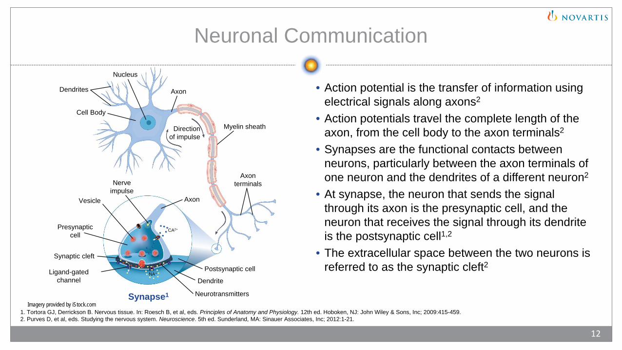
• Action potential is the transfer of information using electrical signals along axons2
• Action potentials travel the complete length of the axon, from the cell body to the axon terminals2
• Synapses are the functional contacts between neurons, particularly between the axon terminals of one neuron and the dendrites of a different neuron2
• At synapse, the neuron that sends the signal through its axon is the presynaptic cell, and the neuron that receives the signal through its dendrite is the postsynaptic cell1,2
• The extracellular space between the two neurons is referred to as the synaptic cleft2
1. Tortora GJ, Derrickson B. Nervous tissue. In: Roesch B, et al, eds. Principles of Anatomy and Physiology. 12th ed. Hoboken, NJ: John Wiley & Sons, Inc; 2009:415-459.2. Purves D, et al, eds. Studying the nervous system. Neuroscience. 5th ed. Sunderland, MA: Sinauer Associates, Inc; 2012:1-21.
12
Synapse1
Cell Body
Dendrites
Axon terminals
Myelin sheath
Axon
Nucleus
Direction of impulse
Nerve impulse
Synaptic cleft
Presynaptic cell
AxonVesicle
Ligand-gated channel Dendrite
Postsynaptic cell
Neurotransmitters
CA2+
NA+
Imagery provided by iStock.com

Electrical and Chemical Synapses
• Electrical synapses allow current to flow through gap junctions, which are specialized membrane channels that allow communication between two cells2
• Chemical synapses use ion channels but require the help of neurotransmitters to transmit information4
• Neurotransmitters are small-molecule neurotransmitters or neuropeptides2
1. Purves D, et al, eds. Synaptic transmission. In: Neuroscience. 5th ed. Sunderland, MA: Sinauer Associates, Inc; 2012:77-108.2. Tortora GJ, Derrickson B. Nervous tissue. In: Roesch B, et al, eds. Principles of Anatomy and Physiology. 12th ed. Hoboken, NJ: John Wiley & Sons, Inc; 2009:415-459.3. Pereda AE. Nat Rev Neurosci. 2014;15(4):250-263.4. Marieb EN, Hoehn K. Fundamentals of the nervous system and nervous tissue. In: Human Anatomy and Physiology. 10th ed. Boston, MA: Pearson; 2016:388-429.
13
Electrical Synapse1-3 Chemical Synapse1-3
Presynaptic neuron
Postsynaptic neuronGap Junction
Connexons
Synaptic cleft
Synaptic vesicles
IonsIons
Presynaptic neuron
Postsynaptic neuron
Neuronal Transmission
Tortora GJ, Derrickson B. Nervous tissue. In: Roesch B, et al, eds. Principles of Anatomy and Physiology. 12th ed. Hoboken, NJ: John Wiley & Sons, Inc; 2009:415-459.
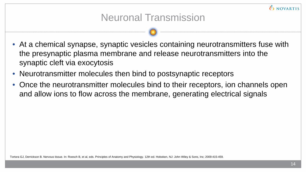
• At a chemical synapse, synaptic vesicles containing neurotransmitters fuse with the presynaptic plasma membrane and release neurotransmitters into the synaptic cleft via exocytosis
• Neurotransmitter molecules then bind to postsynaptic receptors• Once the neurotransmitter molecules bind to their receptors, ion channels open
and allow ions to flow across the membrane, generating electrical signals
14

Glia (Neuroglia)
• Supporting cells of the nervous system that do not play a direct role in synaptic interactions and electrical signaling
• Glial cells have a cell body• Glia have many functions, including aiding or, in some instances, impeding
recovery from neural injury• Have complex glial processes that extend from their cell bodieso Glial processes do not act like axons and dendrites
• Three types of glial cells in the CNSo Astrocyteso Microgliao Oligodendrocytes
Purves D, et al, eds. Studying the nervous system. Neuroscience. 5th ed. Sunderland, MA: Sinauer Associates, Inc; 2012:1-21.
15
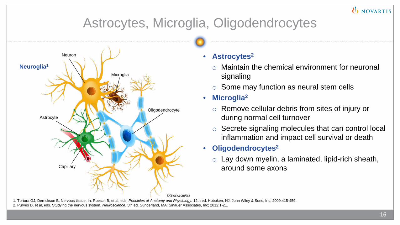
Neuron
Microglia
OligodendrocyteAstrocyte
Capillary
Astrocytes, Microglia, Oligodendrocytes
• Astrocytes2
o Maintain the chemical environment for neuronal signaling
o Some may function as neural stem cells• Microglia2
o Remove cellular debris from sites of injury or during normal cell turnover
o Secrete signaling molecules that can control local inflammation and impact cell survival or death
• Oligodendrocytes2
o Lay down myelin, a laminated, lipid-rich sheath, around some axons
Neuroglia1
© iStock.com/ttsz1. Tortora GJ, Derrickson B. Nervous tissue. In: Roesch B, et al, eds. Principles of Anatomy and Physiology. 12th ed. Hoboken, NJ: John Wiley & Sons, Inc; 2009:415-459.2. Purves D, et al, eds. Studying the nervous system. Neuroscience. 5th ed. Sunderland, MA: Sinauer Associates, Inc; 2012:1-21.
16
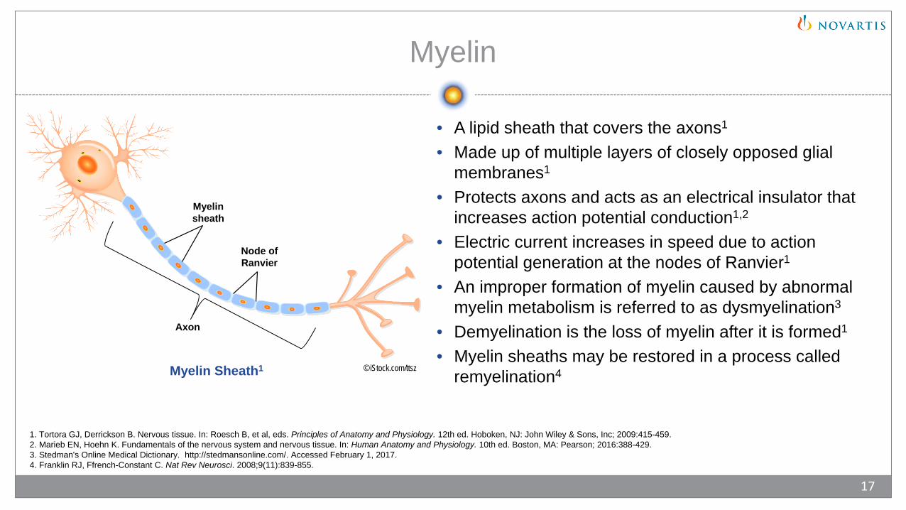
Myelin
• A lipid sheath that covers the axons1
• Made up of multiple layers of closely opposed glial membranes1
• Protects axons and acts as an electrical insulator that increases action potential conduction1,2
• Electric current increases in speed due to action potential generation at the nodes of Ranvier1
• An improper formation of myelin caused by abnormal myelin metabolism is referred to as dysmyelination3
• Demyelination is the loss of myelin after it is formed1
• Myelin sheaths may be restored in a process called remyelination4© iStock.com/ttsz
1. Tortora GJ, Derrickson B. Nervous tissue. In: Roesch B, et al, eds. Principles of Anatomy and Physiology. 12th ed. Hoboken, NJ: John Wiley & Sons, Inc; 2009:415-459.2. Marieb EN, Hoehn K. Fundamentals of the nervous system and nervous tissue. In: Human Anatomy and Physiology. 10th ed. Boston, MA: Pearson; 2016:388-429.3. Stedman's Online Medical Dictionary. http://stedmansonline.com/. Accessed February 1, 2017.4. Franklin RJ, Ffrench-Constant C. Nat Rev Neurosci. 2008;9(11):839-855.
17
Myelin Sheath1
Axon
Node of Ranvier
Myelin sheath

Chapter 2: Immune System
• Innate immunity• Transition From Innate
to Adaptive Immunity• Adaptive Immunity• Inflammatory Response
© iStock.com/RTimages

Innate Immunity
• A defense system present in an individual since birth• Provides initial and immediate protection against pathogenso Nonspecific for any particular pathogen
• Consists of 2 componentso Physical and chemical barriers
• Skin and epithelial surfaces• Antimicrobial proteins secreted at mucosal surfaces
o Cells that destroy or immobilize pathogens• Macrophages• Mast cells• Dendritic cells• Neutrophils
Murphy K. Basic concepts in immunology. In: Janeway’s Immunobiology. 8th ed. New York, NY: Garland Science; 2012:1-36.
19

Transition From Innate to Adaptive Immunity
• The innate immune system transitions to the adaptive immune system through the activation of T-cells and B-cellso Antigen-presenting
cells (APCs) activate T-cells to respond to antigens and initiate the adaptive immune response
Transition From the Innate to the Adaptive Immune Response1
1. Alberts B, et al. The adaptive immune system. In: Molecular Biology of the Cell. 5th ed. New York, NY: Garland Science; 2008:1539-1601.2. Murphy K. T Cell-Mediated Immunity. In: Janeway’s Immunobiology. 8th ed. New York, NY: Garland Science; 2012:334-386.
20
Republished with permission of Garland Science from Alberts B, et al. Molecular Biology of the Cell, 5th ed. © 2008; permission conveyed through Copyright Clearance Center, Inc.
1,2
1 1
1
Adaptive Immunity
• Immune response that is modified over the course of an individual’s life1,2
• Slower to respond than innate response1,2
• Provides a highly specific response that remembers previous pathogen exposure (immunological memory)1,2
• Key components are lymphocytes1
o White blood cells that include T-cells and B-cellso Able to recognize almost any antigen that enters the bodyo When a T-cell recognizes a specific antigen, it undergoes clonal expansion to generate a clone
of effector T-cells of identical antigen specificityo Responsible for long-lasting immunity
• Divided into 2 response systems1,2
o Antibody-mediated, or humoral, immunity (mediated by B-cells) 1,2
o Cell-mediated immunity (mediated by T-cells)1
1. Alberts B, et al. The adaptive immune system. In: Molecular Biology of the Cell. 5th ed. New York, NY: Garland Science; 2008:1539-1601.2. Murphy K. Basic concepts in immunology. In: Janeway’s Immunobiology. 8th ed. New York, NY: Garland Science; 2012:1-36.
21
Humoral Immunity
• Protects the extracellular spaces of the body1
• Mediated by antibodies2
o Antibodies produced by B-cells bind to antigens to initiate the immune response3
o Antibodies are highly specific for each antigen3
o Antibodies can recognize the various components of an antigen3
1. Murphy K. The humoral immune response. In: Janeway’s Immunobiology. 8th ed. New York, NY: Garland Science; 2012:387-428. 2. Murphy K. Basic concepts in immunology. In: Janeway’s Immunobiology. 8th ed. New York, NY: Garland Science; 2012:1-36. 3. Alberts B, et al. The adaptive immune system. In: Molecular Biology of the Cell. 5th ed. New York, NY: Garland Science; 2008:1539-1601.
22
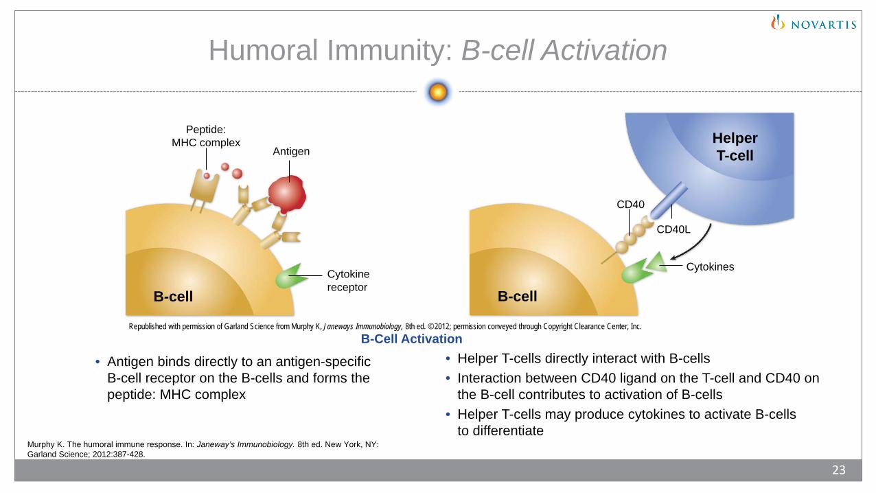
Humoral Immunity: B-cell Activation
• Antigen binds directly to an antigen-specific B-cell receptor on the B-cells and forms the peptide: MHC complex
• Helper T-cells directly interact with B-cells• Interaction between CD40 ligand on the T-cell and CD40 on
the B-cell contributes to activation of B-cells• Helper T-cells may produce cytokines to activate B-cells
to differentiate
B-Cell Activation
Murphy K. The humoral immune response. In: Janeway’s Immunobiology. 8th ed. New York, NY: Garland Science; 2012:387-428.
23
HelperT-cell
B-cell
Cytokines
CD40L
CD40
Cytokinereceptor
B-cell
Peptide:MHC complex
Antigen
Republished with permission of Garland Science from Murphy K, Janeways Immunobiology, 8th ed. © 2012; permission conveyed through Copyright Clearance Center, Inc.

Humoral Immunity: B-cell Differentiation
Once B-cell activation is complete, B-cells can differentiate into1:• Plasma B-cells2
o Secrete antibodies at a high rate, which bind to antigensoMost plasma B-cells live for less than 1 week
• Memory B-cells2
o Provide long-term immune protection against the same antigeno Upon second exposure to the same antigen, memory cells give rise to either
effector cells or more memory cells
1. Murphy K. The humoral immune response. In: Janeway’s Immunobiology. 8th ed. New York, NY: Garland Science; 2012:387-428.2. Alberts B, et al. The adaptive immune system. In: Molecular Biology of the Cell. 5th ed. New York, NY: Garland Science; 2008:1539-1601.
24
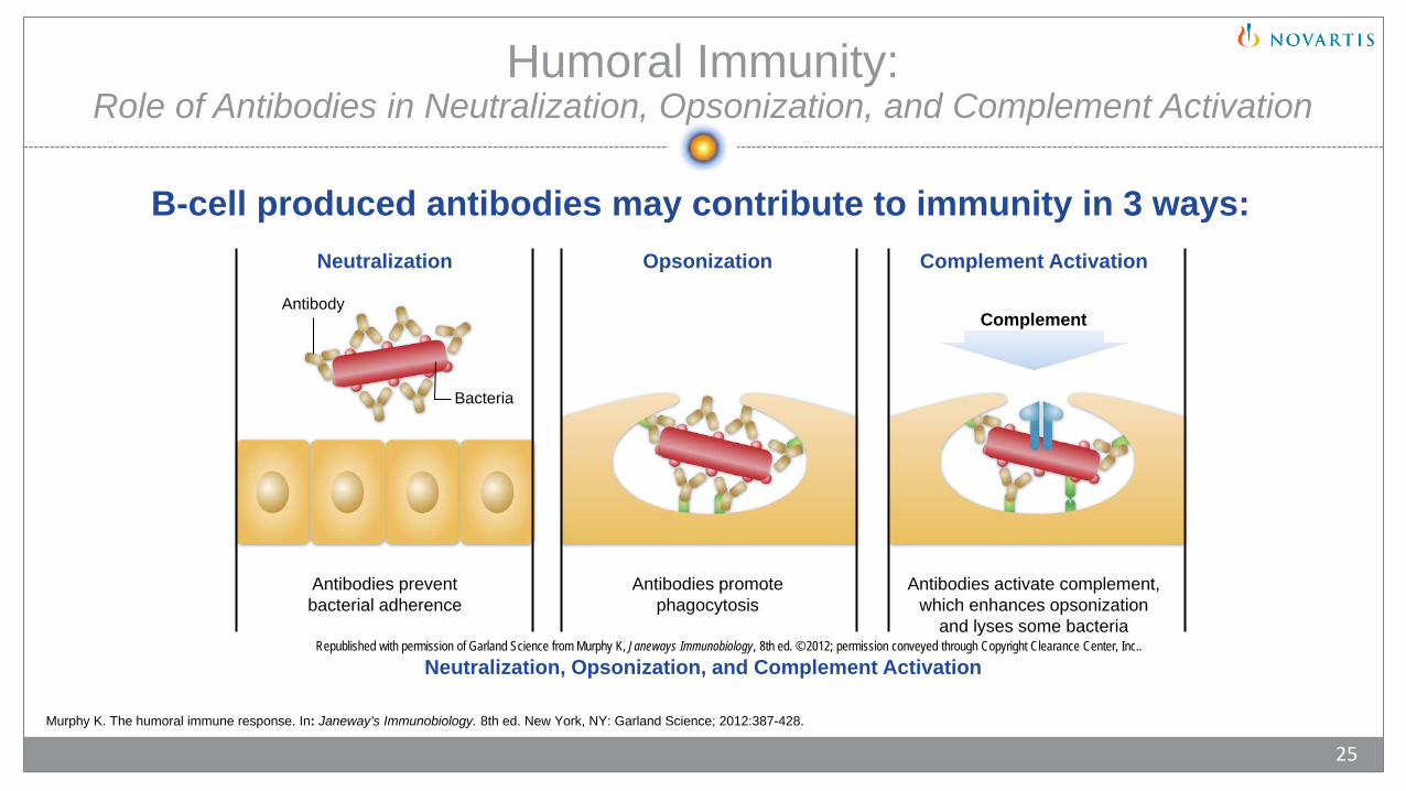
Humoral Immunity: Role of Antibodies in Neutralization, Opsonization, and Complement Activation
B-cell produced antibodies may contribute to immunity in 3 ways:
Neutralization, Opsonization, and Complement Activation
Murphy K. The humoral immune response. In: Janeway’s Immunobiology. 8th ed. New York, NY: Garland Science; 2012:387-428.
25
Antibody
Bacteria
Antibodies preventbacterial adherence
Antibodies promotephagocytosis
Antibodies activate complement,which enhances opsonization
and lyses some bacteria
Complement
Neutralization Opsonization Complement Activation
Republished with permission of Garland Science from Murphy K, Janeways Immunobiology, 8th ed. © 2012; permission conveyed through Copyright Clearance Center, Inc..
Cell-Mediated Immunity
• Immune response mediated by T-cells1
• Mature T-cells circulate between blood and peripheral lymphoid tissues until they encounter their specific antigen1,2
• Mature T-cells that have not encountered their specific antigen are referred to as naïve T-cells1,2
• Once a naïve T-cell recognizes its specific antigen in the form of a peptide: MHC complex on the surface of an APC, it will undergo activation and clonal expansion and produce several types of effector T-cells in a process called priming3
• Proliferation and differentiation into effector T-cells are also dependent on cytokines2
• All effector T-cells either kill infected cells or help stimulate responses of other cells2
1. Murphy K. Basic concepts in immunology. In: Janeway’s Immunobiology. 8th ed. New York, NY: Garland Science; 2012:1-36. 2. Alberts B, et al. The adaptive immune system. In: Molecular Biology of the Cell. 5th ed. New York, NY: Garland Science; 2008:1539-1601.3. Murphy K. T-cell mediated immunity. In: Janeway’s Immunobiology. 8th ed. New York, NY: Garland Science; 2012:335-386.
26

Cell-Mediated Immunity: Effector T-cells
• Effector T-cells1
o Control various aspects of cell-mediated immune response• Cytotoxic T-cells1
o Directly attack and destroy pathogen-infected cells that display peptide fragments of pathogens (eg, viruses), on class I MHC molecules
• Helper T-cells1
o Recognize fragments of antigens and produce cytokines that activate macrophages, which ultimately destroy the intracellular microorganism
o Stimulate B-cells to produce antibodies• Suppressor T-cells1
o Regulatory cells that suppress T-cell activity and help prevent the development of autoimmunity
• Memory T-cells1
o Remember the antigen for future encounters, and ensure that subsequent exposure produces a more rapid response
Cell-Mediated Response1,2
1. Alberts B, et al. The adaptive immune system. In: Molecular Biology of the Cell. 5th ed. New York, NY: Garland Science; 2008:1539-1601.2. Murphy K. The humoral immune response. In: Janeway’s Immunobiology. 8th ed. New York, NY: Garland Science; 2012:387-428.
27
Inflammatory Response: Components of Innate and Adaptive Immunity
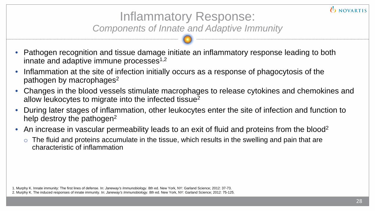
• Pathogen recognition and tissue damage initiate an inflammatory response leading to both innate and adaptive immune processes1,2
• Inflammation at the site of infection initially occurs as a response of phagocytosis of the pathogen by macrophages2
• Changes in the blood vessels stimulate macrophages to release cytokines and chemokines and allow leukocytes to migrate into the infected tissue2
• During later stages of inflammation, other leukocytes enter the site of infection and function to help destroy the pathogen2
• An increase in vascular permeability leads to an exit of fluid and proteins from the blood2
o The fluid and proteins accumulate in the tissue, which results in the swelling and pain that are characteristic of inflammation
1. Murphy K. Innate immunity: The first lines of defense. In: Janeway’s Immunobiology. 8th ed. New York, NY: Garland Science; 2012: 37-73.2. Murphy K. The induced responses of innate immunity. In: Janeway’s Immunobiology. 8th ed. New York, NY: Garland Science; 2012: 75-125.
28
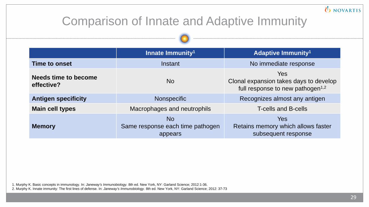
Comparison of Innate and Adaptive Immunity
Innate Immunity1 Adaptive Immunity1
Time to onset Instant No immediate response
Needs time to become effective? No
YesClonal expansion takes days to develop
full response to new pathogen1,2
Antigen specificity Nonspecific Recognizes almost any antigenMain cell types Macrophages and neutrophils T-cells and B-cells
MemoryNo
Same response each time pathogen appears
YesRetains memory which allows faster
subsequent response
1. Murphy K. Basic concepts in immunology. In: Janeway’s Immunobiology. 8th ed. New York, NY: Garland Science; 2012:1-36.2. Murphy K. Innate immunity: The first lines of defense. In: Janeway’s Immunobiology. 8th ed. New York, NY: Garland Science; 2012: 37-73
29

Chapter 3: What Is Multiple Sclerosis?
• Overview of MS• Causes of MS
© iStock.com/adiekoetter
Overview of MS
• What is MS?1
o An autoimmune disease of the CNS targeting myelin, MS is mediated by both the adaptive and innate immune system cells
o Characterized by inflammation, demyelination, and axonal loss• Epidemiology of MSo Approximately 2.3 million people with MS worldwide2
o Approximately 400,000 people with MS in the U.S.3o Predominately affects women2
o Disease onset between 20 and 50 years of age2
1. Birnbaum G. Factors involved in causing multiple sclerosis. In: Multiple Sclerosis: Clinician’s Guide to Diagnosis and Treatment. New York, NY: Oxford University Press, Inc; 2009:7-13.2. National Multiple Sclerosis Society. What Is Multiple Sclerosis? http://www.nationalmssociety.org/NationalMSSociety/media/MSNationalFiles/Brochures/Brochure-What-Is-MS.pdf.
Published June 2016. Accessed February 7, 2017.3. National Multiple Sclerosis Society. 2014 Annual Report: Mid-America Chapter. http://www.nationalmssociety.org/NationalMSSociety/media/Mid-
America/About%20this%20Chapter/Files/KSG-2014-Annual-Report.pdf?ext=.pdf. Accessed February 7, 2017.
31© iStock/Leontura
Causes of MS
• Exact cause of MS is unknown• Theorized to be an autoimmune disease• Studies suggest that the following factors may play a role in disease
development:o Geneticso Environmental factorso Infectionso Immunologic factors
• Please note: the following 4 slides present potential causes of MS that have yet to be proven
Birnbaum G. Factors involved in causing multiple sclerosis. In: Multiple Sclerosis: Clinician’s Guide to Diagnosis and Treatment. New York, NY: Oxford University Press, Inc; 2009:7-13.
32
Causes of MS: Genetics
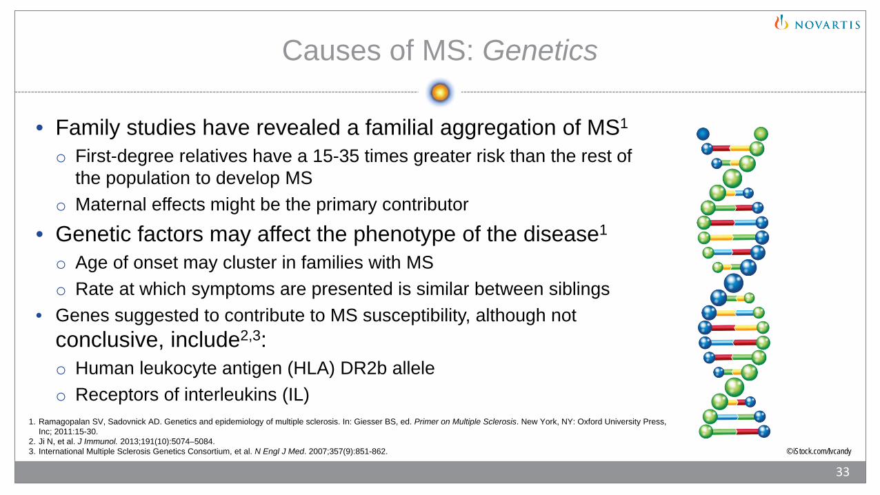
• Family studies have revealed a familial aggregation of MS1
o First-degree relatives have a 15-35 times greater risk than the rest of the population to develop MS
o Maternal effects might be the primary contributor• Genetic factors may affect the phenotype of the disease1
o Age of onset may cluster in families with MSo Rate at which symptoms are presented is similar between siblings
• Genes suggested to contribute to MS susceptibility, although not conclusive, include2,3:o Human leukocyte antigen (HLA) DR2b alleleo Receptors of interleukins (IL)
© iStock.com/Ivcandy
1. Ramagopalan SV, Sadovnick AD. Genetics and epidemiology of multiple sclerosis. In: Giesser BS, ed. Primer on Multiple Sclerosis. New York, NY: Oxford University Press, Inc; 2011:15-30.
3. International Multiple Sclerosis Genetics Consortium, et al. N Engl J Med. 2007;357(9):851-862. 2. Ji N, et al. J Immunol. 2013;191(10):5074–5084.
33
Imagery provided by iStock.com
Causes of MS: Environmental Factors
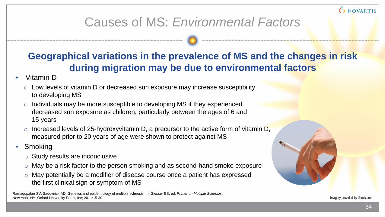
• Vitamin Do Low levels of vitamin D or decreased sun exposure may increase susceptibility
to developing MSo Individuals may be more susceptible to developing MS if they experienced
decreased sun exposure as children, particularly between the ages of 6 and 15 years
o Increased levels of 25-hydroxyvitamin D, a precursor to the active form of vitamin D, measured prior to 20 years of age were shown to protect against MS
• Smokingo Study results are inconclusiveo May be a risk factor to the person smoking and as second-hand smoke exposureo May potentially be a modifier of disease course once a patient has expressed
the first clinical sign or symptom of MSRamagopalan SV, Sadovnick AD. Genetics and epidemiology of multiple sclerosis. In: Giesser BS, ed. Primer on Multiple Sclerosis. New York, NY: Oxford University Press, Inc; 2011:15-30.
34
Geographical variations in the prevalence of MS and the changes in risk during migration may be due to environmental factors
Causes of MS: Infections
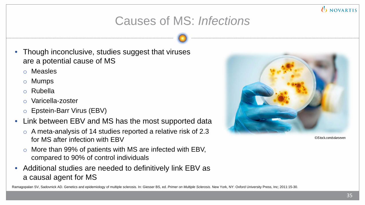
• Though inconclusive, studies suggest that viruses are a potential cause of MSo Measleso Mumpso Rubellao Varicella-zostero Epstein-Barr Virus (EBV)
• Link between EBV and MS has the most supported datao A meta-analysis of 14 studies reported a relative risk of 2.3
for MS after infection with EBVo More than 99% of patients with MS are infected with EBV,
compared to 90% of control individuals• Additional studies are needed to definitively link EBV as
a causal agent for MS
© iStock.com/solarseven
Ramagopalan SV, Sadovnick AD. Genetics and epidemiology of multiple sclerosis. In: Giesser BS, ed. Primer on Multiple Sclerosis. New York, NY: Oxford University Press, Inc; 2011:15-30.
35
Causes of MS: Immunologic Factors
• T-cells and B-cells of the adaptive immune system function by recognizing and reacting against foreign antigens and self-antigens
• T-cells and B-cells normally do not develop an immune response that could be harmful to the host
• When the adaptive immune system targets self-antigens, the result may be the development of an autoimmune disease
• MS is hypothesized to be an autoimmune diseaseo In genetically susceptible individuals, dysregulation of the normal response to one’s
own tissue antigens may be initiated by certain environmental exposureso Immune mediators have been detected at sites of inflammation and demyelination
typical of MS
36
Bar-Or A. Semin Neurol. 2008;28(1):29-45.

© iStock.com/londoneye
Chapter 4: Immunopathogenesis of Multiple Sclerosis

Immunology of MS: Autoantigens
• Myelin proteins primarily have been implicated in the search for MS autoantigens• T-cells reactive to myelin proteins have been found in patients with MS• Myelin proteins include:o Myelin basic proteinso Myelin associated glycoproteinso Myelin oligodendrocyte glycoproteino Proteolipid protein
• Non-myelin antigen has been detectedo Heat-shock protein α-β-crystallin
Sospedra M, Martin R. Annu Rev Immunol. 2005;23:683-747.
38

Immunology of MS: T-cells
• CD4+ T-cellso Studies suggest CD4+ autoreactive T-cells induce inflammation in MSo CD4+ autoreactive T-cells may have a…
• Th1 (helper T-cell) phenotype, characterized by the production of interferon-γ (IFN-gamma)• Th17 (helper T-cell) phenotype, characterized by the production of IL-17
o IFN –γ and IL-17 are pro-inflammatory cytokineso Important for differentiation and/or activation and expansion of Th17 cells: IL-6, TGF-β, IL-21, and IL-23o IL-17 has been detected in MS brains with acute and chronic active lesionso Th17 cells have been shown to migrate across the blood-brain barrier (BBB) and activate a process that can
lead to the initiation and relapses in MS• CD8+ T-cells
o Present in inflammatory MS lesions• Regulatory T-cells
o Regulate autoimmune and inflammatory responses
Boppana S, et al. Mt Sinai J Med. 2011;78(2):207-220.
39
Immunology of MS: Humoral Immunity
• B-cells and immunoglobulins of the humoral immune response are associated with MS pathology and may be detected in early stages of the disease
• Higher levels of oligoclonal bands and cerebrospinal fluid immunoglobulins have been reported in patients with MS
• Studies indicate that antibodies in the CNS of patients with MS are the result of activation by antigens
• Anti-myelin antibodies have been reported in the serum of some patients with MS• Role of B-cells in MS is focused on their potential to react to particular antigens in the CNS and
produce auto-antibodies
Sospedra M, Martin R. Immunology of multiple sclerosis. In: Raine CS, et al, eds. Multiple Sclerosis: A Comprehensive Text. New York, NY: Saunders Elsevier; 2008:192-213.
40
Molecules Associated With MS Pathology: Microglia
• Microglia are quickly activated by myelin injury or pathogens• Microglia function similar to macrophageso Conduct phagocytosiso Participate in antigen presentationo Participate in demyelinationo Produce cytokines and chemokines
• Some are pro-inflammatory
Domingues HS, et al. Front Cell Dev Biol. 2016;4:71. doi: 10.3389/fcell.2016.00071.
41

Molecules Associated With MS: Dendritic Cells
• Function in the periphery as a bridge between the innate and adaptive immune systems1
o Within lymphoid tissue, dendritic cells present a processed antigen to T-cells that lead to T-cell activation and proliferation
• Inflammation in the brain tissue is accompanied by the recruitment and/or development of dendritic cells2
o Dendritic cells accumulate in the cerebrospinal fluid and CNS lesions3
• Dendritic cells are implicated in the regulation of autoimmune responses against myelin antigens3
o In patients with MS, dendritic cells have been observed engulfing myelin components and interacting with T-cells2
1. Alberts B, et al. The adaptive immune system. In: Molecular Biology of the Cell. 5th ed. New York, NY: Garland Science; 2008:1539-1601.2. Piccio L, Cross AH. Immunology of multiple sclerosis. In: Giesser BS, ed. Primer on Multiple Sclerosis. New York, NY: Oxford University Press; 2011:47-59.3. Nuyts AH, et al. Mult Scler. 2013;19(8):995-1002.
42

Molecules Associated With MS: Pro-Inflammatory Cytokines
Pro-inflammatory cytokines• Functions1
o Peripheral immune activationo Improving the passage of activated immune cells into the CNSo Directly damaging oligodendrocytes, myelin, and/or axons
• IFN-γ2
o Anti-T-cell proliferative activity and induces T-cell apoptosis1
• IL-172
o May function to breach the BBB, thereby allowing other immune and inflammatory cells into the brain1
• TNF-α2
o Functions in the expression of B7 and MHC class II, as well as stimulation of IL-12 to produce IFN-γ1
o Drugs initiated to block TNF-α in patients with MS actually resulted in worsening of disease1
• Although reason is unclear, it is suggested that TNF-α in the CNS may have neuroprotective properties• IL-62
o Both pro-inflammatory and anti-inflammatory effects1
1. Piccio L, Cross AH. Immunology of multiple sclerosis. In: Giesser BS. ed. Primer on Multiple Sclerosis. New York, NY: Oxford University Press; 2011:47-59.2. Boppana S, et al. Immunologic aspects of multiple sclerosis. Mt Sinai J Med. 2011;78(2):207-220.
43
Molecules Associated With MS: Anti-Inflammatory Cytokines
Anti-inflammatory cytokines• Function1
o Suppresses pro-inflammatory cytokine production• IL-102
o Suppresses expression of MHC class II, adhesion molecules, and costimulatory molecules on macrophages and dendritic cells1
• TGF-β2
o May suppress inflammation in the late stages of MS1
• IL-6 has both pro-inflammatory and anti-inflammatory effects1
o Anti-inflammatory cytokine effects• Inhibits pro-inflammatory cytokines (IL-1β and TNF-α)1
• Inhibits metalloproteinases1
1. Piccio L, Cross AH. Immunology of multiple sclerosis. In: Giesser BS, ed. Primer on Multiple Sclerosis. New York, NY: Oxford University Press; 2011:47-59.2. Boppana S, et al. Immunologic aspects of multiple sclerosis. Mt Sinai J Med. 2011;78(2):207-220.
44

Molecules Associated With MS: Adhesion Molecules
• Allow circulating cells of the immune system to access the CNS to cause inflammation, demyelination, axonal injury, and gliosis1
• Adhesion molecules such as selectins and integrins, interact with immune cells to initiate migration through the BBB1
• In MS lesions, endothelial cells may express abnormally elevated levels of adhesion molecules2
o Intercellular adhesion molecules (ICAM)-1o Vascular cell adhesion molecules (VCAM)-1
• Antigens for ICAM-1 and VCAM-1 have been observed on inflammatory cells within MS lesions2
1. Piccio L, Cross AH. Immunology of multiple sclerosis. In: Giesser BS, ed. Primer on Multiple Sclerosis. New York, NY: Oxford University Press; 2011:47-59.2. Bar-Or A. The immunology of multiple sclerosis. Semin Neurol. 2008;28(1):29-45
45

Molecules Associated With MS: Chemokines
• Control leukocyte migration from the periphery to the CNS through the BBB• May preferentially traffic pro-inflammatory T-cells into the CNS• Induce and activate leukocyte adhesion molecules, which leads to
recruitment of leukocytes across the CNS• Altered levels of chemokines and their receptors have been detected
in patients with MS
Piccio L, Cross AH. Immunology of multiple sclerosis. In: Giesser BS, ed. Primer on Multiple Sclerosis. New York, NY: Oxford University Press; 2011:47-59.
46

Blood-Brain Barrier
• Protects the brain from toxins circulating in the bloodstream1
• Tight junctions between capillary endothelial cells in the brain restrict certain molecules from entering the CNS1,2
• Leukocyte entry into the CNS is a multistep process2
Blood-Brain Barrier
1. Taber’s Medical Dictionary. http://www.tabers.com/tabersonline/. Accessed February 21, 2017. 2. Man S, et al. Brain Pathol. 2007;17(2):243-250.
47
© iStock.com/CreVis2
Blood-Brain Barrier: Lipid-Mediated and Catalyzed Transport
• Molecules gain access to the brain through the BBB via lipid-mediated or catalyzed transport
• Lipid-mediated transporto Transport of lipid-soluble small molecules to gain access to the brain
• Catalyzed transporto Carrier-mediated transport is used for the passage of small-molecule nutrients,
thyroid hormones, and vitamins to the braino Receptor-mediated transport is used for the passage of endogenous large-molecule
peptides to the braino In contrast, active efflux transporters may be used, such as p-glycoprotein
Pardridge WM. Stroke. 2007;38(2 suppl):686-690.
48
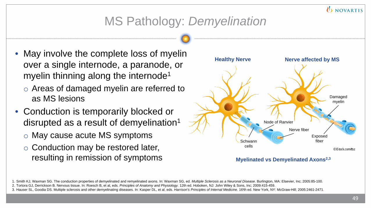
MS Pathology: Demyelination
• May involve the complete loss of myelin over a single internode, a paranode, or myelin thinning along the internode1
o Areas of damaged myelin are referred to as MS lesions
• Conduction is temporarily blocked or disrupted as a result of demyelination1
o May cause acute MS symptomso Conduction may be restored later,
resulting in remission of symptoms Myelinated vs Demyelinated Axons2,3
© iStock.com/ttsz
1. Smith KJ, Waxman SG. The conduction properties of demyelinated and remyelinated axons. In: Waxman SG, ed. Multiple Sclerosis as a Neuronal Disease. Burlington, MA: Elsevier, Inc; 2005:85-100.2. Tortora GJ, Derrickson B. Nervous tissue. In: Roesch B, et al, eds. Principles of Anatomy and Physiology. 12th ed. Hoboken, NJ: John Wiley & Sons, Inc; 2009:415-459.3. Hauser SL, Goodia DS. Multiple sclerosis and other demyelinating diseases. In: Kasper DL, et al, eds. Harrison’s Principles of Internal Medicine. 16tth ed. New York, NY: McGraw-Hill; 2005:2461-2471.
49
Healthy Nerve Nerve affected by MS
Node of Ranvier
Damaged myelin
Nerve fiberExposed
fiberSchwann cells

MS Pathology: Gliosis
• Dense fibrous scar or glial scar1,2
• Caused by the reaction of astrocytes to injury through several mechanisms2,3
o Proliferation3
o Hypertrophy3
o Expansion of their cytoplasm3
o Development of more cytoplasmic organelles3
• Forms in areas of demyelination, producing the lesions that are characteristic of MS2,3
1. Holmes S, et al. Expert Rev Mol Med. 2005;7(3):1-17. 2. Domingues HS, et al. Front Cell Dev Biol. 2016;4:71. doi: 10.3389/fcell.2016.00071.3. Herndon RM. Astrocytes: Structure and function. In: Herndon RM, ed. Multiple Sclerosis: Immunology, Pathology, and Pathophysiology. New York, NY: Demos Medical Publishing, Inc; 2003:25-30.
50

• Regardless of the stage of disease progression, axonal damage occurs during active demyelination1
• Some axonal loss present in all MS lesions, and damage to axons occurs in various degrees2
• Axonal transection, identified by terminalaxonal ovoids, was detected in all lesionsin a study by Trapp et al2o Axons shown undergoing active
demyelination (arrowheads)o Axon ends in a large terminal ovoid (arrow)
MS Pathology: Axonal Damage
Axonal Transection21. Slimp JC. Neurophysiology of multiple sclerosis. In: Giesser BS, ed. Primer on Multiple Sclerosis. New York, NY:
Oxford University Press, Inc; 2011:31-46.2. Trapp BD, et al. N Engl J Med. 1998;338(5):278-285.
51
From N Engl J Med, Trapp BD, et al, Axonal transection in the lesions of multiple sclerosis, 338, 278-285 © 1998 Massachusetts Medical Society. Reprinted with permission from Massachusetts Medical Society.

MS Pathology: Axonal Damage, Cont.
• Inflammation has been implicated in initiating axonal damage1-3
• Axons may be attacked through:o Antigen-specific events1,3
• Axons can be attacked by cytotoxic T-cellso Antigen-independent events1,2
• Damage by toxic products of macrophages and microglia2
• Toxic products may include proteases, cytokines, excitotoxins, and free radicals1
1. Lucchinetti CF. Taking a microscopic look at multiple sclerosis. In: Giesser BS, ed. Primer on Multiple Sclerosis. New York, NY: Oxford University Press, Inc; 2011:61-77.2. Trapp BD, et al. N Engl J Med. 1998;338(5):278-285.3. Boppana S, et al. Mt Sinai J Med. 2011;78(2):207-220.
52
MS Pathology: Atrophy
• May be initiated during the early stages of MS1
• Atrophy progression may occur without obvious lesion formation2
• Rate of atrophy may predict long-term disability3
• CNS atrophy in MS is likely the result of several factors1
o Loss of myelino Axonal loss or thinning
• Atrophy can be1: o Focal – affects the central white matter and can result in ventricular expansiono Global – results in reduced spinal cord area or brain volume
• The rate of brain atrophy in patients with MS is more than 5 times greater than in age-and gender-matched patients without MS4
1. Simon JH. Pathology of multiple sclerosis as revealed by in vivo magnetic resonance-based approaches. In: Multiple Sclerosis: Immunology, Pathology, and Pathophysiology. New York, NY: Demos Medical Publishing, Inc; 2003:199-214.
2. Joy JE, Johnston RB Jr, eds. Clinical and biological features. In: Multiple Sclerosis. Current Status and Strategies for the Future. Washington, DC: National Academy Press; 2001:29-104.3. Popescu V, et al. 10 .4. Rudick RA, et al. J Neurol Sci. 2009;282(1-2):106-111.
J Neurol Neurosurg Psychiatry. 2013;84(10):1082- 91
53
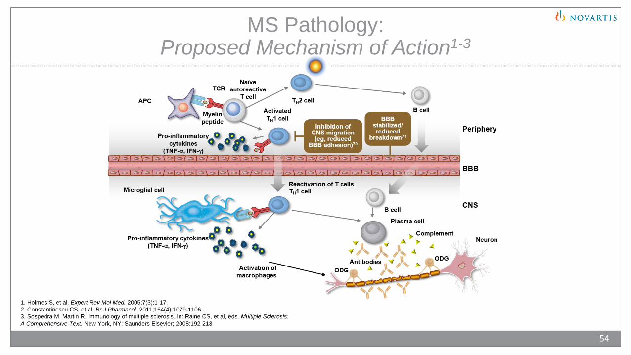
MS Pathology: Proposed Mechanism of Action1-3
1. Holmes S, et al. Expert Rev Mol Med. 2005;7(3):1-17. 2. Constantinescu CS, et al. Br J Pharmacol. 2011;164(4):1079-1106.3. Sospedra M, Martin R. Immunology of multiple sclerosis. In: Raine CS, et al, eds. Multiple Sclerosis: A Comprehensive Text. New York, NY: Saunders Elsevier; 2008:192-213
54

Chapter 5: Clinical Pathology of Multiple Sclerosis
• Common Clinical Symptoms of MS• Role of MRI in Confirming MS Clinical
Pathology• Neuropathophysiology Module Summary
© iStock.com/loco75
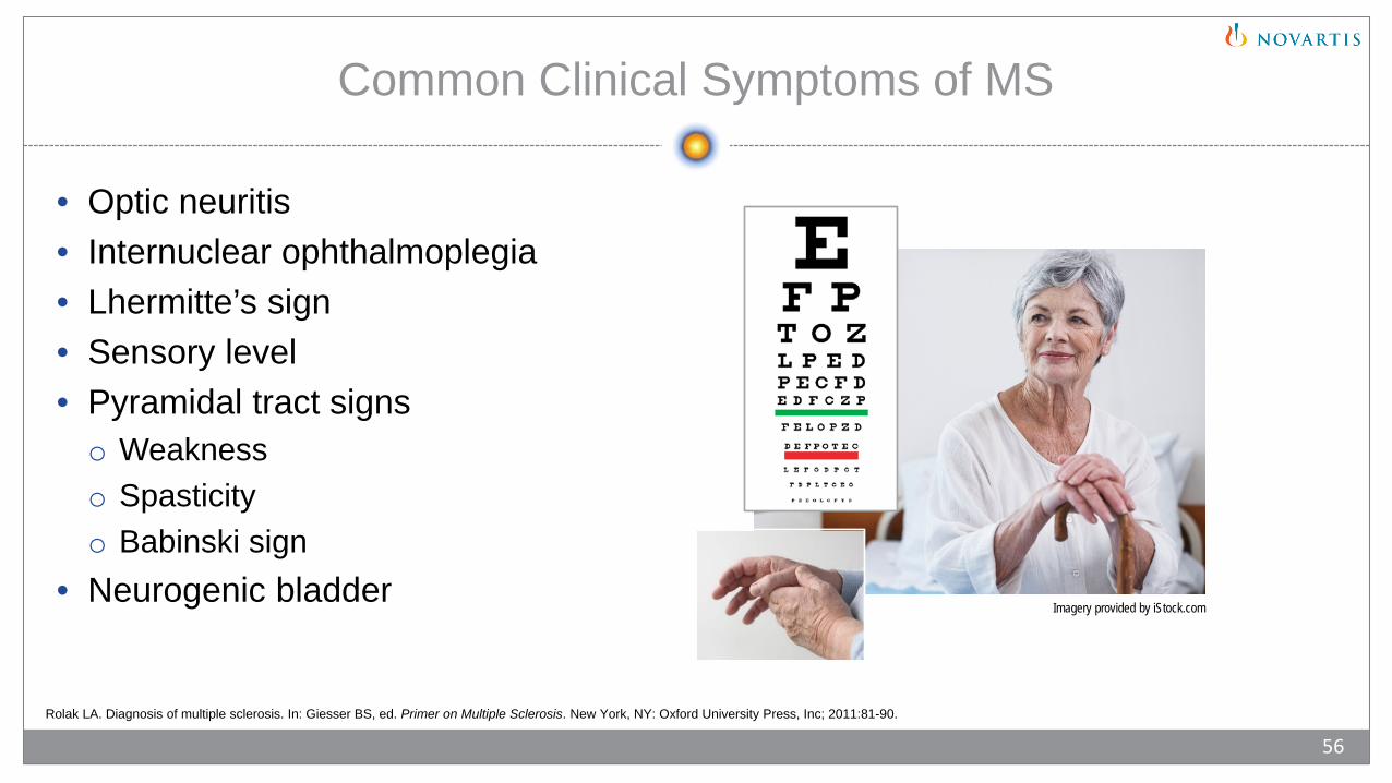
Common Clinical Symptoms of MS
• Optic neuritis• Internuclear ophthalmoplegia• Lhermitte’s sign• Sensory level• Pyramidal tract signso Weaknesso Spasticityo Babinski sign
• Neurogenic bladderImagery provided by iStock.com
Rolak LA. Diagnosis of multiple sclerosis. In: Giesser BS, ed. Primer on Multiple Sclerosis. New York, NY: Oxford University Press, Inc; 2011:81-90.
56
Role of Magnetic Resonance Imaging in Confirming MS Clinical Pathology
© iStock.com/ep_stock
Sicotte NL. Neuroimaging in multiple sclerosis. In: Giesser BS, ed. Primer on Multiple Sclerosis. New York, NY: Oxford University Press, Inc; 2011:91-113.
57
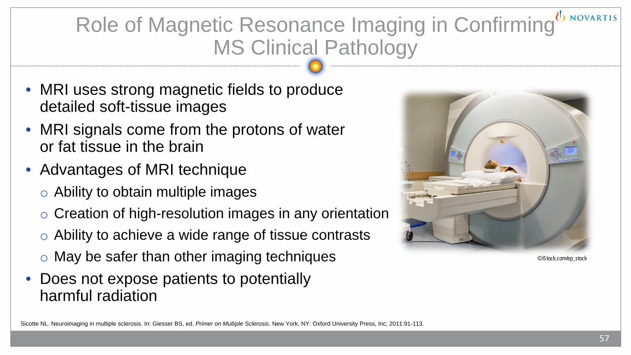
• MRI uses strong magnetic fields to produce detailed soft-tissue images
• MRI signals come from the protons of water or fat tissue in the brain
• Advantages of MRI techniqueo Ability to obtain multiple imageso Creation of high-resolution images in any orientationo Ability to achieve a wide range of tissue contrastso May be safer than other imaging techniques
• Does not expose patients to potentially harmful radiation
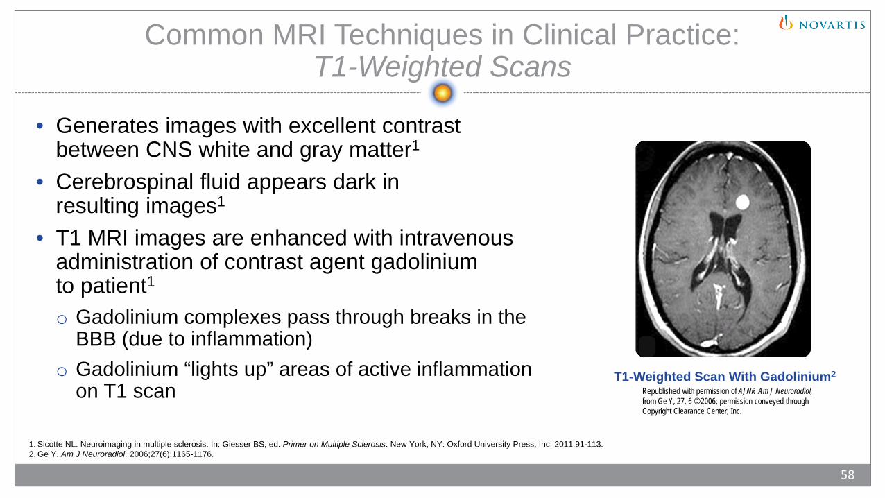
T1-Weighted Scan With Gadolinium2
Common MRI Techniques in Clinical Practice: T1-Weighted Scans
• Generates images with excellent contrast between CNS white and gray matter1
• Cerebrospinal fluid appears dark in resulting images1
• T1 MRI images are enhanced with intravenous administration of contrast agent gadolinium to patient1o Gadolinium complexes pass through breaks in the
BBB (due to inflammation)o Gadolinium “lights up” areas of active inflammation
on T1 scan
1. Sicotte NL. Neuroimaging in multiple sclerosis. In: Giesser BS, ed. Primer on Multiple Sclerosis. New York, NY: Oxford University Press, Inc; 2011:91-113.2. Ge Y. Am J Neuroradiol. 2006;27(6):1165-1176.
58
Republished with permission of AJNR Am J Neuroradiol,from Ge Y, 27, 6 © 2006; permission conveyed through Copyright Clearance Center, Inc.
Common MRI Techniques in Clinical Practice: T2-Weighted Scans
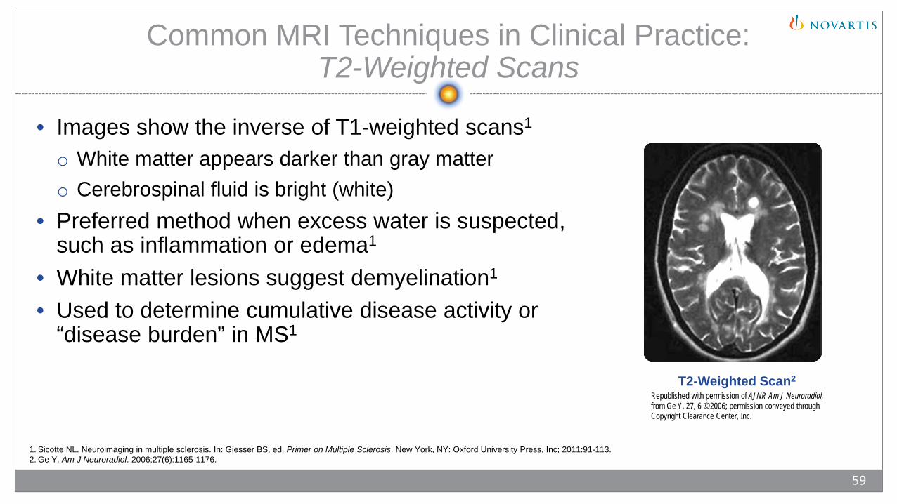
• Images show the inverse of T1-weighted scans1
o White matter appears darker than gray mattero Cerebrospinal fluid is bright (white)
• Preferred method when excess water is suspected, such as inflammation or edema1
• White matter lesions suggest demyelination1
• Used to determine cumulative disease activity or “disease burden” in MS1
59
T2-Weighted Scan2
1. Sicotte NL. Neuroimaging in multiple sclerosis. In: Giesser BS, ed. Primer on Multiple Sclerosis. New York, NY: Oxford University Press, Inc; 2011:91-113.2. Ge Y. Am J Neuroradiol. 2006;27(6):1165-1176.
Republished with permission of AJNR Am J Neuroradiol,from Ge Y, 27, 6 © 2006; permission conveyed through Copyright Clearance Center, Inc.

Common MRI Techniques in Clinical Practice: Fluid Attenuated Inversion Recovery
• Fluid attenuated inversion recovery (FLAIR) images are extensions of T2 images1
o Cerebrospinal fluid is blacko Accentuates areas of high T2 signal within white and
gray matter of braino Enhances periventricular lesions (figure)
• May show images that would not be clearly shown on T2 or protein density scans1
• May reveal the presence of juxtacortical lesions and involvement of the corpus callosum, which are associated with demyelination commonly found in MS1
60
FLAIR Scan2
1. Sicotte NL. Neuroimaging in multiple sclerosis. In: Giesser BS, ed. Primer on Multiple Sclerosis. New York, NY: Oxford University Press, Inc; 2011:91-113.2. Ge Y. Am J Neuroradiol. 2006;27(6):1165-1176.
Republished with permission of AJNR Am J Neuroradiol,from Ge Y, 27, 6 © 2006; permission conveyed through Copyright Clearance Center, Inc.

Neuropathophysiology Summary
• Multiple sclerosis is believed to be caused by autoimmune processes1
• It is hypothesized that MS occurs in genetically susceptible individuals combined with key environmental factors and infectious agents1
• Attempts to isolate a single causative agent have proved unsuccessful1
• A variety of cells and molecules of the innate and adaptive immune systems are involved in the pathology of MSo T-cells2
o B-cells3
o Cytokines2
o Chemokines4
o Adhesion molecules4
• Advances in MRI technology have led to the development of several novel diagnosis techniques5
• Advances in imaging have identified inflammation, demyelination, and axonal loss within the CNS of people with MS and help to explain symptoms of MS5
1. Birnbaum G. Factors involved in causing multiple sclerosis. In: Multiple Sclerosis: Clinician’s Guide to Diagnosis and Treatment. New York, NY: Oxford University Press, Inc; 2009:7-13.2. Boppana S, et al. Mt Sinai J Med. 2011;78(2):207-220.3. Sospedra M, Martin R. Immunology of multiple sclerosis. In: Raine CS, et al, eds. Multiple Sclerosis: A Comprehensive Text. New York, NY: Saunders Elsevier; 2008:192-213.4. Piccio L, Cross AH. Immunology of multiple sclerosis. In: Giesser BS, ed. Primer on Multiple Sclerosis. New York, NY: Oxford University Press; 2011:47-59. 5. Sicotte NL. Neuroimaging in multiple sclerosis. In: Giesser BS, ed. Primer on Multiple Sclerosis. New York, NY: Oxford University Press, Inc; 2011:91-113.
61

Novartis Pharmaceuticals Corporation East Hanover, New Jersey 07936-1080 © 2017 Novartis 5/17 XMG-1339453