Embed Size (px)
Citation preview
Neurons, Nervous System, and the Brain
©Dr. Regis FerriereDepartment of Ecology & Evolutionary Biology
University of Arizona
Lecture 4
ECOL 182 - Spring 2010
Our main questions in this lecture…
• What cells are unique to nervous systems?• How do neurons generate and conduct signals?• How do neurons communicate with one another?• How is the nervous system organized?• How are complex functions controlled by the brain?
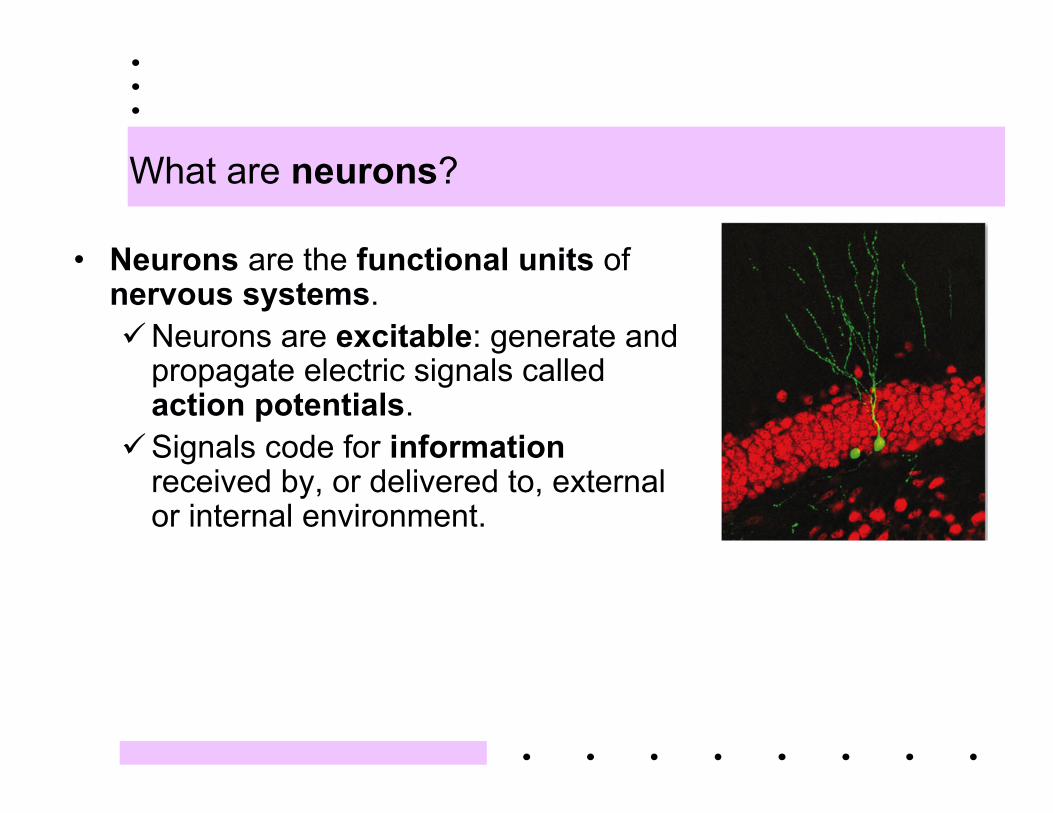
What are neurons?
• Neurons are the functional units ofnervous systems.Neurons are excitable: generate and
propagate electric signals calledaction potentials.
Signals code for informationreceived by, or delivered to, externalor internal environment.
Quiz check 1
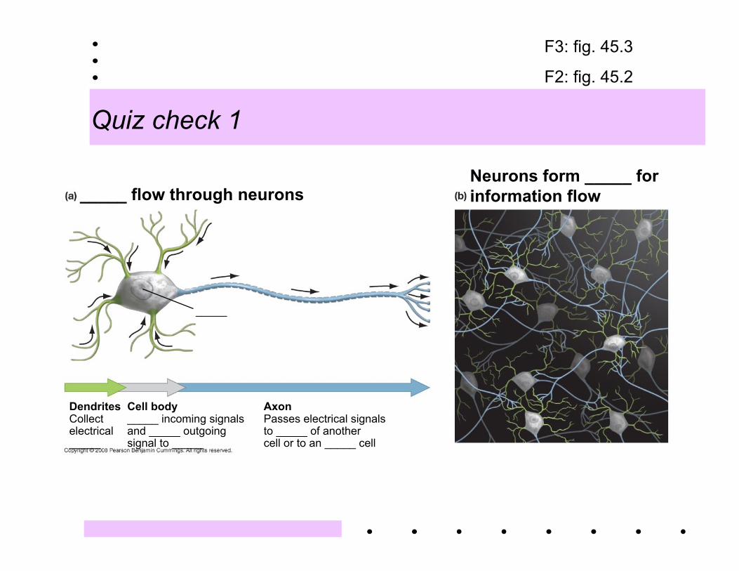
_____ flow through neurons
_____
DendritesCollectelectrical_____
Cell body_____ incoming signalsand _____ outgoingsignal to _____
AxonPasses electrical signalsto _____ of anothercell or to an _____ cell
Neurons form _____ for information flow
F3: fig. 45.3
F2: fig. 45.2
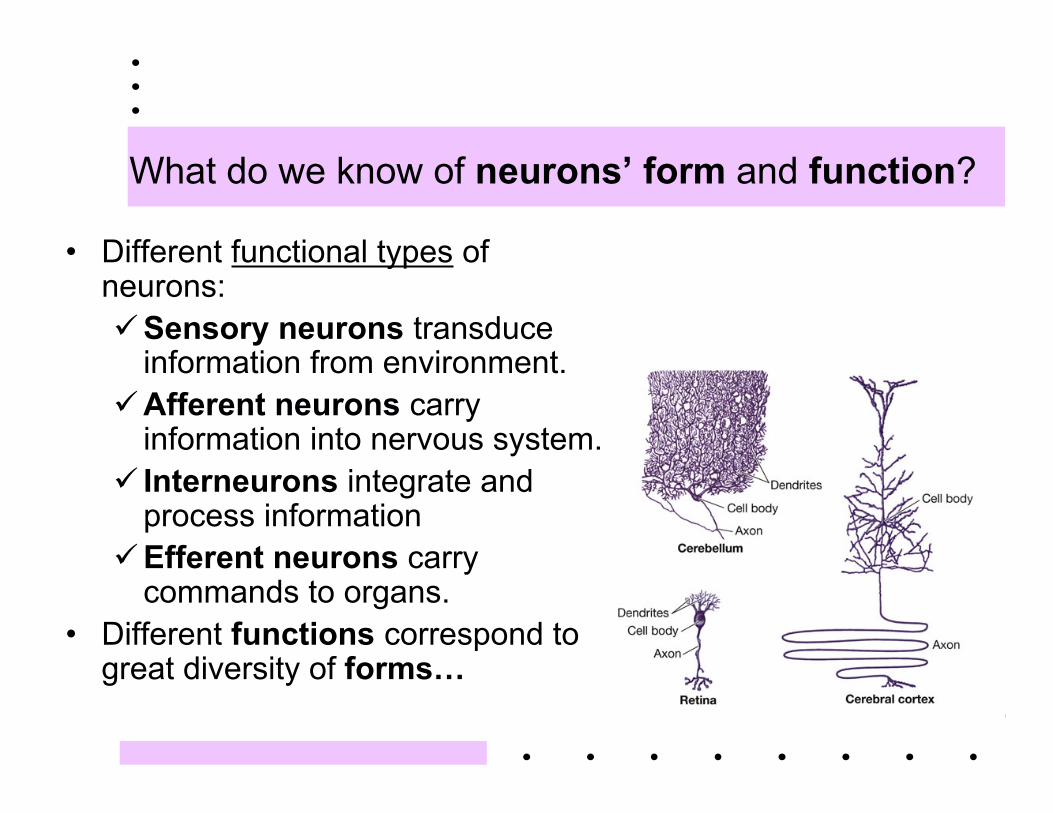
What do we know of neurons’ form and function?
• Different functional types ofneurons:Sensory neurons transduce
information from environment.Afferent neurons carry
information into nervous system. Interneurons integrate and
process informationEfferent neurons carry
commands to organs.• Different functions correspond to
great diversity of forms…
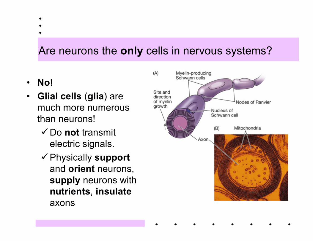
Are neurons the only cells in nervous systems?
• No!• Glial cells (glia) are
much more numerousthan neurons!Do not transmit
electric signals.Physically support
and orient neurons,supply neurons withnutrients, insulateaxons
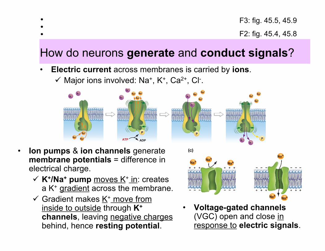
How do neurons generate and conduct signals?
• Ion pumps & ion channels generatemembrane potentials = difference inelectrical charge. K+/Na+ pump moves K+ in: creates
a K+ gradient across the membrane. Gradient makes K+ move from
inside to outside through K+
channels, leaving negative chargesbehind, hence resting potential.
• Electric current across membranes is carried by ions. Major ions involved: Na+, K+, Ca2+, Cl-.
• Voltage-gated channels(VGC) open and close inresponse to electric signals.
F3: fig. 45.5, 45.9
F2: fig. 45.4, 45.8
Quiz check 2
_____ are pores in the membraneof a cell that allow only specific ionsto pass through. In _____ neurons,_____ channels let _____ ions leakout of the cell, and the _____actively moves _____ ions into thecell. The net difference in charge isa _____ _____ of about _____.Active transport by the _____ alsocauses the concentration of _____to be lower _____ than _____. Aparticular kind of _____ called_____ open and close in responseto _____. They play a key role inthe propagation of _____ _____.
A. insideB. outsideC. ion channelsD. voltage-gated channelsE. actionF. electrical signalsG. restingH. K+
I. Na+
J. K+/Na+ pumpK. -0.65mVL. potential
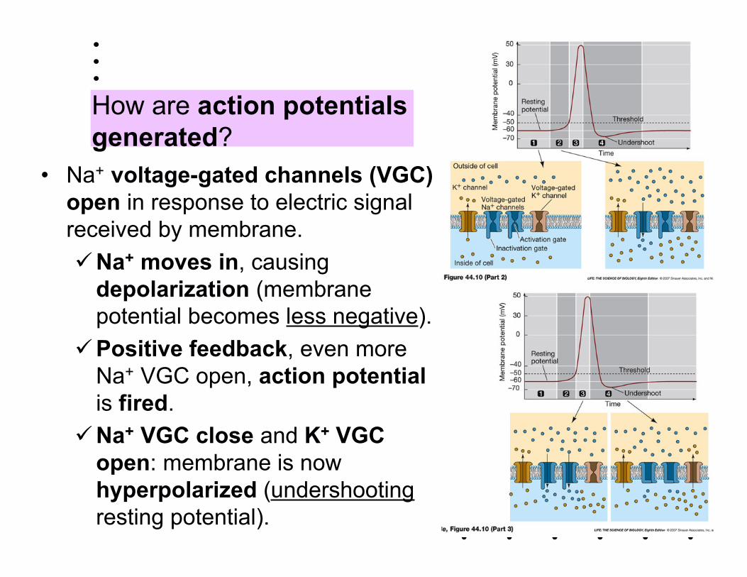
How are action potentialsgenerated?
• Na+ voltage-gated channels (VGC)open in response to electric signalreceived by membrane.Na+ moves in, causing
depolarization (membranepotential becomes less negative).
Positive feedback, even moreNa+ VGC open, action potentialis fired.
Na+ VGC close and K+ VGCopen: membrane is nowhyperpolarized (undershootingresting potential).
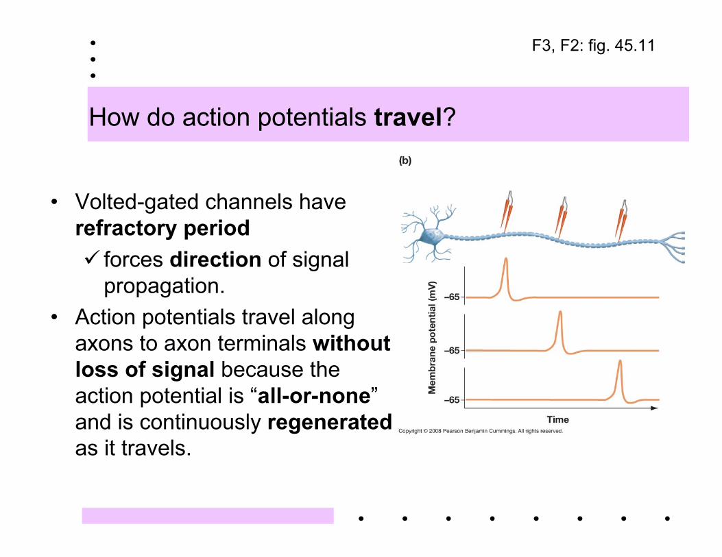
How do action potentials travel?
• Volted-gated channels haverefractory period forces direction of signal
propagation.• Action potentials travel along
axons to axon terminals withoutloss of signal because theaction potential is “all-or-none”and is continuously regeneratedas it travels.
F3, F2: fig. 45.11
Quiz check 3
The propagation of an action potentialinvolves a strong _____ of _____. Insidethe cell, the influx of _____ chargesattracts _____ charges and repels _____charges. As a result, the ______ chargespreads away from the channel where the_____ ions entered, and this _____nearby regions of the _____. __________ channels _____ in response. Asthe membrane potential reaches _____,_____ _____ channels close and __________ channels open. The strong outflowof _____ ions causes the membrane to_____.
A. voltage-gated B. ion pumpC. depolarizesD. hyperpolarizeE. closeF. openG. positiveH. negativeI. axonJ. dendriteK. Na+
L. K+
M. inflowN. outflowO. +40mV
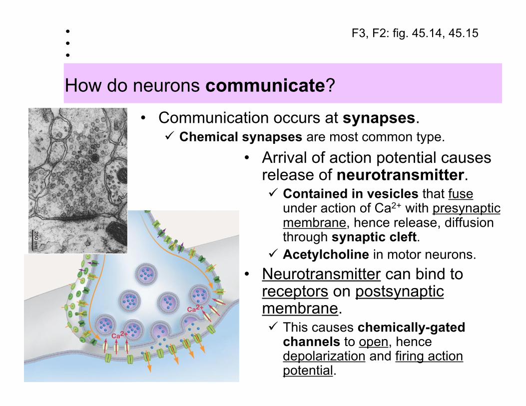
How do neurons communicate?
• Communication occurs at synapses. Chemical synapses are most common type.
• Arrival of action potential causesrelease of neurotransmitter. Contained in vesicles that fuse
under action of Ca2+ with presynapticmembrane, hence release, diffusionthrough synaptic cleft.
Acetylcholine in motor neurons.• Neurotransmitter can bind to
receptors on postsynapticmembrane. This causes chemically-gated
channels to open, hencedepolarization and firing actionpotential.
F3, F2: fig. 45.14, 45.15
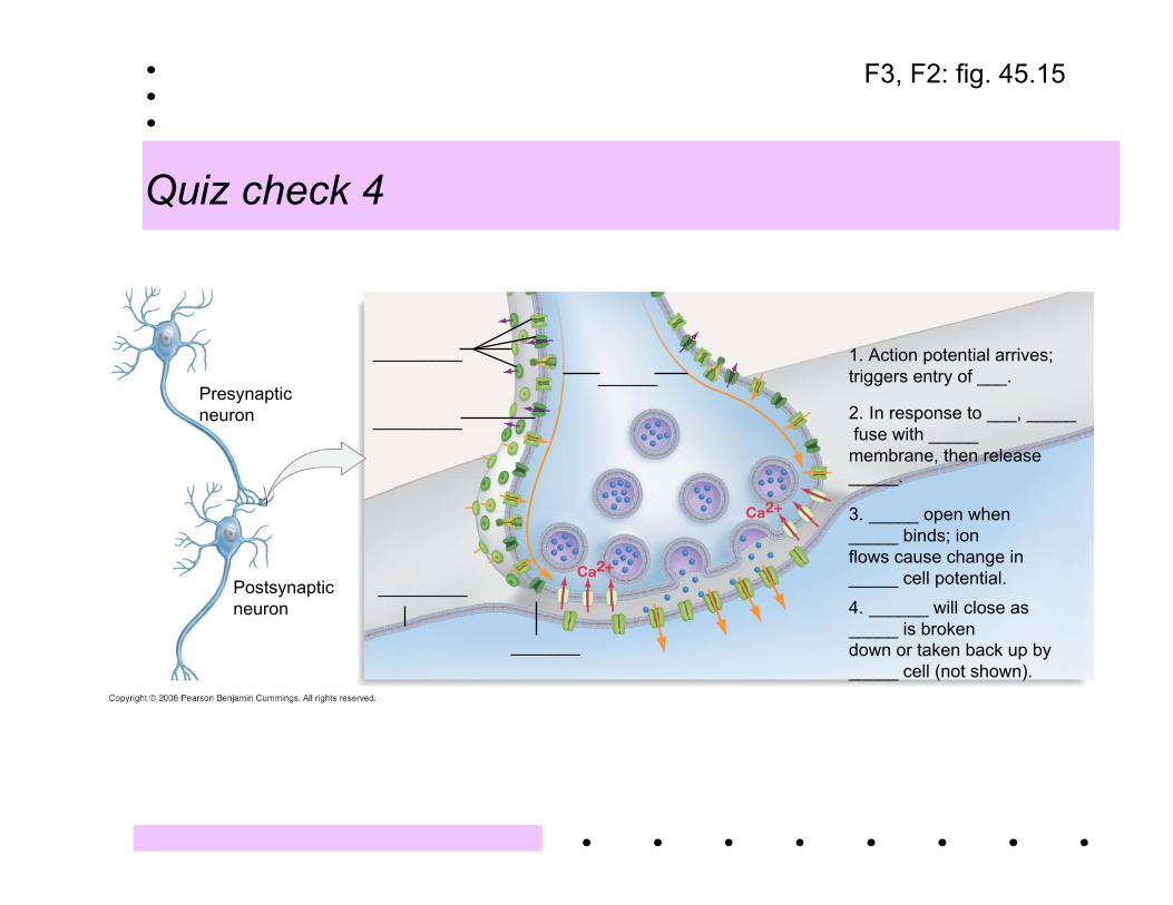
Quiz check 4
Presynapticneuron
Postsynapticneuron
_________
_________
_________
_______
______1. Action potential arrives;triggers entry of ___.
2. In response to ___, _____ fuse with _____membrane, then release_____.
3. _____ open when_____ binds; ionflows cause change in_____ cell potential.4. ______ will close as_____ is brokendown or taken back up by_____ cell (not shown).
F3, F2: fig. 45.15
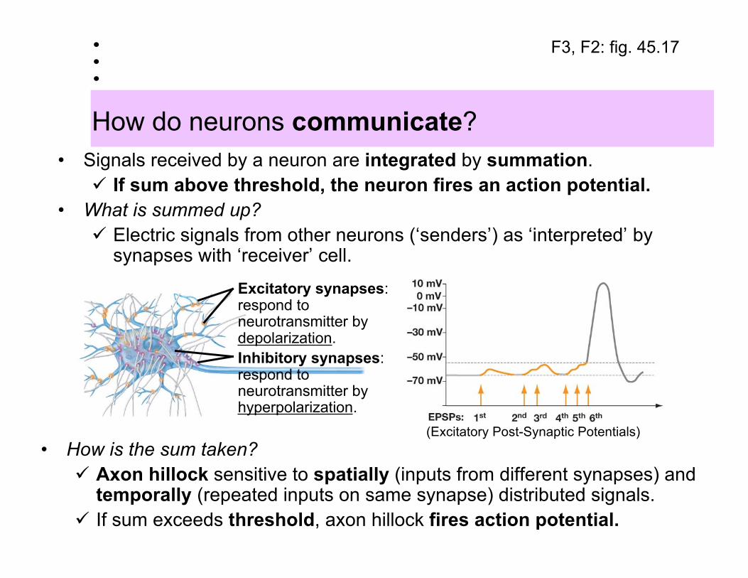
How do neurons communicate?
F3, F2: fig. 45.17
Excitatory synapses:respond toneurotransmitter bydepolarization.Inhibitory synapses:respond toneurotransmitter byhyperpolarization.
• How is the sum taken? Axon hillock sensitive to spatially (inputs from different synapses) and
temporally (repeated inputs on same synapse) distributed signals. If sum exceeds threshold, axon hillock fires action potential.
• Signals received by a neuron are integrated by summation. If sum above threshold, the neuron fires an action potential.
• What is summed up? Electric signals from other neurons (‘senders’) as ‘interpreted’ by
synapses with ‘receiver’ cell.
(Excitatory Post-Synaptic Potentials)
How is the neural network organized?
Structural organization:• Central nervous system (CNS)
Brain and spinal cord.Spinal cord communicates information between
brain and rest of body, can issue commands tobody without input from brain.
• Peripheral nervous system (PNS)Cranial and spinal nerves.
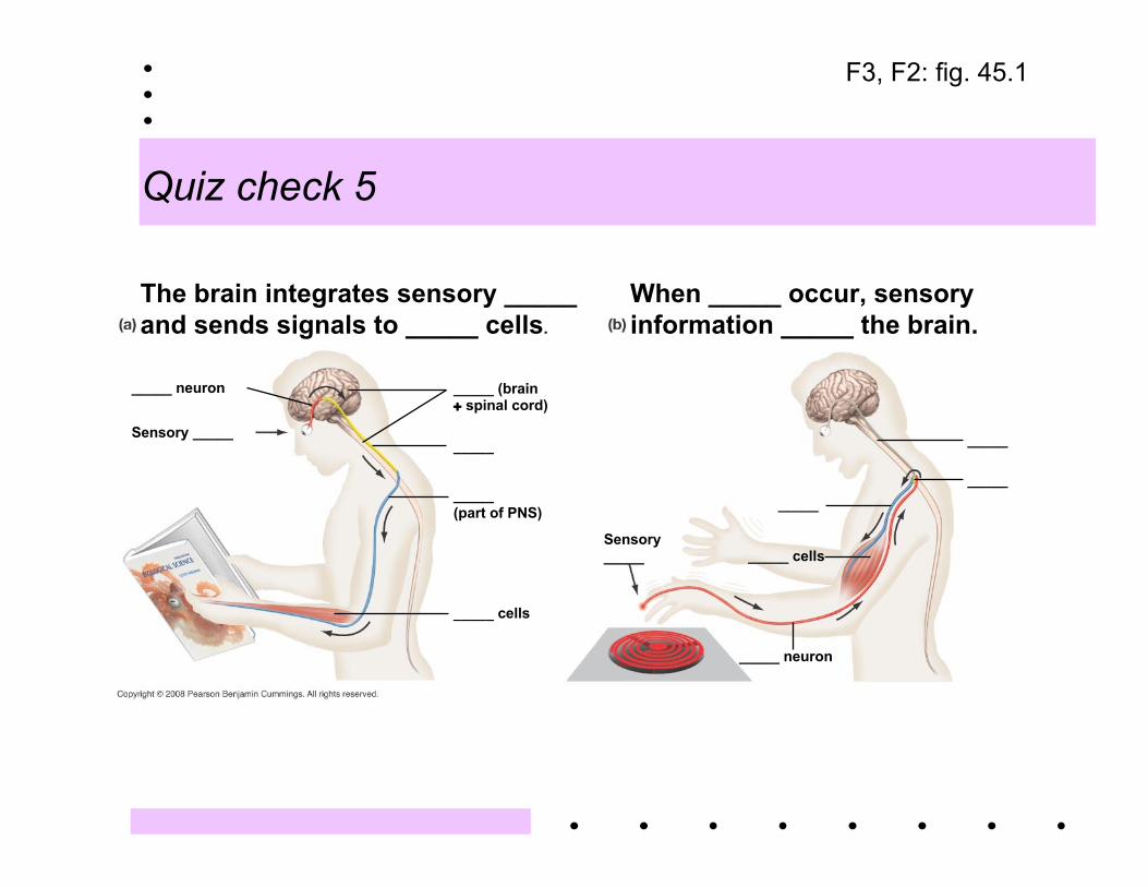
Quiz check 5
The brain integrates sensory _____ and sends signals to _____ cells.
When _____ occur, sensory information _____ the brain.
_____ neuron
Sensory _____
_____ (brain+ spinal cord)
_____
_____(part of PNS)
_____ cells
Sensory_____
_____
_____ cells
_____ neuron
_____
_____
F3, F2: fig. 45.1
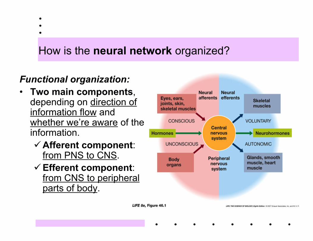
How is the neural network organized?
Functional organization:• Two main components,
depending on direction ofinformation flow andwhether we’re aware of theinformation.Afferent component:
from PNS to CNS.Efferent component:
from CNS to peripheralparts of body.
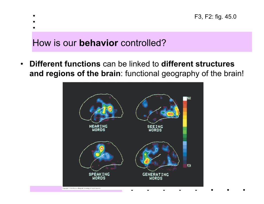
How is our behavior controlled?
• Different functions can be linked to different structuresand regions of the brain: functional geography of the brain!
F3, F2: fig. 45.0
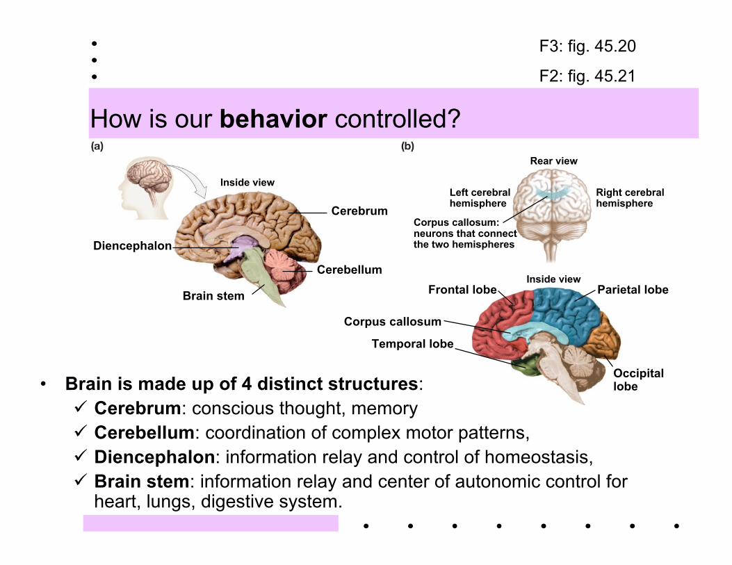
How is our behavior controlled?
Inside view
Diencephalon
Brain stem
Cerebrum
Cerebellum
Rear view
Left cerebralhemisphere
Right cerebralhemisphere
Corpus callosum:neurons that connectthe two hemispheres
Inside viewFrontal lobe Parietal lobe
Occipitallobe
Temporal lobe
Corpus callosum
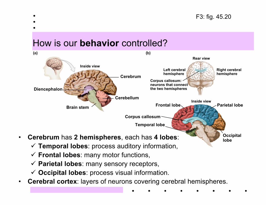
• Brain is made up of 4 distinct structures: Cerebrum: conscious thought, memory Cerebellum: coordination of complex motor patterns, Diencephalon: information relay and control of homeostasis, Brain stem: information relay and center of autonomic control for
heart, lungs, digestive system.
F3: fig. 45.20
F2: fig. 45.21
How is our behavior controlled?
Inside view
Diencephalon
Brain stem
Cerebrum
Cerebellum
Rear view
Left cerebralhemisphere
Right cerebralhemisphere
Corpus callosum:neurons that connectthe two hemispheres
Inside viewFrontal lobe Parietal lobe
Occipitallobe
Temporal lobe
Corpus callosum
• Cerebrum has 2 hemispheres, each has 4 lobes: Temporal lobes: process auditory information, Frontal lobes: many motor functions, Parietal lobes: many sensory receptors, Occipital lobes: process visual information.
• Cerebral cortex: layers of neurons covering cerebral hemispheres.
F3: fig. 45.20
How are our emotions controlled?
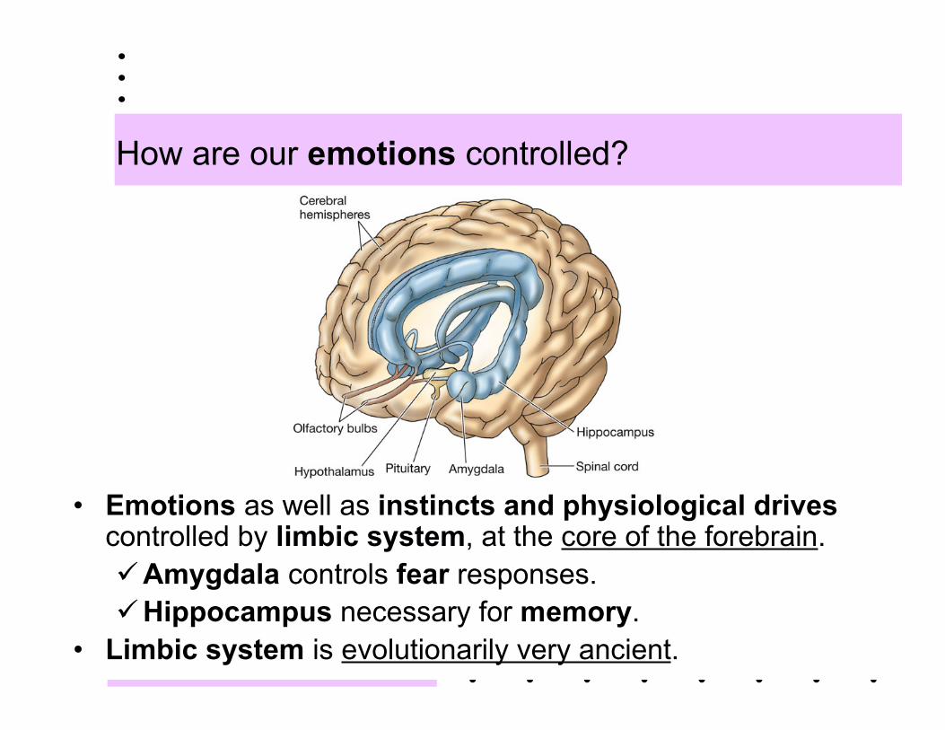
• Emotions as well as instincts and physiological drivescontrolled by limbic system, at the core of the forebrain.Amygdala controls fear responses.Hippocampus necessary for memory.
• Limbic system is evolutionarily very ancient.
How do we learn, memorize things?
• Learning = modification of behavior by experience.Memory = ability to retain what is learned.Some learning and memory processes have been
localized to specific brain areas.• Different types of memory: short-term, long-term.
Short-term memories (10-15 min) are transferred tolong-term memory.
Reinforcement a factor, hippocampus involved.
How do we learn, memorize things?
• Learning leading to long-term memory must involvelong-lasting synaptic changes.Long-term potentiation: high-frequency stimulation
that makes synapses more sensitive to futurestimulations.
Long-term depression: continuous smallstimulation reduces responsiveness.
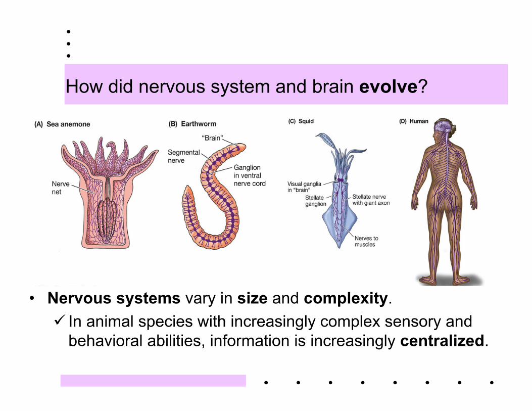
How did nervous system and brain evolve?
• Nervous systems vary in size and complexity. In animal species with increasingly complex sensory and
behavioral abilities, information is increasingly centralized.
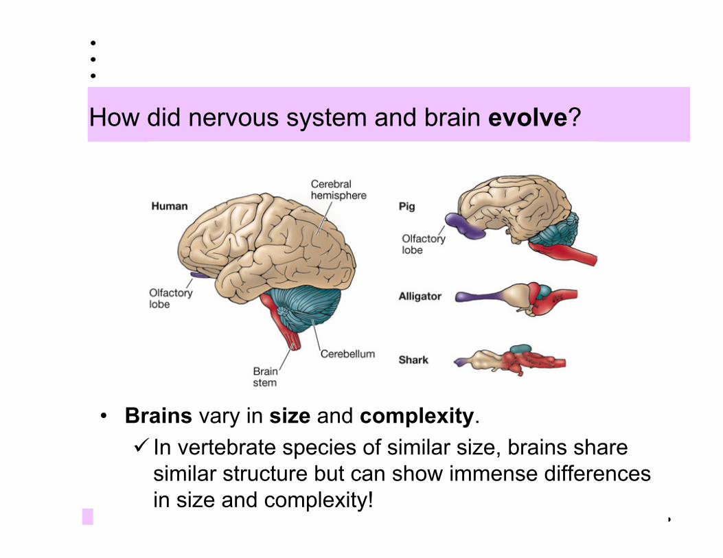
How did nervous system and brain evolve?
• Brains vary in size and complexity. In vertebrate species of similar size, brains share
similar structure but can show immense differencesin size and complexity!
Some recent scientific discoveries…
Up until recently it was thought that the adult human brain could not produce newnerve cells and that there was a steady loss of nerve cells from childhood into oldage. Now we know that new neurons do arise in the adult brain, and thisknowledge could lead to treatments for CNS injury and neurodegenerativediseases. (Kempermann, G. and F. H. Gage. 1999. New nerve cells for the adultbrain. Scientific American, May.)
By genetically engineering a neurotransmitter receptor that is involved in producinglong lasting synaptic changes, scientists have produced a mouse with enhancedlearning and memory. (Tsien, J. 2000. Building a brainier mouse. ScientificAmerican, April.)
The incredible advances in our knowledge of the cellular and molecular mechanismsof brain functions have contributed little insight into the question ofconsciousness. Studies of visual perception are enabling scientists to generatenew ideas about how our brains produce conscious experience. (Logothetis, N.K. 1999. Vision: A window on consciousness. Scientific American, November.)