Embed Size (px)
Citation preview
DRASKO SIMOVIC, M.D.
NEUROMUSCULAR REVIEW
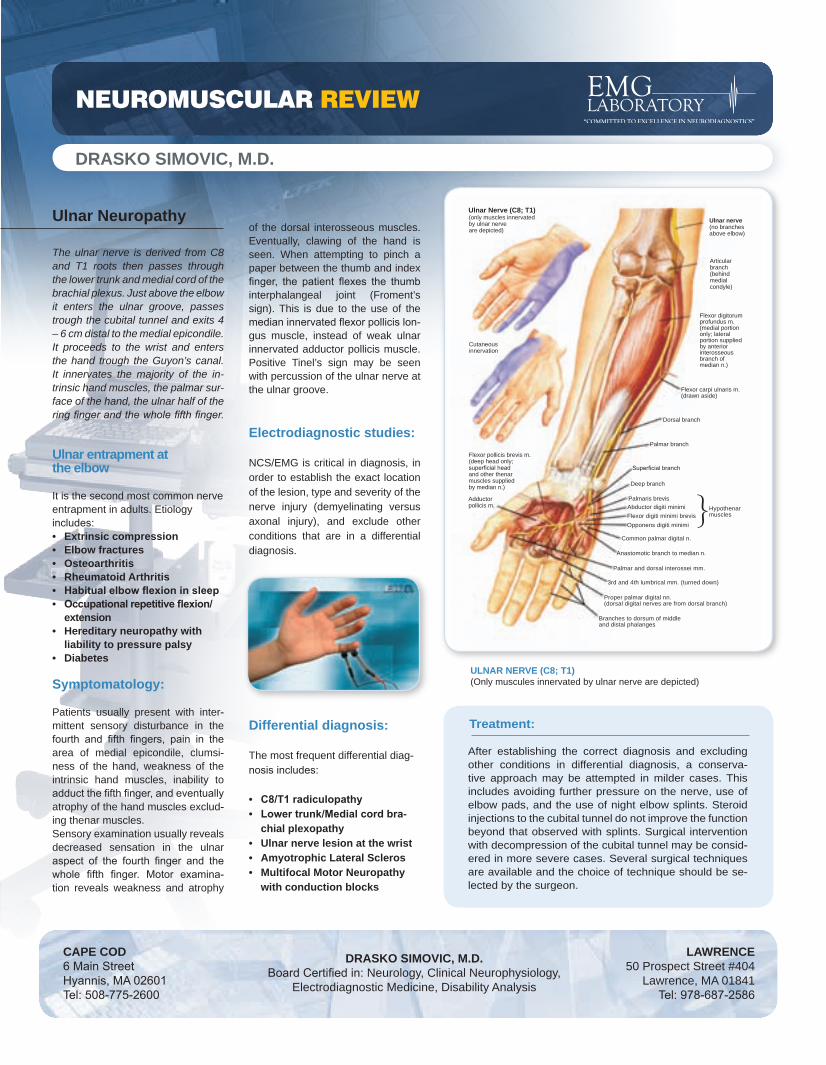
(only muscles innervated by ulnar nerve are depicted)
Ulnar Nerve (C8; T1)
Cutaneousinnervation
Flexor pollicis brevis m.(deep head only;superficial headand other thenarmuscles suppliedby median n.)
Adductorpollicis m.
Ulnar nerve(no branchesabove elbow)
Articularbranch(behindmedialcondyle)
Flexor digitorumprofundus m.(medial portiononly; lateralportion suppliedby anterior interosseousbranch ofmedian n.)
Flexor carpi ulnaris m.(drawn aside)
Dorsal branch
Palmar branch
Superficial branch
Deep branch
Palmaris brevisAbductor digiti minimiFlexor digiti minimi brevisOpponens digiti minimi
Common palmar digital n.
Anastomotic branch to median n.
Palmar and dorsal interossei mm.
3rd and 4th lumbrical mm. (turned down)
Proper palmar digital nn.(dorsal digital nerves are from dorsal branch)
Branches to dorsum of middle and distal phalanges
Hypothenarmuscles}
ULNAR NERVE (C8; T1) (Only muscules innervated by ulnar nerve are depicted)
DRASKO SIMOVIC, M.D.Board Certified in: Neurology, Clinical Neurophysiology,
Electrodiagnostic Medicine, Disability Analysis
LAWRENCE50 Prospect Street #404
Lawrence, MA 01841 Tel: 978-687-2586
CAPE COD6 Main Street Hyannis, MA 02601Tel: 508-775-2600
Ulnar Neuropathy The ulnar nerve is derived from C8 and T1 roots then passes through the lower trunk and medial cord of the brachial plexus. Just above the elbow it enters the ulnar groove, passes trough the cubital tunnel and exits 4 – 6 cm distal to the medial epicondile. It proceeds to the wrist and enters the hand trough the Guyon’s canal. It innervates the majority of the in-trinsic hand muscles, the palmar sur-face of the hand, the ulnar half of the ring finger and the whole fifth finger.
Ulnar entrapment at the elbow
It is the second most common nerve entrapment in adults. Etiology includes:
Extrinsic compression Elbow fracturesOsteoarthritisRheumatoid ArthritisHabitual elbow flexion in sleepOccupational repetitive flexion/extensionHereditary neuropathy with liability to pressure palsyDiabetes
Symptomatology: Patients usually present with inter-mittent sensory disturbance in the fourth and fifth fingers, pain in the area of medial epicondile, clumsi-ness of the hand, weakness of the intrinsic hand muscles, inability to adduct the fifth finger, and eventually atrophy of the hand muscles exclud-ing thenar muscles. Sensory examination usually reveals decreased sensation in the ulnar aspect of the fourth finger and the whole fifth finger. Motor examina-tion reveals weakness and atrophy
••••••
•
•
of the dorsal interosseous muscles. Eventually, clawing of the hand is seen. When attempting to pinch a paper between the thumb and index finger, the patient flexes the thumb interphalangeal joint (Froment’s sign). This is due to the use of the median innervated flexor pollicis lon-gus muscle, instead of weak ulnar innervated adductor pollicis muscle. Positive Tinel’s sign may be seen with percussion of the ulnar nerve at the ulnar groove.
Electrodiagnostic studies:
NCS/EMG is critical in diagnosis, in order to establish the exact location of the lesion, type and severity of the nerve injury (demyelinating versus axonal injury), and exclude other conditions that are in a differential diagnosis.
Differential diagnosis:
The most frequent differential diag-nosis includes:
C8/T1 radiculopathyLower trunk/Medial cord bra-chial plexopathyUlnar nerve lesion at the wristAmyotrophic Lateral SclerosMultifocal Motor Neuropathy with conduction blocks
••
•••
Treatment:
After establishing the correct diagnosis and excluding other conditions in differential diagnosis, a conserva-tive approach may be attempted in milder cases. This includes avoiding further pressure on the nerve, use of elbow pads, and the use of night elbow splints. Steroid injections to the cubital tunnel do not improve the function beyond that observed with splints. Surgical intervention with decompression of the cubital tunnel may be consid-ered in more severe cases. Several surgical techniques are available and the choice of technique should be se-lected by the surgeon.

NEUROMUSCULAR REVIEW
IV
IIIII
Superficial�terminalbranch
Pisiform
Ulnar nerve
I
VMotor branch
Hook of hamate
ULNAR HAND (Ulnar entrapment at the wrist)
Literature:
Kimura J. in: Electrodiagnosis in Diseases of Nerve and MuscleDaube J. in: Clinical NeurophysiologyBrown W., Bolton C. and Aminoff M. in: Neuromuscular Function and DiseaseMendel J., Kissel J. and Cornblath D. in: Diagnosis and Management of Peripheral Nerve Disorders
Treatment: In mild trauma cases, a conservative approach is indicated while for more severe cases or when a mass lesion is found, a surgical exploration is indicated.
Ulnar entrapment at the wrist
Symptomatology:
Patients may complain of wrist pain, intrinsic hand muscle weakness and atrophy, with or without sensory symptoms depending on the ex-act site of lesion. Wu and colleagues classified the lesion into five types:
Type I: The lesion is proximal or within Guyon’s canal, with intrinsic mus-cles weakness and with sensory symptoms in the ulnar innervated fin-gers, but with sparing of the dorsum of the hand, as dorsal ulnar cutane-ous nerve braches off proximal to the wrist. Type II: The lesion is in Guyon’s canal, affecting only the superficial sen-sory branch, leading to sensory disturbance which is limited to the fourth and fifth fingers, with sparing of the dorsum of the hand and the palmar cutaneous distribution.Type III: The lesion is in Guyon’s canal, distal to the superficial sensory branch, leading to a pure motor nerve lesion, with weakness of all intrinsic hand muscle.Type IV: The lesion is in Guyon’s canal, distal to the deep motor branch for hypothenar muscles, leading to weakness of the intrinsic hand mus-cles, excluding the hypothenar muscles. Type V: The lesion is the most distal variety of pure motor lesions, with weakness being limited to the first dorsal interosseous and adductor pol-licis muscles.
•
•
•
•
•
Etiology: • Extrinsic trauma• Ganglion cysts• Tumors• Aberrant Artery• Idiopathic
Newsletter courtesy of:
EMG Laboratory Drasko Simovic, M.D. Lawrence: 978-687-2486
Hyannis: 978-775-2600 www.emglaboratory.com
Comments or [email protected]
ELECTRODIAGNOSTICS: Ulnar Motor Nerve Conduction Study
Electrodiagnostic studies: The electrophysiologic findings vary, depending of the site of the lesion. Nerve conduction techniques with recording from the abductor digiti minimi and first dorsal interosseous may assist in localizing the lesion and excluding alternative diagnosis.
Other studies:
Plain X-ray and CT may exclude pisiform and hook of the hamate frac-tures. When history reveals more insidious onset with no trauma, a MRI is indicated in a search of a ganglion cyst or other mass lesions.


![S ELECTROMYOGRAPHY BASED D NEUROMUSCULAR DISEASES … · 2015-10-08 · considering individual’s neurophysiology, motor-learning and motor development functions [4]. This work primarily](https://img.pdfslide.us/doc/110x75/5e8c7de3247bba12fb4b0564/s-electromyography-based-d-neuromuscular-diseases-2015-10-08-considering-individualas.jpg)