Embed Size (px)
Citation preview
Central Journal of Neurological Disorders & Stroke
Cite this article: Maeda K, Yasuda H (2014) Histological Background of Susceptibility of Oculomotor Nerve to Ischemia. J Neurol Disord Stroke 3(1): 1092.
*Corresponding authorKengo Maeda, Department of Neurology, National Hospital Organization Higashi-ohmi General Medical Center, 255 Gochi, Higashi-ohmi, Shiga 527-8505, Japan, Tel: 81-748-22-3030; Fax: 81-748-23-3383; E-mail:
Submitted: 07 July 2014
Accepted: 16 December 2014
Published: 18 December 2014
Copyright© 2014 Maeda et al.
OPEN ACCESS
Keywords•Diabetic cranial neuropathy•Oculomotor nerve•Microvasculature
Research Article
Histological Background of Susceptibility of Oculomotor Nerve to IschemiaKengo Maeda1,2* and Hitoshi Yasuda1
1Department of Internal Medicine, Shiga University of Medical Science, Japan 2Department of Internal Medicine, Biwako Yoikuin Hospital, Japan
Abstract
Although oculomotor nerve palsy, which is common in diabetes, is considered an ischemic mononeuropathy, the susceptibility of the oculomotor nerve to ischemic insult is not well understood. We analyzed the density of endoneurial microvessels of the oculomotor nerve and compared it with that of the sural nerve in non-diabetic patients. The mean vascular density of the oculomotor nerve (23 ± 8.4/mm2) was significantly smaller than that of the sural nerves (60 ± 29/mm2). The spatial distribution of the microvessels in the inner half (25.3 ± 6.7) was not different from that in the outer half (22.7 ± 9.4) in the oculomotor nerve. The lower density of microvessels in the oculomotor nerve might contribute to its susceptibility to ischemia.
INTRODUCTIONOculomotor nerve palsy is a frequent cranial neuropathy in
diabetic patients. It occurs abruptly and improves spontaneously within a few months. Unlike the third nerve palsy caused by neoplasm or aneurysm, pupillary function is often spared in diabetic oculomotor nerve palsy [1]. Although diabetic third cranial nerve palsy is not uncommon, there are few pathological reports concerning it [2-4] because of its good prognosis. Asbury and colleagues described a demyelinated lesion and occlusion of epineurial arteriole in an 88-year-old diabetic woman who had had an episode of third cranial nerve palsy [3]. Reported lesion sites are intra cavernous segment [2,3] and subarachnoid portion [4]. Smith and Dyck conducted a morphological analysis of myelinated nerve fibers of the oculomotor nerves from diabetic patients who did not have oculomotor nerve palsy [5], but they did not focus on the causes of oculomotor nerve susceptibility to ischemic mononeuropathy with reference to microvasculature. Diabetes causes thickening of the basement membranes of endoneurial micro vessels [6] and these micro vessels respond abnormally to nerve injury in the diabetic state [7]. These vascular characteristics may be systemic in diabetes. However, the susceptibilities to mononeuropathy might differ among peripheral nerves. This clinical observation raises the possibility that there is a nerve specific microenvironment. From this perspective, we analyzed the microvasculature of oculomotor nerves from non-diabetic patients, and compared it with that of the sural nerve. The sural nerve was selected since it is usually used for nerve biopsy.
MATERIALS AND METHODS
Patients
Specimens were collected from non diabetic cadavers autopsied in 1992 and 1993 at Biwako Yoikuin Hospital and Shiga University of Medical Science. The intracranial oculomotor nerve adjacent to the midbrain (subarachnoid portion) was dissected from the bodies of six patients. They had not presented ophthalmoplegia or ptosis during their lives. From seven other cadavers, who had not had any type of sensory neuropathies, the sural nerve was dissected at the lower third of the calf. Their sex, age, and underlying diseases are shown in (Table 1). Informed consent about experimental use was obtained from the patients’ families at the time of autopsy.
Sample processing and morphological examination
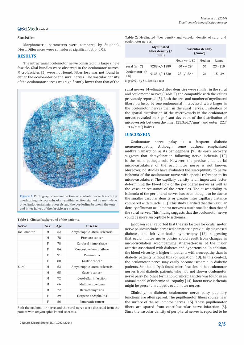
Nerve specimens were fixed in 2.5% glutaraldehyde and 2% paraformaldehyde in 0.025 M cacodylate buffer (pH 7.38) at 4˚C overnight. They were osmicated and embedded in epoxy resin. Transverse semithin sections (1µm) were stained by methylene blue. Total fascicular area and myelinated fiber density of the oculomotor and sural nerves were measured with a LUZEX F (Nireco, Japan). The fiber diameter was automatically calculated from the area of myelinated nerve fiber. Vessel number was counted on photographs based on morphological appearance. For the oculomotor nerves, photographs of a whole fascicle were reconstructed to count vascular number (Figure 1). In addition, the vascular density of the inner half of the oculomotor nerves was compared with that of the outer half as described elsewhere [8].
Central
Maeda et al. (2014)Email:
J Neurol Disord Stroke 3(1): 1092 (2014) 2/5
sural nerves. Myelinated fiber densities were similar in the sural and oculomotor nerves (Table 2) and compatible with the values previously reported [5]. Both the area and number of myelinated fibers perfused by one endoneurial microvessel were larger in the oculomotor nerves than in the sural nerves. Evaluation of the spatial distribution of the microvessels in the oculomotor nerves revealed no significant deviation of the distribution of microvessels between the inner (25.3±6.7/mm2) and outer (22.7 ± 9.4/mm2) halves.
DISCUSSIONOculomotor nerve palsy is a frequent diabetic
mononeuropathy. Although some authors emphasized midbrain infarction as its pathogenesis [9], its early recovery suggests that demyelination following nerve ischemia [10] is the main pathogenesis. However, the precise endoneurial microvasculature of the oculomotor nerve is not known. Moreover, no studies have evaluated the susceptibility to nerve ischemia of the oculomotor nerve with special reference to its microvasculature. The capillary density is an important factor determining the blood flow of the peripheral nerves as well as the vascular resistance of the arterioles. The susceptibility to ischemia of the peripheral nerves has been thought to be due to the smaller vascular density or greater inter capillary distance compared with muscle [11]. This study clarified that the vascular density of human oculomotor nerves is much smaller than that of the sural nerves. This finding suggests that the oculomotor nerve could be more susceptible to ischemia.
Jacobson et al. reported that the risk factors for ocular motor nerve palsies include increased hematocrit, previously diagnosed diabetes, and left ventricular hypertrophy [12], suggesting that ocular motor nerve palsies could result from changes in microcirculation accompanying atherosclerosis of the major arteries associated with diabetes and hypertension. In addition, the blood viscosity is higher in patients with neuropathy than in diabetic patients without this complication [13]. In this context, the oculomotor nerve may easily become ischemic in diabetic patients. Smith and Dyck found microfascicles in the oculomotor nerves from diabetic patients who had not shown oculomotor nerve palsy [5]. Since formation of microfascicles was found in an animal model of ischemic neuropathy [14], latent nerve ischemia might be present in diabetic oculomotor nerves.
Clinically, in diabetic oculomotor nerve palsy pupillary functions are often spared. The pupillomotor fibers course near the surface of the oculomotor nerves [15]. These pupillomotor fibers are spared from centrifascicular nerve infarction [3]. Since the vascular density of peripheral nerves is reported to be
Figure 1 Photographic reconstruction of a whole nerve fascicle by overlapping micrographs of a semithin section stained by methylene blue. Endoneurial microvessels and the borderline between the outer and inner halves of the fascicle are marked.
Nerve Sex Age Disease
Oculomotor M 62 Amyotrophic lateral sclerosis
M 78 Prostate cancer
F 78 Cerebral hemorrhage
F 84 Congestive heart failure
F 91 Pneumonia
F 80 Gastric cancer
Sural M 62 Amyotrophic lateral sclerosis
M 65 Gastric cancer
M 72 Cerebellar infarction
M 66 Multiple myeloma
M 72 Dermatomyositis
F 29 Herpetic encephalitis
F 86 Pancreatic cancer
Table 1: Clinical background of the patients.
Both the oculomotor nerve and the sural nerve were dissected form the patient with amyotrophic lateral sclerosis.
Statistics
Morphometric parameters were compared by Student’s t-test. Differences were considered significant at p<0.05.
RESULTSThe intracranial oculomotor nerve consisted of a large single
fascicle. Glial bundles were observed in the oculomotor nerves. Microfascicles [5] were not found. Fiber loss was not found in either the oculomotor or the sural nerves. The vascular density of the oculomotor nerves was significantly lower than that of the
Myelinated fiber density (/
mm2)
Vascular density (/mm2)
Mean +/- 1 SD Median Range
Sural (n = 7) 9288 +/- 1389 60 +/- 29a 57 23 - 110Oculomotor (n = 6) 9135 +/- 1320 23 +/- 8.4 a 21 15 - 39
Table 2: Myelinated fiber density and vascular density of sural and oculomotor nerves.
a: p<0.01 by Student’s t-test
Central
Maeda et al. (2014)Email:
J Neurol Disord Stroke 3(1): 1092 (2014) 3/5
Maeda K, Yasuda H (2014) Histological Background of Susceptibility of Oculomotor Nerve to Ischemia. J Neurol Disord Stroke 3(1): 1092.
Cite this article
higher in the outer side of the nerves than in the inner side, the outer side is thought to be more resistant to ischemic insult. This hypothesis is extrapolated from the results for rat somatic nerves [8], but our study suggests that this is not the case for the human oculomotor nerve. Although our experiment has the limitation that the two nerves were collected from different patient groups, we did find that the vascular density of the oculomotor nerve was smaller than that of the sural nerve. Although the sural nerve is susceptible to nerve ischemia, as demonstrated by the multifocal fiber loss seen in the sural nerves of diabetic patients, the endoneurial blood flow of the oculomotor nerve might be more easily affected by the change in the arterial blood flow. In addition, these results might provide the pathological basis for the treatment of oculomotor nerve palsy with agents that improve nerve blood flow.
ACKNOWLEDGMENTWe thank Kayoko Ukai for morphometric technical support.
REFERENCES1. Jacobson DM. Pupil involvement in patients with diabetes-associated
oculomotor nerve palsy. Arch Ophthalmol. 1998; 116: 723-727.
2. Dreyfus PM, Hakim S, Adams RD. Diabetic ophthalmoplegia; report of case, with postmortem study and comments on vascular supply of human oculomotor nerve. AMA Arch Neurol Psychiatry. 1957; 77: 337-349.
3. Asbury AK, Aldredge H, Hershberg R, Fisher CM. Oculomotor palsy in diabetes mellitus: a clinico-pathological study. Brain. 1970; 93: 555-566.
4. Weber RB, Daroff RB, Mackey EA. Pathology of oculomotor nerve palsy in diabetics. Neurology. 1970; 20: 835-838.
5. Smith BE, Dyck PJ. Subclinical histopathological changes in the oculomotor nerve in diabetes mellitus. Ann Neurol. 1992; 32: 376-385.
6. Yasuda H, Dyck PJ. Abnormalities of endoneurial microvessels and sural nerve pathology in diabetic neuropathy. Neurology. 1987; 37: 20-28.
7. Maeda K, Yasuda H, Taniguchi Y, Terada M, Kikkawa R. Endoneurial microvasculature of chronically transected sciatic nerves in diabetic rats. J Peripher Nerv Syst. 1999; 4: 13-18.
8. Nukada H, Dyck PJ, Karnes JL. Spatial distribution of capillaries in rat nerves: correlation to ischemic damage. Exp Neurol. 1985; 87: 369-376.
9. Hopf HC, Gutmann L. Diabetic 3rd nerve palsy: evidence for a mesencephalic lesion. Neurology. 1990; 40: 1041-1045.
10. Keane JR, Ahmadi J. Most diabetic third nerve palsies are peripheral. Neurology. 1998; 51: 1510.
11. McManis PG, Low PA, Lagerlund TD. Microenvironment of nerve: blood flow and ischemia. In: Peripheral neuropathy. Dyck PJ and Thomas PK (eds). Saunders Press, Philadelphia, PA. 1993; 453-473.
12. Jacobson DM, McCanna TD, Layde PM. Risk factors for ischemic ocular motor nerve palsies. Arch Ophthalmol. 1994; 112: 961-966.
13. Young MJ, Bennett JL, Liderth SA, Veves A, Boulton AJ, Douglas JT. Rheological and microvascular parameters in diabetic peripheral neuropathy. Clin Sci (Lond). 1996; 90: 183-187.
14. Korthals JK, Gieron MA, Wisniewski HM. Nerve regeneration patterns after acute ischemic injury. Neurology. 1989; 39: 932-937.
15. Kerr FW, Hollowell OW. Location of Pupillomotor and Accommodation Fibres in the Oculomotor Nerve: Experimental Observations on Paralytic Mydriasis. J Neurol Neurosurg Psychiatry. 1964; 27: 473-481.