Embed Size (px)
Citation preview
NV00026448 rev 05 Directions for Use (DFU) Page 1 of 33
Neuroform Atlas® Stent System
Directions for Use
NV00026448 rev 05 Directions for Use (DFU) Page 2 of 33
Caution: Federal Law (USA) restricts this device to sale by or on the order of a physician.
WARNING
Contents supplied STERILE using an ethylene oxide (EO) process. Do not use if sterile barrier is
damaged. If damage is found, call your Stryker Neurovascular representative.
For single use only. Do not reuse, reprocess or resterilize. Reuse, reprocessing or resterilization
may compromise the structural integrity of the device and/or lead to device failure which, in
turn, may result in patient injury, illness or death. Reuse, reprocessing or resterilization may also
create a risk of contamination of the device and/or cause patient infection or cross-infection,
including, but not limited to, the transmission of infectious disease(s) from one patient to
another. Contamination of the device may lead to injury, illness or death of the patient.
After use, dispose of product and packaging in accordance with hospital, administrative and/or
local government policy.
DEVICE DESCRIPTION
The Neuroform Atlas Stent System includes:
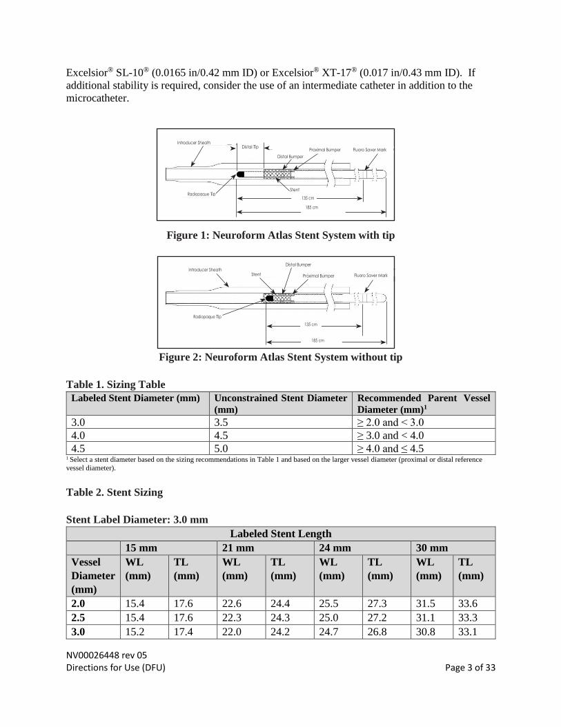
• A self-expanding, open-cell, nitinol stent with three radiopaque markerbands on each end
(proximal and distal) and four interconnects between the central stent segments, designed
to provide support for the coil mass within the aneurysm and minimize stent deflection.
• A stent delivery wire and introducer sheath. The stent is pre-loaded on the stent delivery
wire and protected by an introducer sheath.
• The stent delivery wire comes in two configurations: 1. With an 8.5 mm distal tip, 2.
Without a distal tip. Select a configuration based upon physician preference.
• An accessory pouch containing an optional torque device. The physician may attach the
torque device to the proximal end of the stent delivery wire to facilitate handling and
stabilization. The stent delivery wire is not designed to be torqued.
Contents
• One (1) Neuroform Atlas Stent System
• One (1) Torque Device
Required Accessories
Standard interventional devices, including rotating hemostatic valves ≥ 4.5 F [1.50mm
(0.059in)], a guide catheter, guidewire(s), and Stryker Neurovascular microcatheters specifically
NV00026448 rev 05 Directions for Use (DFU) Page 3 of 33
Excelsior® SL-10® (0.0165 in/0.42 mm ID) or Excelsior® XT-17® (0.017 in/0.43 mm ID). If
additional stability is required, consider the use of an intermediate catheter in addition to the
microcatheter.
Figure 1: Neuroform Atlas Stent System with tip
Figure 2: Neuroform Atlas Stent System without tip
Table 1. Sizing Table
Labeled Stent Diameter (mm) Unconstrained Stent Diameter
(mm)
Recommended Parent Vessel
Diameter (mm)1
3.0 3.5 ≥ 2.0 and < 3.0
4.0 4.5 ≥ 3.0 and < 4.0
4.5 5.0 ≥ 4.0 and ≤ 4.5 1 Select a stent diameter based on the sizing recommendations in Table 1 and based on the larger vessel diameter (proximal or distal reference
vessel diameter).
Table 2. Stent Sizing
Stent Label Diameter: 3.0 mm
Labeled Stent Length
15 mm 21 mm 24 mm 30 mm
Vessel
Diameter
(mm)
WL
(mm)
TL
(mm)
WL
(mm)
TL
(mm)
WL
(mm)
TL
(mm)
WL
(mm)
TL
(mm)
2.0 15.4 17.6 22.6 24.4 25.5 27.3 31.5 33.6
2.5 15.4 17.6 22.3 24.3 25.0 27.2 31.1 33.3
3.0 15.2 17.4 22.0 24.2 24.7 26.8 30.8 33.1
185 cm
Stent Delivery Wire
135 cm
185 cm
Stent Delivery Wire
NV00026448 rev 05 Directions for Use (DFU) Page 4 of 33
Stent Label Diameter: 4.0 mm
Labeled Stent Length
15 mm 21 mm 24 mm 30 mm
Vessel
Diameter
(mm)
WL
(mm)
TL
(mm)
WL
(mm)
TL
(mm)
WL
(mm)
TL
(mm)
WL
(mm)
TL
(mm)
3.0 15.1 17.3 21.7 23.6 24.5 26.6 30.8 32.9
3.5 14.8 17.1 21.0 23.2 24.1 26.3 30.4 32.6
4.0 14.6 16.8 20.4 22.5 23.8 25.4 29.2 31.4
Stent Label Diameter: 4.5 mm
Labeled Stent Length
15 mm
21 mm 24 mm 30 mm
Vessel
Diameter
(mm)
WL
(mm)
TL
(mm)
WL
(mm)
TL
(mm)
WL
(mm)
TL
(mm)
WL
(mm)
TL
(mm)
4.0 14.6 16.3 20.8 23.1 23.7 25.8 29.9 32.3
4.5 14.3 16.1 20.0 22.2 23.1 25.2 29.5 31.8
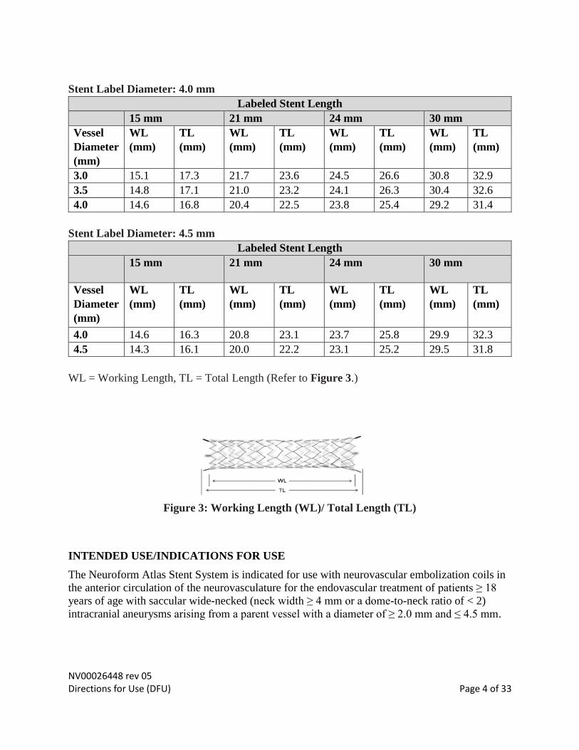
WL = Working Length, TL = Total Length (Refer to Figure 3.)
Figure 3: Working Length (WL)/ Total Length (TL)
INTENDED USE/INDICATIONS FOR USE
The Neuroform Atlas Stent System is indicated for use with neurovascular embolization coils in
the anterior circulation of the neurovasculature for the endovascular treatment of patients ≥ 18
years of age with saccular wide-necked (neck width ≥ 4 mm or a dome-to-neck ratio of < 2)
intracranial aneurysms arising from a parent vessel with a diameter of ≥ 2.0 mm and ≤ 4.5 mm.
NV00026448 rev 05 Directions for Use (DFU) Page 5 of 33
CONTRAINDICATIONS
• Patients in whom the parent vessel size does not fall within the indicated range. • Patients in whom antiplatelet and/or anticoagulation therapy (e.g., aspirin and
clopidogrel) is contraindicated.
• Patients who have not received anti‐platelet agents prior to stent implantation. • Patients with an active bacterial infection. • Patients in whom angiography demonstrates the anatomy is not appropriate for
endovascular treatment due to conditions such as: o Severe intracranial vessel tortuosity or stenosis; o Intracranial vasospasm not responsive to medical therapy.
• Patients in whom a pre-existing stent is in place in the parent artery at the target
intracranial aneurysm location.
WARNINGS
• This device should only be used by physicians who have received appropriate training in
interventional neuroradiology or interventional radiology and preclinical training on the
use of this device as established by Stryker Neurovascular.
• Select a stent size (length) to maintain a minimum of 4 mm on each side of the aneurysm
neck along the parent vessel (see Table 2 for size information). An incorrectly sized stent
may result in damage to the vessel or stent migration. Therefore, the stent is not designed
to treat an aneurysm with a neck greater than 22 mm in length.
• If excessive resistance is encountered during the use of the Neuroform Atlas Stent
System or any of its components at any time during the procedure, discontinue use of the
stent system. Continuing to move the stent system against resistance may result in
damage to the vessel or a system component.
• Use the Neuroform Atlas Stent System with compatible microcatheters. If repeated
friction is encountered during device delivery, verify microcatheter is not kinked or in
extremely tortuous anatomy. Confirm that the microcatheter does not ovalize. Confirm
that there is adequate sterile flush solution.
• Do not torque the delivery wire while advancing or retracting the Neuroform Atlas Stent
System.
• Do not attempt to re-position the Neuroform Atlas stent post-deployment.
• The stent delivery microcatheter and the Neuroform Atlas Stent delivery wire should not
be used to recapture the stent.
• Persons allergic to nickel titanium (Nitinol) may suffer an allergic response to this stent
implant.
• Higher adverse event rates may be experienced for distal aneurysms located in the
anterior and middle cerebral arteries.
• Do not use device to treat patients with ruptured intracranial aneurysms within a
minimum of 30 days from the aneurysm rupture.
NV00026448 rev 05 Directions for Use (DFU) Page 6 of 33
PRECAUTIONS
• Use the Neuroform Atlas Stent System prior to the “Use By” date printed on the package.
• Carefully inspect the sterile package and Neuroform Atlas Stent System prior to use to
verify that neither has been damaged during shipment. Do not use kinked or damaged
components; contact your Stryker Neurovascular representative.
• In cases where multiple aneurysms are to be treated, start at the most distal aneurysm
first.
• After deployment, the stent may foreshorten up to 6.3%.
• The maximum outer diameter (OD) of the coiling microcatheter should not exceed the
maximum OD of the stent delivery microcatheter.
• Standard interventional devices with distal tips > 1.8 F [0.60 mm (0.024 in)] may not be
able to pass through the interstices of the stent.
• Exercise caution when crossing the deployed stent with adjunctive devices.
• Take all necessary precautions to limit X-ray radiation doses to clinical operators by
using sufficient shielding, reducing fluoroscopy times, and modifying X-ray technical
factors whenever possible.
• The Neuroform Atlas stent may create local field inhomogeneity and susceptibility
artifacts during magnetic resonance angiography (MRA), which may degrade the
diagnostic quality to assess effective intracranial aneurysm occlusion.
• Safety and effectiveness of the Neuroform Atlas Stent System in patients below the age
of 18 has not been established.
• The benefits may not outweigh the risks of device use in patients with small and medium
asymptomatic extradural intracranial aneurysms, including those located in the cavernous
internal carotid artery.
• Carefully weigh the benefits vs. risks of device treatment for each individual patient
based on their medical health status and risk factors for intracranial aneurysm rupture
during their expected life time such as age, comorbidities, history of smoking,
intracranial aneurysm size, location, and morphology, family history, history of prior
asymptomatic subarachnoid hemorrhage (aSAH), documented growth of intracranial
aneurysm on serial imaging, presence of multiple intracranial aneurysms, and presence of
concurrent pathology. The benefits may not outweigh the risks associated with device use
in certain patients; therefore, judicious patient selection is recommended based on clinical
practice guidelines or tools to assess the life time risk of intracranial aneurysm rupture.
NV00026448 rev 05 Directions for Use (DFU) Page 7 of 33
POTENTIAL ADVERSE EVENTS
Potential complications include, but are not limited to:
• Aphasia
• Allergic reaction to Nitinol metal and medications
• Aneurysm perforation/rupture, leak or contrast extravasation
• Blindness
• Cardiac arrhythmia
• Coil herniation through stent into parent vessel
• Cranial neuropathy
• Death
• Embolus
• Headache
• Hemiplegia
• Hemorrhage (i.e., intracerebral, subarachnoid, retroperitoneal, or in other locations)
• Hydrocephalus
• In-stent stenosis
• Infection
• Ischemia
• Mass effect
• Myocardial infarction
• Neurological deficit/intracranial sequelae
• Pseudoaneurysm
• Reaction to radiation exposure (i.e., alopecia, burns ranging in severity from skin
reddening to ulcers, cataracts, or delayed neoplasia)
• Reactions to anti-platelet/anti-coagulant agents
• Renal failure
• Seizure
• Stent fracture, migration/embolization, or misplacement
• Stent thrombosis
• Stroke
• Transient ischemic attack
• Vasospasm
• Vessel occlusion or closure including parent vessel or non-target side-branches
• Vessel perforation/rupture, dissection, trauma or damage
• Vessel thrombosis
• Visual impairment
• Other procedural complications including but not limited to anesthetic and contrast media
risks, hypotension, hypertension, access site complications (including pain, hematoma,
local bleeding, local infection, and injury to the artery (i.e. dissection), vein, or adjacent
nerves)
• Unplanned intervention
Refer to the appropriate neurovascular embolization coil instructions for use for other
complications that may occur due to coil embolization.
NV00026448 rev 05 Directions for Use (DFU) Page 8 of 33
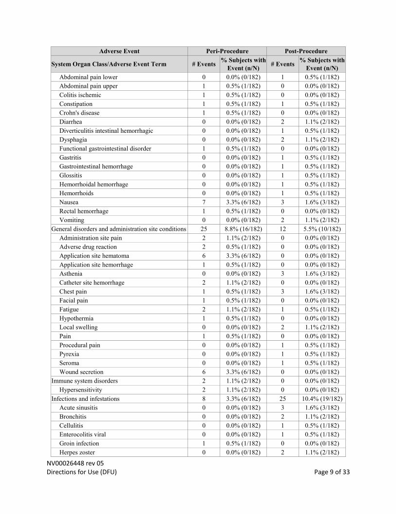
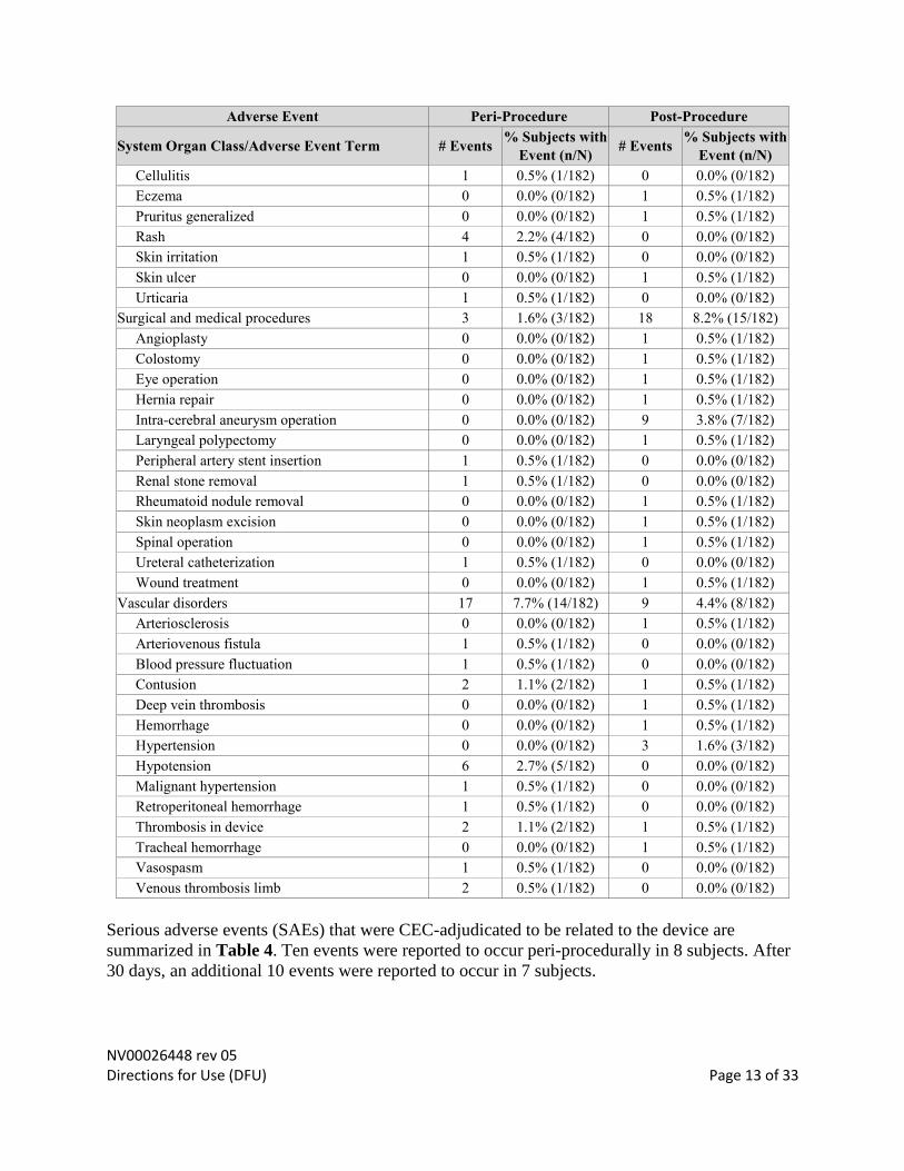
SUMMARY OF ADVERSE EVENTS IN ATLAS STUDY
The ATLAS study utilized a Clinical Events Committee/Data Safety Monitoring Board
(CEC/DSMB) that adjudicated pre-specified clinical events, as they occurred throughout the
study, in accordance with a ratified charter. The CEC/DSMB committee members adjudicated
the adverse event term, stroke severity (if applicable), and event relatedness to the device,
procedure, and/or concomitant medications. No unanticipated adverse device effects (UADE)
occurred during this study. An overall summary of all adverse events (AEs) is shown in Table 3.
A total of 207 events were reported during the peri-procedural period in 98 subjects. After 30
days (31 days to 12 months), 251 events occurred in 104 subjects.
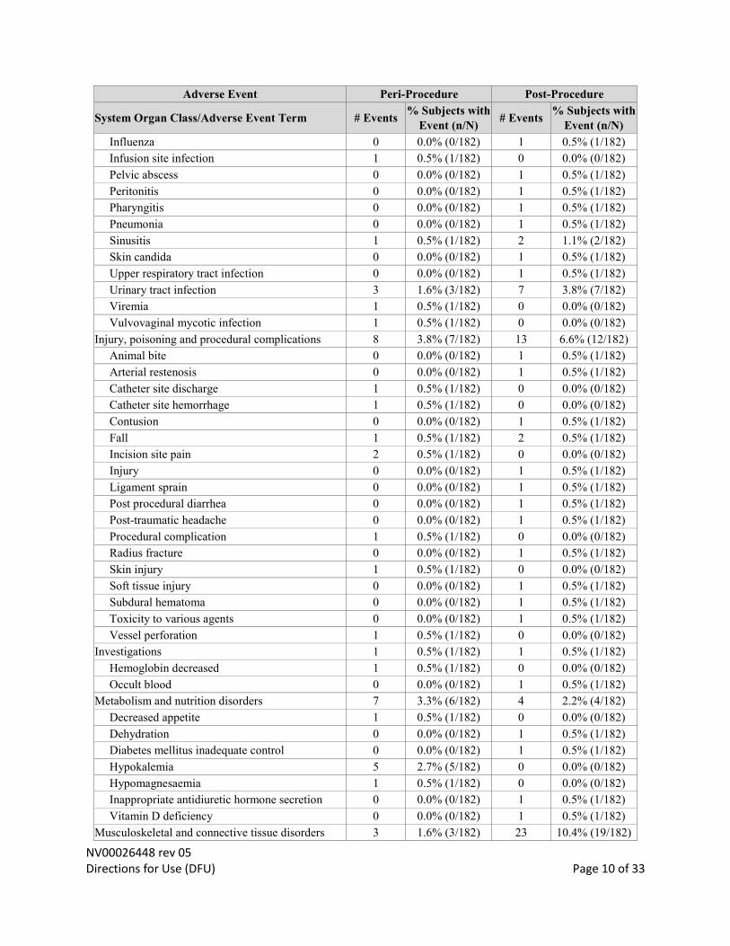
Table 3. Site-reported Overall Summary of Adverse Events - mITT Cohort Adverse Event Peri-Procedure Post-Procedure
System Organ Class/Adverse Event Term # Events % Subjects with
Event (n/N) # Events
% Subjects with
Event (n/N)
Any Adverse Event (AE) 207 53.8% (98/182) 251 57.1% (104/182)
Blood and lymphatic system disorders 3 1.6% (3/182) 5 2.2% (4/182)
Anemia 1 0.5% (1/182) 2 1.1% (2/182)
Increased tendency to bruise 2 1.1% (2/182) 0 0.0% (0/182)
Leukocytosis 0 0.0% (0/182) 1 0.5% (1/182)
Microcytic anemia 0 0.0% (0/182) 1 0.5% (1/182)
Thrombocytopenia 0 0.0% (0/182) 1 0.5% (1/182)
Cardiac disorders 2 1.1% (2/182) 5 2.7% (5/182)
Arrhythmia 0 0.0% (0/182) 2 1.1% (2/182)
Cardiomyopathy 0 0.0% (0/182) 2 1.1% (2/182)
Sinus bradycardia 1 0.5% (1/182) 0 0.0% (0/182)
Supraventricular tachycardia 0 0.0% (0/182) 1 0.5% (1/182)
Tachycardia 1 0.5% (1/182) 0 0.0% (0/182)
Ear and labyrinth disorders 0 0.0% (0/182) 1 0.5% (1/182)
Deafness bilateral 0 0.0% (0/182) 1 0.5% (1/182)
Eye disorders 11 4.9% (9/182) 11 4.9% (9/182)
Blepharospasm 0 0.0% (0/182) 1 0.5% (1/182)
Corneal abrasion 1 0.5% (1/182) 0 0.0% (0/182)
Diplopia 0 0.0% (0/182) 2 1.1% (2/182)
Glaucoma 0 0.0% (0/182) 1 0.5% (1/182)
Macular degeneration 0 0.0% (0/182) 1 0.5% (1/182)
Ocular discomfort 1 0.5% (1/182) 0 0.0% (0/182)
Photophobia 1 0.5% (1/182) 3 1.6% (3/182)
Retinal tear 0 0.0% (0/182) 1 0.5% (1/182)
Scleral hemorrhage 0 0.0% (0/182) 1 0.5% (1/182)
Vision blurred 6 3.3% (6/182) 0 0.0% (0/182)
Visual impairment 1 0.5% (1/182) 0 0.0% (0/182)
Vitreous floaters 1 0.5% (1/182) 1 0.5% (1/182)
Gastrointestinal disorders 13 6.0% (11/182) 20 8.2% (15/182)
Abdominal discomfort 0 0.0% (0/182) 1 0.5% (1/182)
Abdominal pain 0 0.0% (0/182) 2 1.1% (2/182)
NV00026448 rev 05 Directions for Use (DFU) Page 9 of 33
Adverse Event Peri-Procedure Post-Procedure
System Organ Class/Adverse Event Term # Events % Subjects with
Event (n/N) # Events
% Subjects with
Event (n/N)
Abdominal pain lower 0 0.0% (0/182) 1 0.5% (1/182)
Abdominal pain upper 1 0.5% (1/182) 0 0.0% (0/182)
Colitis ischemic 1 0.5% (1/182) 0 0.0% (0/182)
Constipation 1 0.5% (1/182) 1 0.5% (1/182)
Crohn's disease 1 0.5% (1/182) 0 0.0% (0/182)
Diarrhea 0 0.0% (0/182) 2 1.1% (2/182)
Diverticulitis intestinal hemorrhagic 0 0.0% (0/182) 1 0.5% (1/182)
Dysphagia 0 0.0% (0/182) 2 1.1% (2/182)
Functional gastrointestinal disorder 1 0.5% (1/182) 0 0.0% (0/182)
Gastritis 0 0.0% (0/182) 1 0.5% (1/182)
Gastrointestinal hemorrhage 0 0.0% (0/182) 1 0.5% (1/182)
Glossitis 0 0.0% (0/182) 1 0.5% (1/182)
Hemorrhoidal hemorrhage 0 0.0% (0/182) 1 0.5% (1/182)
Hemorrhoids 0 0.0% (0/182) 1 0.5% (1/182)
Nausea 7 3.3% (6/182) 3 1.6% (3/182)
Rectal hemorrhage 1 0.5% (1/182) 0 0.0% (0/182)
Vomiting 0 0.0% (0/182) 2 1.1% (2/182)
General disorders and administration site conditions 25 8.8% (16/182) 12 5.5% (10/182)
Administration site pain 2 1.1% (2/182) 0 0.0% (0/182)
Adverse drug reaction 2 0.5% (1/182) 0 0.0% (0/182)
Application site hematoma 6 3.3% (6/182) 0 0.0% (0/182)
Application site hemorrhage 1 0.5% (1/182) 0 0.0% (0/182)
Asthenia 0 0.0% (0/182) 3 1.6% (3/182)
Catheter site hemorrhage 2 1.1% (2/182) 0 0.0% (0/182)
Chest pain 1 0.5% (1/182) 3 1.6% (3/182)
Facial pain 1 0.5% (1/182) 0 0.0% (0/182)
Fatigue 2 1.1% (2/182) 1 0.5% (1/182)
Hypothermia 1 0.5% (1/182) 0 0.0% (0/182)
Local swelling 0 0.0% (0/182) 2 1.1% (2/182)
Pain 1 0.5% (1/182) 0 0.0% (0/182)
Procedural pain 0 0.0% (0/182) 1 0.5% (1/182)
Pyrexia 0 0.0% (0/182) 1 0.5% (1/182)
Seroma 0 0.0% (0/182) 1 0.5% (1/182)
Wound secretion 6 3.3% (6/182) 0 0.0% (0/182)
Immune system disorders 2 1.1% (2/182) 0 0.0% (0/182)
Hypersensitivity 2 1.1% (2/182) 0 0.0% (0/182)
Infections and infestations 8 3.3% (6/182) 25 10.4% (19/182)
Acute sinusitis 0 0.0% (0/182) 3 1.6% (3/182)
Bronchitis 0 0.0% (0/182) 2 1.1% (2/182)
Cellulitis 0 0.0% (0/182) 1 0.5% (1/182)
Enterocolitis viral 0 0.0% (0/182) 1 0.5% (1/182)
Groin infection 1 0.5% (1/182) 0 0.0% (0/182)
Herpes zoster 0 0.0% (0/182) 2 1.1% (2/182)
NV00026448 rev 05 Directions for Use (DFU) Page 10 of 33
Adverse Event Peri-Procedure Post-Procedure
System Organ Class/Adverse Event Term # Events % Subjects with
Event (n/N) # Events
% Subjects with
Event (n/N)
Influenza 0 0.0% (0/182) 1 0.5% (1/182)
Infusion site infection 1 0.5% (1/182) 0 0.0% (0/182)
Pelvic abscess 0 0.0% (0/182) 1 0.5% (1/182)
Peritonitis 0 0.0% (0/182) 1 0.5% (1/182)
Pharyngitis 0 0.0% (0/182) 1 0.5% (1/182)
Pneumonia 0 0.0% (0/182) 1 0.5% (1/182)
Sinusitis 1 0.5% (1/182) 2 1.1% (2/182)
Skin candida 0 0.0% (0/182) 1 0.5% (1/182)
Upper respiratory tract infection 0 0.0% (0/182) 1 0.5% (1/182)
Urinary tract infection 3 1.6% (3/182) 7 3.8% (7/182)
Viremia 1 0.5% (1/182) 0 0.0% (0/182)
Vulvovaginal mycotic infection 1 0.5% (1/182) 0 0.0% (0/182)
Injury, poisoning and procedural complications 8 3.8% (7/182) 13 6.6% (12/182)
Animal bite 0 0.0% (0/182) 1 0.5% (1/182)
Arterial restenosis 0 0.0% (0/182) 1 0.5% (1/182)
Catheter site discharge 1 0.5% (1/182) 0 0.0% (0/182)
Catheter site hemorrhage 1 0.5% (1/182) 0 0.0% (0/182)
Contusion 0 0.0% (0/182) 1 0.5% (1/182)
Fall 1 0.5% (1/182) 2 0.5% (1/182)
Incision site pain 2 0.5% (1/182) 0 0.0% (0/182)
Injury 0 0.0% (0/182) 1 0.5% (1/182)
Ligament sprain 0 0.0% (0/182) 1 0.5% (1/182)
Post procedural diarrhea 0 0.0% (0/182) 1 0.5% (1/182)
Post-traumatic headache 0 0.0% (0/182) 1 0.5% (1/182)
Procedural complication 1 0.5% (1/182) 0 0.0% (0/182)
Radius fracture 0 0.0% (0/182) 1 0.5% (1/182)
Skin injury 1 0.5% (1/182) 0 0.0% (0/182)
Soft tissue injury 0 0.0% (0/182) 1 0.5% (1/182)
Subdural hematoma 0 0.0% (0/182) 1 0.5% (1/182)
Toxicity to various agents 0 0.0% (0/182) 1 0.5% (1/182)
Vessel perforation 1 0.5% (1/182) 0 0.0% (0/182)
Investigations 1 0.5% (1/182) 1 0.5% (1/182)
Hemoglobin decreased 1 0.5% (1/182) 0 0.0% (0/182)
Occult blood 0 0.0% (0/182) 1 0.5% (1/182)
Metabolism and nutrition disorders 7 3.3% (6/182) 4 2.2% (4/182)
Decreased appetite 1 0.5% (1/182) 0 0.0% (0/182)
Dehydration 0 0.0% (0/182) 1 0.5% (1/182)
Diabetes mellitus inadequate control 0 0.0% (0/182) 1 0.5% (1/182)
Hypokalemia 5 2.7% (5/182) 0 0.0% (0/182)
Hypomagnesaemia 1 0.5% (1/182) 0 0.0% (0/182)
Inappropriate antidiuretic hormone secretion 0 0.0% (0/182) 1 0.5% (1/182)
Vitamin D deficiency 0 0.0% (0/182) 1 0.5% (1/182)
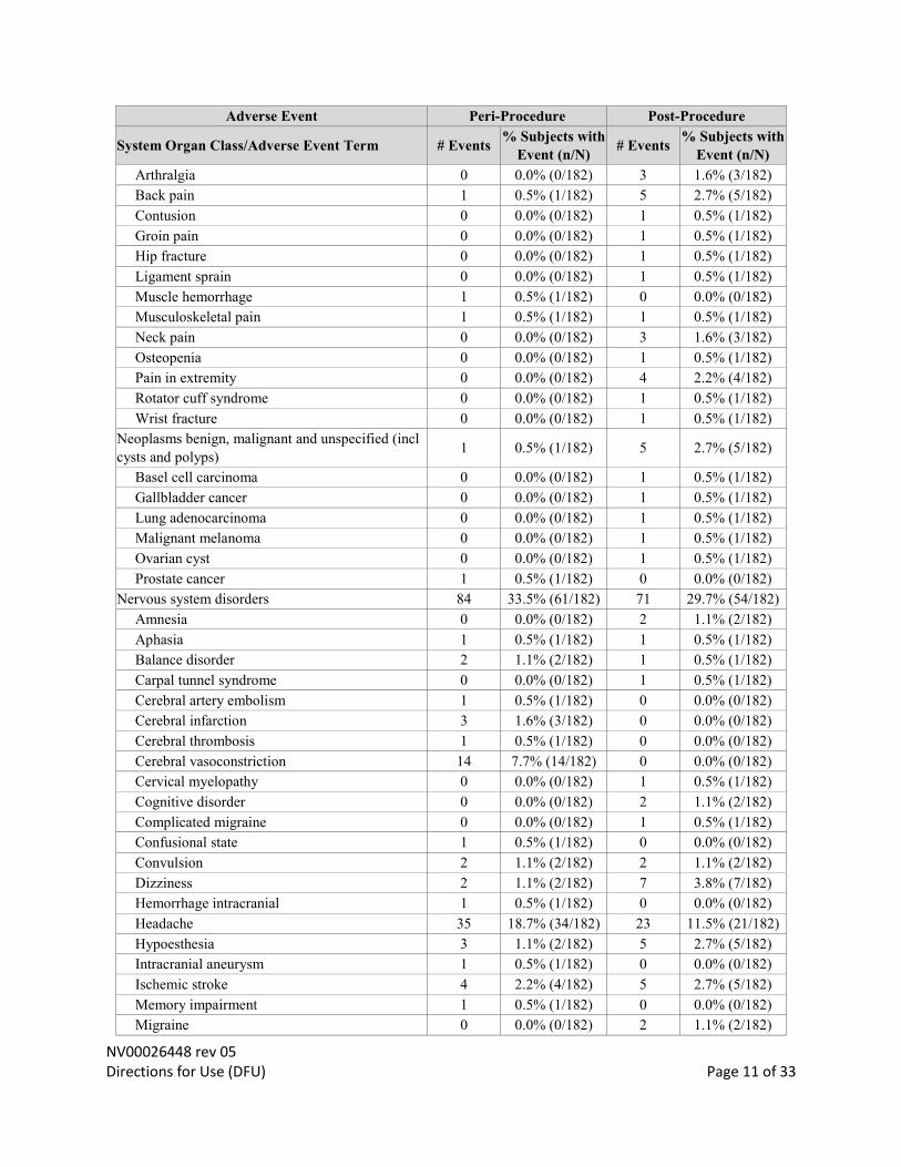
Musculoskeletal and connective tissue disorders 3 1.6% (3/182) 23 10.4% (19/182)
NV00026448 rev 05 Directions for Use (DFU) Page 11 of 33
Adverse Event Peri-Procedure Post-Procedure
System Organ Class/Adverse Event Term # Events % Subjects with
Event (n/N) # Events
% Subjects with
Event (n/N)
Arthralgia 0 0.0% (0/182) 3 1.6% (3/182)
Back pain 1 0.5% (1/182) 5 2.7% (5/182)
Contusion 0 0.0% (0/182) 1 0.5% (1/182)
Groin pain 0 0.0% (0/182) 1 0.5% (1/182)
Hip fracture 0 0.0% (0/182) 1 0.5% (1/182)
Ligament sprain 0 0.0% (0/182) 1 0.5% (1/182)
Muscle hemorrhage 1 0.5% (1/182) 0 0.0% (0/182)
Musculoskeletal pain 1 0.5% (1/182) 1 0.5% (1/182)
Neck pain 0 0.0% (0/182) 3 1.6% (3/182)
Osteopenia 0 0.0% (0/182) 1 0.5% (1/182)
Pain in extremity 0 0.0% (0/182) 4 2.2% (4/182)
Rotator cuff syndrome 0 0.0% (0/182) 1 0.5% (1/182)
Wrist fracture 0 0.0% (0/182) 1 0.5% (1/182)
Neoplasms benign, malignant and unspecified (incl
cysts and polyps) 1 0.5% (1/182) 5 2.7% (5/182)
Basel cell carcinoma 0 0.0% (0/182) 1 0.5% (1/182)
Gallbladder cancer 0 0.0% (0/182) 1 0.5% (1/182)
Lung adenocarcinoma 0 0.0% (0/182) 1 0.5% (1/182)
Malignant melanoma 0 0.0% (0/182) 1 0.5% (1/182)
Ovarian cyst 0 0.0% (0/182) 1 0.5% (1/182)
Prostate cancer 1 0.5% (1/182) 0 0.0% (0/182)
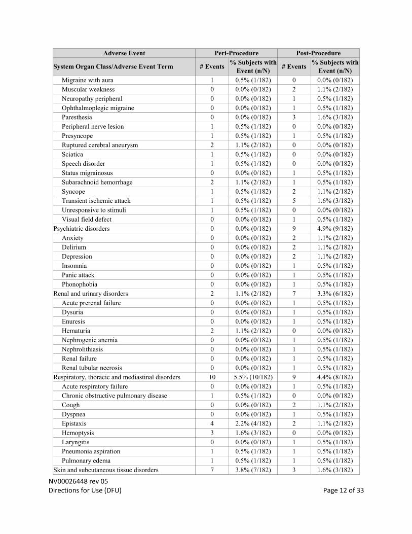
Nervous system disorders 84 33.5% (61/182) 71 29.7% (54/182)
Amnesia 0 0.0% (0/182) 2 1.1% (2/182)
Aphasia 1 0.5% (1/182) 1 0.5% (1/182)
Balance disorder 2 1.1% (2/182) 1 0.5% (1/182)
Carpal tunnel syndrome 0 0.0% (0/182) 1 0.5% (1/182)
Cerebral artery embolism 1 0.5% (1/182) 0 0.0% (0/182)
Cerebral infarction 3 1.6% (3/182) 0 0.0% (0/182)
Cerebral thrombosis 1 0.5% (1/182) 0 0.0% (0/182)
Cerebral vasoconstriction 14 7.7% (14/182) 0 0.0% (0/182)
Cervical myelopathy 0 0.0% (0/182) 1 0.5% (1/182)
Cognitive disorder 0 0.0% (0/182) 2 1.1% (2/182)
Complicated migraine 0 0.0% (0/182) 1 0.5% (1/182)
Confusional state 1 0.5% (1/182) 0 0.0% (0/182)
Convulsion 2 1.1% (2/182) 2 1.1% (2/182)
Dizziness 2 1.1% (2/182) 7 3.8% (7/182)
Hemorrhage intracranial 1 0.5% (1/182) 0 0.0% (0/182)
Headache 35 18.7% (34/182) 23 11.5% (21/182)
Hypoesthesia 3 1.1% (2/182) 5 2.7% (5/182)
Intracranial aneurysm 1 0.5% (1/182) 0 0.0% (0/182)
Ischemic stroke 4 2.2% (4/182) 5 2.7% (5/182)
Memory impairment 1 0.5% (1/182) 0 0.0% (0/182)
Migraine 0 0.0% (0/182) 2 1.1% (2/182)
NV00026448 rev 05 Directions for Use (DFU) Page 12 of 33
Adverse Event Peri-Procedure Post-Procedure
System Organ Class/Adverse Event Term # Events % Subjects with
Event (n/N) # Events
% Subjects with
Event (n/N)
Migraine with aura 1 0.5% (1/182) 0 0.0% (0/182)
Muscular weakness 0 0.0% (0/182) 2 1.1% (2/182)
Neuropathy peripheral 0 0.0% (0/182) 1 0.5% (1/182)
Ophthalmoplegic migraine 0 0.0% (0/182) 1 0.5% (1/182)
Paresthesia 0 0.0% (0/182) 3 1.6% (3/182)
Peripheral nerve lesion 1 0.5% (1/182) 0 0.0% (0/182)
Presyncope 1 0.5% (1/182) 1 0.5% (1/182)
Ruptured cerebral aneurysm 2 1.1% (2/182) 0 0.0% (0/182)
Sciatica 1 0.5% (1/182) 0 0.0% (0/182)
Speech disorder 1 0.5% (1/182) 0 0.0% (0/182)
Status migrainosus 0 0.0% (0/182) 1 0.5% (1/182)
Subarachnoid hemorrhage 2 1.1% (2/182) 1 0.5% (1/182)
Syncope 1 0.5% (1/182) 2 1.1% (2/182)
Transient ischemic attack 1 0.5% (1/182) 5 1.6% (3/182)
Unresponsive to stimuli 1 0.5% (1/182) 0 0.0% (0/182)
Visual field defect 0 0.0% (0/182) 1 0.5% (1/182)
Psychiatric disorders 0 0.0% (0/182) 9 4.9% (9/182)
Anxiety 0 0.0% (0/182) 2 1.1% (2/182)
Delirium 0 0.0% (0/182) 2 1.1% (2/182)
Depression 0 0.0% (0/182) 2 1.1% (2/182)
Insomnia 0 0.0% (0/182) 1 0.5% (1/182)
Panic attack 0 0.0% (0/182) 1 0.5% (1/182)
Phonophobia 0 0.0% (0/182) 1 0.5% (1/182)
Renal and urinary disorders 2 1.1% (2/182) 7 3.3% (6/182)
Acute prerenal failure 0 0.0% (0/182) 1 0.5% (1/182)
Dysuria 0 0.0% (0/182) 1 0.5% (1/182)
Enuresis 0 0.0% (0/182) 1 0.5% (1/182)
Hematuria 2 1.1% (2/182) 0 0.0% (0/182)
Nephrogenic anemia 0 0.0% (0/182) 1 0.5% (1/182)
Nephrolithiasis 0 0.0% (0/182) 1 0.5% (1/182)
Renal failure 0 0.0% (0/182) 1 0.5% (1/182)
Renal tubular necrosis 0 0.0% (0/182) 1 0.5% (1/182)
Respiratory, thoracic and mediastinal disorders 10 5.5% (10/182) 9 4.4% (8/182)
Acute respiratory failure 0 0.0% (0/182) 1 0.5% (1/182)
Chronic obstructive pulmonary disease 1 0.5% (1/182) 0 0.0% (0/182)
Cough 0 0.0% (0/182) 2 1.1% (2/182)
Dyspnea 0 0.0% (0/182) 1 0.5% (1/182)
Epistaxis 4 2.2% (4/182) 2 1.1% (2/182)
Hemoptysis 3 1.6% (3/182) 0 0.0% (0/182)
Laryngitis 0 0.0% (0/182) 1 0.5% (1/182)
Pneumonia aspiration 1 0.5% (1/182) 1 0.5% (1/182)
Pulmonary edema 1 0.5% (1/182) 1 0.5% (1/182)
Skin and subcutaneous tissue disorders 7 3.8% (7/182) 3 1.6% (3/182)
NV00026448 rev 05 Directions for Use (DFU) Page 13 of 33
Adverse Event Peri-Procedure Post-Procedure
System Organ Class/Adverse Event Term # Events % Subjects with
Event (n/N) # Events
% Subjects with
Event (n/N)
Cellulitis 1 0.5% (1/182) 0 0.0% (0/182)
Eczema 0 0.0% (0/182) 1 0.5% (1/182)
Pruritus generalized 0 0.0% (0/182) 1 0.5% (1/182)
Rash 4 2.2% (4/182) 0 0.0% (0/182)
Skin irritation 1 0.5% (1/182) 0 0.0% (0/182)
Skin ulcer 0 0.0% (0/182) 1 0.5% (1/182)
Urticaria 1 0.5% (1/182) 0 0.0% (0/182)
Surgical and medical procedures 3 1.6% (3/182) 18 8.2% (15/182)
Angioplasty 0 0.0% (0/182) 1 0.5% (1/182)
Colostomy 0 0.0% (0/182) 1 0.5% (1/182)
Eye operation 0 0.0% (0/182) 1 0.5% (1/182)
Hernia repair 0 0.0% (0/182) 1 0.5% (1/182)
Intra-cerebral aneurysm operation 0 0.0% (0/182) 9 3.8% (7/182)
Laryngeal polypectomy 0 0.0% (0/182) 1 0.5% (1/182)
Peripheral artery stent insertion 1 0.5% (1/182) 0 0.0% (0/182)
Renal stone removal 1 0.5% (1/182) 0 0.0% (0/182)
Rheumatoid nodule removal 0 0.0% (0/182) 1 0.5% (1/182)
Skin neoplasm excision 0 0.0% (0/182) 1 0.5% (1/182)
Spinal operation 0 0.0% (0/182) 1 0.5% (1/182)
Ureteral catheterization 1 0.5% (1/182) 0 0.0% (0/182)
Wound treatment 0 0.0% (0/182) 1 0.5% (1/182)
Vascular disorders 17 7.7% (14/182) 9 4.4% (8/182)
Arteriosclerosis 0 0.0% (0/182) 1 0.5% (1/182)
Arteriovenous fistula 1 0.5% (1/182) 0 0.0% (0/182)
Blood pressure fluctuation 1 0.5% (1/182) 0 0.0% (0/182)
Contusion 2 1.1% (2/182) 1 0.5% (1/182)
Deep vein thrombosis 0 0.0% (0/182) 1 0.5% (1/182)
Hemorrhage 0 0.0% (0/182) 1 0.5% (1/182)
Hypertension 0 0.0% (0/182) 3 1.6% (3/182)
Hypotension 6 2.7% (5/182) 0 0.0% (0/182)
Malignant hypertension 1 0.5% (1/182) 0 0.0% (0/182)
Retroperitoneal hemorrhage 1 0.5% (1/182) 0 0.0% (0/182)
Thrombosis in device 2 1.1% (2/182) 1 0.5% (1/182)
Tracheal hemorrhage 0 0.0% (0/182) 1 0.5% (1/182)
Vasospasm 1 0.5% (1/182) 0 0.0% (0/182)
Venous thrombosis limb 2 0.5% (1/182) 0 0.0% (0/182)
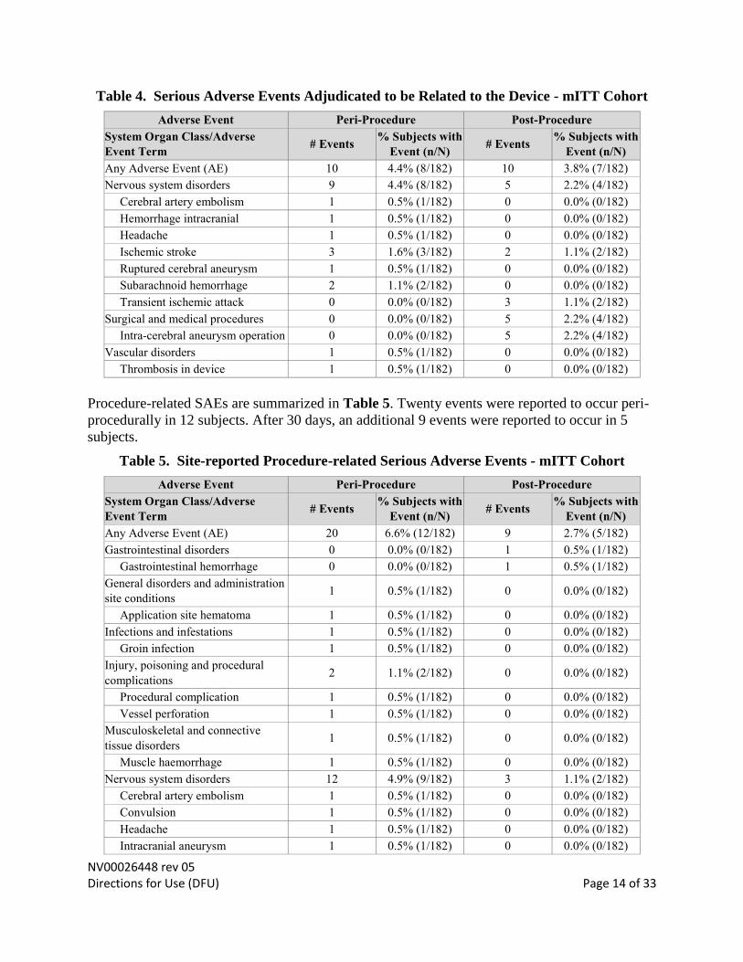
Serious adverse events (SAEs) that were CEC-adjudicated to be related to the device are
summarized in Table 4. Ten events were reported to occur peri-procedurally in 8 subjects. After
30 days, an additional 10 events were reported to occur in 7 subjects.
NV00026448 rev 05 Directions for Use (DFU) Page 14 of 33
Table 4. Serious Adverse Events Adjudicated to be Related to the Device - mITT Cohort
Adverse Event Peri-Procedure Post-Procedure
System Organ Class/Adverse
Event Term # Events
% Subjects with
Event (n/N) # Events
% Subjects with
Event (n/N)
Any Adverse Event (AE) 10 4.4% (8/182) 10 3.8% (7/182)
Nervous system disorders 9 4.4% (8/182) 5 2.2% (4/182)
Cerebral artery embolism 1 0.5% (1/182) 0 0.0% (0/182)
Hemorrhage intracranial 1 0.5% (1/182) 0 0.0% (0/182)
Headache 1 0.5% (1/182) 0 0.0% (0/182)
Ischemic stroke 3 1.6% (3/182) 2 1.1% (2/182)
Ruptured cerebral aneurysm 1 0.5% (1/182) 0 0.0% (0/182)
Subarachnoid hemorrhage 2 1.1% (2/182) 0 0.0% (0/182)
Transient ischemic attack 0 0.0% (0/182) 3 1.1% (2/182)
Surgical and medical procedures 0 0.0% (0/182) 5 2.2% (4/182)
Intra-cerebral aneurysm operation 0 0.0% (0/182) 5 2.2% (4/182)
Vascular disorders 1 0.5% (1/182) 0 0.0% (0/182)
Thrombosis in device 1 0.5% (1/182) 0 0.0% (0/182)
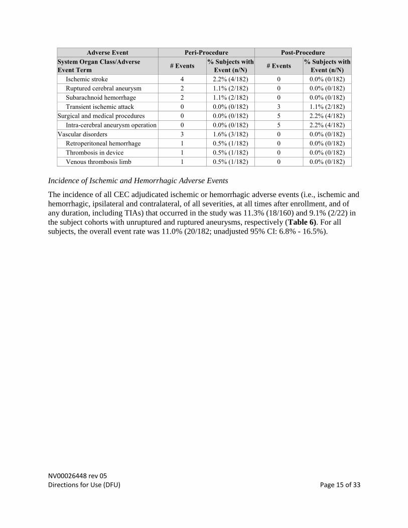
Procedure-related SAEs are summarized in Table 5. Twenty events were reported to occur peri-
procedurally in 12 subjects. After 30 days, an additional 9 events were reported to occur in 5
subjects.
Table 5. Site-reported Procedure-related Serious Adverse Events - mITT Cohort
Adverse Event Peri-Procedure Post-Procedure
System Organ Class/Adverse
Event Term # Events
% Subjects with
Event (n/N) # Events
% Subjects with
Event (n/N)
Any Adverse Event (AE) 20 6.6% (12/182) 9 2.7% (5/182)
Gastrointestinal disorders 0 0.0% (0/182) 1 0.5% (1/182)
Gastrointestinal hemorrhage 0 0.0% (0/182) 1 0.5% (1/182)
General disorders and administration
site conditions 1 0.5% (1/182) 0 0.0% (0/182)
Application site hematoma 1 0.5% (1/182) 0 0.0% (0/182)
Infections and infestations 1 0.5% (1/182) 0 0.0% (0/182)
Groin infection 1 0.5% (1/182) 0 0.0% (0/182)
Injury, poisoning and procedural
complications 2 1.1% (2/182) 0 0.0% (0/182)
Procedural complication 1 0.5% (1/182) 0 0.0% (0/182)
Vessel perforation 1 0.5% (1/182) 0 0.0% (0/182)
Musculoskeletal and connective
tissue disorders 1 0.5% (1/182) 0 0.0% (0/182)
Muscle haemorrhage 1 0.5% (1/182) 0 0.0% (0/182)
Nervous system disorders 12 4.9% (9/182) 3 1.1% (2/182)
Cerebral artery embolism 1 0.5% (1/182) 0 0.0% (0/182)
Convulsion 1 0.5% (1/182) 0 0.0% (0/182)
Headache 1 0.5% (1/182) 0 0.0% (0/182)
Intracranial aneurysm 1 0.5% (1/182) 0 0.0% (0/182)
NV00026448 rev 05 Directions for Use (DFU) Page 15 of 33
Adverse Event Peri-Procedure Post-Procedure
System Organ Class/Adverse
Event Term # Events
% Subjects with
Event (n/N) # Events
% Subjects with
Event (n/N)
Ischemic stroke 4 2.2% (4/182) 0 0.0% (0/182)
Ruptured cerebral aneurysm 2 1.1% (2/182) 0 0.0% (0/182)
Subarachnoid hemorrhage 2 1.1% (2/182) 0 0.0% (0/182)
Transient ischemic attack 0 0.0% (0/182) 3 1.1% (2/182)
Surgical and medical procedures 0 0.0% (0/182) 5 2.2% (4/182)
Intra-cerebral aneurysm operation 0 0.0% (0/182) 5 2.2% (4/182)
Vascular disorders 3 1.6% (3/182) 0 0.0% (0/182)
Retroperitoneal hemorrhage 1 0.5% (1/182) 0 0.0% (0/182)
Thrombosis in device 1 0.5% (1/182) 0 0.0% (0/182)
Venous thrombosis limb 1 0.5% (1/182) 0 0.0% (0/182)
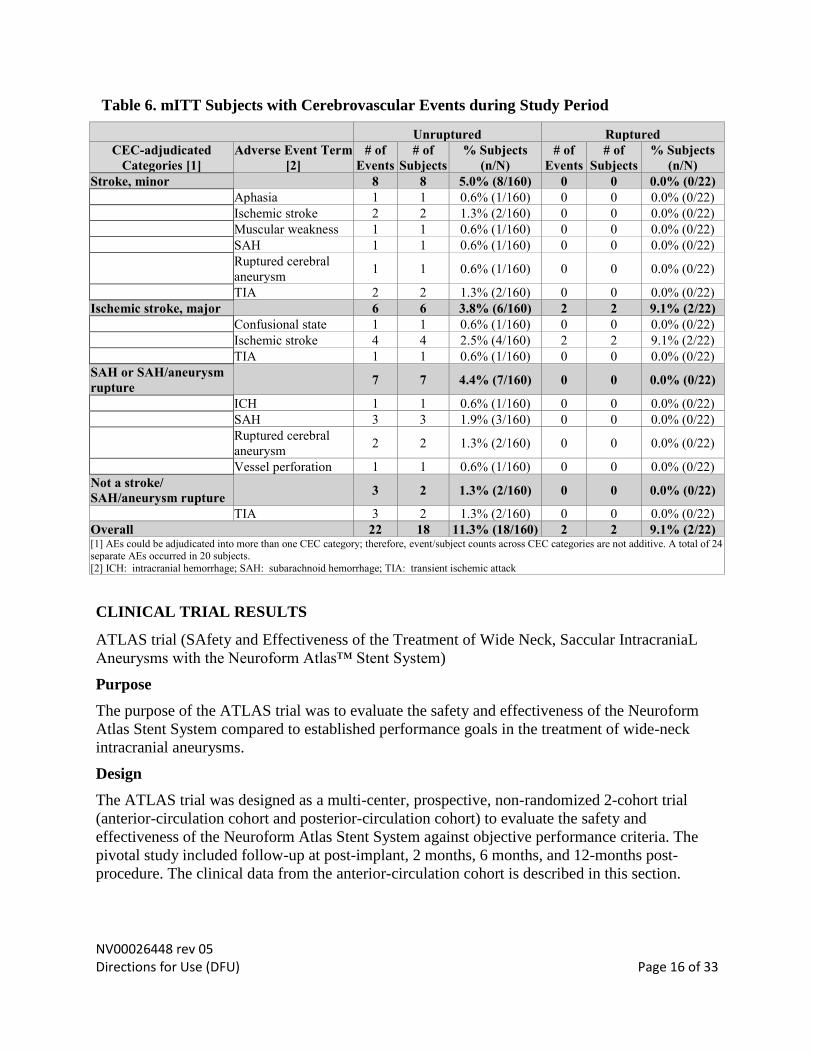
Incidence of Ischemic and Hemorrhagic Adverse Events
The incidence of all CEC adjudicated ischemic or hemorrhagic adverse events (i.e., ischemic and
hemorrhagic, ipsilateral and contralateral, of all severities, at all times after enrollment, and of
any duration, including TIAs) that occurred in the study was 11.3% (18/160) and 9.1% (2/22) in
the subject cohorts with unruptured and ruptured aneurysms, respectively (Table 6). For all
subjects, the overall event rate was 11.0% (20/182; unadjusted 95% CI: 6.8% - 16.5%).
NV00026448 rev 05 Directions for Use (DFU) Page 16 of 33
Table 6. mITT Subjects with Cerebrovascular Events during Study Period
Unruptured Ruptured
CEC-adjudicated
Categories [1]
Adverse Event Term
[2]
# of
Events
# of
Subjects
% Subjects
(n/N)
# of
Events
# of
Subjects
% Subjects
(n/N)
Stroke, minor 8 8 5.0% (8/160) 0 0 0.0% (0/22)
Aphasia 1 1 0.6% (1/160) 0 0 0.0% (0/22)
Ischemic stroke 2 2 1.3% (2/160) 0 0 0.0% (0/22)
Muscular weakness 1 1 0.6% (1/160) 0 0 0.0% (0/22)
SAH 1 1 0.6% (1/160) 0 0 0.0% (0/22)
Ruptured cerebral
aneurysm 1 1 0.6% (1/160) 0 0 0.0% (0/22)
TIA 2 2 1.3% (2/160) 0 0 0.0% (0/22)
Ischemic stroke, major 6 6 3.8% (6/160) 2 2 9.1% (2/22)
Confusional state 1 1 0.6% (1/160) 0 0 0.0% (0/22)
Ischemic stroke 4 4 2.5% (4/160) 2 2 9.1% (2/22)
TIA 1 1 0.6% (1/160) 0 0 0.0% (0/22)
SAH or SAH/aneurysm
rupture 7 7 4.4% (7/160) 0 0 0.0% (0/22)
ICH 1 1 0.6% (1/160) 0 0 0.0% (0/22)
SAH 3 3 1.9% (3/160) 0 0 0.0% (0/22)
Ruptured cerebral
aneurysm 2 2 1.3% (2/160) 0 0 0.0% (0/22)
Vessel perforation 1 1 0.6% (1/160) 0 0 0.0% (0/22)
Not a stroke/
SAH/aneurysm rupture 3 2 1.3% (2/160) 0 0 0.0% (0/22)
TIA 3 2 1.3% (2/160) 0 0 0.0% (0/22)
Overall 22 18 11.3% (18/160) 2 2 9.1% (2/22) [1] AEs could be adjudicated into more than one CEC category; therefore, event/subject counts across CEC categories are not additive. A total of 24 separate AEs occurred in 20 subjects.
[2] ICH: intracranial hemorrhage; SAH: subarachnoid hemorrhage; TIA: transient ischemic attack
CLINICAL TRIAL RESULTS
ATLAS trial (SAfety and Effectiveness of the Treatment of Wide Neck, Saccular IntracraniaL
Aneurysms with the Neuroform Atlas™ Stent System)
Purpose
The purpose of the ATLAS trial was to evaluate the safety and effectiveness of the Neuroform
Atlas Stent System compared to established performance goals in the treatment of wide-neck
intracranial aneurysms.
Design
The ATLAS trial was designed as a multi-center, prospective, non-randomized 2-cohort trial
(anterior-circulation cohort and posterior-circulation cohort) to evaluate the safety and
effectiveness of the Neuroform Atlas Stent System against objective performance criteria. The
pivotal study included follow-up at post-implant, 2 months, 6 months, and 12-months post-
procedure. The clinical data from the anterior-circulation cohort is described in this section.
NV00026448 rev 05 Directions for Use (DFU) Page 17 of 33
Subject Inclusion Criteria
Candidates considered for treatment in the study met the following criteria:
1. Subject is between 18 and 80 years of age.
2. Documented wide neck (neck ≥ 4 mm or a dome-to-neck ratio of < 2) intracranial, saccular
aneurysm arising from a parent vessel with a diameter of ≥ 2 mm and ≤ 4.5 mm, which will be
treated with bare metal coils.
3. Subject or legal representative is willing and able to provide informed consent.
4. Subject is willing and able to comply with protocol follow-up requirements.
Subject Exclusion Criteria
Candidates excluded from the study met the following criteria:
1. Known multiple untreated cerebral aneurysms, other than non-target blister aneurysm,
infundibulum, or aneurysm measuring < 3 mm for each of three dimensions assessed (height,
width, and depth) that will not require treatment during the study period.
2. Target lesion is a blister aneurysm, infundibulum, or aneurysm measuring < 3 mm for each of
three dimensions assessed (height, width, and depth).
3. Target aneurysm that will require an Investigator to intentionally leave a neck remnant in
order to preserve blood flow in a bifurcation or branch.
4. Coiling or stenting of a non-target intracranial aneurysm within 30 days prior to study
treatment.
5. Target aneurysm is in the anterior circulation proximal to the superior hypophyseal internal
carotid artery (ICA).
6. Acute target aneurysm rupture less than 14 days prior to study treatment.
7. Hunt and Hess score ≥ 3 or a pre-morbid mRS score ≥ 4.
8. An admission platelet count of < 50,000, any known coagulopathy, or an International
Normalized Ratio (INR) > 3.0 without oral anticoagulation therapy.
9. A known absolute contraindication to angiography.
10. Evidence of active cancer, terminal illness or any condition which, in the opinion of the
treating physician, would/could prevent subject from completing the study (e.g., a high risk of
embolic stroke, atrial fibrillation, co-morbidities, psychiatric disorders, substance abuse,
major surgery ≤ 30 days pre-procedure, etc.).
11. Known absolute contraindication to the use of required study medications or agents (e.g.,
heparin, aspirin, clopidogrel, and radiographic contrast agents, etc.).
12. Female subject who is pregnant or intends to become pregnant during the study. 13. Moya-Moya disease, arteriovenous malformation(s), arteriovenous fistula(e), intracranial
tumor(s), or intracranial hematoma(s) (unrelated to target aneurysm).
14. Significant atherosclerotic stenosis, significant vessel tortuosity, vasospasm refractory to
medication, unfavorable aneurysm morphology or vessel anatomy, or some other condition(s)
that, in the opinion of the treating physician, would/could prevent or interfere with access to
the target aneurysm and/or successful deployment of the Neuroform Atlas™ Stent.
15. Previous treatment (e.g., surgery, stenting) in the parent artery that, in the opinion of the
treating physician, would/could prevent or interfere with successful use of the Neuroform
Atlas Stent System and/or successful adjunctive deployment of embolic coils.
16. Previous stent-assisted coiling of the target aneurysm.
NV00026448 rev 05 Directions for Use (DFU) Page 18 of 33
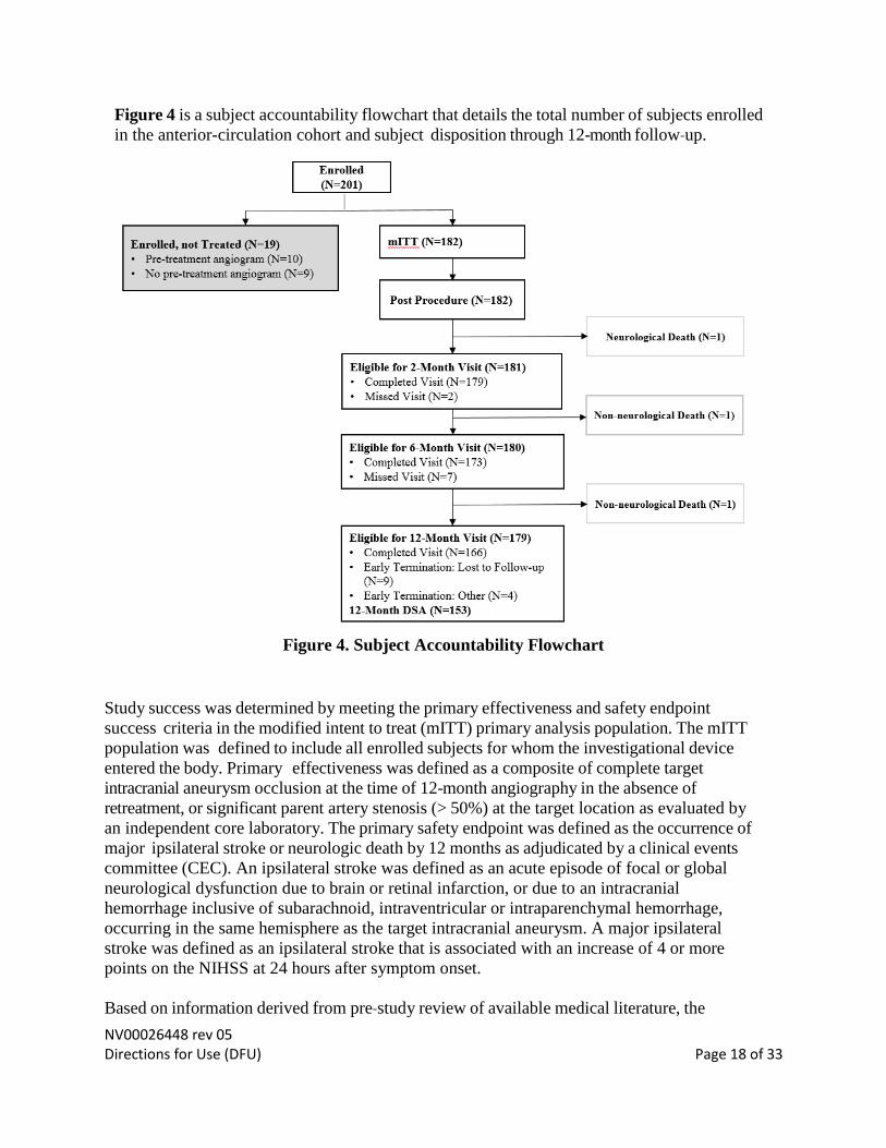
Figure 4 is a subject accountability flowchart that details the total number of subjects enrolled
in the anterior-circulation cohort and subject disposition through 12-month follow‐up.
Figure 4. Subject Accountability Flowchart
Study success was determined by meeting the primary effectiveness and safety endpoint
success criteria in the modified intent to treat (mITT) primary analysis population. The mITT
population was defined to include all enrolled subjects for whom the investigational device
entered the body. Primary effectiveness was defined as a composite of complete target
intracranial aneurysm occlusion at the time of 12-month angiography in the absence of
retreatment, or significant parent artery stenosis (> 50%) at the target location as evaluated by
an independent core laboratory. The primary safety endpoint was defined as the occurrence of
major ipsilateral stroke or neurologic death by 12 months as adjudicated by a clinical events
committee (CEC). An ipsilateral stroke was defined as an acute episode of focal or global
neurological dysfunction due to brain or retinal infarction, or due to an intracranial
hemorrhage inclusive of subarachnoid, intraventricular or intraparenchymal hemorrhage,
occurring in the same hemisphere as the target intracranial aneurysm. A major ipsilateral
stroke was defined as an ipsilateral stroke that is associated with an increase of 4 or more
points on the NIHSS at 24 hours after symptom onset.
Based on information derived from pre‐study review of available medical literature, the
NV00026448 rev 05 Directions for Use (DFU) Page 19 of 33
ATLAS trial was designed to be considered a success if the primary effectiveness endpoint
rate was statistically > 50% and the primary safety endpoint rate was statistically < 20%.
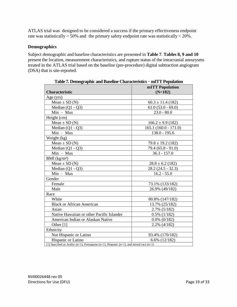
Demographics
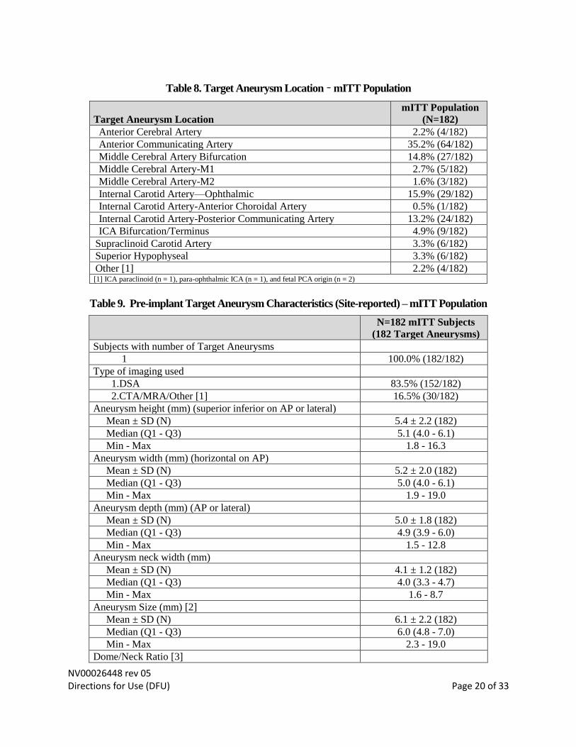
Subject demographic and baseline characteristics are presented in Table 7. Tables 8, 9 and 10
present the location, measurement characteristics, and rupture status of the intracranial aneurysms
treated in the ATLAS trial based on the baseline (pre-procedure) digital subtraction angiogram
(DSA) that is site-reported.
Table 7. Demographic and Baseline Characteristics – mITT Population
Characteristic
mITT Population
(N=182)
Age (yrs)
Mean ± SD (N) 60.3 ± 11.4 (182)
Median (Q1 - Q3) 61.0 (53.0 - 69.0)
Min – Max 23.0 - 80.0
Height (cm)
Mean ± SD (N) 166.2 ± 9.9 (182)
Median (Q1 - Q3) 165.1 (160.0 - 171.0)
Min – Max 138.0 - 195.6
Weight (kg)
Mean ± SD (N) 79.8 ± 19.2 (182)
Median (Q1 - Q3) 79.4 (65.8 - 91.0)
Min – Max 36.3 - 157.0
BMI (kg/m²)
Mean ± SD (N) 28.8 ± 6.2 (182)
Median (Q1 - Q3) 28.2 (24.5 - 32.3)
Min – Max 16.2 - 55.0
Gender
Female 73.1% (133/182)
Male 26.9% (49/182)
Race
White 80.8% (147/182)
Black or African American 13.7% (25/182)
Asian 2.7% (5/182)
Native Hawaiian or other Pacific Islander 0.5% (1/182)
American Indian or Alaskan Native 0.0% (0/182)
Other [1] 2.2% (4/182)
Ethnicity
Not Hispanic or Latino 93.4% (170/182)
Hispanic or Latino 6.6% (12/182) [1] Specified as Arabic (n=1), Portuguese (n=1), Hispanic (n=1), and mixed race (n=1)
NV00026448 rev 05 Directions for Use (DFU) Page 20 of 33
Table 8. Target Aneurysm Location – mITT Population
Target Aneurysm Location
mITT Population
(N=182)
Anterior Cerebral Artery 2.2% (4/182)
Anterior Communicating Artery 35.2% (64/182)
Middle Cerebral Artery Bifurcation 14.8% (27/182)
Middle Cerebral Artery-M1 2.7% (5/182)
Middle Cerebral Artery-M2 1.6% (3/182)
Internal Carotid Artery—Ophthalmic 15.9% (29/182)
Internal Carotid Artery-Anterior Choroidal Artery 0.5% (1/182)
Internal Carotid Artery-Posterior Communicating Artery 13.2% (24/182)
ICA Bifurcation/Terminus 4.9% (9/182)
Supraclinoid Carotid Artery 3.3% (6/182)
Superior Hypophyseal 3.3% (6/182)
Other [1] 2.2% (4/182) [1] ICA paraclinoid (n = 1), para-ophthalmic ICA (n = 1), and fetal PCA origin (n = 2)
Table 9. Pre-implant Target Aneurysm Characteristics (Site-reported) – mITT Population
N=182 mITT Subjects
(182 Target Aneurysms)
Subjects with number of Target Aneurysms
1 100.0% (182/182)
Type of imaging used
1.DSA 83.5% (152/182)
2.CTA/MRA/Other [1] 16.5% (30/182)
Aneurysm height (mm) (superior inferior on AP or lateral)
Mean ± SD (N) 5.4 ± 2.2 (182)
Median (Q1 - Q3) 5.1 (4.0 - 6.1)
Min - Max 1.8 - 16.3
Aneurysm width (mm) (horizontal on AP)
Mean ± SD (N) 5.2 ± 2.0 (182)
Median (Q1 - Q3) 5.0 (4.0 - 6.1)
Min - Max 1.9 - 19.0
Aneurysm depth (mm) (AP or lateral)
Mean ± SD (N) 5.0 ± 1.8 (182)
Median (Q1 - Q3) 4.9 (3.9 - 6.0)
Min - Max 1.5 - 12.8
Aneurysm neck width (mm)
Mean ± SD (N) 4.1 ± 1.2 (182)
Median (Q1 - Q3) 4.0 (3.3 - 4.7)
Min - Max 1.6 - 8.7
Aneurysm Size (mm) [2]
Mean ± SD (N) 6.1 ± 2.2 (182)
Median (Q1 - Q3) 6.0 (4.8 - 7.0)
Min - Max 2.3 - 19.0
Dome/Neck Ratio [3]
NV00026448 rev 05 Directions for Use (DFU) Page 21 of 33
N=182 mITT Subjects
(182 Target Aneurysms)
Mean ± SD (N) 1.2 ± 0.3 (182)
Median (Q1 - Q3) 1.1 (1.0 - 1.3)
Min - Max 0.4 - 2.1
Parent vessel diameter proximal to the aneurysm neck (mm)
Mean ± SD (N) 3.0 ± 0.7 (182)
Median (Q1 - Q3) 2.9 (2.4 - 3.6)
Min - Max 2.0 - 4.5
Parent vessel diameter distal to the aneurysm neck (mm)
Mean ± SD (N) 2.7 ± 0.7 (182)
Median (Q1 - Q3) 2.5 (2.1 - 3.2)
Min - Max 1.6 - 4.4
Parent vessel stenosis pre-implant
No 96.7% (176/182)
Yes 3.3% (6/182)
% stenosis:
25% or less 83.3% (5/6)
26% - 50% 16.7% (1/6) [1] Other includes CTA only (n = 19), MRA only (n = 10), and CTA + MRA + MRI (n = 1) [2] The aneurysm size is defined as the maximum of three dimensions (AP plane, lateral plane, height)
[3] The dome size is defined as the minimum of two widths (AP plane, lateral plane)
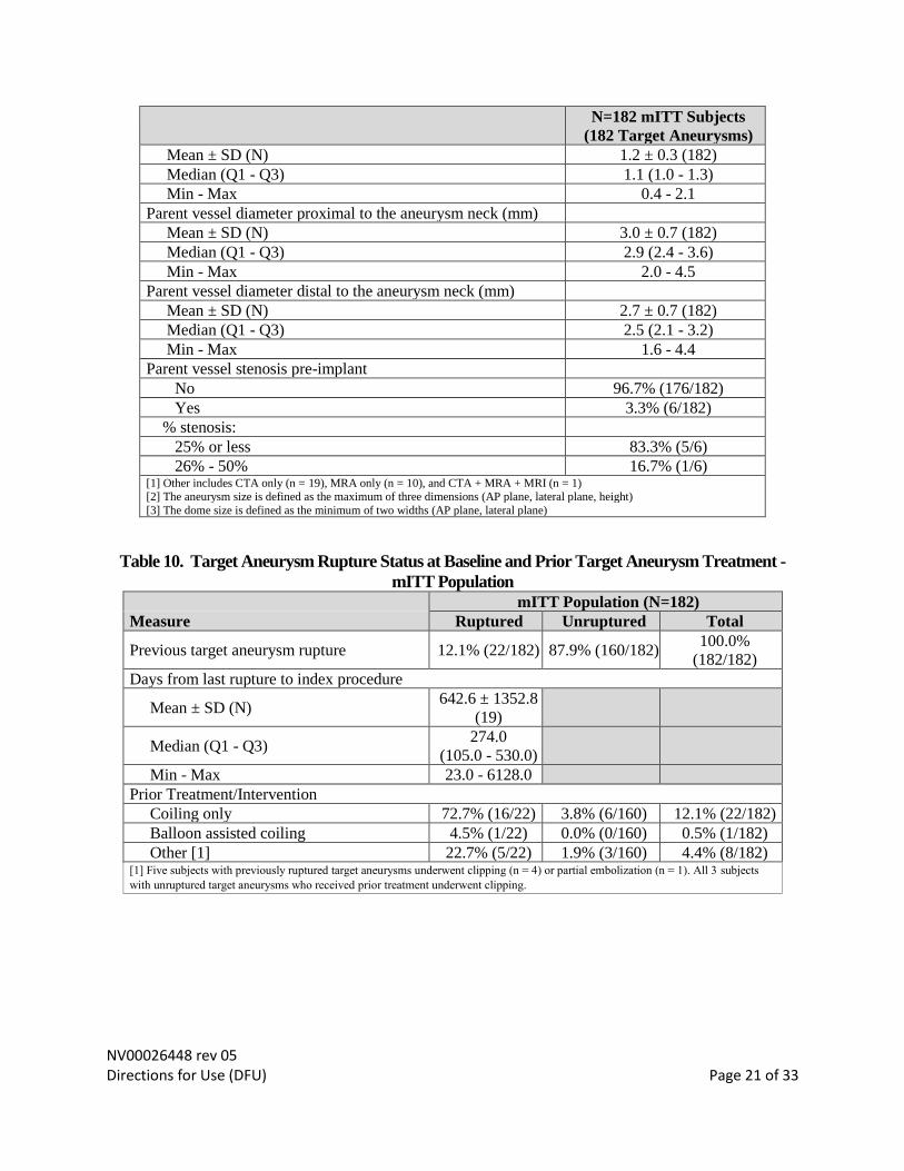
Table 10. Target Aneurysm Rupture Status at Baseline and Prior Target Aneurysm Treatment -
mITT Population
mITT Population (N=182)
Measure Ruptured Unruptured Total
Previous target aneurysm rupture 12.1% (22/182) 87.9% (160/182) 100.0%
(182/182)
Days from last rupture to index procedure
Mean ± SD (N) 642.6 ± 1352.8
(19)
Median (Q1 - Q3) 274.0
(105.0 - 530.0)
Min - Max 23.0 - 6128.0
Prior Treatment/Intervention
Coiling only 72.7% (16/22) 3.8% (6/160) 12.1% (22/182)
Balloon assisted coiling 4.5% (1/22) 0.0% (0/160) 0.5% (1/182)
Other [1] 22.7% (5/22) 1.9% (3/160) 4.4% (8/182) [1] Five subjects with previously ruptured target aneurysms underwent clipping (n = 4) or partial embolization (n = 1). All 3 subjects
with unruptured target aneurysms who received prior treatment underwent clipping.
NV00026448 rev 05 Directions for Use (DFU) Page 22 of 33
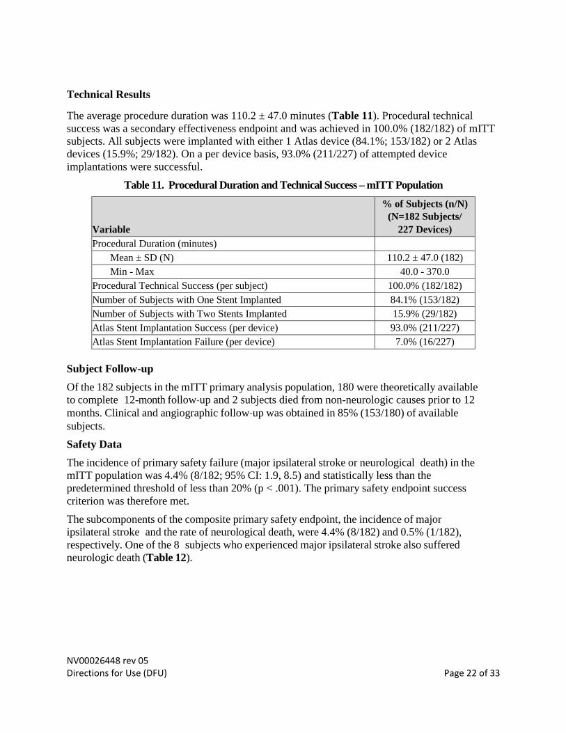
Technical Results
The average procedure duration was 110.2 ± 47.0 minutes (Table 11). Procedural technical
success was a secondary effectiveness endpoint and was achieved in 100.0% (182/182) of mITT
subjects. All subjects were implanted with either 1 Atlas device (84.1%; 153/182) or 2 Atlas
devices (15.9%; 29/182). On a per device basis, 93.0% (211/227) of attempted device
implantations were successful.
Table 11. Procedural Duration and Technical Success – mITT Population
Variable
% of Subjects (n/N)
(N=182 Subjects/
227 Devices)
Procedural Duration (minutes)
Mean ± SD (N) 110.2 ± 47.0 (182)
Min - Max 40.0 - 370.0
Procedural Technical Success (per subject) 100.0% (182/182)
Number of Subjects with One Stent Implanted 84.1% (153/182)
Number of Subjects with Two Stents Implanted 15.9% (29/182)
Atlas Stent Implantation Success (per device) 93.0% (211/227)
Atlas Stent Implantation Failure (per device) 7.0% (16/227)
Subject Follow-up
Of the 182 subjects in the mITT primary analysis population, 180 were theoretically available
to complete 12-month follow‐up and 2 subjects died from non-neurologic causes prior to 12
months. Clinical and angiographic follow‐up was obtained in 85% (153/180) of available
subjects.
Safety Data
The incidence of primary safety failure (major ipsilateral stroke or neurological death) in the
mITT population was 4.4% (8/182; 95% CI: 1.9, 8.5) and statistically less than the
predetermined threshold of less than 20% (p < .001). The primary safety endpoint success
criterion was therefore met.
The subcomponents of the composite primary safety endpoint, the incidence of major
ipsilateral stroke and the rate of neurological death, were 4.4% (8/182) and 0.5% (1/182),
respectively. One of the 8 subjects who experienced major ipsilateral stroke also suffered
neurologic death (Table 12).
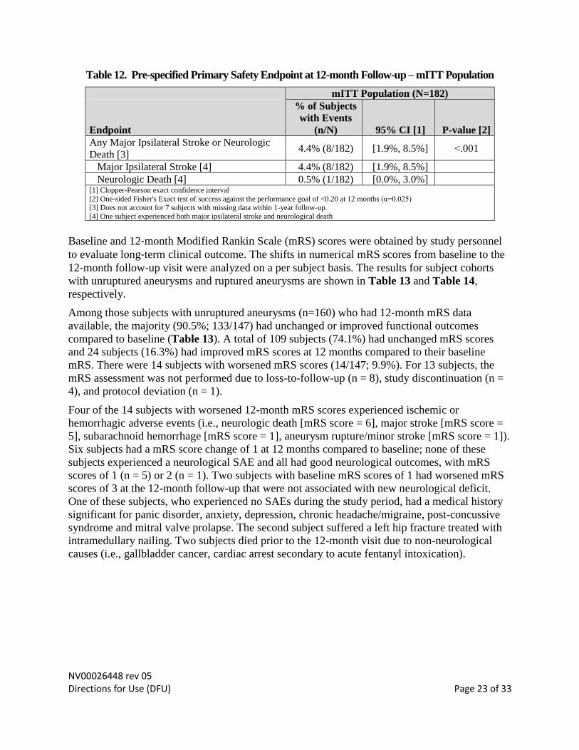
NV00026448 rev 05 Directions for Use (DFU) Page 23 of 33
Table 12. Pre-specified Primary Safety Endpoint at 12-month Follow-up – mITT Population
mITT Population (N=182)
Endpoint
% of Subjects
with Events
(n/N) 95% CI [1] P-value [2]
Any Major Ipsilateral Stroke or Neurologic
Death [3] 4.4% (8/182) [1.9%, 8.5%] <.001
Major Ipsilateral Stroke [4] 4.4% (8/182) [1.9%, 8.5%]
Neurologic Death [4] 0.5% (1/182) [0.0%, 3.0%] [1] Clopper-Pearson exact confidence interval
[2] One-sided Fisher's Exact test of success against the performance goal of <0.20 at 12 months (α=0.025) [3] Does not account for 7 subjects with missing data within 1-year follow-up.
[4] One subject experienced both major ipsilateral stroke and neurological death
Baseline and 12‐month Modified Rankin Scale (mRS) scores were obtained by study personnel
to evaluate long‐term clinical outcome. The shifts in numerical mRS scores from baseline to the
12‐month follow‐up visit were analyzed on a per subject basis. The results for subject cohorts
with unruptured aneurysms and ruptured aneurysms are shown in Table 13 and Table 14,
respectively.
Among those subjects with unruptured aneurysms (n=160) who had 12-month mRS data
available, the majority (90.5%; 133/147) had unchanged or improved functional outcomes
compared to baseline (Table 13). A total of 109 subjects (74.1%) had unchanged mRS scores
and 24 subjects (16.3%) had improved mRS scores at 12 months compared to their baseline
mRS. There were 14 subjects with worsened mRS scores (14/147; 9.9%). For 13 subjects, the
mRS assessment was not performed due to loss-to-follow-up (n = 8), study discontinuation (n =
4), and protocol deviation (n = 1).
Four of the 14 subjects with worsened 12-month mRS scores experienced ischemic or
hemorrhagic adverse events (i.e., neurologic death [mRS score = 6], major stroke [mRS score =
5], subarachnoid hemorrhage [mRS score = 1], aneurysm rupture/minor stroke [mRS score = 1]).
Six subjects had a mRS score change of 1 at 12 months compared to baseline; none of these
subjects experienced a neurological SAE and all had good neurological outcomes, with mRS
scores of 1 (n = 5) or 2 (n = 1). Two subjects with baseline mRS scores of 1 had worsened mRS
scores of 3 at the 12-month follow-up that were not associated with new neurological deficit.
One of these subjects, who experienced no SAEs during the study period, had a medical history
significant for panic disorder, anxiety, depression, chronic headache/migraine, post-concussive
syndrome and mitral valve prolapse. The second subject suffered a left hip fracture treated with
intramedullary nailing. Two subjects died prior to the 12-month visit due to non-neurological
causes (i.e., gallbladder cancer, cardiac arrest secondary to acute fentanyl intoxication).
NV00026448 rev 05 Directions for Use (DFU) Page 24 of 33
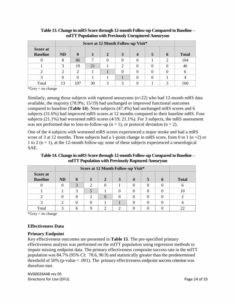
Table 13. Change in mRS Score through 12-month Follow-up Compared to Baseline –
mITT Population with Previously Unruptured Aneurysm
Score at 12 Month Follow-up Visit*
Score at
Baseline ND 0 1 2 3 4 5 6 Total
0 8 86 7 0 0 0 1 2 104
1 3 19 21 1 2 0 0 0 46
2 2 2 1 1 0 0 0 0 6
3 0 0 1 1 1 0 0 1 4
Total 13 107 30 3 3 0 1 3 160
*Grey = no change
Similarly, among those subjects with ruptured aneurysms (n=22) who had 12-month mRS data
available, the majority (78.9%; 15/19) had unchanged or improved functional outcomes
compared to baseline (Table 14). Nine subjects (47.4%) had unchanged mRS scores and 6
subjects (31.6%) had improved mRS scores at 12 months compared to their baseline mRS. Four
subjects (21.1%) had worsened mRS scores (4/19; 21.1%). For 3 subjects, the mRS assessment
was not performed due to loss-to-follow-up (n = 1), or protocol deviation (n = 2).
One of the 4 subjects with worsened mRS scores experienced a major stroke and had a mRS
score of 3 at 12 months. Three subjects had a 1-point change in mRS score, from 0 to 1 (n =2) or
1 to 2 (n = 1), at the 12-month follow-up; none of these subjects experienced a neurological
SAE.
Table 14. Change in mRS Score through 12-month Follow-up Compared to Baseline –
mITT Population with Previously Ruptured Aneurysm
Score at 12 Month Follow-up Visit*
Score at
Baseline ND 0 1 2 3 4 5 6 Total
0 0 3 2 0 1 0 0 0 6
1 1 3 5 1 0 0 0 0 10
2 0 0 2 0 0 0 0 0 2
3 2 0 0 1 1 0 0 0 4
Total 3 6 9 2 2 0 0 0 22
*Grey = no change
Effectiveness Data
Primary Endpoint
Key effectiveness outcomes are presented in Table 15. The pre-specified primary
effectiveness analysis was performed on the mITT population using regression methods to
impute missing endpoint data. The primary effectiveness composite success rate in the mITT
population was 84.7% (95% CI: 78.6, 90.9) and statistically greater than the predetermined
threshold of 50% (p‐value < .001). The primary effectiveness endpoint success criterion was
therefore met.
NV00026448 rev 05 Directions for Use (DFU) Page 25 of 33
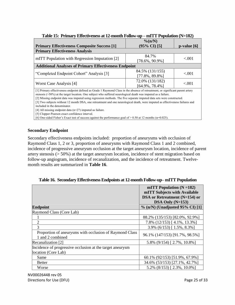
Table 15: Primary Effectiveness at 12-month Follow-up – mITT Population (N=182)
Primary Effectiveness Composite Success [1]
%(n/N)
(95% CI) [5] p-value [6]
Primary Effectiveness Analysis
mITT Population with Regression Imputation [2] 84.7%
[78.6%, 90.9%] <.001
Additional Analyses of Primary Effectiveness Endpoint
“Completed Endpoint Cohort” Analysis [3] 84.5% (131/155)
[77.8%, 89.8%] <.001
Worst Case Analysis [4] 72.0% (131/182)
[64.9%, 78.4%] <.001
[1] Primary effectiveness endpoint defined as Grade 1 Raymond Class in the absence of retreatment, or significant parent artery
stenosis (>50%) at the target location. One subject who suffered neurological death was imputed as a failure.
[2] Missing endpoint data was imputed using regression methods. The five separate imputed data sets were constructed.
[3] Two subjects without 12 month DSA, one retreatment and one neurological death, were imputed as effectiveness failures and
included in the denominator.
[4] All missing endpoint data (n=27) imputed as failure.
[5] Clopper-Pearson exact confidence interval.
[6] One-sided Fisher’s Exact test of success against the performance goal of > 0.50 at 12 months (α=0.025).
Secondary Endpoint
Secondary effectiveness endpoints included: proportion of aneurysms with occlusion of
Raymond Class 1, 2 or 3, proportion of aneurysms with Raymond Class 1 and 2 combined,
incidence of progressive aneurysm occlusion at the target aneurysm location, incidence of parent
artery stenosis (> 50%) at the target aneurysm location, incidence of stent migration based on
follow-up angiogram, incidence of recanalization, and the incidence of retreatment. Twelve-
month results are summarized in Table 16.
Table 16. Secondary Effectiveness Endpoints at 12-month Follow-up– mITT Population
mITT Population (N =182)
mITT Subjects with Available
DSA or Retreatment (N=154) or
DSA Only (N=153)
Endpoint % (n/N) (Unadjusted 95% CI) [1]
Raymond Class (Core Lab)
1 88.2% (135/153) [82.0%, 92.9%]
2 7.8% (12/153) [ 4.1%, 13.3%]
3 3.9% (6/153) [ 1.5%, 8.3%]
Proportion of aneurysms with occlusion of Raymond Class
1 and 2 combined 96.1% (147/153) [91.7%, 98.5%]
Recanalization [2] 5.8% (9/154) [ 2.7%, 10.8%]
Incidence of progressive occlusion at the target aneurysm
location (Core Lab)
Same 60.1% (92/153) [51.9%, 67.9%]
Better 34.6% (53/153) [27.1%, 42.7%]
Worse 5.2% (8/153) [ 2.3%, 10.0%]
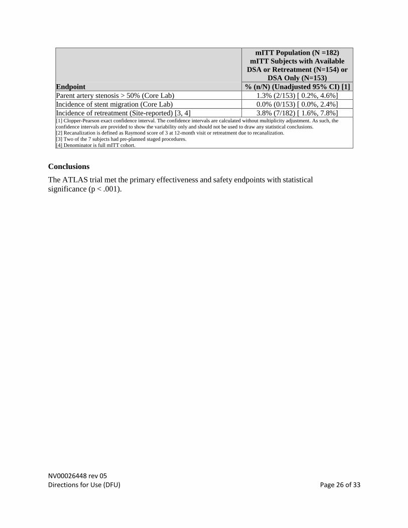
NV00026448 rev 05 Directions for Use (DFU) Page 26 of 33
mITT Population (N =182)
mITT Subjects with Available
DSA or Retreatment (N=154) or
DSA Only (N=153)
Endpoint % (n/N) (Unadjusted 95% CI) [1]
Parent artery stenosis > 50% (Core Lab) 1.3% (2/153) [ 0.2%, 4.6%]
Incidence of stent migration (Core Lab) 0.0% (0/153) [ 0.0%, 2.4%]
Incidence of retreatment (Site-reported) [3, 4] 3.8% (7/182) [ 1.6%, 7.8%] [1] Clopper-Pearson exact confidence interval. The confidence intervals are calculated without multiplicity adjustment. As such, the
confidence intervals are provided to show the variability only and should not be used to draw any statistical conclusions. [2] Recanalization is defined as Raymond score of 3 at 12-month visit or retreatment due to recanalization.
[3] Two of the 7 subjects had pre-planned staged procedures.
[4] Denominator is full mITT cohort.
Conclusions
The ATLAS trial met the primary effectiveness and safety endpoints with statistical
significance (p < .001).
NV00026448 rev 05 Directions for Use (DFU) Page 27 of 33
HOW SUPPLIED
Stryker Neurovascular products are sterile and non-pyrogenic in unopened packaging that is
designed to maintain sterility unless the primary product pouch has been opened or damaged.
Do not use if package is opened or damaged.
Do not use if labeling is incomplete or illegible.
Handling and Storage
Store in a cool, dry, dark place.
OPERATIONAL INSTRUCTIONS
Initial Access, Angiographic Assessment and Stent Selection
1. Gain vascular access according to standard practice. Select a recommended microcatheter
(see Required Accessories section on Page 2). If additional stability is required, consider the
use of an intermediate catheter in addition to the microcatheter. Establish and maintain
continuous flow of appropriate flush solution through the microcatheter per standard vascular
practice. Using angiography, determine the location of the aneurysm and the size of the
aneurysm neck.
2. Navigate the stent delivery microcatheter over an access length guidewire at least 1.2 cm
distal to the aneurysm neck.
Note: The microcatheter tip must be placed sufficiently distal to the aneurysm neck to allow for
slack to be removed from the system after the stent is advanced, while maintaining adequate
stent length (approximately 4 mm) distal to the aneurysm neck. Excessive tortuosity may
necessitate microcatheter tip placement more than 1.2 cm distal to the aneurysm neck.
3. Remove the guidewire.
4. Select an appropriate Neuroform Atlas Stent based on the largest reference vessel diameter
and the sizing recommendations in Tables 1 and 2. Select a stent that is at least 8 mm longer
(referenced off the working length, WL) than the aneurysm neck to maintain a minimum of 4
mm on each side of the aneurysm neck along the parent vessel.
Delivery System Preparation and Stent Transfer
Note: Coiling can be performed by placing the microcatheter into the aneurysm prior to or after
stent deployment, per physician preference.
5. Carefully inspect the stent delivery system packaging for damage.
6. Peel open the pouch using aseptic technique and remove the (sterile) dispenser hoop.
NV00026448 rev 05 Directions for Use (DFU) Page 28 of 33
7. Carefully place the dispenser hoop into the sterile field.
8. Using two hands, one on each side of the wire retention clip, release the stent delivery wire
from the wire retention clip on the dispenser hoop.
9. Remove the device from the dispenser hoop by grabbing the stent delivery wire and the
proximal end of the introducer sheath; holding them together, slowly and carefully remove
the entire wire and introducer sheath.
Note: The stent delivery wire and proximal end of the introducer sheath must be held together
when removing the Neuroform Atlas Stent System from the dispenser hoop to prevent stent
movement and premature deployment.
Note: Ensure that the stent delivery wire does not move relative to the introducer sheath during
removal of the stent system from the dispenser hoop.
10. Inspect the stent delivery system. Confirm that the tip of the stent delivery wire is entirely
within the introducer sheath. Confirm that the stent delivery wire is not kinked and that the
introducer sheath tip is not damaged.
11. Partially insert the distal end of the introducer sheath into the RHV that is connected to the
microcatheter. Tighten the RHV to secure the introducer sheath.
Note: Partial insertion of the introducer sheath into the RHV is necessary to ensure a flow path
for flush. Ensure that the tip of the introducer sheath is inserted into the middle of the RHV.
Note: Under-tightening the RHV may result in inadequate flushing. Over- tightening the RHV
may crush the introducer sheath and may result in inadequate flushing.
12. Open the y- connector valve of the RHV that is connected to the appropriate flush solution
and verify that fluid exits the proximal end of the introducer sheath.
Warning: Purge the system carefully to avoid the accidental introduction of air into the stent
system.
13. Loosen the RHV, then advance the introducer sheath until the tip is fully inserted into the
microcatheter hub. Tighten the RHV firmly.
Warning: Confirm there are no air bubbles trapped anywhere in the stent system.
Note: After tightening the RHV firmly, the introducer sheath tip should not move when pulled
NV00026448 rev 05 Directions for Use (DFU) Page 29 of 33
gently. Failure to secure the introducer sheath may result in premature deployment of the
stent within the microcatheter hub or difficulty in transferring the stent.
Note: The introducer sheath tip must be fully inserted into the microcatheter hub to enable the
stent to move into the microcatheter. Over-tightening the RHV may crush the introducer
sheath, while under-tightening the RHV may result in premature deployment of the stent.
14. Advance the stent delivery wire to transfer the stent from the introducer sheath into the
microcatheter.
Note: Ensure that the introducer sheath does not move while advancing the stent delivery wire.
Movement of the introducer sheath during stent advancement may result in premature
deployment of the stent within the microcatheter hub.
15. Continue advancing the stent delivery wire into the microcatheter until the distal edge of the
fluoro saver mark enters the introducer sheath. The fluoro saver mark is 135 cm from the
stent delivery wire distal tip. When the fluoro saver mark enters the introducer sheath, the
stent is approximately 90 cm inside the microcatheter.
16. Loosen the RHV on the stent delivery microcatheter, remove the introducer sheath from the
proximal end of the stent delivery wire while holding the stent delivery wire fixed in place,
and set the introducer sheath aside.
Note: At this point, fluoroscopy may be used at the physician’s discretion.
17. If desired, place torque device on proximal end of wire (at least 5 cm from proximal end of
fluoro saver marker).
Note: The torque device may be attached to the proximal end of the stent delivery wire to
facilitate handling and stabilization. Be sure to tighten the torque device to secure the stent
delivery wire. Do not use the torque device to torque the stent delivery wire as it is not
designed to be torqued.
18. Slowly advance the delivery wire and stent until the distal edge of the stent delivery wire
fluoro saver mark reaches the stent delivery microcatheter’s RHV.
Note: If resistance is encountered at any point during stent manipulation, do not apply undue
force. Withdraw the microcatheter, stent, and stent delivery wire as a unit and repeat the
procedure with new devices.
NV00026448 rev 05 Directions for Use (DFU) Page 30 of 33
Stent positioning and Deployment
19. Under fluoroscopy, advance the stent delivery wire until the stent’s distal radiopaque markers
are 1 – 2 mm proximal of the distal tip marker of the stent delivery microcatheter.
Note: Maintain adequate stent length (approximately 4 mm) on each side of the aneurysm neck
to ensure appropriate neck coverage.
20. Withdraw the microcatheter slightly to remove any slack from the stent system and position
the stent for deployment by aligning the stent radiopaque markers across the target aneurysm.
21. If stent delivery microcatheter positioning is satisfactory, carefully retract the stent delivery
microcatheter in a continuous movement while maintaining the position of the stent delivery
wire. This will allow the stent to deploy across the neck of aneurysm. The stent’s distal
radiopaque markers will expand as the stent exits the stent delivery microcatheter.
Note: Do not use the stent delivery wire to push the stent out of the microcatheter while
deploying.
Note: Do not deploy the stent if it is not properly positioned in the vessel.
22. Confirm deployed stent position using fluoroscopy.
23. If stent did not adequately cover aneurysm, withdraw the stent delivery wire from the stent
delivery microcatheter. Place additional Neuroform Atlas™ Stents as needed.
24. Once the aneurysm is adequately covered, remove stent delivery wire and stent delivery
microcatheter from patient and establish hemostasis.
25. Perform coiling procedure per appropriate coiling device DFU.
26. Discard used devices.
Additional Information
Patient Information
You should have already provided the patient with a copy of the Patient Information Booklet so
that (s)he has had adequate time to review the information and ask any questions.
Immediately after the procedure, complete the Patient Information Card, which is included in the
carton box, and provide the card to the patient before the patient leaves the hospital. The
Patient Information Card includes important information about the stent that was used and
includes a statement regarding MRI information.
NV00026448 rev 05 Directions for Use (DFU) Page 31 of 33
Concomitant Medical Therapy
Typical antiplatelet and anticoagulation regimen used for interventional intracranial procedure is
recommended at the discretion of the treating physician. Do not use the Neuroform Atlas Stent
System in patients in whom antiplatelet and/or anticoagulation therapy is contraindicated.
MAGNETIC RESONANCE IMAGING (MRI) INFORMATION
Safety Information Magnetic Resonance Conditional
Non-clinical testing and analysis have demonstrated that the Neuroform Atlas Stent is MR
Conditional alone, or when overlapped with a second stent, and adjacent to a Stryker
Neurovascular coil mass. A patient with the Neuroform Atlas Stent can be safely scanned
immediately after placement of this implant, under the following conditions:
• Static magnetic field of 1.5 and 3.0 Tesla
• Maximum spatial gradient field up to 2500 Gauss/cm (25 Tesla/m)
• Maximum MR system reported whole body averaged specific absorption rate of 2 W/kg
(Normal Operating Mode) and head averaged specific absorption rate of 3.2 W/kg.
Under the scan conditions defined above, the Neuroform Atlas Stent is expected to produce a
maximum temperature rise of 4 °C after 15 minutes of continuous scanning. The Neuroform
Atlas Stent should not migrate in this MRI environment.
In non-clinical testing, the image artifact caused by the device extends approximately 2 mm from
the Neuroform Atlas Stent when imaged with a spin echo pulse sequence and 3 Tesla MRI
System. The artifact may obscure the device lumen. It may be necessary to optimize MR
imaging parameters for the presence of this implant. See additional precaution related to the
image artifact from the implant in the “Precautions” section of this labeling.
QUESTIONS AND ANSWERS
Q: What is the optimal position of the stent with respect to the aneurysm?
A: Generally, try to position the stent so that each end of the stent is secured in relatively straight
areas of the parent vessel. The stent will be more stable if each end of the stent is anchored in at
least 4 mm of normal vessel. For example, if an aneurysm is located in the supraclinoid carotid,
it may be better to secure the stent by deploying the distal end in the M1 (middle cerebral artery,
first segment) than trying to deploy it in the few millimeters between the aneurysm and the ICA
(internal carotid artery) bifurcation. When deploying the stent, care should be taken to use a view
that best shows the parent vessel distal to the aneurysm; this enables the distal end of the stent to
be accurately deployed with respect to the aneurysm. This view may be different from the view
used to advance the Neuroform Atlas Stent System, or the view used as a working position for
aneurysm embolization.
NV00026448 rev 05 Directions for Use (DFU) Page 32 of 33
Q: Which stent size should I choose if I intend to place the stent in a vessel that has a different
diameter between the proximal and distal ends of the stent? Example: A vessel proximal
diameter decreases from a 4.0 mm at the right ICA bifurcation terminus to a 2.5 mm MCA distal
diameter.
A: Choose the stent sized for the larger vessel. In this example, choose the 4.0 or 4.5 mm stent.
This stent can be deployed safely in the smaller MCA and will be well anchored in the ICA
terminus.
Q. I have accidently started to deploy the stent, but it is not in the location that I wanted. What
should I do?
A. In general the safest course of action is not to try repositioning the stent. Rather, continue to
deploy the stent where it is and then deploy a second stent at the desired location. Safely
deploying a stent – even in an undesired location – will minimize vascular injury. Animal studies
have demonstrated that the stent endothelializes in less than 30 days.
Q: I misjudged the positioning of the stent and have deployed it with one end adjacent to the
aneurysm rather than in the normal part of the parent vessel. What should I do?
A: Remove the spent stent delivery wire from the microcatheter while maintaining the position
of the microcatheter. Insert and deploy a second Neuroform Atlas Stent starting from inside of
the first stent to the normal portion of the parent vessel (in a telescoping fashion). The second
stent should be of the same diameter or larger than the first.
NV00026448 rev 05 Directions for Use (DFU) Page 33 of 33
Warranty
Stryker Neurovascular warrants that reasonable care has been used in the design and
manufacture of this instrument. This warranty is in lieu of and excludes all other warranties
not expressly set forth herein, whether express or implied by operation of law or otherwise,
including, but not limited to, any implied warranties of merchantability or fitness for a
particular purpose. Handling, storage, cleaning and sterilization of this instrument as well as
other factors relating to the patient, diagnosis, treatment, surgical procedures and other matters
beyond Stryker Neurovascular’s control directly affect the instrument and the results obtained
from its use. Stryker Neurovascular’s obligation under this warranty is limited to the repair or
replacement of this instrument and Stryker Neurovascular shall not be liable for any incidental or
consequential loss, damage or expense directly or indirectly arising from the use of this
instrument. Stryker Neurovascular neither assumes, nor authorizes any other person to assume
for it, any other or additional liability or responsibility in connection with this instrument.
Stryker Neurovascular assumes no liability with respect to instruments reused, reprocessed
or resterilized and makes no warranties, express or implied, including but not limited to
merchantability or fitness for a particular purpose, with respect to such instruments.


![Final Stent[1]](https://img.pdfslide.us/doc/110x75/555e755ed8b42a41328b48b4/final-stent1.jpg)