Embed Size (px)
Citation preview

TheProstate 73:801^812 (2013)
Neuroendocrine-Derived Peptides Promote ProstateCancerCell Survival ThroughActivationof
IGF-1RSignaling
John O. DaSilva,1 George P. Amorino,2 Eli V. Casarez,1 Bradley Pemberton,2 andSarah J. Parsons1*
1DepartmentofMicrobiology,Cancer Center,Universityof VirginiaHealth Sciences Center,Charlottesville,Virginia 22908
2Departmentof RadiationOncologyandCancer Center,Universityof VirginiaHealth Sciences Center,Charlottesville,Virginia 22908
BACKGROUND. Neuroendocrine (NE) cells promote the progression of prostate cancer toa castration-resistant state through the production of paracrine growth factors. We have dem-onstrated this principle using in vitro and in vivo proliferative endpoints; however, the con-tributions of NE-derived pro-survival factors and anti-apoptosis to this phenomenon havenot been thoroughly investigated.METHODS. Here, we utilized conditioned-medium (CM) from LNCaP cells, engineered toundergo NE differentiation, and examined its effects on PC3 and LNCaP cell survival.RESULTS. Statistically significant changes in clonogenic survival, Annexin V staining, PARPcleavage and trypan blue positivity of approximately twofold were observed in the presence ofNE-derived CM relative to control-CM for both LNCaP and PC3 cells. These changes werepartially abrogated by antagonists of the neuropeptides neurotensin, bombesin, and PTHrP.Selective inhibitors of IGF-1R, EGFR or Src caused significant and nearly complete blockade ofprostate cancer cell survival due to NE secretions. Similar increases in cell survival were ob-served for LNCaP or PC3 cells treated with NE-derived medium in the presence of docetaxel.Increased phosphorylation of IGF-1R, following treatment with NE-derived medium, was ac-companied by decreased protein tyrosine phosphatase, receptor type F (PTPRF) mRNA, andprotein levels. Overexpression of PTPRF decreased cell survival, the amplitude and duration ofIGF-1R phosphorylation, and enhanced PARP cleavage in the presence of NE-derived medium.CONCLUSIONS. These data support the hypothesis that NE-derived factors act upon prostatecancer cells to stimulate pro-survival signaling and describe a novel mechanism of cross-talkbetween NE-derived factors and IGF-1R, mediated in part by PTPRF. Prostate 73: 801–812,2013. # 2012 Wiley Periodicals, Inc.
KEY WORDS: neuroendocrine; EGFR; IGF-1R; PTPRF; apoptosis
INTRODUCTION
Early stage prostate cancer (PCa) is responsive toandrogen ablation therapy, but invariably recurs ascastration-resistant and highly aggressive. The molec-ular mechanisms that promote PCa progression arenot fully understood, but both continued androgenreceptor-mediated signaling under conditions of lowcirculating ligand and upregulation of pro-survivalpathways are implicated in this process [1,2].
Neuroendocrine (NE) differentiation has been asso-ciated with progression of PCa to a castration-resistant
Additional supporting information may be found in the online ver-sion of this article.
Grant sponsor: NCI; Grant number: T32-CA009109-33,Grant number: P01CA76465, Grant number: R0176649.
Conflict of interest: The authors declare no conflict of interest.
*Correspondence to: Sarah J. Parsons, PhD, Department of Microbi-ology, University of Virginia Health System, PO Box 800734, Char-lottesville, VA 22908-0734. E-mail: [email protected] 12 October 2012; Accepted 2 November 2012DOI 10.1002/pros.22624Published online 28 November 2012 in Wiley Online Library(wileyonlinelibrary.com).
� 2012WileyPeriodicals,Inc.

phenotype. NE cells are more prevalent in castration-resistant disease, occurring in 30–100% of tumorsstudied [3,4]. The low proliferative capacity of NEcells allows them to resist treatment with most chemo-therapeutic agents, as well as endocrine and radiationtreatments [5,6]. Paracrine factors produced by NEcells bind to G-protein-coupled receptors (GPCR) andactivate signaling pathways that stimulate tumor cellgrowth and may promote survival [7–9]. Among thesepathways is the EGFR/Src/STAT5b axis, which istransactivated upon NE factors binding their cognatereceptors and subsequent metalloproteinase (MMP)liberation of membrane-bound EGFR ligands [9,10].This hypothesis is supported by work demonstratingthat NE-like cells enhance the growth of LNCaP xeno-grafts, with the greatest effects seen under conditionsof androgen deprivation [11,12].
Accumulating evidence suggests that insulin-likegrowth factor 1 (IGF-1) signaling pathways also con-tribute to PCa progression. Studies in murine modelshave correlated altered IGF-1 or IGF-1 receptor (IGF-1R) levels with prostatic dysplasia, tumor growth ormetastasis [13–16]. Human metastatic PCa specimensexhibit increased IGF-1R expression relative to prima-ry tumors [17–19]. Experimental approaches usinglineage-derived PCa progression models demonstratethat increased IGF-1R signaling results in protectionfrom apoptotic stress and enhanced mitotic activitythat correlates with an androgen independent state[19]. Furthermore, long-term androgen-ablation hasbeen shown to cause increased resistance to phospha-tidylinositol 30-kinase (PI3K)/Akt inhibition [20],while reduction of IGF-1R levels in androgen inde-pendent DU145 cells results in increased chemothera-peutic sensitivity [21].
Ligand binding to the IGF-1R a-subunits leads totyrosine phosphorylation of the b-subunits and acti-vation of the receptor tyrosine kinase, resulting insubsequent activation of intracellular signaling path-ways including Ras/MAPK and PI3K/Akt [2]. Acti-vation of PI3K/Akt signaling contributes to PCaprogression by phosphorylation of Bcl-2 family mem-ber BAD and caspase-9 resulting in the inhibition ofapoptosis and promoting cell survival [22]. PI3K/Aktphosphorylation of BAD by Akt prevents the bindingof BAD to BCL-XL, restoring the antiapoptotic func-tion of BCL-XL [23]. In addition, phosphorylation ofpro-caspase 9 by Akt abrogates cytochrome c-inducedproteolytic processing of pro-caspase 9, which in turnprevents the activation of downstream executor cas-pases [24].
GPCR agonists have also been shown to stimulatethe phosphorylation of IGF-1R, suggesting that NE-derived paracrine factors promote crosstalk with theIGF-1 signaling pathway to modulate cell survival
[25,26]. Activation of thrombin, angiotensin II orGABA receptors promotes phosphorylation of IGF-1Ror PI3K/Akt [27–29]. Furthermore, studies of the neu-ropeptides, endothelin-1 and bombesin, indicate theyact as antiapoptotic factors and expression of neutralendopeptidase inhibits their ability to transactivateIGF-1R-mediated apoptosis [7,8,30].
Studies described in this report investigate the con-tribution of NE-derived factors to prostate cancer cellsurvival and resistance to chemotherapeutic agents,such as docetaxel, through the activation of IGF-1R.Our results demonstrate that CM derived from NEcells promotes PCa cell survival and protects from ap-optotic stress through the transcriptional downregula-tion of the protein tyrosine phosphatase, receptortype F (PTPRF). PTPRF has been identified as a metas-tasis-associated gene in prostate cancer cell lines, andits activity is regulated through the interaction withEGFR [31,32]. In both LNCaP and PC3 PCa cell lineswe observed that sustained EGFR and IGF-1R phos-phorylation in response to NE-derived factors corre-lates with downregulation of PTPRF and decreasedapoptosis, overexpression of PTPRF reversed theseeffects. To our knowledge, this is the first report dem-onstrating that IGF-1R activation is regulated down-stream of NE signaling pathways by the modulationof protein tyrosine phosphatase function.
MATERIALSANDMETHODS
Reagents
Annexin-V-FITC flow cytometry kits wereobtained from BD Biosciences (San Diego, CA). Anti-human IGF-1 antibody was purchased from R&D Sys-tems (Minneapolis, MN). pCMV-PTPRF was obtainedfrom Thermo Scientific (Huntsville, AL). Recombi-nant human des(1–3)IGF-1 was obtained fromCell Sciences (Canton, MA). The neurotensin receptorantagonist, SR48692, was a gift from Sanofi-Aventis(Malvern, PA). The bombesin receptor antagonist,(D-Phe6,Leu13-p-chloro-Phe14)-Bombesin [6–14], andPTHrP receptor inhibitor, (Nle8,18,Tyr3,4)-pTH [3–34]amide were obtained from Bachem (King of Prussia,PA). Inhibitors of EGFR (AG1478–100 nM, Millipore,Billerica, MA), IGF-1R (PQ401-5 mM, Tocris Biosci-ence, Ellisville, MO), c-Src (PP2-10mM, Millipore) orMMPs (GM6001-20 mM, Millipore) were added tocells 1 h prior to stimulation.
CellCulture
All cell culture media, fetal bovine serum (FBS),and Lipofectamine 2000 transfection reagents wereobtained from Invitrogen (Carlsbad, CA). PC3 andLNCaP human prostate carcinoma cells were obtained
802 DaSilva et al.
The Prostate

from American Type Tissue Collection (ATTC; Rock-ville, MD) and grown as recommended.
PreparationofNE-CM
NE differentiation of LNCaP-D29 cells by doxycy-cline-induced expression of a constitutively activePKA catalytic subunit has been previously described[11]. Neuroendocrine conditioned-medium (NE-CM)of LNCaP-D29 cells was collected following incuba-tion for 5 days in serum-free medium with 1 mg/mldoxycycline. Control CM (C-CM) was collected fromLNCaP-D29 cells incubated in the absence ofdoxycycline.
WesternBlotting
Immunoblotting was performed as previously de-scribed [9], using the following antibodies: anti-PTPRF(BD Biosciences), anti-EGFRTyr992, and anti-EGFR(Invitrogen), Anti-phospho-IGF-1RTyr1135/1136, anti-IGF-1R, and anti-rabbit-HRP (Cell Signaling Technolo-gy, Beverly, MA).
ClonogenicAssays
Survival assays were performed as previously de-scribed [35]. Colonies were counted after fixation andstaining with 0.5% crystal violet in methanol. The sur-viving fraction was calculated as follows: Survivingfraction (S.F.) ¼ (mean # colonies)/(cells inoculated)(plating fraction), where the plating fraction repre-sents (# colonies)/(cells inoculated). Data were nor-malized such that S.F. ¼ 1 for C-CM samples.
FlowCytometry
Cells were trypsinized, counted, and processedwith binding buffer and Annexin-V-FITC (BD Bio-sciences) according to the manufacturer’s instruc-tions. 10,000 cells/sample were analyzed on a Becton-Dickinson FACS Caliber flow cytometer (FranklinLakes, NJ). Data analysis was performed using Flow-Jo software at the UVA Flow Cytometry Core Facility.
TrypanBlueCellDeathAssay
Floating and adherent cells obtained following tryp-sinization were incubated in 0.2% trypan blue (Sigma,St. Louis, MO), and counted with a hemocytometer.
GeneExpressionAnalysis
Gene expression analysis using GeneChip HGU133Plus 2.0 (Affymetrix, Santa Clara, CA) was performedby the UVA Biomolecular Research Facility. mRNAwas isolated using the RNeasy kit (Qiagen, Hilden,Germany) according to the manufacturer’s instructions.
Triplicates of C-CM-treated LNCaP RNA were com-pared to triplicates of NE-CM-treated LNCaP RNA.Affimetrix GeneChip Operating Software was used tocalculate signal intensity values as well as P-values foreach sample. Two group comparisons were carried outusing the dChip software (http://biosun1.harvar-d.edu/�cli/dchip). Gene expression was deemed sig-nificantly different if the P-value was �0.05, the foldchange in signal intensity was >1.5 or <�1.5, and theabsolute difference in signal intensity was >100.
SignalTransductionPathwayBioinformatics
The genes identified as significantly varied be-tween the controls and the experimental sampleswere used for data mining with the Ingenuity soft-ware. Canonical pathways analysis identified thepathways from the Ingenuity Pathways Analysis li-brary of canonical pathways that were most signifi-cant to the data set. The significance of the associationbetween the data set and the canonical pathway wasmeasured in two ways: (1) a ratio of the number ofmolecules from the data set that map to the pathwaydivided by the total number of molecules in the ca-nonical pathway and (2) Fisher’s exact test was usedto calculate a P-value determining the probability thatthe association between the genes in the dataset andthe canonical pathway is explained by chance alone.
Real-TimeReverseTranscription-PCR
Real-time reverse transcription-PCR was per-formed as previously described [9]. The PCR primersused were the following: PTPRF, 50-GCATGTC-TACTGGAAGCT-30 (forward) and 50-CCTTGCCGAAATCTATGG-30 (reverse); and GUS, 50-CCGACTTCTCTGACAACCGACG-30 (forward) and 50-AGCCGA-CAAAATGCCGCAGACG-30 (reverse).
StatisticalAnalysis
Error bars represent the standard error of the mean(SEM) from N ¼ 3 independent experiments, unlessindicated. P values were obtained using a Student’s t-test; P values <0.05 were considered statisticallysignificant.
RESULTS
NE-CMEnhancesSurvivalof ProstateCancerCells
To determine whether NE-CM confers a survivaladvantage to PCa cells clonogenic survival of LNCaPor PC3 cells was assayed following overnight serumdeprivation and 24 h treatment with C-CM or NE-CM. An increase of approximately two-fold in thesurviving fraction of LNCaP or PC3 cells was
NEFactors StimulateProstateCancerSurvival 803
The Prostate

observed in the presence of NE-CM (Fig. 1A,B).Incubation with NE-CM reduced the number of apo-ptotic LNCaP or PC3 cells by 38% or 47%, respective-ly, as measured by Annexin V flow cytometry(Fig. 1C,D). Under similar conditions as described inFigure 1A, the number of LNCaP or PC3 cells stainingpositively for trypan blue decreased by 55% or 54%,respectively, following treatment with NE-CM relativeto C-CM (Fig. 1E,F). These results indicate that para-crine factors secreted by NE cells contribute to pro-survival signaling in PCa cells.
Docetaxel-based regimens have become the stan-dard of care in the management of castration-resistantPCa [33]. To determine how NE-derived peptides in-fluence the response of PCa cells to chemotherapeu-tics we analyzed the effect of NE-CM treatment on
docetaxel-induced LNCaP or PC3 cell death. Treat-ment of LNCaP or PC3 cells with 10 nM docetaxel inthe presence of C-CM resulted in approximately six-fold increase in the number of cells staining positivefor trypan blue relative to no docetaxel in C-CM. Thislevel of cell death was reduced by 50% in the presenceof NE-CM (Fig. 2A,B). NE-CM treatment also de-creased the extent of PARP cleavage, indicative of re-duced apoptotic induction (Fig. 2C,D). Treatment ofthe PCa cells with docetaxel together with the smallmolecule inhibitors specific for EGFR, IGF-1R, Src orMMP’s in the presence of NE-CM restored PARPcleavage to levels equivalent to docetaxel/C-CM con-ditions implicating the involvement of EGFR transac-tivation and IGF-1R function in the pro-survival effectof NE-CM (Fig. 2C,D).
Fig. 1. NE-CMenhances survival of prostate cancer cells.A,B: LNCaPor PC3 cellswere treated for 48 hwithNE-CMorC-CM, preparedas described inMaterials andMethods Section.Cells were trypsinized, diluted, andplated for colony formation. After10 days, colonieswerestainedwith crystal violet and counted; the surviving fractionwas calculated as described inMaterials andMethods Section.C,D:Detectionof apoptotic celldeathbyAnnexinV-FITC stainingusing flowcytometry following 48 h treatmentwithNE-CMorC-CM.E,F:Trypanblue ex-clusion assayofLNCaPorPC3celldeath following48 h treatmentwithNE-CMorC-CM.Data arepresentedas themean � SEMof threein-dependentexperiments,eachperformedin triplicate;�P < 0.05comparedtoC-CM.
804 DaSilvaet al.
The Prostate

Inhibitionof IGF-1RorEGFRAntagonizesPro-Survival EffectsofNE-CM
To further investigate the role of paracrine factorsproduced by NE cells in promoting survival of PCacells we performed global analysis of gene expressionin LNCaP cells treated for 24 h with NE-CM (TableT1). Canonical pathway analysis of gene expressiondata using Ingenuity Pathways Library revealed thatexpression of IGF-1R and EGFR pathway genes aremost significantly different between NE-CM and C-CM treatments (Fig. 3A).
Inhibition of IGF-1R or EGFR signaling con-firmed the requirement of these signaling pathwaysin mediating the survival signals of NE-CM. Treat-ment of LNCaP or PC3 cells with AG1478, PQ401or the Src inhibitor PP2 for 24 h abolished the pro-survival effects of NE-CM as measured by trypanblue exclusion. In addition, statistically significantincreases in the percentage of cell death were ob-served with combinatorial but not individual an-tagonism of GPCRs mediating the action ofneuropeptides previously shown to be present inNE-CM (Fig. 3B,C, [34]). Antagonism of the PTHrPreceptor significantly reduced the pro-survivaleffects of NE-CM in PC3 cells, suggesting that thisGPCR pathway is a more critical mediator of NE-induced survival signaling in the PC3 cell model(Fig. 3C). Pharmacological inhibition of componentsmediating NE-induced signaling does not alter thepercentage of surviving cells in the presence of C-CM, indicating that their effects on cell viability arespecific to NE-secreted factors (Fig. S1).
NE-InducedActivationof IGF-1RSignaling IsIndependentof IGF-1Binding
To determine whether NE-CM promotes the activa-tion of IGF-1R in LNCaP or PC3 cells, phosphoryla-tion of IGF-1R Tyr1135/1136 was measured byimmunoblotting with phosphospecific antibodies.Phosphorylation of Tyr1135/1136 was increased 1 h afterstimulation with NE-CM and persisted for 24 h butwas not induced by C-CM. Similarly, phosphorylationof EGFR-Tyr992 was sustained throughout the timecourse of analysis in response to NE-CM (Fig. 4A,B).The kinetics of IGF-1R and EGFR activation in re-sponse to NE-CM differs from stimulation with thecognate growth factor. Upon stimulation of LNCaP orPC3 cells with IGF-1, the extent of IGF-1R tyrosinephosphorylation is significantly greater than with NE-CM treatment, peaking at 30 min and returning tobasal levels after 3 h (Fig. S2A,B). In response to EGFthe kinetics of IGF-1R activation are similar to stimula-tion with IGF-1, however the amplitude of receptorphosphorylation is equivalent to NE-CM treatment(Fig. S2A,B and 4E,F). Treatment of LNCaP or PC3cells with AG1478, PQ401, PP2 or GM6001 partially orcompletely inhibited IGF-1R phosphorylation in re-sponse to NE-CM. Interestingly, pIGF-1R inhibitionhad no effect on NE-CM-induced EGFR phosphoryla-tion (Fig. 4C,D). In the AR-positive C4-2B model ofCRPC, NE-CM-induced activation of IGFR, EGFR orSrc was also completely abrogated by the combinedinhibition of the PTHrP, Neurotensin, and Bombesinreceptors, the EGFR inhibitor Lapatinib, the Src inhibi-tor Dasatinib or GM6001 (Fig. S3). Together these
Fig. 2. NE-CMreducesdocetaxel-inducedcelldeath.A,B: Celldeath ofLNCaPorPC3 cells treatedwithNE-CMorC-CMin thepresenceor absence ofdocetaxel (10 nM) for 48 hwasdeterminedby trypanblue staining.Results are expressedas themean � SEMof threeindepen-dentexperiments;��P < 0.05 relative toNE-CM/doc;�P < 0.05compared toNE-CM.C,D:LNCaPorPC3cellswereincubatedwithNE-CMorC-CM � inhibitorsofEGFR(AG1478-AG),IGF-1R(PQ401-PQ),Src(PP2)orMMP’s (GM6001-GM)for48 h.Apoptotic inductionwasdeter-minedbyextentofPARPcleavagewithimmunoblottingusinganti-PARPantibodies.
NEFactors StimulateProstateCancerSurvival 805
The Prostate

observations suggest that the pro-survival advantageconferred by NE-derived factors is associated withlow but sustained phosphorylation of IGF-1R andoccurs in cancer cells that model both pre-castrate-re-sistance (LNCaP) and castrate-resistance (PC-3). In ad-dition, pharmacological inhibition of NE-induced
signaling pathways indicates that EGFR and Src acti-vation are required for IGF-1R phosphorylation.
To uncover the mechanism of IGF-1R activationfollowing NE-CM treatment we first asked whether,in addition to neuropeptide production, cells that un-dergo NE differentiation also increase the productionof IGF-1. ELISA measurement of IGF-1 in C-CM orNE-CM revealed equivalent concentrations rangingfrom 0.012 to 0.013 ng/ml (Fig. S4A). Similarly, sta-tistically significant differences in IGF-1 concentrationwere not observed in the media of LNCaP, or PC3cells following treatment with C-CM or NE-CM for24 h, indicating that the observed increase in IGF-1Rphosphorylation does not result from enhanced auto-crine production of IGF-1 following NE-CM treatment(Fig. S4B,C). To further rule out enhanced IGF-1-me-diated activation of IGF-1R in NE-CM, PCa cells weretreated with hIGF-1 blocking antibody. Neutralizationof IGF-1 did not block IGF-1R phosphorylation in re-sponse to NE-CM, but did significantly reduce IGF-1R phosphorylation induced by IGF-1. Importantly,EGF-induced transactivation of IGF-1R was not neu-tralized by addition of IGF-1 blocking antibody(Fig. 4E,F), indicating that EGFR-mediated activationof IGF-1R is independent of IGF-1 binding to its cog-nate receptor. Together these data suggest that NE-CM-mediated activation of IGF-1R occurs via an in-tracellular mechanism.
TranscriptionalDownregulationof PTPRFinResponsetoNE-CMResultsinDecreased
IGF-1RSignaling
Analysis of global gene expression in response toNE-CM indicated that various components of IGF-1signaling are significantly modulated by NE stimuli(Table I), and identified PTPRF as a potentially impor-tant node in the activation of IGF-1R. This membrane-associated tyrosine phosphatase interacts with andregulates IRS-1, IRS-2, as well as insulin receptor (IR),but its phosphatase activity is regulated by the inter-action with EGFR and Src family kinases [32,36].Using quantitative real-time PCR analysis we verifiedthat PTPRF mRNA levels are reduced by approxi-mately 2.5-fold in both LNCaP and PC3 cells follow-ing treatment for 24 h with NE-CM relative to controlmedia (Fig. 5A,B). Similarly, PTPRF protein levelswere decreased upon treatment with NE-CM for 6 hin both cell lines (Fig. 5C,D). To determine whethermodulation of PTPRF levels alters IGF-1R activationwe transiently overexpressed the protein in eitherLNCaP or PC3 cells and monitored the phosphoryla-tion state of IGF-1R following treatment with NE-CM.Relative to vector transfection control conditions,PTPRF expression levels were not reduced following
Fig. 3. Inhibition of NE-induced signaling antagonizes the pro-survival effect of NE-CM. A: Global gene expression analysis ofLNCaP cells treated with NE-CM for 24 h was performed as de-scribed in Materials and Methods Section. Fisher’s exact test wasused to calculate a P-value determining the probability of associa-tionbetween the genes in the dataset and canonical signaling path-wayswithin the Ingenuity library.The four signalingpathwaysmostsignificantly altered by NE-CM treatment are depicted. B,C:LNCaP or PC3 cells were incubated with C-CM, NE-CM � inhibitors of theneurotensin (NT), bombesin (Bom),PTHrPreceptors or all three inhibitors combined (comb), aswell as inhibi-torsofEGFR(AG1478-AG),IGF-1R(PQ401-PQ),Src (PP2)ormetal-loproteases (GM6001-GM).Cell deathwas analyzedby trypanblueexclusion.Results are expressed as themean � SEMof three inde-pendentexperiments;�P < 0.05relative toNE-CM.
806 DaSilva et al.
The Prostate
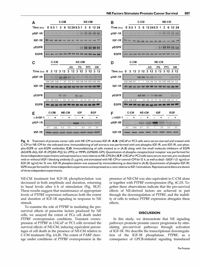
NE-CM treatment but IGF-1R phosphorylation wasdecreased in both amplitude and duration, returningto basal levels after 6 h of stimulation (Fig. 5E,F).These results suggest that maintenance of appropriatelevels of PTPRF expression influences both the extentand duration of IGF-1R signaling in response to NEstimuli.
To examine the role of PTPRF in mediating the pro-survival effects of paracrine factors produced by NEcells, we assayed the extent of PCa cell death underPTPRF overexpression conditions. Transient overex-pression of PTPRF in LNCaP or PC3 cells reversed thesurvival effects of NE-CM, inducing equivalent percen-tages of cell death in the presence of NE-CM relative toC-CM treatment (Fig. 6A,B). The extent of PARP cleav-age under conditions of PTPRF overexpression in the
presence of NE-CM was also equivalent to C-CM aloneor together with PTPRF overexpression (Fig. 6C,D). To-gether these observations indicate that the pro-survivaleffects of NE-derived factors are achieved in partthrough the downregulation of PTPRF, and the inabili-ty of cells to reduce PTPRF expression abrogates theseeffects.
DISCUSSION
In this study, we demonstrate that NE signalingpathways promote prostate cancer progression by stim-ulating pro-survival pathways through activationof IGF-1R. We describe the transcriptional downregula-tion of the IGF-1R phosphatase, PTPRF, as aconsequence of GPCR-initiated signaling transduced
Fig. 4. Treatment of prostate cancer cells with NE-CM activates IGF-1R.A,B: LNCaP or PC3 cells were serum-starved and treated withC-CM orNE-CM for the indicated time. Immunoblotting of cell extracts was performedwith anti-phospho-IGF-1R, anti-IGF-1R, anti-phos-pho-EGFR or anti-EGFR antibodies.C,D: Immunoblotting of cells treated as in (A,B) along with the small molecule inhibitors of EGFR(AG1478-AG), IGF-1R (PQ401-PQ) Src (PP2) or MMPs (GM6001-GM).Quantitation of phospho-receptor/total receptor was performed forthreeindependentexperiments andexpressedas aratiorelative toNE-CM(3hr).E,F:LNCaPorPC3cellswere serum-starved,pre-incubatedwith orwithouthIGF-1blocking antibody (5 mg/ml), and stimulatedwithNE-CMorcontrol-CM for12 h, aswell as des(1^3)IGF-1 (5 ng/ml) orEGF (10 ng/ml) for15 min. IGF-1R phosphorylation was assessed by immunoblotting as described in (A,B).Quantitation of phospho-IGF-1R/IGFRwasperformedfor threeindependentexperiments andexpressedas aratiorelative to IGF-1stimulation.Representativeblotsare shownof threeindependentexperiments.
NEFactors StimulateProstateCancerSurvival 807
The Prostate

through the activation of EGFR/Src, resulting insustained activity of IGF-1R and decreasedapoptotic stress. Results from different experimentalapproaches indicate that activation of IGF-1R in re-sponse to NE-derived factors occurs independently ofIGF-1 binding to its receptor. Cytokine array analysis
of NE-CM did not reveal the increased presence ofany other factors that may directly activate IGF-1R(unpublished data). Although other yet unidentifiedfactors may be present in NE-CM, this observationlends support to the hypothesis that increased phos-phorylation of IGF-1R results from neuropeptide-
TABLE I. IGF-1signalinggenesmodulatedbyNE-CM
Gene Accession Fold change P-value
Protein tyrosine phosphatase, receptor type, F AU158443 �1.47 0.041649Triple functional domain (PTPRF interacting) NM_007118 �1.71 0.036082Phosphoinositide-3-kinase, class 2, alpha polypeptide NM_002645 �1.41 0.03238Phosphoinositide-3-kinase, catalytic, beta polypeptide AA805318 �1.86 0.00372Mitogen-activated protein kinase kinase kinase kinase 4 NM_004834 1.2 0.005317v-akt murine thymoma viral oncogene homolog 1 NM_005163 1.14 0.128342Ribosomal protein S6 kinase, 90 kDa, polypeptide 2 BC002363 1.3 0.004726Ribosomal protein S6 AW205632 �1.42 0.018882Eukaryotic translation initiation factor 4E binding protein 1 AB044548 1.4 0.004413Eukaryotic translation initiation factor 4A, isoform 1 U79273 �1.58 0.015272Protein kinase, AMP-activated, beta 1 non-catalytic subunit AW572402 �1.35 0.045728Phosphoenolpyruvate carboxykinase 2 (mitochondrial) NM_004563 1.45 0.003347
IGF-1 signaling pathway genes up-regulated or down-regulated in LNCaP cells following 24 h NE-CM treatment, as identified byAffymetrix microarray and Ingenuity Pathways analysis.
Fig. 5. Downregulation of PTPRF in response to NE-CM activates IGF-1R.A,B: RNA extracted from LNCaP or PC3 cells treated for 24 hwithNE-CMorC-CMwas analyzedbyquantitativereal-time PCRusingprimers specific to PTPRF.Experimentswereperformedinduplicate andrepeated 3�.Theresultswerenormalized tob-glucuronidasereferencegene and are expressed as fold change fromcontrol treatment. �P < 0.05.C,D: Cell extractsprepared following treatmentwithNE-CMorC-CM for the indicated timeswere immunoblottedwith anti-PTPRF antibodies.E,F: LNCaPor PC3 cells were transiently transfectedwith vector or pCMV-PTPRF, and immunoblot analysis of IGF-1Rphosphorylationwas per-formed following treatmentwithNE-CM for the indicated times.Representativeblots are shownof three independentexperiments.
808 DaSilvaet al.
The Prostate

induced crosstalk with the EGFR/Src signalingpathway.
IGF-1R has previously been implicated in the pro-gression of PCa, but few studies have addressed therole of NE differentiation in modulating IGF-1R sig-naling. We and others have demonstrated that EGFRand Src kinase activities are enhanced by neuropep-tide stimulation of PCa cells and these activities areessential to promote cell proliferation and migration[9,10,37]. Our studies demonstrating that EGFR/Srcactivities modulate IGF-1R activity supports observa-tions made in both Rat aortic smooth muscle and PCacells that Src activation leads to IGF-1R phosphoryla-tion on the same autophosphorylation sites as in-duced by IGF-1 [8,30]. These data suggest thatparacrine factors that stimulate EGFR/Src activity caninduce ligand-independent IGF-1R signaling. Inagreement with this hypothesis, we observed thatEGF-induced signaling results in IGF-1R phosphory-lation in both LNCaP and PC3 cells. In the context ofPCa progression, increased NE-derived peptides thatactivate EGFR/Src signaling contribute to tumor cellproliferation and may also enhance their survivalthrough ligand-independent transactivation of IGF-1R. The effect of NE-CM was observed in both andro-gen-dependent and androgen-independent PCa mod-els and thus may reflect an early event that precedesthe acquisition of castration-resistant properties butcould also enhance survival of castrate-resistant cells.
It is well established that temporal regulation ofEGFR-mediated signaling pathways gives rise tounique patterns of gene expression resulting in differ-ential phenotypic cellular responses. In PC-12 orMCF-7 cells, for example, transient activation of ERKin response to EGF results in a proliferative response,but sustained ERK signaling promotes cellular differ-entiation [38,39]. Distinct cell-fate decisions down-stream of IGF-1R activation are thought to occurprimarily through differential recruitment of sub-strate proteins such as IRS-1 or IRS-2, but temporalregulation of ligand bioavailability mediated by IGFbinding proteins may also be involved [40,41]. We ob-served that modulation of PTPRF levels in responseto NE-derived factors alters the temporal response ofIGF-1R signaling and influences apoptotic induction.These results suggest that cell fate decisions down-stream of IGF-1R activation also involve a novel regu-latory mechanism of temporal signaling controlled byreceptor phosphatase activity.
Many protein tyrosine phosphatases have beenshown to interact with and modulate various receptortyrosine kinase (RTK) signaling pathways. The proteintyrosine kinases IGF-1R, IR, EGFR, HGFR, RET, LCK,and FYN have been reported to be substrates ofPTPRF [36,42–45]. In PTPRF overexpressing micephosphorylation of IRS-2, a signaling mediator IGF-1Rand IR, is reduced and PI3K function associated withIRS-1 and IRS-2 is also decreased [46]. Overexpression
Fig. 6. Overexpression of PTPRF abrogates the anti-apoptotic effect of NE-CM.A,B: LNCaP or PC3 cells were transiently transfectedwith vector or pCMV-PTPRF.FollowingNE-CM or C-CM treatment for 48 h cell deathwas determined by trypan blue staining.Results areexpressed as themean � SEMof three independentexperiments; �P < 0.05 relative toNE-CM.C,D: Apoptotic induction of cells treated asdescribed in (A,B), was determined by extent of PARP cleavage with immunoblotting using anti-PARP antibodies. Representative blots areshownof threeindependentexperiments.
NEFactors StimulateProstateCancerSurvival 809
The Prostate

of PTPRF in our PCa cell model systems resulted inreduced IGF-1R phosphorylation. This result is consis-tent with observations made in vascular smooth mus-cle cells demonstrating that IGF-1R phosphorylation isenhanced in the absence of PTPRF [45]. EGFR associa-tion with PTPRF induces tyrosine phosphorylationand TACE-mediated proteolytic processing of thephosphatase [32]. This post-translational mechanismof PTPRF regulation downstream of EGFR suggeststhat NE-induced modulation of IGF-1R signaling maybe multilayered.
We show that NE-derived factors enhance PCacell viability in the presence of docetaxel, suggest-ing that increased NE differentiation observed inadvance stage tumors may modulate the chemo-therapeutic response. Docetaxel has become thetreatment of choice for management of castration-resistant PCa. Although implementation of doce-taxel-based therapeutic regimens has significantlyimproved patient survival, the response rate hasbeen disappointing [33,47]. In addition, docetaxel isdose-limited due to various adverse side effects[33]. Novel combinations to reduce dose-limitingtoxicity of docetaxel and increase its efficacy couldhave a significant impact on disease-related mor-bidity and mortality. Docetaxel directly binds tothe b-subunit of tubulin and alters microtubule dy-namics, which ultimately leads to mitotic arrestand apoptosis [48]. However, chemosensitivity todocetaxel is also influenced by other pro-survivalsignaling pathways [49–51]. We found that combi-natorial treatment with docetaxel and inhibitors ofNE signaling mediators, including EGFR and IGF-1R, restores PARP cleavage and abrogates the pro-survival effects of NE-derived soluble paracrinefactors. This approach may provide clinical benefitfor patients with tumors that exhibit NE differenti-ation and warrants further investigation.
We conclude from these studies that secreted para-crine factors produced by NE cells contribute to pro-survival signaling of prostate cancer cells and providea survival advantage under hormone-deprived and/or chemotherapeutic conditions. Binding of NE pepti-des to cognate GPCR’s induces Src- and MMP-depen-dent EGFR transactivation that in turn mediates IGF-1R phosphorylation. Concomitantly, NE-initiated sig-naling down-regulates transcription of PTPRF, allow-ing moderate but sustained activation of IGF-1R.These signaling events enhance the ability to prostatecancer cells to survive apoptotic stress. The data sug-gest that the clinical benefit of docetaxel- or hormonedeprivation-based regimens may be enhanced inprostate cancer patients exhibiting NE differentiationby combinatorial inhibition of EGFR or IGF-Rsignaling.
ACKNOWLEDGMENTS
In memory of George Amorino, who dedicated hislife to the study of cancer. We thank Mark Axelrod forexcellent technical assistance and Jill Slack-Davis forcritical review of the manuscript. This work was sup-ported by NCI grants T32-CA009109-33, P01CA76465and R0176649.
REFERENCES
1. Scher HI, Sawyers CL. Biology of progressive, castration-resis-tant prostate cancer: Directed therapies targeting the andro-gen-receptor signaling axis. J Clin Oncol 2005;23(32):8253–8261.
2. Gennigens C, Menetrier-Caux C, Droz JP. Insulin-like growthfactor (IGF) family and prostate cancer. Crit Rev Oncol Hema-tol 2006;58(2):124–145.
3. Hirano D, Okada Y, Minei S, Takimoto Y, Nemoto N. Neuroen-docrine differentiation in hormone refractory prostate cancerfollowing androgen deprivation therapy. Eur Urol 2004;45(5):586–592; discussion 92.
4. Jiborn T, Bjartell A, Abrahamsson PA. Neuroendocrine differ-entiation in prostatic carcinoma during hormonal treatment.Urology 1998;51(4):585–589.
5. Cabrespine A, Guy L, Gachon F, Cure H, Chollet P, Bay JO.Circulating chromogranin a and hormone refractory prostatecancer chemotherapy. J Urol 2006;175(4):1347–1352.
6. Hvamstad T, Jordal A, Hekmat N, Paus E, Fossa SD. Neuroen-docrine serum tumour markers in hormone-resistant prostatecancer. Eur Urol 2003;44(2):215–221.
7. Tallett A, Chilvers ER, Hannah S, Dransfield I, Lawson MF,Haslett C, Sethi T. Inhibition of neuropeptide-stimulated tyro-sine phosphorylation and tyrosine kinase activity stimulatesapoptosis in small cell lung cancer cells. Cancer Res1996;56(18):4255–4263.
8. Sumitomo M, Milowsky MI, Shen R, Navarro D, Dai J, AsanoT, Hayakawa M, Nanus DM. Neutral endopeptidase inhibitsneuropeptide-mediated transactivation of the insulin-likegrowth factor receptor-Akt cell survival pathway. Cancer Res2001;61(8):3294–3298.
9. DaSilva J, Gioeli D, Weber MJ, Parsons SJ. The neuroendo-crine-derived peptide parathyroid hormone-related proteinpromotes prostate cancer cell growth by stabilizing the andro-gen receptor. Cancer Res 2009;69(18):7402–7411.
10. Amorino GP, Deeble PD, Parsons SJ. Neurotensin stimulatesmitogenesis of prostate cancer cells through a novel c-Src/Stat5b pathway. Oncogene 2007;26(5):745–756.
11. Deeble PD, Cox ME, Frierson HF Jr, Sikes RA, Palmer JB,Davidson RJ, Casarez EV, Amorino GP, Parsons SJ. Androgen-independent growth and tumorigenesis of prostate cancer cellsare enhanced by the presence of PKA-differentiated neuroen-docrine cells. Cancer Res 2007;67(8):3663–3672.
12. Jin RJ, Wang Y, Masumori N, Ishii K, Tsukamoto T, ShappellSB, Hayward SW, Kasper S, Matusik RJ. NE-10 neuroendocrinecancer promotes the LNCaP xenograft growth in castratedmice. Cancer Res 2004;64(15):5489–5495.
13. DiGiovanni J, Kiguchi K, Frijhoff A, Wilker E, Bol DK, BeltranL, Moats S, Ramirez A, Jorcano J, Conti C. Deregulated expres-sion of insulin-like growth factor 1 in prostate epithelium leadsto neoplasia in transgenic mice. Proc Natl Acad Sci U S A2000;97(7):3455–3460.
810 DaSilva et al.
The Prostate

14. Burfeind P, Chernicky CL, Rininsland F, Ilan J. Antisense RNAto the type I insulin-like growth factor receptor suppresses tu-mor growth prevents invasion by rat prostate cancer cells invivo. Proc Natl Acad Sci U S A 1996;93(14):7263–7268.
15. Nickerson T, Chang F, Lorimer D, Smeekens SP, Sawyers CL,Pollak M. In vivo progression of LAPC-9 and LNCaP prostatecancer models to androgen independence is associated withincreased expression of insulin-like growth factor I (IGF-I) andIGF-I receptor (IGF-IR). Cancer Res 2001;61(16):6276–6280.
16. Reiss K, D’Ambrosio C, Tu X, Tu C, Baserga R. Inhibition oftumor growth by a dominant negative mutant of the insulin-like growth factor I receptor with a bystander effect. ClinCancer Res 1998;4(11):2647–2655.
17. Scorilas A, Plebani M, Mazza S, Basso D, Soosaipillai AR,Katsaros N, Pagano F, Diamandis EP. Serum human glandularkallikrein (hK2) and insulin-like growth factor 1 (IGF-1)improve the discrimination between prostate cancer and be-nign prostatic hyperplasia in combination with total and %free PSA. Prostate 2003;54(3):220–229.
18. Chan JM, Stampfer MJ, Ma J, Gann P, Gaziano JM, Pollak M,Giovannucci E. Insulin-like growth factor-I (IGF-I) and IGFbinding protein-3 as predictors of advanced-stage prostate can-cer. J Natl Cancer Inst. 2002;94(14):1099–1106.
19. Krueckl SL, Sikes RA, Edlund NM, Bell RH, Hurtado-Coll A,Fazli L, Gleave ME, Cox ME. Increased insulin-like growth fac-tor I receptor expression and signaling are components of an-drogen-independent progression in a lineage-derived prostatecancer progression model. Cancer Res 2004;64(23):8620–8629.
20. Pfeil K, Eder IE, Putz T, Ramoner R, Culig Z, Ueberall F,Bartsch G, Klocker H. Long-term androgen-ablation causes in-creased resistance to PI3K/Akt pathway inhibition in prostatecancer cells. Prostate 2004;58(3):259–268.
21. Hellawell GO, Ferguson DJ, Brewster SF, Macaulay VM.Chemosensitization of human prostate cancer using antisenseagents targeting the type 1 insulin-like growth factor receptor.BJU Int 2003;91(3):271–277.
22. Li L, Ittmann MM, Ayala G, Tsai MJ, Amato RJ, Wheeler TM,Miles BJ, Kadmon D, Thompson TC. The emerging role of thePI3-K-Akt pathway in prostate cancer progression. ProstateCancer Prostatic Dis 2005;8(2):108–118.
23. Datta SR, Dudek H, Tao X, Masters S, Fu H, Gotoh Y, Green-berg ME. Akt phosphorylation of BAD couples survival signalsto the cell-intrinsic death machinery. Cell 1997;91(2):231–241.
24. Cardone MH, Roy N, Stennicke HR, Salvesen GS, Franke TF,Stanbridge E, Frisch S, Reed JC. Regulation of cell death prote-ase caspase-9 by phosphorylation. Science 1998;282(5392):1318–1321.
25. van der Veeken J, Oliveira S, Schiffelers RM, Storm G, vanBergen En Henegouwen PM, Roovers RC. Crosstalk betweenepidermal growth factor receptor- and insulin-like growth fac-tor-1 receptor signaling: implications for cancer therapy. CurrCancer Drug Targets 2009;9(6):748–760.
26. Rozengurt E, Sinnett-Smith J, Kisfalvi K. Crosstalk between in-sulin/insulin-like growth factor-1 receptors and G protein-cou-pled receptor signaling systems: a novel target for theantidiabetic drug metformin in pancreatic cancer. Clin CancerRes 2010;16(9):2505–2511.
27. Tu H, Xu C, Zhang W, Liu Q, Rondard P, Pin JP, Liu J. GABABreceptor activation protects neurons from apoptosis via IGF-1receptor transactivation. J Neurosci 2010;30(2):749–759.
28. Du J, Brink M, Peng T, Mottironi B, Delafontaine P. Thrombinregulates insulin-like growth factor-1 receptor transcription in
vascular smooth muscle: Characterization of the signalingpathway. Circ Res 2001;88(10):1044–1052.
29. Du J, Sperling LS, Marrero MB, Phillips L, Delafontaine P. G-protein and tyrosine kinase receptor cross-talk in rat aorticsmooth muscle cells: Thrombin- and angiotensin II-induced ty-rosine phosphorylation of insulin receptor substrate-1 and in-sulin-like growth factor 1 receptor. Biochem Biophys ResCommun 1996;218(3):934–939.
30. Eberl LP, Valdenaire O, Saintgiorgio V, Jeannin JF, Juillerat-Jeanneret L. Endothelin receptor blockade potentiates FasL-in-duced apoptosis in rat colon carcinoma cells. Int J Cancer2000;86(2):182–187.
31. Trojan L, Schaaf A, Steidler A, Haak M, Thalmann G, Knoll T,Gretz N, Alken P, Michel MS. Identification of metastasis-associated genes in prostate cancer by genetic profiling ofhuman prostate cancer cell lines. Anticancer Res 2005;25(1A):183–191.
32. Ruhe JE, Streit S, Hart S, Ullrich A. EGFR signaling leads todownregulation of PTP-LAR via TACE-mediated proteolyticprocessing. Cell Signal 2006;18(9):1515–1527.
33. Tannock IF, de Wit R, Berry WR, Horti J, Pluzanska A, Chi KN,Oudard S, Theodore C, James ND, Turesson I, Rosenthal MA,Eisenberger MA. Docetaxel plus prednisone or mitoxantroneplus prednisone for advanced prostate cancer. N Engl J Med2004;351(15):1502–1512.
34. Cox ME, Deeble PD, Lakhani S, Parsons SJ. Acquisition of neu-roendocrine characteristics by prostate tumor cells is revers-ible: Implications for prostate cancer progression. Cancer Res1999;59(15):3821–3830.
35. Casarez EV, Dunlap-Brown ME, Conaway MR, Amorino GP.Radiosensitization and modulation of p44/42 mitogen-activat-ed protein kinase by 2-methoxyestradiol in prostate cancermodels. Cancer Res 2007;67(17):8316–8324.
36. Tsujikawa K, Ichijo T, Moriyama K, Tadotsu N, Sakamoto K,Sakane N, Fukada S, Furukawa T, Saito H, Yamamoto H.Regulation of Lck and Fyn tyrosine kinase activities bytransmembrane protein tyrosine phosphatase leukocyte com-mon antigen-related molecule. Mol Cancer Res 2002;1(2):155–163.
37. Lee LF, Guan J, Qiu Y, Kung HJ. Neuropeptide-induced andro-gen independence in prostate cancer cells: Roles of nonreceptortyrosine kinases Etk/Bmx, Src, and focal adhesion kinase. MolCell Biol 2001;21(24):8385–8397.
38. Marshall CJ. Specificity of receptor tyrosine kinase signaling:Transient versus sustained extracellular signal-regulatedkinase activation. Cell 1995;80(2):179–185.
39. Nagashima T, Shimodaira H, Ide K, Nakakuki T, Tani Y,Takahashi K, Yumoto N, Hatakeyama M. Quantitative tran-scriptional control of ErbB receptor signaling undergoes grad-ed to biphasic response for cell differentiation. J Biol Chem2007;282(6):4045–4056.
40. Valverde AM, Lorenzo M, Pons S, White MF, Benito M. Insulinreceptor substrate (IRS) proteins IRS-1 and IRS-2 differentialsignaling in the insulin/insulin-like growth factor-I pathwaysin fetal brown adipocytes. Mol Endocrinol 1998;12(5):688–697.
41. Walter HJ, Berry M, Hill DJ, Logan A. Spatial and temporalchanges in the insulin-like growth factor (IGF) axis indicate au-tocrine/paracrine actions of IGF-I within wounds of the ratbrain. Endocrinology 1997;138(7):3024–3034.
42. Tsujikawa K, Kawakami N, Uchino Y, Ichijo T, Furukawa T,Saito H, Yamamoto H. Distinct functions of the two proteintyrosine phosphatase domains of LAR (leukocyte common
NEFactors StimulateProstateCancerSurvival 811
The Prostate

antigen-related) on tyrosine dephosphorylation of insulin re-ceptor. Mol Endocrinol 2001;15(2):271–280.
43. Kulas DT, Goldstein BJ, Mooney RA. The transmembrane pro-tein-tyrosine phosphatase LAR modulates signaling by multi-ple receptor tyrosine kinases. J Biol Chem 1996;271(2):748–754.
44. Qiao S, Iwashita T, Furukawa T, Yamamoto M, Sobue G,Takahashi M. Differential effects of leukocyte common anti-gen-related protein on biochemical and biological activities ofRET-MEN2A and RET-MEN2B mutant proteins. J Biol Chem2001;276(12):9460–9467.
45. Niu XL, Li J, Hakim ZS, Rojas M, Runge MS, Madamanchi NR.Leukocyte antigen-related deficiency enhances insulin-likegrowth factor-1 signaling in vascular smooth muscle cells andpromotes neointima formation in response to vascular injury. JBiol Chem 2007;282(27):19808–19819.
46. Zabolotny JM, Kim YB, Peroni OD, Kim JK, Pani MA, Boss O,Klaman LD, Kamatkar S, Shulman GI, Kahn BB, Neel BG.Overexpression of the LAR (leukocyte antigen-related)protein-tyrosine phosphatase in muscle causes insulin resis-tance. Proc Natl Acad Sci U S A 2001;98(9):5187–5192.
47. Petrylak DP. New paradigms for advanced prostate cancer.Rev Urol 2007;9(Suppl. 2):S3–S12.
48. Bergstralh DT, Ting JP. Microtubule stabilizing agents: theirmolecular signaling consequences and the potential for en-hancement by drug combination. Cancer Treat Rev 2006;32(3):166–179.
49. Zemskova M, Sahakian E, Bashkirova S, Lilly M. The PIM1kinase is a critical component of a survival pathway activatedby docetaxel and promotes survival of docetaxel-treated prostate cancer cells. J Biol Chem 2008;283(30):20635–20644.
50. Patterson SG, Wei S, Chen X, Sallman DA, Gilvary DL, ZhongB, Pow-Sang J, Yeatman T, Djeu JY. Novel role of Stat1 in thedevelopment of docetaxel resistance in prostate tumor cells.Oncogene 2006;25(45):6113–6122.
51. Mediavilla-Varela M, Pacheco FJ, Almaguel F, Perez J, Saha-kian E, Daniels TR, Leoh LS, Padilla A, Wall NR, Lilly MB, DeLeon M, Casiano CA. Docetaxel-induced prostate cancer celldeath involves concomitant activation of caspase and lysosom-al pathways and is attenuated by LEDGF/p75. Mol Cancer2009;8:68.
812 DaSilva et al.
The Prostate