Embed Size (px)
Citation preview

Neurodegenerative diseases and aging

Neurodegenerative diseases• Neurodegenerative diseases are
defined as hereditary and sporadic
conditions which are characterized
by progressive nervous system
dysfunction.
• These disorders are often associated
with atrophy of the affected central
or peripheral structures of the or peripheral structures of the
nervous system and include:
Alzheimer's Disease and other
dementias, Parkinson's Disease,
Multiple Sclerosis, Amyotrophic
Lateral Sclerosis (ALS or Lou
Gehrig's Disease), Huntington's
Disease, Prion Diseases, and others.
• These diseases rob people of their
ability to remember, speak, write,
ambulate, and control their lives.
There is no cure for these diseases.
Neurodegenerative diseases• Much progress has been made in
identifying genes involved in
familial, or inherited, forms of
different neurodegenerative
diseases, including Alzheimer’s
disease, Parkinson’s disease and
Amyotrophic Lateral Sclerosis Amyotrophic Lateral Sclerosis
(ALS).
• However the majority of these
disease cases are sporadic (not
inherited), where the origin(s) of
sporadic neurodegeneration
remain undetermined.
• Gene candidates, susceptibility
factors (environmental insults
and epigenetic alterations)

Cell death and neurodegeneration
• Among the different modes of cell death, apoptosis (programmed
cell death) has been suggested to occur most frequently in
neurodegenerative disease.
– Parkinson's disease (PD) - dopaminergic cell death
• Familial forms of PD are associated with mutations in α-
synuclein genes. synuclein genes.
• Sporadic PD ~ mitochondrial/environmental toxins (paraquat,
MPP+)
• Upregulation of caspase 3 and Bcl-2 protein members
– Alzheimer's disease (AD) - hippocampal/cortical neurons
• APP, apolipoprotein E, presenilins
• Tau and amyloid β (Aβ)
• DNA fragmentation, caspase and Bcl-2 upregulation
– Amyotrophic lateral sclerosis – Motor neurons. Huntington's
disease (HD) - striatum neuronal death.

Redox signaling, cell death and neurodegeneration
• Oxidative stress is a common event in the pathogenesis of
neurodegenerative diseases
– Parkinson's disease (PD)
• Mitochondrial toxins, dopamine oxidation
• Decreased antioxidant levels (GSH)
• Lipid peroxidation, protein (carbonyls and nitrotyrosines) and • Lipid peroxidation, protein (carbonyls and nitrotyrosines) and
nucleic acid oxidation
– Alzheimer's disease (AD) and mild cognitive impairment.
• Increased lipid-peroxidation (CSF), protein (carbonyls and
nitrotyrosines) and nucleic acid oxidation
• Oxidative stress influences Aβ formation and Aβ has pro-
oxidant effects
– Huntington's disease (HD) – Energy metabolism


Alzheimer’s disease• An estimated 24 million people worldwide have dementia, the majority of
whom are thought to have Alzheimer's disease.
• The two core pathological hallmarks of Alzheimer's disease are amyloid
plaques and neurofibrillary tangles.
• The amyloid cascade hypothesis suggests that deposition of amyloid β (Aβ)
triggers neuronal dysfunction and death in the brain
• Established genetic causes of Alzheimer's disease include dominant • Established genetic causes of Alzheimer's disease include dominant
mutations of the genes encoding amyloid precursor protein (APP) and
presenilin 1 (PSEN1) and PSEN2 (5% or early onset). PSEN1 and PSEN2
mutations affect concentrations of Aβ1–42 because presenilin proteins
form part of γ secretase, which cleaves APP to produce Aβ.
• Environmental risk factors for Alzheimer's disease include cognitive reserve
(a concept combining the benefits of education, occupation, and mental
activities), physical activity and exercise, midlife obesity, alcohol intake, and
smoking.
• Many treatable medical conditions are also associated with an increased
risk of Alzheimer's disease, including stroke, diabetes, TBI.

Amyloid beta• Amyloid precursor protein (APP) is an integral
membrane protein expressed in many tissues and concentrated in the synapses of neurons. Its primary function is not known, though it has been implicated as a regulator of synapse formation, neural plasticity and iron export.
• Non-amyloidogenic cleavage of the β-amyloidpeptide(Aβ) is mediated by α-secretase. A large amyloid precursor protein (sAPPα) and the C83 fragment are generated. fragment are generated.
• Amyloidogenic processing is initiated by β-secretase beta-site amyloid precursor protein–cleaving enzyme 1 (BACE-1), releasing a shortened sAPPβ. The C99 fragment is a γ-secretasesubstrate, generating Aβ and AICD (amyloidprecursor protein intracellular domain).
• Soluble Aβ is prone to aggregation. Protofibrils (upper) and annular or pore-like profiles (lower) are intermediate aggregates.
• Self-association of Aβ monomers into oligomers is dependent on concentration (left immunoblot) and is promoted by oxidizing conditions (lane 2) and divalent metal conditions (lane 3).
NEJM Volume 362:329-344

Tau hyperphosphorylation• Tau proteins are proteins that stabilize
microtubules. Four repeat sequences (R1-
R4) make up the microtubule-binding
domain (MBD).
• Normal phosphorylation of tau occurs on serine and threonine residues.
• When followed by proline (P), these amino
acids are phosphorylated by glycogen
synthase kinase 3 (GSK-3β), cyclin-
dependent kinase (cdk5), or mitogen-
activated protein kinase (MAPK).
Nonproline-directed kinases phophorylating
tau are Akt, Fyn, protein kinase A (PKA),
calcium–calmodulin protein kinase 2
(CaMKII), and microtubule affinity-regulating
kinase (MARK).
• Excessive kinase, reduced phosphataseactivities, or both cause
hyperphosphorylated tau to detach, self-aggregate, and to destabilize microtubules.

Alzheimer’s disease and oxidative stress
• Amyloid β shows peroxidative activity
on cell and organelle membrane lipids
yields the mitochondrial toxins
hydroxynonenal (HNE) and
malondialdehyde.
• Cellular Aβ directly attacks electron
transport complex IV (cytochrome c
oxidase) and key Krebs-cycle oxidase) and key Krebs-cycle
enzymes (α-ketoglutarate and pyruvate
dehydrogenase) and damage to
mitochondrial DNA (mtDNA), leading to
fragmentation.
• Lipid peroxidation products also
promote tau phosphorylation and
aggregation, which in turn inhibit
complex I.
• Contradictory results have been found
with respect to the effect of dietary
intake of antioxidants, such as vitamin E
in reducing the risk or the rate of
progression of Alzheimer's disease.NEJM Volume 362:329-344

Alzheimer’s disease and RNS• NMDAR hyperactivation
triggers generation of NO and subsequent S-nitrosylation of neuronal proteins, contributing to synaptic damage and eventually neuronal death.
• Soluble oligomers of Aβ• Soluble oligomers of Aβoligomers, can facilitate neuronal NO production in both NMDAR-dependent and -independent manners.
• S-Nitrosylation of the fission-inducing protein Drp1 (forming SNO-Drp1) can contribute to synaptic damage and neuronal cell death by triggering excessive mitochondrial fission and bioenergetic impairmentApoptosis. 2010 Nov;15(11):1354-63.
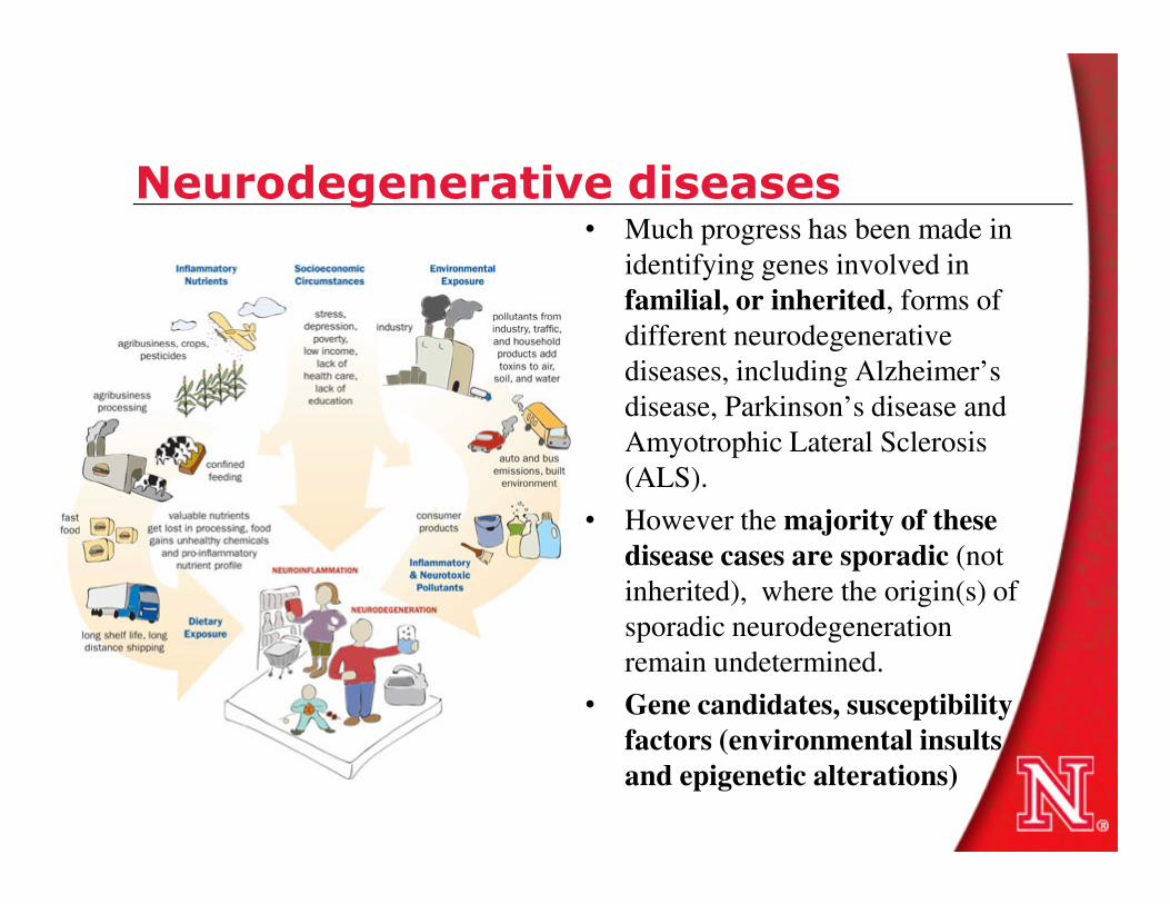
Oxidative stress in triple transgenic AD mouse model
• Tangle formation has been reproduced in P301L tau transgenic pR5 mice, whereas APPswPS2N141I double-transgenic APP152 mice develop Aβ plaques. Cross-breeding generates triple transgenic (tripleAD) mice that combine both pathologies in one model.
• Functional analysis of the consequences of the combined Aβ and tau pathologies, using proteomic analysis followed it was found a massive deregulation of 24 proteins, of which one-third were mitochondrial proteins mainly related to mitochondrial proteins mainly related to complexes I and IV of the oxidative phosphorylation (OXPHOS) system.
• Deregulation of complex I was tau dependent, whereas deregulation of complex IV was Aβdependent, both at the protein and activity levels.
• Synergistic effects of Aβ and tau were evident in 8-month-old tripleAD mice as only they showed a reduction of the mitochondrial membrane potential at this early age. At the age of 12 months, the strongest defects on OXPHOS, synthesis of ATP, and reactive oxygen species were exhibited in the tripleAD mice, again emphasizing synergistic, age-associated effects of Aβ and tau in perishing mitochondria.
Rhein V et al., Proc Natl Acad Sci U S A. 2009 November 24; 106(47): 20057–20062.
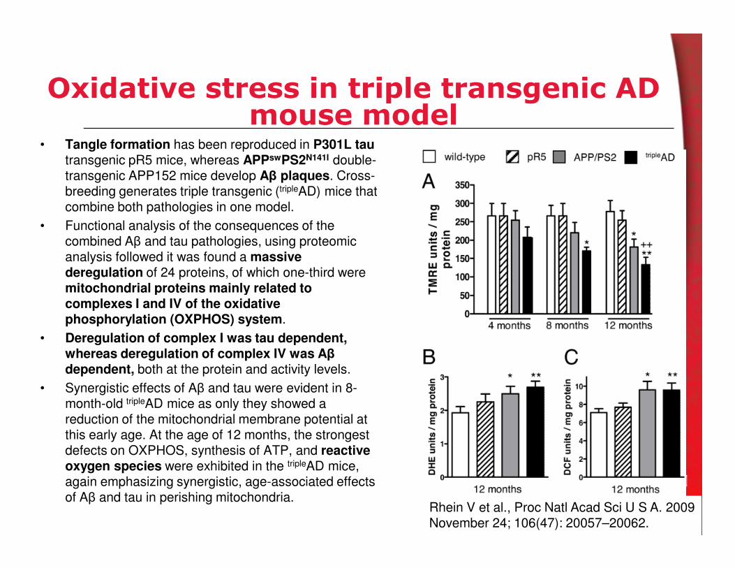
Oxidative modifications and amyloid βtoxicity
Met35 in amyloid β-protein (Aβ) is prone to participating in
redox reactions, and therefore is believed to contribute
significantly Aβ-induced toxicity. Aβ40 and Aβ42 analogues containing Met35→Nle (norleucine) or Met35→Val
substitutions had no significant effect in neurotoxicity.
J Neurochem. 2010 Jun;113(5):1252-62. Epub 2010 Mar 20.
Oxidative damage in brain, including elevated levels of
protein oxidation and lipid peroxidation, was completely
prevented in transgenic AD mouse mice with a M631L mutation in APP. APP mice contains mutations in human
APP corresponding to the Swedish and Indiana familial
forms of AD are expressed (APPSw,In), resulting in Aβ
accumulation, plaque formation, and memory deficits
Free Radic Biol Med. 2010 Jan 1;48(1):136-44.
Parkinson’s disease• Parkinson's disease is a neurodegenerative process characterized by numerous motor and
nonmotor clinical manifestations for which effective, mechanism-based treatments remain
elusive.
• It is characterized by the presence of severe pars-compacta nigral-cell loss, and accumulation
of aggregated α-synuclein in specific brain stem, spinal cord, and cortical regions.
• The main known risk factor is age. However, gene-environment interactions play a significant
role.
• After decades of research, a single cause for Parkinson's disease has not been found and is • After decades of research, a single cause for Parkinson's disease has not been found and is
unlikely to emerge. Whereas some forms of Parkinson's disease are genetic, most cases are
idiopathic, and the underlying environmental causes (if any) remain to be discovered.
• An emerging concept is that SNc homeostasis is vulnerable to different genetic, cellular and
environmental factors that independently or concomitantly cause cell death over time by
mitochondrial dysfunction and oxidative stress, abnormal protein degradation.
• Dopamine metabolism is considered to be critical for the preferential susceptibility of
ventrolateral SNc cells to damage in Parkinson's disease. Dopamine metabolism produces
highly reactive species that oxidize lipids and other compounds, increase oxidative stress
and impair mitochondrial function.

Products of PD-associated genes that affect mitochondrial function and oxidative stress.
Rare inherited mutations in genes encoding electron transport chain components have been associated with parkinsonism.
• Parkin is partially localized to the outer mitochondrial membrane,
• PINK1 is a mitochondrial serine–threonine kinase that affords protection against oxidative stress and acts with Parkin to regulate the balance of mitochondrial fission Parkin to regulate the balance of mitochondrial fission and fusion.
• LRRK2 associates, at least in part, with the outer mitochondrial membrane
• HTRA2 is a mitochondrial serine protease, the release of which might be involved in apoptotic cell death.
• DJ-1 is relocated to mitochondria under conditions of oxidative stress and is thought to be neuroprotectiveunder such conditions.
• The α-synuclein protein has an amino-terminal mitochondrial targeting sequence and, when
overexpressed or under conditions of acidification, is at
least partially associated with the inner mitochondrial
membrane, where it might cause direct damage.
Nature Clinical Practice Neurology (2008) 4, 600-609

Experimental models of PD• 1-methyl-4-phenyl-1,2,3,6-tetrahydropyridine
(MPTP) is a by-product of the chemical
synthesis of a meperidine analogue with
potent heroin-like effects that can induce a
parkinsonian syndrome in humans almost
indistinguishable from Parkinson's disease
(PD).
• It has been used extensively as a model of PD.
MPTP administration causes damage to the MPTP administration causes damage to the
nigrostriatal dopamine (DA) pathway identical
to that seen in PD, with the exception of Lewy
bodies.
• MPTP, crosses the blood–brain barrier and is
metabolized to 1-methyl-4-phenylpyridinium
(MPP+) by the enzyme monoamine oxidase B
(MAO-B) in non-DA cells. MPP+ is then taken
up by DA transporters, for which it has high
affinity. MPP+ impairs mitochondrial
respiration by inhibiting complex I of the
electron transport chain. resulting in an
increased production of free radicals, which
causes oxidative stress
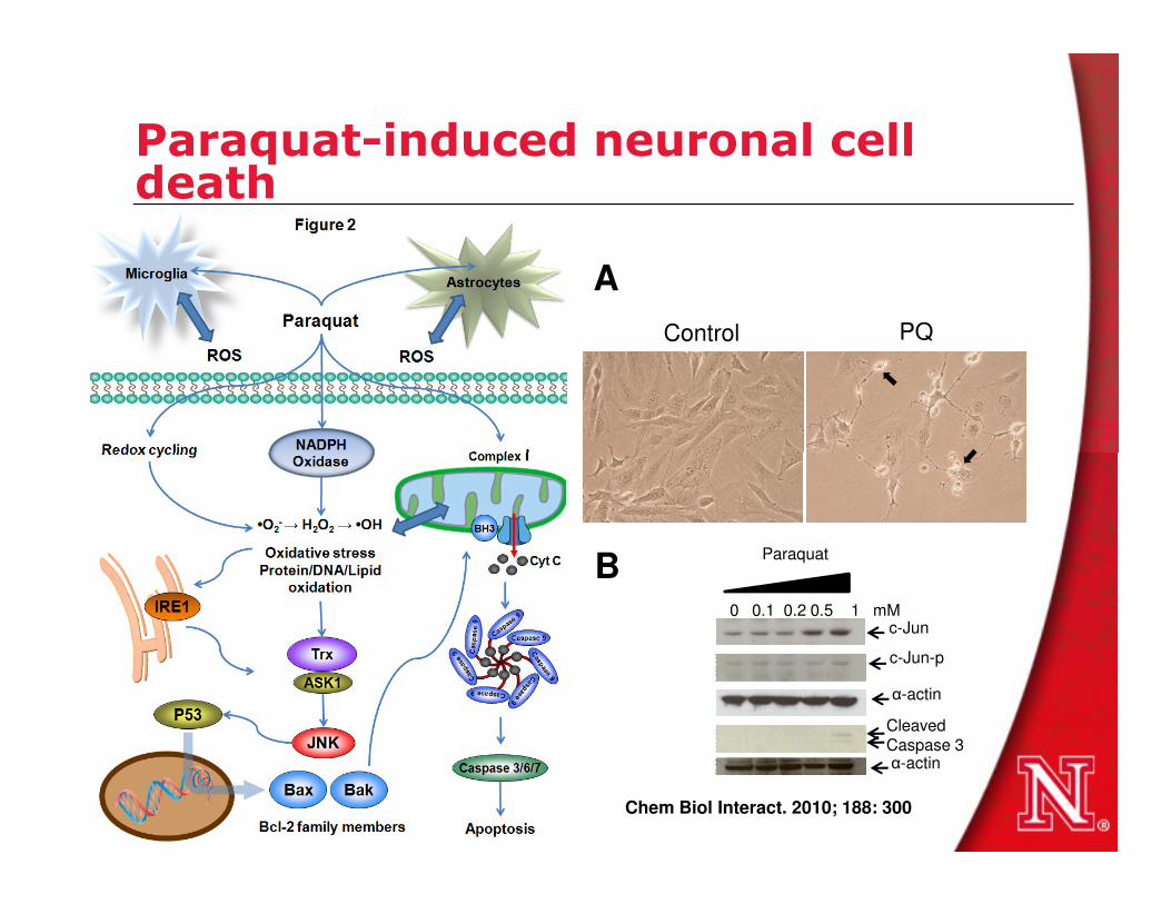
Paraquat-induced neuronal cell death
Control PQ
A
0 0.1 0.2 0.5 1 mM
Paraquat
c-Jun
c-Jun-p
α-actin
α-actin
CleavedCaspase 3
B
Chem Biol Interact. 2010; 188: 300

Cytosol
DA
V
M
A
T
SNO
SOH2
Parkin
DJ-1DA
DA
DA
RS
Extracellular Space
DA
Extracellular sources Glia, environmental toxins (TX), metals, inflammation
DAQ
Microglia Astrocytes
RSRS
+ Fe2+
DAQ-Cys
SOH3DJ-1X X
Misfolded protein degradation(4)
(5)
(6)
Matrix
Intermembrane space
RS
MAO
DOPACDA
H2O H2O2
I II III IV V
H+
H+
H+
H+
H+H+
H+
H+
NADH NAD+
Succinate Fumarate
.O2-
.O2-
.O2-
e-
e- e-
e-
O2 H2O
X
RSDAAuto-oxidation
TXADP ATP
(1)
(2)
(3)

Redox signaling, cell death and PD

DJ-1 in Parkinson’s disease

Oxidative stress in substantia nigra• Using transgenic mice that expressed a redox-
sensitive variant of green fluorescent protein
targeted to the mitochondrial matrix, it was
demonstrated that normal autonomous
pacemaking (responsible for the sustained
release of dopamine necessary for the proper
functioning of target structures, such as the
striatum) created an oxidant stress that was
specific to vulnerable SNc dopaminergic
2;4
68
(73
24
):696
-700
specific to vulnerable SNc dopaminergic
neurons but not in neurons in the ventral
tegmental area.
• The oxidant stress engaged induced transient,
mild mitochondrial depolarization or
uncoupling.
• Knocking out DJ-1 (PARK7), which is a gene
associated with an early-onset form of
Parkinson's disease, down-regulated the
expression of two uncoupling proteins (UCP4
(SLC25A27) and UCP5 (SLC25A14)),
compromised Ca2+-induced uncoupling and
increased oxidation of matrix proteins
specifically in SNc dopaminergic neurons.
Na
ture
.2
01
0 D
ec
2;4
68
(73
24
):696

Peroxiredoxins in PD• Prx2 is the most abundant in
mammalian neurons, making it
a prime candidate to defend
against oxidative stress.
• Prx2 is S-nitrosylated (forming
SNO-Prx2) by reaction with
NO• at two critical cysteine NO• at two critical cysteine
residues (C51 and C172),
preventing its reaction with
peroxides.
• Increased SNO-Prx2 in human
Parkinson's disease (PD) brains,
and S-nitrosylation of Prx2
inhibited both its enzymatic
activity and protective function
from oxidative stress.
Proc Natl Acad Sci U S A. 2007 Nov 20;104(47):18742-7
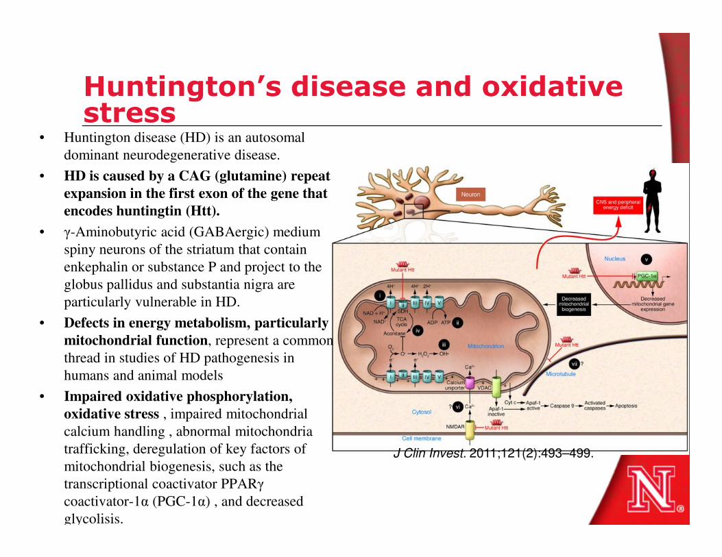
Huntington’s disease and oxidative stress
• Huntington disease (HD) is an autosomal
dominant neurodegenerative disease.
• HD is caused by a CAG (glutamine) repeat
expansion in the first exon of the gene that
encodes huntingtin (Htt).
• γ-Aminobutyric acid (GABAergic) medium
spiny neurons of the striatum that contain
enkephalin or substance P and project to the
globus pallidus and substantia nigra are globus pallidus and substantia nigra are
particularly vulnerable in HD.
• Defects in energy metabolism, particularly
mitochondrial function, represent a common
thread in studies of HD pathogenesis in
humans and animal models
• Impaired oxidative phosphorylation,
oxidative stress , impaired mitochondrial
calcium handling , abnormal mitochondria
trafficking, deregulation of key factors of
mitochondrial biogenesis, such as the
transcriptional coactivator PPARγ
coactivator-1α (PGC-1α) , and decreased
glycolisis.
J Clin Invest. 2011;121(2):493–499.

Huntington’s disease and oxidative stress
• Mitochondrial toxins such as
malonate and 3-nitropropionic acid
(3-NP) inhibit succinate
dehydrogenase (SDH), which is
part of complex II of the electron
transport chain. transport chain.
• Neuroprotection: Coenzyme Q10 is
an electron acceptor from complex
I and complex II that also has
antioxidant activity. Creatine and
triacetyluridine can buffer
intracellular energy stores
Nature Reviews Neuroscience 5, 373-384 (May 2004)

Huntington’s disease, oxidative stress and antioxidant deficiency
• HD knock-in mice (HD140Q/140Q), which have
human huntingtin exon 1 with 140 CAG repeats
inserted into the endogenous mouse huntingtin
gene.
• Elevated ROS in HD neurons
• HD neurons had lower cell surface levels of the
glutamate/cysteine transporter EAAC1 and
were deficient in taking up cysteine. were deficient in taking up cysteine.
• Constitutive trafficking of EAAC1 from recycling
endosomes relies on Rab11 (Ras-related GTP-
binding proteins) activity, which is defective in
the brain of HD140Q/140Q mice.
• Enhancement of Rab11 activity by expression
of a dominant-active Rab11 mutant in primary
HD neurons ameliorated the deficit in cysteine
uptake, increased levels of intracellular
glutathione, normalized clearance of ROS, and
improved neuronal survival
J Neurosci. 2010 Mar 31;30(13):4552-61.

Huntington’s disease and oxidative DNA damage
• Neurodegenerative disorders are
characterized by the accumulation of 8-
oxo-7,8-dihydroguanine (8-oxodG).
• Through direct oxidation of DNA guanine
or via incorporation of the oxidized
nucleotide during replication.
• hMTH1 is the major human hydrolase
that degrades oxidized purine nucleoside that degrades oxidized purine nucleoside
triphosphates
• hMTH1 transgene expression conferred a
dramatic protection against
Huntington's disease-like symptoms,
including striatal degeneration, and
death induced by 3-NP.
• hMTH1 expression protected striatal
cells containing an expanded CAG
repeat of the huntingtin gene (mutant
HdhQ111/Q111) from toxicity associated
with expression of the mutant
huntingtin. PLoS Genet. 2008 Nov;4(11):e1000266.

Amyotrophic lateral sclerosis • Amyotrophic lateral sclerosis (ALS) is a paralytic disorder caused by motor neuron
degeneration in the brain and spinal cord.
• The causes of most cases of ALS are as yet undefined. Excessive excitatory tone, protein
misfolding, impaired energy production, abnormal calcium metabolism, altered axonal
transport and activation of proteases and nucleases.
• Several factors are proposed to instigate these phenomena, including latent infections by viral
and non-viral agents , toxins (for example, insecticides and pesticides) and autoimmune
reactions. reactions.
• Five Mendelian gene defects have been reported to cause ALS The protein products of these
mutated genes are cytosolic Cu/Zn superoxide dismutase (SOD1), alsin, senataxin (SETX),
synaptobrevin/VAMP (vesicle-associated membrane protein)-associated protein B (VAPB)
and dynactin.
• About 20–25% of all familial ALS cases arise because of mutations in SOD1, the protein
product of which accounts for 0.1–0.2% of the cellular proteins in the CNS.
• Abnormal TDP-43 (Trans-activation response [TAR] DNA-binding protein) accumulation in
the cytoplasm is found in familial ALS. Mutations in the TARDBP gene that codes for the
TDP-43 protein have been shown to account for ~2-6% of all familial ALS cases. TDP-43 is
also involved in Frontotemporal lobar dementia with ubiquitin (FTLD-U). Abnormal TDP-43
is not present in cases of ALS with SOD1 mutations.

Amyotrophic lateral sclerosis • The instability of the mutant SOD1 protein
contributes to its toxicity, enhanced by the release of
Zn.
• In the aberrant redox chemistry model, mutant
superoxide dismutase 1 (SOD1) is unstable and
aberrant chemistry is mediated by promiscuous
interaction with non-conventional substrates.
– Hydrogen peroxide (H2O2) or peroxynitrite
(ONOO-) can react with reduced SOD1 (SOD1-
Cu+). Cu+).
– Molecular oxygen (O2) can react aberrantly with
Zn-deficient SOD1 to generate an excess of
superoxide anion (O2-).
– The unstable protein can also release free
copper and/or zinc, which might be toxic.
• In the protein toxicity model, conformationally
altered mutant SOD1 forms toxic, proteinaceous
deposits.
– Aggregated SOD1 inhibits chaperone and/or
proteasome activity, with subsequent misfolding
and insufficient clearance of numerous proteins.
– Alternatively, these aggregates could sequester,
inactivate or enhance the toxicity of other
proteins crucial for cellular processes.
Nat Rev Neurosci. 2006 Sep;7(9):710-23.
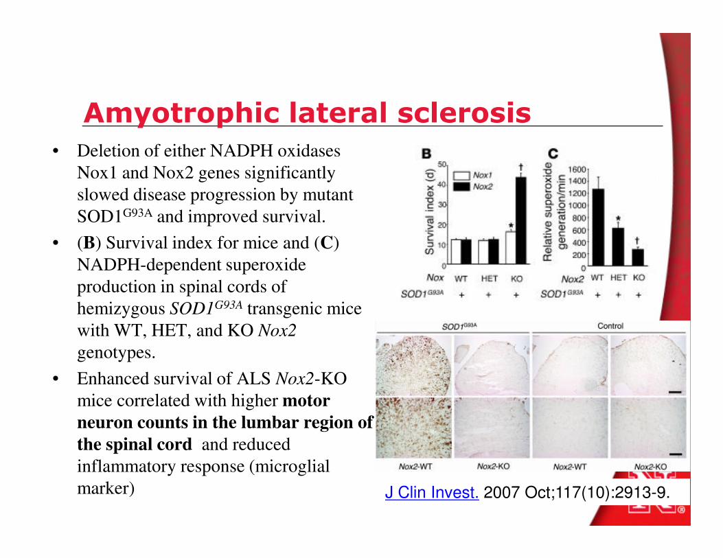
Amyotrophic lateral sclerosis
• Deletion of either NADPH oxidases
Nox1 and Nox2 genes significantly
slowed disease progression by mutant
SOD1G93A and improved survival.
• (B) Survival index for mice and (C)
NADPH-dependent superoxide
production in spinal cords of production in spinal cords of
hemizygous SOD1G93A transgenic mice
with WT, HET, and KO Nox2
genotypes.
• Enhanced survival of ALS Nox2-KO
mice correlated with higher motor
neuron counts in the lumbar region of
the spinal cord and reduced
inflammatory response (microglial
marker) J Clin Invest. 2007 Oct;117(10):2913-9.

Redox control of prion and disease pathogenesis
• The underlying cause of brain pathology in all
prion disorders is PrP-scrapie (PrP(Sc)), a beta-
sheet-rich conformation of a normal
glycoprotein, the prion protein (PrP(C)).
• In prion disorders, imbalance of brain-iron
homeostasis is observed before end-stage
disease and worsens with disease progression,
implicating iron-induced oxidative stress in implicating iron-induced oxidative stress in
disease pathogenesis.
• Increased oxidation, glycoxidation, and
lipoxidation of brain proteins in prion disease.
Free Radic Biol Med. 2008 Oct 15;45(8):1159-66.
• Acute exposure to prion infection induces
transient oxidative stress progressing to be
cumulatively deleterious with chronic propagation
in vitro. Free Radic Biol Med. 2011 Apr 3.
• Cellular prion protein protects against reactive-
oxygen-species-induced DNA damage. Free
Radic Biol Med. 2007 43(6):959-67.
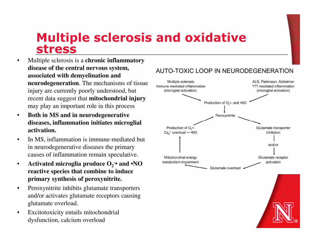
Multiple sclerosis and oxidative stress
• Multiple sclerosis is a chronic inflammatory
disease of the central nervous system,
associated with demyelination and
neurodegeneration. The mechanisms of tissue
injury are currently poorly understood, but
recent data suggest that mitochondrial injury
may play an important role in this process
• Both in MS and in neurodegenerative
diseases, inflammation initiates microglialdiseases, inflammation initiates microglial
activation.
• In MS, inflammation is immune-mediated but
in neurodegenerative diseases the primary
causes of inflammation remain speculative.
• Activated microglia produce O2-• and •NO
reactive species that combine to induce
primary synthesis of peroxynitrite.
• Peroxynitrite inhibits glutamate transporters
and/or activates glutamate receptors causing
glutamate overload.
• Excitotoxicity entails mitochondrial
dysfunction, calcium overload

Ageing and oxidative stress
• The incidence in many
diseases increases with
age
• Ageing can be defined as a
progressive decline in the
efficiency of physiological efficiency of physiological
processes after the
reproductive phase of life
• The ability of cells and
organisms to recover
from an insult such as
oxidative stress decreases
with age, while the risk of
disease increases.

Is the oxidative stress theory of aging dead?


Impaired degradation and repair systems in aging

Calorie restriction and redox balance
• Inverse correlation between
metabolic rates and lifespan.
• Mammalian NAD-dependent
mitochondrial deacetylase SIRT3.
Endogenous SIRT3 is a soluble
protein located in the mitochondrial
matrix
• Fasting increases SIRT3
expression, which increases expression, which increases
respiration and decreases the
production of reactive oxygen
species.
• Calorie restriction promotes the
activity of SIRT3, which
deacetylates SOD2 and isocitrate
dehydrogenase (IDH2), increasing
the activity of these enzymes and
resulting in reduced oxidative
stress. Lowered oxidative stress
leads to a reduced rate of aging.
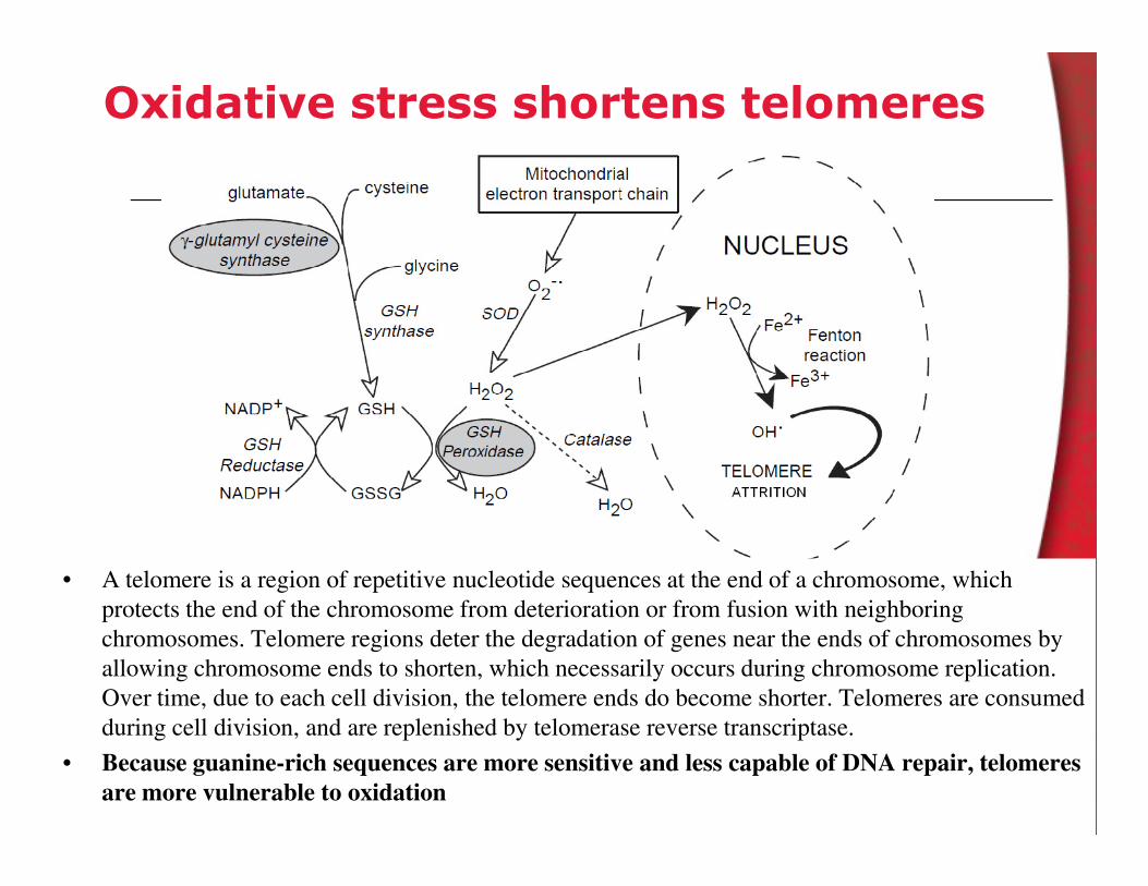
Oxidative stress shortens telomeres
• A telomere is a region of repetitive nucleotide sequences at the end of a chromosome, which
protects the end of the chromosome from deterioration or from fusion with neighboring
chromosomes. Telomere regions deter the degradation of genes near the ends of chromosomes by
allowing chromosome ends to shorten, which necessarily occurs during chromosome replication.
Over time, due to each cell division, the telomere ends do become shorter. Telomeres are consumed
during cell division, and are replenished by telomerase reverse transcriptase.
• Because guanine-rich sequences are more sensitive and less capable of DNA repair, telomeres
are more vulnerable to oxidation

Summary
• Neurodegenerative diseases are characterized by the selective
loss of neuronal populations which is associated with oxidative
stress
• Genetic, environmental factors and aging contribute to the
pathogenesis of neurodegenerative disorders
• Oxidative stress is observed in post-mortem samples of patients. • Oxidative stress is observed in post-mortem samples of patients.
However its relevance to the pathogenesis remains elusive.
Cause or consequence?
• Neurodegenerative disorders are characterized by mitochondrial
dysfunction and abnormal protein aggregation
• Further research is necessary to clearly define the etiology of
neurodegenerative disorders and the role of oxidative stress and
redox signaling in disease progression
– Oxidative damage or redox signaling