Embed Size (px)
Citation preview
A. Bjorklund and M. A. Cenci (Eds.)
Progress in Brain Research, Vol. 183
ISSN: 0079-6123
Copyright � 2010 Elsevier B.V. All rights reserved.
CHAPTER 14
Neurocomputational models of motor and cognitivedeficits in Parkinson’s disease
Thomas V. Wiecki and Michael J. Frank�
Department of Cognitive, Linguistic, and Psychological Sciences, Department of Psychiatry and Human Behavior, andBrown Institute for Brain Science, Brown University, Providence, RI, USA
Abstract: We review the contributions of biologically constrained computational models to ourunderstanding of motor and cognitive deficits in Parkinson’s disease (PD). The loss of dopaminergicneurons innervating the striatum in PD, and the well-established role of dopamine (DA) in reinforcementlearning (RL), enable neural network models of the basal ganglia (BG) to derive concrete and testablepredictions. We focus in this review on one simple underlying principle – the notion that reduced DAincreases activity and causes long-term potentiation in the indirect pathway of the BG. We show how thistheory can provide a unified account of diverse and seemingly unrelated phenomena in PD includingprogressive motor degeneration as well as cognitive deficits in RL, decision making and working memory.DA replacement therapy and deep brain stimulation can alleviate some aspects of these impairments, butcan actually introduce negative effects such as motor dyskinesias and cognitive impulsivity. We discussthese treatment effects in terms of modulation of specific mechanisms within the computationalframework. In addition, we review neurocomputational interpretations of increased impulsivity in theface of response conflict in patients with deep-brain-stimulation.
Keywords: Parkinson’s Disease; Dopamine; Basal Ganglia; Computational Model; Cognition
Introduction
Early onset of Parkinson’s disease (PD) is charac-terized by loss of dopaminergic neurons innervat-ing the striatum in the basal ganglia (BG) (Kishet al., 1988). The symptomatology is most
prominent in the motor domain and progressivelymanifests itself as bradykinesia, akinesia and tre-mor. More recently, however, cognitive and learn-ing deficits have received increased recognition andinterest (e.g. Cools, 2005; Cunha et al., 2009; Frank,2005; Grahn et al., 2009; Moustafa et al., 2008b).Although traditionally cognitive deficits are ofteninterpreted as resulting from decline in prefrontalcortical function, these reviews have highlighted amore central role for the BG in cognitive function.
�Corresponding author.Tel.: (401) 863-6872; Fax: (401) 863-2255;E-mail: [email protected]
DOI: 10.1016/S0079-6123(10)83014-6 275
From a computational and cognitive neu-roscience point of view, PD is a highly intriguingdisorder. Because PD results in depleted striataldopamine (DA) levels, but increased striatal DAlevels following DAmedication (Pavese et al., 2006;Tedroff et al., 1996), researchers can directly testthe influence of different BG DA configurationsin human subjects. Further, in early disease stages,cognitive deficits in PD are linked to depletedstriatal DA levels, with frontal DA levels spared(Nobukatsu et al., 2008). Similarly, cognitive deficitsin healthy ageing are correlated with striatal DAdepletion rather than frontal DA (Bckman et al.,2000, 2006; Kaasinen and Rinne, 2002). Betterunderstanding of this system will ultimately lead tobetter treatment options for PD, but also to otherdiseases involving DA in the BG such as addiction,schizophrenia and Tourette’s syndrome (TS).The BG consists of multiple interconnected
nuclei (Mink, 1996) that are part of several com-plex anatomical/functional loops (Gerfen andWilson, 1996; Graybiel et al., 1994; Haber, 2004;Haber et al., 2000; Nakano et al., 2000). The inher-ent complexity of this dynamic system, the role oflearning and the existence of feedback loops oftenlet classic box-and-arrow diagrams fall short intheir predictive capabilities. Moreover, data aboutthe BG (and PD) are contributed from across dif-ferent domains reaching from psychology to cellu-lar neurobiology. Although not without caveats,biologically constrained computational modelsoffer a disciplined approach to (1) integrate datafrom different domains and (2) derive novel andunintuitive predictions which can then be testedexperimentally to possibly refine the model.These models are inherently dynamic and are gov-erned by concrete activation and learning rules.One example of where these models furthered
our understanding was to reject the notion thatunder chronic DA depletion most synaptic plasti-city in the striatum would be lost (Calabresi et al.,2007b; Kreitzer and Malenka, 2007). The compu-tational model by Frank (2005) challenges thisassumption by hypothesizing that only one classof striatal cells – those that are activated in
response to positive reinforcement – would losesynaptic plasticity; another class of cells activatedin response to negative outcomes would actuallyshow increased synaptic plasticity. This computa-tional prediction has subsequently been confirmedbehaviourally (Frank et al., 2004) and neurobiolo-gically (Shen et al., 2008).This review is structured as follows. First, we
introduce basics of neural network models of theBG, focussing on an intuitive understanding of prin-ciples rather than mathematical formulations(which can be found elsewhere). We then establishthe simple notion of an activation and learningimbalance of the facilitatory and suppressive path-way in the BG and their implication in PD. By thisaccount, the diverse symptomatology of unmedi-cated and medicated PD (caused by a lack andexcess of DA in the striatum, respectively) repre-sent two sides of the same coin. Increased activationand learning in the suppressive pathway(i.e. unmedicated PD) accounts for progressivedecline of motor functions, increased avoidancelearning and reduced updating of working memory(WM). Conversely, increased activation and learn-ing in the facilitatory pathway (i.e. medicated PD)accounts for excess of motor functions(i.e. dyskinesias), increased anticipatory learningand excessive updating of WM. Thus, PD is notonly a motor disorder, but rather a more generaldisorder of action selection, exacerbated by a learn-ing process that induces a bias in the system to avoidselecting actions. This process can lead to a povertyof movement, but also of more cognitive actions.Note that we focus this review mainly on the
predictive power of this dopaminergic account.While this is sufficient for the data we describe,the neurotransmitters noradrenalin, serotonin andacetylcholine have also been implicated with cogni-tive deficits of PD (Calabresi et al., 2006, 2007a).
Neural network models of basal ganglia
Computational models in systems neuroscience(sometimes also called mechanistic or neural
276

network models) consist of layers of simulatedneurons (i.e. units) that are interconnectedaccording to the anatomy of the brain. The unitsused in different models – though varying in theirdegree of biological plausibility – generally try tofocus on the computational properties of real neu-rons and not on all aspects of their anatomy (likebiophysical models do). As such, they are oftenimplemented as point-neurons with the dendritictree and the soma shrunken to a tiny point. Theinfluence of presynaptic inputs is controlled viaweights which model synaptic efficacy (receptoraffinities, densities, number of presynaptic vesiclesreleased, etc). The Leabra framework, for exam-ple, computes the units’ voltage according to exci-tatory, inhibitory and leak conductance channels(O’Reilly and Munakata, 2000). Individual excita-tory and inhibitory channel conductances are
computed by multiplying the presynaptic inputactivity with the respective synaptic weight. Oncethe unit exceeds a certain voltage threshold, itcommunicates output to other downstream units,in the form of either a rate-coded variable (nor-malized firing rate), or discrete spiking.
Architecture of basal ganglia models
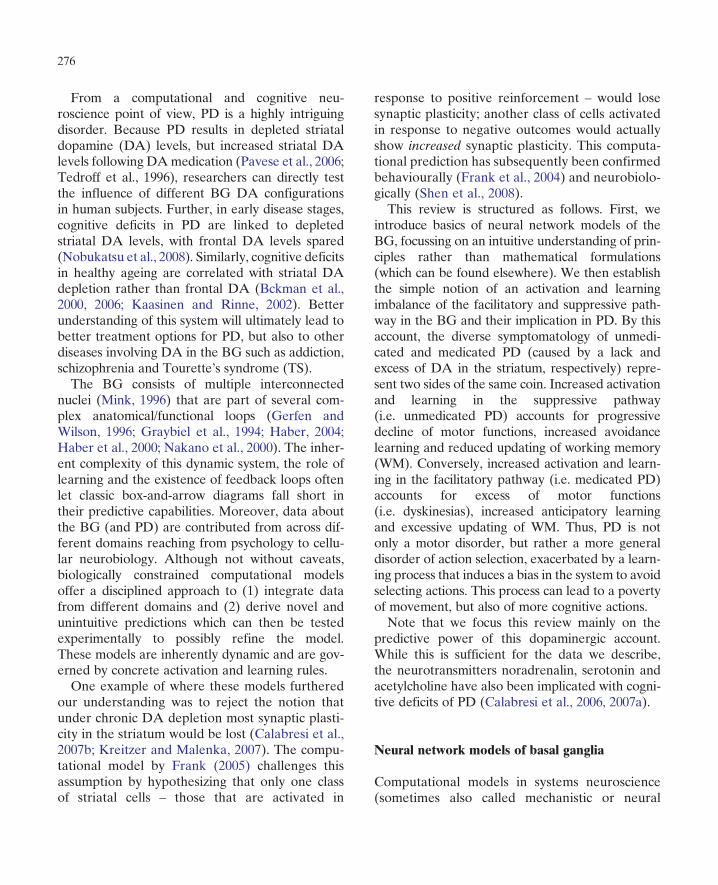
The BG is generally conceptualized as an adaptiveaction selection device gating information flowfrom and to cortex via the thalamus (Graybiel,1996). Its basic anatomy can be appreciated inFig. 1. Two opposing pathways – the direct andindirect pathway – dynamically and selectivelyfacilitate and suppress action representations inthe frontal cortex, respectively (Alexander and
Input StriatumPreSMA
STN
Thalamus
GPi/SNrSNc
Go NoGo GPe
(a) (b)
Striatum
Go NoGo γ-IN
Forntal cortex
STN Thalamus
GPe
GPiSNc
ExcitatoryInhibitoryModulatory
Fig. 1. (a) Box-and-arrow diagram of the basic anatomy of the BG. Frontal cortex projects to striatonigral neurons (Go) of the directpathway and to striatopallidal neurons (NoGo) in the indirect pathway. Dopaminergic projections from the SNc innervate thestriatum and excite and inhibit Go and NoGo neurons, respectively via simulated D1/D2 receptors. Fast-spiking GABAergicinterneurons (g-IN) regulate striatal activity via inhibitory projections. Activation of striatonigral neurons disinhibits the thalamusby inhibiting tonically active GABAergic neurons in the GPi. Activation of striatopallidal neurons removes inhibition of the GPi byinhibiting the GPe – thus ultimately inhibiting the thalamus. The STN is part of the hyperdirect pathway which dynamically activatesBG output, and thereby suppresses behaviour, as a function of cortical response conflict. (b) Implementation of the box-and-arrowdiagram in form of a neural network model by Frank (2006). Cylinders represent individual simulated neurons, their height andcolour encodes their activity level. The computational model complies with current anatomical and physiological BG data.
277
Crutcher, 1990; Brown et al., 2004; Frank, 2005;Frank et al., 2001; Mink, 1996). In the context ofmotor control, the BG were suggested to selec-tively facilitate a single motor command via thedirect pathway while suppressing all others via theindirect pathway (Chevalier and Deniau, 1990;Mink, 1996). The computational models describedbelow retain the basic functionality of the directand indirect pathway proposed in the classicmodel, while also extending the static model toincorporate dynamics, plasticity and updatedaspects of anatomical and physiological data(Cohen and Frank, 2009). As one example, whilethe original model suggested that the subthalamicnucleus (STN) was a key part of the indirect path-way, the updated model places the STN asanother input nucleus from cortex, forming athird ‘hyper-direct’ pathway (Miller, 2008;Nambu et al., 2000) that is functionally distinct.Below we discuss the relevance of this distinctionfor PD.At the heart of BG models is the striatum, a
large structure that consists collectively of thecaudate, putamen and nucleus accumbens. Almostall mechanistic BG models include at least thedirect pathway originating in the striatum, project-ing through BG output nuclei to the thalamus.The main effect of striatal activity in these modelsis to facilitate excitatory thalamic responses, whichin turn amplifies cortical activity associated withthe corresponding action plan. The striatumreceives input from multiple cortical areas andconsists mainly of medium spiny neurons (MSNs)(Gerfen and Wilson, 1996). Direct pathway MSNs(i.e. striatonigral neurons) express excitatorydopaminergic D1 receptors and send inhibitoryprojections to the substantia nigra pars reticulata(SNr) and to the internal segment of the globuspallidus (GPi). In the absence of striatal firing,neurons in SNr and GPi are tonically active andinhibit the thalamus, preventing a frontal actionplan from being executed. Activation of the directpathway leads to disinhibition of the thalamus.Disinhibition implies that thalamic units are notdirectly excited by direct pathway activity, but are
instead enabled to get excited if they also receiveexcitatory glutamatergic input (i.e. from descend-ing cortical signals) (Chevalier and Deniau, 1990;Frank et al., 2001). Striatal MSNs of the directpathway are sometimes labelled as ‘Go’-neurons(e.g. Frank, 2005; O’Reilly and Frank, 2006),because they act to gate or facilitate frontal actionplans, the details of which are specified by corticalrepresentations.The role of the indirect pathway is more con-
tentious, and is sometimes omitted altogether incomputational models (e.g. Arthur et al., 2006;Bogacz and Gurney, 2007). Although debatedfor several years, methodological advances havenow confirmed the original suggestion that D1 andD2 receptors are largely segregated in MSNs, withD1 receptors predominating in the direct pathwayand D2 receptors in the indirect pathway (Gerfenet al., 1990; Gong et al., 2003; Matamales et al.,2009; Surmeier et al., 2007; Valjent et al., 2009).Striatopallidal neurons expressing D2 receptorssend inhibitory projections to the external seg-ment of the globus pallidus (GPe). The GPesends focussed inhibitory projections to GPi/SNr(Bolam et al., 2000; Kincaid et al., 1991; Smith andBolam, 1989, 1990). Due to this additional inhibi-tory projection, activity in the indirect pathwayultimately results in inhibition of the thalamusand thus suppression of frontal action plans.Because of this motor suppression property(Albin et al., 1989), striatal MSNs of the indirectpathway are sometimes labelled as ’NoGo’-neu-rons (Frank, 2005). Electrophysiological studiesfrom different domains support the existence ofboth, facilitatory and suppressive pathways (Api-cella et al., 1992; Jiang et al., 2003; Kimchi andLaubach, 2009a, 2009b; Samejima et al., 2005;Watanabe and Munoz, 2009). Further, selectiveablation of striatopallidal (indirect pathway) cellsleads to increased locomotion (Pierre et al., 2009).Moreover, actions coded specifically in the striatalregion in which the striatopallidal ablation wasadministered are selectively increased (Sanoet al., 2003). These results support the notionthat the indirect pathway acts to suppress
278
behaviours, such that when ablated, these beha-viours are expressed more readily (Miller, 2008).How are only certain actions facilitated or sup-
pressed depending on the context? First, neuronsin these pathways are highly structured accordingto the actions they encode (Deniau et al., 1996;Fger and Crossman, 1984; Mink, 1996). Striatalneurons that receive from a particular corticalregion (e.g. encoding hand movements) recipro-cally, via the loop through BG output and thala-mus, project back to influence activity in that samecortical region (Kelly and Strick, 2004; Middletonand Strick, 2000). Evidence for this ‘closed-loop’has also been reported in humans (Draganski et al.,2008). Second, striatal neurons receive diffuse pro-jections from posterior cortical areas (Frank, 2005).These corticostriatal projections represent theinput to most models and are implemented in theform of units which code for abstract properties ofthe environment (e.g. stimulus colour or context)(Frank, 2005; Guthrie et al., 2009; Wiecki et al.,2009). This many-to-many connection patternenables the model to represent all possible stimu-lus–response pairs and to learn facilitation or sup-pression for each action in response to stimulusproperties. In addition, action selection may befurther contextualized by the cognitive stateencoded in prefrontal cortex (PFC). Indeed, thereappears to be some hierarchical structure to BG–
PFC circuits: in addition to closed loops among BGand particular frontal regions, it is also the case thatPFC areas in a particular loop can innervate striatalareas in more posterior loops (Haber, 2004; Haberand Calzavara, 2009). In this way, cognitive actionplans in PFC can provide additional contextualinput to lower level actions, for example, toinfluence motor control.As mentioned above, multiple cortico-striatal
loops innervate the striatum. The ventral pathway,innervating the ventral striatum (nucleus accum-bens), represents the motivational loop. It plays amajor part in the development of addiction(Dagher and Robbins, 2009). The dorsal pathway,innervating the dorsal striatum (i.e. caudate andputamen), represents the motor loop. It plays a
major role in habit formation (Everitt and Rob-bins, 2005; Henry et al., 2004; Tricomi et al., 2009).In PD, nigrostriatal dopaminergic projectionsinnervating the dorsal striatum are stronglyaffected, while mesolimbic dopaminergic projec-tions innervating the ventral striatum are rela-tively spared (Kish et al., 1988).
Dopamine as a reinforcement learning signal
Recordings of midbrain dopaminergic neurons inawake behaving monkeys reveal phasic firing pat-terns in response to unexpected rewards and pun-ishments (Bayer et al., 2007; Ljungberg et al., 1992;Montague et al., 1997; Pan et al., 2005; Roeschet al., 2007; Schultz, 1998; Waelti et al., 2001).Specifically, a DA burst is observed whenever anoutcome of an action is better than expected, and,conversely, a drop below tonic DA firing (i.e. DAdip) whenever the outcome is worse than expected.Importantly, the same patterns were observed inhuman PD patients who receive abstract (financial)rewards and punishments (Zaghloul et al., 2009).Computational models show that this DA-mediated reward prediction error signal can beused to efficiently learn reward contingencies andto maximize reward intake in simple reinforcementlearning (RL) environments (Barto, 1995; Fristonet al., 1994; Montague et al., 1997; Schultz et al.,1997; Sutton and Barto, 1990).Based on this insight, mechanistic models
explore how such action selection and contingencylearning to maximize rewards is implemented inthe anatomy of the BG. As mentioned above,synaptic strengths are implemented as weightsthat can change dynamically over time. Specifi-cally, co-activation of two connected units resultsin an increase of their connection’s weight [corre-sponding to long-term potentiation (LTP)], other-wise the weight remains stable or is decreased[corresponding to long-term depression (LTD)].In the corticostriatal pathway, this plasticity isstrongly modulated by DA, leading to a ‘3-factor’Hebbian learning rule (Berke and Hyman, 2000;
279
Calabresi et al., 1997, 2000; Kerr and Wickens,2001; Reynolds and Wickens, 2002; Reynoldset al., 2001; Shen et al., 2008). Importantly, DAeffects on postsynaptic activity and plasticitydepend on the receptor class. Active Go neuronsexpressing D1 receptors are depolarized by DA(Hernandez-Lopez et al., 1997), whereas NoGoneurons expressing D2 receptors are inhibited byDA (Hernandez-Lopez et al., 2000). Thus a DAburst in response to reinforcement further acti-vates Go neurons (particularly those that are con-currently excited by corticostriatal glutamatergicinput), but inhibits NoGo neurons. Conversely, aDA dip in response to punishment or lack ofreward activates NoGo neurons by removingtonic inhibition of DA onto postsynaptic D2receptors (Frank, 2005). [See Cohen and Frank(2009) for a detailed discussion of the plausibilityof this mechanism.]In the model, the above plasticity dynamics are
adaptive. Simulated DA activity depends onwhether the network selected the correct responseaccording to the task at hand. If the network chosecorrectly, a DA burst will further activate those Goneurons encoding that action in the current envir-onmental state (stimulus). This increased activity isassociated with synaptic potentiation, such that thecorticostriatal weights from active inputs areincreased. The next time the same stimulus is pre-sented, and the corresponding motor action repre-sented in cortex, striatal Go activity encoding thataction will be stronger, increasing the probabilitythat the rewarded action will be gated. Conversely,if the network is chosen incorrectly, a DA dip willincrease weights between active cortical units andcorresponding NoGo units, ultimately decreasingthe probability that the punished action will berepeated. Across multiple trials of experience, thissystem is able to learn to gate actions that are mostlikely to produce positive outcomes and to suppressthose that are most likely to yield negative out-comes – a corner stone of adaptive behaviour. Arecent study provides direct support for this modelby showing that direct and indirect pathway cellsare required for reward/approach and punishment/
avoidance learning, respectively (Hikida et al.,2010).
Cognitive learning deficits
Parkinson’s disease: too much ‘NoGo’ learning?
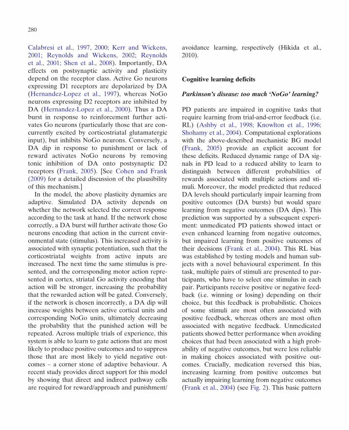
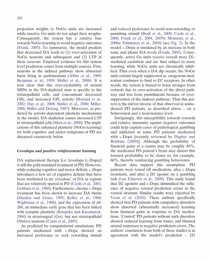
PD patients are impaired in cognitive tasks thatrequire learning from trial-and-error feedback (i.e.RL) (Ashby et al., 1998; Knowlton et al., 1996;Shohamy et al., 2004). Computational explorationswith the above-described mechanistic BG model(Frank, 2005) provide an explicit account forthese deficits. Reduced dynamic range of DA sig-nals in PD lead to a reduced ability to learn todistinguish between different probabilities ofrewards associated with multiple actions and sti-muli. Moreover, the model predicted that reducedDA levels should particularly impair learning frompositive outcomes (DA bursts) but would sparelearning from negative outcomes (DA dips). Thisprediction was supported by a subsequent experi-ment: unmedicated PD patients showed intact oreven enhanced learning from negative outcomes,but impaired learning from positive outcomes oftheir decisions (Frank et al., 2004). This RL biaswas established by testing models and human sub-jects with a novel behavioural experiment. In thistask, multiple pairs of stimuli are presented to par-ticipants, who have to select one stimulus in eachpair. Participants receive positive or negative feed-back (i.e. winning or losing) depending on theirchoice, but this feedback is probabilistic. Choicesof some stimuli are most often associated withpositive feedback, whereas others are most oftenassociated with negative feedback. Unmedicatedpatients showed better performance when avoidingchoices that had been associated with a high prob-ability of negative outcomes, but were less reliablein making choices associated with positive out-comes. Crucially, medication reversed this bias,increasing learning from positive outcomes butactually impairing learning from negative outcomes(Frank et al., 2004) (see Fig. 2). This basic pattern
280
has since been replicated across multiple experi-ments, tasks and labs (Bodi et al., 2009; Coolset al., 2006; Frank et al., 2007b; Moustafa et al.,2008a; Palminteri et al., 2009; Voon et al., 2010).Here, the mechanistic model provides insight
into the neurobiological underpinnings that give
rise to the pattern observed behaviourally.Depleted DA levels (both tonic and phasic) resultsin increased activity of NoGo units expressing inhi-bitory D2 receptors, while Go units expressingexcitatory D1 receptors receive less excitation. Asa result of Hebbian learning, fronto-striatal
(b)(a)
Go A NoGo BTest condition
−0.1
0.0
0.1
0.2
0.3
0.4
0.5
0.6Train
Test
Chosse A?
Aviod B?
A > CDEF
B < CDEF
A (80/20) B (20/80)
C (70/30) D (30/70)
E (60/40) F (40/60)
Str
iata
l Go
/No
Go
act
ivit
y
IntactSim PDSim DA Meds
BG Model Go/NoGo Associations
(d)(c)
Choose A Avoid BTest condition
50
60
70
80
90
100
Acc
ura
cy
SeniorsPD OFFPD ON
Parkinson’s patients on/off meds (’04)
Choose A Avoid BTest condition
50
60
70
80
90A
ccu
racy
SeniorsPD OFFPD ON
Parkinson’s patients on/off meds (’07)
Fig. 2. (a) Probabilistic selection RL task. During training, participants select among each stimulus pair. Probabilities of receivingpositive/negative feedback for each stimulus are indicated in parentheses. In the test phase, all combinations of stimuli are presentedwithout feedback. Go learning is indexed by reliable choice of the most positive stimulus A in these novel pairs, whereas NoGolearning is indexed by reliable avoidance of the most negative stimulus B. (b) Striatal Go and NoGo activation states when presentedwith input stimuli A and B, respectively. Simulated Parkinsons (Sim PD) was implemented by reducing striatal DA levels, whereasmedication (Sim DA Meds) was simulated by increasing DA levels and partially shunting the effects of DA dips during negativefeedback. (c) Behavioural findings in PD patients on/off medication supporting model predictions (Frank et al., 2004). (d) Replicationin another group of patients, where here the most prominent effects were observed in the NoGo learning condition (Frank et al.,2007b).
281
projection weights to NoGo units are increasedwhile inactive Go units do not adapt their weights.Consequently, the system has a relative biastowards NoGo learning to avoid negative outcomes(Frank, 2005). To summarize, the model predictsthat decreased DA leads to (1) over-activation ofNoGo neurons and subsequent and (2) LTP inthese neurons. Empirical evidence for this systemlevel prediction comes from multiple sources. First,neurons in the indirect pathway show abnormalburst firing in parkinsonism (Albin et al., 1989;Bergman et al., 1999; Mallet et al., 2006). It isnow clear that this over-excitability of striatalMSNs in the DA-depleted state is specific to thestriatopallidal cells, and concomitant decreasedGPe, and increased GPi, activity (Boraud et al.,2002; Day et al., 2008; Mallet et al., 2006; Miller,2008; Miller and Delong, 1987). Moreover, as pre-dicted by activity-dependent plasticity mechanismsin the model, DA depletion causes increased LTPin striatopallidal cells (Shen et al., 2008). The impli-cations of this enhanced plasticity (NoGo learning)for both cognitive and motor symptoms of PD arediscussed extensively below.
Levodopa and positive reinforcement learning
DA replacement therapy [i.e. levodopa (L-Dopa)]is still the gold-standard treatment of PD.However,while reducing cognitive andmotor deficits, L-Dopaintroduces a new set of cognitive deficits that havebeen attributed to an ‘overdose’ of DA in regionsthat are relatively spared in PD (Cools et al., 2001;Gotham et al., 1988). Furthermore, chronic L-Dopatreatment has been shown to increase DA bursts(Harden and Grace, 1995; Keller et al., 1988;Wightman et al., 1988), and the expression of zif-268, an immediate early gene that has been linkedwith synaptic plasticity (Knapska and Kaczmarek,2004) in striatonigral (Go), but not striatopallidal(NoGo) neurons (Carta et al., 2005).As predicted by computational simulations, PD
patients medicated with L-Dopa showed anincreased preference to seek rewarding stimuli
and reduced preference to avoid non-rewarding orpunishing stimuli (Bodi et al., 2009; Cools et al.,2006; Frank et al., 2004, 2007b; Moustafa et al.,2008a; Palminteri et al., 2009) (see Fig. 2). In themodel, L-Dopa is simulated by an increase in bothtonic and phasic DA levels (Frank, 2005). Conse-quently, active Go units receive overall more D1-mediated excitation and are thus subject to morelearning, while NoGo units are chronically inhib-ited. Thus even when a DA dip occurs, the NoGounits remain largely suppressed as exogenous med-ication continues to bind to D2 receptors. In otherwords, the system is biased to learn stronger fromrewards due to over-activation of the direct path-way and less from punishments because of over-suppression of the indirect pathway. Thus this pat-tern is the mirror inverse of that observed in unme-dicated PD patients, as described above – on abehavioural and a neuroscience level.Intriguingly, this susceptibility towards rewards
and relative immunity against negative outcomescould help explain cases of pathological gamblingand addiction in some PD patients medicatedwith L-Dopa [recently reviewed by Dagher andRobbins (2009)]. Although the probability offinancial gains at a casino may be roughly 48%,the medicated PD patient’s brain may distort thislearned probability to be closer to, for example,60%, thereby reinforcing gambling behaviours.Recent data support this assumption. PD
patients were tested off medication, after L-Dopatreatment, and after a D2 agonist on a gamblingtask (van Eimeren et al., 2009). This study foundthat D2 agonists and L-Dopa diminished the influ-ence of negative reward prediction errors in theventral striatum. Similar results were reported byVoon et al. (2010). These authors specificallyshowed that PD patients with compulsive disordersshow distorted (abnormally increased) learningfrom financial gains in response to DA medica-tions. ‘Control’ PD patients without such disordersshowed reduced learning from losses, and bluntedstriatal responses to negative prediction errors. Theauthors’ conclusion from both of these studies is inagreement with the model’s prediction – D2
282
agonists and L-Dopa block the effects of DA dipsand thus of negative RL. Another recent fMRIstudy found differences in dorsal striatum activa-tion in medicated PD patients under RL conditions(Schonberg et al., 2010).
Individual differences
Why are only a minority of PD patients susceptibleto pathological gambling in response to medica-tion? It may be that this, too, is explained in accor-dance with the theory proposed above. PD patientswith pathological gambling disorder have lowerbaseline striatal D2 receptor density (Steeveset al., 2009). This result might be inherently linkedto RL learning differences of healthy humans carry-ing different polymorphisms of DA signalling genes(Frank and Hutchison, 2009; Frank et al., 2007a,2009; Klein et al., 2007). Among the tested genes,the polymorphism of the DRD2 gene, associatedwith D2 receptor function, has been reliably linkedto the degree of learning from negative outcomes(i.e. NoGo learning). Those genotypes associatedwith reduced striatal D2 receptor density (Hirvonenet al., 2005) are accordingly associated with reducedNoGo learning (Frank and Hutchison, 2009; Franket al., 2007a, 2009; Klein et al., 2007). Thus it ispossible that the PD patients who are most suscep-tible to pathological gambling from DA medica-tions are those who are genetically predisposed toexhibit reduced learning from negative outcomes.This hypothesis has yet to be directly tested, but theobserved reduced D2 density in pathological gam-bling patients is supportive, whether or not due togenetic factors. Moreover, if this predisposition iscoupled with increased Go learning resulting fromdopaminergic medications (Voon et al., 2010), com-pulsive disorders may be especially evident.This same logic may suggest that a distorted bias
to learn more from positive than negative out-comes in RL may help explain other addictivepersonality types in otherwise healthy individuals.Polymorphism of the DARPP-32 gene relates tosynaptic plasticity in response to D1 stimulation.
Carriers of the polymorphism have increasedsynaptic plasticity and show relatively strongerpositive RL (Frank et al., 2007a, 2009). Similarly,individual differences of baseline striatal DAsynthesis are predictive of the extent to which par-ticipants learn from positive versus negativereward prediction errors (Cools et al., 2009).These biological factors may predispose indivi-duals to have a greater risk for pathological gam-bling and other addictions. Indeed, these samefactors may also play a role in the development ofaddiction to L-Dopa observed in some PD patients(Borek and Friedman, 2005; Dagher and Robbins,2009). For example, a recent review highlightssimilarities between methamphetamine addictionand L-Dopa sensitization (Fornai et al., 2009).Intriguingly, Palminteri et al. (2009) found simi-
lar patterns of RL deficits in patients with TS.Crucially, these patients show the opposite RLpattern – unmedicated TS patients learned betterfrom gains than losses. While this pattern can bestbe explained by DA hyperactivity in TS patients,this evidence for this account of TS remains contro-versial (Albin and Mink, 2006; Leckman, 2002;Singer, 1995; Wong et al., 2008). Nevertheless, TSpatients are treated with D2 antagonists, such thatthe DA system is manipulated in opposite directionto PD. Critically, TS patients treated with D2antagonists exhibited relatively better learningfrom negative than positive outcomes, very similarlyto unmedicated PDpatients (Palminteri et al., 2009).These data are also consistent with model pre-
dictions: D2 antagonism selectively increasesexcitability and plasticity of striatopallidal cells(Centonze et al., 2004; Day et al., 2008; Malletet al., 2006), thereby enhancing NoGo learning.
Motor impairments
Progressive development of motor symptoms:sensitization?
As described above, unmedicated PD patients exhi-bit increased negative RL, inducing avoidance
283
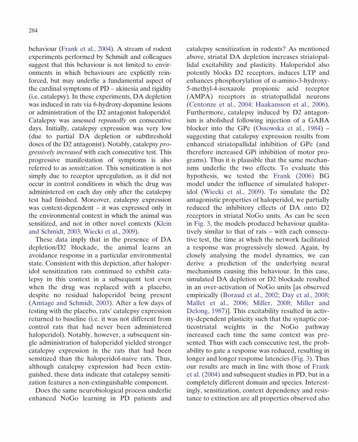
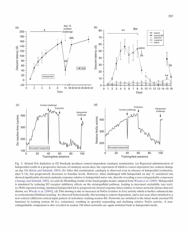
behaviour (Frank et al., 2004). A stream of rodentexperiments performed by Schmidt and colleaguessuggest that this behaviour is not limited to envir-onments in which behaviours are explicitly rein-forced, but may underlie a fundamental aspect ofthe cardinal symptoms of PD – akinesia and rigidity(i.e. catalepsy). In these experiments, DA depletionwas induced in rats via 6-hydroxy-dopamine lesionsor administration of the D2 antagonist haloperidol.Catalepsy was assessed repeatedly on consecutivedays. Initially, catalepsy expression was very low(due to partial DA depletion or subthresholddoses of the D2 antagonist). Notably, catalepsy pro-gressively increased with each consecutive test. Thisprogressive manifestation of symptoms is alsoreferred to as sensitization. This sensitization is notsimply due to receptor upregulation, as it did notoccur in control conditions in which the drug wasadministered on each day only after the catalepsytest had finished. Moreover, catalepsy expressionwas context-dependent – it was expressed only inthe environmental context in which the animal wassensitized, and not in other novel contexts (Kleinand Schmidt, 2003; Wiecki et al., 2009).These data imply that in the presence of DA
depletion/D2 blockade, the animal learns anavoidance response in a particular environmentalstate. Consistent with this depiction, after haloper-idol sensitization rats continued to exhibit cata-lepsy in this context in a subsequent test evenwhen the drug was replaced with a placebo,despite no residual haloperidol being present(Amtage and Schmidt, 2003). After a few days oftesting with the placebo, rats’ catalepsy expressionreturned to baseline (i.e. it was not different fromcontrol rats that had never been administeredhaloperidol). Notably, however, a subsequent sin-gle administration of haloperidol yielded strongercatalepsy expression in the rats that had beensensitized than the haloperidol-naive rats. Thus,although catalepsy expression had been extin-guished, these data indicate that catalepsy sensiti-zation features a non-extinguishable component.Does the same neurobiological process underlie
enhanced NoGo learning in PD patients and
catalepsy sensitization in rodents? As mentionedabove, striatal DA depletion increases striatopal-lidal excitability and plasticity. Haloperidol alsopotently blocks D2 receptors, induces LTP andenhances phosphorylation of a-amino-3-hydroxy-5-methyl-4-isoxazole propionic acid receptor(AMPA) receptors in striatopallidal neurons(Centonze et al., 2004; Haakansson et al., 2006).Furthermore, catalepsy induced by D2 antagon-ism is abolished following injection of a GABAblocker into the GPe (Ossowska et al., 1984) –
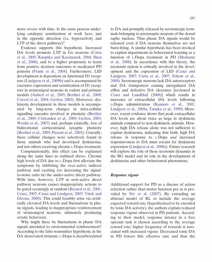
suggesting that catalepsy expression results fromenhanced striatopallidal inhibition of GPe (andtherefore increased GPi inhibition of motor pro-grams). Thus it is plausible that the same mechan-isms underlie the two effects. To evaluate thishypothesis, we tested the Frank (2006) BGmodel under the influence of simulated haloper-idol (Wiecki et al., 2009). To simulate the D2antagonistic properties of haloperidol, we partiallyreduced the inhibitory effects of DA onto D2receptors in striatal NoGo units. As can be seenin Fig. 3, the models produced behaviour qualita-tively similar to that of rats – with each consecu-tive test, the time at which the network facilitateda response was progressively slowed. Again, byclosely analysing the model dynamics, we canderive a prediction of the underlying neuralmechanisms causing this behaviour. In this case,simulated DA depletion or D2 blockade resultedin an over-activation of NoGo units [as observedempirically (Boraud et al., 2002; Day et al., 2008;Mallet et al., 2006; Miller, 2008; Miller andDelong, 1987)]. This excitability resulted in activ-ity-dependent plasticity such that the synaptic cor-ticostriatal weights in the NoGo pathwayincreased each time the same context was pre-sented. Thus with each consecutive test, the prob-ability to gate a response was reduced, resulting inlonger and longer response latencies (Fig. 3). Thusour results are much in line with those of Franket al. (2004) and subsequent studies in PD, but in acompletely different domain and species. Interest-ingly, sensitization, context dependency and resis-tance to extinction are all properties observed also
284
(b)
Des
cent
late
ncy
(S)
$$
$$
saline
**
**
Barpairedpaired-NEpaired-Eunpairedsaline
80
70
60
50
40
30
20
10
00 1 2 3 4 5 6 7 8 9 10 11 12 13 14 15 16
Days
(a)
0−20
0
20
40
60
80
100
120
Des
cent
late
ncy
(S) 140
160
180
200$
$$$
$$
bar
haloperidol
day 10context
challenge
2 4 6 8 10
Days
28 30 32 34 36 38 40 42
Simulated haloperidol
Rel
ativ
e N
oGo
activ
ity
200
300
400
0 20 40 60Training/test sessions
80 100
Context challengeExtinction
Haloperidolchallenge
(d)
0
Simulated haloperidol
Context challenge
100
200
300
Go
reac
tion
time
(cyc
les) 400
20 40 60Training/test sessions
80 100
Extinction
Haloperidolchallenge
(c)
Fig. 3. Striatal DA depletion or D2 blockade produces context-dependent catalepsy sensitization. (a) Repeated administration ofhaloperidol results in a progressive increase of catalepsy across days, the expression of which is context-dependent (see context changeon day 10) (Klein and Schmidt, 2003). (b) After this sensitization, catalepsy is observed even in absence of haloperidol (extinction,days 9–14), but progressively decreases to baseline levels. However, when challenged with haloperidol on day 15, sensitized ratsshowed significantly elevated cataleptic response relative to haloperidol-naive rats, thereby revealing a non-extinguishable component(Amtage and Schmidt, 2003). (c) and (d) Modelling results of the basal ganglia model, adapted fromWiecki et al. (2009). Haloperidolis simulated by reducing D2 receptor inhibitory effects on the striatopallidal pathway, leading to increased excitability (see text).(c) With repeated testing, simulated haloperidol led to progressively slowed response times relative to intact networks [intact data notshown; see Wiecki et al. (2009)]. (d) This slowing is due to increases in NoGo (relative to Go) activity which is further enhanced dueto corticostriatal Hebbian learning. As observed behaviourally, this learning is context-dependent, and is not seen when switched to anew context (different cortical input pattern of activation, training session 40). Networks are switched to the intact mode (normal D2function) in training session 60 (i.e. extinction), resulting in speeded responding and declining relative NoGo activity. A non-extinguishable component is also revealed in session 100 when networks are again switched back to haloperidol mode.
285
in the appetitive domain of addiction (Schmidtand Beninger, 2006). It is possible that the sameprinciples apply in that case, but with sensitizationoccurring in the Go pathway.Moreover, this logic has recently been applied to
a novel RL experiment in human PD patients(Moustafa et al., 2008a). In this study, rather thanchoosing among multiple stimuli to maximizerewards, participants had to press just a single but-ton, but had to learn to speed or slow responses inorder to maximize positive outcomes and minimizenegative outcomes. As predicted by the Go/NoGomodel, unmedicated patients were more adept atlearning to slow down (relative to their baselinespeed) to avoid negative outcomes – that is, theyshowed a bias towards NoGo learning from nega-tive prediction errors leading to slowed responding.In contrast, medicated patients showed the oppo-site pattern, learning better to speed responses toincrease positive outcomes. This same patternnaturally emerged in the computational modelwhen PD and medications were simulated as pre-viously (Moustafa et al., 2008a).A recent study found learning impairments in a
rotarod movement task of mice with selectivelydenervated dorso-striatal DA (due to PITx3genetic knockout) which could be rescued byL-Dopa administration (Beeler et al., in print). Cru-cially, cessation of L-Dopa treatment in trainedmice did not result in an immediate performancedrop, but rather a progressive decline. Relatedly,healthy mice treated with a D2 antagonist showedthe same progressive decline. However, treatmentwith a D1 antagonist resulted in an immediateperformance deficit. In light of our computationalframework, this pattern can be explained in termsof learning. D1 antagonists would reduce the sig-nal-to-noise ration in Go neurons such that thosecells encoding learned motor associations in therotarod task are relatively suppressed. This sameeffect would also diminish synaptic plasticity inthese cells (indeed, unpublished simulations of aD1 antagonist in the same model as describedabove also show an immediate impairment ofmotor function). D2 antagonists, on the other
hand, would increase NoGo activity while still leav-ing Go expression of learned associations intact.Initially, learned Go activity may be sufficient toovercome the drug-induced NoGo activity. Afterrepeated exposure, however, NoGo neurons wouldbecome progressively active due to LTP and leadto the progressive decline in performance observedexperimentally. In sum, these data further supportthe hypothesis that synaptic plasticity in the indirectpathway is the root of sensitization under low levelsof DA (Wiecki et al., 2009).The progressive worsening of symptoms in PD is
generally attributed to the progressive cell death ofdopaminergic neurons. However, the datareviewed above, along with modelling results, letthis symptom progression appear in a differentlight. Even though it might sound counter intuitive,it seems that motor (and cognitive?) symptoms inPD are, at least partially, learned. To better treatPD patients, we have to explore if and howmuch ofthe motor and cognitive symptoms of PD are actu-ally learned due to a dysfunctional learning signal.Ultimately, this could open the door to a wholenew set of treatment options if we manage to finda way to unlearn these symptoms.
Dyskinesia and Go learning
While L-Dopa is quite effective in the beginningof treatment, following progressive treatment,L-Dopa-induced dyskinesia (LID) begin to appearin certain patients. LID are characterized byexcessive and uncontrollable movements. ChronicL-Dopa treatment has been shown to increase DAbursts (Harden and Grace, 1995; Keller et al.,1988; Wightman et al., 1988), and the expressionof zif-268, an immediate early gene that has beenlinked with synaptic plasticity (Knapska and Kacz-marek, 2004) in striatonigral (Go), but not striato-pallidal (NoGo) neurons (Carta et al., 2005). Inthis regard, LID can be seen as the opposite ofsome PD symptoms (e.g. catalepsy). Like the pro-gressive manifestation of catalepsy in parkinsonianrats, LID are also not present at first but get
286
more severe with time. Is the same process under-lying catalepsy sensitization at work here, justin the opposite direction (i.e. hyperactivity andLTP of the direct pathway)?Evidence supports this hypothesis. Increased
DA levels promote LTP in Go neurons (Cartaet al., 2005; Knapska and Kaczmarek, 2004; Shenet al., 2008), and to a higher propensity to learnfrom positive decision outcomes in medicated PDpatients (Frank et al., 2004). Furthermore, LIDdevelopment is dependent on functional D1 recep-tors (Lindgren et al., 2009b) and is accompanied byexcessive expression and sensitization of D1 recep-tors in striatonigral neurons in rodent and primatemodels (Aubert et al., 2005; Berthet et al., 2009;Corvol et al., 2004; Gerfen, 2003). Moreover, dys-kinesia development in these models is accompa-nied by long-term changes in intra-cellularsignalling cascades involved in plasticity (Berthetet al., 2009; Crittenden et al., 2009; Gerfen, 2003;Westin et al., 2007) and, perhaps relatedly, loss ofbidirectional corticostriatal synaptic plasticity(Berthet et al., 2009; Picconi et al., 2003). Crucially,these cellular changes were selectively found inthose animals who had developed dyskinesias,and not others receiving chronic L-Dopa treatment.Computationally, this effect can be explained
along the same lines as outlined above. Chronichigh levels of DA due to L-Dopa first alleviate thesymptoms by inhibiting the over-active indirectpathway and exciting (or increasing the signal-to-noise ratio in) the under-active direct pathway.With time, however, LTP in now-active directpathway neurons causes inappropriate actions tobe gated seemingly at random (Bezard et al., 2001;Cenci, 2007; Cenci and Lindgren, 2007; Vitek andGiroux, 2000). This could feasibly arise via artifi-cially elevated DA levels and fluctuations in pha-sic signals, leading to inappropriate reinforcementof striatonigral neurons, ultimately producingerratic behaviour.Why might there be fluctuations in phasic DA
signals unrelated to environmental reinforcement?According to the false-transmitter hypothesis, in theDA denervated striatum, L-Dopa is decarboxylated
to DA and promptly released by serotonergic term-inals belonging to presynaptic neurons of the dorsalraphe nucleus. Thus phasic DA signals would bereleased even if DA neurons themselves are notburst-firing. A similar hypothesis has been invokedto explain impairments in behavioural learning as afunction of L-Dopa treatment in PD (Shohamyet al., 2006). In accordance with this theory, theserotonin system is critically involved in the devel-opment and the expression of LID (Cenci andLindgren, 2007; Carta et al., 2007; Eskow et al.,2009). Serotonergic neurons lack DA autoreceptorsand DA transporters causing unregulated DAefflux and defective DA clearance [reviewed inCenci and Lundblad (2006)] which results inincreases of extracellular DA levels followingL-Dopa administration (Kannari et al., 2001;Lindgren et al., 2009a; Tanaka et al., 1999). More-over, recent evidence shows that peak extracellularDA levels are about twice as large in dyskineticanimals compared to non-dyskinetic animals. How-ever, high DA release alone was not sufficient toexplain dyskinesias, indicating that both, high DArelease in response to L-Dopa and increasedresponsiveness to DA must coexist for dyskinesiaexpression (Lindgren et al., 2009a). Future researchwill explore the role of these serotonin dynamics inthe BG model and its role in the development ofdyskinesias and other behavioural phenomena.
Response vigour
Additional support for PD as a disease of actionselection rather than motor function per se is pro-vided by Niv et al. (2007). By extending anabstract model of RL to include the averageexpected reward rate (hypothesized to be encodedby tonic DA activity), the authors explain reducedresponse vigour observed in PD patients. Accord-ing to their model, response latency in a freeoperant task is chosen according to the averagereward rate: higher frequency of rewards is asso-ciated with increased vigour. Decreased tonic DAin PD lowers this effective rate and thus the
287
vigour. Experiments support this hypothesis. In agrasping task, PD patients were able to achievethe same maximal movement speeds as healthyindividuals, their average speed was just loweroverall (Mazzoni et al., 2007). In a review high-lighting the close connection between these find-ings, the authors conclude that ‘it is not that PDpatients cannot move, it is that their DA circuitrydoes not “want” to’. (Niv and Rivlin-Etzion, 2007).
Working memory impairments
Among movement and learning defects, PD isalso characterized by WM impairments (Cools,2005; Frank, 2005; Owen et al., 1992, 1998). Neu-roimaging studies reveal that WM impairments inPD patients are associated with decreased BGactivity (Lewis et al., 2003; Owen et al., 1998;Postle et al., 1997). Can the same hypothesis thatexplains cognitive and motor deficits (as outlinedabove) also explain specific WM impairments ofPD? The conceptualization of the BG as a gatingdevice of motor commands (Mink, 1996) ishypothesized to also gate information flow intoWM (Frank and O’Reilly, 2006; Frank et al.,2001; Moustafa et al., 2008b; O’Reilly and Frank,2006). The mechanism is very similar to thatdescribed above for gating action plans, onlyhere we simulate BG circuits interacting withPFC rather than premotor cortex, and the ‘action’is whether or not to maintain the current stimulusin PFC. In this context, Go activity indicates that arepresentation is task-relevant and should bestored in memory, whereas NoGo activity indi-cates that the stimulus should be ignored or fil-tered out of WM. Recent neuroimaging dataprovide support for this notion (Cools et al.,2007; McNab and Klingberg, 2007).According to this framework, DA depletion as
in PD would lead to an increased threshold forupdating WM (because of too much NoGo activ-ity), such that most information is treated as irre-levant. In contrast, chronic DA elevations byreplacement therapy would result in too much
WM updating and the gating of distracting infor-mation into WM. This specific pattern was foundin a conjoint behavioural WM task (Moustafaet al., 2008b). Medicated patients were alsoimpaired at ignoring stimulus information thathad previously been relevant but is subsequentlydistracting, consistent with the hypothesis that Goactivity for initially relevant information, com-bined with medication-induced suppression ofNoGo activity, results in difficulty filtering outstimuli from WM. Recent evidence further sup-ports this hypothesis. In a task where subjects hadto keep certain stimuli in WM while ignoring dis-tracting stimuli, unmedicated PD patients showedabnormally enhanced resistance to distractors(Cools et al., 2010). PD patients were impaired,however, in a task which required repeated updat-ing of WM contents. Did DA depletion blockgating of relevant and distracting informationinto WM? Indeed, susceptibility to distractorswas reintroduced by DA replacement medication.Furthermore, a recent study showed that PDpatients specifically showed reduced transient(phasic) activation of the BG during WM updat-ing, consistent with impaired gating functionality(Petter et al., 2009).
The subthalamic nucleus, deep brain stimulationand behavioural inhibition
A critical aspect of controlled cognition and beha-viour is not only knowing which action to select,but also knowing when to cancel a plannedresponse, or to slow down to take more time tomake a more considered decision. The original2005 BG model was extended to include theSTN (Frank, 2006). In the model, this nucleus isconceptualized as a dynamic brake on the outputstructures of the BG. Rather than being part ofthe classical indirect pathway, the STN receivesinput directly from cortex and sends diffuseexcitatory projections to GPi – the so-calledhyperdirect pathway (Nambu et al., 2000). Thecomputational model simulates the dynamics of
288
STN activation in response to cortical activity, andhow this may be adaptive. Specifically, the STNreceives excitatory input from presupplementarymotor area (preSMA), which in turn is most activeunder conditions of response conflict. In themodel, preSMA represents the candidate motoractions available in a given context and conveysthis information to the striatum, which then gatesone of the responses and suppresses others.Response conflict occurs when multiple motoractions are represented concurrently in preSMAin response to a particular environmental stimu-lus. The resultant increased STN activation pro-vides a temporary brake on action selection byexciting BG output (which then inhibits actionselection in the thalamus), allowing more time toresolve conflict such that the optimal decision canbe made (Frank, 2006).In PD patients, the STN is pathologically hyper-
active (DeLong, 1990; Miller and Delong, 1987),leading to global inhibition of motor programmes(in addition to the NoGo pathway) deep brainstimulation (DBS) of the STN has been success-fully applied in PD patients where other therapyoptions have failed. In this surgical procedure, astimulating electrode is placed into the pathologi-cally hyperactive STN, which is thought to actsimilarly to an STN lesion (e.g. Bergman et al.,1990). However, as predicted by the simulationsand subsequently confirmed behaviourally (Franket al., 2007b), the chronic STN stimulation comesat the cost of increased impulsivity because it pre-vents adaptive slowing in the face of responseconflict. This was tested in a version of the prob-abilistic selection task as described above (Franket al., 2004). The model and subjects were againtrained to select stimuli with different probabilisticreward contingencies. In a successive test it wasfound that healthy individuals, PD patients on andoff medication and PD patients off DBS exhibitedrelatively slowed responding when selectingamong stimuli associated with conflict (i.e. bothstimuli had been associated with similar reinforce-ment contingencies). In contrast, patients on DBSdid not exhibit such slowing and even showed
speeded responding under conflict (Frank et al.,2007b). This same pattern was predicted when theSTN was disabled in the models to simulate theDBS. Without the dynamic braking signal, modelshad no way to slow down in high conflict scenariosuntil the conflict was resolved.For the first time, a link between DBS and
impulsive personality changes, so far onlyreported to neurologists on an individual basis,had been made. Recently, it was reported thatDBS induces impulsivity to patients in theirevery day lives (Hlbig et al., 2009). More recentour lab has recorded EEG from both mediofrontalscalp electrodes and local field potentials in STNdepth electrodes in PD patients undergoing DBSsurgery. In both scalp EEG and STN local fieldpotentials, power in the theta band (4–8 Hz) isenhanced under conditions of response conflict(Cavanagh et al., in progress). Furthermore,patients off DBS exhibit slower response timeswhen cortical theta power is high, suggesting thatcortical conflict produces controlled behaviour.When DBS stimulators were turned on, patientsno longer slowed responses with increased corticaltheta and the relationship between theta powerand conflict was also reduced. These data supportthe hypothesis that mediofrontal cortical signalsrecruit the STN to slow behavioural respondingunder conditions of conflict, and that DBS dis-rupts this mechanism by preventing the STNfrom responding naturally to its cortical inputs.In the future, computational models might aid
the development of a new generation of DBSsystems, which, instead of disabling the STN, willstimulate the STN dynamically, depending on thetask at hand. One could imagine, for example, aclosed-loop system in which STN stimulation is setaccording to recorded electrophysiological activitycorrelating with response conflict.The STN does not solely respond to response
conflict. A recent rat study identified neural sub-populations in the STN that respond to differentreward types available to the animal (Lardeuxet al., 2009). Theoretically, if multiple rewardswould be made available, multiple subpopulations
289
should get activated simultaneously and thusresult in higher overall STN activation. In light ofthe putative response inhibition role of STN, thiswould mean that the STN not only halts actionselection in the BG during motor response con-flict, but also when multiple rewards are present.This conceptualization is still hypothetical andneeds further exploration, but may also be adap-tive to enable controlled selection of action plansthat would produce the most desirable reward.
Conclusion and outlook
Neural network models allow us to bridge the gapbetween the behavioural and neuronal level. Byintegrating data from different domains into oneconglomerate model, we might start to see the‘bigger picture’. For this approach to be successful,it must stay close to empirical data and provideconcrete predictions which have to be testedexperimentally to possibly refine the model. Thesemodels pose an advantage to the classic box-and-arrow diagrams: neural network models provide amore disciplined approach that is grounded bymathematics and allows exploration of more com-plex dynamics than are considered by static anato-mical diagrams. As the research described abovehas hopefully shown, this approach has alreadyproven to be very valuable in understanding theBG and associated disorders. Nevertheless, welook forward to revising the models to incorporateother existing and future biological data.
Acknowledgements
This work was funded by the Michael J Fox Foun-dation for Parkinson’s Research.
Abbreviations
BG basal gangliaDA dopamine
DBS deep brain stimulationGPe external segment of the globus
pallidusGPi internal segment of the globus
pallidusL-Dopa levodopaLID L-Dopa-induced dyskinesiaLTD long-term depressionLTP long-term potentiationMSN medium spiny neuronPD Parkinson’s diseasePFC prefrontal cortexpreSMA presupplementary motor areaRL reinforcement learningSNr substantia nigra pars reticulataSTN subthalamic nucleusTS Tourette’s syndromeWM working memory
References
Albin, R. L., & Mink, J. W. (2006). Recent advances in tourettesyndrome research. Trends in Neurosciences, 29(3), 175–182,URL http://dx.doi.org/10.1016/j.tins.2006.01.001.
Albin, R. L., Young, A. B., & Penney, J. B. (1989). The func-tional anatomy of basal ganglia disorders. Trends in Neuros-ciences, 1(2), 366–375.
Alexander, G. E., & Crutcher, M. D. (1990). Functional archi-tecture of basal ganglia cicuits: Neural substrates of parallelprocessing. Trends in Neuroscience, 1(3), 266–271.
Amtage, J., & Schmidt, W. J. (2003). Context-dependent cata-lepsy intensification is due to classical conditioning and sen-sitization. Behavioural Pharmacology, 14(7), 563–567, URLhttp://www.ncbi.nlm.nih.gov/pubmed/14557724.
Apicella, P., Scarnati, E., Ljungberg, T., & Schultz, W. (1992).Neuronal activity in monkey striatum related to the expecta-tion of predictable environmental events. Journal of Neuro-physiology, 68, 945–960, URL http://www.ncbi.nlm.nih.gov/pubmed/1432059.
Arthur, L., Thomas, B., Wassilios, M., Hagai, B., & David, H.(2006). Competition between feedback loops underlies nor-mal and pathological dynamics in the basal ganglia. TheJournal of Neuroscience, 26(13), 3567–3583.
Ashby, F. G., Alfonso-Reese, L. A., Turken, A. U., &Waldron, E. M. (1998). A neuropsychological theory of mul-tiple systems in category learning. Psychological Review, 105(3), 442–481, URL http://view.ncbi.nlm.nih.gov/pubmed/9697427.
290
Aubert, I., Guigoni, C., H �akansson, K., Li, Q., Dovero, S.,Barthe, N., et al. (2005). Increased d1 dopamine receptorsignaling in levodopa-induced dyskinesia. Annals of Neurol-ogy, 57(1), 17–26, URL http://dx.doi.org/10.1002/ana.20296.
Barto, A. G. (1995). Adaptive critics and the basal ganglia. InJ. C. Houk, J. L. Davis, & D. G. Beiser (Eds.), Models ofinformation processing in the basal ganglia (pp. 215–232).Cambridge, MA: MIT Press.
Bayer, H. M., Lau, B., & Glimcher, P. W. (2007). Statistics ofmidbrain dopamine neuron spike trains in the awake pri-mate. Journal of Neurophysiology, 98(3), 1428–1439, URLhttp://dx.doi.org/10.1152/jn.01140.2006.
Bckman, L., Ginovart, N., Dixon, R. A., Wahlin, T. B., Wahlin,A., Halldin, C., et al. (2000). Age-related cognitive deficitsmediated by changes in the striatal dopamine system. TheAmerican Journal of Psychiatry, 157(4), 635–637, URLhttp://www.ncbi.nlm.nih.gov/pubmed/10739428.
Bckman, L., Nyberg, L., Lindenberger, U., Li, S. -C., & Farde, L.(2006). The correlative triad among aging, dopamine, andcognition: Current status and future prospects. Neuroscienceand Biobehavioral Reviews, 30(6), 791–807, URL http://www. ncbi.nlm.nih.gov/pubmed/16901542.
Beeler, J.A., Cao, Z.F.H., Kheirbek,M.A., Ding, Y., Koranda, J.,Murakami, M., et al. (in print). Dopamine-dependent motorlearning: Insight into L-dopa’s long-duration response. Annalsof Neurology, 9999(999A), NAþ. URL http://dx.doi.org/10.1002/ana.21947
Bergman, H., Feingold, A., Nini, A., Raz, A., Slovin, H.,Abeles, M., et al. (1999). Physiological aspects of informationprocessing in the basal ganglia of normal and parkinsonianprimates. Trends in Neurosciences, 21, 32–38, URL http://www.ncbi.nlm.nih.gov/pubmed/9464684.
Bergman, H., Wichmann, T., & DeLong, M. R. (1990). Rever-sal of experimental parkinsonism by lesions of the subthala-mic nucleus. Science (New York), 249, 1436–1438, URLhttp://www.ncbi.nlm.nih.gov/pubmed/2402638.
Berke, J. D., & Hyman, S. E. (2000). Addiction, dopamine, andthemolecularmechanisms ofmemory.Neuron, 25(3), 515–532.
Berthet, A., Porras, G., Doudnikoff, E., Stark, H., Cador, M.,Bezard, E., et al. (2009). Pharmacological analysis demon-strates dramatic alteration of d1 dopamine receptor neuronaldistribution in the rat analog of L-dopa induced dyskinesia.The Journal of Neuroscience: The Official Journal of theSociety for Neuroscience, 29(15), 4829–4835, URL http://dx.doi.org/10.1523/JNEUROSCI.5884-08.2009.
Bezard, E., Brotchie, J. M., & Gross, C. E. (2001). Pathophy-siology of levodopa-induced dyskinesia: Potential for newtherapies. Nature Reviews. Neuroscience, 2(8), 577–588,URL http://dx.doi.org/10.1038/35086062.
Bodi, N., Keri, S., Nagy, H., Moustafa, A., Myers, C. E., Daw,N., et al. (2009). Reward-learning and the novelty-seekingpersonality: A between-and within-subjects study of theeffects of dopamine agonists on young parkinson’s patients.
Brain, 132, 2385–2395, URL http://brain.oxfordjournals.org/cgi/content/abstract/awp094v1.
Bogacz, R., & Gurney, K. (2007). The basal ganglia and corteximplement optimal decision making between alternativeactions. Neural Computation, 19(2), 442–477, URL http://www.ncbi.nlm.nih.gov/pubmed/17206871.
Bolam, J. P., Hanley, J. J., Booth, P. A., & Bevan, M. D. (2000).Synaptic organisation of the basal ganglia. Journal of Anat-omy, 196(Pt 4), 527–542, URL http://www.ncbi.nlm.nih.gov/pubmed/10923985.
Boraud, T., Bezard, E., Bioulac, B., & Gross, C. E. (2002).From single extracellular unit recording in experimentaland human parkinsonism to the development of a functionalconcept of the role played by the basal ganglia in motorcontrol. Progress in Neurobiology, 66(4), 265–283, URLhttp://www.ncbi.nlm.nih.gov/pubmed/11960681.
Borek, L. L., & Friedman, J. H. (2005). Levodopa addiction inidiopathic parkinson disease. Neurology, 65(9), 1508, URLhttp://www.ncbi.nlm.nih.gov/pubmed/16275855.
Brown, J., Bullock, D., & Grossberg, S. (2004). How laminarfrontal cortex and basal ganglia circuits interact to controlplanned and reactive saccades. Neural Networks, 17, 471–510,URL http://www.ncbi.nlm.nih.gov/pubmed/15109680.
Calabresi, P., Galletti, F., Saggese, E., Ghiglieri, V., & Picconi, B.((2007a)). Neuronal networks and synaptic plasticity in parkin-son’s disease: Beyond motor deficits. Parkinsonism & RelatedDisorders, 13(Suppl. 3), S259–S262, URL http://dx.doi.org/10.1016/S1353-8020(08)70013-0.
Calabresi, P., Gubellini, P., Centonze,D., Picconi, B., Bernardi,G.,Chergui, K., et al. (2000). Dopamine and camp-regulated phos-phoprotein 32 kda controls both striatal long-term depressionand long-term potentiation, opposing forms of synaptic plasti-city. The Journal of Neuroscience: The Official Journal of theSociety for Neuroscience, 20(22), 8443–8451, URL http://www.ncbi.nlm.nih.gov/pubmed/11069952.
Calabresi, P., Picconi, B., Parnetti, L., & Di Filippo, M. (2006).A convergent model for cognitive dysfunctions in parkin-son’s disease: The critical dopamine-acetylcholine synapticbalance. Lancet Neurology, 5(11), 974–983, URL http://dx.doi.org/10.1016/S1474-4422(06)70600-7.
Calabresi, P., Picconi, B., Tozzi, A., & Filippo, M. D. (2007b).Dopamine-mediated regulation of corticostriatal synapticplasticity. Trends in Neurosciences, 30, 211–219, URL http://www.ncbi.nlm.nih.gov/pubmed/17367873.
Calabresi, P., Pisani, A., Centonze, D., & Bernardi, G. (1997).Synaptic plasticity and physiological interactions betweendopamine and glutamate in the striatum. Neuroscience andBiobehavioral Reviews, 21, 519–523, URL http://www.ncbi.nlm.nih.gov/pubmed/9195611.
Carta, M., Carlsson, T., Kirik, D., & Bjorklund, A. (2007).Dopamine released from 5-ht terminals is the cause ofL-dopa-induced dyskinesia in parkinsonian rats. Brain, 130(7),1819–1833, URL http://dx.doi.org/10.1093/brain/awm082.
291
Carta, A. R., Tronci, E., Pinna, A., & Morelli, M. (2005). Differ-ent responsiveness of striatonigral and striatopallidal neuronsto L-dopa after a subchronic intermittent L-dopa treatment.The European Journal of Neuroscience, 21(5), 1196–1204,URL http://www.ncbi.nlm.nih.gov/pubmed/15813929.
Cavanagh, J., Sherman, S., Cohen, M., Figueroa, C., & Frank,M.(in progress). Electrophysiological markers of medifrontal-subthalamic interactions during high conflict decisions inparkinson’s disease.
Cenci, M. A. (2007). Dopamine dysregulation of movementcontrol in L-dopainduced dyskinesia. Trends in Neuros-ciences, 30(5), 236–243, URL http://dx.doi.org/10.1016/j.tins.2007.03.005.
Cenci, M. A., & Lindgren, H. S. (2007). Advances in under-standing L-dopainduced dyskinesia. Current Opinion in Neu-robiology, 17(6), 665–671, URL http://dx.doi.org/10.1016/j.conb.2008.01.004.
Cenci, M. A., & Lundblad, M. (2006). Post-versus presynapticplasticity in L-dopa-induced dyskinesia. Journal of Neuro-chemistry, 99(2), 381–392, URL http://dx.doi.org/10.1111/j.1471-4159.2006.04124.x.
Centonze, D., Usiello, A., Costa, C., Picconi, B., Erbs, E.,Bernardi, G., et al. (2004). Chronic haloperidol promotescorticostriatal long-term potentiation by targeting dopamined2l receptors. Journal of Neuroscience, 2(4), 8214–8222.
Chevalier, G., & Deniau, J. M. (1990). Disinhibition as a basicprocess in the expression of striatal functions. Trends inNeurosciences, 13, 277–280, URL http://www.ncbi.nlm.nih.gov/pubmed/1695403.
Cohen, M. X., & Frank, M. J. (2009). Neurocomputationalmodels of basal ganglia function in learning, memory andchoice. Behavioural Brain Research, 199(1), 141–156, URLhttp://www.ncbi.nlm.nih.gov/pubmed/18950662.
Cools, R. (2005). Dopaminergic modulation of cognitive func-tion – implications for L-dopa treatment in parkinson’s dis-ease. Neuroscience and Biobehavioral Reviews, 30(1), 1–23,URL http://www.ncbi.nlm.nih.gov/pubmed/15935475.
Cools, R., Altamirano, L., & D’Esposito, M. (2006). Reversallearning in parkinson’s disease depends on medication statusand outcome valence. Neuropsychologia, 44, 1663–1673,URL http://www.ncbi.nlm.nih.gov/pubmed/16730032.
Cools, R., Barker, R. A., Sahakian, B. J., & Robbins, T. W.(2001). Mechanisms of cognitive set flexibility in parkinson’sdisease. Brain: A Journal of Neurology, 124, 2503–2512,URL http://www.ncbi.nlm.nih.gov/pubmed/11701603.
Cools, R., Frank, M. J., Gibbs, S. E., Miyakawa, A., Jagust, W.,& D’Esposito, M. (2009). Striatal dopamine predicts out-come-specific reversal learning and its sensitivity to dopami-nergic drug administration. The Journal of Neuroscience: TheOfficial Journal of the Society for Neuroscience, 29(5), 1538,URL http://www.ncbi.nlm.nih.gov/pubmed/19193900.
Cools, R., Miyakawa, A., Sheridan, M., & D’Esposito, M.(2010). Enhanced frontal function in parkinson’s disease.
Brain: A Journal of Neurology, 133, 225–233, URL http://dx.doi.org/10.1093/brain/awp301.
Cools, R., Sheridan, M., Jacobs, E., & D’Esposito, M. (2007).Impulsive personality predicts dopamine-dependent changesin frontostriatal activity during component processes ofworking memory. The Journal of Neuroscience: The OfficialJournal of the Society for Neuroscience, 27(20), 5506–5514,URL http://www.ncbi.nlm.nih.gov/pubmed/17507572.
Corvol, J.-C. C., Muriel, M.-P. P., Valjent, E., Féger, J.,Hanoun, N., Girault, J.-A. A., et al. (2004). Persistentincrease in olfactory type g-protein alpha subunit levelsmay underlie d1 receptor functional hypersensitivity in par-kinson disease. The Journal of Neuroscience: The OfficialJournal of the Society for Neuroscience, 24(31), 7007–7014,URL http://dx.doi.org/10.1523/JNEUROSCI.0676-04.2004.
Crittenden, J. R., Cantuti-Castelvetri, I., Saka, E., Keller-McGandy, C. E., Hernandez, L. F., Kett, L. R., et al.(2009). Dysregulation of caldag-gefi and caldag-gefii predictsthe severity of motor side-effects induced by anti-parkinso-nian therapy. Proceedings of the National Academy ofSciences of the United States of America, 106(8), 2892–2896URL http://dx.doi.org/10.1073/pnas.0812822106.
Cunha, C.D.,Wietzikoski, E. C., Dombrowski, P., Bortolanza,M.,Santos, L. M., Boschen, S. L., et al. (2009). Learning processingin the basal ganglia: A mosaic of broken mirrors. BehaviouralBrain Research, 199(1), 157–170, URL http://www.ncbi.nlm.nih.gov/pubmed/18977393.
Dagher, A., &Robbins, T. (2009). Personality addiction dopamine:Insights from parkinson’s disease. Neuron, 61(4), 502–510.
Day, M., Wokosin, D., Plotkin, J. L., Tian, X., & Surmeier,D. J. (2008). Differential excitability and modulation of stria-tal medium spiny neuron dendrites. The Journal of Neu-roscience, 28(45), 11603, URL http://www.ncbi.nlm.nih.gov/pubmed/18987196.
DeLong, M. R. (1990). Primate models of movement disordersof basal ganglia origin. Trends in Neurosciences, 13, 281–285.
Deniau, J. M., Menetrey, A., & Charpier, S. (1996). The lamel-lar organization of the rat substantia nigra pars reticulata:Segregated patterns of striatal afferents and relationship tothe topography of corticostriatal projections. Neuroscience,73(3), 761–781.
Draganski, B., Kherif, F., Klppel, S., Cook, P. A., Alexander,D. C., Parker, G. J.M., et al. (2008). Evidence for segregatedand integrative connectivity patterns in the human basalganglia. The Journal of Neuroscience: The Official Journalof the Society for Neuroscience, 28(28), 7143–7152, URLhttp://www.ncbi.nlm.nih.gov/pubmed/18614684.
Eskow, K. L., Dupre, K. B., Barnum, C. J., Dickinson, S. O.,Park, J. Y., & Bishop, C. (2009). The role of the dorsal raphenucleus in the development, expression, and treatment ofL-dopa-induced dyskinesia in hemiparkinsonian rats.Synapse (New York), 63(7), 610–620, URL http://dx.doi.org/10.1002/syn.20630.
292
Everitt, B. J., & Robbins, T. W. (2005). Neural systems ofreinforcement for drug addiction: From actions to habits tocompulsion. Nature Neuroscience, 8(11), 1481–1489, URLhttp://www.ncbi.nlm.nih.gov/pubmed/16251991.
Fger, J., & Crossman, A. R. (1984). Identification of differentsubpopulations of neostriatal neurones projecting to globuspallidus or substantia nigra in the monkey: A retrograde fluor-escence double-labelling study. Neuroscience Letters, 49(1-2),7–12, URL http://www.ncbi.nlm.nih.gov/pubmed/6493600.
Fornai, F., Biagioni, F., Fulceri, F., Murri, L., Ruggieri, S., &Paparelli, A. (2009). Intermittent dopaminergic stimulationcauses behavioral sensitization in the addicted brain and par-kinsonism. International Review of Neurobiology, 88, 371–398,URL http://dx.doi.org/10.1016/S0074-7742(09)88013-6.
Frank, M. J. (2005). Dynamic dopamine modulation in thebasal ganglia: A neurocomputational account of cognitivedeficits in medicated and non-medicated parkinsonism. Jour-nal of Cognitive Neuroscience, 17, 51–72.
Frank, M. J. (2006). Hold your horses: A dynamic computa-tional role for the subthalamic nucleus in decision making.Neural Networks: The Official Journal of the InternationalNeural Network Society, 19, 1120–1136, URL http://www.ncbi.nlm.nih.gov/pubmed/16945502.
Frank, M. J., Doll, B., Oas-Terpstra, J., & Moreno, F. (2009).Prefrontal and striatal dopaminergic genes predict individualdifferences in exploration and exploitation. Nature Neu-roscience, 12, 1062–1068.
Frank, M., & Hutchison, K. (2009). Genetic contributions toavoidance-based decisions: Striatal d2 receptor polymorph-isms. Neuroscience, 164(1), 131–140, URL http://www.ncbi.nlm.nih.gov/pubmed/19393722.
Frank, M. J., Loughry, B., & O’Reilly, R. C. (2001). Interac-tions between the frontal cortex and basal ganglia in workingmemory: A computational model. Cognitive, Active, andBehavioral Neuroscience, 1, 137–160.
Frank, M. J., Moustafa, A. A., Haughey, H. M., Curran, T., &Hutchison, K. E. (2007a). Genetic triple dissociation revealsmultiple roles for dopamine in reinforcement learning. Pro-ceedings of the National Academy of Sciences of the UnitedStates of America, 104, 16311–16316, URL http://www.ncbi.nlm.nih.gov/pubmed/17913879.
Frank, M. J., & O’Reilly, R. C. (2006). A mechanistic accountof striatal dopamine function in human cognition: Psycho-pharmacological studies with cabergoline and haloperidol.Behavioral Neuroscience, 120, 497–517, URL http://www.ncbi.nlm.nih.gov/pubmed/16768602.
Frank, M. J., Samanta, J., Moustafa, A. A., & Sherman, S. J.(2007b). Hold your horses: Impulsivity, deep brain stimulation,and medication in parkinsonism. Science (New York), 318, 1309–1312, URL http://www.ncbi.nlm.nih.gov/pubmed/17962524.
Frank, M. J., Seeberger, L. C., & O’Reilly, R. C. (2004). Bycarrot or by stick: Cognitive reinforcement learning in par-kinsonism. Science, 306, 1940–1943.
Friston, K. J., Tononi, G., Reeke, G. N., Sporns, O., & Edel-man, G. M. (1994). Value-dependent selection in the brain:Simulation in a synthetic neural model. Neuroscience, 59(2),229–243, URL http://view.ncbi.nlm.nih.gov/pubmed/8008189.
Gerfen, C. R. (2003). D1 dopamine receptor supersensitivity inthe dopamine-depleted striatum animal model of parkinson’sdisease. Neuroscientist, 9, 455–462.
Gerfen,C.R.,Engber, T.M.,Mahan,L.C., Susel, Z., Chase, T.N.,Monsma, F. J., et al. (1990). D1 and d2 dopamine receptor-regulated gene expression of striatonigral and striatopallidalneurons. Science, 250(4986), 1429–1432.
Gerfen, C. R., & Wilson, C. (1996). The basal ganglia. InL. Swanson, A. Bjorkland, & T. Hokfelt (Eds.), Handbookof chemical neuroanatomy. Vol 12: Integrated systems of theCNS (pp. 371–468). Amsterdam: Elsevier.
Gong, S., Zheng, C., Doughty, M. L., Losos, K., Didkovsky, N.,Schambra, U. B., et al. (2003). A gene expression atlas of thecentral nervous system based on bacterial artificial chromo-somes. Nature, 425(6961), 917–925, URL http://www.ncbi.nlm.nih.gov/pubmed/14586460.
Gotham, A. M., Brown, R. G., & Marsden, C. D. (1988).’Frontal’ cognitive function in patients with parkinson’s dis-ease ‘on’ and ‘off’ levodopa. Brain, 111, 299–321.
Grahn, J. A., Parkinson, J. A., & Owen, A. M. (2009). The roleof the basal ganglia in learning and memory: Neuropsycho-logical studies. Behavioural Brain Research, 199(1), 53–60,URL http://dx.doi.org/10.1016/j.bbr.2008.11.020.
Graybiel, A. M. (1996). Building action repertoires: Memoryand learning functions of the basal ganglia. Current Opinionin Neurobiology, 5, 733, URL http://www.ncbi.nlm.nih.gov/pubmed/8805417.
Graybiel, A. M., Aosaki, T., Flaherty, A. W., & Kimura, M.(1994). The basal ganglia and adaptive motor control.Science (New York), 265(5180), 1826–1831, URL http://view.ncbi.nlm.nih.gov/pubmed/8091209.
Guthrie, M., Myers, C. E., & Gluck, M. A. (2009). A neuro-computational model of tonic and phasic dopamine in actionselection: A comparison with cognitive deficits in parkinson’sdisease. Behavioural Brain Research, 200(1), 48–59, URLhttp://www.ncbi.nlm.nih.gov/pubmed/19162084.
Haakansson, K., Galdi, S., Hendrick, J., Snyder, G., Green-gard, P., & Fisone, G. (2006). Regulation of phosphorylationof the glur1 ampa receptor by dopamine d2 receptors. Jour-nal of Neurochemistry, 96(2), 482–488, URL http://dx.doi.org/10.1111/j.1471-4159.2005.03558.x.
Haber, S. N. (2004). The primate basal ganglia: Parallel and inte-grative networks. Journal of Chemical Neuroanatomy, 26(4),317–330, URL http://www.ncbi.nlm.nih.gov/pubmed/14729134.
Haber, S. N., & Calzavara, R. (2009). The cortico-basal gangliaintegrative network: The role of the thalamus. BrainResearch Bulletin, 78, 69–79. URL http://www.ncbi.nlm.nih.gov/pubmed/18950692.
293
Haber, S. N., Fudge, J. L., & McFarland, N. R. (2000). Striato-nigrostriatal pathways in primates form an ascending spiralfrom the shell to the dorsolateral striatum. The Journal ofNeuroscience, 20, 2369–2382, URL http://www.ncbi.nlm.nih.gov/pubmed/10704511.
Harden, D. G., & Grace, A. A. (1995). Activation of dopaminecell firing by repeated L-dopa administration to dopamine-depleted rats: Its potential role in mediating the therapeuticresponse to L-dopa treatment. Journal of Neuroscience, 15(9), 6157–6166.
Henry, H. Y., Knowlton, B. J., & Balleine, B. W. (2004).Lesions of dorsolateral striatum preserve outcome expec-tancy but disrupt habit formation in instrumental learning.The European Journal of Neuroscience, 19, 181–189. URLhttp://www.ncbi.nlm.nih.gov/pubmed/14750976.
Hernandez-Lopez, S., Bargas, J., Surmeier, D. J., Reyes, A., &Galarraga, E. (1997). D1 receptor activation enhancesevoked discharge in neostriatal medium spiny neurons bymodulating an l-type Ca2þ conductance. Journal of Neu-roscience, 17, 3334–3342.
Hernandez-Lopez, S., Tkatch, T., Perez-Garci, E., Galarraga, E.,Bargas, J., Hamm, H., et al. (2000). D2 dopamine receptors instriatal medium spiny neurons reduce l-type Ca2þ currentsand excitability via a novel plc1-ip3-calcineurin-signalingcascade. Journal of Neuroscience, 20(24), 8987–8995.
Hikida, T., Kimura, K., Wada, N., Funabiki, K., Nakanishi, S.(2010). Distinct Roles of synaptic transmission in direct andindirect striatal pathways to reward and aversive behavior.Neuron, 66(6), 896–907.
Hirvonen, M., Laakso, A., Nagren, K., Rinne, J., Pohjalainen, T.,& Hietala, J. (2005). C957t polymorphism of the dopamine d2receptor (drd2) gene affects striatal drd2 availability in vivo(corrigendum). Molecular Psychiatry, 10, 889.
Hlbig, T. D., Tse, W., Frisina, P. G., Baker, B., Hollander, E.,Shapiro, H., et al. (2009). Subthalamic deep brain stimulationand impulse control in parkinson’s disease. European Jour-nal of Neurology, 16(4), 493–497.
Jiang, H., Stein, B. E., & McHaffie, J. G. (2003). Opposingbasal ganglia processes shape midbrain visuomotor activitybilaterally. Nature, 423, 982–985, URL http://www.ncbi.nlm.nih.gov/pubmed/12827201.
Kaasinen, V., & Rinne, J. O. (2002). Functional imaging studiesof dopamine system and cognition in normal aging and par-kinson’s disease. Neuroscience and Biobehavioral Reviews,26(7), 785–793, URL http://www.ncbi.nlm.nih.gov/pubmed/12470690.
Kannari, K., Yamato, H., Shen, H., Tomiyama, M., Suda, T., &Matsunaga, M. (2001). Activation of 5-HTiA but not 5-HTiBreceptors attenuates an increase in extracellular dopaminederived from exogenously administered L-DOPA in thestriatum with nigrostriatal denervation. Journal of Neuro-chemistry, 76(5), 1346–1353, URL http://dx.doi.org/10.1046/j.1471-4159.2001.00184.x.
Keller, R. W., Kuhr, W. G., Wightman, R. M., & Zigmond,M. J. (1988). The effect of L-dopa on in vivo dopaminerelease from nigrostriatal bundle neurons. Brain Research,447(1), 191–194, URL http://www.ncbi.nlm.nih.gov/pubmed/3382951.
Kelly, R. M., & Strick, P. L. (2004). Macro-architecture ofbasal ganglia loops with the cerebral cortex: Use of rabiesvirus to reveal multisynaptic circuits. Progress in BrainResearch, 143, 449–459, URL http://www.ncbi.nlm.nih.gov/pubmed/14653187.
Kerr, J. N., & Wickens, J. R. (2001). Dopamine d-1/d-5receptor activation is required for long-term potentiationin the rat neostriatum in vitro. Journal of Neurophysiology,85, 117–124, URL http://www.ncbi.nlm.nih.gov/pubmed/11152712.
Kimchi, E. Y., & Laubach, M. (2009a). The dorsomedial stria-tum reflects response bias during learning. The Journal ofNeuroscience, 29(47), 14891–14902
Kimchi, E. Y., & Laubach, M. (2009b). Dynamic encoding ofaction selection by the medial striatum. The Journal of Neu-roscience, 29(10), 3148, URL http://www.ncbi.nlm.nih.gov/pubmed/19279252.
Kincaid, A. E., Penney, J. B., Young, A. B., & Newman, S. W.(1991). The globus pallidus receives a projection from theparafascicular nucleus in the rat. Brain Research, 553(1), 18–26, URL http://view.ncbi.nlm.nih.gov/pubmed/1933274.
Kish, S. J., Shannak, K., & Hornykiewicz, O. (1988). Unevenpattern of dopamine loss in the striatum of patients withidiopathic parkinson’s disease. New England Journal ofMedecine, 318, 876–880.
Klein, T. A., Neumann, J., Reuter, M., Hennig, J., von Cramon,D. Y., & Ullsperger, M. (2007). Genetically determined dif-ferences in learning from errors. Science (New York), 318(5856), 1642–1645, URL http://www.ncbi.nlm.nih.gov/pubmed/18063800.
Klein, A., & Schmidt, W. J. (2003). Catalepsy intensifies con-text-dependently irrespective of whether it is induced byintermittent or chronic dopamine deficiency. BehaviouralPharmacology, 14(1), 49–53, URL http://dx.doi.org/10.1097/01.fbp.0000053068.81452.74.
Knapska, E., & Kaczmarek, L. (2004). A gene for neuronal plas-ticity in the mammalian brain: Zif268/Egr-1/NGFI-A/Krox-24/TIS8/ZENK? Progress in Neurobiology, 74(4), 183–211, URLhttp://dx.doi.org/10.1016/j.pneurobio.2004.05.007.
Knowlton, B. J., Mangels, J. A., & Squire, L. R. (1996). A neos-triatal habit learning system in humans. Science (New York),273, 1399,URLhttp://www.ncbi.nlm.nih.gov/pubmed/8703077.
Kreitzer, A. C., & Malenka, R. C. (2007). Endocannabinoid-mediated rescue of striatal ltd and motor deficits in parkin-son’s disease models. Nature, 445(7128), 643–647, URLhttp://www.ncbi.nlm.nih.gov/pubmed/17287809.
Lardeux, S., Pernaud, R., Paleressompoulle, D., & Baunez, C.(2009). Beyond the reward pathway: Coding reward
294
magnitude and error in the rat subthalamic nucleus. Journalof Neurophysiology, 102(4), 2526–2537, URL http://dx.doi.org/10.1152/jn.91009.2008.
Leckman, J. F. (2002). Tourette’s syndrome. Lancet, 360(9345),1577–1586, URL http://dx.doi.org/10.1016/S0140-6736(02)11526-1.
Lewis, S. J.G., Dove, A., Robbins, T.W., Barker, R.A., &Owen,A. M. (2003). Cognitive impairments in early parkinson’s dis-ease are accompanied by reductions in activity in frontostriatalneural circuitry. The Journal of Neuroscience: The OfficialJournal of the Society for Neuroscience, 23, 6351– 6384URLhttp://www.ncbi.nlm.nih.gov/pubmed/12867520.
Lindgren, H. S., Andersson, D. R., Lagerkvist, S., Nissbrandt,H., & Cenci, M. A. (2009a). L-dopa-induced dopamineefflux in the striatum and the substantia nigra in a ratmodel of parkinson’s disease: Temporal and quantitativerelationship to the expression of dyskinesia. Journalof Neurochemistry, 112(6), 1465–1476, URL http://dx.doi.org/10.1111/j.1471-4159.2009.06556.x.
Lindgren, H. S., Ohlin, K. E., & Cenci, M. A. (2009b). Differ-ential involvement of d1 and d2 dopamine receptors inL-dopa-induced angiogenic activity in a rat model of parkin-son’s disease. Neuropsychopharmacology: Official Publicationof the American College of Neuropsychopharmacology, 34(12), 2477–2488, URL http://dx.doi.org/10.1038/npp.2009.74.
Ljungberg, T., Apicella, P., & Schultz, W. (1992). Responses ofmonkey dopamine neurons during learning of behavioralreactions. Journal of Neurophysiology, 67, 145–163, URLhttp://www.ncbi.nlm.nih.gov/pubmed/1552316.
Mallet, N., Ballion, B., Moine, C. L., & Gonon, F. (2006).Cortical inputs and gaba interneurons imbalance projectionneurons in the striatum of parkinsonian rats. The Journal ofNeuroscience: The Official Journal of the Society for Neu-roscience, 26(14), 3875–3884, URL http://www.ncbi.nlm.nih.gov/pubmed/16597742.
Matamales, M., Bertran-Gonzalez, J., Salomon, L., Degos, B.,Deniau, J.-M., Valjent, E., et al. (2009). Striatal medium-sized spiny neurons: Identification by nuclear staining andstudy of neuronal subpopulations in bac transgenic mice.PloS One, 4(3), e4770, URL http://www.ncbi.nlm.nih.gov/pubmed/19274089.
Mazzoni, P., Hristova, A., & Krakauer, J. W. (2007). Whydon’t we move faster? parkinson’s disease, movementvigor, and implicit motivation. Journal of Neuroscience,27(27), 7105–7116, URL http://dx.doi.org/10.1523/JNEUR-OSCI.0264-07.2007.
McNab, F., & Klingberg, T. (2007). Prefrontal cortex and basalganglia control access to working memory. Nature Neu-roscience, 11(1), 103–107, URL http://www.ncbi.nlm.nih.gov/pubmed/18066057.
Middleton, F. A., & Strick, P. L. (2000). Basal ganglia andcerebellar loops: Motor and cogntive circuits. Brain ResearchReviews, 31, 236–250.
Miller, R. (2008) A theory of the basal ganglia and their dis-orders. CRC Press.
Miller, W., & Delong, M. R. (1987). Altered tonic activity ofneurons in the globus pallidus and subthalamic nucleus in theprimate mptp model of parkinsonism. In M. B. Carpenter, &A. Jayaraman (Eds.), The basal ganglia (Vol. II, pp.415–427). New York: Plenum Press.
Mink, J. W. (1996). The basal ganglia: Focused selection andinhibition of competing motor programs. Progress in Neuro-biology, 50, 381–425, URL http://www.ncbi.nlm.nih.gov/pubmed/9004351.
Montague, P. R., Dayan, P., & Sejnowski, T. J. (1997). Aframework for mesencephalic dopamine systems based onpredictive hebbian learning. The Journal of Neuroscience,16, 1936–1947, URL http://www.ncbi.nlm.nih.gov/pubmed/8774460.
Moustafa, A. A., Cohen, M. X., Sherman, S. J., & Frank, M. J.(2008a). A role for dopamine in temporal decision makingand reward maximization in parkinsonism. Journal of Neu-roscience, 28(47), 12294–12304, URL http://www.ncbi.nlm.nih.gov/pubmed/19020023.
Moustafa, A. A., Sherman, S. J., & Frank, M. J. (2008b). Adopaminergic basis for working memory, learning, and atten-tional shifting in parkinson’s disease. Neuropsychologia, 46,3144–3156.
Nakano, K., Kayahara, T., Tsutsumi, T., & Ushiro, H. (2000).Neural circuits and functional organization of the striatum.Journal of Neurology, 247(Suppl. 5), V1–V15.
Nambu, A., Tokuno, H., Hamada, I., Kita, H., Imanishi, M.,Akazawa, T., et al. (2000). Excitatory cortical inputs to palli-dal neurons via the subthalamic nucleus in the monkey.Journal of Neurophysiology, 84, 289–300, URL http://www.ncbi.nlm.nih.gov/pubmed/10899204.
Niv, Y., Daw, N. D., Joel, D., & Dayan, P. (2007). Tonicdopamine: Opportunity costs and the control of responsevigor. Psychopharmacology, 191(3), 507–520, URL http://www.ncbi.nlm.nih.gov/pubmed/17031711.
Niv, Y., & Rivlin-Etzion, M. (2007). Parkinson’s disease: Fight-ing the will? The Journal of Neuroscience: The Official Jour-nal of the Society for Neuroscience, 27(44), 11777–11779,URL http://www.ncbi.nlm.nih.gov/pubmed/17978012.
Nobukatsu, S., Paola, P., Gary, H., Nicola, P., Kris, T., &David, B. (2008). Cognitive deficits and striato-frontal dopa-mine release in parkinson’s disease. Brain, 131(5),1294–1302, URL http://brain.oxfordjournals.org/cgi/con-tent/abstract/131/5/1294.
O’Reilly, R. C., & Frank, M. J. (2006). Making working memorywork: A computational model of learning in the prefrontalcortex and basal ganglia. Neural Computation, 18, 283–328,URL http://www.ncbi.nlm.nih.gov/pubmed/16378516.
O’Reilly, R. C., & Munakata, Y. (2000). Computational explora-tions in cognitive neuroscience: Understanding the mind bysimulating the brain. Cambridge, MA: The MIT Press.
295
Ossowska, K., Wedzony, K., & Wolfarth, S. (1984). The role ofthe gaba mechanisms of the globus pallidus in mediatingcatalepsy stereotypy and locomotor activity. PharmacologyBiochemistry and Behavior, 21(6), 825–831.
Owen, A. M., Doyon, J., Dagher, A., Sadikot, A., & Evans,A. C. (1998). Abnormal basal ganglia outflow inparkinson’s disease identified with pet. Implications forhigher cortical functions. Brain: A Journal of Neurology,121(Pt 5), 949–965, URL http://www.ncbi.nlm.nih.gov/pubmed/9619196.
Owen, A., James, M., Leigh, P., Summers, B., Marsden, C.,Quinn, N., et al. (1992). Fronto-striatal cognitive deficits atdifferent stages of parkinson’s disease. Brain, 115(6), 1727,URL http://www.ncbi.nlm.nih.gov/sites/entrez.
Palminteri, S., Lebreton, M., Worbe, Y., Grabli, D., Hartmann,A., & Pessiglione, M. (2009). Pharmacological modulation ofsubliminal learning in parkinson’s and tourette’s syndromes.Proceedings of the National Academy of Sciences, 106(45),19179–19184.
Pan, W. -X., Schmidt, R., Wickens, J. R., & Hyland, B. I.(2005). Dopamine cells respond to predicted events duringclassical conditioning: Evidence for eligibility traces in thereward-learning network. Journal of Neuroscience, 25(26),6235–6242, URL http://www.ncbi.nlm.nih.gov/pubmed/15987953.
Pavese, N., Evans, A. H., Tai, Y. F., Hotton, G., Brooks, D. J.,Lees, A. J., et al. (2006). Clinical correlates of levodopa-induced dopamine release in parkinson disease: A petstudy. Neurology, 67(9), 1612–1617, URL http://www.ncbi.nlm.nih.gov/pubmed/17101892.
Petter, M., Anne, L., Eva, E., Jan, L., Atextparagraphm,R. K.A., Lars, F., et al. (2009). Temporal dynamics of basalganglia under-recruitment in parkinson’s disease: Transientcaudate abnormalities during updating of working memory.Brain, 132(Pt 2), 336–346.
Picconi, B., Centonze, D., Hkansson, K., Bernardi, G.,Greengard, P., Fisone, G., et al. (2003). Loss of bidirectionalstriatal synaptic plasticity in L-dopa-induced dyskinesia. Nat-ure Neuroscience, 6, 501–506, URL http://www.ncbi.nlm.nih.gov/pubmed/12665799.
Pierre, D., Bertrand, B., Stefania, G., Thorsten, B., Ari, W.,Michele, Z., et al. (2009). D2r striatopallidal neurons inhibitboth locomotor and drug reward processes. Nature Neu-roscience Advanced Online Publication, 12, 393–395, URLhttp://www.nature.com/neuro/journal/vaop/ncurrent/full/nn.2286.html.
Postle, B. R., Jonides, J., Smith, E. E., Corkin, S., & Growdon,J. H. (1997). Spatial, but not object, delayed response isimpaired in early parkinson’s disease. Neuropsychology, 11,171–179.
Reynolds, J. N.J., Hyland, B. I., & Wickens, J. R. (2001). Acellular mechanism of reward-related learning. Nature, 412,67–69.
Reynolds, J. N., &Wickens, J. R. (2002). Dopamine-dependentplasticity of corticostriatal synapses. Neural Networks, 15,507–521.
Roesch, M. R., Calu, D. J., & Schoenbaum, G. (2007). Dopa-mine neurons encode the better option in rats decidingbetween differently delayed or sized rewards. Nature Neu-roscience, 10(12), 1615–1624, URL http://www.ncbi.nlm.nih.gov/pubmed/18026098.
Samejima, K., Ueda, Y., Doya, K., & Kimura, M. (2005).Representation of action-specific reward values in the stria-tum. Science (New York), 310(5752), 1337–1340, URL http://www.ncbi.nlm.nih.gov/pubmed/16311337.
Sano, H., Yasoshima, Y., Matsushita, N., Kaneko, T., Kohno, K.,Pastan, I., et al. (2003). Conditional ablation of striatal neuro-nal types containing dopamine d2 receptor disturbs coordina-tion of basal ganglia function. The Journal of Neuroscience:The Official Journal of the Society for Neuroscience, 23(27),9078–9088, URL http://www.ncbi.nlm.nih.gov/pubmed/14534241.
Schmidt, W. J., & Beninger, R. J. (2006). Behavioural sensiti-zation in addiction, schizophrenia, parkinson’s disease anddyskinesia. Neurotoxicity Research, 10(2), 161–166, URLhttp://www.ncbi.nlm.nih.gov/pubmed/17062377.
Schonberg, T., O’Doherty, J. P., Joel, D., Inzelberg, R., Segev, Y.,& Daw, N. D. (2010). Selective impairment of prediction errorsignaling in human dorsolateral but not ventral striatum inparkinson’s disease patients: Evidence from a model-basedfmri study. NeuroImage, 49(1), 772–781, URL http://dx.doi.org/10.1016/j.neuroimage.2009.08.011.
Schultz, W. (1998). Predictive reward signal of dopamine neu-rons. Journal of Neurophysiology, 80, 1, URL http://www.ncbi.nlm.nih.gov/pubmed/9658025.
Schultz, W., Dayan, P., & Montague, P. R. (1997). A neuralsubstrate of prediction and reward. Science, 275, 1593, URLhttp://www.ncbi.nlm.nih.gov/pubmed/9054347.
Shen, W., Flajolet, M., Greengard, P., & Surmeier, D. J. (2008).Dichotomous dopaminergic control of striatal synaptic plas-ticity. Science (New York), 321(5890), 848–851, URL http://www.ncbi.nlm.nih.gov/pubmed/18687967.
Shohamy, D., Myers, C. E., Geghman, K. D., Sage, J., &Gluck, M. A. (L-dopa impairs learning, but spares general-ization, in parkinson’s disease. 2006). Neuropsychologia,44(5), 774–784, URL http://www.ncbi.nlm.nih.gov/pubmed/16150469.
Shohamy, D., Myers, C. E., Grossman, S., Sage, J., Gluck,M. A., & Poldrack, R. A. (2004). Cortico-striatal contribu-tions to feedback-based learning: Converging data from neu-roimaging and neuropsychology. Brain: A Journal ofNeurology, 127, 851–859, URL http://www.ncbi.nlm.nih.gov/pubmed/15013954.
Singer, W. (1995). Development and plasticity of cortical pro-cessing architectures. Science (New York), 270, 758, URLhttp://www.ncbi.nlm.nih.gov/pubmed/7481762.
296
Smith, Y., & Bolam, J. (1989). Neurons of the substantia nigrareticulata receive a dense gaba-containing input from theglobus pallidus in the rat. Brain Research, 493(1), 160.
Smith, A. D., & Bolam, J. P. (1990). The neural network of thebasal ganglia as revealed by the study of synaptic connectionsof identified neurones. Trends in Neuroscience, 13, 259–265,URL http://www.ncbi.nlm.nih.gov/pubmed/1695400.
Steeves, T. D.L., Miyasaki, J., Zurowski, M., Lang, A. E.,Pellecchia, G., Eimeren, T. V., et al. (2009). Increased stria-tal dopamine release in parkinsonian patients with patholo-gical gambling: A 11c raclopride pet study. Brain, 132(5),1376, URL http://www.ncbi.nlm.nih.gov/pubmed/19346328.
Surmeier, D. J., Ding, J., Day, M., Wang, Z., & Shen, W.(2007). D1 and d2 dopamine-receptor modulation of striatalglutamatergic signaling in striatal medium spiny neurons.Trends Neuroscience, 30(5), 228–235, URL http://dx.doi.org/10.1016/j.tins.2007.03.008.
Sutton, R. S., & Barto, A. G. (1990). Time-derivative models ofpavlovian reinforcement. In J. W. Moore, & M. Gabriel(Eds.), Learning and computational neuroscience (pp. 497–537). Cambridge, MA: MIT Press.
Tanaka, H., Kannari, K., Maeda, T., Tomiyama, M., Suda, T.,& Matsunaga, M. (1999). Role of serotonergic neurons inL-dopa-derived extracellular dopamine in the striatum of6-ohda-lesioned rats. NeuroReport, 10(3), 631–634, URLhttp://www.ncbi.nlm.nih.gov/pubmed/10208602.
Tedroff, J., Pedersen, M., Aquilonius, S. M., Hartvig, P.,Jacobsson, G., & Lngstrm, B. (1996). Levodopa-inducedchanges in synaptic dopamine in patients with parkinson’sdisease as measured by raclopride displacement and pet.Neurology, 46(5), 1430–1436, URL http://www.ncbi.nlm.nih.gov/pubmed/8628494.
Tricomi, E., Balleine, B., & O’Doherty, J. (2009). A specificrole for posterior dorsolateral striatum in human habit learn-ing. European Journal of Neuroscience, 29(11), 2225–2232.
Valjent, E., Bertran-Gonzalez, J., Herv, D., Fisone, G., &Girault, J. -A. (2009). Looking bac at striatal signaling:Cell-specific analysis in new transgenic mice. Trends in Neu-rosciences, 32(10), 538–547, URL http://www.ncbi.nlm.nih.gov/pubmed/19765834.
van Eimeren, T., Ballanger, B., Pellecchia, G., Miyasaki, J. M.,Lang, A. E., & Strafella, A. P. (2009). Dopamine agonistsdiminish value sensitivity of the orbitofrontal cortex: A trig-ger for pathological gambling in parkinson’s disease? Neu-ropsychopharmacology, 34(13), 2758–2766, URL http://www.ncbi.nlm.nih.gov/pubmed/19741594.
Vitek, J. L., & Giroux, M. (2000). Physiology of hypokineticand hyperkinetic movement disorders: Model for dyskinesia.Annals of Neurology, 47(4 Suppl. 1), S131–S140, URL http://view.ncbi.nlm.nih.gov/pubmed/10762140.
Voon, V., Pessiglione, M., Brezing, C., Gallea, C., Fernandez,H. H., Dolan, R. J., et al. (2010). Mechanisms underlyingdopamine-mediated reward bias in compulsive behaviors.Neuron, 65(1), 135–142.
Waelti, P., Dickinson, A., & Schultz, W. (2001). Dopamineresponses comply with basic assumptions of formal learningtheory. Nature, 412, 43–48, URL http://www.ncbi.nlm.nih.gov/pubmed/11452299.
Watanabe, M., & Munoz, D. P. (2009). Neural correlatesof conflict resolution between automatic and volitionalactions by basal ganglia. European Journal of Neuro-science, 30, 2165–2176, URL http://dx.doi.org/10.1111/j.1460-9568.2009.06998.x.
Westin, J. E., Vercammen, L., Strome, E. M., Konradi, C., &Cenci, M. A. (2007). Spatiotemporal pattern of striatal erk1/2 phosphorylation in a rat model of L-dopa-induced dyski-nesia and the role of dopamine d1 receptors. BiologicalPsychiatry, 62(7), 800–810, URL http://dx.doi.org/10.1016/j.biopsych.2006.11.032.
Wiecki, T. V., Riedinger, K., von Ameln-Mayerhofer, A.,Schmidt, W. J., & Frank, M. J. (2009). A neurocomputationalaccount of catalepsy sensitization induced by d2 receptorblockade in rats: Context dependency, extinction, andrenewal. Psychopharmacology, 204, 1–13, URL http://www.ncbi.nlm.nih.gov/pubmed/19169674.
Wightman, R. M., Amatore, C., Engstrom, R. C., Hale, P. D.,Kristensen, E. W., Kuhr, W. G., et al. (1988). Real-timecharacterization of dopamine overflow and uptake in therat striatum. Neuroscience, 25(2), 513–523, URL http://www.ncbi.nlm.nih.gov/pubmed/3399057.
Wong, D. F., Brasíc, J. R., Singer, H. S., Schretlen, D. J.,Kuwabara, H., Zhou, Y., et al. (2008). Mechanisms of dopa-minergic and serotonergic neurotransmission in tourette syn-drome: Clues from an in vivo neurochemistry study with pet.Neuropsychopharmacology: Official Publication of theAmerican College of Neuropsychopharmacology, 33(6),1239–1251, URL http://dx.doi.org/10.1038/sj.npp.1301528.
Zaghloul, K. A., Blanco, J. A., Weidemann, C. T., McGill, K.,Jaggi, J. L., Baltuch, G. H., et al. (2009). Human substantianigra neurons encode unexpected financial rewards. Science,323(5920), 1496–1499, URL http://www.sciencemag.org/cgi/content/abstract/323/5920/1496.
297