Embed Size (px)
Citation preview
Neuroblastoma
Neuroblastoma
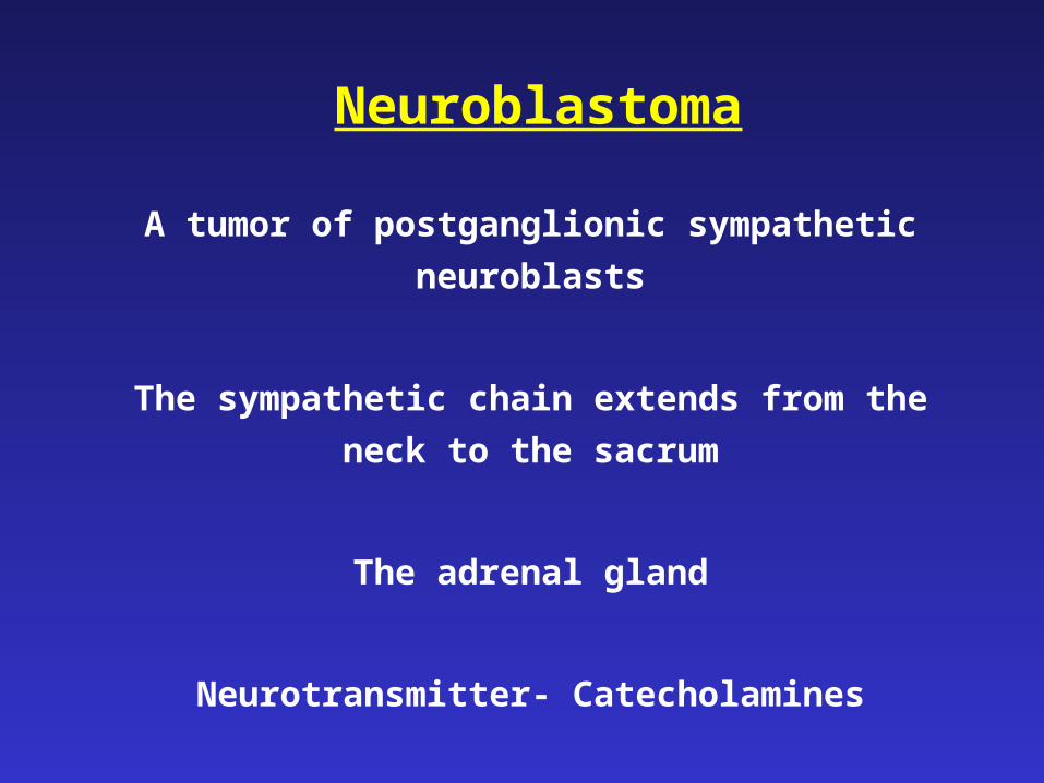
A tumor of postganglionic sympathetic neuroblasts
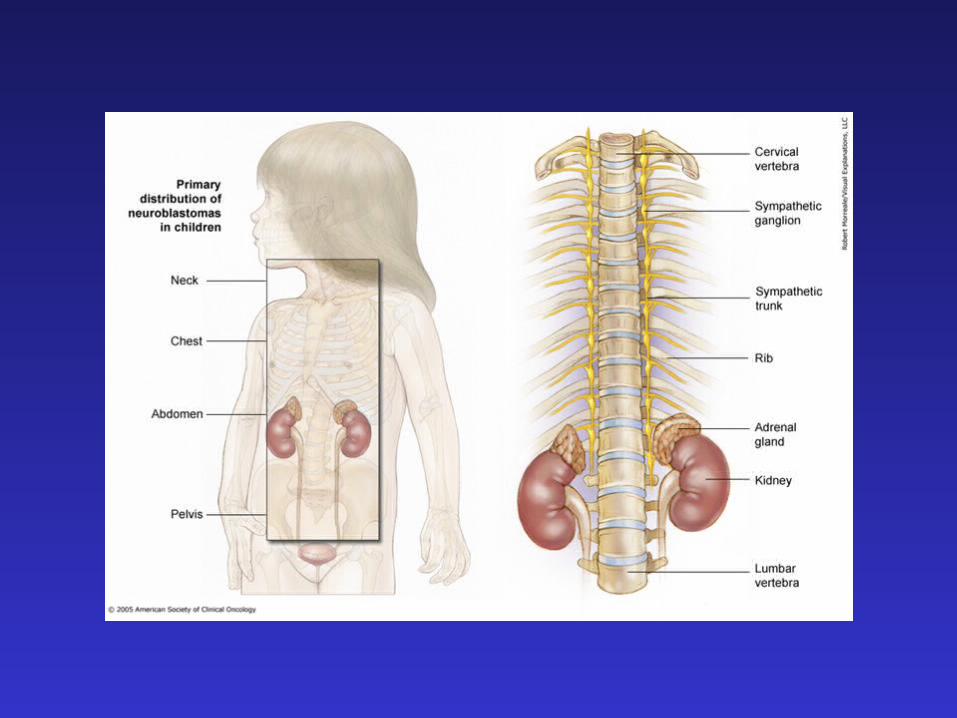
The sympathetic chain extends from the neck to the
sacrum
The adrenal gland
Neurotransmitter- Catecholamines
Neuroblastoma - Epidemiology
• Most common extracranial solid tumor
• Incidence – 10/million per year
• ~10% of pediatric tumors
• Peak age – 2-3 years
• >90% cases before 5 years
• Mostly sporadic – rare genetic associations
• Most common neonatal tumor
Neuroblastoma - Genetics
• Mostly sporadic – rare (2%) genetic associations (NF1, Hirschsprung, congenital central hypoventilation syndrome)
• Mutations in ALK (tyrosine kinase)
• Environment??
Neuroblastoma – Biology
• Neuroblastoma is a tumor of undifferentiated (embryonal) neuroectodermal cells, derived from the neural crest
• An aberration of normal differentiation
• An abnormal response to normal neurotropic signals (TRK-A, NGF)
• Spontaneous regression
• Differentiation – Ganglioneuroma, ganglioneuroblastoma
Neuroblastoma – Clinical features I
• Tumor originates from sympathetic ganglia/adrenal gland
• Lymphatic and hematogenous spread
• Metastases to bone, liver, bone marrow, skin
• Most patients present with advanced disease (40% of all patients, 55% of patients over 1 year)
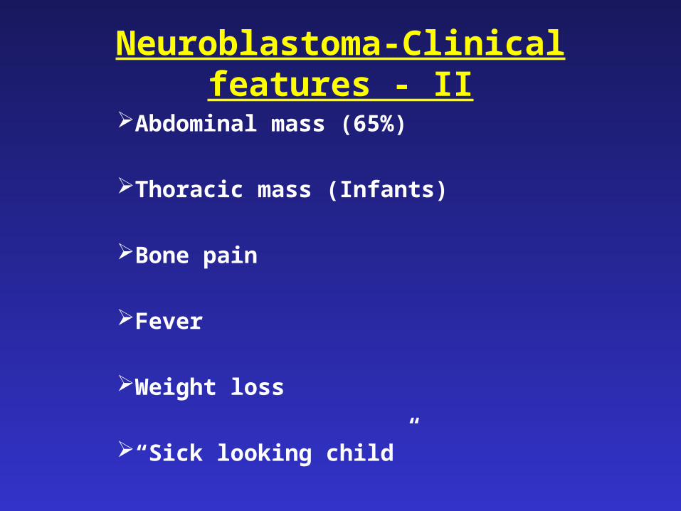
Neuroblastoma-Clinical features - II
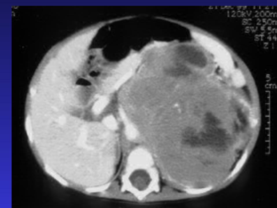
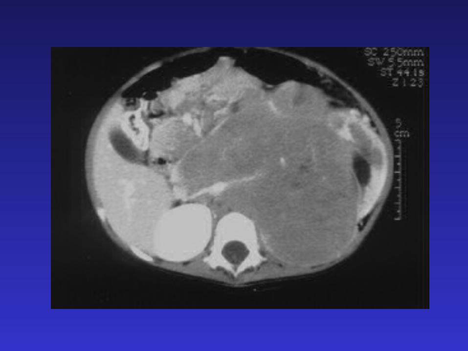
Abdominal mass (65%)
Thoracic mass (Infants)
Bone pain
Fever
Weight loss
“Sick looking child”
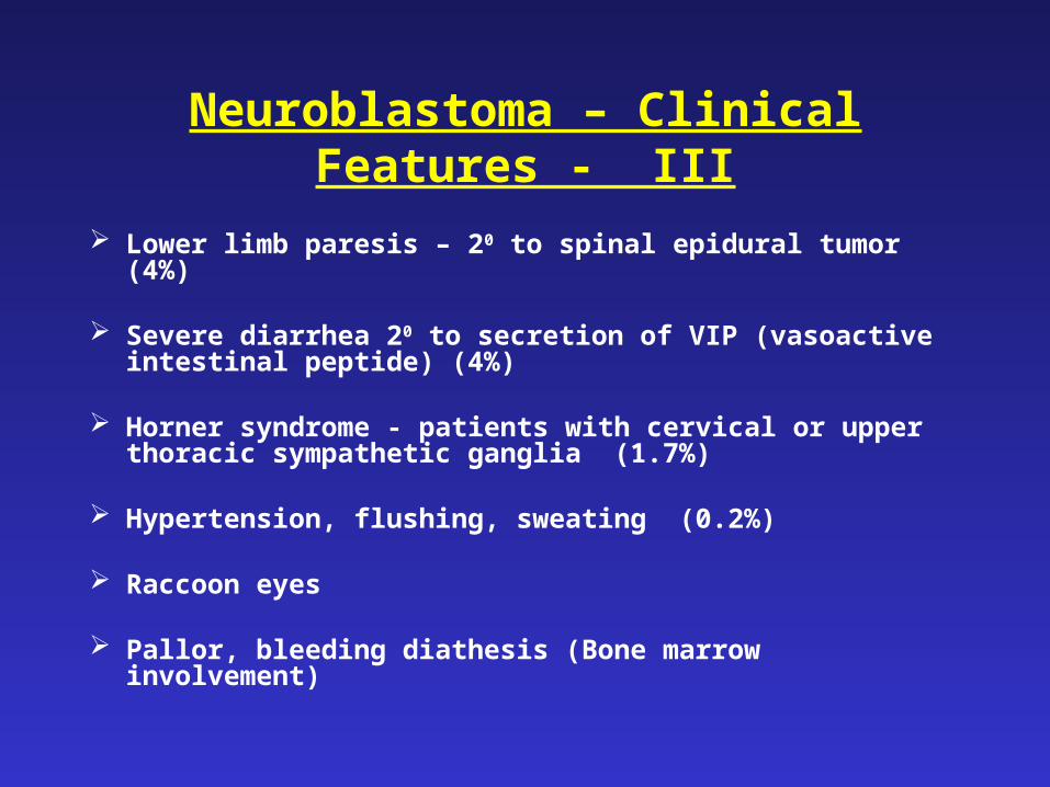
Neuroblastoma – Clinical Features - III
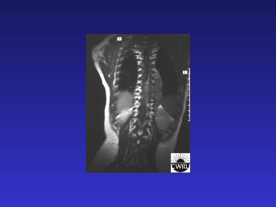
Lower limb paresis – 20 to spinal epidural tumor (4%)
Severe diarrhea 20 to secretion of VIP (vasoactive intestinal peptide) (4%)
Horner syndrome - patients with cervical or upper thoracic sympathetic ganglia (1.7%)
Hypertension, flushing, sweating (0.2%)
Raccoon eyes
Pallor, bleeding diathesis (Bone marrow involvement)
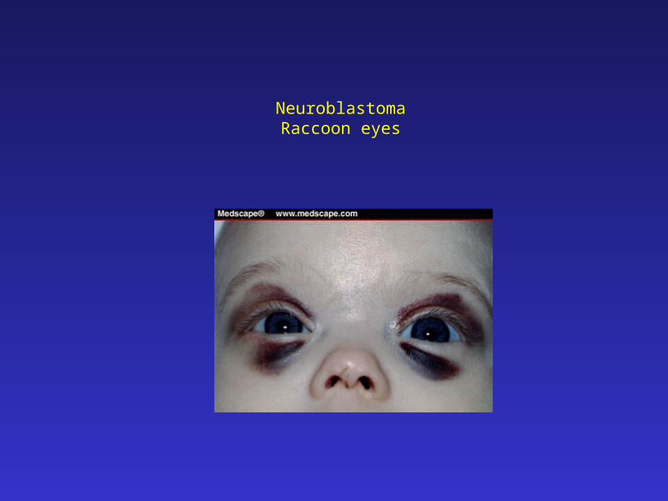
NeuroblastomaRaccoon eyes
Neuroblastoma – Clinical features - IV
Opsoclonus-Myoclonus syndrome
Acute cerebellar encephalopathy characterized by cerebellar ataxia, rapid and random eye movements (opsoclonus) and myoclonic jerks (2.8%)
Good oncological but poor neurological outcome
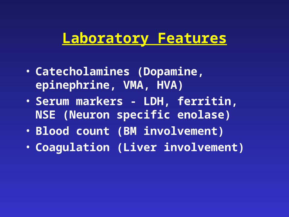
Laboratory Features
• Catecholamines (Dopamine, epinephrine, VMA, HVA)
• Serum markers - LDH, ferritin, NSE (Neuron specific enolase)
• Blood count (BM involvement)• Coagulation (Liver involvement)
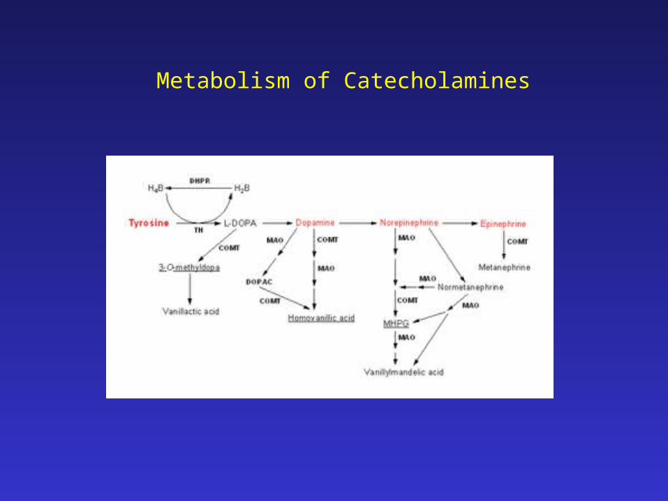
Metabolism of Catecholamines
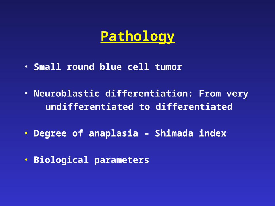
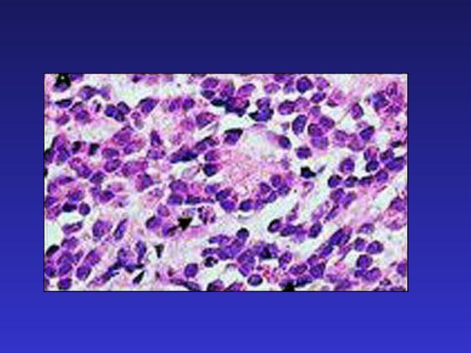
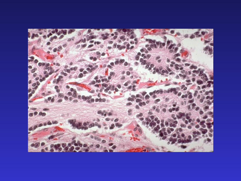
Pathology
• Small round blue cell tumor
• Neuroblastic differentiation: From very
undifferentiated to differentiated
• Degree of anaplasia – Shimada index
• Biological parameters
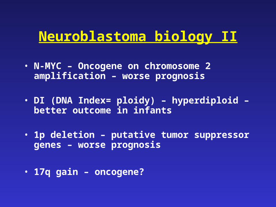
Neuroblastoma biology II
• N-MYC – Oncogene on chromosome 2 amplification – worse prognosis
• DI (DNA Index= ploidy) – hyperdiploid – better outcome in infants
• 1p deletion – putative tumor suppressor genes – worse prognosis
• 17q gain – oncogene?
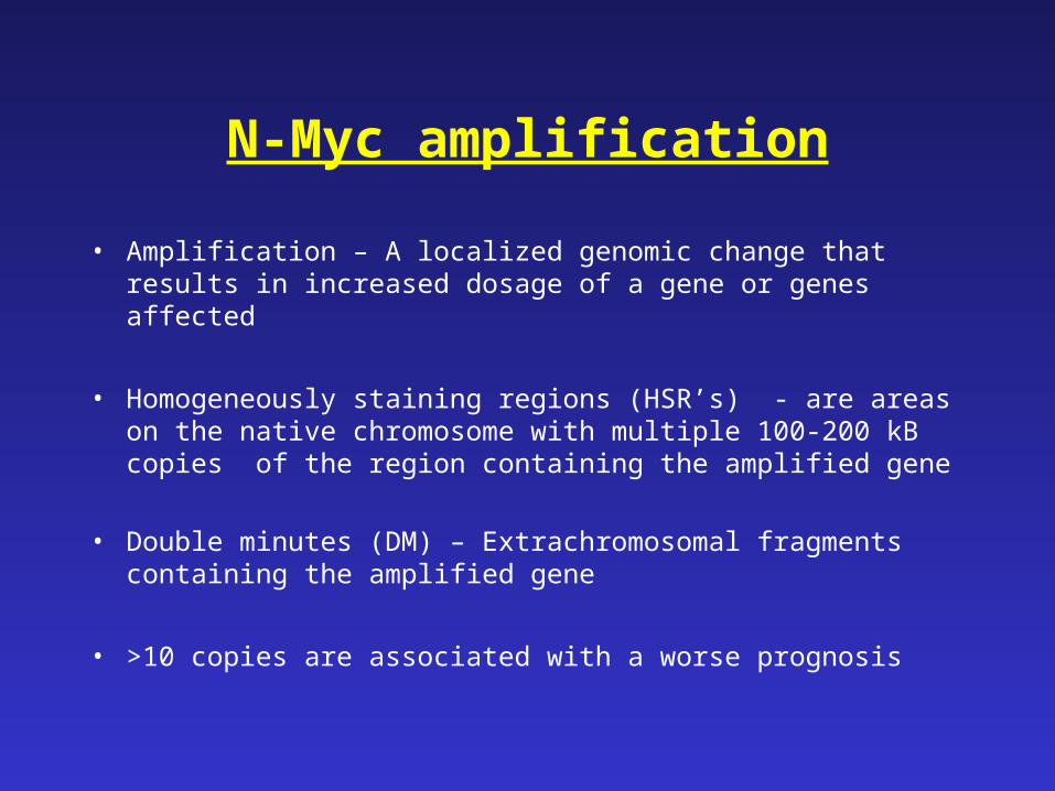
N-Myc amplification
• Amplification – A localized genomic change that results in increased dosage of a gene or genes affected
• Homogeneously staining regions (HSR’s) - are areas on the native chromosome with multiple 100-200 kB copies of the region containing the amplified gene
• Double minutes (DM) – Extrachromosomal fragments containing the amplified gene
• >10 copies are associated with a worse prognosis
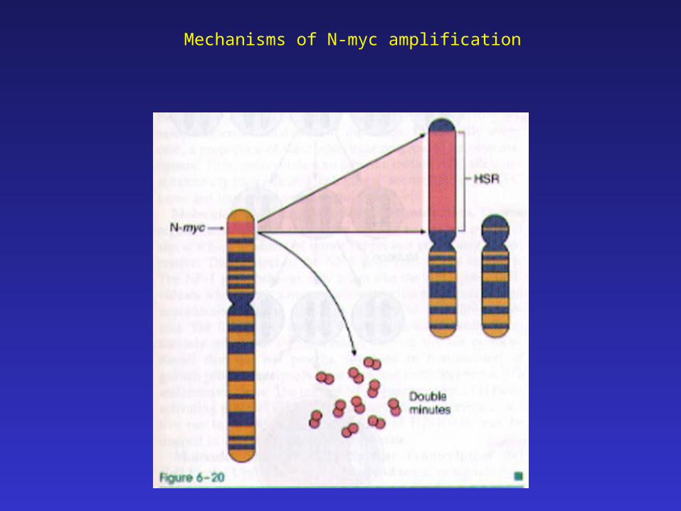
Mechanisms of N-myc amplification
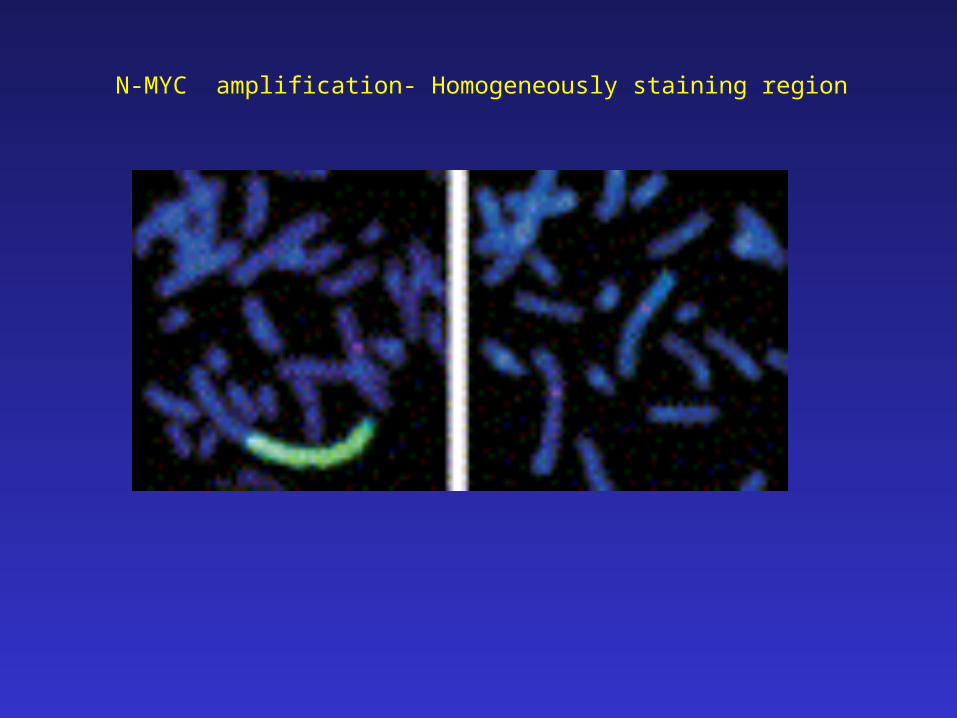
N-MYC amplification- Homogeneously staining region
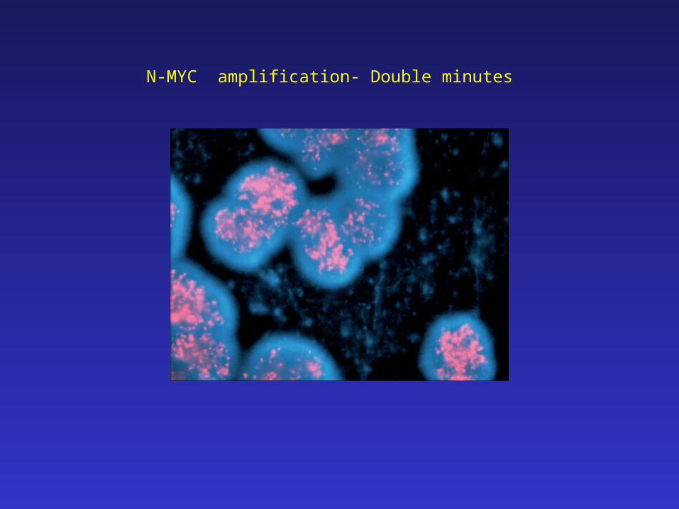
N-MYC amplification- Double minutes
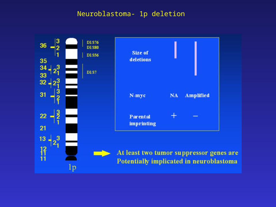
Neuroblastoma- 1p deletion
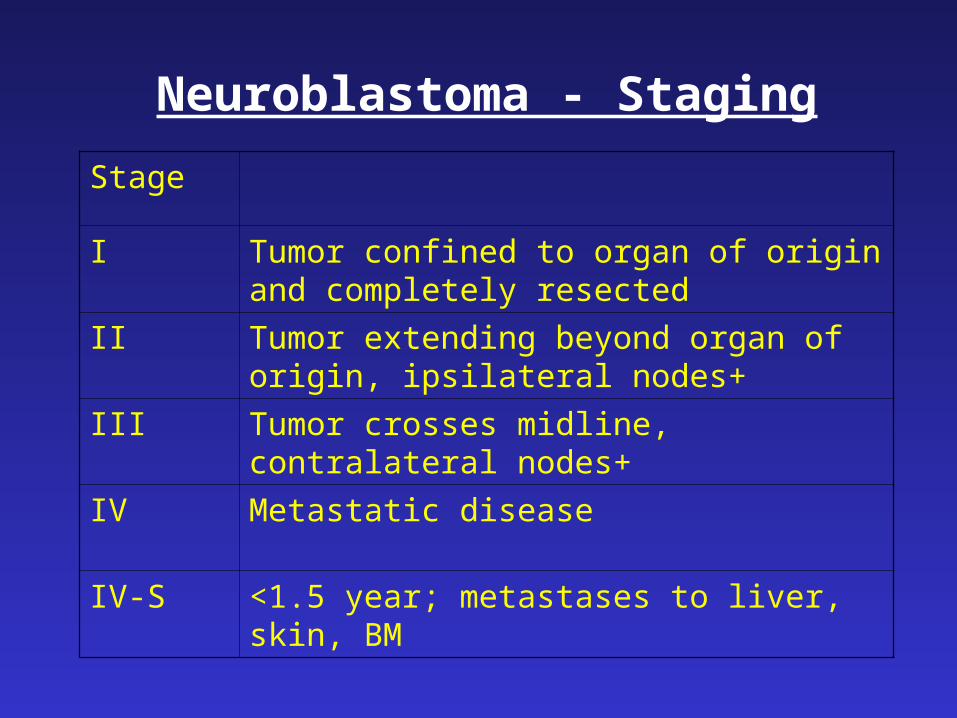
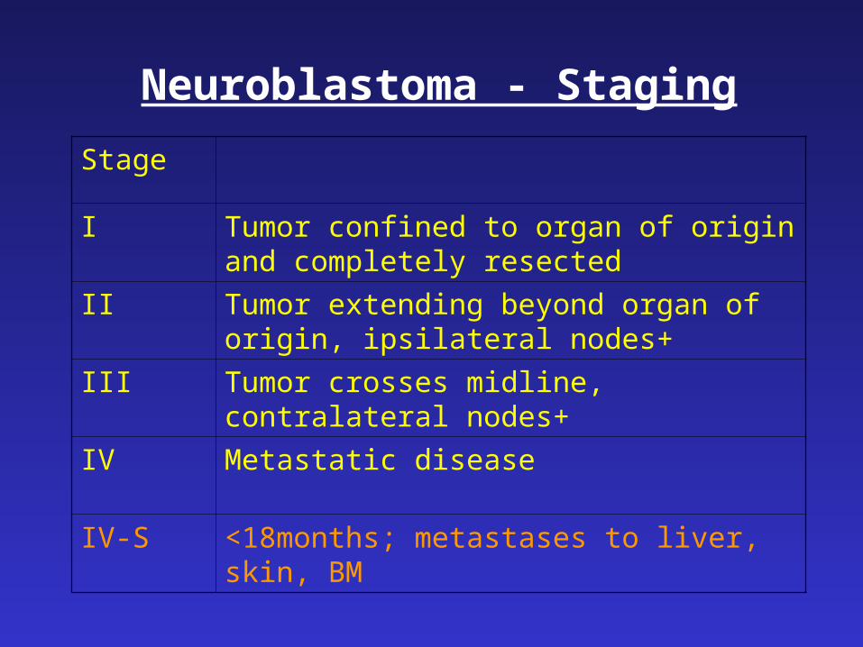
Neuroblastoma - Staging
• CT/MRI
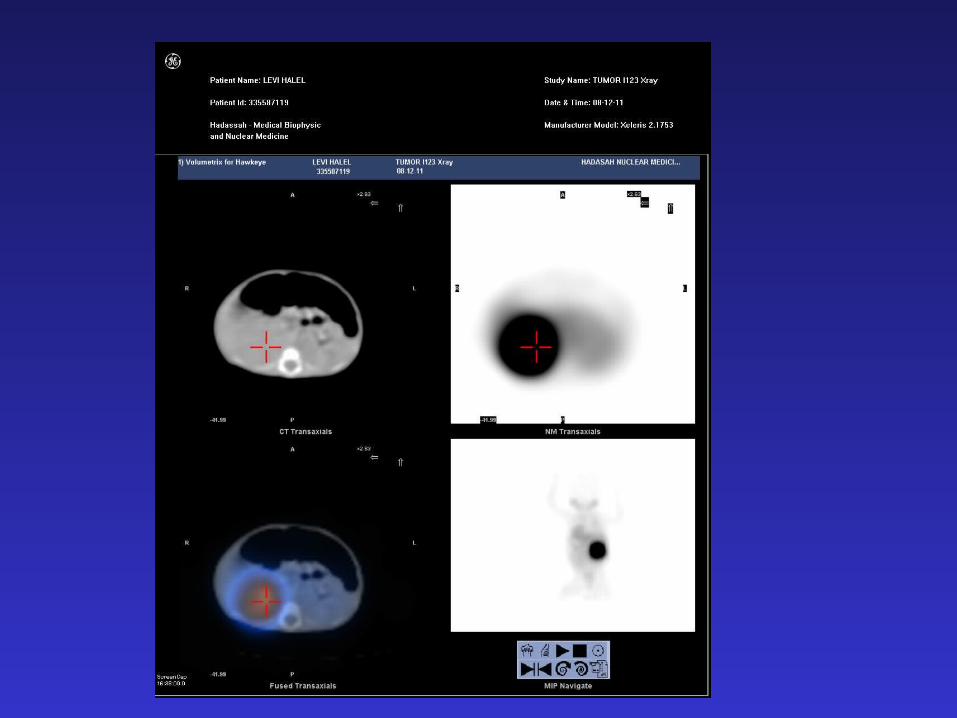
• MIBG (Meta-iodo-benzyl-guanidine)
• Bone scan
• Bone marrow aspirate and biopsy
• Surgery
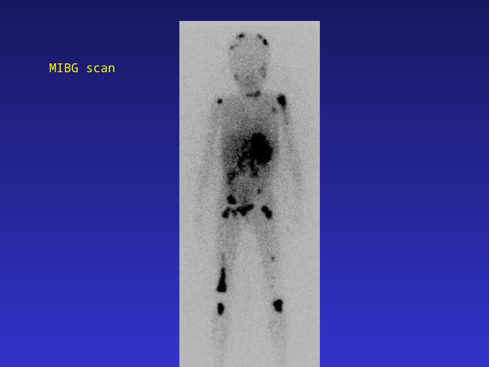
MIBG scan
Neuroblastoma - Staging
Stage
I Tumor confined to organ of origin and completely resected
II Tumor extending beyond organ of origin, ipsilateral nodes+
III Tumor crosses midline, contralateral nodes+
IV Metastatic disease
IV-S <1.5 year; metastases to liver, skin, BM
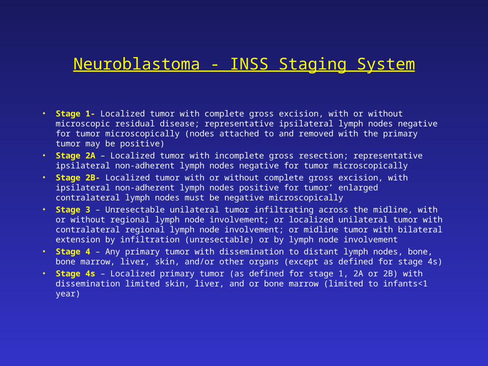
Neuroblastoma - INSS Staging System
• Stage 1- Localized tumor with complete gross excision, with or without microscopic residual disease; representative ipsilateral lymph nodes negative for tumor microscopically (nodes attached to and removed with the primary tumor may be positive)
• Stage 2A – Localized tumor with incomplete gross resection; representative ipsilateral non-adherent lymph nodes negative for tumor microscopically
• Stage 2B- Localized tumor with or without complete gross excision, with ipsilateral non-adherent lymph nodes positive for tumor’ enlarged contralateral lymph nodes must be negative microscopically
• Stage 3 – Unresectable unilateral tumor infiltrating across the midline, with or without regional lymph node involvement; or localized unilateral tumor with contralateral regional lymph node involvement; or midline tumor with bilateral extension by infiltration (unresectable) or by lymph node involvement
• Stage 4 – Any primary tumor with dissemination to distant lymph nodes, bone, bone marrow, liver, skin, and/or other organs (except as defined for stage 4s)
• Stage 4s – Localized primary tumor (as defined for stage 1, 2A or 2B) with dissemination limited skin, liver, and or bone marrow (limited to infants<1 year)

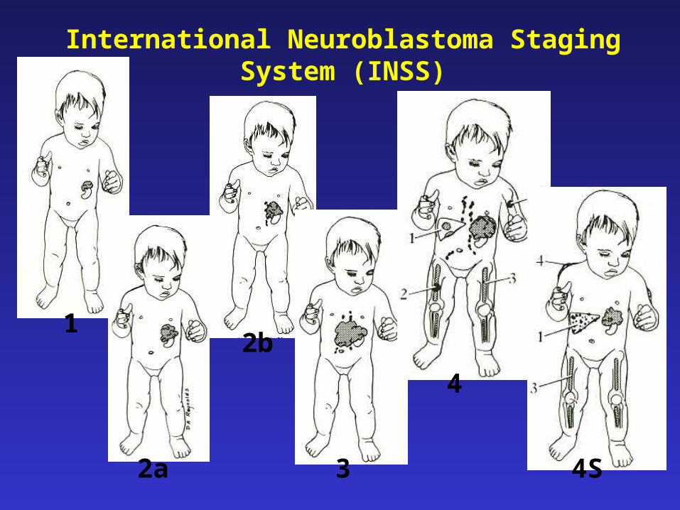
2b1
2a 3
International Neuroblastoma Staging System (INSS)
4
4S
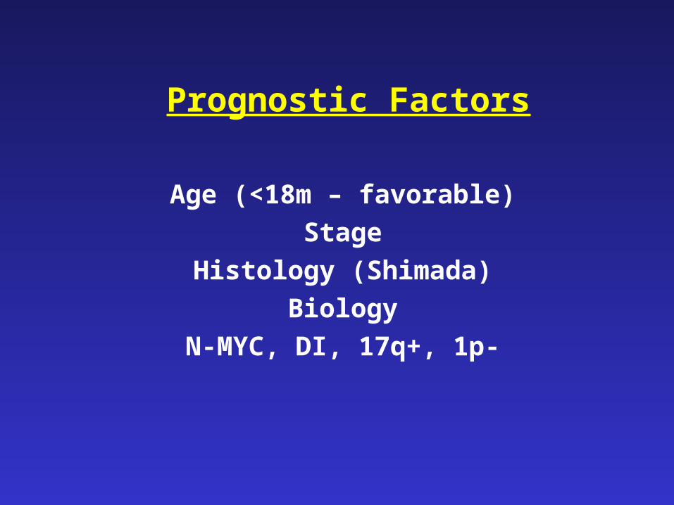
Prognostic Factors
Age (<18m – favorable)
Stage
Histology (Shimada)
Biology
N-MYC, DI, 17q+, 1p-
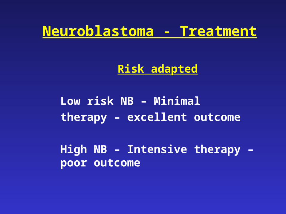
Neuroblastoma - Treatment
Risk adapted
Low risk NB – Minimal
therapy – excellent outcome
High NB – Intensive therapy – poor outcome
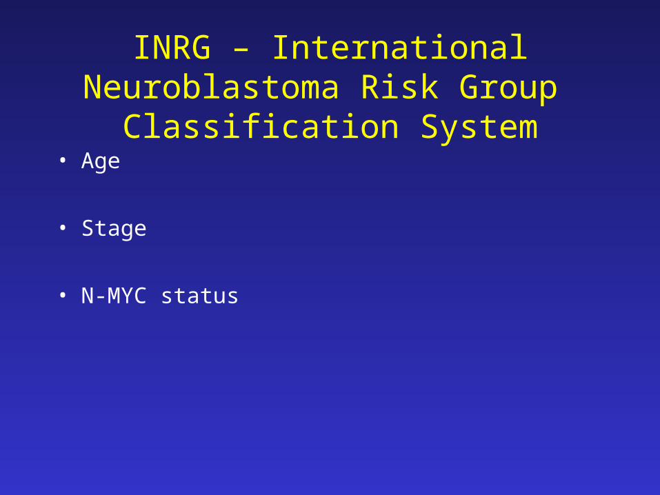
INRG – International Neuroblastoma Risk Group Classification System
• Age
• Stage
• N-MYC status
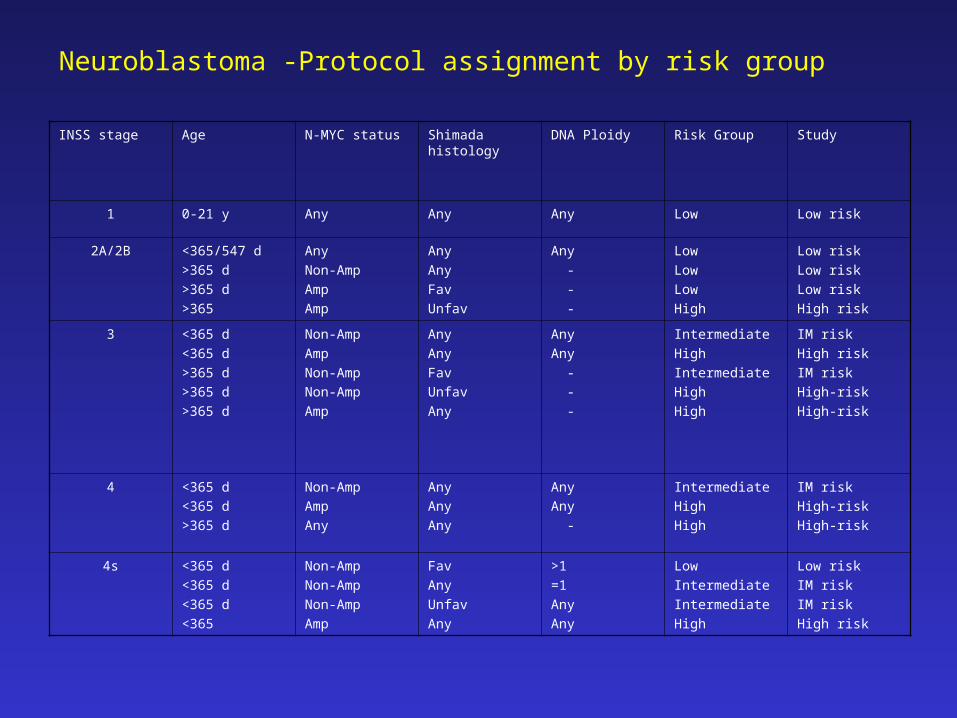
Neuroblastoma -Protocol assignment by risk group
INSS stage Age N-MYC status Shimada histology DNA Ploidy Risk Group Study
1 0-21 y Any Any Any Low Low risk
2A/2B <365/547 d
>365 d
>365 d
>365
Any
Non-Amp
Amp
Amp
Any
Any
Fav
Unfav
Any
-
-
-
Low
Low
Low
High
Low risk
Low risk
Low risk
High risk
3 <365 d
<365 d
>365 d
>365 d
>365 d
Non-Amp
Amp
Non-Amp
Non-Amp
Amp
Any
Any
Fav
Unfav
Any
Any
Any
-
-
-
Intermediate
High
Intermediate
High
High
IM risk
High risk
IM risk
High-risk
High-risk
4 <365 d
<365 d
>365 d
Non-Amp
Amp
Any
Any
Any
Any
Any
Any
-
Intermediate
High
High
IM risk
High-risk
High-risk
4s <365 d
<365 d
<365 d
<365
Non-Amp
Non-Amp
Non-Amp
Amp
Fav
Any
Unfav
Any
>1
=1
Any
Any
Low
Intermediate
Intermediate
High
Low risk
IM risk
IM risk
High risk
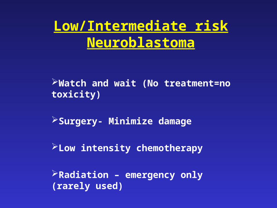
Low/Intermediate risk Neuroblastoma
Watch and wait (No treatment=no toxicity)
Surgery- Minimize damage
Low intensity chemotherapy
Radiation – emergency only (rarely used)
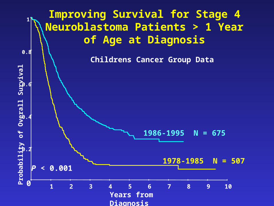
Improving Survival for Stage 4 Neuroblastoma Patients > 1 Year of Age
at Diagnosis
Childrens Cancer Group Data
P < 0.001
0
0.2
0.4
0.6
0.8
1
1 2 3 4 5 6 7 8 9 10
Years from Diagnosis
Pro
bab
ility
of
Ov
eral
l Su
rviv
al
1978-1985 N = 507
1986-1995 N = 675
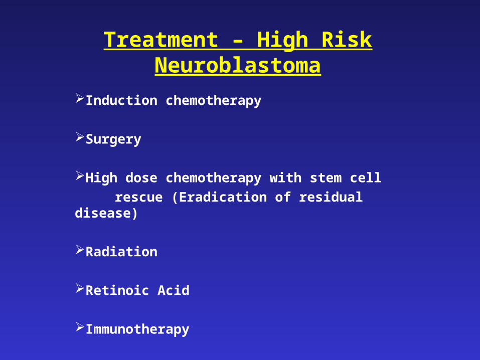
Treatment – High Risk Neuroblastoma
Induction chemotherapy
Surgery
High dose chemotherapy with stem cell
rescue (Eradication of residual disease)
Radiation
Retinoic Acid
Immunotherapy
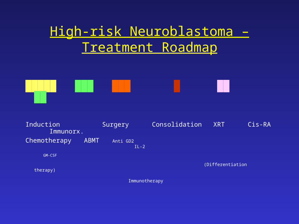
High-risk Neuroblastoma – Treatment Roadmap
█████ ███ ███ █ ██ ██
Induction Surgery Consolidation XRT Cis-RA Immunorx.
Chemotherapy ABMT Anti GD2
IL-2
GM-CSF
(Differentiation therapy)
Immunotherapy
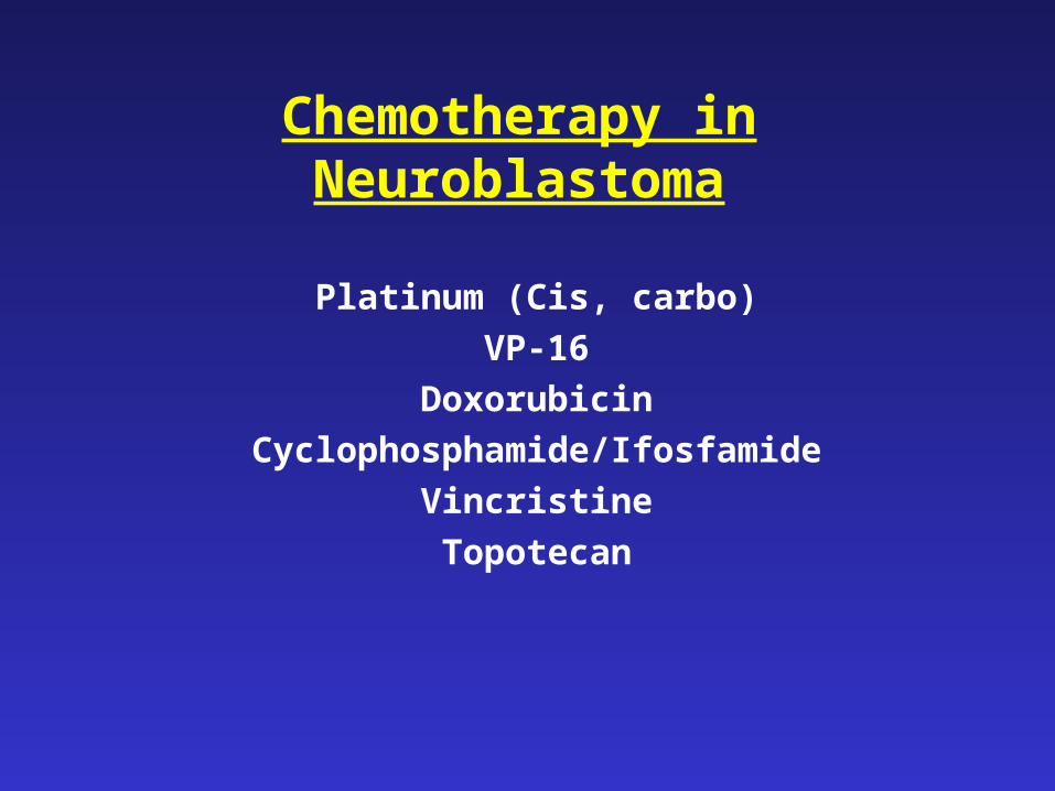
Chemotherapy in Neuroblastoma
Platinum (Cis, carbo)
VP-16
Doxorubicin
Cyclophosphamide/Ifosfamide
Vincristine
Topotecan
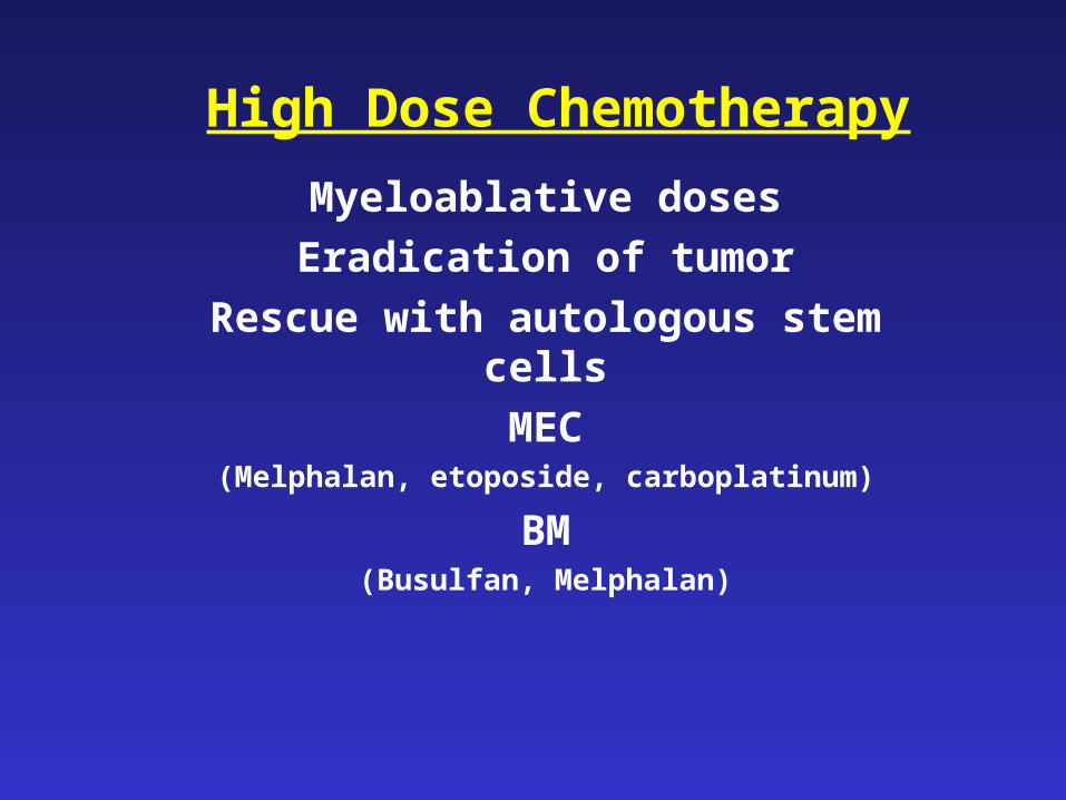
High Dose Chemotherapy
Myeloablative doses
Eradication of tumor
Rescue with autologous stem cells
MEC(Melphalan, etoposide, carboplatinum)
BM(Busulfan, Melphalan)
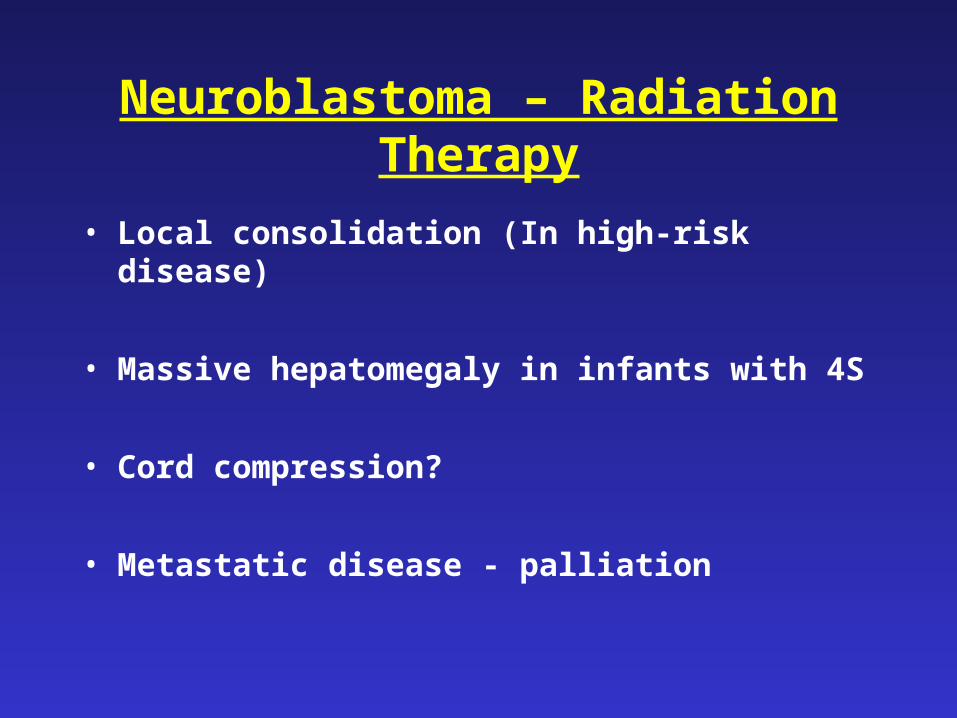
Neuroblastoma – Radiation Therapy
• Local consolidation (In high-risk disease)
• Massive hepatomegaly in infants with 4S
• Cord compression?
• Metastatic disease - palliation
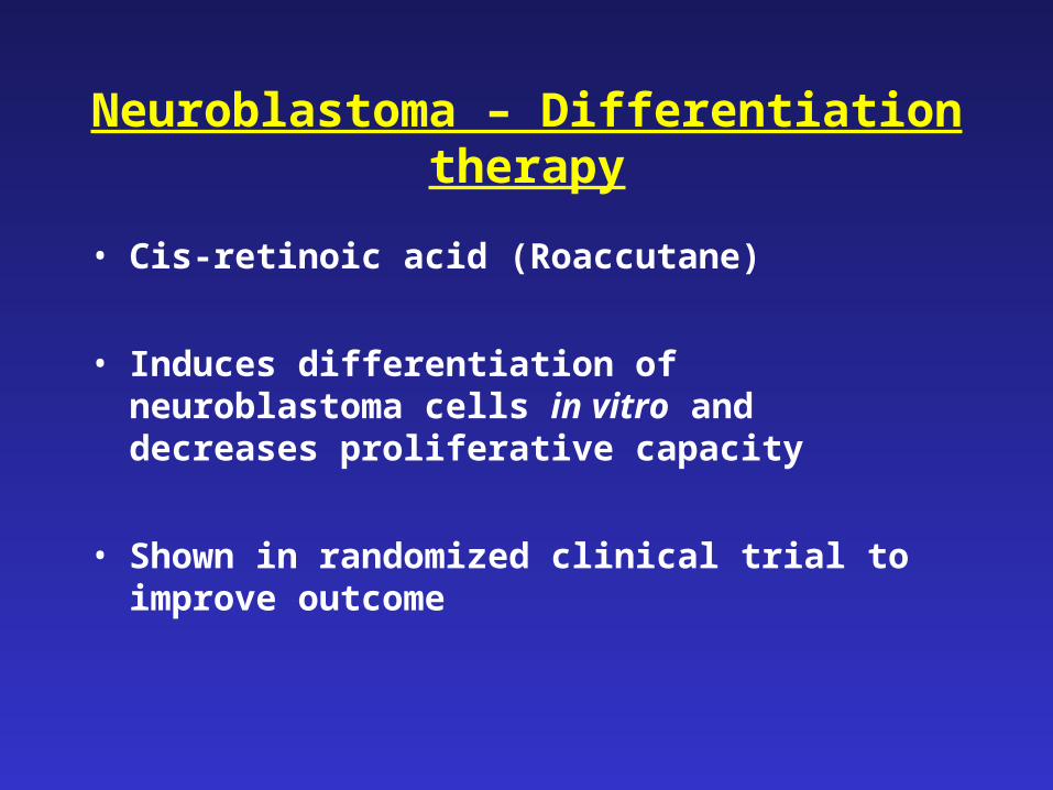
Neuroblastoma – Differentiation therapy
• Cis-retinoic acid (Roaccutane)
• Induces differentiation of neuroblastoma cells in vitro and decreases proliferative capacity
• Shown in randomized clinical trial to improve outcome
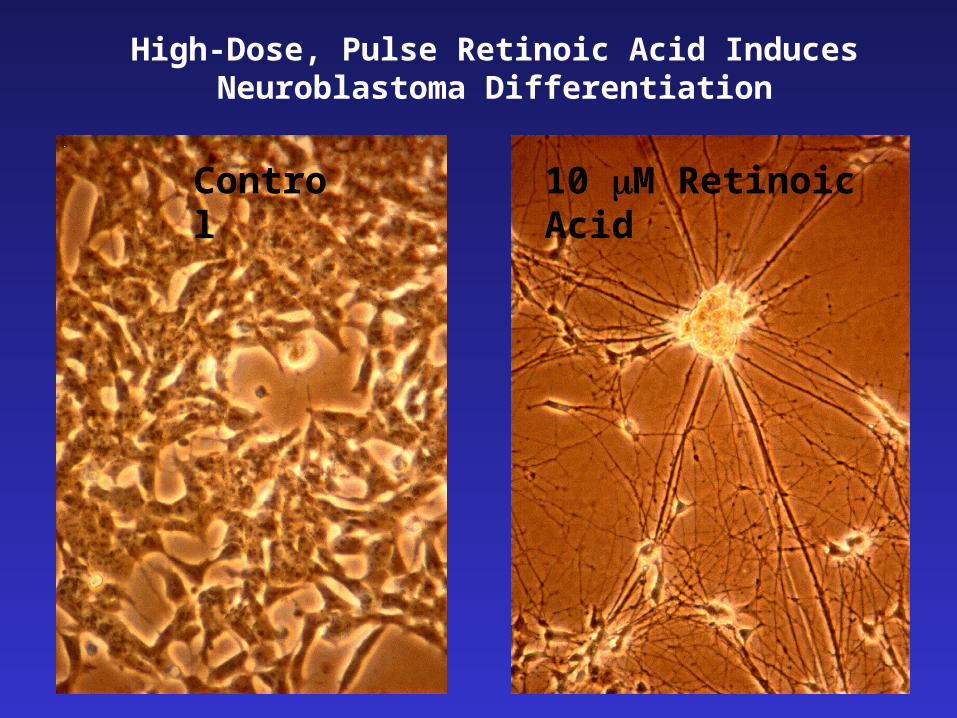
Control 10 M Retinoic Acid
High-Dose, Pulse Retinoic Acid Induces Neuroblastoma Differentiation

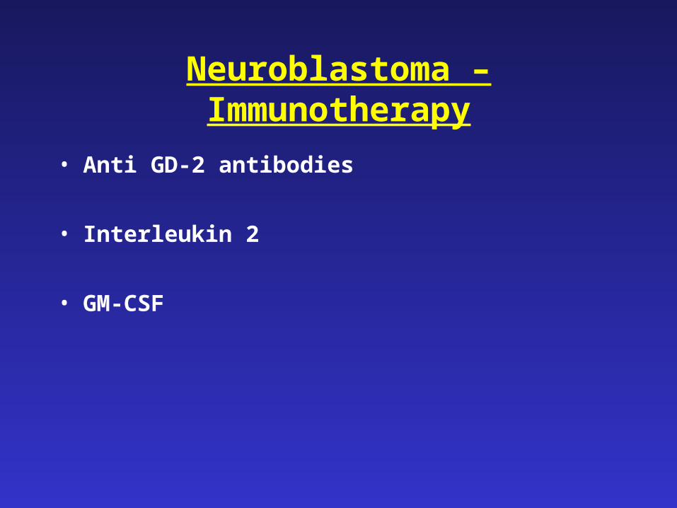
Neuroblastoma – Immunotherapy
• Anti GD-2 antibodies
• Interleukin 2
• GM-CSF
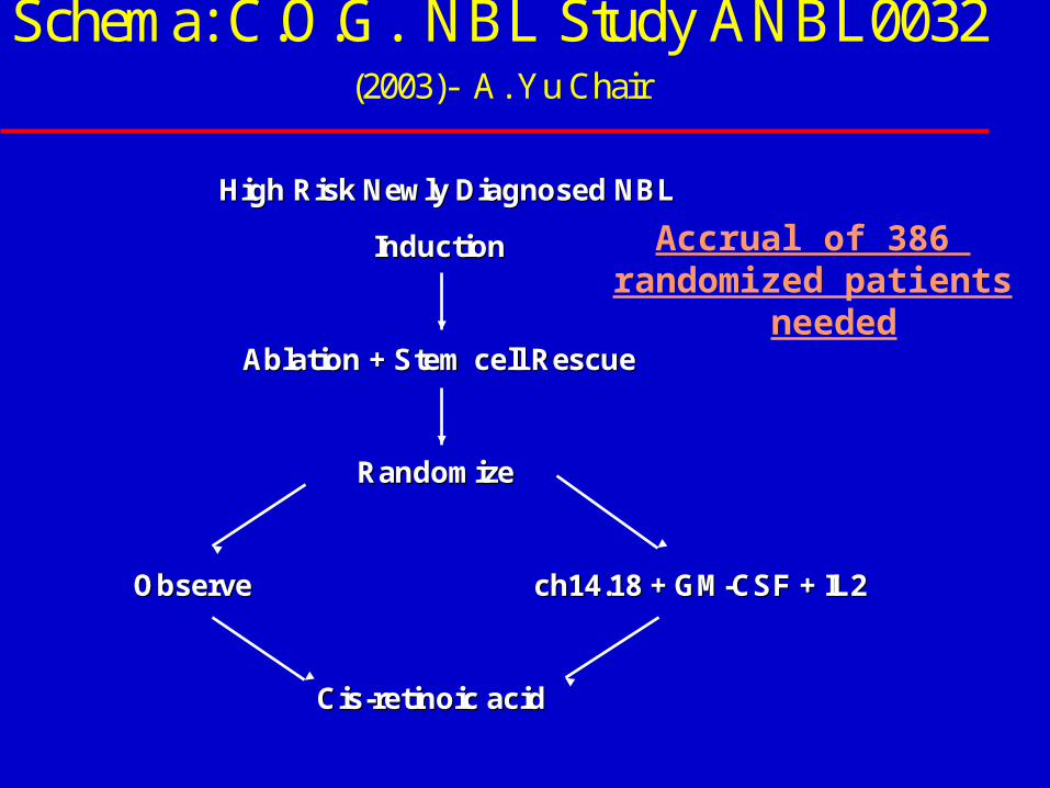
Schema: C.O.G. NBL Study ANBL0032 (2003)- A. Yu Chair
High Risk Newly Diagnosed NBLHigh Risk Newly Diagnosed NBL
InductionInduction
Ablation + Stem cell RescueAblation + Stem cell Rescue
RandomizeRandomize
ObserveObserve ch14.18 + GMch14.18 + GM--CSF + IL2CSF + IL2
CisCis--retinoic acidretinoic acid
Accrual of 386 randomized patients
needed
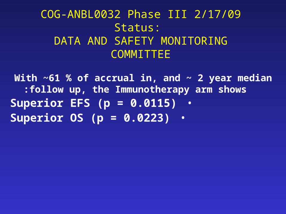
COG-ANBL0032 Phase III 2/17/09 Status: DATA AND SAFETY MONITORING
COMMITTEE
With ~61 % of accrual in, and ~ 2 year median follow up, the Immunotherapy arm shows:
•Superior EFS (p = 0.0115)•Superior OS (p = 0.0223)
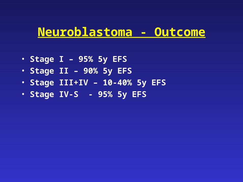
Neuroblastoma - Outcome
• Stage I – 95% 5y EFS
• Stage II – 90% 5y EFS
• Stage III+IV – 10-40% 5y EFS
• Stage IV-S - 95% 5y EFS
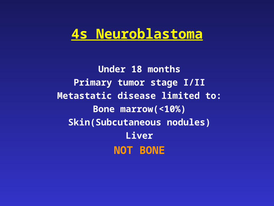
4s Neuroblastoma
Under 18 months
Primary tumor stage I/II
Metastatic disease limited to:
Bone marrow(<10%)
Skin(Subcutaneous nodules)
Liver
NOT BONE
Bone and Bone Marrow
Neuroblastoma - Staging
Stage
I Tumor confined to organ of origin and completely resected
II Tumor extending beyond organ of origin, ipsilateral nodes+
III Tumor crosses midline, contralateral nodes+
IV Metastatic disease
IV-S <18months; metastases to liver, skin, BM
4s Neuroblastoma
Spontaneous regression
Cure with minimal therapy
Exception: Massive hepatomegaly in <2 months
(May require chemotherapy, RT)
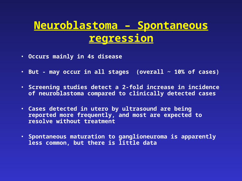
Neuroblastoma – Spontaneous regression
• Occurs mainly in 4s disease
• But - may occur in all stages (overall ~ 10% of cases)
• Screening studies detect a 2-fold increase in incidence of neuroblastoma compared to clinically detected cases
• Cases detected in utero by ultrasound are being reported more frequently, and most are expected to resolve without treatment
• Spontaneous maturation to ganglioneuroma is apparently less common, but there is little data
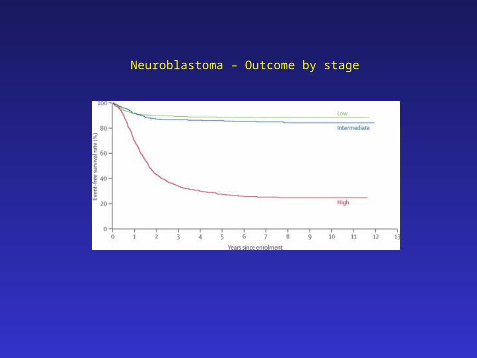
Neuroblastoma – Outcome by stage
0
0.1
0.2
0.3
0.4
0.5
0.6
0.7
0.8
0.9
1
0 1 2 3 4 5 6 7 8
Years from Diagnosis
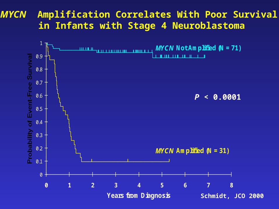
MYCN Not Amplified (N = 71)
MYCN Amplified (N = 31)
Schmidt, JCO 2000
P < 0.0001
MYCN Amplification Correlates With Poor Survival in Infants with Stage 4 Neuroblastoma
Potential Therapy
• Radioactive MIBG
• Anti-GD2 antibodies
• Allogeneic BMT
• Sequential auto-BMT x2-3
• Rapid sequence induction
• Early detection
• Targeted therapy - ALK inhibition
Neuroblastoma - Screening
Rationale:
Poor results in advanced-stage tumors
Early stage – better outcome
Available tumor marker (CA)
Method
Universal screening by urinary CA at 6 months
Neuroblastoma Screening - Results
• More early stage tumors
• No increase in detection of advanced tumors
• No improvement in overall cure
• Do advanced tumors evolve from early stage less aggressive tumors?
• Or are they rapidly evolving metastatic tumors that occur after 1 year of age?
Perinatal Neuroblastoma
• Tumors discovered during routine antenatal ultrasound as adrenal masses
• Differential diagnosis: Adrenal hemorrhage, pulmonary sequestration
• Traditional approach: Surgery
• Most tumors – stage 1, favorable histology, N-MYC non-amp
• Postnatal MIBG scan. If positive:
• Close observation; surgery for growing tumors
• Excellent outcome
Neuroblastoma
• A tumor of sympathetic neuroblasts that arises along
the sympathetic chain and in the adrenal gland
• Heterogeneous clinical behavior and prognosis
• Different subtypes present different challenges