Embed Size (px)
Citation preview
Neurobiology of Disease
Absence of Functional Peroxisomes from Mouse CNS CausesDysmyelination and Axon Degeneration
Leen Hulshagen,1 Olga Krysko,1 Astrid Bottelbergs,1 Steven Huyghe,1 Rudiger Klein,5 Paul P. Van Veldhoven,2
Peter P. De Deyn,6 Rudi D’Hooge,6 Dieter Hartmann,3,4,7 and Myriam Baes1
1Laboratory of Cell Metabolism, Department of Pharmaceutical Sciences, 2Laboratory for Lipid Biochemistry and Protein Interactions, Department ofMolecular Cell Biology, and 3Center for Human Genetics, Katholieke Universiteit Leuven, and 4Department of Molecular and Developmental Genetics,Flanders Institute for Biotechnology, B-3000 Leuven, Belgium, 5Max-Planck-Institut fur Neurobiologie, D-82152 Martinsried, Germany, 6Laboratory ofNeurochemistry and Behaviour, University of Antwerp, B-2610 Antwerp, Belgium, and 7Department of Anatomy, University of Bonn, D-53115 Bonn,Germany
Peroxisomal metabolism is essential for normal brain development both in men and in mice. Using conditional knock-out mice, werecently showed that peroxisome deficiency in liver has a severe and persistent impact on the formation of cortex and cerebellum,whereas absence of functional peroxisomes from the CNS only causes developmental delays without obvious alteration of brainarchitecture.
We now report that a substantial fraction of the latter Nes-Pex5 knock-out mice survive into adulthood but develop progressive motoricand coordination problems, impaired exploration, and a deficit in cognition and die before the age of 6 months. Histopathologically, boththe white and gray matter of the CNS displayed a region-specific accumulation of neutral lipids, astrogliosis and microgliosis, upregula-tion of catalase, and scattered cell death. Nes-Pex5 knock-out mice featured a dramatic reduction of myelin staining in corpus callosum,whereas cerebellum and other white matter tracts were less affected or unchanged. This was accompanied by a depletion of alkenylphos-pholipids in myelin and differentially reduced immunoreactivity of myelin proteins. EM analysis revealed that myelin wrappings aroundaxons did still form, but they showed a reduction in thickness relative to axon diameters. Remarkably, multifocal axonal damage occurredin the corpus callosum. Thereby, debris accumulated between axolemma and inner myelin surface and axons collapsed, although myelinsheaths remained present. These anomalies of myelinated axons were already present in juvenile mice but aggravated in adulthood.Together, loss of CNS peroxisomal metabolism both affects myelin sheaths and axonal integrity possibly via independent pathways.
Key words: peroxisome; myelin; Zellweger syndrome; plasmalogen; fatty acid; knock-out mice; astrocyte; axon; microglia
IntroductionPeroxisomes fulfill multiple tasks in metabolism and adapt con-tents and functions according to cell type, age, and organism.Among the metabolic reactions that take place in peroxisomes,oxygen metabolism, �-oxidation of a number of carboxylatesthat cannot be handled by mitochondria, �-oxidation of3-methyl branched chain and 2-hydroxy fatty acids, ether lipidsynthesis, and detoxification of glyoxylate are the most important(Mannaerts and Van Veldhoven, 1993; Wanders and Waterham,2006).
Loss or impairment of peroxisomal function, as seen in per-oxisome biogenesis disorders, or mutations of individual perox-isomal enzymes or transporter proteins result in characteristicpatterns of CNS lesions (Powers and Moser, 1998).
First, the most severe peroxisome biogenesis disorder, Zell-weger syndrome, and some single peroxisomal �-oxidation en-zyme deficiencies cause cytoarchitectonic abnormalities includ-ing cortical neuronal migration defects, malformations of thecerebellum and of the inferior medullary olives (Evrard et al.,1978; Gould et al., 2001). These defects were recapitulated inZellweger mouse models (Baes and Van Veldhoven, 2006). Sec-ond, disturbed function of peroxisomes is associated with whitematter anomalies that can be both demyelinative and dysmyeli-native in nature (Powers and Moser, 1998). Finally, peroxisomaldisorders are linked with neuronal degenerations, including de-generation and loss of Purkinje and granule cells (Powers et al.,1999; Bams-Mengerink et al., 2006), lesions in the spinal cord(Kumar et al., 1995; Powers et al., 2000), and loss of sensoryneurons (Wanders et al., 2001). Although in some cases the neu-ropathologic alterations are associated with specific metabolicperturbations, no causal links have been experimentally proven
Received June 19, 2007; revised Feb. 6, 2008; accepted Feb. 18, 2008.This work was supported by grants from Fonds Wetenschappelijk Onderzoek Vlaanderen (G.0235.01 and
G.0385.05), Geconcerteerde Onderzoeksacties (2004/08), the European Union (“MMPD,” QLG1-CT2001-01277, FP5;“Peroxisomes,” LSHG-CT-2004-512018, FP6), and the European Leukodystrophy Association (2007-00414). Theexcellent technical help of Benno Das, Lies Pauwels, Els Meyhi, Sven Terclavers, Nico Smets, Birgit Rau, Barbel Balzer,Ulrike Schwaab, Elke Maes, An Snellinx, Koen Poesen, and An Verheyen is gratefully acknowledged.
Correspondence should be addressed to Prof. Dr. Myriam Baes, Laboratory of Cell Metabolism, Department ofPharmaceutical Sciences, Herestraat 49 O/N Box 823, B-3000 Leuven, Belgium. E-mail: [email protected].
R. D’Hooge’s present address: Laboratory of Biological Psychology, Department of Psychology, Katholieke Uni-versiteit Leuven, B-3000 Leuven, Belgium.
DOI:10.1523/JNEUROSCI.4968-07.2008Copyright © 2008 Society for Neuroscience 0270-6474/08/284015-13$15.00/0
The Journal of Neuroscience, April 9, 2008 • 28(15):4015– 4027 • 4015

and the pathomechanisms of the neurological abnormalities re-main unresolved.
Analysis of peroxisome function in brain development andmaintenance is further complicated by our still poor knowledgeof peroxisome distribution by brain region, cell type(s), and de-velopmental kinetics. Initial electron microscopical studies (Ar-nold and Holtzman, 1978; Holtzman, 1982) using cytochemicalvisualization of catalase (and thus perhaps not revealing all per-oxisomes) reported microperoxisomes for all cell types, with apreponderance for glial cells and early postnatal stages, but theirabsence from several adult brain regions. Peroxisomes wereabundant in oligodendrocytes during myelination (Adamo et al.,1986). More recent immunohistochemical data indicated con-stant brain expression of peroxisomal proteins from the juvenileperiod into adulthood (Adamo et al., 1986; Imamura et al., 1994;Itoh et al., 1999, 2000; Huyghe et al., 2001; Ahlemeyer et al.,2007).
Recent data from our own group indicate that the role ofperoxisomes for CNS development may not only depend on theirpresence and dynamics in neurons and glial cells. Indeed, weshowed that elimination of functional peroxisomes from fetalliver, by �-fetoprotein promoter driven Cre recombination of thePex5 gene, had a dramatic and permanent impact on the forma-tion of cortex and cerebellum. In contrast, local ablation of Pex5from the CNS using Nestin-Cre mice, only caused delays in neu-ronal positioning during the prenatal and early postnatal period(Krysko et al., 2007). Because a substantial fraction of the Nes-Pex5 knock-out mice survived into early adulthood, we have fur-ther exploited this model to investigate the long-term effects of acomplete deficiency of functional peroxisomes within the CNS.
Materials and MethodsAnimalsNestin-Cre (Nes-Cre) mice (Tronche et al., 1999) were mated withPex5FL/� mice (Baes et al., 2002), and selected Nes-Cre Pex5FL/� offspringwere crossed with Pex5FL/FL mice to obtain Nes-Cre Pex5FL/FL, furtherdenoted as Nes-Pex5 knock-out mice (Krysko et al., 2007).
Twenty-five percent of the pups born from Nes-Cre Pex5FL/� andPex5FL/FL parents were Cre-positive and homozygous for the floxed Pex5allele as expected for Mendelian inheritance of the alleles. Littermateswith one of the other possible genotypes were considered as control mice.Mice were bred under conventional conditions, a 12 h light/dark cyclewith standard rodent food chow and water ad libitum. All animal exper-iments were approved by the Institutional Animal Ethical Committee ofthe University of Leuven.
Genotypes were determined on tail DNA via PCR analysis using theCre primers (5�-GCCTGCATTACCGGTCGATGCAACGA and 5�-GTGGCAGATGGCGCGGCAACACCATT) and two Pex5 primers en-compassing a DNA fragment containing the 3�-loxP site (5�-CTCT-GGTTCCCATTGGCCAGGGTGG and 5�-GGGGAGTACGACA-AGGCTGTGGACTG), to distinguish a shorter wild-type band and alonger loxP-containing band.
Biochemical analysisNes-Pex5 knock-out and control mice were anesthetized with an over-dose of Nembutal (150 �g/g body weight). Brains were removed, andcerebella and cerebra were separated, sagitally cut, snap frozen in liquidnitrogen, and stored at �80°C.
RNA isolation and quantitative PCR. RNA was isolated from cerebel-lum using the Trizol reagent (Invitrogen, Merelbeke, Belgium) and re-verse transcribed with Superscript II Reverse Transcriptase (Invitrogen).Quantitative PCRs were then performed by using an ABI Prism 7700(Gene-Amp PCR system 9600; Applied Biosystems, Foster City, CA) andthe TaqMan system.
Primers and probes for calbindinD-28K [forward primer, 5�-AACTG-ACAGAGATGGCCAGGTTA (603– 625); probe, 5�-ACCAGTG-
CAGGAAAATTTCCTTCTTAAATTCCA (628 – 660); and reverseprimer, 5�-TGAACTCTTTCCCACACATTTTGAT (665– 689)] andHPRT [forward primer, 5�-TTATCAGACTGAAGAGCTACTGTAA-TGATC (383– 412); probe, 5�-TGAGAGATCATCTCCACCAATA-ACTTTTATGTCCC (422– 456); and reverse primer 5�-TTACCAG-TGTCAATTATATCTTCAACAATC (480 –509)], and catalase [forwardprimer, 5�-CGCAGAGACCTGATGTCCTGA (145–164); probe, 5�-CACCGGAGGCGGGAACCCAATA (166 –185); and reverse primer, 5�-CCCCGCGGTCATGAATATTAA (205–223)] were designed by usingthe program Primer Express (Applied Biosystems) and were purchasedfrom GE Healthcare (Diegem, Belgium), Sigma-Aldrich (St. Louis, MO),and Eurogentec (Seraing, Belgium). The relative expression level of thetarget gene calbindinD-28K and catalase was calculated as a ratio to thehousekeeping gene HPRT.
Western blot analysis. After homogenizing cerebrum, cerebellum, ormicrodissected corpus callosum in 0.25 M sucrose, 5 mM MOPS, and 1mM EDTA, 30 �g of proteins were separated by 10% SDS-PAGE [12% formyelin basic protein (MBP)]. The monoclonal antibodies glial fibrillaryacidic protein (GFAP), proteolipid protein (PLP) (Millipore BioscienceResearch Reagents, Temecula, CA), and the polyclonal rabbit antibodiesto MBP (Millipore Bioscience Research Reagents) and catalase (Rock-land Immunochemicals, Gilbertsville, PA) were used. The monoclonalantibody myelin-associated glycoprotein (MAG) (Millipore BioscienceResearch Reagents) was used under nonreducing conditions.
Lipid analysis. Total lipid extracts were processed for fatty acid analysisas previously described (Baes et al., 1997). For plasmalogen analysis,aliquots of dried lipid extracts were dissolved in 200 �l of 10 mM dinitro-phenylhydrazine (in ethanol/1N H2SO4, 1/1, v/v) and placed at 50°C for30 min. After adding water, hydrazones were extracted into hexane, driedshielded from light, and dissolved in 100 �l of acetonitrile of which 50 �lwas injected on a C18-Symmetry column (5 �m; 100 Å; 4.6 � 150 mm;Waters, Milford, MA) run in acetonitrile at 0.8 ml/min and monitored at360 nm as described previously (Foulon et al., 2005). For calculation, theareas corresponding to the eluted nitrophenylhydrazone derivatives ofhexadecanal, octadecanal, and octadecenal were summed up and com-pared with an external standard (gravimetric solution of hexadecanaldimethyl acetal). Triglycerides, cholesterol, and cholesterylesters wereisolated by thin-layer chromatography and analyzed with coupled enzy-matic assays as described previously (Van Veldhoven et al., 1997, 1998;Huyghe et al., 2006a) except for cholesterylesters and triglycerides thatwere hydrolyzed chemically (5% 5 M KOH in ethanol; 75°C; 90 min).Statistical analysis was done by unpaired Student’s t test.
Isolation of myelinMyelin was isolated according to Kwik-Uribe et al. (2000), and the lipidcontent was further analyzed as described for cerebrum.
Behavioral studiesBehavioral performance was assessed repeatedly in 16 Nes-Pex5 knock-out and 16 control mice between 7 and 19 weeks of age, as describedpreviously (D’Hooge et al., 2001; Caeyenberghs et al., 2006; Goddyn etal., 2006). Neuromotor tests included rotarod, wire suspension, and cageactivity. The rotarod test comprised a 2 min adaptation trial (4 rpm) andfour test trials on an accelerating rotarod apparatus (4 – 40 rpm in 5 min;Ugo Basile, Comerio, Italy). In the wire suspension test, mice were put ona horizontal wire (0.6 mm thick) that was suspended 46 cm above tabletop. Mice were held in front of the wire and allowed to grab it with theirfront paws while being slightly held by their tail (120 s trial). Cage activitywas recorded in single-housed mice during 23 h using infrared photocellsand a laboratory-built beam interruption counter. Exploration tests in-cluded open-field and dark/light transition recordings. Open-field ex-ploration was assessed in a 50 � 50 cm 2 brightly lit arena during the darkphase of their activity cycle. After 1 min adaptation, exploratory activitywas video tracked for 10 min (Chromotrack; San Diego Instruments, SanDiego, CA). The dark/light transition box consisted of a small dark and(4.5 times) larger illuminated chamber. Mice were placed in the darkside, and the number of transitions between the compartments was reg-istered for 10 min with infrared photocells. Passive avoidance learningwas measured in a step-through box. The avoidance learning procedure
4016 • J. Neurosci., April 9, 2008 • 28(15):4015– 4027 Hulshagen et al. • Peroxisome Elimination from CNS
does not allow repeated testing and was therefore only performed at 11weeks of age. During the dark phase of their cycle, mice were put in theilluminated compartment, a door leading to the dark compartment wasopened after 5 s, and the mice received a brief footshock (0.3 mA; 1 s) onentry. Twenty-four hours later followed a test trial that allowed reassess-ment of step-through latency (up to 300 s).
Measurement of compound motor action potentialsFor electrophysiological recordings, 5-month-old animals were anesthe-tized with Nembutal (70 �g/g body weight). The stimulating electrodeswere inserted at the hip near the sciatic nerve (active electrode) and inbase of the tail (reference electrode). The detection electrodes were in-serted in the gastrocnemius muscle (active electrode), under the skin ofvertebral column (grounding), and in the Achilles tendon (referenceelectrode). After stimulation of the sciatic nerve, the velocity of nerveconduction to the gastrocnemius muscle was analyzed. This was done bymeasuring the amplitude (in microvolts) and latency (in milliseconds) ofthe recorded patterns of motor unit action potentials.
Histological analysisThree to five Nes-Pex5 knock-out and control mice at an age of 3, 6, 12,and 20 weeks were anesthetized with an overdose of Nembutal (150 �g/gbody weight) and intracardially perfused with 10% neutral buffered for-malin (Prosan, Merelbeke, Belgium).
The brain and vertebral column were removed from the animal, post-fixed overnight in the same fixative, and embedded in paraffin.
Immunohistochemical analysis. Five-micrometer-thick sections wereprocessed as previously described (Huyghe et al., 2006b) except that,depending on the antibody, trypsinization or heating of tissues in citratebuffer was applied for antigen retrieval. The following primary antibod-ies were used: monoclonal mouse antibodies against GFAP (Sigma-Aldrich), PLP (Millipore Bioscience Research Reagents), nonphospho-rylated neurofilament SMI32 (Sternberger, Baltimore, MD), amyloidprecursor protein (APP) (Millipore Bioscience Research Reagents),cleaved caspase-3 (Cell Signaling Technology, Danvers, MA), CC1 (Cal-biochem, San Diego, CA), CD45 (BD Pharmingen, San Diego, CA),2�,3�-cyclic nucleotide 3�-phosphodiesterase (CNP) (Sternberger);monoclonal rat antibody against F4/80 (Serotec, Oxford, UK), and poly-clonal rabbit antibodies against CD3 (BD Pharmingen), MBP, and cata-lase already described for Western blot analysis. The secondary antibod-ies were conjugated with peroxidase (horse anti-mouse IgGs; VectorLaboratories, Burlingame, CA; goat anti-rabbit IgGs, Dako, High Wy-combe, UK) or biotin (rabbit anti-rat IgGs; Vector Laboratories). Tyra-mide conjugated fluorochromes were used for detection with the tyra-mide signal amplification kits (PerkinElmer Life and Analytical Sciences,Waltham, MA). Sections were coverslipped with Vectashield (VectorLaboratories) and images were obtained by fluorescence microscopy[Carl Zeiss (Jena, Germany) Axioplan 2 Imaging equipped with a CarlZeiss AxioCam HRc camera and a Nikon (Tokyo, Japan) 90i microscopewith a Nikon DM 1200 camera].
When double stainings were performed, antibodies were added con-secutively and the cyanine 3 amplification kit TSA (PerkinElmer Life andAnalytical Sciences) was used for amplification.
For determining the density of oligodendrocytes in the corpus callo-sum, sections from comparable anatomical regions were stained withCC1 antibody and counterstained with DAPI (4�,6�-diamidino-2-phenylindole). To avoid regional and experimental variations in labeling,sections from control and knock-out mice were treated simultaneously.For each mouse, cells were counted in three different fields.
Five-micrometer-thick cryostat sections of fresh frozen brain werefixed in ice-cold pure acetone and stained according to the proceduredescribed above. Antibodies for MAG (Millipore Bioscience ResearchReagents) were diluted 1/1000, and antibodies for MBP and CNP werediluted 1/5000.
Luxol fast blue staining. Myelin was detected using the Luxol fast blue(LFB) stain (Solvent Blue 38; Sigma-Aldrich). Sections were deparaf-finized and incubated overnight at 56°C in 0.1% LFB solution. The nextday, slides were cooled at 4°C and stained sections were then differenti-ated in Li2CO3 for 5 min and briefly in 70% ethanol, followed by a
counterstaining with Nuclear Fast Red. Dehydration was performed ingraded alcohol baths and xylene and sections were coverslipped withDPX (Prosan). Care was taken to process knock-out samples with theirrespective controls in parallel.
Visualization of lipids. Five-micrometer-thick cryostat sections werestained with the lipid-soluble dye Oil Red O (color index no. 26125; BDHLaboratory Supplies, Poole, UK) 0.24% w/v in isopropanol/water (3:2)for 18 min. Sections were mounted with glycerol– gelatin solution.
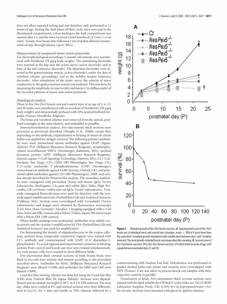
Figure 1. Retarded growth of Nes-Pex5 knock-out mice. A, Typical growth curve of Nes-Pex5knock-out (4 individual mice) and control mice (triangles, mean � SEM of 4 mice) born fromthe same litter. A marked growth retardation with variable onset between day 3 and 7 could beobserved. The most growth-retarded knock-out mouse died after weaning. B, Survival curve ofNes-Pex5 knock-out mice. Of 63 Nes-Pex5 knock-out mice, 57% died within 6 weeks of age, witha peak between postnatal days 21 and 30.
Hulshagen et al. • Peroxisome Elimination from CNS J. Neurosci., April 9, 2008 • 28(15):4015– 4027 • 4017

Electron microscopical analysisAt least two mice of each genotype were anes-thetized with an overdose of Nembutal and per-fused with 6% glutaraldehyde in Sorensenbuffer at pH 7.2. Tissues were postfixed for 24 hin 6% glutaraldehyde, osmicated, and embed-ded in glycidether (Serva, Heidelberg,Germany). Semithin sections used for lightmicroscopy were stained with 1% paraphe-nylenediamine or toluidine blue. Ultrathin sec-tions were cut at 70 nm, contrasted with leadcitrate, and examined either in a JEOL (Tokyo,Japan) JEM 2100 or a Carl Zeiss EM 910 trans-mission electron microscope. Axonal and mye-lin sheaths cross-sectional areas were quantifiedby measuring corpus callosum cross sections at5000� magnification using the Image Pro Plus(Media Cybernetics, Bethesda, MD) morphom-etry software. Myelin sheath thickness was plot-ted over axon cross-sectional area using the sta-tistics and data presentation software XACT!(SciLab, Hamburg, Germany).
ResultsMacroscopic phenotype of Nes-Pex5knock-out miceThe generation of Nes-Pex5 knock-outmice and demonstration of Pex5 inactiva-tion, which was completed between em-bryonic day 14.5 (E14.5) and E18.5, weredescribed previously (Baes et al., 2002;Krysko et al., 2007). Newborn Nes-Pex5knock-out pups were indistinguishablefrom control littermates but they displayeda marked postnatal growth retardation(Fig. 1A). The weight gain of �50% of Nes-Pex5 knock-out mice was extremely small,and these mice died within 6 weeks of age,with a peak in the postweaning period (Fig.1B). Three-week-old Nes-Pex5 knock-outmice developed cataract, abnormal plantarreflexes, and hindlimb flexion on liftingthem by their tails. After �4 months, mo-toric problems aggravated and food pelletswere placed inside the cages as mice hadproblems to hang on the grid. Subse-quently, the mice became lethargic, lostweight, and always died before or at the ageof 6 months. At the time of death, adultknock-outs weighed �44 – 46% less com-pared with their littermate controls [fe-male control (Ct) mice, 36.5 � 5.4 g, n �14; male Ct mice, 37.4 � 4.5 g, n � 12;female Nes-Pex5�/�, 19.8 � 2.4 g, n � 14;male Nes-Pex5�/�, 21.0 � 3.3 g, n � 15].
Behavioral phenotype of Nes-Pex5knock-out miceLongitudinal behavior assessment between 7 and 19 weeks re-vealed progressive ataxia, dyskinesia, and decreased explorationin the Nes-Pex5 knock-out mice (Fig. 2). Asthenia and/or neuro-motor defects are manifested in the knock-outs by their impairedmotor performance in the rotarod, wire suspension, and cageactivity tests (Fig. 2A–C). All three tests demonstrated significanteffects of genotype and genotype by age interaction (two-way
ANOVA, p � 0.001) on the neuromotor measures. For example,the significant effect of genotype on the rotarod measure indi-cates that neuromotor and coordination defects were already ob-vious at an age of 7 weeks (F(1,73) � 760; p � 0.001), but deteri-orated henceforth (interaction genotype by age; F(2,73) � 38; p �0.001).
Exploration in the open-field and dark–light transition testsindicated a defect in the knock-out group that was only slightly
Figure 2. Behavioral analysis of Nes-Pex5 knock-out mice. A–C, Neuromotor test performance indicates progressive impair-ment in Nes-Pex5 knock-out mice (dark gray bars) compared with controls (white bars). Latency of falling off the rod during thefourth accelerating rotarod trial (A) and of grip release during the wire suspension test (B), as well as locomotor cage activity (C),progressively decreased in Nes-Pex5 knock-outs. D, E, Exploratory behavior of Nes-Pex5 knock-out mice was also reduced. Open-field test revealed less entries in the central circle at all ages tested (D), and Nes-Pex5 knock-out mice generally produced lessbeam crossings in the dark–light transition box (E). F, Finally, Nes-Pex5 knock-out mice displayed reduced latency to reenter thedark compartment in the passive avoidance learning task. The histograms depict mean values � SEM. The asterisks indicatesignificance of differences versus control means in pairwise comparison ( post hoc Fisher’s least significant difference): *p �0.05,***p � 0.001.
4018 • J. Neurosci., April 9, 2008 • 28(15):4015– 4027 Hulshagen et al. • Peroxisome Elimination from CNS

progressive and indicative of emotional dulling at an early age(Fig. 2D,E). Total path length in the open-field test and numberof corner crossings (measures of locomotor activity) were notsignificantly altered (data not shown in figure), whereas the num-ber of entries in the center (Fig. 2D) and the percentage of dis-tance in the center (data not shown) were significantly reduced( p � 0.05). Accordingly, Nes-Pex5 knock-out mice made signif-icantly less exploratory transitions in the dark–light box at all agestested (Fig. 2E).
In addition to these motoric and coordination performancetests, cognitive abilities were examined in 11-week-old mice. De-spite their generally reduced activity and normal reaction to theelectric shock, Nes-Pex5 knock-out mice entered the dark com-partment faster than controls during the test trial of the passiveavoidance task, which suggests learning/memory impairments inNes-Pex5 knock-out mice as well (Fig. 2F). Together, these latterdata sets demonstrate that peroxisome deficiency in brain se-verely affects central functions like exploratory and cognitivebehaviors.
Metabolic profile of Nes-Pex5 knock-out miceA number of metabolites that are either generated or degraded byperoxisomal pathways were analyzed in entire cerebra of5-month-old mice. Plasmalogens, a subclass of etherphospholip-ids that were reduced by 65% in brain of newborn Nes-Pex5knock-out mice (Krysko et al., 2007), were more profoundly de-pleted in 5-month-old mice (Table 1). Conversely, levels of the
very long chain fatty acid (VLCFA) C26:0 were threefold increasedat birth and sixfold in adulthood (Table 1). The concentration ofdocosahexaenoic acid (DHA) (C22:6n-3), the most abundant poly-unsaturated fatty acid (PUFA) in brain that depends on one cycleof peroxisomal �-oxidation for its synthesis, was reduced in brainof newborn (Krysko et al., 2007) but not in adult mice (Table 1).Although astrocytes are capable of synthesizing DHA from n-3PUFA precursors, these data confirm previous suggestions thatmost of the DHA in brain is acquired from the diet or fromsynthesis in the liver (Williard et al., 2001). Finally, there was nodecrease of cholesterol levels in Nes-Pex5 knock-out mice, con-
tradicting previously proposed roles forperoxisomes in isoprenoid biosynthesis(Biardi and Krisans, 1997) (Table 1). Amore generalized lipid profile analysis re-vealed that the concentration of phospho-lipids was unaltered but that triglyceridesand cholesterylesters were 2- and 10-foldincreased in cerebra of Nes-Pex5 knock-out mice, respectively (Table 1).
Multiple pathologies in the CNS but noanomalies in the peripheralneuromuscular systemBecause the nestin promoter drives Cre ex-pression in brain and spinal cord, we firstevaluated whether the motoric impair-ments could be attributable to neuromus-cular lesions (e.g., by affecting spinal mo-toneurons). Histological analysis of high-resolution semithin sections of peripheralnerves, light microscopy of muscle (datanot shown), and measurement of com-pound motor action potentials did not re-veal differences between control (17.73 �1.74 mV) and Nes-Pex5 knock-out mice at
the age of 5 months (19.45 � 3.11 mV) (n � 3). Latencies ofcompound motor action potentials were 0.82 � 0.04 ms in con-trols versus 0.85 � 0.02 ms in Nes-Pex5 knock-out mice.
In marked contrast, as described in detail below, the CNS ofNes-Pex5 knock-out mice develops a complex phenotype charac-terized by region-specific lipid storage, gliosis, axon degenera-tion, and changes in myelin sheath composition (summarized inTable 2).
Lipids accumulate in brain and spinal cordOil Red O stainings were done to define the site of neutral lipidaccumulations (Table 1) in 5-month-old Nes-Pex5 knock-outmice. Lipid inclusions were most prominent in the ependymallining of cerebral ventricles (Fig. 3A,B,G) and the central canal ofthe spinal cord (Fig. 3C,D). No lipid accumulations were ob-served in the choroid plexus. In addition to ependymal cells, OilRed O-positive lipid droplets extensively accumulated in the mo-lecular layer of the cerebellar cortex (Fig. 3E,F). More specifi-cally, they were present in Bergmann glia fibers, neurite pro-cesses, and glial endfeet at the pial basement membrane andstrikingly in cerebellar meningeal cells, whereas meningeal cellselsewhere in the CNS were unaffected (Fig. 3H). To a much lesserextent, lipid droplets were spread over all gray matter areas suchas the granular layer of the cerebellum, the cerebral cortex, hip-pocampus, the hypothalamus and thalamus, and the superiorand inferior colliculi (Table 2). In addition, lipid droplets werefound in the corpus callosum and white matter of cerebellum.
Table 1. Lipid analyses in cerebrum of 5-month-old Nes-Pex5 knock-out and controlmice
Control Nes-Pex5 knock-out
Plasmalogens (pmol/nmol phospholipids) 23.4 � 5.5 3.8 � 1.3***C26:0/C22:0 0.027 � 0.006 0.153 � 0.015***C22:6n-3 (pmol/nmol phospholipids) 190 � 14 180 � 4Cholesterol (�mol/g tissue) 16.92 � 0.33 17.20 � 2.21Cholesterylesters (�mol/g tissue) 0.05 � 0.01 0.49 � 0.20*Triglycerides (�mol/g tissue) 0.27 � 0.03 0.46 � 0.05**
Values represent means � SEM of three or four independent samples.
*p � 0.1, **p � 0.05, ***p � 0.005, unpaired Student’s t test.
Table 2. Summary of observed pathologies in CNS of 5-month-old Nes-Pex5 knock-out mice
Lipid accu-mulation Astrogliosis Microgliosis Myelin defect Axonal damage Cell death
Catalaseupregulation
Optic nerve � � � � � � �Cortex x xx (V) xx (V) x xx (V) � xCc x xx xxx xxx xxx x xxxFornix x xx xxx (V) xxx (V) xx (V) � xxAnterior commissure x xx xx x (V) � � xxHippocampus x xx xx � xx � xThalamus x xx xx � xx � xxHypothalamus x xx xx � xx � xxSup coll x xx xx � xx � xxInf coll x xx xx � xx � xxMidbrain x xx xx � xx � xxPons x xx xx � xx � xxMedulla x xx xx � xx � xxCerebellum
ML xxx x x � xx � xxGL x x x � xx � xxWhm x x x xx xxx � xxx
Spinal cord x xx xx x (V) x (V) � /
�, Not present; x, present; xx, more present; xxx, most present; V, variable between mice; /, not investigated. Abbreviations: Cc, corpus callosum; sup coll,superior colliculus; inf coll, inferior colliculus; ML, molecular layer; GL, granular layer; Whm, white matter.
Hulshagen et al. • Peroxisome Elimination from CNS J. Neurosci., April 9, 2008 • 28(15):4015– 4027 • 4019

Peroxisome deficiency causes severe gliosis in brain andspinal cordBoth as a readout for any kind of pathological changes in the CNSas well as to investigate whether the elimination of functionalperoxisomes affects the glial compartment, immunohistochemi-cal detection of astrocyte and microglial markers was performedbetween the ages of 3 weeks and 5 months.
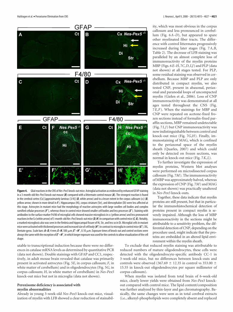
Increased expression of GFAP, the common marker of astro-glial activation, was clearly visible from 3 weeks on in spinal cord(supplemental Fig. 1A,B, available at www.jneurosci.org as sup-plemental material) and from 6 weeks on in gray and white mat-ter of all brain regions of Nes-Pex5 knock-out mice (Fig. 4B,shown for 3-month-old mouse). Marked regional differences inseverity of astrogliosis were observed being most extensive incerebral cortex and corpus callosum (Fig. 4B,F). In Table 2, theseverity and distribution of gliosis in 5-month-old mice is shown.
Astrocytes in mutant mice had the morphology of reactiveastrocytes with large swollen cell bodies and complex ramifiedcellular processes (Fig. 4F�). In contrast, astroglial cells identifiedin control mice had small cell bodies and less cellular processes(Fig. 4E�). Western blotting of brain homogenates confirmed theupregulation of the astrocytic marker in Nes-Pex5 knock-outmice (data not shown).
Immunoreactivity of the macrophage/microglia surface mol-ecule F4/80 was massively increased at an age of 3 weeks in spinalcord (supplemental Fig. 1C,D, available at www.jneurosci.org assupplemental material) and 6 weeks in brain of Nes-Pex5 knock-out mice, pointing to the activation of microglial cells. This mi-crogliosis was observed in all brain regions but was most pro-nounced in white matter (Fig. 4D,H, Table 2). Microglial cells inmutant mice exhibited thickened processes and increased cellbody size, but ameboid microglia were not observed (Fig. 4H�).In contrast, microglia identified in control mice had small cellbodies and finer cytoplasmatic ramifications (Fig. 4G�). Both theexpression of F4/80 and GFAP increased during the lifetime ofNes-Pex5 knock-out mice.
To determine whether astrogliosis and microgliosis was asso-ciated with lymphocyte influx, we tested for infiltrating T cellswith anti-CD3 antibody. No T cells were found at any age tested.In addition, an immunohistochemical staining with CD45, apan-leukocyte marker gave a very similar staining pattern to F4/80, suggesting that the vast majority of CD45 staining was attrib-utable to microglia/macrophages (data not shown).
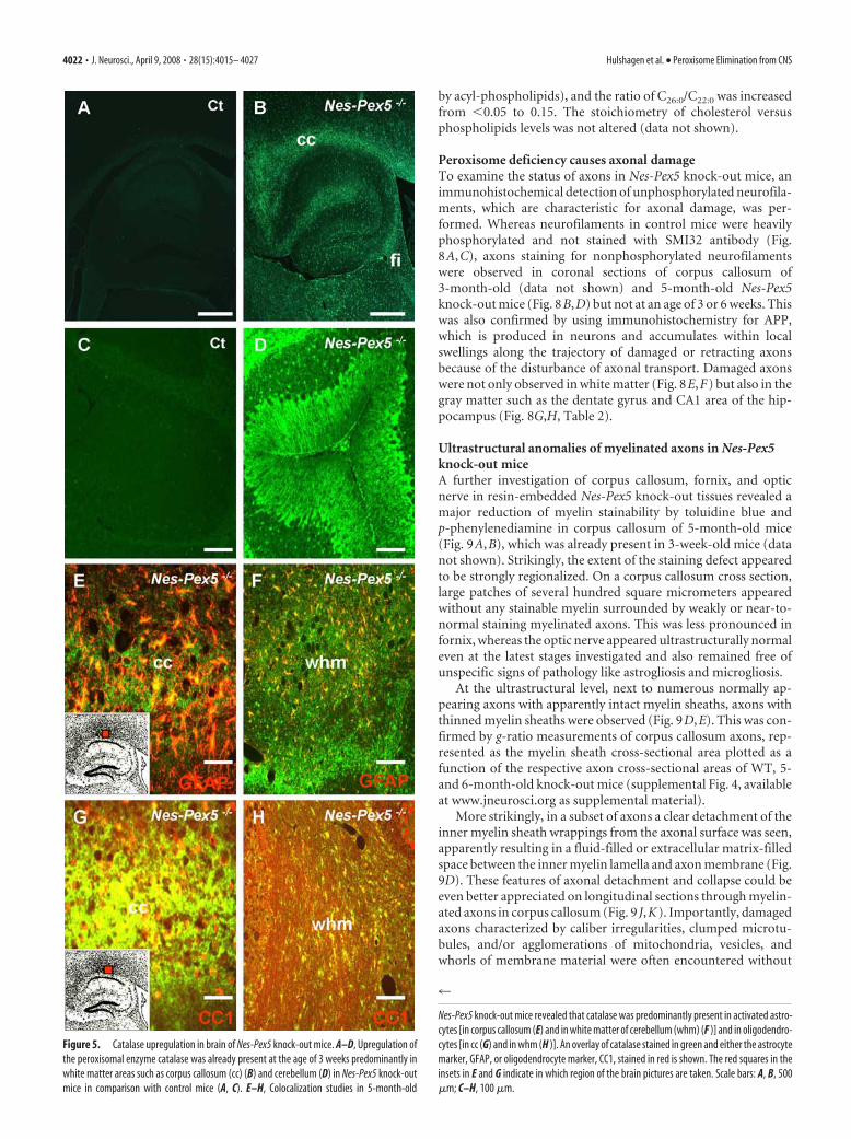
Peroxisomal catalase is upregulatedCatalase, the enzyme that inactivates toxic peroxides produced byperoxisomal oxidases and other cellular sources, is one of theexceptional peroxisomal enzymes that are stable and active whenmislocalized to the cytosol. Immunohistochemical stainings re-vealed that catalase was extensively upregulated in white matterareas (Fig. 5A,B) of Nes-Pex5 knock-out mice and to a variableextent in Bergmann glial cells of the cerebellum (Fig. 5C,D) al-ready from an age of 3 weeks. Western blot analysis on cerebralhomogenates confirmed the upregulation starting from an age of3 weeks in Nes-Pex5 knock-out mice. The increase was not attrib-
Figure 3. Accumulation of lipids in the CNS of Nes-Pex5 knock-out mice. Oil Red O stainingreveals neutral lipid accumulations in ependymal cells surrounding the fourth ventricle (B) andthe central canal of the spinal cord (D) but not in the choroid plexus epithelium in Nes-Pex5knock-out mice. In control mice, no lipid droplets were detected (A, C). Lipid accumulations
4
were also abundant in the molecular layer of the cerebellum of Nes-Pex5 knock-out mice (F ) butwere absent from control mice (E). The arrows point to the lipid droplets that appear yellow/pink in dark field (B, F ) and red in light field (D) optics. G, H, Electron micrograph of lipiddroplets in an ependymal cell (G) and in Bergmann glia endfeet (arrowhead) and cerebellarmeningeal cells (arrow) (H ). Scale bars: A, B, E, F, 100 �m; C, D, 50 �m; G, 2 �m; H, 5 �m. GL,Granular layer; ML, molecular layer.
4020 • J. Neurosci., April 9, 2008 • 28(15):4015– 4027 Hulshagen et al. • Peroxisome Elimination from CNS
utable to transcriptional induction because there were no differ-ences in catalase mRNA levels as determined by quantitative PCR(data not shown). Double stainings with GFAP and CC1, respec-tively, in adult mouse brain revealed that catalase was primarilypresent in activated astrocytes (Fig. 5E, in corpus callosum; F, inwhite matter of cerebellum) and in oligodendrocytes (Fig. 5G, incorpus callosum; H, in white matter of cerebellum) in Nes-Pex5knock-out mice but not in microglia (data not shown).
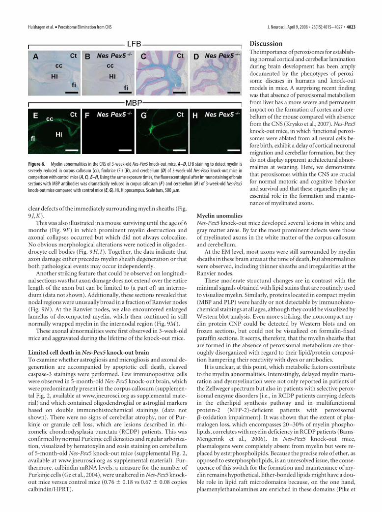
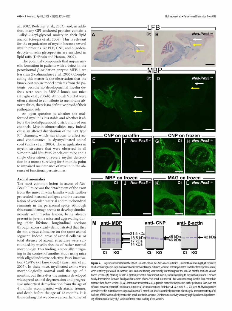
Peroxisome deficiency is associated withmyelin abnormalitiesAlready in young 3-week-old Nes-Pex5 knock-out mice, visual-ization of myelin with LFB showed a clear reduction of stainabil-
ity, which was most obvious in the corpuscallosum and less pronounced in cerebel-lum (Fig. 6A–D), but appeared to spareother myelinated fiber tracts. The differ-ence with control littermates progressivelyincreased during later stages (Fig. 7A,B,Table 2). The decrease of LFB staining wasparalleled by an almost complete loss ofimmunoreactivity of the myelin proteinsMBP (Figs. 6E–H, 7C,D, I,J) and PLP (datanot shown) at all stages tested. For PLP,some residual staining was observed in cer-ebellum. Because MBP and PLP are onlydistributed in compact myelin, we alsotested CNP, present in abaxonal, periax-onal and paranodal loops of uncompactedmyelin (Gielen et al., 2006). Loss of CNPimmunoreactivity was demonstrated at allages tested throughout the CNS (Fig.7E,F). When the stainings for MBP andCNP were repeated on acetone-fixed fro-zen sections instead of formalin-fixed par-affin sections, MBP remained undetectable(Fig. 7 I, J) but CNP immunoreactivity wasnow indistinguishable between control andknock-out mice (Fig. 7G,H). Finally, im-munostaining of MAG, which is confinedto the periaxonal space of the myelinsheath (Quarles, 2007) and which couldonly be detected on frozen sections, wasnormal in knock-out mice (Fig. 7K,L).
To further investigate the expression ofmyelin proteins, Western blot analyseswere performed on microdissected corpuscallosum (Fig. 7M). The immunoreactivityof MBP was approximately halved, whereasthe expression of CNP (Fig. 7M) and MAG(data not shown) was practically unalteredin Nes-Pex5 knock-outs.
Together, these data indicate that myelinproteins are still present, but that in particu-lar the immunohistochemical detection ofproteins present in compact myelin is se-verely impaired. Although the loss of MBPimmunoreactivity in the sections might beattributable to a sensitivity problem, the dif-ferential detection of CNP, depending on theprocedure used, might indicate that the pro-teins are embedded in an altered lipid envi-ronment within the myelin sheath.
To exclude that reduced myelin staining was attributable toreduced numbers of mature oligodendrocytes, these cells weredetected with the oligodendrocyte-specific antibody CC-1 in3-week-old mice, but no differences between knock-outs andcontrols were observed (367.69 � 12.35 in control vs 353.00 �15.55 in knock-out oligodendrocytes per square millimeter ofcorpus callosum).
When myelin was isolated from total brain of 6-week-oldmice, clearly lower yields were obtained from Nes-Pex5 knock-out compared with control mice. The lipid content/compositionwas further analyzed by thin-layer and gas chromatography. Ba-sically, the same changes were seen as in total cerebral extracts(i.e., alkenyl-phospholipids were completely absent and replaced
Figure 4. Glial reactions in the CNS of Nes-Pex5 knock-out mice. Astroglial activation as evidenced by enhanced GFAP stainingin a 3-month-old Nes-Pex5 knock-out mouse (B) compared with a littermate control mouse (A). The strongest reaction is foundin the cerebral cortex (Cx) [approximately laminas (3/4)] (B, white arrow) and to a lesser extent in the corpus callosum (cc) (B,yellow arrow; shown in more detail in F ). Hippocampus (Hi), corpus striatum (Str), and diencephalon (Di) were less affected atthis stage. Astrocytes in mutant mice had the morphology of reactive astrocytes with large swollen cell bodies and complexramified cellular processes (F�), whereas those in control mice showed smaller cell bodies and less processes (E�). Staining withantibodies to the surface marker F4/80 of microglial cells showed massive microgliosis in cc (yellow arrow) and less pronouncedreactions in the Cx (white arrow) of 3-month-old Nes-Pex5 knock-out mice (D, H ) in comparison with control mice (C, G). Notably,a marked microgliosis also was seen in the fimbria and hippocampal fissure of Hi, Str, and less so in Di. Microglial cells in mutantmice were activated with thickened processes and increased size of cell body (H�) in contrast to microglia in control mice (G�). DG,Dentate gyrus. Scale bars: A–D, 4 mm; E–H, 500 �m; E�–H�, 0.10 �m. Exposure times of knock-out and control sections werealways the same with the exception of E�–H�, in which longer exposure was used for the controls to allow visualization of the cellshape.
Hulshagen et al. • Peroxisome Elimination from CNS J. Neurosci., April 9, 2008 • 28(15):4015– 4027 • 4021
by acyl-phospholipids), and the ratio of C26:0/C22:0 was increasedfrom �0.05 to 0.15. The stoichiometry of cholesterol versusphospholipids levels was not altered (data not shown).
Peroxisome deficiency causes axonal damageTo examine the status of axons in Nes-Pex5 knock-out mice, animmunohistochemical detection of unphosphorylated neurofila-ments, which are characteristic for axonal damage, was per-formed. Whereas neurofilaments in control mice were heavilyphosphorylated and not stained with SMI32 antibody (Fig.8A,C), axons staining for nonphosphorylated neurofilamentswere observed in coronal sections of corpus callosum of3-month-old (data not shown) and 5-month-old Nes-Pex5knock-out mice (Fig. 8B,D) but not at an age of 3 or 6 weeks. Thiswas also confirmed by using immunohistochemistry for APP,which is produced in neurons and accumulates within localswellings along the trajectory of damaged or retracting axonsbecause of the disturbance of axonal transport. Damaged axonswere not only observed in white matter (Fig. 8E,F) but also in thegray matter such as the dentate gyrus and CA1 area of the hip-pocampus (Fig. 8G,H, Table 2).
Ultrastructural anomalies of myelinated axons in Nes-Pex5knock-out miceA further investigation of corpus callosum, fornix, and opticnerve in resin-embedded Nes-Pex5 knock-out tissues revealed amajor reduction of myelin stainability by toluidine blue andp-phenylenediamine in corpus callosum of 5-month-old mice(Fig. 9A,B), which was already present in 3-week-old mice (datanot shown). Strikingly, the extent of the staining defect appearedto be strongly regionalized. On a corpus callosum cross section,large patches of several hundred square micrometers appearedwithout any stainable myelin surrounded by weakly or near-to-normal staining myelinated axons. This was less pronounced infornix, whereas the optic nerve appeared ultrastructurally normaleven at the latest stages investigated and also remained free ofunspecific signs of pathology like astrogliosis and microgliosis.
At the ultrastructural level, next to numerous normally ap-pearing axons with apparently intact myelin sheaths, axons withthinned myelin sheaths were observed (Fig. 9D,E). This was con-firmed by g-ratio measurements of corpus callosum axons, rep-resented as the myelin sheath cross-sectional area plotted as afunction of the respective axon cross-sectional areas of WT, 5-and 6-month-old knock-out mice (supplemental Fig. 4, availableat www.jneurosci.org as supplemental material).
More strikingly, in a subset of axons a clear detachment of theinner myelin sheath wrappings from the axonal surface was seen,apparently resulting in a fluid-filled or extracellular matrix-filledspace between the inner myelin lamella and axon membrane (Fig.9D). These features of axonal detachment and collapse could beeven better appreciated on longitudinal sections through myelin-ated axons in corpus callosum (Fig. 9 J,K). Importantly, damagedaxons characterized by caliber irregularities, clumped microtu-bules, and/or agglomerations of mitochondria, vesicles, andwhorls of membrane material were often encountered without
4
Nes-Pex5 knock-out mice revealed that catalase was predominantly present in activated astro-cytes [in corpus callosum (E) and in white matter of cerebellum (whm) (F )] and in oligodendro-cytes [in cc (G) and in whm (H )]. An overlay of catalase stained in green and either the astrocytemarker, GFAP, or oligodendrocyte marker, CC1, stained in red is shown. The red squares in theinsets in E and G indicate in which region of the brain pictures are taken. Scale bars: A, B, 500�m; C–H, 100 �m.
Figure 5. Catalase upregulation in brain of Nes-Pex5 knock-out mice. A–D, Upregulation ofthe peroxisomal enzyme catalase was already present at the age of 3 weeks predominantly inwhite matter areas such as corpus callosum (cc) (B) and cerebellum (D) in Nes-Pex5 knock-outmice in comparison with control mice (A, C). E–H, Colocalization studies in 5-month-old
4022 • J. Neurosci., April 9, 2008 • 28(15):4015– 4027 Hulshagen et al. • Peroxisome Elimination from CNS
clear defects of the immediately surrounding myelin sheaths (Fig.9 J,K).
This was also illustrated in a mouse surviving until the age of 6months (Fig. 9F) in which prominent myelin destruction andaxonal collapses occurred but which did not always colocalize.No obvious morphological alterations were noticed in oligoden-drocyte cell bodies (Fig. 9H, I). Together, the data indicate thataxon damage either precedes myelin sheath degeneration or thatboth pathological events may occur independently.
Another striking feature that could be observed on longitudi-nal sections was that axon damage does not extend over the entirelength of the axon but can be limited to (a part of) an interno-dium (data not shown). Additionally, these sections revealed thatnodal regions were unusually broad in a fraction of Ranvier nodes(Fig. 9N). At the Ranvier nodes, we also encountered enlargedlamellas of decompacted myelin, which then continued in stillnormally wrapped myelin in the internodal region (Fig. 9M).
These axonal abnormalities were first observed in 3-week-oldmice and aggravated during the lifetime of the knock-out mice.
Limited cell death in Nes-Pex5 knock-out brainTo examine whether astrogliosis and microgliosis and axonal de-generation are accompanied by apoptotic cell death, cleavedcaspase-3 stainings were performed. Few immunopositive cellswere observed in 5-month-old Nes-Pex5 knock-out brain, whichwere predominantly present in the corpus callosum (supplemen-tal Fig. 2, available at www.jneurosci.org as supplemental mate-rial) and which contained oligodendroglial or astroglial markersbased on double immunohistochemical stainings (data notshown). There were no signs of cerebellar atrophy, nor of Pur-kinje or granule cell loss, which are lesions described in rhi-zomelic chondrodysplasia punctata (RCDP) patients. This wasconfirmed by normal Purkinje cell densities and regular arboriza-tion, visualized by hematoxylin and eosin staining on cerebellumof 5-month-old Nes-Pex5 knock-out mice (supplemental Fig. 2,available at www.jneurosci.org as supplemental material). Fur-thermore, calbindin mRNA levels, a measure for the number ofPurkinje cells (Ge et al., 2004), were unaltered in Nes-Pex5 knock-out mice versus control mice (0.76 � 0.18 vs 0.67 � 0.08 copiescalbindin/HPRT).
DiscussionThe importance of peroxisomes for establish-ing normal cortical and cerebellar laminationduring brain development has been amplydocumented by the phenotypes of peroxi-some diseases in humans and knock-outmodels in mice. A surprising recent findingwas that absence of peroxisomal metabolismfrom liver has a more severe and permanentimpact on the formation of cortex and cere-bellum of the mouse compared with absencefrom the CNS (Krysko et al., 2007). Nes-Pex5knock-out mice, in which functional peroxi-somes were ablated from all neural cells be-fore birth, exhibit a delay of cortical neuronalmigration and cerebellar formation, but theydo not display apparent architectural abnor-malities at weaning. Here, we demonstratethat peroxisomes within the CNS are crucialfor normal motoric and cognitive behaviorand survival and that these organelles play anessential role in the formation and mainte-nance of myelinated axons.
Myelin anomaliesNes-Pex5 knock-out mice developed several lesions in white andgray matter areas. By far the most prominent defects were thoseof myelinated axons in the white matter of the corpus callosumand cerebellum.
At the EM level, most axons were still surrounded by myelinsheaths in these brain areas at the time of death, but abnormalitieswere observed, including thinner sheaths and irregularities at theRanvier nodes.
These moderate structural changes are in contrast with theminimal signals obtained with lipid stains that are routinely usedto visualize myelin. Similarly, proteins located in compact myelin(MBP and PLP) were hardly or not detectable by immunohisto-chemical stainings at all ages, although they could be visualized byWestern blot analysis. Even more striking, the noncompact my-elin protein CNP could be detected by Western blots and onfrozen sections, but could not be visualized on formalin-fixedparaffin sections. It seems, therefore, that the myelin sheaths thatare formed in the absence of peroxisomal metabolism are thor-oughly disorganized with regard to their lipid/protein composi-tion hampering their reactivity with dyes or antibodies.
It is unclear, at this point, which metabolic factors contributeto the myelin abnormalities. Interestingly, delayed myelin matu-ration and dysmyelination were not only reported in patients ofthe Zellweger spectrum but also in patients with selective perox-isomal enzyme disorders [i.e., in RCDP patients carrying defectsin the etherlipid synthesis pathway and in multifunctionalprotein-2 (MFP-2)-deficient patients with peroxisomal�-oxidation impairment]. It was shown that the extent of plas-malogen loss, which encompasses 20 –30% of myelin phospho-lipids, correlates with myelin deficiency in RCDP patients (Bams-Mengerink et al., 2006). In Nes-Pex5 knock-out mice,plasmalogens were completely absent from myelin but were re-placed by esterphospholipids. Because the precise role of ether, asopposed to esterphospholipids, is an unresolved issue, the conse-quence of this switch for the formation and maintenance of my-elin remains hypothetical. Ether-bonded lipids might have a dou-ble role in lipid raft microdomains because, on the one hand,plasmenylethanolamines are enriched in these domains (Pike et
Figure 6. Myelin abnormalities in the CNS of 3-week-old Nes-Pex5 knock-out mice. A–D, LFB staining to detect myelin isseverely reduced in corpus callosum (cc), fimbriae (Fi) (B), and cerebellum (D) of 3-week-old Nes-Pex5 knock-out mice incomparison with control mice (A, C). E–H, Using the same exposure times, the fluorescent signal after immunostaining of brainsections with MBP antibodies was dramatically reduced in corpus callosum (F ) and cerebellum (H ) of 3-week-old Nes-Pex5knock-out mice compared with control mice (E, G). Hi, Hippocampus. Scale bars, 500 �m.
Hulshagen et al. • Peroxisome Elimination from CNS J. Neurosci., April 9, 2008 • 28(15):4015– 4027 • 4023
al., 2002; Rodemer et al., 2003), and, in addi-tion, many GPI anchored proteins contain a1-alkyl-2-acyl-glycerol moiety in their lipidanchor (Gorgas et al., 2006). This is relevantfor the organization of myelin because severalmyelin proteins like PLP, CNP, and oligoden-drocyte–myelin glycoprotein are enriched inlipid rafts (DeBruin and Harauz, 2007).
The potential compounds that impair my-elin formation in patients with a defect in theperoxisomal �-oxidation enzyme MFP-2 areless clear (Ferdinandusse et al., 2006). Compli-cating this matter is the observation that theknock-out mouse model deviates from the pa-tients, because no developmental myelin de-fects were seen in MFP-2 knock-out mice(Huyghe et al., 2006b). Although VLCFA wereoften claimed to contribute to membrane ab-normalities, there is no definitive proof of theirpathogenic role.
An open question is whether the mal-formed myelin is less stable and whether it af-fects the nodal/paranodal distribution of ionchannels. Myelin abnormalities may indeedcause an altered distribution of the Kv1 typeK� channels, which was shown to affect ax-onal conductance in dysmyelinated spinalcord (Sinha et al., 2005). The irregularities inmyelin structure that were observed in all5-month-old Nes-Pex5 knock-out mice and asingle observation of severe myelin destruc-tion in a mouse surviving for 6 months pointto impaired maintenance of myelin in the ab-sence of functional peroxisomes.
Axonal anomaliesThe most common lesion in axons of Nes-Pex5�/� mice was the detachment of the axonfrom the inner myelin lamella which furtherproceeded in axonal collapse and the accumu-lation of vesicular material and mitochondrialremnants in the periaxonal space. Althoughthis axonal damage seems to develop simulta-neously with myelin lesions, being alreadypresent in juvenile mice and aggravating dur-ing their lifetime, longitudinal sectionsthrough axons clearly demonstrated that theydo not always colocalize on the same axonalsegment. Indeed, areas of axonal collapse ortotal absence of axonal structures were sur-rounded by myelin sheaths of rather normalmorphology. This finding is especially intrigu-ing in the context of another study using micewith oligodendrocyte-selective Pex5 inactiva-tion (CNP-Pex5 knock-out) (Kassmann et al.,2007). In these mice, myelinated axons weremorphologically normal until the age of 2months, but thereafter the animals developedwidespread axonal degeneration and progres-sive subcortical demyelination from the age of4 months accompanied with ataxia, tremor,and death before the age of 12 months. It isthus striking that we observe an earlier onset of
Figure 7. Myelin abnormalities in the CNS of 3-month-old old Nes-Pex5 knock-out mice. Luxol fast blue staining (A, B) producedmuchweakersignals incorpuscallosum(whitearrow)ofknock-outmice,whereasothermyelinatedtracts likefornix(yellowarrow)were relatively preserved. In contrast, MBP immunostaining was virtually lost throughout the CNS on paraffin sections (D) andfrozen sections (J ). Staining for CNP, a protein present in noncompact myelin, varied according to the fixation protocol. CNP wasbarely detectable in formalin-fixed paraffin sections of Nes-Pex5 knock-out mice (F ) but was not distinguishable from controls inacetone-fixed frozen sections (G, H ). Immunoreactivity for MAG, a protein that exclusively occurs in the periaxonal loop, was notdifferent between control (K ) and knock-out mice (L) on frozen sections. Scale bars: A–D, 4 mm; E–L, 500 �m. M, Myelin proteinswere detected in microdissected corpus callosum of 5-month-old knock-out mice by Western blot analysis. Immunoreactivity of allisoforms of MBP was markedly reduced in knock-out brain, whereas CNP immunoreactivity was only slightly reduced. Equal inten-sity of immunoreactivity of �-actin confirmed equal loading of the samples.
4024 • J. Neurosci., April 9, 2008 • 28(15):4015– 4027 Hulshagen et al. • Peroxisome Elimination from CNS

disease and a faster disease progression with much shorter lifespan inNes-Pex5 compared with CNP-Pex5 knock-out mice. This mightindicate that peroxisomes in other cell types of the CNS play also acrucial role in the formation and stability of myelinated axons. How-ever, the severe pathology in CNP-Pex5 knock-out mice proves thatperoxisomes in oligodendrocytes are essential for the maintenanceof myelinated axons. To clarify the precise pathogenesis, additionalcell type-selective knock-out mice with elimination of functionalperoxisomes from either neurons or astrocytes are now urgentlyneeded. Indeed, perinodal astrocytes might be of crucial importance,because nodal regions are heavily affected, and also the contributionof autonomous neuronal effects impairing axon stability need to bedefined.
However, it cannot be excluded that the earlier ablation ofperoxisomes from oligodendrocytes when using the Nestin pro-moter as opposed to the CNP promoter also plays a role in thedifference in phenotype between the two mouse models. In Nes-Pex5 knock-out mice, functional peroxisomes were absent fromthe brain at birth (Krysko et al., 2007). In CNP-Pex5 knock-outmice, oligodendrocyte-specific Cre expression and Pex5 gene re-combination in brain was shown in 7-d-old mice (Kassmann etal., 2007). This is before the major wave of myelination but it isnot impossible that the already formed etherlipids can stay in theCNS for several days.
In patients with peroxisomal disorders, axonopathies havebeen reported in the peripheral nervous system and in the spinalcord but no information is available on the brain, probably re-lated to the very few ultrastructural analyses that have been done.Interestingly, adrenomyeloneuropathy, caused by a defect in theperoxisomal transporter ABCD1, is postulated to be a fundamen-tal defect in the axonal or neuronal membrane, which leads todegeneration but not to apoptotic cell death (Powers et al., 2000,2001). Also in a mouse model with ABCD1 deficiency, it wasdemonstrated that axonal damage occurs as first pathologicalevent followed by myelin degeneration (Pujol et al., 2004).
Regional differences in pathologiesThe diverse lesions in Nes-Pex5 knock-out mice show regional dif-ferences in severity that are not always linked to the distribution ofperoxisomes. The areas with the most distinct lipid accumulations(ependymal cells and molecular layer of cerebellum) maintain highlevels of peroxisomes in postnatal life (Ahlemeyer et al., 2007) but donot seem to be hit by the other pathologies. The cerebral cortex andits main commissural system, and to a lesser degree the cerebellum,are most affected with regard to gliosis and damaged myelinatedaxons. Although the dysmyelinated areas always exhibit severe mi-crogliosis and a less pronounced astrogliosis, it should be empha-sized that glial activation also occurs in gray matter. The sparing ofthe optic nerve from all pathologies is an enigmatic observation,which remains unexplained. Interestingly, in CNP-Pex5 knock-outmice, a similar pattern of lesions was observed whereby white matterabnormalities and axonal swellings first occurred in the genu of cor-pus callosum extending to fimbriae and anterior commissure,whereas other white matter tracts remained intact (Kassmann et al.,2007).
Pathogenesis of motoric and cognitive impairmentWhat is the pathological basis of the progressive loss of motoricand cognitive abilities of Nes-Pex5 knock-out mice? It is quitestriking that the phenotypic appearance and course of life of Nes-Pex5 is very similar to that of MFP-2 knock-out mice with im-paired peroxisomal �-oxidation. Indeed, they both develop ab-normal reflexes of the paws on lifting, impaired coordination,
Figure 8. Axonal damage and cell degeneration in the CNS of Nes-Pex5 knock-out mice.A–D, Coronal sections of corpus callosum (cc) (A, B) and sagittal sections of cerebellum (C, D)were stained with SMI32 antibody to detect unphosphorylated neurofilament, a sign of axonaldamage, in 5-month-old mice. Strongly enhanced staining was observed in Nes-Pex5 knock-outmice (B, D) compared with control mice (A, C). This was further confirmed by increased APPimmunostaining on sagittal brain sections in the corpus callosum (F ) and in the hippocampus(H ) of 5-month-old knock-out mice in comparison with respective areas in control mice (E, G).The red squares in the insets in A, E, and G indicate in which region of the brain pictures aretaken, and arrows point to immunopositive cells in Nes-Pex5 knock-out mice. CA1, Cornu am-monis 1; DG, dentate gyrus; whm, white matter. Scale bars, 100 �m.
Hulshagen et al. • Peroxisome Elimination from CNS J. Neurosci., April 9, 2008 • 28(15):4015– 4027 • 4025

progressive motoric inability, lethargy, anddeath before the age of 6 months (Huygheet al., 2006b). We found no evidence forneuromuscular abnormalities in eithermouse model. In the brain, Nes-Pex5 andMFP-2 knock-out mice share lipid accu-mulations, astrogliosis and microgliosis inthe gray matter, and increased expressionof catalase (Huyghe et al., 2006b).
However, the developmental and sus-tained myelin abnormalities that were ac-companied with massive microgliosis inthe corpus callosum and cerebellar whitematter were only seen in Nes-Pex5 and didnot occur in MFP-2 knock-outs. New ob-servations in MFP-2 knock-out brains re-vealed extensive axonal damage at differentages (L. Hulshagen, unpublished observa-tions). We therefore propose that axonaldamage and/or abnormalities in gray mat-ter that are common between Nes-Pex5 andMFP-2 knock-out mice, rather than dys-myelination, are responsible for the behav-ioral problems and early death of bothmouse models. The present data supportthe conclusion that peroxisomes are indis-pensable organelles for the integrity of theCNS, for normal motoric activity and cog-nitive abilities. Additional investigationsare required to pinpoint the essential per-oxisomal metabolic conversions and thecell types in which they occur in the CNS.
ReferencesAdamo AM, Aloise PA, Pasquini JM (1986) A
possible relationship between concentration ofmicroperoxisomes and myelination. Int J DevNeurosci 4:513–517.
Ahlemeyer B, Neubert I, Kovacs WJ, Baumgart-Vogt E (2007) Differential expression of per-oxisomal matrix and membrane proteins dur-ing postnatal development of mouse brain.J Comp Neurol 505:1–17.
Arnold G, Holtzman E (1978) Microperoxisomes
Figure 9. Semithin and ultrastructural analysis of myelin and axon lesions in Nes-Pex5 knock-out mice. A, B, Sagittallyoriented semithin cross sections of the corpus callosum of 5-month-old animals stained with p-phenylenediamine show a clearreduction of overall stainability of white matter in Pex5-deficient mice. C–E, On the ultrastructural level, Nes-Pex5 knock-out micecontain a majority of structurally almost normal appearing myelinated axons (D, E), albeit the thickness of the myelin sheet wasoften reduced compared with controls (C). Note the detachment of an axon from the inner myelin lamella (D, arrow) and theaccumulation of multivesicular material in a pathological space between axon and inner myelin sheath surface (D, asterisk). In a
4
6-month-old mouse (F ), more prominent axonal damage andeven complete destruction of axon segments (arrowheads)were seen in still preserved myelin sheaths, whereas in theimmediate neighborhood, also a “co-destruction” of axonwith their myelin sheaths (arrows) was observed. These fea-tures of axonal detachment and collapse were confirmed onlongitudinal sections through axons in the corpus callosum (G,J–N ). In the periaxonal space, degenerated mitochondria anddebris accumulated in a segmental manner (J, K, arrows). Atthe nodes of Ranvier, the exposed myelin-free nodal regionwas often enlarged (L; N, arrows), whereas the decompactmyelin loops (L–N, ML) were irregularly swollen. Note alsosignificant accumulation of cellular debris in the nodal region(M, asterisk) that could represent degenerated perinodal as-trocytes. It is striking thereby that oligodendrocyte cell bodies(H, WT; I, Nes-Pex5�/�; both shown at 5 months of age) re-main normal, without, for example, degenerative changes inthe cytoplasm. Scale bars: A, B, 20 �m; C–E, 2 �m; F, 5 �m;G, 1 �m; H, I, 5 �m; J, K, 2 �m; L, 0.5 �m; M, N, 2 �m.
4026 • J. Neurosci., April 9, 2008 • 28(15):4015– 4027 Hulshagen et al. • Peroxisome Elimination from CNS

in the central nervous system of the postnatal rat. Brain Res 155:1–17.Baes M, Van Veldhoven PP (2006) Generalised and conditional inactivation
of Pex genes in mice. Biochim Biophys Acta 1763:1785–1793.Baes M, Gressens P, Baumgart E, Carmeliet P, Casteels M, Fransen M, Evrard
P, Fahimi D, Declercq PE, Collen D, Van Veldhoven PP, Mannaerts GP(1997) A mouse model for Zellweger syndrome. Nat Genet 17:49 –57.
Baes M, Dewerchin M, Janssen A, Collen D, Carmeliet P (2002) Generationof Pex5-IoxP mice allowing the conditional elimination of peroxisomes.Genesis 32:177–178.
Bams-Mengerink AM, Majoie CB, Duran M, Wanders RJ, Van HJ, ScheurerCD, Barth PG, Poll-The BT (2006) MRI of the brain and cervical spinalcord in rhizomelic chondrodysplasia punctata. Neurology 66:798 – 803.
Biardi L, Krisans SK (1997) Compartmentalization of cholesterol biosyn-thesis. J Biol Chem 271:1784 –1788.
Caeyenberghs K, Balschun D, Roces DP, Schwake M, Saftig P, D’Hooge R(2006) Multivariate neurocognitive and emotional profile of a man-nosidosis murine model for therapy assessment. Neurobiol Dis23:422– 432.
DeBruin L, Harauz G (2007) White matter rafting—membrane microdo-mains in myelin. Neurochem Res 32:213–228.
D’Hooge R, Van Dam D, Franck F, Gieselmann V, De Deyn PP (2001) Hy-peractivity, neuromotor defects, and impaired learning and memory in amouse model for metachromatic leukodystrophy. Brain Res 907:35– 43.
Evrard P, Caviness VS, Prats-Vinas J, Lyon G (1978) The mechanism of arrest ofneuronal migration in the Zellweger malformation: an hypothesis basedupon cytoarchitectonic analysis. Acta Neuropathol 41:109–117.
Ferdinandusse S, Denis S, Mooyer PAW, Dekker C, Duran M, Soorani-Lunsing RJ, Boltshauser E, Macaya A, Gartner J, Majoie CBLM, Barth PG,Wanders RJA, Poll-The BT (2006) Clinical and biochemical spectrumof D-bifunctional protein deficiency. Ann Neurol 59:92–104.
Foulon V, Sniekers M, Huysmans E, Asselberghs S, Mahieu V, Mannaerts GP,Van Veldhoven PP, Casteels M (2005) Breakdown of 2-hydroxylatedstraight chain fatty acids via peroxisomal 2-hydroxyphytanoyl-CoA lyase.J Biol Chem 280:9802–9812.
Ge Y, Belcher SM, Pierce DR, Light KE (2004) Detection of Purkinje cell lossfollowing drug exposures to developing rat pups using reversetranscriptase-polymerase chain reaction (RT-PCR) analysis forcalbindin-D28k mRNA expression. Toxicol Lett 150:325–334.
Gielen E, Baron W, Vandeven M, Steels P, Hoekstra D, Ameloot M (2006)Rafts in oligodendrocytes: evidence and structure-function relationship.Glia 54:499 –512.
Goddyn H, Leo S, Meert T, D’Hooge R (2006) Differences in behaviouraltest battery performance between mice with hippocampal and cerebellarlesions. Behav Brain Res 173:138 –147.
Gorgas K, Teigler A, Komljenovic D, Just WW (2006) The ether lipid-deficient mouse: tracking down plasmalogen functions. Biochim BiophysActa 1763:1511–1526.
Gould SJ, Raymond GV, Valle D (2001) The peroxisome biogenesis disor-ders. In: The metabolic and molecular bases of inherited disease (ScriverCR, Beaudet AL, Valle D, Sly WS, eds), pp 3181–3217. New York:McGraw-Hill.
Holtzman E (1982) Peroxisomes in nervous tissue. Ann NY Acad Sci386:523–525.
Huyghe S, Casteels M, Janssen A, Meulders L, Mannaerts GP, Declercq PE,Van Veldhoven PP, Baes M (2001) Prenatal and postnatal developmentof peroxisomal lipid-metabolizing pathways in the mouse. Biochem J353:673– 680.
Huyghe S, Schmalbruch H, De Gendt K, Verhoeven G, Guillou F, Van Veld-hoven PP, Baes M (2006a) Peroxisomal multifunctional protein 2 is es-sential for lipid homeostasis in Sertoli cells and for male fertility in mice.Endocrinology 147:2228 –2236.
Huyghe S, Schmalbruch H, Hulshagen L, Van Veldhoven PP, Baes M, HartmannD (2006b) Peroxisomal multifunctional protein-2 deficiency causes motordeficits and glial lesions in the adult CNS. Am J Pathol 168:1321–1334.
Imamura A, Kamei A, Suzuki Y, Orii T, Takashima S (1994) Developmentalimmunohistochemistry of bifunctional protein in human brain. BrainRes 640:236 –239.
Itoh M, Suzuki Y, Takashima S (1999) A novel peroxisomal enzyme, D-3-hydroxyacyl-CoA dehydratase/D-3-hydroxyacyl-CoA dehydrogenase bi-functional protein: its expression in the developing human brain. MicroscRes Tech 45:383–388.
Itoh M, Suzuki Y, Akaboshi S, Zhang Z, Miyabara S, Takashima S (2000)
Developmental and pathological expression of peroxisomal enzymes:their relationship of D-bifunctional protein deficiency and Zellweger syn-drome. Brain Res 858:40 – 47.
Kassmann CM, Lappe-Siefke C, Baes M, Brugger B, Mildner A, Werner HB,Natt O, Michaelis T, Prinz M, Frahm J, Nave K-A (2007) Axonal lossand neuroinflammation caused by peroxisome-deficient oligodendro-cytes. Nat Genet 39:969 –976.
Krysko O, Hulshagen L, Janssen A, Schutz G, Klein R, De Bruycker M, EspeelM, Gressens P, Baes M (2007) Neocortical and cerebellar developmentalabnormalities in conditions of selective elimination of peroxisomes frombrain or from liver. J Neurosci Res 85:58 –72.
Kumar AJ, Kohler W, Kruse B, Naidu S, Bergin A, Edwin D, Moser HW(1995) MR findings in adult-onset adrenoleukodystrophy. AJNR Am JNeuroradiol 16:1227–1237.
Kwik-Uribe CL, Gietzen D, German JB, Golub MS, Keen CL (2000) Chronicmarginal iron intakes during early development in mice result in persis-tent changes in dopamine metabolism and myelin composition. J Nutr130:2821–2830.
Mannaerts GP, Van Veldhoven PP (1993) Metabolic role of mammalianperoxisomes. In: Peroxisomes: biology and importance in toxicology andmedicine (Gibson G, Lake B, eds), pp 19 – 62. London: Taylor & Francis.
Pike LJ, Han X, Chung KN, Gross RW (2002) Lipid rafts are enriched inarachidonic acid and plasmenylethanolamine and their composition isindependent of caveolin-1 expression: a quantitative electrospray ioniza-tion/mass spectrometric analysis. Biochemistry 41:2075–2088.
Powers JM, Moser HW (1998) Peroxisomal disorders: genotype, pheno-type, major neuropathologic lesions, and pathogenesis. Brain Pathol8:101–120.
Powers JM, Kenjarski TP, Moser AB, Moser HW (1999) Cerebellar atrophyin chronic rhizomelic chondrodysplasia punctata: a potential role forphytanic acid and calcium in the death of its Purkinje cells. Acta Neuro-pathol 98:129 –134.
Powers JM, DeCiero DP, Ito M, Moser AB, Moser HW (2000) Adrenomy-eloneuropathy: a neuropathologic review featuring its noninflammatorymyelopathy. J Neuropathol Exp Neurol 59:89 –102.
Powers JM, DeCiero DP, Cox C, Richfield EK, Ito M, Moser AB, Moser HW(2001) The dorsal root ganglia in adrenomyeloneuropathy: neuronal at-rophy and abnormal mitochondria. J Neuropathol Exp Neurol60:493–501.
Pujol A, Ferrer I, Camps C, Metzger E, Hindelang C, Callizot N, Ruiz M,Pampols T, Giros M, Mandel J-L (2004) Functional overlap betweenABCD1 (ALD) and ABCD2 (ALDR) transporters: a therapeutic target forX-adrenoleukodystrophy. Hum Mol Genet 13:2997–3006.
Quarles RH (2007) Myelin-associated glycoprotein (MAG): past, presentand beyond. J Neurochem 100:1431–1448.
Rodemer C, Thai TP, Brugger B, Kaercher T, Werner H, Nave K-A, WielandF, Gorgas K, Just WW (2003) Inactivation of ether lipid biosynthesiscauses male infertility, defects in eye development and optic nerve hyp-oplasia in mice. Hum Mol Genet 12:1881–1895.
Sinha K, Karimi-Abdolrezaee S, Velumian AA, Fehlings MG (2005) Func-tional changes in genetically dysmyelinated spinal cord axons of Shiverermice: role of juxtaparanodal Kv1 family K � channels. J Neurophys95:1683–1695.
Tronche F, Kellendonk C, Kretz O, Gass P, Anlag K, Orban PC, Bock R, KleinR, Schutz G (1999) Disruption of the glucocorticoid receptor gene in thenervous system results in reduced anxiety. Nat Genet 23:99 –103.
Van Veldhoven PP, Swinnen JV, Esquenet M, Verhoeven G (1997) Lipase-based quantitation of triacylglycerols in cellular lipid extracts: require-ment for presence of detergent and prior separation by thin-layer chro-matography. Lipids 32:1297–1300.
Van Veldhoven PP, Meyhi E, Mannaerts GP (1998) Enzymatic quantitationof cholesterol esters in lipid extracts. Anal Biochem 258:152–155.
Wanders RJA, Waterham HR (2006) Biochemistry of mammalian peroxi-somes revisited. Annu Rev Biochem 75:295–332.
Wanders RJA, Vreken P, Ferdinandusse S, Jansen GA, Waterham HR, VanRoermund CWT, van Grunsven EG (2001) Peroxisomal fatty acid �-and �-oxidation in humans: enzymology, peroxisomal metabolite trans-porters and peroxisomal diseases. Biochem Soc Trans 29:250 –267.
Williard D-E, Harmon SD, Kaduce TL, Preuss M, Moore SA, Robbins MEC,Spector AA (2001) Docosahexaenoic acid synthesis from n-3 polyunsat-urated fatty acids in differentiated rat brain astrocytes. J Lipid Res 42:1368 –1376.
Hulshagen et al. • Peroxisome Elimination from CNS J. Neurosci., April 9, 2008 • 28(15):4015– 4027 • 4027