Embed Size (px)
Citation preview

Neurobiology of Disease
Suppression of Microglial Activation Is Neuroprotective in aMouse Model of Human Retinitis Pigmentosa
X Bo Peng,1 Jia Xiao,1,4 Ke Wang,1 Kwok-Fai So,2,3 George L. Tipoe,1 and Bin Lin1,2,3
1Department of Anatomy, 2Department of Ophthalmology, and 3State Key Laboratory of Brain and Cognitive Sciences, The University of Hong Kong, Li KaShing Faculty of Medicine, Pokfulam, Hong Kong, and 4Department of Immunobiology, Institute of Tissue Transplantation and Immunology, JinanUniversity, 510632 Guangzhou, China
Retinitis pigmentosa (RP) is a photoreceptor-degenerative disease caused by various mutations and is characterized by death of rodphotoreceptor cell followed by gradual death of cone photoreceptors. The molecular mechanisms that lead to rod and cone death are notyet fully understood. Neuroinflammation contributes to the progression of many chronic neurodegenerative disorders. However, itremains to be determined how microglia contribute to photoreceptor disruption in RP. In this study, we explored the role of microglia asa contributor to photoreceptor degeneration in the rd10 mouse model of RP. First, we demonstrated that microglia activation was an earlyalteration in RP retinas. Inhibition of microglia activation by minocycline reduced photoreceptor apoptosis and significantly improvedretinal structure and function and visual behavior in rd10 mice. Second, we identified that minocycline exerted its neuroprotective effectsthrough both anti-inflammatory and anti-apoptotic mechanisms. Third, we found that Cx3cr1 deficiency dysregulated microglia activa-tion and subsequently resulted in increased photoreceptor vulnerability in rd10 mice, suggesting that the Cx3cl1/Cx3cr1 signalingpathway might protect against microglia neurotoxicity. We concluded that suppression of neuroinflammatory responses could be apotential treatment strategy aimed at improving photoreceptor survival in human RP.
Key words: Cx3cr1; microglia; minocycline; photoreceptor degeneration; rd10 mice
IntroductionRetinitis pigmentosa (RP) is a photoreceptor-degenerative dis-ease caused by various mutations that result in rod death followedby gradual death of cones (Hartong et al., 2006). However, themolecular mechanisms that lead to rod and cone death are notfully understood. There are currently few effective treatmentsthat can halt or reverse progressive photoreceptor degenerationfor human RP. Thus, it becomes critical to investigate what de-termines the progressive nature of RP, and to develop therapeuticstrategies aimed at suppressing its progression. Neuroinflamma-tion is now considered a hallmark of many chronic neurodegen-erative disorders (Glass et al., 2010). Microglia mediate an innateimmune response and contribute to the progression of diseases(Hanisch and Kettenmann, 2007). However, it remains to bedetermined how neuroinflammation contributes to photorecep-tor degeneration in RP.
Microglia react promptly to a wide variety of neural injuries.On the one hand, microglia function as specialized scavengers
that eliminate pathogens (Hanisch and Kettenmann, 2007). Onthe other hand, microglia have been implicated in the pathologyof neurodegenerative diseases (Block et al., 2007). Previous stud-ies have shown that microglia activation contributes to Parkin-son’s and Alzheimer’s diseases (Lucin and Wyss-Coray, 2009;Saijo and Glass, 2011). Inherited retinal degeneration is also as-sociated with microglia activation (Langmann, 2007), which isidentified in RP animal models and human RP (Roque et al.,1996; Gupta et al., 2003; Zeiss and Johnson, 2004; Zeng et al.,2005; Gehrig et al., 2007; Sasahara et al., 2008; Ebert et al., 2009;Zhou et al., 2011; Sheets et al., 2013; Yoshida et al., 2013a,b).Moreover, microglia activation has been found to coincide withor to precede the occurrence of peak photoreceptor apoptosis inRP (Roque et al., 1996; Gupta et al., 2003; Zeiss and Johnson,2004; Zeng et al., 2005; Gehrig et al., 2007), suggesting that mi-croglia might play an active role in the pathogenesis of RP. How-ever, the identification of activated microglia in RP retinas doesnot, in itself, confirm the role of activated microglia in the pro-gression of photoreceptor apoptosis.
In this study, we asked whether activated microglia contrib-uted to RP neurodegenerative process. For this purpose, we usedrd10 mice, a well characterized mouse model of RP, to study therole of microglia in photoreceptor apoptosis. To validate the im-portance of neuroinflammation in RP, minocycline, an inhibitorof microglial activation, was injected into the peritoneum of rd10mice. Moreover, chemokine receptor Cx3cr1, which is specifi-cally expressed by microglia, has been suggested to play an im-portant role in regulating microglia activation and neuronal
Received Dec. 12, 2013; revised April 25, 2014; accepted May 5, 2014.Author contributions: B.P. and B.L. designed research; B.P., J.X., and K.W. performed research; B.P., J.X., K.-F.S.,
G.L.T., and B.L. analyzed data; B.P., G.L.T. and B.L. wrote the paper.This work was supported by The University of Hong Kong Seed Funding Program for Basic Research and General
Research Fund from the Hong Kong Research Grants Council (772810). We thank Dr. Gary Pickard for criticallyreading and improving English text and Davy Lee and Guoyin Xiong for technical support.
The authors declare no competing financial interests.Correspondence should be addressed to Dr. Bin Lin, Department of Anatomy, The University of Hong Kong, Li Ka
Shing Faculty of Medicine, 21 Sassoon Road, Pokfulam, Hong Kong. E-mail: [email protected]:10.1523/JNEUROSCI.5200-13.2014
Copyright © 2014 the authors 0270-6474/14/348139-12$15.00/0
The Journal of Neuroscience, June 11, 2014 • 34(24):8139 – 8150 • 8139

survival (Meucci et al., 2000; Cardona etal., 2006). It also has been reported thatCx3cr1 deficiency causes pronouncedaccumulation of activated microglia inretinas, leading to retinal macular degen-eration (Combadiere et al., 2007). How-ever, it remains unclear whether thissignaling pathway contributed to the pathol-ogy in RP. We backcrossed Cx3cr1GFP/GFP
mice, in which the Cx3cr1 gene was re-placed with a cDNA encoding greenfluorescent protein, into the rd10 back-ground, and investigated the effect ofCx3cr1 deficiency on microglia activationand photoreceptor survival in rd10retinas.
Materials and MethodsAnimals and treatment. Wild-type (WT;C57BL/6J) mice, rd10 mice, and Cx3cr1GFP/GFP
mice were obtained from The Jackson Labo-ratory. Rd10 mice were backcrossed withCx3cr1GFP/GFP mice, and the littermates fromrd10/Cx3cr1�/GFP mice and rd10/Cx3cr1GFP/GFP
mice of either sex were used for experiments.Animals were housed on a 12 h light/dark cycleand maintained at the Laboratory AnimalUnit, The University of Hong Kong. All exper-imental procedures were approved by the Committee on the Use of LiveAnimals in Teaching and Research at The University of Hong Kong andconducted in accordance with the ARVO statement for the use of ani-mals. The Laboratory Animal Unit of the University of Hong Kong isfully accredited by the Association for Assessment and Accreditation ofLaboratory Animal Care International.
Minocycline (Sigma-Aldrich) or vehicle PBS was injected intraperito-neally. rd10 mice were treated with minocycline (45 mg/kg) twice daily(12 h apart), starting from postnatal day 13 (P13) and continuingthrough to P24 or P29. Another set of rd10 mice was similarly prepared toreceive intraperitoneal injections of cyclooxygenase (COX)-1 selectiveinhibitor SC-560 (30 mg/kg; Sigma-Aldrich) or vehicle (24% DMSO in0.1 M phosphate buffer, pH 7.4) once a day from P13 to P24.
Immunocytochemistry and confocal imaging. The animals were deeplyanesthetized with a mixture of ketamine hydrochloride (30 – 40 mg/kg)and xylazine (3– 6 mg/kg) at different time points. Eyes were quicklyenucleated after a reference point was made to label the superior pole andthe retinas were dissected free of vitreous and sclera in carboxygenatedAmes’ Medium (Sigma-Aldrich), and then fixed in 4% paraformalde-hyde (PFA) in 0.1 M phosphate buffer, pH 7.4, for 0.5–1 h. Some of theretinas were sectioned serially at a thickness of 10 –12 �m using a cryo-stat. Both whole-mounted retinas and cross sections were blocked in asolution containing 3% normal goat serum (NGS), 1% bovine serumalbumin (BSA), and 0.3% Triton X-100 in PBS, pH 7.4, for 1 h. Primaryantibodies were from rabbit antibody to GFP (1:500, Invitrogen; catalog#A11122, RRID: AB_10073917), rat anti-mouse CD68 (1:500; AbD Se-rotec; catalog #MCA1957, RRID: AB_322219), and rabbit anti-red/greenopsin (1:500; Millipore Bioscience Research Reagents; catalog #AB5405,RRID: AB_177456).
The primary antibodies were diluted with a blocking solution (1%NGS, 1% BSA, and 0.1% Triton X-100 in PBS) and applied to sections orwhole-mounted retinas from overnight to 3 d at 4°C. After blocking andrinsing, a secondary antibody conjugated to either Alexa 488 (1:500;Invitrogen; catalog #A11008, RRID: AB_10563748) or Alexa 568 (1:500;Invitrogen; catalog #A11077, RRID: AB_2313592) was applied to sec-tions or whole-mounted retinas for 2 h at room temperature. In double-labeling experiments using primary antibodies from different hosts,primary antibodies were applied to sections or whole-mounted retinassimultaneously and then visualized by application of appropriate sec-
ondary antibodies. Sections and whole-mounted retinas were rinsed andcoverslipped in Vectashield mounting medium (Vector Laboratories).
Confocal micrographs of fluorescent specimens from retinal flat-mounted preparations and vertical sections were captured using a ZeissLSM 700 Meta Axioplan 2 laser scanning confocal microscope (CarlZeiss) equipped with argon and helium-neon lasers. Plan-Apochromat63�/1.4 or 40�/1.4 oil-immersion objectives were used. Image scale wascalibrated, and if necessary, brightness and contrast were adjusted usingPhotoshop CS3 software (Adobe Systems).
Terminal deoxynucleotidyl transferase biotin-dUTP nick end labelingassay. Terminal deoxynucleotidyl transferase biotin-dUTP nick end la-beling (TUNEL) assay was conducted for detecting apoptotic photore-ceptor cells. In brief, retina sections (10 –12 �m in thickness) weredigested by proteinase K at 37°C for 30 min, and then reacted with amixture of terminal deoxynucleotidyl transferase and biotin-dUTPbuffer (Millipore) at 37°C for 1 h. After terminating the reaction, thebiotin-labeled cleavage sites were visualized by reaction with AlexaFluor 568-conjugated Streptavidin (1:100; Invitrogen) in PBS for 2 hat room temperature. After several washings with PBS, the slides weremounted in Vectashield mounting medium with DAPI for counterstaining nuclei.
Electroretinographic analysis. Electroretinographs (ERGs) were re-corded from both minocycline-treated rd10 mice and age-matched PBS-treated rd10 controls, rd10/Cx3cr1�/GFP mice, and rd10/Cx3cr1GFP/GFP
mice, as well as Cx3cr1�/GFP, Cx3cr1GFP/GFP, and C57BL/6 mice with anEspion ERG Diagnosys machine (Diagnosys). Mice were dark adaptedovernight and anesthetized intraperitoneally with a mixture of Dormitor(1 mg/kg medetomidine hydrochloride; Pfizer) and ketamine before theprocedures. Pupils were dilated with 1% Mydriacyl (Alcon). The head ofthe mouse was held in a standardized position in a Ganzfeld bowl illumi-nator. Flash ERG was measured using a gold wire corneal electrode, aforehead reference electrode, and a ground electrode near the tail. Sco-topic, rod-mediated responses were obtained from dark-adapted ani-mals at the following increasing light intensities: 0.01 and 3 cd-s/m 2.Photopic, cone-mediated responses were performed following 10 minlight adaptation on the background light intensity of 30 cd/m 2. Record-ings were obtained at the light intensity of 3 cd-s/m 2. Fifteen waveformsfrom each animal were recorded and the values were averaged. The mea-surement for the a- and b-waves was done in MATLAB (MathWorks).The ERG a-wave amplitudes were measured from the baseline to the
Figure 1. Temporal course of photoreceptor apoptosis in rd10 retinas. Each retinal section is from the mid-peripheral region ofsuperior retina for this and the rest of the figures. A–C, To label the nuclear layers, we stained cell nuclei with DAPI (blue) at P16 (A),P19 (B), and P22 (C). D, No TUNEL-positive photoreceptor cells (red) are observed in the ONL by P16. E, TUNEL-positive photore-ceptor cells begin to appear in the ONL at P19. F, More TUNEL-positive photoreceptor cells are seen in the ONL at P22. OPL, outerplexiform layer; INL, inner nuclear layer; IPL, inner plexiform layer; GCL, ganglion cell layer. Scale bar, 20 �m.
8140 • J. Neurosci., June 11, 2014 • 34(24):8139 – 8150 Peng et al. • The Role of Microglia in RP Retinas

negative peak and the b-wave was measured from the trough of thea-wave to the peak of the positive wave or, when the a-wave was notpresent, from baseline to the peak of the first positive wave.
Optokinetic tracking. The methods were adapted from previous studies(Iwakabe et al., 1997; Umino et al., 2008). This test measures the ten-dency of an animal to follow with the head and eyes against the move-ment of the surrounding environment. In practice, it was tested byplacing the animal on a platform positioned in the middle of an arenacreated by a quad square of computer monitors. Vertical sine wave grat-ings (100% contrast) written in MATLAB were projected on the com-puter monitors. The spatial frequencies tested were 0.05, 0.075, 0.1, 0.2,0.3, 0.4, 0.5, and 0.6 cycles per degree, at a constant speed of 12 degrees/s.The image of eye movements was monitored by an infrared-sensitivesmall camera.
Flow cytometry. Twenty-four hours after the final intraperitoneal in-jection of minocycline or PBS, rd10 mice were anesthetized. Eyes wererapidly removed, and retinas were dissected in an oxygenated Ames’solution (Sigma-Aldrich). Retinas from seven mice in each group werepooled together and minced, and then trypsinized for 30 min at 37°C.After quenching and fixation, the cells were incubated with fluorescentlyconjugated CD11b (1:250; BD PharMingen; catalog #557396, RRID:AB_396679) and CD45 antibodies (1:250; BD PharMingen; catalog#559864, RRID: AB_398672) in FACS buffer on ice for 2 h. Fluores-cently labeled cell populations were separated using a Becton-Dickinson FACSAria I Flow Sorter. Activated microglia were definedas CD11b-positive/CD45-high, whereas resting microglia wereCD11b-positive/CD45-low.
ELISA measurement. ELISA for tumor ne-crosis factor-� (TNF-�), COX-1, COX-2,caspase-3/7 and Bax levels in retinas werequantified using ELISA kits from PeproTech,Cell Signaling Technology, and EIAab, respec-tively, according to manufacturer’s instruc-tions as previously described (Xiao et al., 2013).
Data analysis. For measurement of outer nu-clear layer (ONL) thickness at P25, only verti-cal sections passing through the optic nervehead were analyzed by counting the number ofphotoreceptor nuclei in the ONL. Three ver-tical sections per eye were analyzed and mea-surements were taken at 200 �m (centralregion) and 1 mm (mid-peripheral region)from the optic nerve on both sides. Quantifica-tion of activated microglia was conducted inretinal whole mounts, using a 40� objective(NA 0.85). Sampling areas were four 240 � 240�m squares along the dorsoventral axis of ret-inal whole mounts per retina, at 200 �m (cen-tral region) and 1 mm (mid-peripheral region)from the optic nerve on both sides.
Statistical analyses. All data were expressedas mean � SD. ANOVAs with Bonferroni’sand Dunnett’s post hoc tests for multiple com-parisons were performed with Origin (Origin-Lab) and programs written in MATLAB(MathWorks) on full datasets to detect signifi-cant differences in the mean. A p value �0.05was considered statistically significant.
ResultsAssociation of microglia activation withphotoreceptor apoptosis in rd10 retinasTo identify the initiation of photoreceptorcell apoptosis in rd10 retinas, we per-formed TUNEL assay on retinal sections.No TUNEL-positive photoreceptor nu-clei were observed in rd10 retinas at P16(Fig. 1A,D). TUNEL-positive photore-ceptor nuclei were initially detected in the
ONL of the rd10 retina at P19 (Fig. 1B,E, arrows). At P22,TUNEL-positive photoreceptor nuclei were detectable at all lev-els of the ONL (arrows, Fig. 1C,F). On P30, only scatteredTUNEL-positive photoreceptor cells were observed. In contrast,no TUNEL-positive photoreceptor nuclei were detected in WTmouse retinas (data not shown).
During CNS inflammation, microglial cells are activated andcontribute to the pathological processes. To visualize and charac-terize early microglia activation in rd10 retinas, we bred rd10 micewith Cx3cr1GFP/GFP mice. Retinal sections and whole-mounted reti-nas from WT Cx3cr1�/GFP mice and rd10/Cx3cr1�/GFP mice wereanalyzed for GFP fluorescence. Microglia in WT Cx3cr1�/GFP
mouse retinas were exclusively localized in the inner and outerplexiform layers and ganglion cell layer. The number and distri-bution of microglia in the rd10/Cx3cr1�/GFP retina were compa-rable to those in the control WT Cx3cr1�/GFP retinas at P13 (Fig.2A). From P16 onward, activated microglia/macrophage wereobserved to migrate into the inner portion of the ONL (Fig. 2B,arrowheads), with an increasing microglia infiltration into theentire ONL by P19 (Fig. 2C, arrowheads). To confirm the pres-ence of microglia activation, we performed CD68 staining, amarker for microglia activation, to label these activated microgliain whole-mounted retinas of rd10/Cx3cr1�/GFP mice (Fig. 2D–I).CD68 staining initially appeared at P16 (Fig. 2H), and became
Figure 2. The initiation of microglia activation and migration in rd10 retinas. In retinal sections (A–C), GFP-expressing micro-glia are observed to migrate into the inner portion of the ONL at P16 (B, arrowheads), and infiltrate the entire ONL by P19 (C,arrowheads). In retinal whole mounts (D–I ), GFP-expressing microglia in the OPL are observed to have a ramified morphology atP13 (D). At P16, microglia begin to change their morphology by retracting their processes (E), and turn into an amoeboid shapewith few processes by P19 (F ). CD68 staining (G–I ), a marker for microglia activation, first appeared at P16 (H ) and became moreintense by P19 (I ). OPL, outer plexiform layer; IPL, inner plexiform layer; INL, inner nuclear layer; GCL, ganglion cell layer. Scale bars:A–C, 20 �m; D–I, 40 �m.
Peng et al. • The Role of Microglia in RP Retinas J. Neurosci., June 11, 2014 • 34(24):8139 – 8150 • 8141

intense by P19 (Fig. 2I). Microglia tookon a ramified appearance at P13 (Fig.2A,D). Then, microglia gradually under-went a morphological change into anamoeboid shape with retracted processesand increasing age (Fig. 2C,F).
Together, the retinal microglia wereactivated and infiltrated the ONL at P16(Fig. 2B,H), whereas photoreceptor apo-ptosis started at P19 (Fig. 1B,E), indicat-ing that microglia activation preceded theinitiation of photoreceptor apoptosis.Our results were consistent with severalprevious studies (Zeiss and Johnson,2004; Zeng et al., 2005; Gehrig et al.,2007), indicating that microglia activationwas an early alteration in the rd10 retinaand might potentially contribute to dis-ease progression.
Minocycline treatment inhibitsmicroglia activation in rd10 retinasTo confirm the involvement of microgliain photoreceptor apoptosis, we adminis-tered minocycline into rd10 mice to sup-press microglia activation. Minocycline,which effectively crosses the blood– brainbarrier, has been reported to be an inhib-itor of microglial activation in the brainand retina (Wu et al., 2002; Shen et al.,2011). Minocycline administration startedfrom P13, right before the initiation ofmicroglia activation, and continued untilthe animals were killed. Control rd10 an-imals received injections of PBS on thesame schedule. The majority of photore-ceptors are degenerated by P25 in the rd10retina (Gargini et al., 2007). Thus, we chose P25 as the end pointto examine the preservation of photoreceptor structure and func-tion in rd10 mice following 12 d (P13 to P24) of minocyclinetreatment. At P25, we found less intense CD68 immunoreactivityin minocycline-treated rd10/Cx3cr1�/GFP retinas (Fig. 3B) whencompared with PBS-treated rd10/Cx3cr1�/GFP mice (Fig. 3E).Microglia in minocycline-treated rd10/Cx3cr1�/GFP retinasmaintained a ramified appearance at P25 (Fig. 3A), while a sub-stantial number of activated amoeboid microglial cells with re-tracted processes and rounded cell bodies were observed in theONL of PBS-treated rd10/Cx3cr1�/GFP retinas (Fig. 3D, arrow-heads). The percentage of CD68-positive microglia over totalmicroglial cells was significantly different between the two groups(15.0 � 2.6% following minocycline treatment vs 94.1 � 8.7% inPBS-treated controls, p � 0.01; Fig. 3G). In addition, we observedfewer number of microglia infiltration to the ONL comparedwith PBS-treated rd10/Cx3cr1�/GFP retinas at both P16 and P19(Fig. 3H). Therefore, our data suggested that minocycline treat-ment significantly reduced microglial activation in rd10 retinas.
Resting microglia expressed CD11b and a low level of CD45(CD45 lo) when compared with those activated microglia, whichexpressed CD11b-positive and CD45-high (CD45 hi). Activatedand resting microglia can thus be separated by two-color flowcytometry based on relative levels of CD45 expression (Fig. 4A–C). Flow cytometry analysis of the expression of CD45 confirmedthat the percentage of CD45 hi microglia over total retinal cells
was significantly lower in minocycline-treated rd10 mice than inPBS-treated rd10 mice (0.307 � 0.005% following minocycline-treatment vs 0.693 � 0.043% in controls, p � 0.01; Fig. 4D),suggesting that minocycline treatment suppressed microglialactivation.
Minocycline treatment attenuates photoreceptordegenerationWe investigated whether the inhibition of microglia activationcould slow down photoreceptor degeneration in rd10 retinas. Weobserved that administration of minocycline delayed the onset ofphotoreceptor apoptosis. No apoptotic photoreceptors were de-tected in the ONL by P19 (Fig. 5A,D,B,E), the time when apo-ptotic photoreceptors initially appeared in PBS-treated rd10retinas (Figs. 1B,E, 5G). Compared with rd10 controls at P22(Fig. 1C,F), the extent of photoreceptor apoptosis in the ONLwas substantially reduced in minocycline-treated rd10 retinas(Fig. 5C,F), suggesting that minocycline treatment reduced pho-toreceptor apoptosis.
The measurement of ONL thickness confirmed the resultsobtained from TUNEL staining. Following minocycline treat-ment from P13 to P24, the numbers of photoreceptor nuclei inthe ONL were counted in vertical sections at P25. Photoreceptordegeneration in rd10 retina started from the central-to-peripheral region. The study of rod and cone cell death thusrequired observation in the same region of the retina between
Figure 3. Minocycline treatment reduces microglia activation and infiltration in rd10 retinas. A–C, At P25, microglia maintaina ramified morphology in minocycline (Mino)-treated rd10 retinas (A), and CD68 staining is dramatically reduced (B). D–F, At P25,microglia show an amoeboid morphology with few processes in PBS-treated rd10 retinas (D), and CD68 staining is intense andwidespread (E). G, The percentage of CD68-positive microglia over total microglial cells. H, Quantification of microglial cells thatmigrated into the outer retina in PBS-treated and minocycline-treated animals at three time points before P25. Results arepresented as mean � SD; *p � 0.05, **p � 0.01, n � number of retinas in each group. Scale bar, 20 �m.
8142 • J. Neurosci., June 11, 2014 • 34(24):8139 – 8150 Peng et al. • The Role of Microglia in RP Retinas

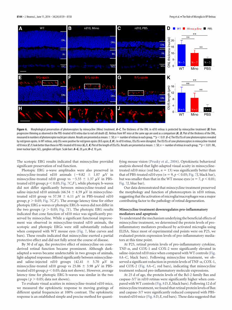
groups. The regions in the superior retina that were located at 200�m from the ONL were targeted for measurements (Fig. 6A–C).Rd10 mice treated with minocycline had a significantly thickerphotoreceptor nuclear layer (10.3 � 2.4 rows, n � 7 retinas; Fig.6D, red bar) when compared with the control rd10 eyes injectedwith PBS (5.9 � 1.1 rows, n � 5 retinas; p � 0.01; Fig. 6D, blackbar). A similar phenomenon was observed in the peripheral ret-ina (data not shown). The ONL thickness from WT C57BL/6retinas (17.0 � 3.2 rows, n � 5 retinas; Fig. 6D, blue bar) was alsomeasured for comparison. These results showed that minocy-
cline treatment provided a significantmorphological preservation of rodsthroughout the retina. In normal WTC57BL/6 mice, administration of minocy-cline did not affect ONL thickness (datanot shown).
In addition to the preservation of rods,we also observed relatively preservation ofcone inner and outer segments inminocycline-treated rd10 retinas. To bet-ter characterize this protective effect oncones, we stained vertical sections withanti-red/green opsin antibody. At the ageof 25 d, cones in the rd10 retina showeddisrupted inner segments (ISs)/outer seg-ments (OSs), which were not like the WT(Fig. 6E,H). Although the length of theOS/IS was shorter than that in the WTC57BL/6 mice (p � 0.01; Fig. 6E,H,K), itwas significantly longer than that in thesaline-injected controls (p � 0.01; Fig.6G, J,K). Therefore, minocycline pre-served both rods and cone morphology inrd10 retinas.
Minocycline treatment preservesphotoreceptor functionTo assess whether minocycline treatmentpreserved photoreceptor cell function andvisual behavior in rd10 mice, we per-formed functional studies by measuringscotopic and photopic ERGs and optomo-tor responses at P25. Retinal function inminocycline-treated rd10 eyes was mark-edly better preserved with larger scotopicand photopic ERG responses than in thePBS-treated rd10 eyes at P25 (Fig. 7). Mi-
nocycline treatment partially prevented the reduction in scotopicERG a-wave amplitudes when compared with PBS-treated rd10controls at two light intensities (p � 0.01; Fig. 7A,B,D,E). Com-pared with those treated with PBS, rd10 mice treated with mino-cycline also had greater mean amplitude of b-wave (p � 0.01; Fig.7A,B,D,E). A similar trend was also observed for average latencytime (time to peak). The latency time for scotopic ERG a- andb-waves in minocycline-treated rd10 mice was significantlyshorter than in PBS-injected rd10 controls (p � 0.01; Fig. 7G,H).
Figure 4. Quantification of activated microglial cells by flow cytometry analysis. A–C, Microglia analyzed by two-color flow cytometry displayed different patterns of CD11b and CD45 expressionin rd10 retinas treated with minocycline (Mino) treatment (B) and PBS (C). Retinas from WT mice at the same age are used as a comparison (A). CD45 expression is shown on the y-axis and CD11bexpression is shown on the x-axis. Region 2 shows the percentage of the subpopulation of CD11b �CD45 hi-activated microglia/macrophages. D, The percentages of activated microglia/macro-phages over total retinal cells were quantified. Mean � SD; **p � 0.01.
Figure 5. Minocycline (Mino) treatment reduces photoreceptor apoptosis. The nuclear layers were stained with DAPI (A–C,blue). No TUNEL-positive photoreceptor cells (red) are observed in the ONL at both P16 (D) and P19 (E). TUNEL-positive photore-ceptor cells begin to appear in the ONL at P22 (F ). G, Quantification of TUNEL-positive photoreceptor cells in the ONL at three timepoints. Note that the number of TUNEL-positive photoreceptor cells significantly decreased in minocycline-treated rd10 mice atP19 and P22. Results are presented as mean � SD; **p � 0.01, n � number of retinas in each group. OPL, outer plexiform layer;INL, inner nuclear layer; IPL, inner plexiform layer; GCL, ganglion cell layer. Scale bar, 20 �m.
Peng et al. • The Role of Microglia in RP Retinas J. Neurosci., June 11, 2014 • 34(24):8139 – 8150 • 8143
The scotopic ERG results indicated that minocycline providedsignificant preservation of rod function.
Photopic ERG a-wave amplitudes were also preserved inminocycline-treated rd10 animals (�9.82 � 1.07 �V inminocycline-treated rd10 group vs �5.55 � 1.37 �V in PBS-treated rd10 group; p � 0.05; Fig. 7C,F), while photopic b-wavesdid not differ significantly between minocycline-treated andsaline-injected rd10 animals (66.54 � 4.59 �V in minocycline-treated rd10 group vs 57.58 � 6.11 �V in PBS-treated rd10group; p � 0.05; Fig. 7C,F). The average latency time for eitherphotopic ERG a-waves or photopic ERG b-waves did not differ inthe two groups (p � 0.05; Fig. 7I). The photopic ERG resultsindicated that cone function of rd10 mice was significantly pre-served by minocycline. While a significant functional improve-ment was observed in minocycline-treated rd10 animals, thescotopic and photopic ERGs were still substantially reducedwhen compared with WT mouse eyes (Fig. 7, blue curves andbars). These results indicated that minocycline exerted a partialprotective effect and did not fully arrest the course of disease.
By 30 d of age, the protective effect of minocycline on cone-derived retinal function became prominent. Although dark-adapted a-waves became undetectable in two groups of animals,light-adapted responses differed significantly between minocycline-and saline-injected rd10 groups (42.41 � 5.70 �V inminocycline-treated rd10 group vs 25.06 � 3.98 �V in PBS-treated rd10 group; p � 0.05; data not shown). However, averagelatency time for photopic ERG b-waves was similar in the twogroups (p � 0.05; data not shown).
To evaluate visual acuities in minocycline-treated rd10 mice,we measured the optokinetic response to moving gratings ofdifferent spatial frequencies at 100% contrast. The optokineticresponse is an established simple and precise method for quanti-
fying mouse vision (Prusky et al., 2004). Optokinetic behavioralanalysis showed that light-adapted visual acuity in minocycline-treated rd10 mice (red bar, n � 13) was significantly better thanthat of PBS-treated rd10 eyes (n � 9, p � 0.05; Fig. 7J, black bar),but was smaller than that in the WT mouse eyes (n � 7, p � 0.01;Fig. 7J, blue bar).
Our data demonstrated that minocycline treatment preservedthe morphology and function of photoreceptors in rd10 retinas,suggesting that the activation of microglia/macrophages was a majorcontributing factor to the pathology of retinal degeneration.
Minocycline treatment downregulates pro-inflammatorymediators and apoptosisTo understand the mechanism underlying the beneficial effects ofminocycline treatment, we determined the protein levels of pro-inflammatory mediators produced by activated microglia usingELISA. Since most of experimental end points were on P25, weevaluated protein expression levels of pro-inflammatory media-tors at this time point.
At P25, retinal protein levels of pro-inflammatory cytokine,TNF-�, and COX-1 and COX-2 were significantly elevated insaline-injected rd10 mice when compared with WT controls (Fig.8A–C, black bars). Following minocycline treatment, we ob-served a significant reduction in protein levels of TNF-�, COX-1,and COX-2 (Fig. 8A–C, red bars), indicating that minocyclinetreatment reduced pro-inflammatory molecule expressions.
At 25 d of age, the protein levels of the Bcl-2 family Bax andcaspase-3/7 in rd10 retinas were significantly higher when com-pared with WT controls (Fig. 8D,E, black bars). Following 12 d ofminocycline treatment, we found that retinal protein levels of Baxand caspase-3/7 were significantly reduced compared with PBS-treated rd10 mice (Fig. 8D,E, red bars). These data suggested that
Figure 6. Morphological preservation of photoreceptors by minocycline (Mino) treatment. A–C, The thickness of the ONL in rd10 retinas is protected by minocycline treatment (B) fromprogressive thinning as observed in the PBS-treated rd10 retina due to rod cell death (C). Retinas from WT mice at the same age are used as a comparison (A). D, Plot of the thickness of the ONL,measured in numbers of photoreceptor nuclei per column. Results are presented as means � SD; n � number of retinas in each group, **p � 0.01. E–J, The OSs/ISs of cone photoreceptors revealedby red/green opsins. In WT retinas, only OSs were positive for red/green opsins (R/G opsin; E, H ). In rd10 retinas, OSs/ISs were disrupted. The OS/ISs of cone photoreceptors in minocycline-treatedrd10 mice (F, I ) look better than those in PBS-treated rd10 mice (G, J ). K, Plot of the length of OSs/ISs. Results are presented as means � SD; n � number of retinas in each group, **p � 0.01. INL,inner nuclear layer; GCL, ganglion cell layer. Scale bars: A–G, 20 �m; H–J, 10 �m.
8144 • J. Neurosci., June 11, 2014 • 34(24):8139 – 8150 Peng et al. • The Role of Microglia in RP Retinas

the protective effects of minocycline were mediated through itsanti-inflammatory and anti-apoptotic properties, leading topreservation of structure and function of photoreceptors in rd10mice.
COX inhibition provides significant protection ofphotoreceptor functionMinocycline has been shown to reduce microglial activation andimprove neuronal survival in different models of neurodegenera-tive disease (Yrjanheikki et al., 1998; Yong et al., 2004; Zhang etal., 2004; Fan et al., 2007; Lazarini et al., 2012; Schafer et al., 2012).We observed the same phenomenon in rd10 retinas. Addition-ally, we found that minocycline could directly exert its neuropro-tective effects. To further confirm that the suppression ofmicroglial activation was neuroprotective in rd10 retinas, we ap-plied the COX-1-specific inhibitor SC-560 to suppress microgliaactivation (Choi et al., 2008; García-Bueno et al., 2009; Griffin etal., 2013). Because of its predominant expression in microglia,COX-1 facilitates activation of microglia and supports neuroin-flammatory processes in several models of neurodegenerativedisorders (García-Bueno et al., 2009; Griffin et al., 2013). Wefound that SC-560 significantly reduced the expression of pro-
inflammatory cytokine TNF-� in rd10retinas when compared with vehicle-treated rd10 mice (Fig. 8F). Interestingly,we found that SC-560 slightly downregu-lated the expression of COX-2 protein(Fig. 8G; p � 0.05). Meanwhile, the ex-pression levels of BAX and caspase-3/7were also reduced in SC-560-treatedrd10 mice compared with vehicle-treatedrd10 mice (Fig. 8H, I). Quantification ofthe number of photoreceptor nucleiwithin the ONL showed that the photore-ceptors in SC-560-treated rd10 retinaswere significantly preserved (Fig. 8J; p �0.05). Therefore, suppression of microgliaactivation by SC-560 reduced the productionof microglia-derived pro-inflammatory fac-tors, leading to the preservation of photo-receptors and subsequent reduction ofBAX and caspase-3/7 production.
Effect of Cx3cr1 on microglialactivation in rd10 retinasIn the CNS, fractalkine (Cx3cl1) is pri-marily expressed by neurons, whereas itsunique G-protein-coupled receptor (Cx3cr1)is specifically expressed by microglia. Ithas been suggested that Cx3cl1 is impor-tant for microglial regulation and com-munication between microglia andneurons (Cardona et al., 2006). We thusinvestigated whether Cx3cr1 receptor wasinvolved in regulating microglia activa-tion in rd10 retinas. Previous studies haveshown that retinas do not exhibit struc-tural differences in retinal lamination oroverall morphology and distribution ofmicroglia in the absence of Cx3cr1 signal-ing in early adulthood (Liang et al., 2009).Our ERG measurements confirmed themorphological analysis. We showed that
scotopic and photopic ERG responses were indistinguishable be-tween Cx3cr1GFP/GFP mice and Cx3cr1�/GFP mice (Fig. 9, bluecurves and bars), suggesting Cx3cr1 deletion did not affect retinalstructure and function during retina development.
We then studied whether Cx3cr1 deficiency affected microgliaresponse in rd10 retinas. To this end, we backcrossed Cx3cr1knock-out mice (Cx3cr1GFP/GFP) into rd10 background and gen-erated a new line of rd10/Cx3cr1GFP/GFP mice. At P25, scotopicand photopic ERG a-wave amplitudes were virtually indistin-guishable between rd10/Cx3cr1�/GFP and rd10/Cx3cr1GFP/GFP
mice (p � 0.05; Fig. 9, black curves and bars), indicating thesimilar level of loss of functional rods in rd10 mice with or with-out Cx3cr1 at P25. However, scotopic and photopic ERG b-waveamplitudes were significantly smaller in rd10/Cx3cr1GFP/GFP micethan in rd10/Cx3cr1�/GFP mice (Fig. 9, black curves and bars),indicating that Cx3cr1 deficiency exaggerated cone photorecep-tor degeneration in rd10 mice.
To correlate ERG results with morphological changes, we thencounted the number of ONL rows. There was a significant differ-ence in the ONL thickness between rd10/Cx3cr1�/GFP and rd10/Cx3cr1GFP/GFP mice at P25 (5.8 � 1.2 rows in rd10/Cx3cr1�/GFP
group vs 4.2 � 1.0 rows in rd10/Cx3cr1GFP/GFP group; n � 10; p �
Figure 7. Functional preservation of photoreceptors by minocycline (Mino) treatment. A, B, Representative scotopic ERGresponses to 0.01 and 3 cd-s/m 2 light intensities from rd10 mice at P25 treated with minocycline (red curve) or PBS (dark curve)from P13 to P24. Scotopic ERG responses from C57BL/6 (blue curve) are shown as comparisons. D, E, Average scotopic a-wave andb-wave amplitudes elicited at 0.01cd-s/m 2 (D) and 3 cd-s/m 2 (E) light intensities from normal C57 BL/6 (blue), minocycline-treated (red), and PBS-treated (dark) rd10 eyes. Data are expressed as the mean � SD. C, Representative photopic ERGs elicited at3 cd-s/m 2 light intensity from the same three groups of mice as scotopic recording. F, Averaged photopic a-wave and b-waveamplitudes elicited at 3 cd-s/m 2 light intensity from normal C57 BL/6 (blue), minocycline-treated (red), and PBS-treated (dark)rd10 eyes. G–I, Average latency time (time to peak) for scotopic ERG a-waves and b-waves at 0.01cd-s/m 2 (G) and 3 cd-s/m 2 (H )light intensities, and for photopic ERG a-waves and b-waves at 3 cd-s/m 2 (I ). J, Photopic visual acuity measured by the optokineticresponse. Values are means and SDs. n � animal numbers; *p � 0.05 and **p � 0.01.
Peng et al. • The Role of Microglia in RP Retinas J. Neurosci., June 11, 2014 • 34(24):8139 – 8150 • 8145

0.05; Fig. 10A,C,F). In addition, the sig-nificant difference in the ONL thicknesswas detected at P19 as well (17.3 � 2.1rows in rd10/Cx3cr1�/GFP group vs 11.2 �1.7 rows in rd10/Cx3cr1GFP/GFP group;n � 10; p � 0.01; Fig. 10F). Therefore,Cx3cr1 deficiency caused more rod loss inrd10 retinas. On the other hand, conedensity was found to be comparablebetween rd10/Cx3cr1�/GFP and rd10/Cx3cr1GFP/GFP mice at P25. However, conemorphology was severely damaged inrd10/Cx3cr1GFP/GFP mice. At P25, the IS ofcones was seen in rd10/Cx3cr1�/GFP mice(Fig. 10B), while cones had flattened ISsin rd10/Cx3cr1GFP/GFP mice (Fig. 10D).Moreover, the red/green-opsins were ex-pressed primarily in the OS of cones. Thecone axons and pedicles were positive forred/green-opsins in rd10/Cx3cr1GFP/GFP
mice (Fig. 10D), while red/green-opsinswere restricted to the ISs of cones in rd10/Cx3cr1�/GFP mice. The redistribution ofthe red/green-opsins in cones is a sign ofdamage to cone photoreceptors (Haverkamp et al., 2005; Lin etal., 2009). The redistributions of the opsins in cones of rd10/Cx3cr1GFP/GFP mice indicated the additional neurotoxic effect ofCx3cr1 deficiency in cones.
To understand the mechanisms underlying the acceleratedphotoreceptor degeneration in rd10/Cx3cr1GFP/GFP mice, we ex-amined the state of microglial activation. We found that Cx3cr1deletion led to increased numbers of microglia/macrophages inthe ONL when compared with rd10/Cx3cr1�/GFP mice at bothP16 and P19 (Fig. 10E), suggesting that Cx3cr1 deficiency dys-regulated microglia activation in rd10 retinas. Therefore, the ad-ditional neurotoxic effect of Cx3cr1 deficiency on photoreceptorsin rd10 retinas was probably associated with increased inflamma-tory responses.
Furthermore, we tested whether Cx3cr1 deficiency affectedminocycline treatment. We treated rd10/Cx3cr1GFP/GFP micewith minocycline or PBS for 12 d, starting from P13. We per-
formed functional studies by measuring scotopic and photopicERGs at P25. Compared with those treated with PBS,minocycline-treated rd10/Cx3cr1GFP/GFP mice had greater meanamplitudes of a- and b-waves under scotopic conditions (p �0.01; Fig. 11A,B,D,E). A similar trend was also observed for av-erage latency time for b-waves under scotopic conditions (p �0.01; Fig. 11G,H). Under photopic condition, however, we foundthat b-waves did not differ between minocycline-treated andPBS-injected rd10/Cx3cr1GFP/GFP mice (p � 0.05; Fig. 11C,F),whereas a-wave amplitudes were significantly smaller in PBS-injected rd10/Cx3cr1GFP/GFP mice than in minocycline-injectedrd10/Cx3cr1GFP/GFP mice (p � 0.05; Fig. 11C,F). We did notobserve any change in average latency times for both a- andb-waves under photopic condition (p � 0.05; Fig. 11I). There-fore, minocycline treatment also delayed the progression of pho-toreceptor degeneration in Cx3cr1-deficient rd10 retinas,indicating that minocycline exerted its neuroprotective effectsthrough Cx3cr1-independent mechanisms.
Figure 8. Downregulation of pro-inflammatory cytokines and pro-apoptotic molecules by minocycline (Mino) treatment or by COX-1-specific inhibitor SC-560. The protein levels of thesemolecules were assessed using ELISA. A–E, Results from minocycline treatment. Pro-inflammatory cytokine TNF-� (A), COX-1 (B), and COX-2 (C) are upregulated in rd10 retinas (black bars) anddownregulated following minocycline treatment (red bars). The similar trends were observed for pro-apoptotic molecules Bax (D) and caspase-3/7 (E). F–J, Results from SC-560 treatment.Pro-inflammatory cytokines TNF-� (F ) and COX-2 (B) are downregulated following SC-560 treatment. The similar trends are observed in Bax (H ) and caspase-3/7 (I ). J, Plot of the thickness of theONL, measured in numbers of photoreceptor nuclei per column. Results are presented as means � SD; n � number of retinas in each group, *p � 0.05.
Figure 9. Cx3cr1 deficiency leads to additional loss of photoreceptor function in rd10 retinas. Cx3cr1 deletion did not affectretinal function in WT retinas. Scotopic and photopic ERG responses are indistinguishable between Cx3cr1GFP/GFP mice andCx3cr1�/GFP mice (blue curves in A–C and bars in D–F ). Scotopic and photopic ERG a-wave amplitudes are virtually comparablebetween rd10 mice with or without Cx3cr1 deletion (black curves and bars). However, scotopic and photopic ERG b-wave ampli-tudes are significantly smaller in rd10 mice with Cx3cr1 deletion, compared with rd10 mice without Cx3cr1 deletion (black curvesin A–C and bars in D–F ). Values are means and SDs. n � animal numbers; *p � 0.05 and **p � 0.01.
8146 • J. Neurosci., June 11, 2014 • 34(24):8139 – 8150 Peng et al. • The Role of Microglia in RP Retinas

DiscussionThe main finding of this study was that microglia activationplayed pathological roles in the course of retinal degeneration inthe rd10 mouse model of RP and inhibition of microglia activa-tion by minocycline protected photoreceptors against apoptosis.First, we demonstrated that microglia activation was an earlyalteration in the rd10 retina, potentially contributing to diseaseprogression. Second, suppression of microglia activation by mi-nocycline and COX-1 specific inhibitor SC-560 resulted in reduc-tion of both pro-inflammatory cytokines and pro-apoptoticmediators, leading to significant structural and functional pres-ervation in rd10 retinas. Third, we identified that minocyclineexerted its neuroprotective effects through both anti-apoptoticand anti-inflammatory mechanisms. Finally, we found thatCx3cr1 deficiency dysregulated microglia activation and subse-quently increased photoreceptor vulnerability in rd10 mice, sug-gesting Cx3cr1 might protect against microglial neurotoxicity.Collectively, our data demonstrated that activated microglia con-tributed to the severity of RP retinas. Therefore, modulating mi-
croglia activation could be a potentialtreatment strategy aimed at improvingphotoreceptor survival in human RP.
Microglia activation has been reportedto be an early event in the development ofretinal degeneration (Roque et al., 1996;Gupta et al., 2003; Zeiss and Johnson,2004; Zeng et al., 2005; Gehrig et al., 2007;Sasahara et al., 2008; Ebert et al., 2009;Zhou et al., 2011; Sheets et al., 2013; Yo-shida et al., 2013a, b). Our data from rd10retinas were in agreement with these pre-vious reports. Microglia activation and in-filtration in the subretinal space occurredin rd10 mice at P16, whereas photorecep-tor apoptosis started at P19, 3 d later. Toconfirm whether microglia activationcontributed to the progression of photo-receptor atrophy, we treated rd10 micewith minocycline from P13, before theinitiation of microglia activation. Mino-cycline has been widely used as an inhibi-tor of microglial activation for differentCNS neurodegenerative diseases (Yrjan-heikki et al., 1998; Yong et al., 2004; Zhanget al., 2004; Fan et al., 2007; Lazarini et al.,2012; Schafer et al., 2012). Indeed, we ob-served that minocycline significantlyreduced microglia activation and infiltra-tion to the ONL in the rd10 retinas. More-over, minocycline treatment significantlyreduced TUNEL positivity and providedsignificant morphological preservation ofphotoreceptors throughout the retina.ERG recordings and optomotor responsesfurther confirmed that minocycline treat-ment provided functional photoreceptorpreservation in rd10 mice. These resultsprovided clear evidence for the involve-ment of microglial activation in exacer-bating photoreceptors’ death in rd10retinas. Similarly, inhibition of microglialactivation using minocycline has beendemonstrated in other experimental
models of retinal degeneration caused by light (Kohno et al.,2013), subretinal hemorrhage (Zhao et al., 2011), and geneticimpairments (rds mice; Hughes et al., 2004).
We identified that minocycline treatment exerted neuropro-tective effects on photoreceptor apoptosis through two mecha-nisms. The first mechanism was through direct anti-apoptoticeffect on photoreceptors. A previous study has reported thatsome of the photoreceptors in the ONL of rd10 retinas express anactivated form of caspase-3 (Gargini et al., 2007). Similarly, theincreased activity of caspase-3 has also been reported in the rd1mouse model of RP (Yoshizawa et al., 2002; Zeiss et al., 2004;Sharma and Rohrer, 2007). Minocycline has previously beenshown to inhibit the caspase-3 activation (Chen et al., 2000;Hughes et al., 2004) and the release of cytochrome c from themitochondria in mice (Zhu et al., 2002). In agreement with pre-vious findings, we found that expression level of caspase-3/7protein was significantly increased in rd10 retinas. Adminis-tration of minocycline led to a significant reduction ofcaspase-3/7 and Bax proteins in rd10 retinas when compared
Figure 10. Cx3cr1 deficiency dysregulates microglia activation and increases photoreceptor apoptosis in rd10 retinas.A–D, Cx3cr1 deficiency caused more rod loss in rd10 retinas (C) than in rd10 retinas with Cx3cr1 (A). At P25, conephotoreceptors in rd10/Cx3cr1�/GFP retinas maintain ISs (B), and the ISs of cone photoreceptors in rd10/Cx3cr1GFP/GFP
retinas become flattened, and redistribution of red/green opsins (R/G opsin) to cell bodies and cone axonal terminals occursin cones of rd10/Cx3cr1GFP/GFP retinas (D). E, Quantification of microglial cells that migrated into the ONL in vertical sectionsof rd10 retinas with or without Cx3cr1 deficiency at P13, P16, and P19. F, The ONL thickness, measured by photoreceptorlayers, is significantly different between rd10 retinas with or without Cx3cr1 deficiency at both P19 and P25. Results arepresented as means � SD; n � number of retinas in each group, *p � 0.05 and **p � 0.01. INL, inner nuclear layer; GCL,ganglion cell layer; Scale bar, 20 �m.
Peng et al. • The Role of Microglia in RP Retinas J. Neurosci., June 11, 2014 • 34(24):8139 – 8150 • 8147

with PBS-treated rd10 controls, whichmight reflect its direct action on photore-ceptor apoptosis. Since comparableamounts of apoptotic photoreceptorswere still observed in minocycline-treatedrd10 retinas, we thus believed that its anti-apoptotic effect might not be the primarymechanism involved in our study.
The second mechanism was throughits anti-inflammatory effect. Previously, ithas been reported that minocycline re-duces microglial activation and improvesneuronal survival in models of glaucoma,subretinal hemorrhage, and rds mice(Yang et al., 2007; Bosco et al., 2008; Zhaoet al., 2011). Similarly, minocycline hasbeen shown to prevent microglial activa-tion against cerebral ischemia and Parkin-son’s disease (Yrjanheikki et al., 1999; Wuet al., 2002). Consistent with previousfindings, we also observed suppression ofmicroglia activation and reduction of pro-inflammatory cytotoxic mediator TNF-�,COX-1, and COX-2 in rd10 retinas fol-lowing minocycline treatment.
Moreover, inhibition of microglial ac-tivation using minocycline resulted fromthe partial inhibition of COX-1 andCOX-2 induction, key enzymes involvedin the production of pro-inflammatoryprostanoids (García-Bueno et al., 2009;Griffin et al., 2013; Jiang et al., 2013). The upregulation of COX-2has been demonstrated in other chronic neurodegenerative dis-eases, such as Parkinson’s disease (Teismann et al., 2003a,b; Choiet al., 2009). Moreover, it has been shown that genetic deletionor pharmacological inhibition of COX-2 increases survival ofdopaminergic neurons in animal models of Parkinson’s dis-ease (Teismann et al., 2003a). Here we showed that COX-2protein level was upregulated in rd10 retinas. After minocy-cline treatment, we observed a significant reduction of COX-2protein. COX-1 has been reported to facilitate neuroinflam-matory processes in several models of neurodegenerative dis-orders (García-Bueno et al., 2009; Griffin et al., 2013). Geneticdeletion or pharmacological inhibition of COX-1 attenuatesinflammatory response and neuronal damage (Choi et al.,2008; Choi and Bosetti, 2009; García-Bueno et al., 2009; Grif-fin et al., 2013). Minocycline treatment led to a significantreduction of COX-1 protein in rd10 retinas. Therefore, mino-cycline might partially inhibit COX-1 and COX-2 activation,which led to the reduction of microglial-derived mediatorsand survival of photoreceptors.
It has been proposed that Cx3cl1/Cx3cr1 signaling pathwayregulates the communication of microglia with neurons in thenervous system (Harrison et al., 1998). However, the studies onthe Cx3cl1/Cx3cr1 pathway in neurological disorders producedcontrasting results. Cx3cr1 knock-out has been shown to eitherincrease (Meucci et al., 2000; Cardona et al., 2006) or decrease(Fuhrmann et al., 2010) neurotoxicity in several models of theCNS insult. We found that Cx3cr1 deficiency led to an increasedaccumulation of activated microglia in the ONL and increasedphotoreceptor vulnerability, which was confirmed by scotopicand photopic ERGs (Fig. 9) and immunocytochemistry (Fig. 10).Less ONL were observed in rd10 mice without Cx3cr1 than in
rd10 mice with Cx3cr1, suggesting Cx3cr1 deficiency caused ad-ditional rod loss. However, similar a-wave amplitudes under sco-topic conditions were observed in rd10 mice with or withoutCx3cr1 at P25 (Fig. 9). Given the fact that the majority of photo-receptors degenerated by P25 in the rd10 retina (Gargini et al.,2007), which largely contributed to the dramatic reduction in thea-wave, it was not surprising that additional rod loss in Cx3cr1-deficient rd10 mice did not cause any significant change ina-wave amplitudes by P25. A significant drop in the ONL wasobserved in rd10 mice without Cx3cr1 by P19 (Fig. 10F). After thepeak of rod death, the difference in the ONL thickness was nar-rowed between rd10 mice with or without Cx3cr1 by P25 (Fig.10F). Therefore, the loss of more ONL rows in rd10 mice withoutCx3cr1 by P25 did not cause any further significant reduction ina-wave amplitudes. On the other hand, although Cx3cr1 defi-ciency did not result in any cone loss in rd10 mice, it did exacer-bate morphological changes and functional loss of cones.Photopic ERG b-wave amplitudes were significantly smallerwhen compared with rd10 without Cx3cr1 deficiency. TheCx3cl1/Cx3cr1 signaling pathway appeared to restrain microgliaactivation in rd10 retinas, and Cx3cr1 deficiency resulted in extramicroglia activation. The activated Cx3cr1-deficient microgliahad an additional neurotoxic effect on photoreceptor survival inrd10 retinas.
In summary, our study provided strong support to the con-cept that activated microglia were important contributors to theoverall apoptosis of photoreceptors in rd10 retinas. The suppres-sion of microglia activation by minocycline or SC-560 reducedmicroglia-mediated photoreceptor death. Our data suggestedthat therapeutic interventions aimed at preventing the loss ofphotoreceptor cells can be through the inhibition of microgliaactivation.
Figure 11. Functional preservation of photoreceptors in Cx3cr1GFP/GFP mice following minocycline (Mino) treatment. A–B,Representative scotopic ERG responses to 0.01 and 3 cd-s/m 2 light intensities from rd10 mice treated with minocycline (red curves)or PBS (dark curves) from P13 to P24. D, E, Average scotopic a-wave and b-wave amplitudes elicited at 0.01 cd-s/m 2 (D) and 3cd-s/m 2 (E) light intensities. C, Representative photopic ERG responses to 3 cd-s/m 2 light intensity. F, Averaged photopic a-waveand b-wave amplitudes elicited at 3 cd-s/m 2 light intensity. G–I, Average latency time (time to peak) for scotopic ERG a-waves andb-waves at 0.01 cd-s/m 2 (G) and 3 cd-s/m 2 (H ) light intensities, and for photopic ERG a-waves and b-waves at 3 cd-s/m 2 (I ). Dataare expressed as the mean � SD. n � animal numbers; **p � 0.01.
8148 • J. Neurosci., June 11, 2014 • 34(24):8139 – 8150 Peng et al. • The Role of Microglia in RP Retinas

ReferencesBlock ML, Zecca L, Hong JS (2007) Microglia-mediated neurotoxicity: un-
covering the molecular mechanisms. Nat Rev Neurosci 8:57– 69. CrossRefMedline
Bosco A, Inman DM, Steele MR, Wu G, Soto I, Marsh-Armstrong N, Hub-bard WC, Calkins DJ, Horner PJ, Vetter ML (2008) Reduced retina mi-croglial activation and improved optic nerve integrity with minocyclinetreatment in the DBA/2J mouse model of glaucoma. Invest OphthalmolVis Sci 49:1437–1446. CrossRef Medline
Cardona AE, Pioro EP, Sasse ME, Kostenko V, Cardona SM, Dijkstra IM,Huang D, Kidd G, Dombrowski S, Dutta R, Lee JC, Cook DN, Jung S, LiraSA, Littman DR, Ransohoff RM (2006) Control of microglial neurotox-icity by the fractalkine receptor. Nat Neurosci 9:917–924. CrossRefMedline
Chen M, Ona VO, Li M, Ferrante RJ, Fink KB, Zhu S, Bian J, Guo L, FarrellLA, Hersch SM,Hobbs W, Vonsattel JP, Cha JH, Friedlander RM (2000)Minocycline inhibits caspase-1 and caspase-3 expression and delays mor-tality in a transgenic mouse model of Huntington disease. Nat Med 6:797–801. CrossRef Medline
Choi SH, Bosetti F (2009) Cyclooxygenase-1 null mice show reduced neu-roinflammation in response to beta-amyloid. Aging 1:234 –244. Medline
Choi SH, Langenbach R, Bosetti F (2008) Genetic deletion or pharmacolog-ical inhibition of cyclooxygenase-1 attenuate lipopolysaccharide-inducedinflammatory response and brain injury. FASEB J 22:1491–1501. Medline
Choi SH, Aid S, Bosetti F (2009) The distinct roles of cyclooxygenase-1 and-2 in neuroinflammation: implications for translational research. TrendsPharmacol Sci 30:174 –181. CrossRef Medline
Combadiere C, Feumi C, Raoul W, Keller N, Rodero M, Pezard A, Lavalette S,Houssier M, Jonet L, Picard E, Debre P, Sirinyan M, Deterre P, FerroukhiT, Cohen SY, Chauvaud D, Jeanny JC, Chemtob S, Behar-Cohen F, Senn-laub F (2007) CX3CR1-dependent subretinal microglia cell accumula-tion is associated with cardinal features of age-related maculardegeneration. J Clin Invest 117:2920 –2928. CrossRef Medline
Ebert S, Weigelt K, Walczak Y, Drobnik W, Mauerer R, Hume DA, Weber BH,Langmann T (2009) Docosahexaenoic acid attenuates microglial activa-tion and delays early retinal degeneration. J Neurochem 110:1863–1875.CrossRef Medline
Fan R, Xu F, Previti ML, Davis J, Grande AM, Robinson JK, Van Nostrand WE(2007) Minocycline reduces microglial activation and improves behav-ioral deficits in a transgenic model of cerebral microvascular amyloid.J Neurosci 27:3057–3063. CrossRef Medline
Fuhrmann M, Bittner T, Jung CK, Burgold S, Page RM, Mitteregger G, HaassC, LaFerla FM, Kretzschmar H, Herms J (2010) Microglial Cx3cr1knockout prevents neuron loss in a mouse model of Alzheimer’s disease.Nat Neurosci 13:411– 413. CrossRef Medline
García-Bueno B, Serrats J, Sawchenko PE (2009) Cerebrovascularcyclooxygenase-1 expression, regulation, and role in hypothalamic-pituitary-adrenal axis activation by inflammatory stimuli. J Neurosci 29:12970 –12981. CrossRef Medline
Gargini C, Terzibasi E, Mazzoni F, Strettoi E (2007) Retinal organization inthe retinal degeneration 10 (rd10) mutant mouse: a morphological andERG study. J Comp Neurol 500:222–238. CrossRef Medline
Gehrig A, Langmann T, Horling F, Janssen A, Bonin M, Walter M, Poths S,Weber BH (2007) Genome-wide expression profiling of the retinoschisin-deficient retina in early postnatal mouse development. Invest OphthalmolVis Sci 48:891–900. CrossRef Medline
Glass CK, Saijo K, Winner B, Marchetto MC, Gage FH (2010) Mechanismsunderlying inflammation in neurodegeneration. Cell 140:918 –934.CrossRef Medline
Griffin EW, Skelly DT, Murray CL, Cunningham C (2013) Cyclooxygenase-1-dependent prostaglandins mediate susceptibility to systemic inflammation-induced acute cognitive dysfunction. J Neurosci 33:15248 –15258.CrossRef Medline
Gupta N, Brown KE, Milam AH (2003) Activated microglia in human reti-nitis pigmentosa, late-onset retinal degeneration, and age-related macu-lar degeneration. Exp Eye Res 76:463– 471. CrossRef Medline
Hanisch UK, Kettenmann H (2007) Microglia: active sensor and versatileeffector cells in the normal and pathologic brain. Nat Neurosci 10:1387–1394. CrossRef Medline
Harrison JK, Jiang Y, Chen S, Xia Y, Maciejewski D, McNamara RK, Streit WJ,Salafranca MN, Adhikari S, Thompson DA, Botti P, Bacon KB, Feng L(1998) Role for neuronally derived fractalkine in mediating interactions
between neurons and CX3CR1-expressing microglia. Proc Natl Acad SciU S A 95:10896 –10901. CrossRef Medline
Hartong DT, Berson EL, Dryja TP (2006) Retinitis pigmentosa. Lancet 368:1795–1809. CrossRef Medline
Haverkamp S, Wassle H, Duebel J, Kuner T, Augustine GJ, Feng G, Euler T(2005) The primordial, blue-cone color system of the mouse retina.J Neurosci 25:5438 –5445. CrossRef Medline
Hughes EH, Schlichtenbrede FC, Murphy CC, Broderick C, van Rooijen N,Ali RR, Dick AD (2004) Minocycline delays photoreceptor death in therds mouse through a microglia-independent mechanism. Exp Eye Res78:1077–1084. CrossRef Medline
Iwakabe H, Katsuura G, Ishibashi C, Nakanishi S (1997) Impairment ofpupillary responses and optokinetic nystagmus in the mGluR6-deficientmouse. Neuropharmacology 36:135–143. CrossRef Medline
Jiang J, Quan Y, Ganesh T, Pouliot WA, Dudek FE, Dingledine R (2013)Inhibition of the prostaglandin receptor EP2 following status epilepticusreduces delayed mortality and brain inflammation. Proc Natl Acad SciU S A 110:3591–3596. CrossRef Medline
Kohno H, Chen Y, Kevany BM, Pearlman E, Miyagi M, Maeda T, PalczewskiK, Maeda A (2013) Photoreceptor proteins initiate microglial activationvia Toll-like receptor 4 in retinal degeneration mediated by all-trans-retinal. J Biol Chem 288:15326 –15341. CrossRef Medline
Langmann T (2007) Microglia activation in retinal degeneration. J LeukocBiol 81:1345–1351. CrossRef Medline
Lazarini F, Gabellec MM, Torquet N, Lledo PM (2012) Early activation ofmicroglia triggers long-lasting impairment of adult neurogenesis in theolfactory bulb. J Neurosci 32:3652–3664. CrossRef Medline
Liang KJ, Lee JE, Wang YD, Ma W, Fontainhas AM, Fariss RN, Wong WT(2009) Regulation of dynamic behavior of retinal microglia by CX3CR1signaling. Invest Ophthalmol Vis Sci 50:4444 – 4451. CrossRef Medline
Lin B, Masland RH, Strettoi E (2009) Remodeling of cone photoreceptorcells after rod degeneration in rd mice. Exp Eye Res 88:589 –599. CrossRefMedline
Lucin KM, Wyss-Coray T (2009) Immune activation in brain aging andneurodegeneration: too much or too little? Neuron 64:110 –122. CrossRefMedline
Meucci O, Fatatis A, Simen AA, Miller RJ (2000) Expression of CX3CR1chemokine receptors on neurons and their role in neuronal survival. ProcNatl Acad Sci U S A 97:8075– 8080. CrossRef Medline
Prusky GT, Alam NM, Beekman S, Douglas RM (2004) Rapid quantifica-tion of adult and developing mouse spatial vision using a virtual optomo-tor system. Invest Ophthalmol Vis Sci 45:4611– 4616. CrossRef Medline
Roque RS, Imperial CJ, Caldwell RB (1996) Microglial cells invade the outerretina as photoreceptors degenerate in Royal College of Surgeons rats.Invest Ophthalmol Vis Sci 37:196 –203. Medline
Saijo K, Glass CK (2011) Microglial cell origin and phenotypes in health anddisease. Nat Rev Immunol 11:775–787. CrossRef Medline
Sasahara M, Otani A, Oishi A, Kojima H, Yodoi Y, Kameda T, Nakamura H,Yoshimura N (2008) Activation of bone marrow-derived microglia pro-motes photoreceptor survival in inherited retinal degeneration. Am JPathol 172:1693–1703. CrossRef Medline
Schafer DP, Lehrman EK, Kautzman AG, Koyama R, Mardinly AR, YamasakiR, Ransohoff RM, Greenberg ME, Barres BA, Stevens B (2012) Micro-glia sculpt postnatal neural circuits in an activity and complement-dependent manner. Neuron 74:691–705. CrossRef Medline
Sharma AK, Rohrer B (2007) Sustained elevation of intracellular cGMPcauses oxidative stress triggering calpain-mediated apoptosis in photore-ceptor degeneration. Curr Eye Res 32:259 –269. CrossRef Medline
Sheets KG, Jun B, Zhou Y, Zhu M, Petasis NA, Gordon WC, Bazan NG(2013) Microglial ramification and redistribution concomitant with theattenuation of choroidal neovascularization by neuroprotectin D1. MolVis 19:1747–1759. Medline
Shen D, Cao X, Zhao L, Tuo J, Wong WT, Chan CC (2011) Naloxone ame-liorates retinal lesions in Ccl2/Cx3cr1 double-deficient mice via modula-tion of microglia. Invest Ophthalmol Vis Sci 52:2897–2904. CrossRefMedline
Teismann P, Tieu K, Choi DK, Wu DC, Naini A, Hunot S, Vila M, Jackson-Lewis V, Przedborski S (2003a) Cyclooxygenase-2 is instrumental inParkinson’s disease neurodegeneration. Proc Natl Acad Sci U S A 100:5473–5478. CrossRef Medline
Teismann P, Vila M, Choi DK, Tieu K, Wu DC, Jackson-Lewis V, Przedborski
Peng et al. • The Role of Microglia in RP Retinas J. Neurosci., June 11, 2014 • 34(24):8139 – 8150 • 8149

S (2003b) COX-2 and neurodegeneration in Parkinson’s disease. AnnN Y Acad Sci 991:272–277. Medline
Umino Y, Solessio E, Barlow RB (2008) Speed, spatial, and temporal tuningof rod and cone vision in mouse. J Neurosci 28:189 –198. CrossRefMedline
Wu DC, Jackson-Lewis V, Vila M, Tieu K, Teismann P, Vadseth C, Choi DK,Ischiropoulos H, Przedborski S (2002) Blockade of microglial activationis neuroprotective in the 1-methyl-4-phenyl-1,2,3,6-tetrahydropyridinemouse model of Parkinson disease. J Neurosci 22:1763–1771. Medline
Xiao J, Ching YP, Liong EC, Nanji AA, Fung ML, Tipoe GL (2013) Garlic-derived S-allylmercaptocysteine is a hepato-protective agent in non-alcoholic fatty liver disease in vivo animal model. Eur J Nutr 52:179 –191.CrossRef Medline
Yang LP, Li Y, Zhu XA, Tso MO (2007) Minocycline delayed photoreceptordeath in rds mice through iNOS-dependent mechanism. Mol Vis 13:1073–1082. Medline
Yong VW, Wells J, Giuliani F, Casha S, Power C, Metz LM (2004) Thepromise of minocycline in neurology. Lancet Neurol 3:744 –751. CrossRefMedline
Yoshida N, Ikeda Y, Notomi S, Ishikawa K, Murakami Y, Hisatomi T, EnaidaH, Ishibashi T (2013a) Clinical evidence of sustained chronic inflamma-tory reaction in retinitis pigmentosa. Ophthalmology 120:100 –105.CrossRef Medline
Yoshida N, Ikeda Y, Notomi S, Ishikawa K, Murakami Y, Hisatomi T, EnaidaH, Ishibashi T (2013b) Laboratory evidence of sustained chronic in-flammatory reaction in retinitis pigmentosa. Ophthalmology 120:e5– e12.CrossRef Medline
Yoshizawa K, Kiuchi K, Nambu H, Yang J, Senzaki H, Kiyozuka Y, Shikata N,Tsubura A (2002) Caspase-3 inhibitor transiently delays inherited reti-nal degeneration in C3H mice carrying the rd gene. Graefes Arch Clin ExpOphthalmol 240:214 –219. CrossRef Medline
Yrjanheikki J, Keinanen R, Pellikka M, Hokfelt T, Koistinaho J (1998) Tet-racyclines inhibit microglial activation and are neuroprotective in global
brain ischemia. Proc Natl Acad Sci U S A 95:15769 –15774. CrossRefMedline
Yrjanheikki J, Tikka T, Keinanen R, Goldsteins G, Chan PH, Koistinaho J(1999) A tetracycline derivative, minocycline, reduces inflammation andprotects against focal cerebral ischemia with a wide therapeutic window.Proc Natl Acad Sci U S A 96:13496 –13500. CrossRef Medline
Zeiss CJ, Johnson EA (2004) Proliferation of microglia, but not photorecep-tors, in the outer nuclear layer of the rd-1 mouse. Invest Ophthalmol VisSci 45:971–976. CrossRef Medline
Zeiss CJ, Neal J, Johnson EA (2004) Caspase-3 in postnatal retinal develop-ment and degeneration. Invest Ophthalmol Vis Sci 45:964 –970. CrossRefMedline
Zeng HY, Zhu XA, Zhang C, Yang LP, Wu LM, Tso MO (2005) Identifica-tion of sequential events and factors associated with microglial activation,migration, and cytotoxicity in retinal degeneration in rd mice. InvestOphthalmol Vis Sci 46:2992–2999. CrossRef Medline
Zhang C, Lei B, Lam TT, Yang F, Sinha D, Tso MO (2004) Neuroprotectionof photoreceptors by minocycline in light-induced retinal degeneration.Invest Ophthalmol Vis Sci 45:2753–2759. CrossRef Medline
Zhao L, Ma W, Fariss RN, Wong WT (2011) Minocycline attenuates pho-toreceptor degeneration in a mouse model of subretinal hemorrhage mi-croglial: inhibition as a potential therapeutic strategy. Am J Pathol 179:1265–1277. CrossRef Medline
Zhou Y, Sheets KG, Knott EJ, Regan CE Jr, Tuo J, Chan CC, Gordon WC,Bazan NG (2011) Cellular and 3D optical coherence tomography assess-ment during the initiation and progression of retinal degeneration in theCcl2/Cx3cr1-deficient mouse. Exp Eye Res 93:636 – 648. CrossRefMedline
Zhu S, Stavrovskaya IG, Drozda M, Kim BY, Ona V, Li M, Sarang S, Liu AS,Hartley DM, Wu DC, Gullans S, Ferrante RJ, Przedborski S, Kristal BS,Friedlander RM (2002) Minocycline inhibits cytochrome c release anddelays progression of amyotrophic lateral sclerosis in mice. Nature 417:74 –78. CrossRef Medline
8150 • J. Neurosci., June 11, 2014 • 34(24):8139 – 8150 Peng et al. • The Role of Microglia in RP Retinas