Embed Size (px)
Citation preview

Neuroanatomy, Neurochemistry and Behavioral Health
Robert Walker, M.S.W., L.C.S.W.
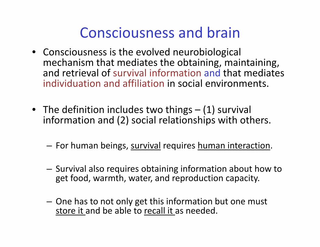
Consciousness and brain• Consciousness is the evolved neurobiological mechanism that mediates the obtaining, maintaining, and retrieval of survival information and that mediates individuation and affiliation in social environments.
• The definition includes two things – (1) survival information and (2) social relationships with others.
– For human beings, survival requires human interaction.
– Survival also requires obtaining information about how to get food, warmth, water, and reproduction capacity.
– One has to not only get this information but one must store it and be able to recall it as needed.
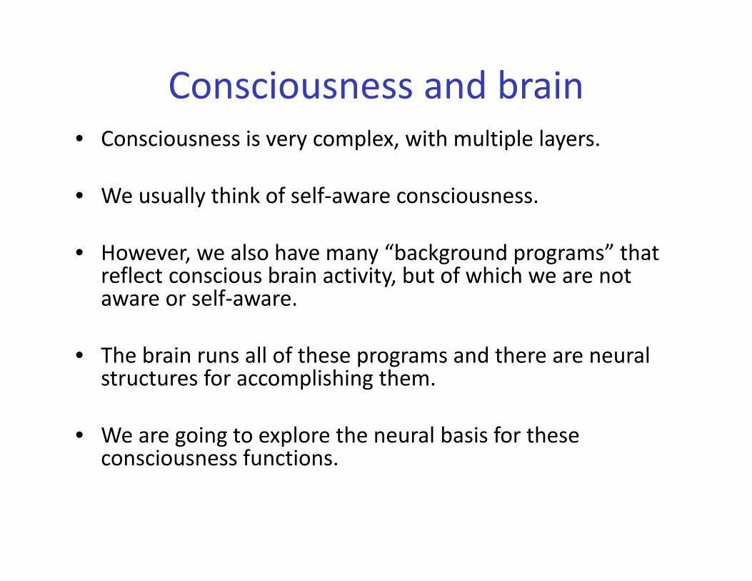
Consciousness and brain• Consciousness is very complex, with multiple layers.
• We usually think of self‐aware consciousness.
• However, we also have many “background programs” that reflect conscious brain activity, but of which we are not aware or self‐aware.
• The brain runs all of these programs and there are neural structures for accomplishing them.
• We are going to explore the neural basis for these consciousness functions.
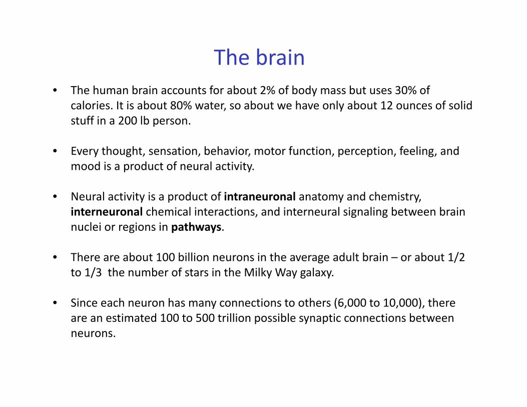
• The human brain accounts for about 2% of body mass but uses 30% of calories. It is about 80% water, so about we have only about 12 ounces of solid stuff in a 200 lb person.
• Every thought, sensation, behavior, motor function, perception, feeling, and mood is a product of neural activity.
• Neural activity is a product of intraneuronal anatomy and chemistry, interneuronal chemical interactions, and interneural signaling between brain nuclei or regions in pathways.
• There are about 100 billion neurons in the average adult brain – or about 1/2 to 1/3 the number of stars in the Milky Way galaxy.
• Since each neuron has many connections to others (6,000 to 10,000), there are an estimated 100 to 500 trillion possible synaptic connections between neurons.
The brain
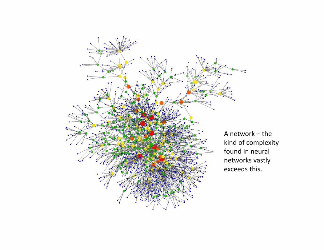
A network – the kind of complexity found in neural networks vastly exceeds this.
Development• Contrary to what used to be thought, brain development only
occurs in the context of environmental stimuli.
• Development parallels brain structure complexity in that the most primitive areas are instantiated first and higher cortical functions last.
• The brain develops from bottom up.
• The “bottom” includes the most basic survival emotions –fight/flight and related arousal systems.
• Each developmental phase is marked by being “set” – that is some basal rates of activation and brain region interaction get hard‐wired to certain levels based on what experience says is necessary.
Development• Each developmental phase is marked by being “set” – that is some
basal rates of activation and brain region interaction get hard‐wired to certain levels based on what experience says is necessary.
• As each level of brain anatomy and chemistry is fixed, all subsequent levels are predicated on factors in the preceding level.
• Thus, an arousal system that is fixed to over‐react to any fearful stimuli sets some constraints on limbic system development and on to higher cortical development as well.
• The foundation affects what kind of building, how high, how resilient to what kinds of winds and earthquakes.


The basic structures involved throughout development
• We will discuss four central levels of the brain in the context of development.
• First is the brain stem, where basic survival functions are carried out.
• Next is the diencephalon that mediates key autonomic functions and arousability. Many different functions are linked to this region including the sensory systems such as the auditory, somatic, visceral, gustatory and visual systems.
• Next higher up is the limbic system where other basic emotions and declarative memory are mediated.
• Last, is the neocortex where logic, language, pattern detecting and analyzing occur.
The natural development: Nature/nurture
• By age 2 one has the greatest number of neurons than at any other point in life.
• The efficient functioning of the brain requires pruning of excessive connections and too many neurons.
• Thus beginning around age 2, the neurons begin this pruning process in response to external stimulation of certain pathways.
• Plus, all environmental contributions to brain development trigger development of homeostatic mechanisms that are adjusted to the experiences of the infant/toddler’s exposures. More on this later...

The natural development: nature/nurture
• Healthy infant stimulation guides this process toward nurture of positive neurobehavior and neurodevelopment.
• Mirroring enhances development of affective and cognitive pathways.
• Each stimulative act strengthens a neural pathway.
• Violent and/or under‐stimulated (neglectful) environments affect this development very differently.
• Environments characterized by aggression, stimulate fight/flight pathways and the arousal system over‐develops in one form or another.
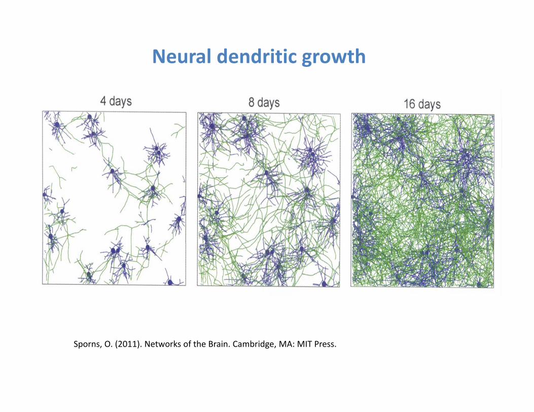
Sporns, O. (2011). Networks of the Brain. Cambridge, MA: MIT Press.
Neural dendritic growth

Sporns, O. (2011). Networks of the Brain. Cambridge, MA: MIT Press.
Neural networks
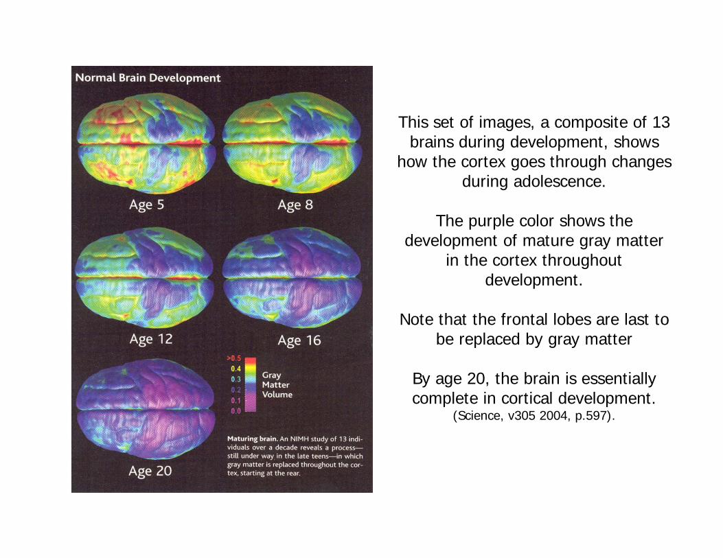
This set of images, a composite of 13 brains during development, shows
how the cortex goes through changes during adolescence.
The purple color shows the development of mature gray matter
in the cortex throughout development.
Note that the frontal lobes are last to be replaced by gray matter
By age 20, the brain is essentially complete in cortical development.
(Science, v305 2004, p.597).
Moving from neurons to larger anatomical structures
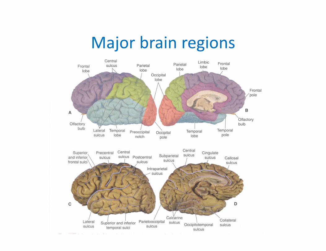
Major brain regions

Orbital frontal cortex

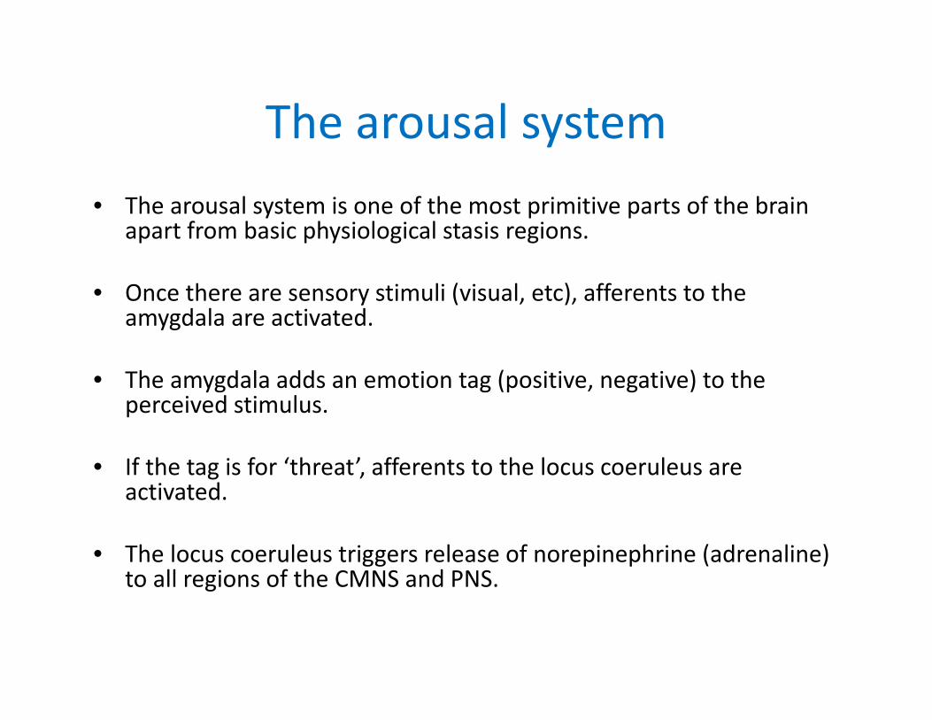
The arousal system• The arousal system is one of the most primitive parts of the brain
apart from basic physiological stasis regions.
• Once there are sensory stimuli (visual, etc), afferents to the amygdala are activated.
• The amygdala adds an emotion tag (positive, negative) to the perceived stimulus.
• If the tag is for ‘threat’, afferents to the locus coeruleus are activated.
• The locus coeruleus triggers release of norepinephrine (adrenaline) to all regions of the CMNS and PNS.
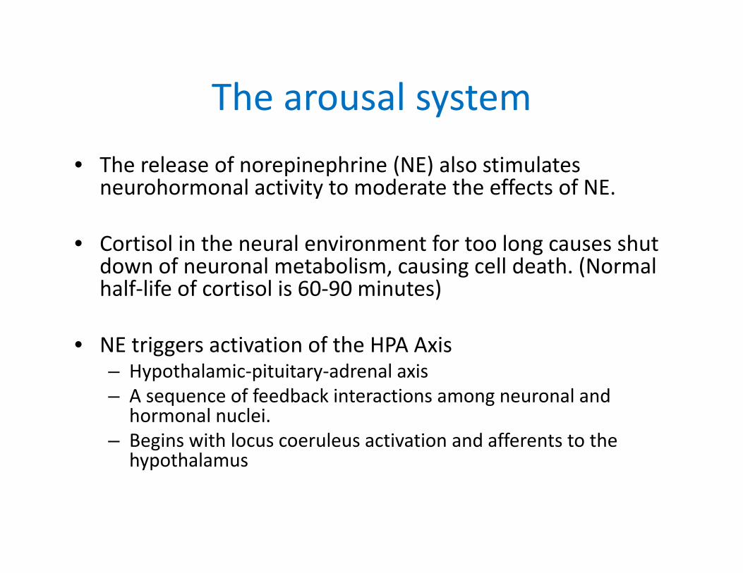
The arousal system• The release of norepinephrine (NE) also stimulates
neurohormonal activity to moderate the effects of NE.
• Cortisol in the neural environment for too long causes shut down of neuronal metabolism, causing cell death. (Normal half‐life of cortisol is 60‐90 minutes)
• NE triggers activation of the HPA Axis– Hypothalamic‐pituitary‐adrenal axis– A sequence of feedback interactions among neuronal and
hormonal nuclei.– Begins with locus coeruleus activation and afferents to the
hypothalamus

The arousal system• The hypothalamus releases corticotrophin‐releasing hormone (CRH) to the
pituitary gland and to the adrenal gland.
• In response, the pituitary gland secretes adrenocorticotropic hormone (ACTH) which then triggers ‐
• The adrenal gland to secrete corticosteroids such as cortisol which suppresses the hypothalamus.
• Cortisol increases glucose in the blood stream and decreases it in other tissues. It also regulates vascular smooth muscle tension to increase blood pressure.
• Cortisol decreases REM sleep and reduces sleep length.
• Cortisol reduces T‐cell development and thus has a negative impact on the immune system.
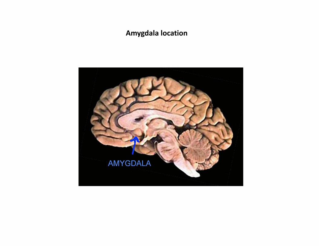
Amygdala location
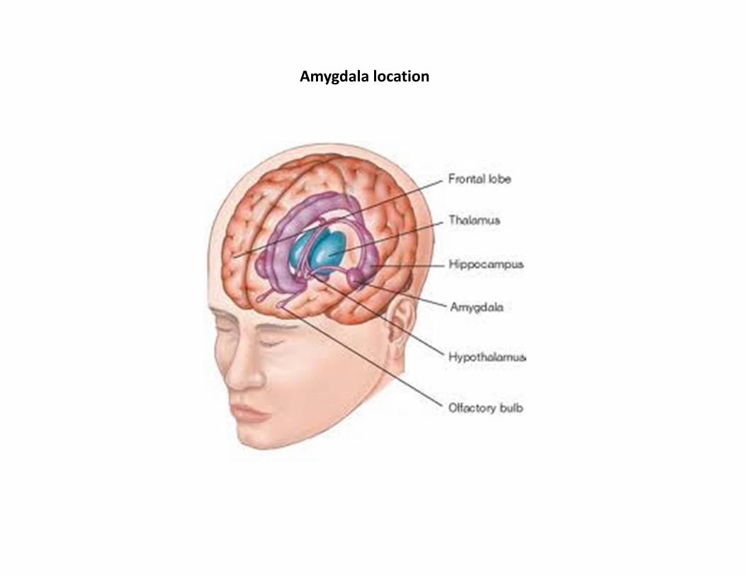
Amygdala location
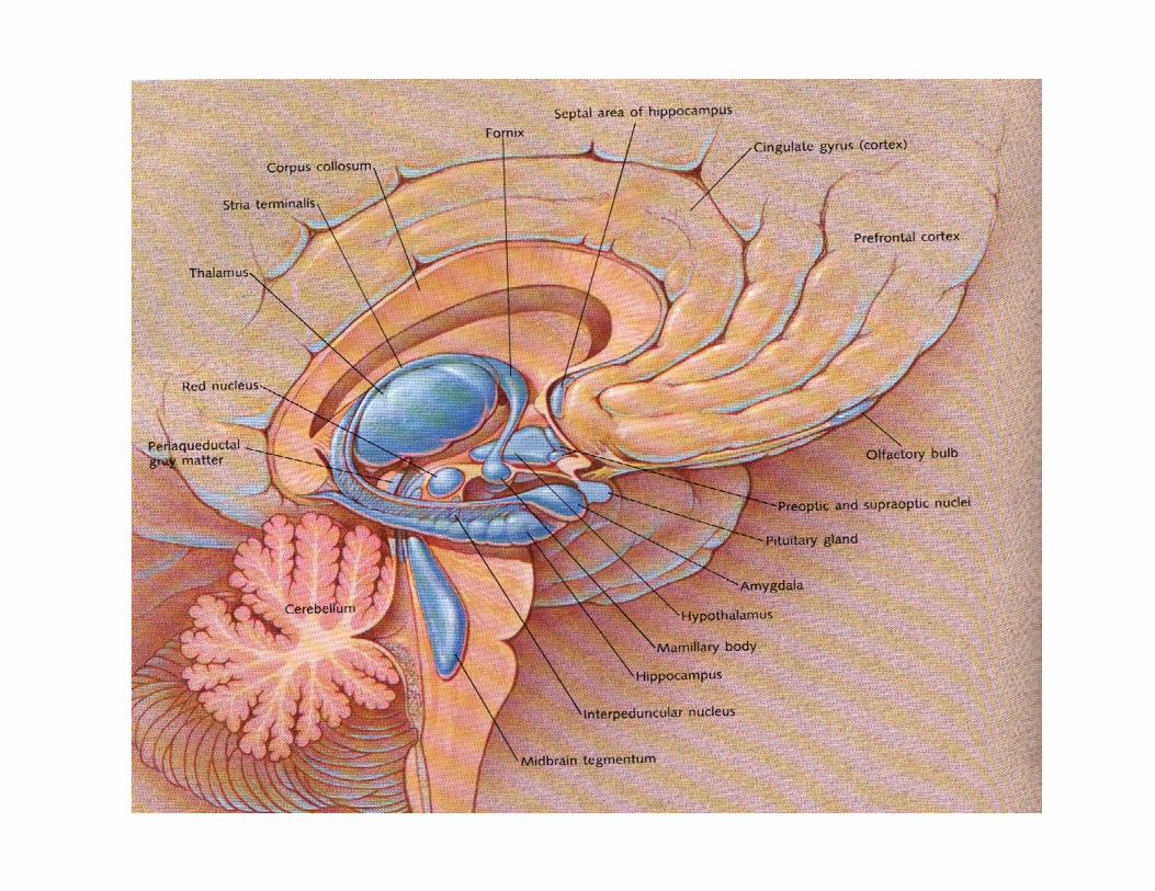
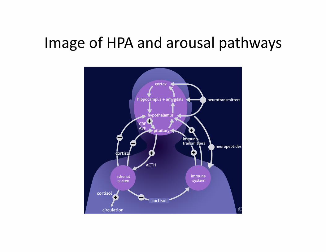
Image of HPA and arousal pathways

Arousal and development
• When the HPA axis is highly activated in early development (up to age 2‐3), it tends to be ‘set’ at a different level than for those who are not frequently activated.
• The brain achieves its set points for arousal as part of homeostasis‐seeking.
• Thus, chronically disturbed arousal systems can arise from environmentally induced fear states.

Arousal and development• Protracted and heightened arousal can result in one of two forms:
(1) a chronically hyper‐arousable type and (2) chronically hypo‐arousable type.
• Both types also inherit temperamental contributions and perhaps even temperamental influence over which type arises from extended fear states.
• Extended periods of arousal lead to extended cortisol secretion and subsequent cell death in the hippocampus and amygdala.
• Also, overloaded burden on the arousal system is associated with allostatic load where the set point for arousal is modified and resource deployment from cortical areas is diminished.

Arousal• One other factor to consider – the striatum, part of the basal
ganglia, is comprised of nuclei that detect rank order of other persons.
• It receives afferents from the frontal lobes and the emotion system to ascertain social rank – dominance or submission patterns.
• Canines have this brain function, as do even more primitive animals such as reptiles (e.g., the adage about the ‘reptilian brain’).
• Detection of rank order can contribute to stress as evident among Sapolosky’s baboons.
• Detection of rank order is a very primitive function and it plays a role in Marmot’s study of social determinates of health and well‐being.

Chronically disturbed arousal conditions
• Chronically disturbed arousal conditions become major risk factors for mental disorders and substance abuse.
• The brain is evolved to seek pleasure and avoid pain.
• Disturbed arousal conditions (either type) lead to are anhedonic states that people try to modify.

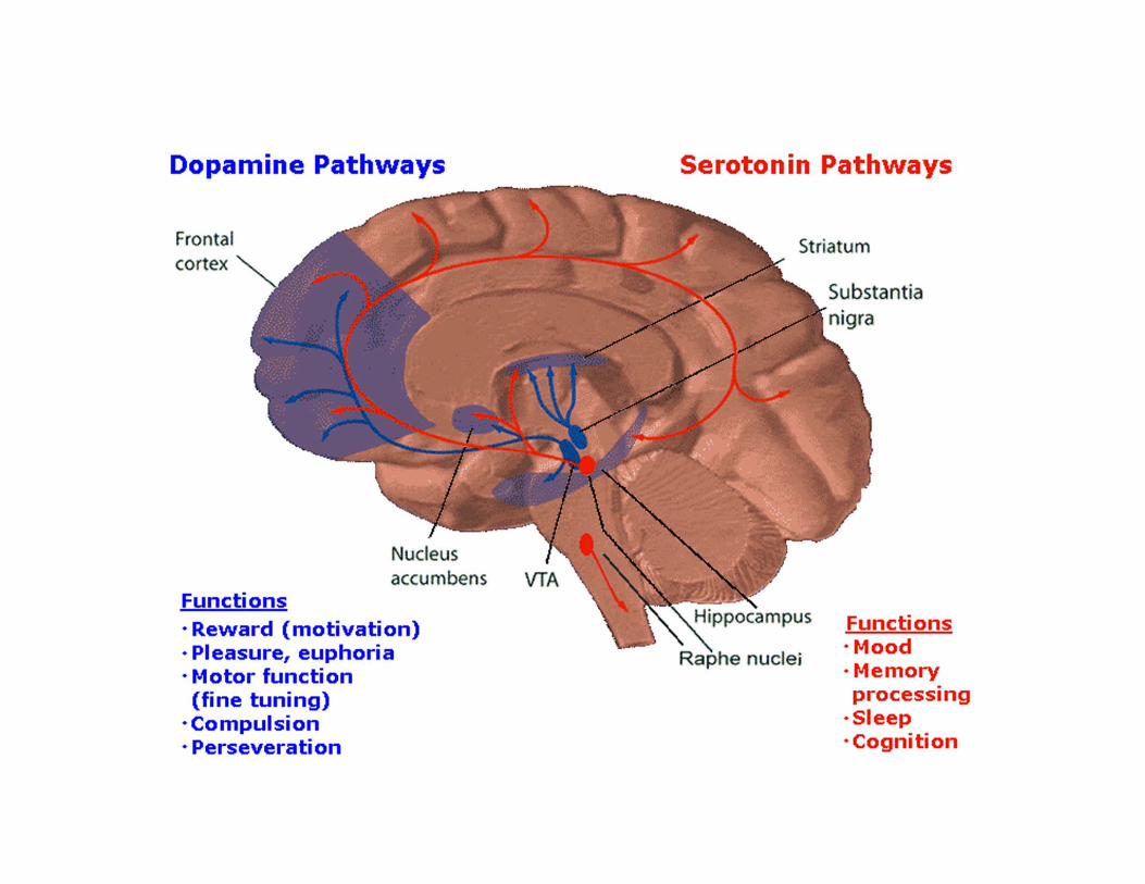
Reward, pleasure, memory• The brain regions responsible for mediating pleasure are located very close to memory centers and other key emotion sensing areas –
• The hippocampus – memory encoding and retrieving and short‐term memory
• The amygdala – emotion‐tagging system• Raphe nuclei – the serotonin system
• They are also closely related to centers that mediate intentional actions.– Striatum –mediated by dopamine and to some extent by serotonin

Brain structures and addiction• The human brain has reward centers that mediate the
experience of pleasure.
• The striatum, the ventral tegmental area (VTA), and the nucleus accumbens (NAc) are the primary locations for core pleasure experiences. – These are the primary regions where pleasure is mediated.
• When a person experiences pleasure from chocolate, a ride in a fast car, a buzz off a drug, the NAc has been activated. The striatum and substantia nigra engages motivational patterns to re‐activate the reward.
• The stimulation to activate reward centers can be increased by experiencing negative effects of the arousal system (either over activation of it or under activation)
Brain anatomy and addiction• All intoxicating substances are made of molecules that
are shaped much like the brain’s natural neurotransmitter molecules.
• Several neurotransmitters are affected by addictive substances. Sometimes the initial effect is on one neurotransmitter, but this activation can kick off something like a cascade of other events.
• The end result is dopamine activation of the striatum and most importantly:– Ventral tegmental area (VTA)– The nucleus accumbens (NAc)
• The VTA also triggers release of endorphin which is distributed in the anterior cingulate and frontal lobes.

NAc• Reward‐seeking is facilitated by the release of the neurotransmitter dopamine in the nucleus accumbens (NAc).
• Nac has decreased activity in depressive states.
• Subpopulations of NAc neurons even respond to predictive cues to promote reward‐seeking behavior.
• Even cues about a drug (such as talking about it, or even thinking about incidents related to drug use) can mobilize brain centers to begin pleasure expectations.

SPECT image of alcoholic brain
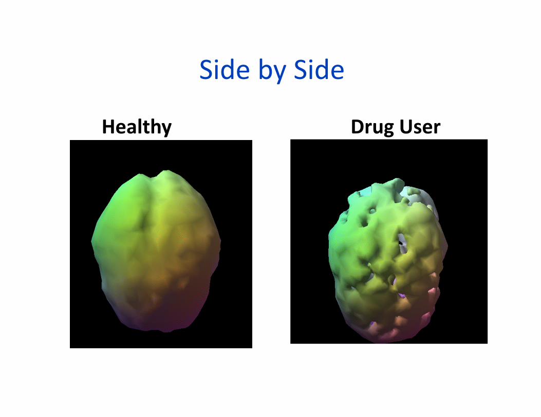
Side by Side
Healthy Drug User
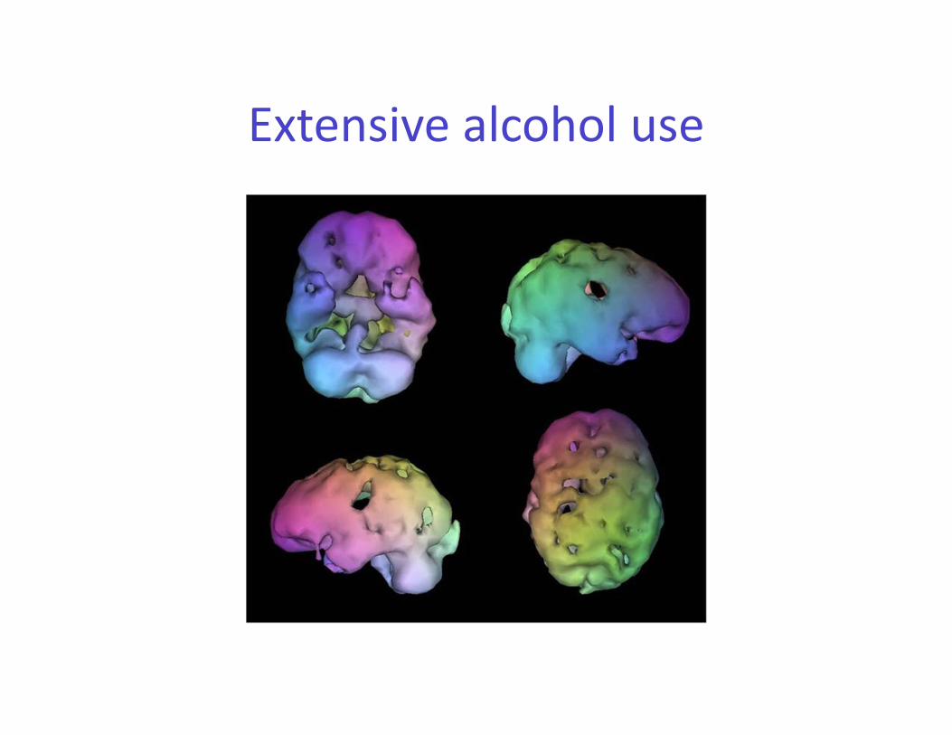
Extensive alcohol use

Buddha was right
Buddha was right• If we consider the findings from contemporary neuroscience, there is no
such thing as a perdurant self.
• We are a flux of continually changing neurotransmitters, dying of neurons, pruning of neural connections, growth of new connections, alterations in pathways, and even neurogenesis.
• One is not, at the end of this presentation, the same brain that one was at the beginning.
• Every memory you have is on the ‘hard drive’ of neural cells and pathways. Take them away and your memory is gone.
• If you have a different brain, can you be the same self you were before?
• Remember, whatever we are, we are our brain tissue – apart from it we are mere red meat.


![[Nancy_Claxton]_Using_Deliberative_Techniques_in_english class.pdf](https://img.pdfslide.us/doc/110x75/577c7c041a28abe05498f0f0/nancyclaxtonusingdeliberativetechniquesinenglish-classpdf.jpg)