Embed Size (px)
Citation preview
NeuroaNatomy aNd Neurophysiology for Speech and Hearing Sciences

NeuroaNatomy aNd Neurophysiology for Speech and Hearing Sciences
J. aNthoNy seikel, phd • kostas koNstaNtopoulos, phd • david g. drumright, Bs
5521 Ruffin RoadSan Diego, CA 92123
e-mail: [email protected]: http://www.pluralpublishing.com
Copyright © 2020 by Plural Publishing, Inc.
Typeset in 10/12 Minion Pro by Flanagan’s Publishing Services, Inc.Printed in China by Spectrum Printing, LLC
All rights, including that of translation, reserved. No part of this publication may be reproduced, stored in a retrieval system, or transmitted in any form or by any means, electronic, mechanical, recording, or otherwise, including photocopying, recording, taping, Web distribution, or information storage and retrieval systems without the prior written consent of the publisher.
For permission to use material from this text, contact us byTelephone: (866) 758-7251Fax: (888) 758-7255e-mail: [email protected]
Every attempt has been made to contact the copyright holders for material originally printed in another source. If any have been inadvertently overlooked, the publishers will gladly make the necessary arrangements at the first opportunity.
Library of Congress Cataloging-in-Publication Data
Names: Seikel, John A., author. | Konstantopoulos, Kostas, author. | Drumright, David G., author.Title: Neuroanatomy and neurophysiology for speech and hearing sciences / J. Anthony Seikel, Kostas Konstantopoulos, David G. Drumright.Description: San Diego, CA : Plural, [2020] | Includes bibliographical references and index.Identifiers: LCCN 2018021961| ISBN 9781635500714 (alk. paper) | ISBN 1635500710 (alk. paper)Subjects: | MESH: Speech—physiology | Hearing—physiology | Nervous System—anatomy & histologyClassification: LCC QP306 | NLM WV 501 | DDC 612.7/8—dc23LC record available at https://lccn.loc.gov/2018021961
v
ContentS
Preface xiiiAcknowledgments xvContributors xviiReviewers xixAbout the Authors xxi
1 iNtroductioN aNd overview 1overview of the nervous System 2Divisions of the nervous System 7
Autonomic Nervous System 7Somatic Nervous System 7
Developmental organization 8Development 8
terminology Related to neuroanatomy and neurophysiology 16Terms of Movement 17Terms of Neuropathology in Speech-Language Pathology 18
Chapter Summary 19Case Study 1–1 21Case Study 1–2 24References 28
2 NeuroNs aNd glial cells 31Introduction 31neurons 31
Cellular Components of the Soma 32Gross Structure of the Neuron 33
Soma 33Dendrites 34Axons 34
Neuronal Cell Types 34Classification Based on Number of Dendrites 34Classification Based on Dendrite Arborization 34Classification Based on Axon Length 36Classification Based on Conduction Velocity 36Classification Based on Functional Connection 36
vi n e u R o a n a t o m y a n D n e u R o p H y S I o l o g y f o R S p e e C H a n D H e a R I n g S C I e n C e S
glial Cells 37Oligodendrocytes and Schwann Cells 40Radial Glia 41Satellite Cells and Enteric Glial Cells 41Ependymal Cells 41
action potential 41The Physical Synapse 42Stimulation of a Neuron 42Generating the Action Potential 44
Ion Channels and Gradients 44Resting State 44Stimulation 45Generation of the Action Potential 45Propagation 45
Communication across the Synapse 47Excitation and Inhibition 47Summation 48Neurotransmitters 48
Chapter Summary 51Case Study 2–1 52References 60
3 Basic reflex aNd seNsory fuNctioN: how we kNow the world 61Introduction 61the Spinal Reflex arc 61golgi tendon organs 65other Sensation and Sensors 65
Somatosensors 65Representation of the Somatic Sensation in the Spinal Cord 68Special Senses 69
Visual Sensation 69Olfactory Sensation 71Gustatory Sensation 71Auditory Sensation 72Vestibular Sensors 73
Chapter Summary 74References 80
4 cereBral cortex 81Introduction 81general Structures and landmarks of the Cerebral Cortex 85
Major Sulci and Fissures 85Meningeal Linings 89The Ventricles and Cerebrospinal Fluid 90
Circulation of CSF 93Cell Types of the Cerebral Cortex 93Layers of the Cerebral Cortex 94
lobes of the Cerebral Cortex 100
C o n t e n t S vii
Frontal Lobe 100Parietal Lobe 104Temporal Lobe 104Occipital Lobe 106Insula or Insular Cortex 106Limbic System 107
medial Surface of the Cerebral Cortex 107Inferior Surface of the Cerebral Cortex 108
Posterior-Inferior (Ventral) Cerebral Cortex 108Anterior-Inferior Cerebral Cortex 108
myelinated fibers 109Projection Fibers 109
Corticobulbar and Corticospinal Tracts 110Corticobulbar Tract 111Corticospinal Tract 111
Association Fibers 111Commissural Fibers 112
the other Half: Hemispheric Specialization 113Chapter Summary 118Case Study 4–1 119Case Study 4–2 121Case Study 4–3 123Case Study 4–4 125References 133
5 aNatomy of the suBcortex 137Introduction 137Basal ganglia 137Hippocampus 143Diencephalon 145
Thalamus 145Epithalamus 145Subthalamus 145Hypothalamus 145
Chapter Summary 148Case Study 5–1 149Case Study 5–2 153Case Study 5–3 156References 161
6 aNatomy of the BraiNstem 163Introduction 163Superficial Brainstem landmarks 163
Superficial Medulla Oblongata 163Superficial Pons 167Superficial Midbrain 167
Deep Structures of the Brainstem 167
viii n e u R o a n a t o m y a n D n e u R o p H y S I o l o g y f o R S p e e C H a n D H e a R I n g S C I e n C e S
Deep Structures of the Medulla Oblongata 167Deep Structures of the Pons 172Deep Structures of the Midbrain 176
auditory pathway 179Cochlear Nucleus 179Superior Olivary Complex 179Inferior Colliculus 180Lateral Lemniscus 182Medial Geniculate Body 182
auditory Reception at temporal lobe 182Efferent Pathways 184Vestibular Pathway 184Acoustic Reflex 185
Chapter Summary 185Case Study 6–1 186References 192
7 the craNial Nerves 193Introduction 193Cranial nerve Classification 193
General Somatic Afferent (GSA) Nerves 194Special Somatic Afferent (SSA) Nerves 194General Visceral Afferent (GVA) Nerves 194Special Visceral Afferent (SVA) Nerves 194General Visceral Efferent (GVE) Nerves 197General Somatic Efferent (GSE) Nerves 197
Specific Cranial nerves 198I Olfactory Nerve (SVA) 198II Optic Nerve (SSA) 198Eye Movement: III Oculomotor Nerve (GSE, GVE), IV Trochlear Nerve (GSE), 202
VI Abducens Nerve (GSE)V Trigeminal Nerve (GSA, SVE) 204VII Facial Nerve (SVE, SVA, GVE) 205VIII Vestibulocochlear Nerve (SSA) 207
Acoustic Branch 208Vestibular Branch 209Efferent Component 211
IX Glossopharyngeal Nerve (GSA, GVA, SVA, GVE, SVE) 211X Vagus Nerve (GSA, GVA, SVA, GVE, SVE) 212XI Accessory Nerve (SVE) 214XII Hypoglossal Nerve (GSE) 214
Chapter Summary 215Case Study 7–1 216Case Study 7–2 218References 227
8 cereBellar aNatomy aNd physiology 229Introduction 229Structure of the Cerebellum 229
C o n t e n t S ix
Cellular Structure of the Cerebellum 238nuclei of the Cerebellum 240
Fastigial Nucleus 241Globose and Emboliform Nuclei 241Dentate Nucleus 241
tracts Serving the Cerebellum 242Input to the Cerebellum 242
Vestibulocerebellar Pathways 242Dorsal Spinocerebellar Tract 242Cuneocerebellar Tract 242Ventral Spinocerebellar Tract 242Rostral Spinocerebellar Tract 242Pontocerebellar Tract 243Olivocerebellar Tract 244
Cerebellar Peduncles 244Superior Cerebellar Peduncle 245Middle Cerebellar Peduncle 245Inferior Cerebellar Peduncle 246
Cerebellum and motor Control 247Chapter Summary 248Case Study 8–1 249Case Study 8–2 253Case Study 8–3 257References 263
9 spiNal cord aNd pathways 265Introduction 265Vertical anatomy of the Spinal Cord 266transverse anatomy of the Spinal Cord 270pathways of the Spinal Cord 273
Ascending Pathways 276Posterior Funiculus: Fasciculus Gracilis and Fasciculus Cuneatus 276Anterior Funiculus: Anterior and Lateral Spinothalamic Tracts 278Lateral Funiculus 278Anterior and Posterior Spinocerebellar Tracts 278
Descending Pathways 278Pyramidal Pathways 278
Corticospinal Tract 278Corticobulbar Tract 281
Other Descending Pathways 283Extrapyramidal System 283Corticostriate Pathway 284Corticothalamic Fibers 285Corticopontocerebellar Fibers 285
Chapter Summary 285Case Study 9–1 286Case Study 9–2 289References 295
x n e u R o a n a t o m y a n D n e u R o p H y S I o l o g y f o R S p e e C H a n D H e a R I n g S C I e n C e S
10 cereBrovascular supply 297Introduction 297Carotid artery Supply 297
External Carotid Artery Supply 297Internal Carotid Artery Supply 303
Anterior Cerebral Artery 304Posterior Communicating Artery 304Middle Cerebral Artery 304
Vertebrobasilar System 307Vertebral and Basilar Arteries 307Posterior Cerebral Artery 307
Venous Drainage of the Cerebrovascular Supply 307Chapter Summary 309Case Study 10–1 310References 318
11 Neural coNtrol of speech aNd swallowiNg 319Introduction 319neural Control of Speech 319
Feedback and Correction 319models of Speech production 321neural Control of mastication and Deglutition 322
Development of Swallowing Function 322adult patterns of mastication and Deglutition 323
Oral Stage 323Pharyngeal Stage 324Relaxation of the Upper Esophageal Sphincter 326Esophageal Stage 326
Reflexes and their Integration into Central pattern generators 327Oral Stage Reflexes 327
Chewing Reflex 327Pharyngeal Stage Reflexes 327
Vomit Reflex 327Cough Reflex 328
Reflexes of Respiration and Apnea 328Sensation in mastication and Deglutition 328
Sensation 328Gustation 328Olfaction 330Tactile, Proprioceptive, and Thermal Sensation 330Complex Motor Responses 330
Chapter Summary 331Case Study 11–1 332Case Study 11–2 335Case Study 11–3 337

C o n t e n t S xi
Case Study 11–4 339References 345
Appendix. Answers to Study Questions 347Glossary 357Index 373

xiii
pRefaCe
the study of the brain and its functions is at the heart of communication sciences and its disorders. While there are many neurological conditions that immediately come
to mind when we think of maladies affecting the brain, the nervous system is intrinsically involved in most of the activi-ties of our field, from the cognitive and motoric aspects of phonology or the impact of myelination on normal language development to (central) auditory processing disorder. All human actions arise from processes of the nervous system, and we can trace many of the deficits treated in our profes-sions to some type of failure affecting this nervous system.
It is with this understanding that we sought to create this textbook and study materials. Neuroscience is the study of what is arguably the most complex phenomenon in the known universe, the nervous system. We, as humans, have brains that are uniquely complex in structure and, most importantly, in function. The human brain has evolved to work in complex networks that entrain multiple areas of the brain, giving it a capacity for problem solving that outstrips our nearest evolutionary neighbors.
As audiologists, speech-language pathologists, and speech and hearing scientists we are in a position to see the inner workings of the brain firsthand through the many neuropathologies with which we are presented. The basal ganglia circuits are uniquely revealed in the tremor, hyper-kinesia, and hypokinesia of conditions such as Parkinson’s disease, Huntington’s disease, or hepatolenticular degenera-tion. The impact of disease conditions such as multiple scle-rosis on hearing function, cognition, and speech production can provide evidence for site of lesion activity if we are able to recognize the signs and symptoms related to the brain region affected. We are challenged on a daily basis to provide meaningful therapy to individuals who have suffered cere-brovascular accident or trauma, and we must work to pro-vide treatment to help overcome the life-changing effects of those lesions. To do this requires a deep knowledge of this extraordinarily complex nervous system but also requires that the clinician develop the intention to continually learn about the nervous system and new treatments that emerge. As an example, behavioral treatments are emerging that have been shown to differentially increase the brain volume and function in areas shown to be active during attention activi-
ties, expression of compassion, and awareness of others (the-ory of mind). Therapies directed toward these dysfunctions could directly affect the lives of those with right hemisphere dysfunction, and the wise clinician will keep a close eye on developments such as these. To do this requires knowledge, desire, and intention. It is our deep hope that these materials can provide at least some of the motivation for a lifetime of study in neuroscience. A central component of this text is the Neuroquest software. We owe a deep debt of gratitude to Dr. Sadanand Singh, who, many years ago as our first publisher of another book, insisted that study software was a critically important component of any text. Now, 20 years later, we are pleased and humbled to continue with his charge to make the current textbook as powerful a learning tool as we can.
The purpose of this textbook is to help the undergradu-ate and graduate student of speech-language pathology learn about the structure and function of the brain. This knowledge will aid not only in accurate clinical diagnosis but also in the correct use of evidence-based practice methods for speech therapy. There are many neurological diseases in which the primary signs and symptoms are within the domains of speech, language, or hearing disorders, so there is fertile ground for application of the knowledge acquired through study of neuro-science. We have included a number of clinical cases at the end of each chapter to prime the student’s problem-solving clinical skills in his or her future profession. Most of the cases include neurological assessments that were performed over the course of treatment (sometimes even 10 years after initial neurologi-cal diagnosis), which we have included to help the reader rec-ognize the timing of the speech/language disorder as related to the timing of the other neurological symptomatology.
This textbook is divided into 11 chapters. Chapter 1 briefly overviews the nervous system, starting from embry-onic development to aging and including disorders of speech and language that the students in audiology and speech-language pathology need to be aware of. Chapters 2 and 3 discuss the structure and function of cellular components of the central nervous system, including how the signals are propagated (Chapter 2) and the function of basic reflexes (Chapter 3). Chapter 4 discusses the cerebral cortex, includ-ing landmarks and components and their relation to our disciplines. Chapters 5 and 6 discuss areas and structures
xiv n e u R o a n a t o m y a n D n e u R o p H y S I o l o g y f o R S p e e C H a n D H e a R I n g S C I e n C e S
beneath the cortex (subcortex), including the basal ganglia, hippocampus, thalamus (Chapter 5), brainstem (Chapter 6), as well as their associated connections to the cortex. Chapter 7 is dedicated to presentation of the cranial nerves, many of which are critical to hearing and speech. Chapters 8 and 9 discuss the cerebellum, the spinal cord, and their fiber con-nections. Chapter 10 focuses on the cerebrovascular supply to the brain, elaborating on the vascular supply critical for speech, language, and hearing. Chapter 11 aims to provide to the student the knowledge about the function for the neural control of speech and swallowing, including theoretical mod-els of speech production.
J. Tony Seikel is co-author of two textbooks in anatomy and physiology of speech, hearing, and language and has taught neuroscience and neurogenic coursework for 30 years. Kostas Konstantopoulos is an assistant professor in the Euro-pean University Cyprus and teaches all neurogenic courses
and neuroanatomy. He currently serves as the coordinator for the Bachelor's degree of Speech and Language Therapy and is the coordinator for the Master's degree in Speech Language Pathology. He has extensive clinical and research experience in neurogenic communication disorders spanning 15 years. For the past 6 years he has provided clinical assessment and treatment of speech and dysphagia at the Cyprus Institute of Neurology and Genetics (CING). The majority of the case histories utilized in the chapters have been drawn from his files and referred from all four neurology clinics in the Cyprus Institute of Neurology and Genetics. David Drum-right is co-author of two textbooks in anatomy and papers on pedagogy, and has developed software for study of anatomy (Anima and Anatesse for anatomy and physiology, Audin for auditory physiology, and now Neuroquest for study of neuroscience).
297
10
Learning Outcomes for Chapter 10
• Identify the two major sources of vascular supply to the brain.
• Identify the major arteries arising from the vertebrobasilar and carotid supplies and the regions of the brain being served by those arteries.
• Discuss the hypothesized function of the circle of Willis.
• Identify the deep arteries and branches supplying each lobe and surface of the brain as well as the brainstem, spinal cord, cerebellum, basal ganglia, and thalamus.
• Discuss the potential effects of loss of supply to any of the major branches of the vertebrobasilar or carotid supplies.
297
CerebrovasCular supply
IntrOduCtIOn
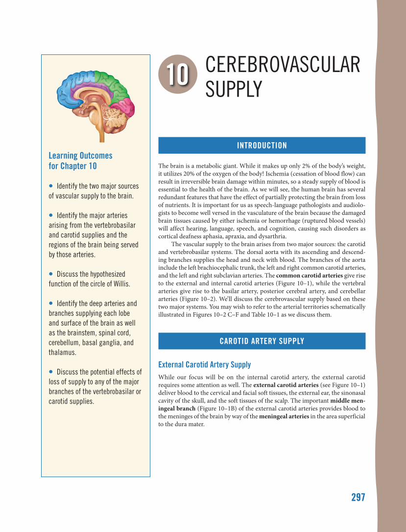
The brain is a metabolic giant. While it makes up only 2% of the body’s weight, it utilizes 20% of the oxygen of the body! Ischemia (cessation of blood flow) can result in irreversible brain damage within minutes, so a steady supply of blood is essential to the health of the brain. As we will see, the human brain has several redundant features that have the effect of partially protecting the brain from loss of nutrients. It is important for us as speech-language pathologists and audiolo-gists to become well versed in the vasculature of the brain because the damaged brain tissues caused by either ischemia or hemorrhage (ruptured blood vessels) will affect hearing, language, speech, and cognition, causing such disorders as cortical deafness aphasia, apraxia, and dysarthria.
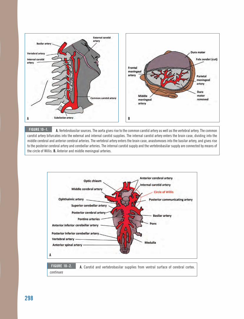
The vascular supply to the brain arises from two major sources: the carotid and vertebrobasilar systems. The dorsal aorta with its ascending and descend-ing branches supplies the head and neck with blood. The branches of the aorta include the left brachiocephalic trunk, the left and right common carotid arteries, and the left and right subclavian arteries. The common carotid arteries give rise to the external and internal carotid arteries (Figure 10–1), while the vertebral arteries give rise to the basilar artery, posterior cerebral artery, and cerebellar arteries (Figure 10–2). We’ll discuss the cerebrovascular supply based on these two major systems. You may wish to refer to the arterial territories schematically illustrated in Figures 10–2 C–F and Table 10–1 as we discuss them.
CarOtId artery SuppLy
external Carotid artery SupplyWhile our focus will be on the internal carotid artery, the external carotid requires some attention as well. The external carotid arteries (see Figure 10–1) deliver blood to the cervical and facial soft tissues, the external ear, the sinonasal cavity of the skull, and the soft tissues of the scalp. The important middle men-ingeal branch (Figure 10–1B) of the external carotid arteries provides blood to the meninges of the brain by way of the meningeal arteries in the area superficial to the dura mater.
298
a B
figure 10–1. a. Vertebrobasilar sources. the aorta gives rise to the common carotid artery as well as the vertebral artery. the common carotid artery bifurcates into the external and internal carotid supplies. the internal carotid artery enters the brain case, dividing into the middle cerebral and anterior cerebral arteries. the vertebral artery enters the brain case, anastomoses into the basilar artery, and gives rise to the posterior cerebral artery and cerebellar arteries. the internal carotid supply and the vertebrobasilar supply are connected by means of the circle of willis. B. anterior and middle meningeal arteries.
a
figure 10–2. a. Carotid and vertebrobasilar supplies from ventral surface of cerebral cortex. continues
299
B c
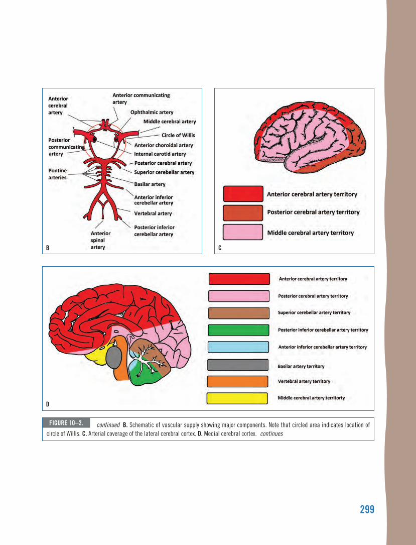
figure 10–2. continued B. Schematic of vascular supply showing major components. note that circled area indicates location of circle of willis. c. arterial coverage of the lateral cerebral cortex. d. medial cerebral cortex. continues
d
300
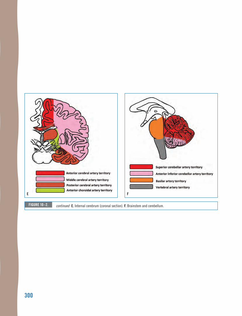
e f
figure 10–2. continued e. Internal cerebrum (coronal section). f. Brainstem and cerebellum.
301
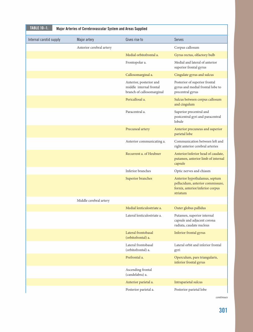
taBle 10–1. major arteries of cerebrovascular system and areas supplied
Internal carotid supply major artery gives rise to Serves
Anterior cerebral artery Corpus callosum
Medial orbitofrontal a. Gyrus rectus, olfactory bulb
Frontopolar a. Medial and lateral of anterior superior frontal gyrus
Callosomarginal a. Cingulate gyrus and sulcus
Anterior, posterior and middle internal frontal branch of callosomarginal
Posterior of superior frontal gyrus and medial frontal lobe to precentral gyrus
Pericallosal a. Sulcus between corpus callosum and cingulum
Paracentral a. Superior precentral and postcentral gyri and paracentral lobule
Precuneal artery Anterior precuneus and superior parietal lobe
Anterior communicating a. Communication between left and right anterior cerebral arteries
Recurrent a. of Heubner Anterior/inferior head of caudate, putamen, anterior limb of internal capsule
Inferior branches Optic nerves and chiasm
Superior branches Anterior hypothalamus, septum pellucidum, anterior commissure, fornix, anterior/inferior corpus striatum
Middle cerebral artery
Medial lenticulostriate a. Outer globus pallidus
Lateral lenticulostriate a. Putamen, superior internal capsule and adjacent corona radiata, caudate nucleus
Lateral frontobasal (orbitofrontal) a.
Inferior frontal gyrus
Lateral frontobasal (orbitofrontal) a.
Lateral orbit and inferior frontal gyri
Prefrontal a. Operculum, pars triangularis, inferior frontal gyrus
Ascending frontal (candelabra) a.
Anterior parietal a. Intraparietal sulcus
Posterior parietal a. Posterior parietal lobe
continues
302
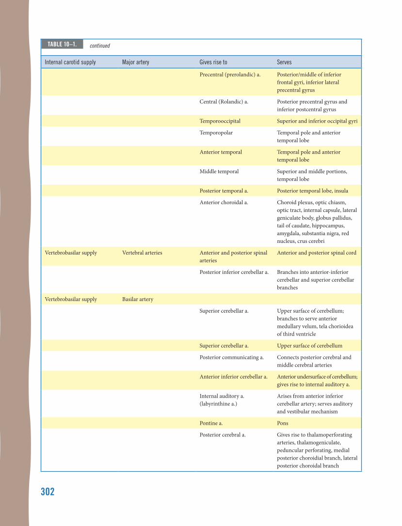
Internal carotid supply major artery gives rise to Serves
Precentral (prerolandic) a. Posterior/middle of inferior frontal gyri, inferior lateral precentral gyrus
Central (Rolandic) a. Posterior precentral gyrus and inferior postcentral gyrus
Temporooccipital Superior and inferior occipital gyri
Temporopolar Temporal pole and anterior temporal lobe
Anterior temporal Temporal pole and anterior temporal lobe
Middle temporal Superior and middle portions, temporal lobe
Posterior temporal a. Posterior temporal lobe, insula
Anterior choroidal a. Choroid plexus, optic chiasm, optic tract, internal capsule, lateral geniculate body, globus pallidus, tail of caudate, hippocampus, amygdala, substantia nigra, red nucleus, crus cerebri
Vertebrobasilar supply Vertebral arteries Anterior and posterior spinal arteries
Anterior and posterior spinal cord
Posterior inferior cerebellar a. Branches into anterior-inferior cerebellar and superior cerebellar branches
Vertebrobasilar supply Basilar artery
Superior cerebellar a. Upper surface of cerebellum; branches to serve anterior medullary velum, tela chorioidea of third ventricle
Superior cerebellar a. Upper surface of cerebellum
Posterior communicating a. Connects posterior cerebral and middle cerebral arteries
Anterior inferior cerebellar a. Anterior undersurface of cerebellum; gives rise to internal auditory a.
Internal auditory a. (labyrinthine a.)
Arises from anterior inferior cerebellar artery; serves auditory and vestibular mechanism
Pontine a. Pons
Posterior cerebral a. Gives rise to thalamoperforating arteries, thalamogeniculate, peduncular perforating, medial posterior choroidial branch, lateral posterior choroidal branch
taBle 10–1. continued
C H a p t e R 1 0 • C e R e B R o V a S C u l a R S u p p l y 303
the meningeal artery and traumatic Brain injury
The meningeal vessels, located in the sulci of the skull, are susceptible to penetrating
traumatic brain injury (TBI). The middle meningeal artery, which covers a large portion of the skull, is fre-quently affected, with penetrating injury rupturing the artery and causing release of blood into the area above the dura mater (epidural hematoma). Clinically, the development of such a mass creates increased intracra-nial pressure and a concomitant herniation and swell-ing of the brain, with symptomatology involving speech, mobility, vision, and consciousness.
internal carotid artery supplyThe internal carotid arteries give rise to the anterior cerebral arteries, posterior communicating arteries, and middle cere-bral arteries as well as a number of other smaller branches. For speech-language pathology and audiology, the middle cerebral artery (MCA) is extraordinarily important, as it serves all of the speech, language, and hearing territory of the brain.
internal carotid artery Branching and stroke
The internal carotid arteries and the verte-bral arteries have tortuous bends and branch-
ing, which can lead to problems as we age. One of the most significant problem spots in the cerebrovascular system is at the point of bifurcation (dividing) of the common carotid artery into the internal and exter-nal carotid arteries. There is a significant narrowing at this branch in the artery, and if an embolus (float-ing blood clot) lodges at this location, it may starve the downstream tissue of oxygen. This is a critical emer-gency, since irreversible brain damage begins within five minutes of blood stoppage. In this case, blocking the entire internal carotid artery deprives blood to the anterior two-thirds of one entire cerebral hemisphere. An infarct covering this much of the MCA territory can result in profound aphasia if it involves the dominant hemisphere.
This is not the end of the problems with this bifur-cation. Atherosclerotic plaques develop as we age as a result of diet, exercise level, and genetics. These plaques plaster themselves to the arterial walls, reducing the flex-ibility of the wall and thereby promoting hypertension
(high blood pressure). Further, turbulence at the bifur-cation of the common carotid artery increases the like-lihood that plaques will become dislodged and become emboli that can lodge elsewhere within the bloodstream. The arterial supply mimics the branching in trees, with larger branches splitting into smaller branches at bifur-cations, which then branch again to create even smaller branches. In the vascular supply, arteries give rise to arterioles, which give rise to capillaries, with each suc-cessive generation at a branching becoming smaller in diameter. The terminal point of this branching is the union of veins and arteries, with veins showing increas-ing diameter as the blood makes its way back to the heart for oxygenation. (This is why there are rarely problems with emboli in the venous system, although clots may enter the heart and cause significant problems there.) Thus, any foreign body in the arterial system has a high probability of lodging somewhere downstream of where it is released. The larger a floating clot is, the greater the area it will affect when it does lodge in the bloodstream. Doppler and magnetic arteriography are essential tools for the assessment of blood flow, as they provide a non-invasive and low cost means of determin-ing the presence of plaques and restrictions before they cause ischemic events.
arterial Branches from the carotid supply
While we tend to focus on the anterior and middle cerebral arteries in our discussion
of the carotid supply, it’s worth reminding ourselves of other very relevant branches for speech-language pathol-ogists and audiologists. The ophthalmic artery provides oxygenated blood to the eyeball, while the meningeohy-pophyseal artery supplies the pituitary gland. The infero-lateral artery is very important, in that it supplies the III oculomotor, IV trochlear, and VI abducens nerves, and ischemia associated with this blood vessel can result in oculomotor paralysis (Capo, Kupersmith, Berenstein, Choi, & Diamond, 1991). The capsular arteries serve the internal capsule and the basal ganglia. Ischemia affect-ing the posterior limb of the internal capsule can result in severe paralysis due to the effect on the pyramidal pathway, although recovery can occur (Fries, Danek, Scheidtmann, & Hamburger, 1993), and involvement of the basal ganglia can result in motor control deficit (Boyd, Edwards, Siengsukon, Vidoni, Wessel, & Linsdell, 2009) and cognitive impairment (e.g., Seidel, Gronewold, Wicking, Bellebaum, & Hermann, 2016; Westmacott et al., 2017).
304 n e u R o a n a t o m y a n D n e u R o p H y S I o l o g y f o R S p e e C H a n D H e a R I n g S C I e n C e S
The posterior communicating artery is partially responsible for regulating blood flow and pressure within the cerebrovascular supply. Specifically, this artery con-nects the internal carotid and vertebrobasilar supplies, so that an occlusion in one supply can be compensated by the other supply. Occlusion of the posterior commu-nicating artery threatens the ability of the circle of Willis to compensate for blood flow fluctuations (Liebeskind, 2003; Schomer et al., 1994). The anterior choroidal artery also serves the internal capsule (posterior limb), thala-mus, and optic chiasm, so an infarct involving this artery can result in hemiplegia on the contralateral side of the body.
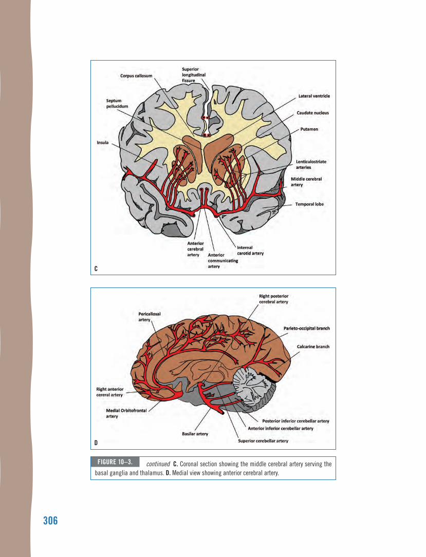
Anterior Cerebral ArteryAs seen in Figure 10–2A, the anterior cerebral artery (ACA)comprises a portion of the circle of Willis, coursing through the superior longitudinal fissure along the dorsal surface of the corpus callosum. The circle of Willis acts as a vascular backup system. Look at the schematic in Figure 10–2B, and first identify the basilar artery and then the internal carotid arteries so that you have located the two sources of blood for the brain. Now examine the way they are connected. The circle of Willis connects these two vascular supplies, and if the internal carotid artery supply is cut off due to infarct, the vertebrobasilar supply has the potential to supply those areas that are not receiving blood due to the infarct. The circle of Willis includes the anterior cerebral artery and the anterior and posterior communicating arteries. The anterior cere-bral artery continues into the superior longitudinal fissure, serving the medial surface of the cerebral cortex, as well as a portion of the dorsal surface of the frontal and parietal lobes (Figures 10–3A, B, and C). The ACA is divided into three major segments. Section A1 includes the portion of the ACA within the circle of Willis and supplies blood to the basal gan-glia, while A2 extends distally into the anterior two-thirds of the medial surface of the cerebral hemispheres to the genu of the corpus callosum. Segment A3 extends to the superior sur-face of the corpus callosum and supplies blood to the medial cerebral cortex.
ischemia of the anterior cerebral artery
A number of clinical signs arise from an infarction involving the anterior cerebral
artery (ACA), depending on the anatomi-cal sites involved, although collateral blood supply often reduces the signs of ACA blockage. The ACA serves the medial surface of the cerebral cortex, which supplies the areas controlling the legs. An infarct involving the ACA
can result in hemiplegia, particularly if the supplemen-tary motor area is involved, as well as cortical sensory loss. With paralysis, a person may also find that inhibited reflexes are released and become active, including grasp and sucking reflexes. If the frontal pole, corpus callo-sum, and superior frontal gyrus are involved, the patient may have reduced ability to make decisions (hypobulia) or other cognitive involvement (Kang & Kim, 2008). Infarcts of the ACA are related to emotional lability, which is uncontrollable laughing or crying in absence of a relevant stimulus.
Posterior Communicating ArteryThe posterior communicating artery sends collateral arteri-oles into the deep tissues of the brain, serving the thalamus, internal capsule, and optic tract. It gives rise to the anterior choroidal artery, which supplies the optic tract, posterior internal capsule, cerebral peduncles, medial temporal lobe, thalamus, and a portion of the corpus striatum.
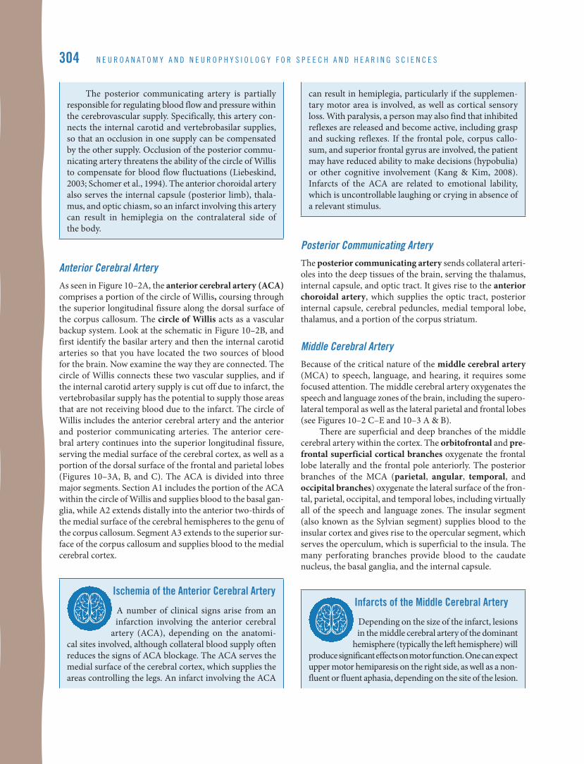
Middle Cerebral ArteryBecause of the critical nature of the middle cerebral artery (MCA) to speech, language, and hearing, it requires some focused attention. The middle cerebral artery oxygenates the speech and language zones of the brain, including the supero-lateral temporal as well as the lateral parietal and frontal lobes (see Figures 10–2 C–E and 10–3 A & B).
There are superficial and deep branches of the middle cerebral artery within the cortex. The orbitofrontal and pre-frontal superficial cortical branches oxygenate the frontal lobe laterally and the frontal pole anteriorly. The posterior branches of the MCA (parietal, angular, temporal, and occipital branches) oxygenate the lateral surface of the fron-tal, parietal, occipital, and temporal lobes, including virtually all of the speech and language zones. The insular segment (also known as the Sylvian segment) supplies blood to the insular cortex and gives rise to the opercular segment, which serves the operculum, which is superficial to the insula. The many perforating branches provide blood to the caudate nucleus, the basal ganglia, and the internal capsule.
infarcts of the middle cerebral artery
Depending on the size of the infarct, lesions in the middle cerebral artery of the dominant
hemisphere (typically the left hemisphere) will produce significant effects on motor function. One can expect upper motor hemiparesis on the right side, as well as a non-fluent or fluent aphasia, depending on the site of the lesion.
305
a
B
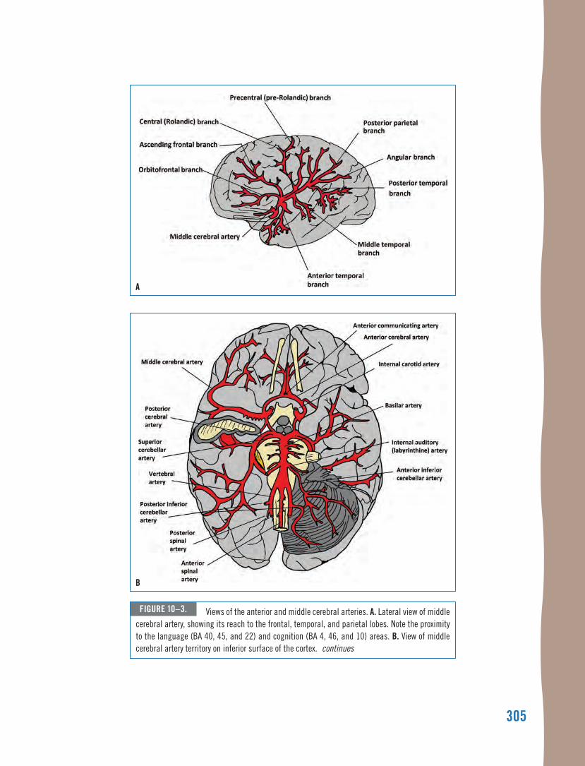
figure 10–3. Views of the anterior and middle cerebral arteries. a. lateral view of middle cerebral artery, showing its reach to the frontal, temporal, and parietal lobes. note the proximity to the language (Ba 40, 45, and 22) and cognition (Ba 4, 46, and 10) areas. B. View of middle cerebral artery territory on inferior surface of the cortex. continues
306
c
d
figure 10–3. continued c. Coronal section showing the middle cerebral artery serving the basal ganglia and thalamus. d. medial view showing anterior cerebral artery.
C H a p t e R 1 0 • C e R e B R o V a S C u l a R S u p p l y 307
Lesions in the insular and Sylvian fissure segments of the middle cerebral artery can affect Broca’s area, Wernicke’s area, and the precentral gyrus, resulting in aphasia, apraxia, and dysarthria. Infarct involving the insula can result in apraxia of speech, as well as loss of thermal sensation (Kodumuri et al., 2016). If the lesion extends to the cortical white matter beneath the in- sular cortex, one may see sensory disturbances, transcor-tical motor aphasia, phonatory deficiency, and dyspha-gia. A lesion in the area of the basal ganglia and internal capsule may create dysfunction in the direct and indi-rect pathways responsible for the initiation and control of movement, as well as dysfunction of the corticobulbar tract.
In summary, the brain uses a disproportionate volume of blood, and ischemia can result in irreversible brain damage within minutes.
• The vascular supply to the brain arises from two major sources: the carotid and vertebrobasilar systems. The com-mon carotid arteries give rise to the external and inter-nal carotid arteries, and the vertebral arteries give rise to the basilar artery, posterior cerebral artery, and cerebellar arteries.
• The external carotid arteries deliver blood to the cervical and facial soft tissues, the external ear, the sinonasal cavity of the skull, and the soft tissues of the scalp. The middle meningeal branch of the external carotid arteries provides blood to the meninges of the brain by way of the menin-geal arteries in the area superficial to the dura mater.
• The internal carotid arteries give rise to the anterior and middle cerebral arteries as well as a number of other smaller branches. The middle cerebral artery is particu-larly relevant to speech-language pathologists and audiolo-gists because of the territory it serves.
• The anterior cerebral artery courses through the superior longitudinal fissure along the dorsal surface of the cor-pus callosum. The anterior cerebral artery continues into the superior longitudinal fissure, serving the medial sur- face of the cerebral cortex, corpus callosum, a portion of the dorsal surface of the frontal and parietal lobes, and basal ganglia.
• The circle of Willis provides vascular redundancy to the blood supply to the brain. The circle of Willis includes the anterior cerebral artery and the anterior and posterior communicating arteries.
• The posterior communicating artery serves the thalamus, internal capsule, and optic tract.
• Superficial branches of the middle cerebral artery serve the superior temporal lobe, as well as the lateral and most of the dorsal surfaces of the frontal and parietal lobes, and the insula and operculum. Perforating branches serve the cau-date nucleus, the basal ganglia, and the internal capsule.
verteBroBasilar system
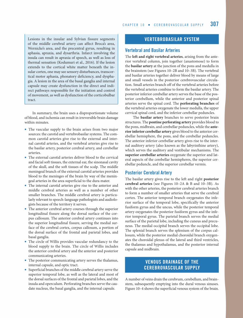
vertebral and Basilar arteriesThe left and right vertebral arteries, arising from the ante-rior vertebral column, join together (anastomose) to form the basilar artery at the junction of the pons and medulla in the brainstem (see Figures 10–2B and 10–3B). The vertebral and basilar arteries together deliver blood by means of large and small vessels in the posterior cerebrovascular circula-tion. Small arteries branch off of the vertebral arteries before the vertebral arteries combine to form the basilar artery. The posterior inferior cerebellar artery serves the base of the pos-terior cerebellum, while the anterior and posterior spinal arteries serve the spinal cord. The perforating branches of the vertebral arteries oxygenate the lower medulla, the upper cervical spinal cord, and the inferior cerebellar peduncles.
The basilar artery branches to serve posterior brain structures. The pontine perforating artery provides blood to the pons, midbrain, and cerebellar peduncles, while the ante-rior inferior cerebellar artery gives blood to the anterior cer-ebellar hemisphere, the pons, and the cerebellar peduncles. The anterior inferior cerebellar artery gives rise to the inter-nal auditory artery (also known as the labyrinthine artery), which serves the auditory and vestibular mechanisms. The superior cerebellar arteries oxygenate the superior and lat-eral aspects of the cerebellar hemispheres, the superior cer-ebellar peduncle, and the superior cerebellar vermis.
posterior cerebral arteryThe basilar artery gives rise to the left and right posterior cerebral arteries (see Figures 10–2A & B and 10–3B). As with the other arteries, the posterior cerebral arteries branch to form a number of smaller arteries that serve the cerebral cortex. The anterior temporal branch oxygenates the infe-rior surface of the temporal lobe, specifically the anterior fusiform gyrus and the uncus, while the posterior temporal artery oxygenates the posterior fusiform gyrus and the infe-rior temporal gyrus. The parietal branch serves the medial surface of the parietal lobe, including the cuneus and precu-neus. The medial occipital branch serves the occipital lobe. The splenial branch serves the splenium of the corpus cal-losum, while the posterior medial choroidal branch oxygen-ates the choroidal plexus of the lateral and third ventricles, the thalamus and hypothalamus, and the posterior internal capsule and midbrain.
veNous draiNage of the cereBrovascular supply
A number of veins drain the cerebrum, cerebellum, and brain-stem, subsequently emptying into the dural venous sinuses. Figure 10–4 shows the superficial venous system of the brain.