Embed Size (px)
Citation preview

Pupil
The pupil is the central portion of the iris plane. It both constricts(miosis) and dilates (mydriasis). Mydriasis is controlled by thedilator muscle, which is controlled primarily by sympatheticinnervation. Miosis is controlled by the sphincter muscle, whichprimarily receives parasympathetic innervation.
� Sympathetic Innervation
Sympathetic innervation (Figure 6-1) traverses a long path fromthe brain to the dilator muscle via the first-, second-, and third-order sympathetic innervation. The first-order neuron of thesympathetic chain begins in the ipsilateral posterolateral hypo-thalamus and traverses through the brain stem to synapse in theintermediolateral gray matter of the spinal cord at the level ofC8, T1, and T2 (ciliospinal center of budge). The second-orderneuron then exits the spinal cord and travels under the subclavianartery before passing over the pulmonary apex. It then passesthrough the stellate ganglion, without synapsing, to synapse inthe superior cervical ganglion. Then, originating from the supe-rior cervical ganglion, the third-order postganglionic neuron joinsthe internal carotid plexus, enters the cavernous sinus, and travelswith the ophthalmic division of cranial nerve V (CN V) throughthe superior orbital fissure to the orbit and then to the dilatormuscle.
� Parasympathetic Innervation (Figure 6-2)
The sphincter muscle is composed of a circular band of smoothmuscle fibers located near the pupillary margin. It has dual inner-vation, but it receives its primary innervation from parasympa-thetic fibers that originate in the nucleus of CN III. Thesympathetic innervation to the sphincter muscle appears to servean inhibitory role dilating the pupil in darkness. The parasympa-thetic pathway regulates the pupil size in different levels of light-ing conditions.
6 Chapter
Neuro-Ophthalmology
Thomas E. Bournias, MD
59
Ch_06.qxd 18/08/2004 09:35 AM Page 59

Afferent LimbThe pupillary response to light originates in the rods and cones.The afferent pupillomotor fibers are transmitted via the opticnerve and hemidecussate at the chiasm. These fibers follow thevisual sensory pathway through the optic tracts, exiting justbefore the lateral geniculate body (where the visual fiberssynapse) to enter the brain stem by way of the brachium of thesuperior colliculus. The pupillomotor fibers synapse at the pre-tectal nuclei. The pretectal nuclei then project to both ipsilateraland contralateral Edinger-Westphal nuclei in the oculomotornuclear complex in the midbrain.
Efferent LimbThe efferent pupillary fibers exit the Edinger-Westphal nucleusto join the third cranial nerve. Initially these fibers are located onthe superior surface of the nerve. The third nerve rests on theedge of the tentorium cerebrum on its way to the cavernoussinus, where the uncal portion of the undersurface of the tempo-ral lobe rests. Here, a supratentorial space-occupying mass maycompress the efferent fibers (uncal herniation) and result in adilated and fixed pupil (Hutchinson’s pupil). The third nerve alsotravels lateral to the posterior communicating artery here and is
60 • Blueprints Ophthalmology
Lung
Inferior cervicalganglion
Ciliospinal centerof budge (C8, T1, T2)
Superior cervicalganglion
Middle cervicalganglion
Internal carotid
Pons
Medulla
External carotid
Hypothalmus
Trigeminal nerve (CN V)To Mueller’s muscleof eyelids
Pupil dilator to iris via long ciliary nerve
Sudomotor andvasoconstrictor fibersto face (sweating)
Subclavian artery
Intermediate (second order)Peripheral (first order)
Central (third order)
. .
Figure 6-1 • The pathway of the first-, second-, and third-order neurons of theoculosympathetic pathway from the hypothalamus to the iris dilator muscle. Notethe proximity of the apex of the lung to the sympathetic chain. Therefore, aPancoast tumor may result in an ipsilateral Horner’s syndrome.
Ch_06.qxd 18/08/2004 09:35 AM Page 60

also vulnerable to compression by an aneurysm, especially at thejunction of the posterior communicating and the internal carotidarteries.
As the third nerve courses forward in the subarachnoid spaceand cavernous sinus, the pupillary fibers move down around the outside of the nerve to enter the inferior division of CN III.These efferent fibers then leave the inferior division of the thirdnerve and synapse at the ciliary ganglion. Parasympathetic postgan-glionic fibers are then distributed to the iris sphincter and ciliarybody by way of the short ciliary nerves. Thus, light informationpresented to one eye is transmitted to both pupils equally.
An intact parasympathetic pathway results in a normalswinging flashlight test. The examiner projects light on the (forexample) right eye and observes both pupils constrict. At thispoint, the examiner quickly swings the light to the left eye, whichwill remain constricted (except for some subsequent escape to an intermediate size). Consider an afferent pupillary defect (for example) in the left eye. After shining the light in the right
Ch. 6: Neuro-Ophthalmology • 61
Optic nerveAfferent pathway
Efferent pathway
Optic track
Parasympatheticefferents enter CN III
Lateralgeniculatenucleus
(Visual fiberssynapse)
Posteriorcommissure
Pretectalnucleus
Superior colliculus
Brachium of the superior colliculus
Pretecto-oculomotortract
Edinger Westphalnucleus
Level of midbrain
Rednucl
CN III
Ciliary ganglion
To pupil constriction
Light source
Figure 6-2 • The afferent and efferent parasympathetic pathway of the pupillaryreaction to light. —— afferent; - - - - - efferent
Ch_06.qxd 18/08/2004 09:35 AM Page 61

eye resulting in constriction of both pupils, the light is swung overquickly to the opposite side, and the previously constricted leftpupil will show anywhere from initial constriction with greaterescape to immediate dilation (RAPD or Marcus Gunn pupil).
Anisocoria
During distance fixation and with constant, moderate, ambientillumination the pupils tend to be of equal and constant size.There is a small bilateral, symmetric, nonrhythmic variation insize (usually less than 1 mm) termed hippus. When shifting tonear fixation, equal miosis of the pupils is noted. Miosis is alsonoted when bright light is placed before one or both eyes duringdistance fixation. This “light” miosis is equal to or greater than“near” miosis. About 20% of the population has clearly dis-cernible pupils of unequal size (termed anisocoria).
� Etiology
• Abnormal pupil is constricted:-Unilateral use of miotic eye drops such as pilocarpine-Iritis: anterior chamber cell and flare (Plate 1) usually present;may have eye pain or redness
-Horner’s syndrome: mild ipsilateral ptosis usually presentwith a positive cocaine test
-Argyll Robertson pupil: secondary to syphilis, typically bilat-eral but usually with a mild degree of anisocoria
-Long-standing Adie’s tonic pupil: initially pupil is dilated inAdie’s, but may constrict over time. It reacts poorly to light butoccasionally mildly to prolonged near effort (convergence).
• Abnormal pupil is dilated:-Trauma to iris sphincter: may see transillumination defects atpupillary border on slit lamp examination.
-Adie’s tonic pupil: pupil is dilated in initial Adie’s.-Unilateral use of mydriatic drop such as atropine: will notconstrict to pilocarpine during first week.
-Third-nerve palsy: associated with extraocular muscle palsiesand ipsilateral ptosis. Pupil will react to regular strength pilo-carpine (1%), but not to weaker strengths.
• Physiologic anisocoria: size difference between the two pupilsis usually 1 mm or less. The pupils react normally to light, andanisocoria is the same in both light and dark situations.
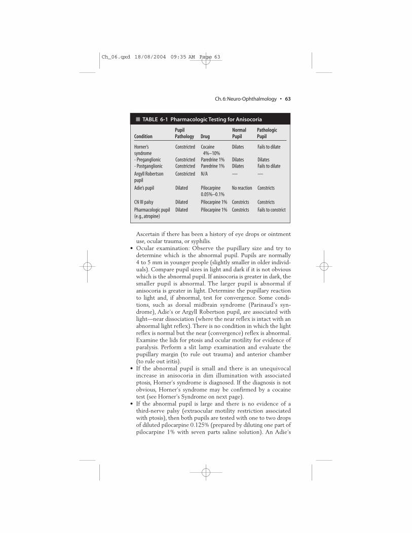
� Diagnostic Evaluation (Table 6-1)
• Ask when anisocoria was first noted and if associated with signsor symptoms or decreased vision. Examine old photographs.
62 • Blueprints Ophthalmology
Ch_06.qxd 18/08/2004 09:35 AM Page 62
Ascertain if there has been a history of eye drops or ointmentuse, ocular trauma, or syphilis.
• Ocular examination: Observe the pupillary size and try todetermine which is the abnormal pupil. Pupils are normally4 to 5 mm in younger people (slightly smaller in older individ-uals). Compare pupil sizes in light and dark if it is not obviouswhich is the abnormal pupil. If anisocoria is greater in dark, thesmaller pupil is abnormal. The larger pupil is abnormal ifanisocoria is greater in light. Determine the pupillary reactionto light and, if abnormal, test for convergence. Some condi-tions, such as dorsal midbrain syndrome (Parinaud’s syn-drome), Adie’s or Argyll Robertson pupil, are associated withlight—near dissociation (where the near reflex is intact with anabnormal light reflex). There is no condition in which the lightreflex is normal but the near (convergence) reflex is abnormal.Examine the lids for ptosis and ocular motility for evidence ofparalysis. Perform a slit lamp examination and evaluate thepupillary margin (to rule out trauma) and anterior chamber(to rule out iritis).
• If the abnormal pupil is small and there is an unequivocalincrease in anisocoria in dim illumination with associatedptosis, Horner’s syndrome is diagnosed. If the diagnosis is notobvious, Horner’s syndrome may be confirmed by a cocainetest (see Horner’s Syndrome on next page).
• If the abnormal pupil is large and there is no evidence of athird-nerve palsy (extraocular motility restriction associatedwith ptosis), then both pupils are tested with one to two dropsof diluted pilocarpine 0.125% (prepared by diluting one part ofpilocarpine 1% with seven parts saline solution). An Adie’s
Ch. 6: Neuro-Ophthalmology • 63
� TABLE 6-1 Pharmacologic Testing for Anisocoria
Pupil Normal Pathologic Condition Pathology Drug Pupil Pupil
Horner’s Constricted Cocaine Dilates Fails to dilatesyndrome 4%–10%- Preganglionic Constricted Paredrine 1% Dilates Dilates- Postganglionic Constricted Paredrine 1% Dilates Fails to dilate
Argyll Robertson Constricted N/A — —pupil
Adie’s pupil Dilated Pilocarpine No reaction Constricts0.05%–0.1%
CN III palsy Dilated Pilocarpine 1% Constricts Constricts
Pharmacologic pupil Dilated Pilocarpine 1% Constricts Fails to constrict(e.g., atropine)
Ch_06.qxd 18/08/2004 09:35 AM Page 63

pupil constricts significantly more than the normal pupil after15 minutes (see Adie’s Tonic Pupil on p. 67). In a chronicAdie’s pupil, the pupil may not react to dilute pilocarpine0.125%.
• If pharmacologic dilation is suspected (e.g., atropine) or thepupil did not respond to pilocarpine 0.125%, one drop of pilo-carpine 1% may be placed in both eyes. A pharmacologicallydilated pupil will constrict much less and more slowly than anormal pupil.
� Treatment/Follow-up
• For further information on managing the above causes ofanisocoria, see Horner’s Syndrome, Argyll Robertson Pupil,Adie’s Tonic Pupil, and Isolated Third-Nerve Palsy on p. 68.
Horner’s Syndrome
A defect of the sympathetic pathway resulting in ipsilateralmiosis, ptosis, enophthalmos (apparent recession of the globe intothe orbit), and possibly anhydrosis (lack of sweating).
� Etiology
• First-order neuron: stroke (vertebrobasilar artery insuffi-ciency); tumor.
• Second-order neuron: tumor (e.g., lung tumor, metastasis, thy-roid adenoma, neurofibroma). Suspect Pancoast’s tumor if armpain present. In children, suspect neuroblastoma, lymphoma,or metastasis.
• Third-order neuron: headache (Raeder’s paratrigeminal neu-ralgia, migraine, cluster), internal carotid dissection (superiorcervical ganglion lies near the bifurcation of the carotid artery),herpes zoster virus, Tolosa-Hunt syndrome, otitis media.
• Congenital: trauma (e.g., Erb’s palsy from delivery).
� History
• Pupil size disparity (smaller pupil is abnormal), ipsilateraldroopy eyelid (ptosis)
• Often asymptomatic
� Clinical Manifestations
• Anisocoria (greater in dim illumination because smaller pupilunable to dilate well due to defective sympathetic innervation)
• Ipsilateral drooping of upper eyelid (ptosis) and elevated lowereyelid (reverse ptosis)
64 • Blueprints Ophthalmology
Ch_06.qxd 18/08/2004 09:35 AM Page 64

• Ipsilateral anhydrosis (loss of sweating ability)• Iris heterochromia (lighter iris color in affected eye) in congen-
ital cases• Increased accommodation in involved eye• Intact light and near reaction
� Differential Diagnosis
• See Anisocoria above.
� Diagnostic Evaluation
• Determine the duration of Horner’s syndrome (old photo-graphs may help). New-onset Horner’s syndrome requiresmore extensive workup (an old Horner’s syndrome is morelikely to be benign). Ask about headache, arm pain, or previousstroke. Determine if there was a history of damage to the sym-pathetic chain such as previous surgery including thoracic, car-diac, thyroid, or neck or any head or neck trauma. Determine ifthere is a history of stroke, arm pain, or headache.
• Physical examination: Check for palpable supraclavicularnodes, thyroid enlargement, or a neck mass.
• Cocaine test if diagnosis is uncertain: place one drop of cocaine4% to 10% in both eyes and repeat in 1 minute.After 15 minutescheck pupillary size; if no change is present, place an additionaldrop in both eyes and reevaluate in another 15 minutes. AHorner’s pupil will dilate less than the normal pupil (cocaineblocks reuptake of norepinephrine).
• Paredrine test (hydroxyamphetamine 1%): may be used whenHorner’s syndrome is confirmed to distinguish a first- andsecond-order (preganglionic) neuron disorder from a third-order (postganglionic) neuron disorder. Wait at least 24 hoursif cocaine test was performed. Place one drop of hydroxyam-phetamine 1% into both eyes, and repeat 1 minute later. If theHorner’s pupil does not dilate to an equivalent amount as thenormal eye, a third-order neuron lesion is present. (Paredrinereleases norepinephrine from the presynaptic terminal.)
• Some of the following tests may be performed if a new-onsetHorner’s is discovered, a tumor is suspected, or a preganglioniclesion is present.-Chest x-ray or CT of lung with special attention to the apex-CT (axial and coronal) or MRI of brain and neck-CBC with differential-Lymph node biopsy in presence of lymphadenopathy-Carotid angiogram, magnetic resonance angiography (MRA),or carotid Doppler ultrasound in cases of neck pain wherecarotid dissection is suspected
Ch. 6: Neuro-Ophthalmology • 65
Ch_06.qxd 18/08/2004 09:35 AM Page 65

� Treatment
• Management of underlying disorder if possible• Ptosis surgery if necessary
� Follow-up
• As soon as possible in acute situation to rule out a life-threateningcause
• As soon as possible in a child with ptosis to treat for possibleamblyopia (see amblyopia treatment, p. 57)
• Chronic Horner’s syndrome is evaluated routinely.
Argyll Robertson Pupil
A rare syndrome that occurs in some patients with tertiary syphilisinvolving the central nervous system (CNS). Over time bothpupils become small (less than 2 mm) and irregular. This is one ofthe rare situations where a very miotic pupil may react briskly.
� Etiology
• Tertiary syphilis [positive fluorescent treponemal antibody,absorbed (FTA-ABS)]
� History
• Usually asymptomatic
� Clinical Manifestations
• Small, irregular pupils that react poorly to light but normallyto convergence (light-near dissociation). Pupils dilate poorly.
• Initially unilateral, but later becomes bilateral. Often asymmetric.• Normal vision.
� Differential Diagnosis
• See Anisocoria above.
� Diagnostic Evaluation
• Test pupillary reaction to light and convergence (determine iflight-near dissociation exists).
• Look for evidence of interstitial keratitis on slit lamp examination.• Look for uveitis, chorioretinitis, and papillitis on dilated fundus
examination.• Check FTA-ABS and rapid plasma reagin (RPR) or Venereal
Disease Research Laboratory (VDRL) results.• Possible lumbar puncture if positive diagnosis of syphilis.
66 • Blueprints Ophthalmology
Ch_06.qxd 18/08/2004 09:35 AM Page 66

� Treatment
• See Chapter 11, Acquired Syphilis, p. 157• The decision to treat is based on the presence of active disease
and if the patient had previously been treated appropriately.
� Follow-up
• Not emergent.• Workup patient in a few days to 2 weeks to determine if
syphilitic activity is present.
Adie’s Tonic Pupil
Dilated pupil that appears to be benign in nature. The pupil mayget smaller with time. The cause is unknown, but the ciliary gan-glion has been found to have a dramatic reduction in ganglioncells.
� Etiology
• Idiopathic• Orbital trauma• Infection with herpes zoster, varicella, syphilis• May be bilateral in diabetes, chronic alcoholism, familial
dysautonomia (Riley-Day syndrome)
� Epidemiology
• 70% women• 80% unilateral, second pupil may become involved later.
� History
• Anisocoria, larger pupil dilated• Blurred vision• May be asymptomatic
� Clinical Manifestations
• Irregularly dilated pupil.• Affected pupil demonstrates minimal to no reaction to light
and slow constriction to convergence with slow redilationafterwards (light-near dissociation).
• Affected pupil is supersensitive (constricts) to weak choliner-gics (e.g., one drop of pilocarpine 0.125%).
• Normal dilation with mydriatics.• Chronically, the involved pupil may become smaller than the
normal pupil.• Adie’s syndrome: when associated with absent deep tendon
reflexes (ankles and knees).
Ch. 6: Neuro-Ophthalmology • 67
Ch_06.qxd 18/08/2004 09:35 AM Page 67

� Differential Diagnosis
See Anisocoria above.
� Diagnostic Evaluation
• See diagnostic evaluation for anisocoria for general workup.• Examine the suspicious pupil with bright light at the slit lamp
to confirm a pupil that reacts slowly and irregularly.• Measure the size of each pupil as the patient fixates at dis-
tance. Check for a supersensitive pupil by placing one drop ofpilocarpine 0.125% in both eyes.
• An Adie’s tonic pupil constricts significantly more than thenormal contralateral pupil. Occasionally, the supersensitivitymay not be present immediately after developing an Adie’spupil; therefore, supersensitivity testing may be repeated 2 to 3weeks later.
• Dilute pilocarpine may yield a positive result in ArgyllRobertson pupil and familial dysautonomia.
Note: Parinaud’s syndrome: may cause bilateral middilatedpupils that react poorly to light but constrict normally duringconvergence (unlike Adie’s), upgaze paralysis, convergence-retraction nystagmus, and lid retraction (Collier’s sign). Withthese signs, perform an MRI to rule out pinealoma or other mid-brain abnormality.
� Treatment
• May treat with pilocarpine 0.125% two to four times per dayto constrict the pupil for cosmetic purposes or to improveaccommodation.
� Follow-up
• Routine follow-up if diagnosis is certain• If the patient is younger than 1 year, and Adie’s pupil or super-
sensitivity is present, refer the patient to a pediatric neurologistto rule out familial dysautonomia.
Isolated Third-Nerve Palsy
Defect in CN III resulting in motility impairment (external oph-thalmoplegia) and possibly impairment of pupillary reaction(internal ophthalmoplegia). Pupillary involvement may occursecondary to aneurysm, tumor, ischemia, trauma, or giant cellarteritis (GCA). The pupil is controlled by the efferent parasym-pathetics (miosis) that run in the inferior division of CN III,which innervates the medial and inferior recti.
68 • Blueprints Ophthalmology
Ch_06.qxd 18/08/2004 09:35 AM Page 68

� History
• Double vision that disappears when one eye is occluded• Ptosis (droopy eye lid) with/without pain
� Clinical Manifestations
• Limitation of ocular movements, especially superior, inferior,and nasal.
• Ptosis may be present.• Pupil may be dilated with diminished reactivity to light.• Eye may appear to be looking down and/or out.• May see aberrant regeneration (elevation of the upper lid and
possibly constriction of the pupil with superior, medial, or infe-rior gaze). If this sign occurs spontaneously (primary), it mayindicate a cavernous sinus aneurysm or tumor.
� Differential Diagnosis
• GCA: pupil may or may not be involved but may developocular motility dysfunction.
• Myasthenia gravis: no pupil involvement; fluctuation in ptosisand diplopia; ptosis worsens with sustained gaze; positiveresult on edrophonium chloride (e.g., Tensilon) test.
• Thyroid eye disease: have lid retraction and not ptosis; mayhave ocular motility disorders. May have proptosis. Also mayhave injection over recti muscle insertions and resistance onforced-duction testing.
• Orbital inflammatory pseudotumor: idiopathic orbital inflam-mation with pain on eye movement. Patients may have doublevision with restriction of ocular motility. May have proptosisand/or decreased vision.
• Internuclear ophthalmoplegia: unilateral or bilateral adductiondeficit with horizontal nystagmus in the opposite abductingeye. A lesion of the medial longitudinal fasciculus. Ptosis is notpresent. Seen in multiple sclerosis.
• Chronic progressive ophthalmoplegia: pupil is spared. Patientsdemonstrate progressive bilateral ptosis and ocular motilitydeficits. May or may not have diplopia.
• Parinaud’s syndrome: dorsal midbrain lesion with bilateralinability to look up and pupils demonstrating normal reactionto accommodation but abnormal reaction to light (light-neardissociation). There is no ptosis, and lid retraction and conver-gence-retraction nystagmus may be present.
� Diagnostic Evaluation
• Ascertain if there is a history of trauma or medical illness suchas diabetes, hypertension, or cancer.
Ch. 6: Neuro-Ophthalmology • 69
Ch_06.qxd 18/08/2004 09:35 AM Page 69

• Complete ocular examination with careful attention to pupilexamination. Check for ptosis and signs of aberrant regeneration.
• Check exophthalmometer for presence of proptosis.• Check for signs of other cranial nerve abnormalities.• Order immediate brain CT scan or MRI to rule out a
mass/aneurysm if:-There is pupillary involvement.-The pupil is not involved but:
-Other cranial nerves are involved.-The patient is younger than 50 years without a history ofdiabetes or hypertension.
-The third-nerve palsy has been present for more than 3months without improvement.
-There is spontaneous aberrant regeneration.• Check fasting blood sugar/Hb A1c and blood pressure if suspi-
cious of ischemic disease.• Check ESR if suspicious of GCA.• Check edrophonium chloride (e.g., Tensilon) test if suspicious
of myasthenia gravis (and pupil is not involved).
� Treatment
• Manage underlying abnormality or vascular disease if present.• If double vision is present, may patch involved eye (patching
not generally done in children under 10 years of age for fear ofamblyopia).
� Follow-up
• Pupil-involved third-nerve palsy: workup as described aboveand immediate hospitalization.
• Pupil-spared third-nerve palsy: workup as described and, ifnew, observe for 1 week. Recheck every 6 weeks.
• Normal function should return in about 3 months.• If function is not restored by 3 months or additional neurologic
abnormality results, an MRI of the brain is indicated.
Papilledema
Bilateral optic disc swelling caused by increased intracranialpressure
� Etiology
• Intracranial tumors (primary and metastatic) • Pseudotumor cerebri (benign intracranial hypertension): usually
occurs in women, often overweight. CT scan or MRI of the brainis normal, but may have exotropia secondary to CN VI palsy.
70 • Blueprints Ophthalmology
Ch_06.qxd 18/08/2004 09:35 AM Page 70

• Aqueductal stenosis: may produce hydrocephalus• Subdural and epidural hematoma: from trauma, especially if
on anticoagulation therapy• Subarachnoid hemorrhage: causes severe headache. May develop
preretinal and vitreous hemorrhage (Terson’s syndrome)• Brain abscess: especially if HIV positive; may cause fever• Arteriovenous malformation (AVM)
� History
• Transient loss of vision (lasting seconds) usually bilateral andcaused by changes in posture
• Decrease in visual acuity acutely (if associated with macularedema)
• Severe decrease of visual acuity and visual field defects withchronic papilledema (secondary to optic nerve pallor)
• Double vision (if CN VI palsy present)• Headache, nausea, vomiting
� Clinical Manifestations
• Early: bilateral, swollen, hyperemic optic nerves with blurringof the disc margin (Plate 7)-Normal pupil response to light and normal color vision.-Papillary and peripapillary retinal hemorrhages (often flameshaped) and cotton-wool spots may be present.
-Dilated, tortuous retinal veins.-Absent venous pulsation at optic disc (absent in 20% ofnormal persons).
-Visual field testing may reveal an enlarged blind spot.• Chronic: optic atrophy (Plate 5)
-Decreased central visual acuity and color vision-Esotropia (secondary to unilateral or bilateral sixth-nervepalsy)
-Collateral vessels on the optic disk-Resolution of peripapillary hemorrhages and cotton-woolspots
-Narrowing of peripapillary retinal vessels with peripapillarygliosis
-Visual field defects
� Differential Diagnosis (causes of bilateral disc edema)
• Pseudopapilledema: e.g., optic nerve drusen. Disc margins aresharp and vessels are not obscured. The neuroretinal rim is nothyperemic and the surrounding nerve fiber layer is normal.
• Malignant hypertensive retinopathy (Plate 8): fundus exami-nation reveals bilateral narrowed arterioles with arteriovenous
Ch. 6: Neuro-Ophthalmology • 71
Ch_06.qxd 18/08/2004 09:35 AM Page 71

nicking. There are associated scattered hemorrhages withcotton-wool spots. Blood pressure is extremely high.
• Ischemic optic neuropathy (ION): sudden, severe, unilateral loss invision that may soon involve the second eye (e.g., GCA and nonar-teritic ION). Optic nerve swelling is pale and not hyperemic.
• Diabetic papillitis: usually bilateral disc edema possibly associ-ated with diabetic retinopathy changes.Typically seen in youngpersons with type I diabetes.
• Infiltration of the optic nerve: usually unilateral but can bebilateral. Seen in sarcoidosis, leukemia, tuberculosis, metastasis,and other inflammatory diseases. If seen in a leukemia patient,immediate radiation therapy is indicated to preserve vision.
• Graves’ ophthalmopathy: other signs of thyroid dysfunctionwould be evident (see Chapter 11, Thyroid disease, p. 150).
• Leber’s optic neuropathy: begins unilaterally but rapidlybecomes bilateral. There is a rapid, progressive loss of visionwith disc swelling associated with peripapillary telangiectasiathat later becomes atrophic. Occurs primarily in men in theirsecond to third decade of life. Family history may or may notbe present.
� Diagnostic Evaluation
• Determine if there is a history of systemic disease, especiallyhypertension, thyroid disease, and diabetes.
• Ophthalmic examination should include pupillary and colorvision testing. Dilated fundus examination should carefullyobserve the vitreous for signs of inflammation, and the opticnerve appearance should be carefully noted.
• Check blood pressure.• Immediate CT (axial and coronal) or MRI of the brain (and
possibly the orbit) in all cases of bilateral disc edema(papilledema) to rule out a space occupying lesion.
• Lumbar puncture if CT/MRI result is unremarkable.
� Treatment
•Address underlying cause.
� Follow-up
• Initially weekly; later less frequently.• Follow visual acuity and visual field carefully.
Optic Neuritis
An inflammation of the optic nerve of unknown cause associatedwith rapid deterioration of vision followed by a steady recovery.
72 • Blueprints Ophthalmology
Ch_06.qxd 18/08/2004 09:35 AM Page 72

� Etiology
• Idiopathic• Multiple sclerosis• Viral infections, such as mononucleosis, herpes zoster,
encephalitis, measles, mumps, chickenpox• Granulomatous inflammations, such as syphilis, sarcoidosis,
and tuberculosis• Intraocular inflammations
� Epidemiology
• Usually occurs between ages 20 to 45 years• Occurs in nearly 100% of patients with long-standing multiple
sclerosis
� History
• Visual loss usually deteriorating over days until about 1 week(visual loss may deteriorate rarely over hours and may besubtle to extreme).
• Usually unilateral, 90% with orbital pain especially with eyemovement.
• Loss of color vision• Uhthoff’s symptom (increased symptoms with exercise or
increased body temperature)• Pulfrich’s phenomenon (altered perception of moving objects)• May have had a preceding viral syndrome (upper respiratory,
gastrointestinal, or other flulike symptoms).• Other: focal neurologic symptoms (e.g., numbness or tingling
in extremities).
� Clinical Manifestations
• Decreased visual acuity with a relative afferent pupillarydefect (RAPD).
• Decreased color vision and visual field defects.• Swollen optic nerve [may have peripapillary flame-shaped
hemorrhages (Plate 9)].• Disc may appear normal in retrobulbar optic neuritis; usually
seen in adults.• Rarely, may see vitreous cells.
� Differential Diagnosis
• Ischemic optic neuropathy (ION): sudden, painless loss of vision.Optic nerve swelling primarily pale. Visual field defects are usu-ally inferior altitudinal. Usually no pain with ocular motility.
• Acute papilledema: disc edema is bilateral. Visual acuity andcolor vision are usually normal unless macular edema is present.Spontaneous venous pulsations absent. No pain with ocular
Ch. 6: Neuro-Ophthalmology • 73
Ch_06.qxd 18/08/2004 09:35 AM Page 73

motility. No vitreous cells. Visual field testing often revealsenlarged blind spot.
• Severe systemic hypertension: bilateral disc edema with flame-shaped hemorrhages and cotton-wool spots. Blood pressure iselevated (Plate 8).
• Intracranial mass compressing the afferent visual pathway:Disc appears normal but there is loss of color vision with a pos-itive RAPD. Neuroimaging of the brain reveals a mass.
• Orbital tumor compressing the optic nerve: optic nerveswelling (if present) would be unilateral. Proptosis or restric-tion of extraocular motility may be evident. Vitreous cells arenot present.
• Toxic or metabolic optic neuropathy: progressive painlessbilateral loss of vision secondary to malnutrition (vitamin B1),alcohol, tobacco, pernicious anemia, or numerous toxins (e.g.,heavy metals, ethambutol, isoniazid, chloroquine, digitalis).Optic discs appear pale, especially temporally.
• Leber’s optic neuropathy: See discussion under differentialdiagnosis of papilledema above.
� Diagnostic Evaluation
• Determine whether acute or gradual loss of vision is present.Inquire if there have been previous episodes or pain withocular motility. Ask the patient’s age.
• Perform ophthalmic examination including pupillary and colorvision assessment. Perform dilated fundus examination to lookfor vitreous cells and swelling or hemorrhage in the retina oroptic nerve.
• Visual field testing.• Check blood pressure and perform neurologic examination
looking for abnormalities.• Consider blood tests such as CBC, ESR, RPR, FTA-ABS, and
antinuclear antibody (ANA) if presentation is unusual such asno pain on eye movement or patient’s age range is atypical.
• MRI with gadolinium of the brain and orbits for unusual casesand first episodes.
� Treatment
• Observation if patient has a history of optic neuritis or MS.• Intravenous steroids (e.g., methylprednisolone 250 mg IV
every six hours) for 3 days followed by oral prednisone (e.g.,40–100 mg PO every day) if the patient is seen acutely andthere is no previous history of optic neuritis or MS, and subse-quent MRI of brain reveals at least one area of demyelination.
• Consider intravenous steroids to accelerate visual recovery insome patients.
74 • Blueprints Ophthalmology
Ch_06.qxd 18/08/2004 09:35 AM Page 74

• No oral prednisone as a primary treatment because this hasbeen associated with an increased risk of recurrences.
� Follow-up
• Every 1 to 3 months.• Follow more closely and check IOP if on steroids.• Neurology consult if CNS demyelination is seen on MRI.
Arteritic Ischemic Optic Neuropathy (Giant Cell Arteritis)
Ischemia to the optic nerve head as a result of giant cell arteritis(GCA). GCA is an autoimmune disease that affects arteries withan internal elastic lamina. GCA can result in myocardial infarc-tion, stroke, and ocular complications.
� Epidemiology
• Increased incidence with older age (more than 55 years)• Usually occurs in whites (rare in blacks)
� History
• Sudden, painless loss of vision without progression.• Initially unilateral, but may rapidly become bilateral.• May experience headache, jaw claudication (pain with chewing),
or scalp tenderness.• May have proximal muscle and joint pain (e.g., polymyalgia
rheumatica), anorexia, weight loss, fever.• Usually occurs in patients older than 50 years.
� Clinical Manifestations
• Devastating loss of vision with an RAPD.• Funduscopic examination reveals pale, swollen optic nerve
(often associated with flame-shaped hemorrhages). As opticnerve edema resolves, optic atrophy ensues.
• Central retinal artery occlusion may occur.• Visual field defects may occur revealing altitudinal or central
visual loss.• Palpated temporal artery may be tender and nonpulsatile.• May acquire cranial nerve palsies (primarily CN VI).• ESR may be very elevated.
� Differential Diagnosis
• Nonarteritic ION. Usually patient is younger with less severeloss of vision. Usually do not have above symptoms of GCA;ESR is usually normal.
Ch. 6: Neuro-Ophthalmology • 75
Ch_06.qxd 18/08/2004 09:35 AM Page 75

• Inflammatory optic neuritis (papillitis): Usually visual loss isless severe and less sudden. Pain is usually associated with eyemovements. No symptoms of GCA. Associated with morehemorrhage of the optic disc. No vitreous cells. Usuallyyounger persons affected.
• Central retinal vein occlusion (CRVO): severe loss of visionwith associated RAPD. Optic nerve swelling is present as wellas diffuse retinal hemorrhages (Blood and Thunder) extendingto periphery.
• Compressive optic nerve tumor: slow progressive loss of visualacuity. No symptoms of GCA. Optic nerve slowly becomespale.
• Central retinal artery occlusion (CRAO): sudden, severe, pain-less loss of vision associated with an RAPD. However, opticdisc is not swollen. Retinal edema may be observed with a clas-sic cherry-red spot in the fovea.
� Diagnostic Evaluation
• Try to elicit above symptoms and ascertain the age of thepatient, as older patients have a higher risk of developingGCA.
• Ocular examination: color vision testing and pupillary assess-ment to rule out RAPD. Dilated fundus examination for opticnerve evaluation and to rule out retinal causes of severe loss ofvision.
• Stat ESR (Westergren). Upper limit of normal: men = age/2;women = (age + 10)/2.
• Temporal artery biopsy if symptoms or signs suspicious ofGCA or ESR elevated.
• ESR may be normal (perform biopsy within 1 to 2 weeks ofinstituting steroid therapy).
• If biopsy is negative (at least a 2-cm section), a contralateralbiopsy may be taken if a high clinical suspicion exists (somephysicians prefer initial bilateral temporal artery biopsiesbecause of skip areas and possible false negatives).
� Treatment
• Once suspected, start intravenous steroids (e.g., methylpred-nisolone 250 mg IV every 6 hours for 12 doses) in the hospital.Switch to oral prednisone (e.g., 80–100 mg PO every day)after 12 doses.
• An oral antiulcer medication (e.g., ranitidine 150 mg PO twiceper day) is often given concurrently with the steroid.
• Obtain temporal artery biopsy, often while the patient is still inthe hospital.
76 • Blueprints Ophthalmology
Ch_06.qxd 18/08/2004 09:35 AM Page 76

• If biopsy is positive, continue oral prednisone. Steroid may beslowly tapered over 3 to 12 months as ESR declines or symp-toms improve. Patient’s internist should manage complicationsof steroid therapy, but an ophthalmologist should adjust andtaper the steroid level based on clinical findings and the ESR.
• Discontinue steroids after obtaining negative biopsies onadequate specimens.
� Follow-up
• Evaluate and treat emergently if GCA suspected.• After diagnosis confirmed by biopsy, initial steroid dosage is
continued for 4 weeks until symptoms improve or ESRdecreases.
• Taper steroid very slowly over 3 to 12 months.• Decrease dosage monthly as symptoms reverse or ESR
decreases.• Check ESR monthly with each dosage change.
-If symptoms return or ESR elevates, steroid dose must beincreased.
-Use minimal steroid dose to achieve desired result.
Nonarteritic Ischemic Optic Neuropathy
Ischemia of the optic nerve head due to compromise of the pos-terior ciliary vessels.
� History
• Sudden moderate, nonprogressive loss of vision; painless• Initially unilateral but may become bilateral
� Clinical Manifestations
• RAPD and decreased color vision• Optic disc with flame-shaped hemorrhages and often pale
swelling involving only one sector• Optic atrophy after edema resolves (Plate 5)• Altitudinal or central visual field defect• Normal ESR
� Risk Factors
• Often associated with hypertension, diabetes, arteriosclerosis• Patients usually 40 to 60 years old
� Differential Diagnosis
• See Arteritic Ischemic Optic Neuropathy above.
Ch. 6: Neuro-Ophthalmology • 77
Ch_06.qxd 18/08/2004 09:35 AM Page 77

� Diagnostic Evaluation
• Ask about above symptoms of GCA. Determining age is veryimportant (usually occurs in 40- to 60-year-olds).
• Ocular examination: evaluate pupils for RAPD and checkcolor vision with color plates. Dilated fundus examination toevaluate the optic nerve and to rule out retinal causes of severevisual loss.
� Treatment/Follow-up
• Observation• Follow-up in 1 month
78 • Blueprints Ophthalmology
Ch_06.qxd 18/08/2004 09:35 AM Page 78

Plate 7 • Discedema secondaryto increasedintracranialhypertensiondemonstrates anelevated disc with ahyperemic neuro-retinal rim anddilated capillaries.
Plate 8 • Disc edema secondary to uncontrolled hypertension. Note the blurreddisc margins and hemorrhages and evidence of hypertensive retinopathy. Leakinglipids travel in Henle’s layer toward the macula forming a “macular star.”
Plate 9 • Disc edema secondary to malignant hypertension, optic neuritis, orischemic optic neuropathy demonstrates hemorrhagic disc swelling with exudatesand cotton-wool spots surrounding the optic nerve.The disc margin is very blurred.
Bournias_plates.qxd 23/08/2004 12:57 PM Page 3