Embed Size (px)
Citation preview
12 Jun 2003 14:58 AR AR191-BE05-11.tex AR191-BE05-11.sgm LaTeX2e(2002/01/18)P1: GJB10.1146/annurev.bioeng.5.011303.120731
Annu. Rev. Biomed. Eng. 2003. 5:293–347doi: 10.1146/annurev.bioeng.5.011303.120731
Copyright c© 2003 by Annual Reviews. All rights reservedFirst published online as a Review in Advance on June 4, 2003
NEURAL TISSUE ENGINEERING: Strategies forRepair and Regeneration
Christine E. Schmidt1 and Jennie Baier Leach21Department of Biomedical Engineering and2Department of Chemical Engineering,The University of Texas at Austin, Austin, Texas 78712; email: [email protected],[email protected]
Key Words nerve regeneration, nerve guidance channels, peripheral nerve repair,spinal cord injury
■ Abstract Nerve regeneration is a complex biological phenomenon. In the periph-eral nervous system, nerves can regenerate on their own if injuries are small. Largerinjuries must be surgically treated, typically with nerve grafts harvested from elsewherein the body. Spinal cord injury is more complicated, as there are factors in the bodythat inhibit repair. Unfortunately, a solution to completely repair spinal cord injuryhas not been found. Thus, bioengineering strategies for the peripheral nervous systemare focused on alternatives to the nerve graft, whereas efforts for spinal cord injuryare focused on creating a permissive environment for regeneration. Fortunately, recentadvances in neuroscience, cell culture, genetic techniques, and biomaterials provideoptimism for new treatments for nerve injuries. This article reviews the nervous systemphysiology, the factors that are critical for nerve repair, and the current approaches thatare being explored to aid peripheral nerve regeneration and spinal cord repair.
CONTENTS
INTRODUCTION . . . . . . . . . . . . . . . . . . . . . . . . . . . . . . . . . . . . . . . . . . . . . . . . . . . . . 294Physiology of the Nervous System. . . . . . . . . . . . . . . . . . . . . . . . . . . . . . . . . . . . . . 294Nerve Injury and Regeneration. . . . . . . . . . . . . . . . . . . . . . . . . . . . . . . . . . . . . . . . . 296Current Clinical Approaches for Treating Nerve Injuries. . . . . . . . . . . . . . . . . . . . . 297Challenges and Bioengineering Strategies for Nerve Repair. . . . . . . . . . . . . . . . . . 299
GUIDANCE THERAPIES . . . . . . . . . . . . . . . . . . . . . . . . . . . . . . . . . . . . . . . . . . . . . . 300Historical Introduction to Guidance Therapies. . . . . . . . . . . . . . . . . . . . . . . . . . . . . 300Autologous Tissue Grafts. . . . . . . . . . . . . . . . . . . . . . . . . . . . . . . . . . . . . . . . . . . . . 302Nonautologous Tissue and Acellular Grafts. . . . . . . . . . . . . . . . . . . . . . . . . . . . . . . 302Natural-Based Materials. . . . . . . . . . . . . . . . . . . . . . . . . . . . . . . . . . . . . . . . . . . . . . 303Synthetic Materials . . . . . . . . . . . . . . . . . . . . . . . . . . . . . . . . . . . . . . . . . . . . . . . . . . 304Applications in the Central Nervous System. . . . . . . . . . . . . . . . . . . . . . . . . . . . . . . 307
BIOMOLECULAR THERAPIES . . . . . . . . . . . . . . . . . . . . . . . . . . . . . . . . . . . . . . . . . 309Neurotrophic Factors to Promote Regeneration. . . . . . . . . . . . . . . . . . . . . . . . . . . . 309Biomolecule Delivery Techniques. . . . . . . . . . . . . . . . . . . . . . . . . . . . . . . . . . . . . . . 311Intrinsic Neuronal Factors to Promote Regeneration. . . . . . . . . . . . . . . . . . . . . . . . 313
1523-9829/03/0815-0293$14.00 293

12 Jun 2003 14:58 AR AR191-BE05-11.tex AR191-BE05-11.sgm LaTeX2e(2002/01/18)P1: GJB
294 SCHMIDT ¥ BAIER LEACH
Blocking Inhibitory Biomolecules in the CNS. . . . . . . . . . . . . . . . . . . . . . . . . . . . . 316CELLULAR THERAPIES . . . . . . . . . . . . . . . . . . . . . . . . . . . . . . . . . . . . . . . . . . . . . . 317
Glial Cells and Macrophages. . . . . . . . . . . . . . . . . . . . . . . . . . . . . . . . . . . . . . . . . . . 317Olfactory Ensheathing Cells. . . . . . . . . . . . . . . . . . . . . . . . . . . . . . . . . . . . . . . . . . . 319Stem Cells . . . . . . . . . . . . . . . . . . . . . . . . . . . . . . . . . . . . . . . . . . . . . . . . . . . . . . . . . 321Genetically Modified Cells . . . . . . . . . . . . . . . . . . . . . . . . . . . . . . . . . . . . . . . . . . . . 321
ADVANCED THERAPIES . . . . . . . . . . . . . . . . . . . . . . . . . . . . . . . . . . . . . . . . . . . . . . 322Advanced Guidance Channel Fabrication Techniques. . . . . . . . . . . . . . . . . . . . . . . 322Combination Approaches and Novel Stimuli. . . . . . . . . . . . . . . . . . . . . . . . . . . . . . 327Convergence of Neural Regeneration and Neural Prostheses. . . . . . . . . . . . . . . . . . 327
CONCLUSION . . . . . . . . . . . . . . . . . . . . . . . . . . . . . . . . . . . . . . . . . . . . . . . . . . . . . . . 329
INTRODUCTION
Physiology of the Nervous System
The physiology of the nervous system presents unique challenges to bioengineer-ing research addressing nerve injuries. This section briefly describes the generalorganization and the cellular components of the nervous system as well as theanatomy of the peripheral nerve and spinal cord. Discussions of nerve injuries andthe currently available clinical treatments are also presented.
ORGANIZATION OF THE NERVOUS SYSTEM The nervous system is classified intothe central nervous system (CNS) and the peripheral nervous system (PNS). TheCNS, which includes the brain, spinal cord, optic, and olfactory and auditorysystems, conducts and interprets signals as well as provides excitatory stimuli to thePNS. The PNS consists of the cranial nerves arising from the brain, the spinal nervesarising from the spinal cord, and sensory nerve cell bodies (dorsal root ganglia)and their processes. Peripheral nerves innervate muscle tissue, transmitting sensoryand excitatory input to and from the spinal column.
CELLULAR COMPONENTS OF THE NERVOUS SYSTEM The nervous system is com-posed of two cell types: neurons and neuroglia. Neurons are the basic structuraland functional elements of the nervous system and consist of a cell body (soma)and its extensions (axons and dendrites). Clusters of sensory nerve soma, knownas ganglia, are located just outside the spinal column. Dendrites transmit electricalsignals to the neuron cell body and the axon conducts impulses away. Glial cells, orneuroglia, are support cells that aid the function of neurons and include Schwanncells in the PNS and astrocytes and oligodendrocytes in the CNS. Glial cells aremore abundant than neurons, and unlike neurons, which cannot undergo mitosis,glial cells have some capacity for cell division. Although neurons cannot divideby mitosis, they can regenerate a severed portion or sprout new processes undercertain conditions.
In the PNS, sheaths of living Schwann cells surround all axons. On the outersurface of this Schwann cell layer is the neurilemma, a basement membrane similar

12 Jun 2003 14:58 AR AR191-BE05-11.tex AR191-BE05-11.sgm LaTeX2e(2002/01/18)P1: GJB
NEURAL TISSUE ENGINEERING 295
to that found in epithelial layers. In contrast to axons in the PNS, CNS axons do notpossess this continuous basement membrane and sheath of Schwann cells. Manyaxons are instead surrounded by an insulating myelin sheath, which is formed fromdense layers of successive wrappings of the cell membrane of Schwann cells (PNS)or oligodendrocytes (CNS). Myelin serves to increase the propagation velocity ofthe nerve impulse, which is particularly important for those axons that extend longdistances (up to 1 m).
ANATOMY OF THE PERIPHERAL NERVE AND SPINAL CORD A peripheral nerveconsists of motor and sensory axons bundled together by support tissue into ananatomically defined trunk (Figure 1). Endoneurium surrounds individual axonsand their Schwann cell sheaths and is composed predominantly of oriented col-lagen fibers. Next, the perineurium, formed from many layers of flattened cells(i.e., fibroblasts) and collagen, surrounds groups of axons to form fascicles. Fi-nally, epineurium, an outer sheath of loose fibrocollagenous tissue, binds individualnerve fascicles into a nerve trunk. Peripheral nerves are well vascularized by cap-illaries within the support tissue of the nerve trunk or by vessels that penetrate thenerve from surrounding arteries and veins.
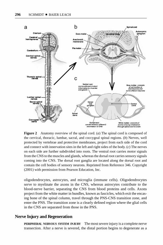
The spinal cord is composed of dendrites, axons, and cell bodies (Figure 2). Thecenter of the spinal cord, a butterfly-shaped region referred to as gray matter, con-tains the cell bodies of excitatory neurons, as well as glial cells and blood vessels.The gray matter is surrounded by white matter, which helps to protect and in-sulate the spinal cord. White matter consists of axons and glial cells, including
Figure 1 Anatomical overview of the PNS. Axons, surrounded by myelinatingSchwann cell sheaths, are enclosed by endoneurium. Next, the perineurium binds in-dividual axons together to form fascicles. Several axons are contained in each fascicle.Lastly, epineurium groups fascicles to one another, forming the nerve cable. Reprintedfrom Reference 343, pp. 375–415. Copyright (2002) with permission from MarcelDekker, Inc.
12 Jun 2003 14:58 AR AR191-BE05-11.tex AR191-BE05-11.sgm LaTeX2e(2002/01/18)P1: GJB
296 SCHMIDT ¥ BAIER LEACH
Figure 2 Anatomy overview of the spinal cord. (a) The spinal cord is composed ofthe cervical, thoracic, lumbar, sacral, and coccygeal spinal regions. (b) Nerves, wellprotected by vertebrae and protective membranes, project from each side of the cordand connect with innervation sites in the left and right sides of the body. (c) The nerveson each side are further subdivided into roots. The ventral root carries motor signalsfrom the CNS to the muscles and glands, whereas the dorsal root carries sensory signalscoming into the CNS. The dorsal root ganglia are located along the dorsal root andcontain the cell bodies of sensory neurons. Reprinted from Reference 346. Copyright(2001) with permission from Pearson Education, Inc.
oligodendrocytes, astrocytes, and microglia (immune cells). Oligodendrocytesserve to myelinate the axons in the CNS, whereas astrocytes contribute to theblood-nerve barrier, separating the CNS from blood proteins and cells. Axonsproject from the white matter in bundles, known as fascicles, which exit the encas-ing bone of the spinal column, travel through the PNS-CNS transition zone, andenter the PNS. The transition zone is a clearly defined region where the glial cellsin the CNS are separated from those in the PNS.
Nerve Injury and Regeneration
PERIPHERAL NERVOUS SYSTEM INJURY The most severe injury is a complete nervetransection. After a nerve is severed, the distal portion begins to degenerate as a

12 Jun 2003 14:58 AR AR191-BE05-11.tex AR191-BE05-11.sgm LaTeX2e(2002/01/18)P1: GJB
NEURAL TISSUE ENGINEERING 297
result of protease activity and separation from the metabolic resources of the nervecell bodies (Figure 3a). The cytoskeleton begins to breakdown, followed by thedissolution of the cell membrane. The proximal end of the nerve stump swells,but experiences only minimal damage via retrograde degradation. After the cy-toskeleton and membrane degrade, Schwann cells surrounding the axons in thedistal end shed their myelin lipids. Phagocytotic cells, such as macrophages andSchwann cells, clear myelin and axonal debris (1). In addition to clearing myelindebris, macrophages and Schwann cells also produce cytokines, which enhanceaxon growth (2). Following debris clearance, regeneration begins at the proximalend and continues toward the distal stump. New axonal sprouts usually emanatefrom the nodes of Ranvier, nonmyelinated areas of axons located between Schwanncells. Functional reinnervation requires that axons extend until they reach their dis-tal target, and in humans, axon regeneration occurs at a rate of about 2–5 mm/day;thus significant injuries can take many months to heal (3).
When a hollow nerve conduit is used to repair a severed peripheral nerve (dis-cussed further below), an additional step for regeneration is required (4, 5). Afterinjury, a fibrin bridge is formed through the conduit and across the defect site. Thisfibrin cable includes macrophages and other cells thought to be involved in debrisclearance. The fibrin bridge retracts as Schwann cells and capillaries begin to growacross the gap, and regeneration proceeds as normal. It is not clear if the formationof a fibrin cable also occurs in the absence of a conduit or when a conduit containsan internal matrix.
CENTRAL NERVOUS SYSTEM INJURY A key difference between the PNS and CNSis the capacity for peripheral nerves to regenerate; CNS axons do not regenerateappreciably in their native environment. Several glycoproteins in the native extra-cellular environment (myelin) of the CNS are inhibitory for regeneration (6–8).The physiological response to injury in the CNS is also different compared to thatof the PNS. After injury in the CNS, macrophages infiltrate the site of injury muchmore slowly compared to macrophage infiltration in the PNS, delaying the removalof inhibitory myelin (9). This is largely a result of the blood-spine barrier, whichlimits macrophage entry into the nerve tissue to just the site of injury, where barrierintegrity is weakened. In addition, cell adhesion molecules in the distal end of theinjured spinal cord are not upregulated appreciably as they are in the PNS, limitingmacrophage recruitment. Finally, astrocytes proliferate in a manner similar to thatof Schwann cells in the PNS, but instead become “reactive astrocytes,” producingglial scars that inhibit regeneration (Figure 3b) (10).
Current Clinical Approaches for Treating Nerve Injuries
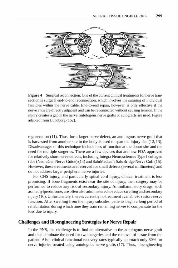
For peripheral nerve injury, treatment typically consists of either direct end-to-end surgical reconnection of the damaged nerve ends (Figure 4) or the use of anautologous nerve graft. Suturing the ends of the two nerve ends together can repairsmall defects or gaps in the nerve. For longer nerve gaps, this approach is notdesired because any tension introduced into the nerve cable would inhibit nerve

12 Jun 2003 14:58 AR AR191-BE05-11.tex AR191-BE05-11.sgm LaTeX2e(2002/01/18)P1: GJB
298 SCHMIDT ¥ BAIER LEACH
Figure 3 Responses to axotomy in the PNS and spinal cord. (a) In the PNS, supportcells aid neuronal regeneration. Proliferating Schwann cells, macrophages, and mono-cytes work together to remove myelin debris, release neurotrophins, and lead axonstoward their synaptic targets, resulting in restored neuronal function. (b) In the CNS,however, the few neurons that survive axotomy attempt regeneration and subsequentlymeet an impenetrable glial scar composed of myelin and cellular debris, as well as as-trocytes, oligodendrocytes, and microglia. Fibroblasts, monocytes, and macrophagesmay also be present in the glial scar. Consequently, regenerating neurons in the spinalcord are blocked from reaching their synaptic target. Figure adapted from Bahr &Bonhoeffer (345).
12 Jun 2003 14:58 AR AR191-BE05-11.tex AR191-BE05-11.sgm LaTeX2e(2002/01/18)P1: GJB
NEURAL TISSUE ENGINEERING 299
Figure 4 Surgical reconnection. One of the current clinical treatments for nerve tran-section is surgical end-to-end reconnection, which involves the suturing of individualfascicles within the nerve cable. End-to-end repair, however, is only effective if thenerve ends are directly adjacent and can be reconnected without causing tension. If theinjury creates a gap in the nerve, autologous nerve grafts or autografts are used. Figureadapted from Lundborg (162).
regeneration (11). Thus, for a larger nerve defect, an autologous nerve graft thatis harvested from another site in the body is used to span the injury site (12, 13).Disadvantages of this technique include loss of function at the donor site and theneed for multiple surgeries. There are a few devices that are now FDA approvedfor relatively short nerve defects, including Integra Neurosciences Type I collagentube (NeuraGen Nerve Guide) (14) and SaluMedica’s SaluBridge Nerve Cuff (15).However, these treatments are reserved for small defects (several millimeters) anddo not address larger peripheral nerve injuries.
For CNS injury, and particularly spinal cord injury, clinical treatment is lesspromising. If bone fragments exist near the site of injury, then surgery may beperformed to reduce any risk of secondary injury. Antiinflammatory drugs, suchas methylprednisone, are often also administered to reduce swelling and secondaryinjury (16). Unfortunately, there is currently no treatment available to restore nervefunction. After swelling from the injury subsides, patients begin a long period ofrehabilitation during which time they train remaining nerves to compensate for theloss due to injury.
Challenges and Bioengineering Strategies for Nerve Repair
In the PNS, the challenge is to find an alternative to the autologous nerve graftand thus eliminate the need for two surgeries and the removal of tissue from thepatient. Also, clinical functional recovery rates typically approach only 80% fornerve injuries treated using autologous nerve grafts (17). Thus, bioengineering
12 Jun 2003 14:58 AR AR191-BE05-11.tex AR191-BE05-11.sgm LaTeX2e(2002/01/18)P1: GJB
300 SCHMIDT ¥ BAIER LEACH
strategies for the PNS have focused on developing alternative treatments to thenerve graft (e.g., nerve guidance channels), especially for larger defects, and im-proving recovery rates and functional outcome.
The CNS is a greater challenge for new therapies. The ability of spinal nerves toregenerate was not decisively shown until 1980 (18), and it was not until after thistime that research in this area rapidly developed. In addition, results from variousstudies have been controversial (19, 20), complicating developments. It has beenshown that both embryonic spinal cord grafts and peripheral nerve tissue grafts cansupport regenerating fibers in the CNS, but the fibers often do not successfully growback across the PNS-CNS transition zone (21, 22). Thus, bioengineering effortsare focused on creating a permissive environment for regeneration and providinga seamless interface between the CNS and PNS.
These challenges provide fertile ground for the development of therapies anddevices to enhance regeneration. Many researchers are presently focusing effortson creating physical or chemical pathways for regenerating axons. These devicesinclude physical or mechanical guidance cues, cellular components, and biomolec-ular signals, as reviewed individually below. Future therapies will incorporate mul-tiple cues into unique devices that more closely mimic native nerve. They will alsobe interactive and programmable, and thus capable of seamless communicationwith surrounding tissues.
GUIDANCE THERAPIES
Historical Introduction to Guidance Therapies
It is commonly accepted that physical guidance of axons is a vital component ofnerve repair. During the nineteenth century, many materials were used in an attemptto physically guide the regeneration of damaged peripheral nerves, including au-tologous nerve grafts (23), bone (24), metal tubes (25), and fat sheaths (26). It wasnot until the 1960s that Millesi pioneered microsurgical techniques to accuratelyalign nerve fascicles in the direct resection of nerve ends, with improved functionaloutcomes (11). He also determined that the use of nerve grafts reduced tensionon the damaged nerves in many cases and further enhanced functional recovery.These results also supported the need for physical guidance as an essential elementin nerve regeneration. Later research demonstrated that biochemical signals (seeBiomolecular Therapies, below) as well as physical guidance are critical for nerveregeneration (27–30).
Currently, the autologous nerve graft is the gold standard for repair of a pe-ripheral nerve defect (12, 13). Current research is focused on developing improvedscaffolds that can be used to physically guide regeneration of nerves across lesions.Similar techniques are also being explored for the repair of transected nerves inthe spinal cord. These “nerve guides” or “nerve guidance channels” serve to directaxons sprouting from the proximal nerve end, provide a conduit for the diffusionof growth factors secreted by the injured nerve ends, and reduce the infiltrationof scar tissue. Past research in this area has focused either on existing natural or

12 Jun 2003 14:58 AR AR191-BE05-11.tex AR191-BE05-11.sgm LaTeX2e(2002/01/18)P1: GJB
NEURAL TISSUE ENGINEERING 301
synthetic materials; however, none of the materials studied to date have matched orexceeded the performance of the nerve autograft. As a result, researchers are nowfocusing on the combination of materials and desired biomolecules to create newcomposite materials that can actively stimulate nerve regeneration. In addition,methods to minimize the immune response to nonautologous tissue could providea source of natural material for nerve repair.
Note: For additional reviews on nerve regeneration, nerve grafts, and nerveguidance channels, refer to (31–38). Table 1 provides a summary of materials
TABLE 1 Nerve grafts and nerve conduit materials
Graft Reference
Autologous tissue grafts1. Nerve grafts (gold standard) (12, 13)2. Vein grafts (17, 43–45)3. Muscle grafts (41, 42)4. Epineurial sheaths (46)5. Tendon grafts (47)
Nonautologous/acellular grafts1. Immunosuppression with allografts (373)2. Acellular allografts and xenografts
Thermal decellularization (52, 53, 57)Radiation treatment (54, 58)Chemical decellularization (55, 56)
3. Small intestinal submucosa (SIS) (65, 66, 70)4. Human amnion (71, 75, 76)
Natural-based materials1. ECM protein-based materials
Fibronectin (84, 85)Laminin (82, 88)Collagen (90–92)
2. Hyaluronic acid-based materials (95)3. Fibrin/fibrinogen (96, 97)4. Other materials (alginate, agarose. . . ) (99, 100, 102)
Synthetic materials1. Biodegradable synthetic materials
Poly(lactic acid) (PLA) (109, 110)Poly(lactic-co-glycolic acid) PLGA (107)Poly(caprolactone) (111, 113)Poly(urethane) (114)Poly(organo)phosphazene (112)Poly(3-hydroxybutyrate) (116)Poly(ethylene glycol) “glue” (128, 156)Biodegradable glass (117, 118)
2. Electrically active materialsPiezoelectric (119)Electrically conducting (120)
3. Nonbiodegradable synthetic materialsSilicone (122, 127)Gore-Tex or ePTFE (123–125)
12 Jun 2003 14:58 AR AR191-BE05-11.tex AR191-BE05-11.sgm LaTeX2e(2002/01/18)P1: GJB
302 SCHMIDT ¥ BAIER LEACH
in use or under investigation for nerve repair applications, as described in detailbelow.
Autologous Tissue Grafts
Natural tissues, including autologous tissue grafts, possess several advantages.Natural materials are more likely to be biocompatible than artificial materials, areless toxic, and provide a support structure to promote cell adhesion and migration.Drawbacks, on the other hand, include potential difficulties with isolation andcontrolled scale-up.
Autologous tissue grafts have been used extensively for nerve repair applica-tions. Nerve autografts are typically derived from one of several cutaneous nerves,such as the sural or saphenous nerve, with an available length up to about 40 cmand a cable diameter of 2–3 cm (39, 40). For a more thorough synopsis of the earlyhistory of nerve repair using nerve grafts, refer to the review by Chiu (17), and foradditional information on the surgical techniques used in nerve grafting, refer to(13, 38).
In addition to the nerve graft, other natural tissues, such as autologous muscle(41, 42) and vein grafts (17, 43–45), have been used to limited extents in the clinic.Furthermore, some current research efforts are focused on natural tissue graftsfor peripheral nerve repair, including the use of epineurial sheaths (46), tendongrafts (47), muscle-vein combined grafts (48, 49), inside-out vein grafts (50), andvein grafts impregnated with autologous Schwann cells (51). All have exhibitedencouraging results in research but still suffer from the key drawback that tissuemust be removed from the patient.
Nonautologous Tissue and Acellular Grafts
As a result of the limitations with using autologous tissue, attention has turnedtoward nonautologous tissue and extracellular matrix (ECM)-based materials.Allogenic and xenogeneic tissues (donor tissue from cadavers and animals,respectively) have the advantages that supplies can be large and their use doesnot require harvest from the patient. However, these tissues possess some risk ofdisease transmission and must either be used in conjunction with immunosuppres-sants or must be processed to remove immunogenic components. Many efforts arebeing made to process intact nonautologous tissue, rendering it less immunogenicfor clinical use. These methods focus on removal or destruction of the immunogeniccells and the preservation of the ECM components that are essentially conservedbetween species. Many different methods have been explored, including thermaltechniques (52, 53), radiation (54), and chemical processes (55, 56).
The most common decellularization technique is thermal decellularization,which involves repeated freeze-thaw cycles to kill and fragment the cells. Nervegrafts processed using this approach have been shown to be generally nonimmuno-genic (57); however, the structure of the ECM is typically damaged and the cellularremnants are not completely extracted, resulting in inflammation when implanted.

27 Jun 2003 14:12 AR AR191-BE05-11.tex AR191-BE05-11.sgm LaTeX2e(2002/01/18)P1: GJB
NEURAL TISSUE ENGINEERING 303
Radiation treatments destroy cells in tissues and produce relatively little damageto the matrix structure, but they also fail to extract all cellular components (58).Several chemical (e.g., detergent) treatments have also been developed that aremore effective in the complete removal of cell debris. Recent approaches, usingoptimized combinations of detergents, show good cellular clearance and excel-lent structural preservation (Figure 5) (T.W. Hudson & C.E. Schmidt, unpublishedresults). Similar thermal and chemical decellularization methods have also beenapplied to muscle tissue for use in nerve repair applications (59–62). These effortsto develop acellular sources of tissue for nerve repair applications appear quitepromising, especially in light of recent successes to create other acellular tissuesfor clinical applications, such as cardiovascular tissue [reviewed in (63)] and skin(64). Thus, the use of acellular tissues for clinical nerve repair may become aviable option in the future.
Other natural tissues explored for nerve repair applications include small in-testinal submucosa (SIS) and amniotic tissue grafts. SIS is an acellular matrixderived from small intestine, typically of porcine origin. SIS is prepared from themucosa and muscle layers of the small intestine, which are treated with a hypotonicsolution to lyse and wash away the cells. The resultant ECM material is composedof collagen, fibronectin, growth factors, glycosaminoglycans, proteoglycans, andglycoproteins (65, 66). SIS has been used with encouraging results as a regener-ative scaffold for a number of tissues including vascular grafts (67), urinary tract(68), and tendon (69). Recently, SIS derived from rats has been used in conjunctionwith Schwann cells to create nerve grafts that promote regeneration almost as wellas the nerve autograft (70).
Amnion harvested from human placental tissues has also received attentionfor its potential use in nerve regeneration applications (71–76). The amnion is anatural, biodegradable tissue that exhibits low immunogenicity and stimulates newvascularization (77). This material is also readily available in large quantities anddoes not require surgical procedures for harvest. To process this tissue, the epithelialcell layer of the amnion membrane is removed while the basement membrane andstromal surfaces remain intact. After removing the epithelial cells, the resultingacellular connective tissue matrix can be manufactured into thin dry sheets and thensubsequently processed into conduits. Amnion tubes have been shown to promoteregeneration comparable to that of the nerve autograft across 1 cm defects in thesciatic nerves of rats, and the tubes completely degrade by 4 months (73).
Natural-Based Materials
In addition to intact acellular tissues, a great deal of research has focused on the useof purified natural ECM proteins and glycosaminoglycans, which can be modifiedto serve as appropriate scaffolding. ECM molecules, such as laminin, collagen,and fibronectin, have been shown to play a significant role in axonal developmentand repair in the body (78, 79). Furthermore, many other proteoglycans and gly-cosaminoglycans of the ECM are known to modulate neural activity and neurite

12 Jun 2003 14:58 AR AR191-BE05-11.tex AR191-BE05-11.sgm LaTeX2e(2002/01/18)P1: GJB
304 SCHMIDT ¥ BAIER LEACH
extension; some provide stimulatory cues, whereas others provide inhibitory cues(80, 81). Thus, ECM components are obvious candidates for use in nerve guides.
There are a number of examples in which the ECM proteins laminin, fibronectin,and collagen have been used for nerve repair applications (82–90). For example,silicone tubes filled with laminin, fibronectin, and collagen show improved regen-eration over a 10 mm rat sciatic nerve gap compared to empty silicone controls(86). Oriented mats or strands of fibronectin have been used to bridge 10 mmnerve defects in rats, with results close to that of the nerve autograft (84). Colla-gen filaments have also been used to guide regenerating axons across 20–30 mmdefects in rats (87, 90). Further studies have shown that oriented fibers of colla-gen within gels, aligned using magnetic fields, provide an improved template forneurite extension compared to randomly oriented collagen fibers (91, 92). Rates ofregeneration comparable to those using a nerve autograft have been achieved usingcollagen tubes containing a porous collagen-glycosaminoglycan matrix (93, 94). Itis believed that providing a suitable matrix for Schwann cell and neurite migrationenhances nerve repair.
Other naturally derived molecules investigated for their application in nerve re-pair include hyaluronic acid (95), fibrinogen (96), fibrin gels (97), self-assemblingpeptide scaffolds (98), alginate (99), agarose (100, 101), and chitosan (102). Cur-rent studies are under way to further modify these materials for tissue engineeringapplications, such as the chemical cross-linking of hyaluronic acid, an ECM gly-cosaminoglycan, to allow it to be photopolymerized into porous, three-dimensionalhydrogels and to stabilize it against rapid degradation (103). Other studies includethe modulation of fibrin gels either using magnetic fields to align the polymer fibers(104) or with appropriate biomolecules to enhance neurite extension (105, 106).
Synthetic Materials
Research is also under way to identify synthetic materials that can be used for nerverepair applications. Synthetic materials are attractive because their chemical andphysical properties (e.g., degradation rate, porosity, mechanical strength) can bespecifically optimized for a particular application. However, the biocompatibilityof synthetic materials poses a challenge because the body’s inflammatory responsecan vary considerably from one material to another. In addition, some syntheticmaterials that are tolerated by the body’s immune system are unfortunately incom-patible with cell adhesion and tissue repair. These materials are often modified torender them more “cell friendly.”
To select an appropriate synthetic material, there are several general propertiesthat all nerve guidance channels should possess: (a) They must be readily formedinto a conduit with desired dimensions, (b) they must be sterilizable, (c) they mustbe tear resistant, and (d) they must be easy to handle and suture. Permanent mate-rials pose a higher risk for infection, are more likely to provoke a chronic inflam-matory response, and have the potential to compress the nerve over time. Thus,a nerve guide that degrades as the nerve regenerates is preferred. Additionally,
12 Jun 2003 14:58 AR AR191-BE05-11.tex AR191-BE05-11.sgm LaTeX2e(2002/01/18)P1: GJB
NEURAL TISSUE ENGINEERING 305
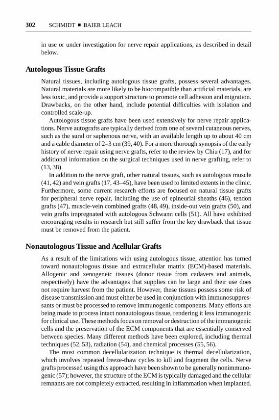
Figure 6 Properties of the ideal nerve guidance channel. The desired physical prop-erties of a nerve conduit include (clockwise from top left): a biodegradable and porouschannel wall; the ability to deliver bioactive factors, such as growth factors; the in-corporation of support cells; an internal oriented matrix to support cell migration;intraluminal channels to mimic the structure of nerve fascicles; and electrical activ-ity. Reprinted from Reference 34. Copyright (1999), with permission from ElsevierScience.
guidance channels should be pliable, but should maintain their shape and resistcollapse during implantation and over the time course for regeneration. Researchhas also shown that guidance channels should be semipermeable and should havea smooth inner wall. Hudson et al. (34) review the desired physical properties ofthe nerve guidance channel (Figure 6).
A number of different synthetic materials have been explored for use in aidingnerve regeneration. Poly(esters), such as poly(glycolic acid) (PGA), poly(lacticacid) (PLA), and poly(lactic-co-glycolic acid) (PLGA) (107, 108), were some ofthe first synthetic polymers studied because of their availability, ease of process-ing, biodegradation characteristics, and approval by the FDA. These materialscontinue to be researched to date and have been processed into foams (Figure 7)and seeded with Schwann cells to improve their regenerative potential (109, 110).Other biodegradable poly(esters), such as poly(caprolactones), have also demon-strated promise for nerve regeneration applications (111–113). In addition topoly(esters), biodegradable poly(urethane) (114), poly(organo phosphazene) (112),methacrylate-based hydrogels (115), and poly(3-hydroxybutyrate) (116) haveshown a capacity for guiding regeneration. Biodegradable glass tubes have alsobeen studied, but results have not been optimistic (117, 118). More advanced
12 Jun 2003 14:58 AR AR191-BE05-11.tex AR191-BE05-11.sgm LaTeX2e(2002/01/18)P1: GJB
306 SCHMIDT ¥ BAIER LEACH
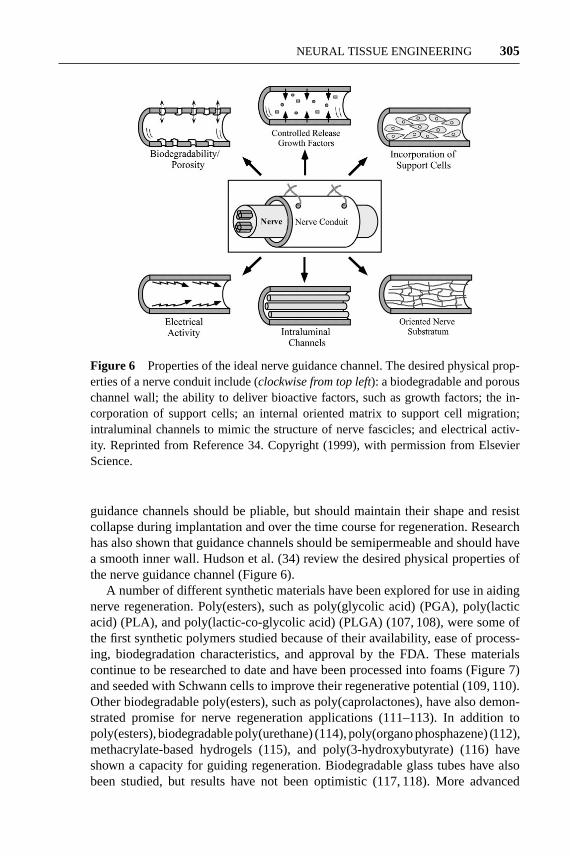
Figure 7 Poly(L-lactic acid) foam nerve guidance channels. Porous biodegradablepoly(L-lactic acid) (PLL) conduits were synthesized using a solvent casting, extrusion,and particulate leaching technique. (a) Nerve guidance channels from 10 mm to 22 mmin length were used to repair transected rat sciatic nerves, a common model for studyingperipheral nerve regeneration. (b) After 4 months, the PLL conduits remained struc-turally intact, supported tissue infiltration and vascularization, and resulted in structuraland functional regeneration comparable to isografts, the current clinical gold standard(109). Figure courtesy of C.W. Patrick, Jr., Department of Plastic Surgery, The Uni-versity of Texas M.D. Anderson Cancer Center and G.R. Evans, Division of PlasticSurgery, University of California-Irvine.
materials processing techniques to create three-dimensional channels and uniquepores or fiber structures are discussed below in Advanced Therapies.
Research has also shown that electrical charges play a significant role in stim-ulating the cellular differentiation for several tissue types. Neurite extension, forexample, is significantly enhanced on piezoelectric materials (i.e., materials thatgenerate a surface charge with small deformations), such as poly(vinylidene fluo-ride) (PVDF) (119), and on electrically conducting polymers, such as poly(pyrrole)

12 Jun 2003 14:58 AR AR191-BE05-11.tex AR191-BE05-11.sgm LaTeX2e(2002/01/18)P1: GJB
NEURAL TISSUE ENGINEERING 307
(120). Further modification of these materials with biological stimuli (e.g.,hyaluronic acid) may provide interactive biomaterials for use as nerve guidancechannels (121).
Several nondegradable synthetic materials have been used in nerve repair appli-cations, including silicone tubing (122), which has been applied in clinical settingsas well as in research settings, and expanded poly(tetrafluoroethylene) or ePTFE(Gore-Tex) (123–125). Silicone, in particular, has been studied since the 1960s(126, 127), and much fundamental insight into nerve regeneration has come fromthe use of this model system. In general, inert silicone tubes can be used to bridgeshort gaps with some success. However, it is commonly accepted that imperme-able, inert guidance channels, such as silicone, do not support regeneration acrossdefects larger than 10 mm (in rats) without the presence of exogenous growth fac-tors. Instead, research is now focused on developing semipermeable or degradableguidance channels that can actively stimulate improved regeneration over longer,more clinically relevant defect lengths. The development of nondegradable guid-ance channels is not under very active pursuit because of the limitations associatedwith permanent materials, as described earlier.
Poly(ethylene glycol) (PEG) has also been applied to nerve regeneration appli-cations. In one unique approach, PEG has been used to “fuse” the membranes ofsevered nerve ends of sciatic and spinal nerves (128). These studies have shownthat conduction of axon potentials can be restored immediately with this proce-dure. Unfortunately, this process can only be applied if the severed nerve ends aredirectly adjacent, and therefore is not useful for large nerve defects. Additionally,cross-linked PEG hydrogels that are modified with factors to mimic the ECM areunder active development, particularly for cardiovascular applications (129, 130).Recent studies on these systems are also looking into their ability to aid nerveregeneration (131). For example, PC12 cells are able to extend neurites on a PEGhydrogel when the cell adhesion motif Arg-Gly-Asp-Ser (RGDS) is covalentlyincorporated into the material (Figure 8) (B.K. Mann, personal communication).
Applications in the Central Nervous System
Although regeneration of the mammalian CNS was once thought to be impossible(132), studies over the past two decades have shown that axonal growth afterspinal cord injury can occur when provided with the correct substratum (133–135).Research has focused mainly on the use of peripheral nerve grafts and embryonicspinal cord grafts (18, 136–140), and recent studies have looked to embryonicneural progenitor or stem cells (141, 142). Research suggests that embryonic spinalcord grafts both rescue neurons from injury-induced cell death and serve as asubstrate to support new axonal growth (143, 144). On the other hand, peripheralnerve grafts appear to only provide a permissive substrate for the ingrowth of CNSaxons (145).
Cultured Schwann cells (146–148), ECM-based materials (149, 150), and syn-thetic polymers (128, 131, 151–158) have also been investigated for their ability to

12 Jun 2003 14:58 AR AR191-BE05-11.tex AR191-BE05-11.sgm LaTeX2e(2002/01/18)P1: GJB
308 SCHMIDT ¥ BAIER LEACH
Figure 8 Biomimetic polyethylene glycol (PEG) hydrogels for nerve regeneration.PEG hydrogels are an alternative synthetic material that could be used for nerve regen-eration. Bioactive factors, such as cell adhesion ligands, growth factors, and proteolyticdegradation sites, can be incorporated into PEG hydrogels in order to render a morebiomimetic scaffold (129, 130). PC12 cells are able to adhere to and extend neurites onPEG hydrogels with covalently incorporated cell adhesion ligands (RGDS, YIGSR,and IKVAV) but not on hydrogels with a nonadhesive control peptide RGES. Thisimage shows PC12 cells extending neurites on a PEG hydrogel with RGDS covalentlyincorporated into the material. The amount of neurite extension was dependent onthe type and concentration of adhesion ligand incorporated into the hydrogel. Figurecourtesy of B.K. Mann, Keck Graduate Institute.
serve as templates for spinal nerve regeneration. For example, Woerly et al. (151)have shown that a poly[N-(2-hydroxypropyl)methacrylamide] (pHPMA) hydro-gel containing the cell-adhesive region of fibronectin Arg-Gly-Asp (RGD) is ableto support tissue development within a lesion created in a rat spinal cord. Areasof angiogenesis and axonal growth were observed within the tissue, and necro-sis was reduced in the adjacent white and gray matter, suggesting that pHPMAhydrogel matrices could potentially serve as a substrate for spinal nerve regenera-tion across a defect. Additional synthetic matrices under investigation in the CNSinclude: poly(lactic acid) or poly(D,L-lactide) (154, 155), poly(2-hydroxyethyl

12 Jun 2003 14:58 AR AR191-BE05-11.tex AR191-BE05-11.sgm LaTeX2e(2002/01/18)P1: GJB
NEURAL TISSUE ENGINEERING 309
methacrylate) or pHEMA (131, 153), poly(lactic-co-glycolic acid) (PLGA) andblock copolymer of poly(lactic-co-glycolic acid)-poly(lysine) (159), and poly(ethylene glycol) “glue” (128, 152, 156). Results to date from these various studiesgive great hope for the therapeutic reconstruction of neural connections after spinalcord injury; systems can be successfully designed to provide a permissive substratefor regenerating spinal nerve fibers. However, major challenges that remain includethe growth of the nerve fibers back into the spinal cord and the functional inte-gration of the nerve fibers with the host synaptic pathways. These hurdles may beaddressed in the future by combining guidance therapy approaches with variousbiomolecular therapies, as described below. For reviews of tissue engineering andbiomaterials strategies applied to spinal cord injury, see (157, 160).
BIOMOLECULAR THERAPIES
Neurotrophic Factors to Promote Regeneration
The role of neurotrophic factors in neural regeneration has been the focus of exten-sive research [for reviews see (161–165)]. The influence of these factors in neuraldevelopment, survival, outgrowth, and branching has been explored on variouslevels, from molecular interactions to macroscopic tissue responses. One familyof neurotrophic factors, the neurotrophins, has been heavily investigated in nerveregeneration studies. The neurotrophins include nerve growth factor (NGF), brain-derived neurotrophic factor (BDNF), neurotrophin-3 (NT-3), and neurotrophin-4/5(NT-4/5). Outside of the neurotrophin family, other factors of importance are cil-iary neurotrophic factor (CNTF), glial cell line-derived growth factor (GDNF),and acidic and basic fibroblast growth factor (aFGF, bFGF). As discussed below,these factors promote a range of neural responses (summarized in Table 2). A
TABLE 2 Neural responses to neurotrophic factors
Neural response promoted Neurotrophic factors
Motor neuron survival BDNF, NT-3, NT-4/5, CNTF, GDNF
Motor neuron outgrowth BDNF, NT-3, NT-4/5, CNTF, GDNF
Sensory neuron survival NGF, NT-4/5, GDNF
Sensory neuron outgrowth NGF, BDNF, NT-3
Spinal cord regeneration NGF, NT-3, CNTF, FGFs
Peripheral nerve regeneration NGF, NT-3, NT-4/5, CNTF, GDNF, FGFs
Sensory nerve growth across the NGF, NT-3, GDNF, FGFsPNS-CNS transition zone
Abbreviations: Brain-derived neurotrophic factor (BDNF), neurotrophin-3 (NT-3), neurotrophin-4/5(NT-4/5), ciliary neurotrophic factor (CNTF), glial cell line-derived growth factor (GDNF), nervegrowth factor (NGF), acidic and basic fibroblast growth factors (FGFs).

12 Jun 2003 14:58 AR AR191-BE05-11.tex AR191-BE05-11.sgm LaTeX2e(2002/01/18)P1: GJB
310 SCHMIDT ¥ BAIER LEACH
detailed discussion of all neurotrophic factors, their additive effects, and their di-rect influence on glial cells is not within the scope of this review; see (161–164,166) for more information on these topics.
NGF is vital to the development and regeneration of the nervous system; conse-quently, NGF is the most thoroughly characterized neurotrophic factor (162, 167).NGF is expressed at low levels in healthy peripheral nerve and is upregulated inthe distal stump upon injury (168). Similarly, following spinal cord transection,NGF accumulates in both the distal and proximal stumps (169). On the cellularlevel, NGF promotes survival, outgrowth, and branching in sensory neurons, butdoes not aid motor neuron regeneration (170–172). Nonetheless, much work hasfocused on delivering NGF to neuronal injuries. Studies using nerve guidancechannels filled with NGF solutions have provided conflicting results (173–175),perhaps due to leakage from the channel or NGF inactivation. Continuous de-livery devices (discussed in more detail below) offer a more reliable means ofadministering NGF, and such work has been associated with increased regenera-tion in both the PNS (176–178) and the spinal cord (171, 179, 180). Applicationof exogenous NGF has also been linked to increased sensory neuron regenerationfrom the dorsal root ganglia, through the PNS-CNS transition zone, and into thespinal cord (180–182). However, the use of NGF is not without disadvantages:the application of exogenous NGF to spinal cord injuries has been associated withsignificant sprouting of uninjured sensory axons (181). This sprouting has beenlinked to serious side effects, including chronic pain (181–183) and inappropriateneuronal reflexes (181, 184).
BDNF supports motor neuron survival (185–187) and promotes the axonalgrowth of motor (188) and sensory (189) neurons. However, research investigatingthe effects of BDNF on nerve regeneration has provided inconclusive results inboth the PNS (178, 190, 191) and the spinal cord (179, 189, 192–196). Similarto NGF, these inconsistencies likely stem from different methods used to deliverBDNF, as it has been noted that BDNF must be delivered locally and at highconcentrations to have an effect on nerve regeneration (165).
NT-3, like BDNF, promotes motor neuron survival (187) and outgrowth (188)as well as sensory axon growth (197). In vivo, NT-3 plays a vital role in aidingthe regeneration of peripheral nerves (178, 198) and spinal cord (179, 189, 194–196). NT-3 has also been associated with the increased ability of sensory axons togrow from the dorsal root ganglia, across the PNS-CNS transition zone, and intothe spinal cord (180, 182, 195). NT-4/5 has not been studied in as much detail asNT-3. However, NT-4/5 has been shown to promote the survival of motor neurons(187, 199) and sensory neurons (200). NT-4/5 also supports the axonal outgrowthof motor neurons (188) and has been associated with improved regeneration ofsevered peripheral nerve (201).
CNTF promotes motor neuron survival (202, 203), outgrowth (204), and sprout-ing (205). CNTF is thought to play a role in the response of spinal cord to injury,as CNTF mRNA is found at increased levels adjacent to spinal cord lesions (206).The application of exogenous CNTF has been associated with increased levels of

12 Jun 2003 14:58 AR AR191-BE05-11.tex AR191-BE05-11.sgm LaTeX2e(2002/01/18)P1: GJB
NEURAL TISSUE ENGINEERING 311
regeneration following injury in both the spinal cord (196) and peripheral nerve(207). A drawback, however, is that CNTF has been demonstrated to play a rolein glial scarring (i.e., an injury response that results in a nonpermissive growthenvironment in the CNS; see below for more information) (208, 209).
GDNF promotes the survival of motor (210), sensory (172, 209), and autonomic(211) neurons. GDNF also promotes the growth of motor neurons in the CNS(212) and has been correlated with improved peripheral nerve regeneration (177).In comparison to NGF and NT-3, GDNF was shown to promote more extensivegrowth of sensory neurons from the dorsal root ganglia, through the PNS-CNStransition zone, and into the spinal cord (180, 182).
aFGF and bFGF have been associated with enhanced regeneration followinginjuries in the peripheral nerve (213, 214) and spinal cord (19, 179). The fibroblastgrowth factors are strong promoters of angiogenesis (215) and could, therefore,directly and indirectly aid in the healing of injured nerves. Like other neurotrophins(i.e., NGF, NT-3, and GDNF), bFGF has been associated with increased outgrowthof sensory neurons from the dorsal root ganglia, through the PNS-CNS transitionzone, and into the spinal cord (181).
In summary, neurotrophic factors promote a variety of neural responses: sur-vival and outgrowth of the motor and sensory nerves, spinal cord and peripheralnerve regeneration, and sensory nerve growth across the PNS-CNS transition zone.However, in vivo responses can vary due to the method of delivering the growth fac-tor. Therefore, the continued use and development of highly controllable deliverydevices are required for the study of these extremely complex systems.
Biomolecule Delivery Techniques
The delivery of biomolecules to support regeneration has several intrinsic chal-lenges, including the toxicities and poor stability associated with many bioactivefactors. A variety of techniques to deliver therapeutics to the nervous system havebeen established, including osmotic pumps (216) and silicone reservoirs (217).However, these methods are often associated with drawbacks, including devicefailure and higher potentials for inflammation and infection due to their non-degradable components (218). Polymer matrices, microspheres, and gene therapy,as described below, are effective delivery methods that overcome these challenges.For a recent review on these and other methods of delivering biomolecules to thenervous system, see Maysinger & Morinville (218).
Synthetic and naturally derived polymers are widely used in controlled releasedevices for protein delivery. These devices are designed such that bioactive factorsare released in a spatially and temporally controlled manner; for example, releasecan occur as the polymer degrades or by diffusion through pores in the polymermatrix. In neural applications, two types of delivery devices have been primar-ily used: polymer matrices and microspheres. One example of a polymer matrixdelivery device is the nerve guidance channel. These tubular conduits have beenprimarily used as a model system to study peripheral nerve repair (see Guidance

12 Jun 2003 14:58 AR AR191-BE05-11.tex AR191-BE05-11.sgm LaTeX2e(2002/01/18)P1: GJB
312 SCHMIDT ¥ BAIER LEACH
Figure 9 Poly(lactide-co-glycolide) microspheres. Poly(lactide-co-glycolide) mi-crospheres prepared using a water/oil/water double emulsion solvent evaporation pro-cess. The number average size of these particles is about 1 mm. Particles were freeze-dried and sputter coated for observation under scanning electron microscopy. Figurecourtesy of K. Roy, S.P. Kasturi, and J. Mendenhall, Department of Biomedical En-gineering and Institute of Cellular and Molecular Biology, The University of Texas atAustin.
Therapies above for more detail). By incorporating growth factors into the conduitwall, the nerve guidance channel itself becomes a delivery device. Ethylene vinylacetate (177, 178) and fibronectin mats (198) are examples of polymeric nerveguidance channel materials that have been successfully modified to deliver growthfactors to regenerating peripheral nerve.
Microspheres (Figure 9) are suitable for a variety of delivery applications andare commonly used to deliver molecules to the CNS. Like polymeric matrices,microspheres aid in the controlled release of active biomolecules; however, thesesmall devices typically have diameters in the range of 1µm to 1 mm and can beapplied less invasively (e.g., injection). Growth factors encapsulated in chitosan,alginate, poly(lactic acid), poly(glycolic acid), poly(lactic-co-glycolic acid), andpoly(caprolactone) have been investigated and show promise for further study inmodels of spinal cord injury (219, 220).
Though polymers can be used for controlled release, they only provide a finitereservoir of active biomolecular agents. Thus, researchers are turning toward genetherapy techniques for the long-term production of active growth factors in situ(see Cellular Therapies below for a discussion of genetically modified cells fortransplant applications). Recent reviews provide an excellent overview of genetherapy in the nervous system (221–223); therefore, this section only provides abrief summary.
A number of viral and nonviral gene delivery techniques are available (221, 222).Viral vectors have been investigated in a variety of tissue systems and include
12 Jun 2003 14:58 AR AR191-BE05-11.tex AR191-BE05-11.sgm LaTeX2e(2002/01/18)P1: GJB
NEURAL TISSUE ENGINEERING 313
methods based on retrovirus, herpes virus, adenovirus, and adeno-associated virus;efforts in the nervous system have primarily implemented herpes virus (224, 225)and adenovirus (197, 203). Viral gene transfer is able to promote high levels ofgene expression from the vector (221). Nonetheless, some level of risk is associ-ated with administering a therapy based on a viral agent. Before they can be used inthe clinic, viral vectors must be proven to be safe and any inflammatory responsemust be minimized (222, 223).
Given the drawbacks inherent to viral vectors, researchers are also investigatingnonviral transfection techniques. In the absence of viral protein machinery to en-ter cells, transfections with nonviral vectors rely on direct delivery or nonspecificinternalization methods (221). Naked DNA can be injected directly, but this tech-nique often results in low expression. Gene guns increase transfection efficienciesby attaching the DNA to a gold particle and then shooting the complex into thecell with a high-voltage arc or high-pressure gas. Therefore, the use of gene gunscan result in higher transfection efficiencies, but at the cost of increased tissuedamage.
Cationic lipids and polymers can also be used to assist transfection; these meth-ods primarily depend upon nonspecific internalization to deliver DNA into neu-ronal cells (221). Lipoplexes, or complexes of DNA with cationic lipids, are oneof the most common and successful nonviral gene delivery methods (221). How-ever, the mechanisms by which nonviral vectors are able to transfect cells is notwell understood; thus, the optimization of these systems is yet very challenging.[For an excellent review of the current challenges to nonviral gene therapy in theCNS, see Berry et al. (221)]. Nevertheless, through new means of delivering andtargeting the complexes, scientists are able to increase the transfection efficienciesof nonviral vectors. For example, cell-specific targeting ligands (e.g., the nontoxicneuronal-specific fragment C of tetanus toxin) (226) allow transfection only in thedesired cell types, and gene-activated matrices (GAMs; biodegradable matricesloaded with DNA) allow repeated transfection as neurites extend throughout thematrix (221).
Intrinsic Neuronal Factors to Promote Regeneration
As described above, the application of exogenous neurotrophic factors can be aneffective means of promoting regeneration. Through an intricate and synergisticcascade of signaling events, neurotrophic factors affect the expression of genesthat promote neural survival and axonal outgrowth. Because of the complexity as-sociated with delivering active neurotrophic factors, researchers are also lookingtoward other means of manipulating the genes that control nerve regeneration. Oneapproach gains insight from contrasting the intrinsic neuronal mechanisms of twosystems: the mature response to injuries in adults and the embryonic developmentof the nervous system. A separate but related approach considers gene expressionfollowing axotomy in the PNS (where regeneration does occur) compared to thespinal cord (where regeneration typically does not occur). Based on the analysisof these systems, researchers are investigating methods of optimally controlling

12 Jun 2003 14:58 AR AR191-BE05-11.tex AR191-BE05-11.sgm LaTeX2e(2002/01/18)P1: GJB
314 SCHMIDT ¥ BAIER LEACH
regeneration-associated genes (RAGs), neuronal cytoskeletal components, and an-tiapoptosis factors.
RAGs are first expressed during the development of the nervous system [forreviews, see (227, 228)]. The developmental expression of RAGs is transient, andin healthy adult nerve, the expression of these genes is negligible. However, whenperipheral nerves are damaged, a selection of these genes is re-expressed, oftenresulting in successful regeneration. This is not the case following spinal cordinjury and may be one of the reasons that regeneration is not successful in this sys-tem. Many have hypothesized, therefore, that inducing the upregulation of RAGsmay be an effective treatment of spinal cord injury. Two of the most abundant andwell-studied RAGs are GAP-43 and CAP-23 (227, 229). The overexpression ofeach of these factors alone can induce axonal sprouting (230–232), but not to adegree suitable for the support of regeneration (233). Based on this information,it has been suggested that an effective therapy to treat spinal cord regenerationmay not rely on the upregulation of a single RAG, but will require the coordinatedoverexpression of two or more RAGs acting together (227, 228). Such a therapycould be based on in situ transfection, such as the work with adenoviral vectorsfor the overexpression of GAP-43 (234), or rely on the administration of neu-rotrophins (e.g., BDNF or NT-4/5), which are known to stimulate RAG expression(193).
Another means of promoting the intrinsic neuronal mechanisms of regenerationrelies on an understanding of the cytoskeletal dynamics driving axonal growth.Actin polymerization and rearrangement are crucial to the migration of cells, suchas fibroblasts (235), and the elongation of nerve processes (236). However, themechanism of this process is not well understood. Furnish et al. (237) suggestthat actin accessory proteins, such as gelsolin, could play an important role inregulating axonal growth (Figure 10). Information gained from such studies couldlead to a better understanding of the cytoskeleton’s role in nerve regeneration anduncover new possible targets for clinical therapies.
Before regeneration and cytoskeletal reorganization begin, apoptosis is one ofthe main obstacles to overcome following nerve injury. Therefore, scientists haveattempted overexpressing antiapoptosis factors in neurons as a means of aidingnerve regeneration. Work in this area has primarily focused on overexpressingbcl-2 (225, 238–242). Interestingly, the expression of bcl-2 has also been impli-cated in aiding axonal outgrowth (238, 240). However, the exact nature of thisgene is not well understood, as some have suggested that bcl-2 expression doesnot directly enhance nerve regeneration (241, 242). While the majority of thesestudies were carried out in transgenic mice, promising work has demonstrated thesuitability of viral vectors for the overexpression of antiapoptosis factors (225).
Studies of neuronal development have helped to expose other factors thatcould also aid adult nerve regeneration. These include adhesion molecules (L1,NCAM, N-cadherin) (223, 243), molecules for axon guidance and path-finding(semaphorins, Slits, netrins, ephrins) (244), and synaptogenic factors (agrin, s-laminin, and ARIA) (245). For example, several studies have associated the

12 Jun 2003 14:58 AR AR191-BE05-11.tex AR191-BE05-11.sgm LaTeX2e(2002/01/18)P1: GJB
NEURAL TISSUE ENGINEERING 315
Figure 10 Enhancing the intrinsic neuronal mechanisms to improve regeneration:optimizing cytoskeletal dynamics for axonal outgrowth. The overexpression of gel-solin, an actin accessory protein, was associated with improved neurite outgrowth inPC12 cells. After three days, (a) PC12 cells transfected to overexpress gelsolin pos-sessed longer neurites than (b) mock clones (cells that received the transfection vectorwithout the gene for gelsolin) (237). Scale bar, 100µm. Figure courtesy of E.J. Furnish& C.E. Schmidt, Departments of Chemical Engineering and Biomedical Engineering,The University of Texas at Austin.
12 Jun 2003 14:58 AR AR191-BE05-11.tex AR191-BE05-11.sgm LaTeX2e(2002/01/18)P1: GJB
316 SCHMIDT ¥ BAIER LEACH
overexpression of the adhesion molecule L1 with increased axonal regenerationfollowing injury in the CNS (246, 247).
Blocking Inhibitory Biomolecules in the CNS
As described above, some nerve regeneration studies consider the differential in-jury responses found in the PNS and the spinal cord. Furthermore, as studies withneurotrophins, RAGs, and antiapoptosis factors have shown, neurons in the spinalcord have not necessarily lost their intrinsic ability to grow. However, one of thegreatest challenges facing spinal cord regeneration still remains: glial scarring [seeNerve Injury and Regeneration above; for reviews, see (166, 248, 249–251)]. Thisresponse is characterized by the formation of a nonpermissive environment that in-hibits axon growth and myelination. The main cell types involved are macrophages,microglia, oligodendrocytes, and astrocytes. The noncellular components of theglial scar include myelin-associated molecules (Nogo, myelin associated glyco-protein, oligodendrocyte-myelin glycoprotein), chondroitin sulfate proteoglycans(phosphacan, neurocan, brevican), axon guidance molecules (semaphorin, ephrin,netrin), and tenascin. Therefore, many bioengineering therapies for treating spinalcord injuries have focused upon attenuating the inhibitory glial scar components;these methods attempt to reduce the synthesis of the inhibitory components, toblock their effects, or to remove them altogether with enzymatic degradationtreatments.
The mechanisms that control the upregulation of inhibitory molecules follow-ing spinal cord injury are not well understood. However, factors, such as trans-forming growth factor-β2 (TGF-β2) (252) and ciliary neurotrophic factor (CNTF)(208, 209), have been associated with the stimulation of this process. Therapeutictreatments to block the effects of these growth factors are likely to aid in pre-venting the synthesis of glial scar components. For example, the administration ofanti-TGF-β2 antibody was associated with decreased scarring following injury inthe CNS (252).
Blocking the effects of glial scar critically relies upon identifying its inhibitorycomponents. Studies by Caroni & Schwab (253) were the first to confirm that oligo-dendrocyte myelin inhibits nerve regeneration. In this work, IN-1, a monoclonalantibody raised against the myelin protein NI-35, was shown to block the myelin-associated inhibition of axonal growth. Later work indicated that the IN-1 antibodymight indeed improve regeneration following spinal cord injury (254–256). NI-35,also called Nogo, was found to contain two inhibitory domains, Nogo-A (7, 257)and Nogo-66 (258). Shortly after the identification of NgR, the receptor for Nogo-66 (258), molecules were discovered that could block its interaction with Nogo.These include a soluble truncated form of NgR (259) and a competitive antagonistpeptide that binds with high affinity to NgR (260). Both systems were subsequentlyshown to decrease the Nogo inhibition of neurite outgrowth in vitro (259, 260).Also, administration of the antagonist peptide was shown to improve functional re-covery following spinal cord injury (260). Interestingly, NgR is a receptor for two

27 Jun 2003 14:29 AR AR191-BE05-11.tex AR191-BE05-11.sgm LaTeX2e(2002/01/18)P1: GJB
NEURAL TISSUE ENGINEERING 317
other growth inhibitors found in myelin, myelin-associated glycoprotein (MAG)(261), and oligodendrocyte-associated glycoprotein (OMgp) (262). This findinghas prompted some to suggest that methods targeting NgR could lead to futureclinical therapies (261, 263).
In addition to Nogo, MAG, and OMgp, there are likely a number of other in-hibitory molecules in oligodendrocyte myelin left to be discovered. Therefore,therapeutic vaccination shows promise toward blocking multiple inhibitory com-ponents in one treatment. Vaccination stimulates the body’s own immune systemto produce polyclonal antibodies against an antigen. In such studies, myelin im-munization in mice was associated with the long-distance regeneration of largenumbers of axons and improved motor function following spinal cord injury (264).Many refinements will be required before this method can be translated into a ther-apy for humans; however, the polyclonal antibodies will likely prove useful foridentifying any yet unknown inhibitory components of myelin.
Protease treatments degrade the inhibitory components in glial scar, facilitatingtheir removal and increasing the ability of axons to grow through the injured area.Several extracellular matrix components, including chondroitin sulfate proteogly-cans (CSPGs), are upregulated by oligodendrocytes and astrocytes following spinalcord injury, and are inhibitory towards axonal outgrowth (248). Treatment of spinalcord injuries with chondroitinase ABC, an enzyme that degrades the side chainsof CSPGs, has been associated with improved regeneration (265) and improvedfunctional recovery (266). CSPGs have also been found to be inhibitors of ax-onal outgrowth in the PNS (267), and chondroitinase ABC treatment was likewiselinked to improved regeneration following sciatic nerve transection (268).
CELLULAR THERAPIES
As detailed in earlier sections, nerve regeneration can be greatly aided by thesupplementation of supportive ECM components, neurotrophic factors, and celladhesion molecules. Cells are effective and appropriate vehicles for supplyingthese factors. Glial cells (i.e., Schwann cells, astrocytes, and oligodendrocytes) andmacrophages support regeneration by clearing debris and secreting neurotrophicfactors to aid axonal outgrowth. In addition to these cells, olfactory ensheathingcells (OECs) and stem cells are being extensively investigated as transplants tosupport nerve regeneration. Transfection of these cells has further expanded theirpotential to aid repair in the nervous system.
Glial Cells and Macrophages
As stated earlier, peripheral nerve grafts have the ability to promote regenerationin PNS and spinal cord injuries (165). Schwann cells are primarily responsible forthe supportive environment within this tissue, as they produce ECM, cell adhesionmolecules, integrins, and neurotrophins (158, 243, 269). Schwann cells also play acritical role in leading peripheral axons to the distal nerve stump (Figure 11) and in
27 Jun 2003 14:29 AR AR191-BE05-11.tex AR191-BE05-11.sgm LaTeX2e(2002/01/18)P1: GJB
318 SCHMIDT ¥ BAIER LEACH
synapse formation (270). Furthermore, highly pure cultures of Schwann cells canbe reliably cultured from nerve autografts (165), allowing for the transplantationof autologous support cells. For all of these reasons, the ability of Schwann cells topromote nerve regeneration has been a research area of intense focus [for reviews,see (165, 269)].
Recent studies of Schwann cells in central and peripheral nerve injuries haveprimarily centered on techniques to deliver these cells to the injury site. For ex-ample, Hadlock et al. demonstrated that rolled Schwann cell monolayer graftsimplanted in a 7 mm gap in ratsciatic nerve increased functional regenerationafter 10.5 weeks compared to acellular controls (70). Xu et al. have shown thatSchwann cells, when implanted with Matrigel, can aid axonal regeneration acrossspinal cord transections in adult rats at levels above that of Matrigel alone (271).Kierstead et al. extended these studies to show that Schwann cell transplantation incombination with demyelination enhanced axonal growth in rat spinal cord injuries(272). In this study, demyelination allowed the Schwann cells to migrate beyond thesite of implantation, facilitating longer regeneration distances. Moreover, researchby Guest et al. (273) supports the extension of these studies to human therapies:grafts of human Schwann cells in the injured nude rat spinal cord were found tocreate a highly integrated cord-graft interface and allowed a small population ofneurons to regenerate across the graft and reenter the spinal cord (273).
There are several challenges facing Schwann cell therapies for spinal cord repair.Notably, while regenerating axons grow into Schwann cell grafts, the axons failto leave the hospitable environment, possibly due to unfavorable interactions withcomponents in the glial scar (269). Schwann cells have likewise not been found toremyelinate axons beyond the injury site (274). Furthermore, Schwann cells mayexacerbate chondroitin sulfate proteoglycan production (275) (a nonpermissivecomponent of glial scar; see Biomolecular Therapies above) and may not aid nerveregeneration in astrocyte-rich environments (276). Thus, while Schwann cells havebeen associated with some success in promoting spinal cord repair, evidence in-dicates that Schwann cells hold their greatest promise when combined with otherfactors (such as Matrigel) or demyelination treatments. For comprehensive reviewsof combination Schwann cell therapies, see Bunge (269) and Jones et al. (165).
Work with CNS glia (i.e., astrocytes, oligodendrocytes, and microglia) presentssimilarly conflicting data. For example, while one study has demonstrated associ-ations between microglia and axonal regeneration (277), the effect of microglialactivation and the role of these cells in neuronal function is not well understood(161). Furthermore, CNS glial cells are capable of contributing to the formation ofglial scar tissue, which is inhibitory toward axonal growth (see the sections NerveInjury and Regeneration in the Introduction and Biomolecular Therapies abovefor more information). Therefore, a greater understanding of CNS glia must beobtained before dependable therapies can be implemented for spinal cord repair.For a review of transplantation studies with CNS glia, see Houweling et al. (161).
Like Schwann cells, macrophages play a vital role in promoting peripheralnerve repair by clearing myelin debris. With this in mind, researchers have trans-planted macrophages into peripheral nerve and spinal cord injuries (162). Such

12 Jun 2003 14:58 AR AR191-BE05-11.tex AR191-BE05-11.sgm LaTeX2e(2002/01/18)P1: GJB
NEURAL TISSUE ENGINEERING 319
transplants have been associated with a significant decrease in myelin-associatedglycoproteins, as well as increased angiogenesis, Schwann cell infiltration, andaxonal regeneration (278). However, other than clearing myelin debris, the de-gree to which macrophages aid regeneration by other means is unclear: Somesuggest that macrophages produce factors that aid peripheral nerve regeneration(279), whereas others have found no evidence in rat spinal cord compressioninjuries that transplanted macrophages directly synthesize neurotrophins (278).Researchers have also found that macrophages can inhibit nerve repair followingspinal cord injury (280). Popovich et al. hypothesized that macrophages contributeto a detrimental inflammatory response following spinal cord injury. In this work,enhanced regeneration was associated with systemic depletion of macrophages(280). By providing conflicting evidence for the role of macrophages in nerveregeneration, these results underscore the complexity of macrophage activation;thus, further work is required to develop a more thorough understanding of therole of macrophages in spinal cord injuries (280).
Olfactory Ensheathing Cells
One of the most promising new types of cellular transplants is OECs. The use ofthese cells in spinal cord regeneration has been extensively reviewed (276, 281,282). OECs share phenotypic similarities to Schwann cells and astrocytes (281),but are only found in the olfactory system and are a distinct lineage from thesecell types as well as oligodendrocytes (283). Normally, OECs aid axon outgrowthfrom the nasal epithelium, through the olfactory bulb PNS-CNS transition zone, tothe central nerves in the olfactory glomeruli (281, 282). To do this, OECs migratealong with growing axons (284, 285) and support axonal outgrowth and survivalby producing neurotrophins and cell adhesion molecules (282). Moreover, OECsprovide a permissive substrate for axon growth (275), effectively supporting axonaloutgrowth through glial scars (284–286). For all of these reasons, this uniqueglial cell has been found to benefit regeneration in both the PNS (287) and CNS(284, 285, 288). [For a broad list of OEC transplant studies, see Wewetzer et al.(289).]
Most work in this area has been carried out with centrally derived rat OECs,which are obtained from the olfactory bulb within the animal’s skull (282). Trans-plants of centrally derived OECs support the regeneration of axons following spinalcord injury (286, 288) and dorsal root transection (290). Furthermore, Ramon-Cueto et al. found that centrally derived OECs enhance the regenerative effect ofSchwann cells after complete spinal cord transection by enabling extensive regen-eration through the glial scar and allowing long-distance axonal growth (284). Inlong-term studies, OECs injected into transected rat spinal cord were associatedwith extended axonal regeneration and regained sensory and motor function sevenmonths after transplantation (291).
Promising studies have also shown that human OECs behave in a similar man-ner to that of rat OECs (292, 293). However, because of the invasiveness of theharvest procedure, which consequently compromises the host’s sense of smell, it is

12 Jun 2003 14:58 AR AR191-BE05-11.tex AR191-BE05-11.sgm LaTeX2e(2002/01/18)P1: GJB
320 SCHMIDT ¥ BAIER LEACH
Figure 12 Centrally and peripherally derived olfactory ensheathing cells. Centrallyderived olfactory ensheathing cells (OECs) are harvested from the relatively largeolfactory bulb in rats through a procedure that would be not acceptable in humans (dueto the invasive nature of the surgery that would leave the host with a compromised abilityto smell). To address this problem, work with peripherally derived OECs, derived fromhuman olfactory epithelium and harvested during a simple biopsy procedure throughthe nose, has indicated that these cells are a promising alternative to centrally derivedOECs. Figure adapted from Lu & Ashwell (282).
not acceptable to remove the human olfactory bulb as a source of autografted OECs(282). Therefore, researchers have investigated an alternative source of OECs: pe-ripherally derived OECs for autologous transplantation (Figure 12). Peripherallyderived OECs can be obtained from biopsy of the olfactory epithelium in the nose(282), purified, and expanded in culture (294). Recent studies by Lu et al. haveassociated peripherally derived OEC transplants with partial functional recoveryfollowing spinal cord transection (282, 295). Clearly, this work must be corrob-orated and more thoroughly investigated, but these studies favorably indicate thefuture role of peripherally derived OEC transplants in nerve repair therapies.
OECs demonstrate a few distinct advantages over Schwann cell transplants inspinal cord injuries. As discussed above, Schwann cells have unfavorable interac-tions with astrocytes and have been associated with increased proteoglycan syn-thesis. On the other hand, OECs can favorably coexist with astrocytes (275, 276)and may aid in preventing an astrocytic response to injury (including proteoglycansynthesis) (276, 296, 297).
12 Jun 2003 14:58 AR AR191-BE05-11.tex AR191-BE05-11.sgm LaTeX2e(2002/01/18)P1: GJB
NEURAL TISSUE ENGINEERING 321
On the other hand, recent studies have challenged the claim that OECs offeradvantages over Schwann cells. Takami et al. transplanted Schwann cells, OECs,or both cell types into contused rat spinal cord (298). At 12 weeks, the cords withSchwann cells had more myelinated axons and were associated with increasedfunctional recovery compared to those with OECs (alone or in combination withSchwann cells). Moreover, Plant et al. suggest that OEC myelination studies couldhave been misinterpreted because of Schwann cell contaminants (299). In theirwork, purified OEC cultures do not form myelin and otherwise do not displaySchwann cell-like associations with axons. Studies with clearly defined cell trans-plants are required to understand the abilities of Schwann cells and OECs (289). Inthe case of peripheral nerve repair, where OECs have shown promise in only onestudy (287), a detailed description of the differences between OECs and Schwanncells will be particularly helpful, as the transplantation of OECs only makes sensein cases where they pose distinct advantages over Schwann cells (289).
Stem Cells
Recently, researchers have begun to investigate the potential of stem cells in nerveregeneration applications (300). Neural stem cells have been isolated from rodentbrain (301, 302), spinal cord (302, 303), skeletal muscle (304), and bone marrow(305). [However, recent studies have called into question the observations of bonemarrow cells dedifferentiating into neurons (306, 307).] Interestingly, 2–5 weeksafter transplant, stem cells implanted in injured rat spinal cord have survived;differentiated into neurons, astrocytes, and oligodendrocytes; and migrated up to8 mm from the lesion; moreover, rats that had the transplanted stem cells showedimproved functional recovery (141). Similarly, other studies have also found thatstem cells implanted into injured spinal cord differentiate into neurons and glialcells (308, 309). It has consequently been suggested that the environment is agreater factor in neural stem cell fate than the intrinsic properties of the cell (302).Greater control over stem cell differentiation, by in vitro treatments (308, 309) orby using stem cells that are restricted to the neuronal lineage (310), may allowstem cell transplantation to yield more predictable results.
Glial progenitor cells have also been isolated from throughout the spinal cord(311) and have been shown to act as a source of astrocytes and oligodendrocytesin rat spinal cord following demyelination (312) or injury (313). Though glialprogenitors have been used successfully in remyelination applications [for review,see Bartolomei & Greer (281)], they have not been extensively explored as atherapy following spinal cord injury (314, 315).
Genetically Modified Cells
The application of neurotrophins can result in significant increases in nerve re-generation (see Biomolecular Therapies above). However, it is difficult to deliveractive growth factors controllably over the entire duration of regeneration (164).Transplanted genetically modified cells, on the other hand, pose advantages as a
27 Jun 2003 14:30 AR AR191-BE05-11.tex AR191-BE05-11.sgm LaTeX2e(2002/01/18)P1: GJB
322 SCHMIDT ¥ BAIER LEACH
means to deliver a continual supply of active neurotrophins (164, 218). Further-more, if the gene expression in the modified cells can be turned on and off, it isprobable that expression could be directed in a complex manner. For example, acascade of neurotrophin expression could lead axons to grow into a graft, switchpatterns of expression, and then lead axons out of the graft (164).
Genetically modified fibroblasts have been a highly studied model for the de-livery of neurotrophins. For example, fibroblasts have been engineered to pro-duce NGF (170, 179, 194), BDNF (179, 194, 317, 318), NT-3 (179, 194), CNTF(194, 316), GDNF (212), and bFGF (179, 194). Implants of transfected fibroblastshave provided information regarding the application of active neurotrophins lo-cally to the nerve injury site. More advanced studies have included a molecular“on switch” to control the genetically modified expression of NGF in fibroblasts(Figure 13); such methods have included Muristerone A–inducible expression(321, 322) and tetracycline-responsive promoters (323, 324).
Schwann cells, OECs, and neural stem cells have also been transfected to over-express neurotrophins, but have received less attention than fibroblasts. Schwanncells have been modified to release BDNF alone (192) or in combination withNT-3 (325), as well as modified to secrete NGF (326). Researchers have begun totransfect OECs to express colored markers for tracing experiments (328) or usetransgenic animals expressing a xenogeneic protein as a source for modified OECs(328). Such studies indicate promise for future work using modified OECs to ex-press neurotrophins. Finally, the neural stem cell clone C17.2 has been transfectedto release NT-3 in spinal cord injury sites (329), further demonstrating the versa-tility of delivering neurotrophins through a variety of cell types. For a summary ofgenetically modified cells for transplantation, see Table 3.
ADVANCED THERAPIES
Advanced Guidance Channel Fabrication Techniques
To more accurately mimic natural repair in the body, recent studies have focusedon the use of various advanced approached to create complex guidance channelsand to combine multiple stimuli into a single therapy. With regard to advancedguidance channel fabrication, most techniques have focused on creating intricateinternal structures that more accurately mimic the nerve architecture, such as theinclusion of fibers and channels to guide individual nerve fibers. These guidancechannels are fabricated in a number of ways, including magnetic polymer fiberalignment, injection molding, phase separation, solid free-form fabrication, andink-jet polymer printing. Each technique is desctibed in more detail below.
MAGNETIC ALIGNMENT, INJECTION MOLDING, AND PHASE SEPARATION Nerveguidance channels have been predominantly fabricated as hollow tubes or as porousfoam rods because of the ease in manufacturing these devices. For hollow tubes, itis known that the body normally generates an oriented fibrin matrix after placement
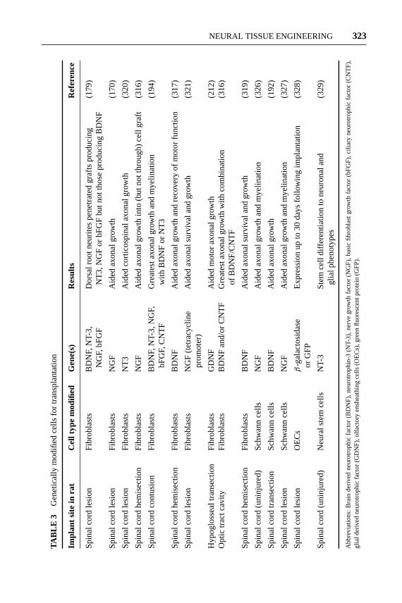
12 Jun 2003 14:58 AR AR191-BE05-11.tex AR191-BE05-11.sgm LaTeX2e(2002/01/18)P1: GJB
NEURAL TISSUE ENGINEERING 323
TAB
LE3
Gen
etic
ally
mod
ified
cells
for
tran
spla
ntat
ion
Impl
ants
itein
rat
Cel
ltyp
em
odifi
edG
ene(
s)R
esul
tsR
efer
ence
Spi
nalc
ord
lesi
onF
ibro
blas
tsB
DN
F,N
T-3,
Dor
salr
ootn
eurit
espe
netr
ated
graf
tspr
oduc
ing
(179
)N
GF,
bFG
FN
T3,
NG
For
bFG
Fbu
tnot
thos
epr
oduc
ing
BD
NF
Spi
nalc
ord
lesi
onF
ibro
blas
tsN
GF
Aid
edax
onal
grow
th(1
70)
Spi
nalc
ord
lesi
onF
ibro
blas
tsN
T3
Aid
edco
rtic
ospi
nala
xona
lgro
wth
(320
)
Spi
nalc
ord
hem
isec
tion
Fib
robl
asts
NG
FA
ided
axon
algr
owth
into
(but
nott
hrou
gh)
cell
graf
t(3
16)
Spi
nalc
ord
cont
usio
nF
ibro
blas
tsB
DN
F,N
T-3,
NG
F,G
reat
esta
xona
lgro
wth
and
mye
linat
ion
(194
)bF
GF,
CN
TF
with
BD
NF
orN
T3
Spi
nalc
ord
hem
isec
tion
Fib
robl
asts
BD
NF
Aid
edax
onal
grow
than
dre
cove
ryof
mot
orfu
nctio
n(3
17)
Spi
nalc
ord
lesi
onF
ibro
blas
tsN
GF
(tet
racy
clin
eA
ided
axon
alsu
rviv
alan
dgr
owth
(321
)pr
omot
er)
Hyp
oglo
ssea
ltra
nsec
tion
Fib
robl
asts
GD
NF
Aid
edm
otor
axon
algr
owth
(212
)O
ptic
trac
tcav
ityF
ibro
blas
tsB
DN
Fan
d/or
CN
TF
Gre
ates
taxo
nalg
row
thw
ithco
mbi
natio
n(3
16)
ofB
DN
F/C
NT
F
Spi
nalc
ord
hem
isec
tion
Fib
robl
asts
BD
NF
Aid
edax
onal
surv
ival
and
grow
th(3
19)
Spi
nalc
ord
(uni
njur
ed)
Sch
wan
nce
llsN
GF
Aid
edax
onal
grow
than
dm
yelin
atio
n(3
26)
Spi
nalc
ord
tran
sect
ion
Sch
wan
nce
llsB
DN
FA
ided
axon
algr
owth
(192
)
Spi
nalc
ord
lesi
onS
chw
ann
cells
NG
FA
ided
axon
algr
owth
and
mye
linat
ion
(327
)
Spi
nalc
ord
lesi
onO
EC
sβ
-gal
acto
sida
seE
xpre
ssio
nup
to30
days
follo
win
gim
plan
tatio
n(3
28)
orG
FP
Spi
nalc
ord
(uni
njur
ed)
Neu
rals
tem
cells
NT-
3S
tem
cell
diffe
rent
iatio
nto
neur
onal
and
(329
)gl
ialp
heno
type
s
Abb
revi
atio
ns:
Bra
inde
rived
neur
otro
phic
fact
or(B
DN
F),
neur
otro
phin
-3(N
T-3)
,ne
rve
grow
thfa
ctor
(NG
F),
basi
cfib
robl
ast
grow
thfa
ctor
(bF
GF
),ci
liary
neur
otro
phic
fact
or(C
NT
F),
glia
lder
ived
neur
otro
phic
fact
or(G
DN
F),
olfa
ctor
yen
shea
thin
gce
lls(O
EC
s),g
reen
fluor
esce
ntpr
otei
n(G
FP
).

27 Jun 2003 14:31 AR AR191-BE05-11.tex AR191-BE05-11.sgm LaTeX2e(2002/01/18)P1: GJB
324 SCHMIDT ¥ BAIER LEACH
of a hollow conduit across a nerve defect, which serves as a critical precursor toaxonal regeneration. With regard to foam rods, it is questionable whether an amor-phous irregular structure optimally guides axonal regeneration. Thus, to mimicnatural repair in the body and to shorten the time required for regeneration, morerecent studies have focused on the modification of nerve guidance channels withinternal matrices of longitudinally aligned fibers or channels. These devices oftenrequire more advanced processing techniques.
In one approach, guidance channels containing smaller chambers, or sublumina,to mimic the natural fascicular organization of the nerve have shown the capacityto support nerve regeneration (330). Tubes of poly(lactic acid) and poly(lactic-co-glycolic acid) were formed using a dip-molding technique. They were created tocontain 1, 2, 4, or 5 sublumina, or “fascicular analogs.” With this approach, it maybe possible to incorporate particular neurotrophic factors into separate subluminalcompartments in an effort to guide the elongation of specific subsets of nerve fibers(e.g., motor versus sensory fibers).
Several experiments have also investigated the use of extruded or spun fibers ofcollagen or other biodegradable materials that are then placed within the lumen ofconduits. For example, Tong et al. (83) have shown good regeneration using colla-gen tubes that were filled with laminin and fibronectin double-coated collagen fiberbundles. Similarly, eight longitudinally oriented polyamide filaments (i.e., sutures)placed inside silicone tubes supported regeneration over 15 mm gaps in rats (15).Matsumoto et al. (331) showed that poly(glycolic acid)-collagen conduits (4 mminner diameter, 50µm wall thickness, and 90 mm in length) filled with collagenfibers (80 fibers/conduit; 50µm in diameter, and 90 mm in length) permitted regen-eration over 80 mm defect lengths in dogs. Previous reports illustrate regenerationup to a maximum length of 50 mm, suggesting that the internal collagen fibers in-deed enhanced axonal elongation. Later, a study from this same laboratory showedthat a conduit with an internal collagen sponge supported similar levels of regen-eration compared to the collagen fiber-filled conduits (332). The authors claimthat the collagen sponge is a preferred substrate because of its ease of preparationcompared to the fibers. Indeed, these fiber approaches, as well as the sublumina ap-proach mentioned earlier, are tedious and the fiber or chamber alignment may notbe well controlled because of the handmade fabrication techniques. Thus, there isa push to devise other methods that can be used to create aligned internal structureand that are also not too tedious or difficult to prevent eventual scale-up.
As a means to provide the necessary matrix alignment and also improve uponprocessing conditions, magnetic fields have been used to orient protein poly-mers. Magnetically aligned fibrin and collagen matrices (Figure 14) have beenshown to promote enhanced axonal outgrowth in vitro and in vivo comparedto random fibrin and collagen matrices, respectively (91, 92, 104). In a differ-ent approach, a novel foam-processing technique, utilizing low-pressure injectionmolding, created highly porous conduits from poly(lactic-co-glycolic acid) withcontinuous longitudinal channels (333). Conduits were constructed containing 1,5, 16, 45, or more longitudinally aligned channels. In similar studies, poly(lactic-co-glycolic acid) was subjected to injection molding followed by a thermally
27 Jun 2003 14:32 AR AR191-BE05-11.tex AR191-BE05-11.sgm LaTeX2e(2002/01/18)P1: GJB
NEURAL TISSUE ENGINEERING 325
induced phase transition process to produce conduits with longitudinally alignedinternal channels (334). As part of a separate study from this same laboratory, a“two-phase” conduit was fabricated for spinal cord repair (159). The scaffold’sinner portion emulated the gray matter and was composed of a porous polymerseeded with neural stem cells, which provided trophic support. The outer portionmimicked the white matter with long, axially oriented pores for axonal guidanceand radial porosity to allow for fluid transport while inhibiting the ingrowth ofscar tissue (Figure 15). The inner-oriented pores were created from poly(lacticco-glycolic acid) using a solid-liquid phase separation technique.
SOLID FREEFORM FABRICATION AND INK-JET LIQUID POLYMER PRINTING Recen-tly, the emergence of advanced techniques for polymer processing, such as solidfreeform fabrication (SFF) and ink-jet printing for liquid polymer solutions, hascreated even more opportunities for the design of intricate devices for peripheralnerve and spinal cord repair. There are several types of freeform fabrication de-vices, and currently these devices can produce features as small as 6µm (157).Three-dimensional printing (3DP) is one type of solid freeform fabrication tech-nique that employs powder processing in the construction of devices in a layer-wise manner (335–337). 3DP, like other SFF methods, is capable of fabricating astructure directly from a computer model and can handle complex features, suchas internal walls, porosity gradients, tortuous channels, and multiple material re-gions. The process begins by spreading a thin layer of polymer powder onto apiston plate. A liquid binder solution is passed through a nozzle affixed to thefast-axis carriage, and the nozzle is rastered back and forth over the powder bedto selectively print droplets, which bind the powder particles together, generatinga two-dimensional pattern. The piston is lowered, another thin layer of powder isspread, and the process is repeated. Polymers can be printed directly or one canfabricate a mold using SFF. The latter approach provides enhanced control overthe type of material used to fabricate a desired device. Scaffolds created usingSFF can contain biomimetic internal architectures that prove valuable for tissueengineering. For example, conduits with internal channels corresponding to keyspinal cord tracts could be created using these approaches (157). Some limitationsdo exist, including the high cost of the instrument and the inability to incorporatebiological components under some processing conditions.
MicroFab Inc., in Plano, Texas, has devised an ink-jet print station designedto precisely deliver solutions of polymers, such as poly(lactic acid), for tissue en-gineering applications. Three-dimensional structures can be created with desiredthickness, dimensions, and incorporated biomolecules. In one study, they havegenerated bifurcated degradable polymer tubes that have ridges or support ribs(Figure 16a). The ridges provide strength to resist compressive forces, whereasthe remaining polymer tube is thinner and allows for nutrient exchange and anoptimum degradation rate. In a second study, MicroFab has created a nonbifur-cated ridged tube that is impregnated with a gradient of fluorescent dye (Figure16; note: the gradient is increasing from left to right). Similar gradients can becreated with biomolecules and neurotrophic factors. These examples illustrate

12 Jun 2003 14:58 AR AR191-BE05-11.tex AR191-BE05-11.sgm LaTeX2e(2002/01/18)P1: GJB
326 SCHMIDT ¥ BAIER LEACH

12 Jun 2003 14:58 AR AR191-BE05-11.tex AR191-BE05-11.sgm LaTeX2e(2002/01/18)P1: GJB
NEURAL TISSUE ENGINEERING 327
the powerful opportunities provided with advanced instrumentation, such as SFFand ink-jet polymer printing.
Combination Approaches and Novel Stimuli
Many approaches to enhance nerve repair in the past have focused on mechanical,chemical, biological, or electrical stimuli. More research today is focused on usingmultiple stimuli in an effort to better mimic the complex milieu of signals nor-mally found in the body. The challenge is assessing the optimal levels of differentsignals in such a complicated environment. For example, Miller et al. (338) havecreated micropatterned substrates for in vitro neurite elongation studies. Compres-sion molded and solvent cast biodegradable polymer substrates made of poly(lacticacid) were micropatterned to form grooves on the substrate surfaces. Laminin waslocalized in the grooves and rat Schwann cells were seeded on the substrates.The micropatterns provide physical guidance, laminin provides chemical cues,and the Schwann cells provide biological cues to the axons. The synergistic com-bination of physical, chemical, and cellular guidance enabled greater than 98%alignment of neurites and accelerated outgrowth of nerve fibers in the direction ofthe microgrooves. In fact, the synergistic effect of physical and chemical guidancecues has been found in other studies to be more effective than individual cues inpromoting directional outgrowth of neurites (339).
Beyond the typical signals found in the body, other stimuli are also beinginvestigated for their ability to impact neuronal growth. For example, Ehrlicheret al. (340) have shown that weak optical forces can be used to guide the movementof an axon. To do this, a laser spot is placed in front of the actively growingneuron, enhancing growth into the beam focus and resulting in guided turns aswell as enhanced growth. The power of the laser is chosen so that gradient forcesare sufficiently powerful to bias actin polymerization and neuronal extension, buttoo weak to actually hold and move any part of the cell. This demonstrates theability of light to control biological processes, which could prove useful in nerveregeneration (e.g., in laser-assisted surgical repair of damaged nerves).
Convergence of Neural Regeneration and Neural Prostheses
Neural prostheses are assistive devices that restore functions lost as a result ofneural damage. These devices, which electrically stimulate nerves and are either
←−−−−−−−−−−−−−−−−−−−−−−−−−−−−−−−−−−−−−−−−−−−−−−−−−−−−−−−−−Figure 15 Oriented poly(lactide-co-glycolide) scaffolds. Oriented scaffolds werecreated using solid-liquid phase separation (159). Poly(lactide-co-glycolide) was dis-solved in dioxane. The dioxane was induced to crystallize in an oriented manner throughthe use of a thermal gradient and then was removed by sublimation. The scaffoldswere sputter coated for observation under scanning electron microscopy. (a) Scale bar,200µm. (b) Scale bar, 50µm. Images courtesy of R. Langer & E. Lavik, Depart-ments of Chemical Engineering and Biomedical Engineering, Massachusetts Instituteof Technology.
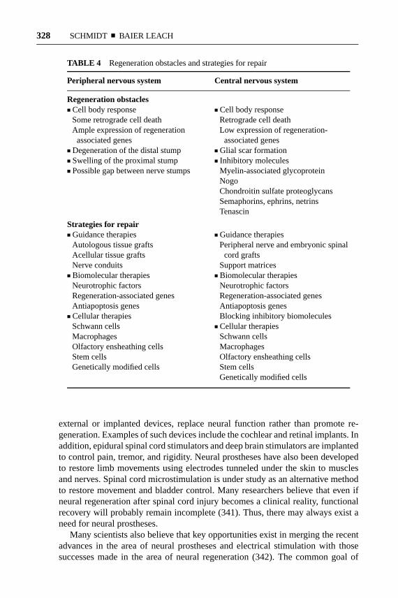
12 Jun 2003 14:58 AR AR191-BE05-11.tex AR191-BE05-11.sgm LaTeX2e(2002/01/18)P1: GJB
328 SCHMIDT ¥ BAIER LEACH
TABLE 4 Regeneration obstacles and strategies for repair
Peripheral nervous system Central nervous system
Regeneration obstaclesj Cell body response j Cell body response
Some retrograde cell death Retrograde cell deathAmple expression of regeneration Low expression of regeneration-associated genes associated genes
j Degeneration of the distal stump j Glial scar formationj Swelling of the proximal stump j Inhibitory moleculesj Possible gap between nerve stumps Myelin-associated glycoprotein
NogoChondroitin sulfate proteoglycansSemaphorins, ephrins, netrinsTenascin
Strategies for repairj Guidance therapies j Guidance therapies
Autologous tissue grafts Peripheral nerve and embryonic spinalAcellular tissue grafts cord graftsNerve conduits Support matrices
j Biomolecular therapies j Biomolecular therapiesNeurotrophic factors Neurotrophic factorsRegeneration-associated genes Regeneration-associated genesAntiapoptosis genes Antiapoptosis genes
j Cellular therapies Blocking inhibitory biomoleculesSchwann cells j Cellular therapiesMacrophages Schwann cellsOlfactory ensheathing cells MacrophagesStem cells Olfactory ensheathing cellsGenetically modified cells Stem cells
Genetically modified cells
external or implanted devices, replace neural function rather than promote re-generation. Examples of such devices include the cochlear and retinal implants. Inaddition, epidural spinal cord stimulators and deep brain stimulators are implantedto control pain, tremor, and rigidity. Neural prostheses have also been developedto restore limb movements using electrodes tunneled under the skin to musclesand nerves. Spinal cord microstimulation is under study as an alternative methodto restore movement and bladder control. Many researchers believe that even ifneural regeneration after spinal cord injury becomes a clinical reality, functionalrecovery will probably remain incomplete (341). Thus, there may always exist aneed for neural prostheses.
Many scientists also believe that key opportunities exist in merging the recentadvances in the area of neural prostheses and electrical stimulation with thosesuccesses made in the area of neural regeneration (342). The common goal of

12 Jun 2003 14:58 AR AR191-BE05-11.tex AR191-BE05-11.sgm LaTeX2e(2002/01/18)P1: GJB
NEURAL TISSUE ENGINEERING 329
both research fields is the restoration of function after neurological damage. Areasof research that have been identified to have the greatest impact on achieving thisgoal include: enhancement of axonal regeneration with electric fields, developmentof hybrid neural interfaces combining silicon and biologically derived elements,and investigation of the role of patterned neural activity in regulating neuronalprocesses and neurorehabilitation (342). Some of this research, especially thatof combining microelectronics technology and biologically derived elements, isreviewed in (343).
CONCLUSION
The requirements for functional nerve regeneration are complex. However, throughthe combined efforts of scientists and engineers from a variety of disciplines, ex-perimental work in this field has made great progress. This review of nerve regener-ation divided recent advances into guidance therapies, biomolecular therapies, andcellular therapies (summarized in Table 4). An additional section about advancedtherapies outlined sophisticated guidance channel fabrication techniques and com-bination therapies. Yet, a single approach on its own has not allowed an effectiveclinical therapy. For example, although synthetic nerve guidance channels effec-tively guide axonal growth, these conduits have not been a widely used replace-ment for autografts, particularly in long defects. In spinal cord repair, Schwann celltransplantation to the site of injury has yielded improved recoveries, but challengesremain to implementing this treatment clinically.
On the other hand, many studies have been incrementally successful. Newpotential targets for novel therapies have been discovered through an increasedunderstanding of the molecular biology of neural development and regeneration.Furthermore, advances in the areas of drug delivery, gene therapy, and bioma-terials are able to apply these therapies with increased efficacy and biocom-patibility. As the separate experimental approaches are melded together, futureresearch will allow more dramatic successes in peripheral nerve regeneration andspinal cord repair. Multiple treatments, perhaps coordinated at different timesor locations within the patient, will be needed to promote improved functionalregeneration.
The Annual Review of Biomedical Engineeringis online athttp://bioeng.annualreviews.org
LITERATURE CITED
1. Stoll G, Griffin JW, Li CY, Trapp BD.1989. Wallerian degeneration in the pe-ripheral nervous system: participation ofboth Schwann cells and macrophagesin myelin degradation.J. Neurocytol.18:671–83
2. Chaudhry V, Glass JD, Griffin JW. 1992.Wallerian degeneration in peripheralnerve disease.Neurol. Clin. 10:613–27
3. Jacobsen S, Guth L. 1965. An electro-physiological study of the early stages of

12 Jun 2003 14:58 AR AR191-BE05-11.tex AR191-BE05-11.sgm LaTeX2e(2002/01/18)P1: GJB
330 SCHMIDT ¥ BAIER LEACH
peripheral nerve regeneration.Exp. Neu-rol. 11:48–60
4. Williams LR, Longo FM, Powell HC,Lundborg G, Varon S. 1983. Spatial-temporal progress of peripheral nerve re-generation within a silicone chamber: pa-rameters for a bioassay.J. Comp. Neurol.218:460–70
5. Fields RD, Ellisman MH. 1986. Axons re-generated through silicone tube splices. I.Conduction properties.Exp. Neurol.92:48–60
6. Mukhopadhyay G, Doherty P, Walsh FS,Crocker PR, Filbin MT. 1994. A novel rolefor myelin-associated glycoprotein as aninhibitor of axonal regeneration.Neuron13:757–67
7. Chen MS, Huber AB, van der Haar ME,Frank M, Schnell L, et al. 2000. Nogo-Ais a myelin-associated neurite outgrowthinhibitor and an antigen for monoclonalantibody IN-1.Nature403:434–39
8. Morgenstern DA, Asher RA, Fawcett JW.2002. Chondroitin sulphate proteoglycansin the CNS injury response.Prog. BrainRes.137:313–32
9. Avellino AM, Hart D, Dailey AT, Mac-Kinnon M, Ellegala D, et al. 1995. Dif-ferential macrophage responses in theperipheral and central nervous systemduring wallerian degeneration of axons.Exp. Neurol.136:183–98
10. McKeon RJ, Schreiber RC, Rudge JS,Silver J. 1991. Reduction of neurite out-growth in a model of glial scarring fol-lowing CNS injury is correlated with theexpression of inhibitory molecules on re-active astrocytes.J. Neurosci.11:3398–411
11. Millesi H, Ganglberger J, Berger A. 1967.Erfahrungen mit der Mikrochirurgie pe-ripherer Nerven.Chir. Plast.3:47
12. Lundborg G. 1988.Nerve Injury and Re-pair. New York: Longman Group UK
13. Mackinnon SE, Dellon AL. 1988.Surgeryof the Peripheral Nerve.New York:Thieme Med. Publ.
14. Archibald SJ, Shefner J, Krarup C, Madi-
son RD. 1995. Monkey median nerve re-paired by nerve graft or collagen nerveguide tube.J. Neurosci.15:4109–23
15. Lundborg G, Dahlin L, Dohi D, Kanje M,Terada N. 1997. A new type of “bioarti-ficial” nerve graft for bridging extendeddefects in nerves.J. Hand Surg. [Br.]22:299–303
16. McDonald JW. 1999. Repairing the dam-aged spinal cord.Sci. Am.281:64–73
17. Chiu DT. 1995. Special article: the devel-opment of autogenous venous nerve con-duit as a clinical entity. InP&S MedicalReview, 3(1). New York: Columbia-Presbyterian Med. Cent.
18. Richardson PM, McGuinness UM, Agu-ayo AJ. 1980. Axons from CNS neu-rons regenerate into PNS grafts.Nature284:264–65
19. Cheng H, Cao Y, Olson L. 1996. Spinalcord repair in adult paraplegic rats: partialrestoration of hind limb function.Science273:510–13
20. Davies SJ, Fitch MT, Memberg SP, HallAK, Raisman G, et al. 1997. Regenerationof adult axons in white matter tracts of thecentral nervous system.Nature390:680–83
21. Bernstein JJ, Goldberg WJ. 1995. Exper-imental spinal cord transplantation as amechanism of spinal cord regeneration.Paraplegia33:250–53
22. Carlstedt T. 1997. Nerve fibre regenera-tion across the peripheral-central transi-tional zone.J. Anat.190:51–56
23. Albert E. 1885. Einige Operationen anNerven.Wien Med.26:1285
24. Gluck T. 1880. Ueber Neuroplastik aufdem Wege der Transplantation.Arch.Klin. Chir. 25:606–16
25. Payr E. 1900. Beitr¨age zur Technik derBlutgefass und Nervennaht nebst Mit-teilungen uber die Verwendung einesresorbibaren Metalles in der Chirurgie.Arch. Klin. Chir.62:67
26. Kirk EG, Lewis D. 1915. Fascial tubuliza-tion in the repair of nerve defects.JAMA65:486–92

12 Jun 2003 14:58 AR AR191-BE05-11.tex AR191-BE05-11.sgm LaTeX2e(2002/01/18)P1: GJB
NEURAL TISSUE ENGINEERING 331
27. Lundborg G, Dahlin LB, Danielsen N,Nachemson AK. 1986. Tissue specificityin nerve regeneration.Scand. J. Plast. Re-constr. Surg.20:279–83
28. Mackinnon SE, Dellon AL, Lundborg G,Hudson AR, Hunter DA. 1986. A studyof neurotrophism in a primate model.J.Hand Surg. [Am.]11:888–94
29. Politis MJ, Ederle K, Spencer PS. 1982.Tropism in nerve regeneration in vivo.Attraction of regenerating axons by dif-fusible factors derived from cells in dis-tal nerve stumps of transected peripheralnerves.Brain Res.253:1–12
30. Williams LR. 1987. Rat aorta isograftspossess nerve regeneration-promotingproperties in silicone Y chambers.Exp.Neurol.97:555–63
31. Bellamkonda R, Aebischer P. 1994. Re-view: tissue engineering in the nervoussystem.Biotechnol. Bioeng.43:543–54
32. Valentini RF. 2000. Nerve guidance chan-nels. In The Biomedical EngineeringHandbook, ed. JD Bronzino, pp. 2:135-1–12. Boca Raton: CRC. 2nd ed.
33. Furnish EJ, Schmidt CE. 1998. Tissue en-gineering of the peripheral nervous sys-tem. In Frontiers in Tissue Engineering,ed. CW Patrick Jr, AG Mikos, LV McIn-tire, pp. 514–35. New York: Elsevier Sci.
34. Hudson TW, Evans GR, Schmidt CE.1999. Engineering strategies for periph-eral nerve repair.Clin. Plast. Surg.26:617–28
35. Tresco PA. 2000. Tissue engineeringstrategies for nervous system repair.Prog.Brain Res.128:349–63
36. Strauch B. 2000. Use of nerve conduitsin peripheral nerve repair.Hand Clin.16:123–30
37. Evans GR. 2000. Challenges to nerve re-generation.Semin. Surg. Oncol.19:312–18
38. Evans GR. 2001. Peripheral nerve injury:a review and approach to tissue engi-neered constructs.Anat. Rec.263:396–404
39. Seddon HJ. 1975.Surgical Disorders
of the Peripheral Nerves.New York:Churchill Livingston
40. Sunderland S. 1991.Nerve Injuries andTheir Repair. London: Churchill Liv-ingston
41. Battiston B, Tos P, Cushway TR, Geuna S.2000. Nerve repair by means of vein filledwith muscle grafts I. Clinical results.Mi-crosurgery20:32–36
42. Meek MF, Varejao AS, Geuna S. 2002.Muscle grafts and alternatives for nerverepair.J. Oral Maxillofac. Surg.60:1095–96
43. Chiu DT, Janecka I, Krizek TJ, WolffM, Lovelace RE. 1982. Autogenous veingraft as a conduit for nerve regeneration.Surgery91:226–33
44. Walton RL, Brown RE, Matory WE Jr,Borah GL, Dolph JL. 1989. Autogenousvein graft repair of digital nerve defects inthe finger: a retrospective clinical study.Plast. Reconstr. Surg.84:944–49
45. Risitano G, Cavallaro G, Merrino T, Cop-polino S, Ruggeri F. 2002. Clinical re-sults and thoughts on sensory nerve repairby autologous vein graft in emergencyhand reconstruction.Chir. Main.21:194–97
46. Karacaoglu E, Yuksel F, Peker F, GulerMM. 2001. Nerve regeneration throughan epineurial sheath: its functional aspectcompared with nerve and vein grafts.Mi-crosurgery21:196–201
47. Brandt J, Dahlin LB, Lundborg G. 1999.Autologous tendons used as grafts forbridging peripheral nerve defects.J. HandSurg. [Br.] 24:284–90
48. Geuna S, Tos P, Battiston B, GuglielmoneR, Giacobini-Robecchi MG. 2000. Astereological study of long-term regenera-tion of rat severed sciatic nerve repaired bymeans of muscle-vein-combined grafts.Ital. J. Anat. Embryol.105:65–73
49. Pagnotta A, Tos P, Fornaro M, Gigante A,Geuna S, et al. 2002. Neurotrophins andtheir receptors in early axonal regenera-tion along muscle-vein-combined grafts.Microsurgery22:300–3

12 Jun 2003 14:58 AR AR191-BE05-11.tex AR191-BE05-11.sgm LaTeX2e(2002/01/18)P1: GJB
332 SCHMIDT ¥ BAIER LEACH
50. Rodrigues Ade C, Silva MD. 2001. Inside-out versus standard artery graft to re-pair a sensory nerve in rats.Microsurgery21:102–7
51. Zhang F, Blain B, Beck J, Zhang J, ChenZ, et al. 2002. Autogenous venous graftwith one-stage prepared Schwann cellsas a conduit for repair of long segmen-tal nerve defects.J. Reconstr. Microsurg.18:295–300
52. Ide C, Tohyama K, Tajima K, Endoh K,Sano K, et al. 1998. Long acellular nervetransplants for allogeneic grafting and theeffects of basic fibroblast growth factoron the growth of regenerating axons indogs: a preliminary report.Exp. Neurol.154:99–112
53. Frerichs O, Fansa H, Schicht C, Wolf G,Schneider W, et al. 2002. Reconstructionof peripheral nerves using acellular nervegrafts with implanted cultured Schwanncells.Microsurgery22:311–15
54. Hiles RW. 1972. Freeze dried irradiatednerve homograft: a preliminary report.Hand4:79–84
55. Sondell M, Lundborg G, Kanje M. 1998.Regeneration of the rat sciatic nerve intoallografts made acellular through chemi-cal extraction.Brain Res.795:44–54
56. Sondell M, Lundborg G, Kanje M. 1999.Vascular endothelial growth factor stimu-lates Schwann cell invasion and neovascu-larization of acellular nerve grafts.BrainRes.846:219–28
57. Gulati AK, Cole GP. 1994. Immunogenic-ity and regenerative potential of acellularnerve allografts to repair peripheral nervein rats and rabbits.Acta Neurochir. (Wien)126:158–64
58. Marmor L. 1964. The repair of peripheralnerves by irradiated homografts.Clin. Or-thop.34:161–69
59. Lawson GM, Glasby MA. 1998. Periph-eral nerve reconstruction using freeze-thawed muscle grafts: a comparison withgroup fascicular nerve grafts in a largeanimal model.J. R. Coll. Surg. Edinb.43:295–302
60. Liu XL, Arai T, Sondell M, Lundborg G,Kanje M, et al. 2001. Use of chemicallyextracted muscle grafts to repair extendednerve defects in rats.Scand. J. Plast. Re-constr. Surg. Hand Surg.35:337–45
61. Fansa H, Schneider W, Wolf G, KeilhoffG. 2002. Host responses after acellularmuscle basal lamina allografting used as amatrix for tissue engineered nerve grafts.Transplantation74:381–87
62. Mligiliche N, Kitada M, Ide C. 2001.Grafting of detergent-denatured skeletalmuscles provides effective conduits forextension of regenerating axons in the ratsciatic nerve.Arch. Histol. Cytol.64:29–36
63. Schmidt CE, Baier JM. 2000. Acellularvascular tissues: natural biomaterials fortissue repair and tissue engineering.Bio-materials21:2215–31
64. Costantino PD, Wolpoe ME, GovindarajS, Chaplin JM, Sen C, et al. 2000. Humandural replacement with acellular dermis:clinical results and a review of the litera-ture.Head Neck22:765–71
65. Voytik-Harbin SL, Brightman AO, KraineMR, Waisner B, Badylak SF. 1997. Iden-tification of extractable growth factorsfrom small intestinal submucosa.J. CellBiochem.67:478–91
66. Badylak SF, Record R, Lindberg K,Hodde J, Park K. 1998. Small intestinalsubmucosa: a substrate for in vitro cellgrowth. J. Biomater. Sci. Polym. Ed.9:863–78
67. Lantz GC, Badylak SF, Hiles MC, Cof-fey AC, Geddes LA, et al. 1993. Smallintestinal submucosa as a vascular graft:a review.J. Invest. Surg.6:297–310
68. Badylak SF, Kropp B, McPherson T,Liang H, Snyder PW. 1998. Small in-testional submucosa: a rapidly resorbedbioscaffold for augmentation cystoplastyin a dog model.Tissue Eng.4:379–87
69. Badylak SF, Tullius R, Kokini K, Shel-bourne KD, Klootwyk T, et al. 1995. Theuse of xenogeneic small intestinal submu-cosa as a biomaterial for Achilles tendon
12 Jun 2003 14:58 AR AR191-BE05-11.tex AR191-BE05-11.sgm LaTeX2e(2002/01/18)P1: GJB
NEURAL TISSUE ENGINEERING 333
repair in a dog model.J. Biomed. Mater.Res.29:977–85
70. Hadlock TA, Sundback CA, Hunter DA,Vacanti JP, Cheney ML. 2001. A newartificial nerve graft containing rolledSchwann cell monolayers.Microsurgery21:96–101
71. Davis GE, Blaker SN, Engvall E, VaronS, Manthorpe M, et al. 1987. Human am-nion membrane serves as a substratum forgrowing axons in vitro and in vivo.Sci-ence236:1106–9
72. Ozcan G, Shenaq S, Spira M. 1993. Vascu-larized nerve tube: an experimental alter-native for vascularized nerve grafts overshort gaps.J. Reconstr. Microsurg.9:405–13
73. Mohammad J, Shenaq J, Rabinovsky E,Shenaq S. 2000. Modulation of peripheralnerve regeneration: a tissue-engineeringapproach. The role of amnion tube nerveconduit across a 1-centimeter nerve gap.Plast. Reconstr. Surg.105:660–66
74. Mohammad JA, Warnke PH, Pan YC,Shenaq S. 2000. Increased axonal regen-eration through a biodegradable amnionictube nerve conduit: effect of local de-livery and incorporation of nerve growthfactor/hyaluronic acid media.Ann. Plast.Surg.44:59–64
75. Meek MF, Coert JH, Nicolai JP. 2001.Amnion tube for nerve regeneration.Plast. Reconstr. Surg.107:622–23
76. Mligiliche N, Endo K, Okamoto K, Fuji-moto E, Ide C. 2002. Extracellular matrixof human amnion manufactured into tubesas conduits for peripheral nerve regen-eration.J. Biomed. Mater. Res.63:591–600
77. Gray KJ, Shenaq SM, Engelmann UH,Fishman IJ, Jeraj K, et al. 1987. Use ofhuman amnion for microvascular inter-positional grafts.Plast. Reconstr. Surg.79:778–85
78. Rutishauser U. 1993. Adhesion moleculesof the nervous system.Curr. Opin. Neu-robiol. 3:709–15
79. Grimpe B, Silver J. 2002. The extracel-
lular matrix in axon regeneration.Prog.Brain Res.137:333–49
80. Bovolenta P, Fernaud-Espinosa I. 2000.Nervous system proteoglycans as modu-lators of neurite outgrowth.Prog. Neuro-biol. 61:113–32
81. Asher RA, Morgenstern DA, Moon LD,Fawcett JW. 2001. Chondroitin sulphateproteoglycans: inhibitory components ofthe glial scar.Prog. Brain Res.132:611–19
82. Kauppila T, Jyvasjarvi E, Huopaniemi T,Hujanen E, Liesi P. 1993. A laminin graftreplaces neurorrhaphy in the restorativesurgery of the rat sciatic nerve.Exp. Neu-rol. 123:181–91
83. Tong XJ, Hirai K, Shimada H, Mizutani Y,Izumi T, et al. 1994. Sciatic nerve regen-eration navigated by laminin-fibronectindouble coated biodegradable collagengrafts in rats.Brain Res.663:155–62
84. Whitworth IH, Brown RA, Dore C, GreenCJ, Terenghi G. 1995. Orientated mats offibronectin as a conduit material for usein peripheral nerve repair.J. Hand Surg.(Br.) 20:429–36
85. Ahmed Z, Brown RA. 1999. Adhesion,alignment, and migration of culturedSchwann cells on ultrathin fibronectin fi-bres.Cell Motil. Cytoskelet.42:331–43
86. Chen YS, Hsieh CL, Tsai CC, Chen TH,Cheng WC, et al. 2000. Peripheral nerveregeneration using silicone rubber cham-bers filled with collagen, laminin and fi-bronectin.Biomaterials21:1541–47
87. Yoshii S, Oka M. 2001. Peripheral nerveregeneration along collagen filaments.Brain Res.888:158–62
88. Toba T, Nakamura T, Lynn AK, Matsu-moto K, Fukuda S, et al. 2002. Evaluationof peripheral nerve regeneration acrossan 80-mm gap using a polyglycolic acid(PGA)–collagen nerve conduit filled withlaminin-soaked collagen sponge in dogs.Int. J. Artif. Organs25:230–37
89. Itoh S, Takakuda K, Kawabata S, Aso Y,Kasai K, et al. 2002. Evaluation of cross-linking procedures of collagen tubes used
12 Jun 2003 14:58 AR AR191-BE05-11.tex AR191-BE05-11.sgm LaTeX2e(2002/01/18)P1: GJB
334 SCHMIDT ¥ BAIER LEACH
in peripheral nerve repair.Biomaterials23:4475–81
90. Yoshii S, Oka M, Shima M, Taniguchi A,Akagi M. 2002. 30 mm regeneration ofrat sciatic nerve along collagen filaments.Brain Res.949:202–8
91. Ceballos D, Navarro X, Dubey N,Wendelschafer-Crabb G, Kennedy WR,et al. 1999. Magnetically aligned collagengel filling a collagen nerve guide improvesperipheral nerve regeneration.Exp. Neu-rol. 158:290–300
92. Dubey N, Letourneau PC, TranquilloRT. 1999. Guided neurite elongation andSchwann cell invasion into magneticallyaligned collagen in simulated peripheralnerve regeneration.Exp. Neurol. 158:338–50
93. Chamberlain LJ, Yannas IV, Hsu HP,Strichartz G, Spector M. 1998. Collagen-GAG substrate enhances the quality ofnerve regeneration through collagen tubesup to level of autograft.Exp. Neurol.154:315–29
94. Chamberlain LJ, Yannas IV, Hsu HP,Strichartz GR, Spector M. 2000. Near-terminus axonal structure and functionfollowing rat sciatic nerve regenerationthrough a collagen-GAG matrix in a ten-millimeter gap.J. Neurosci. Res.60:666–77
95. Seckel BR, Jones D, Hekimian KJ, WangKK, Chakalis DP, et al. 1995. Hyaluronicacid through a new injectable nerveguide delivery system enhances periph-eral nerve regeneration in the rat.J. Neu-rosci. Res.40:318–24
96. Ahmed Z, Underwood S, Brown RA.2000. Low concentrations of fibrino-gen increase cell migration speed onfibronectin/fibrinogen composite cables.Cell Motil. Cytoskelet.46:6–16
97. Herbert CB, Nagaswami C, Bittner GD,Hubbell JA, Weisel JW. 1998. Effectsof fibrin micromorphology on neuritegrowth from dorsal root ganglia cul-tured in three-dimensional fibrin gels.J.Biomed. Mater. Res.40:551–59
98. Holmes TC, de Lacalle S, Su X, Liu G,Rich A, et al. 2000. Extensive neurite out-growth and active synapse formation onself-assembling peptide scaffolds.Proc.Natl. Acad. Sci. USA97:6728–33
99. Hashimoto T, Suzuki Y, Kitada M,Kataoka K, Wu S, et al. 2002. Peripheralnerve regeneration through alginate gel:analysis of early outgrowth and late in-crease in diameter of regenerating axons.Exp. Brain Res.146:356–68
100. Balgude AP, Yu X, Szymanski A, Bel-lamkonda RV. 2001. Agarose gel stiffnessdetermines rate of DRG neurite extensionin 3D cultures.Biomaterials22:1077–84
101. Mosahebi A, Simon M, Wiberg M,Terenghi G. 2001. A novel use of alginatehydrogel as Schwann cell matrix.TissueEng.7:525–34
102. Haipeng G, Yinghui Z, Jianchun L, Yan-dao G, Nanming Z, et al. 2000. Studies onnerve cell affinity of chitosan-derived ma-terials.J. Biomed. Mater. Res.52:285–95
103. Leach JB, Bivens KA, Patrick CW Jr,Schmidt CE. 2003. Photocrosslinkedhyaluronic acid hydrogels: natural,biodegradable tissue engineering scaf-folds.Biotechnol. Bioeng.82:578–89
104. Dubey N, Letourneau PC, Tranquillo RT.2001. Neuronal contact guidance in mag-netically aligned fibrin gels: effect of vari-ation in gel mechano-structural proper-ties.Biomaterials22:1065–75
105. Sakiyama SE, Schense JC, Hubbell JA.1999. Incorporation of heparin-bindingpeptides into fibrin gels enhances neu-rite extension: an example of designermatrices in tissue engineering.Faseb J.13:2214–24
106. Schense JC, Bloch J, Aebischer P, HubbellJA. 2000. Enzymatic incorporation ofbioactive peptides into fibrin matrices en-hances neurite extension.Nat. Biotechnol.18:415–19
107. Molander H, Olsson Y, Engkvist O,Bowald S, Eriksson I. 1982. Regenerationof peripheral nerve through a polyglactintube.Muscle Nerve5:54–57

12 Jun 2003 14:58 AR AR191-BE05-11.tex AR191-BE05-11.sgm LaTeX2e(2002/01/18)P1: GJB
NEURAL TISSUE ENGINEERING 335
108. Nyilas E, Chiu TH, Sidman RL, HenryEW, Brushart TM, et al. 1983. Peripheralnerve repair with bioresorbable prosthe-sis.Trans. Am. Soc. Artif. Intern. Organs.29:307–13
109. Evans GR, Brandt K, Niederbichler AD,Chauvin P, Herrman S, et al. 2000. Clini-cal long-term in vivo evaluation of poly(L-lactic acid) porous conduits for periph-eral nerve regeneration.J. Biomater. Sci.Polym. Ed.11:869–78
110. Evans GR, Brandt K, Katz S, Chauvin P,Otto L, et al. 2002. Bioactive poly(L-lacticacid) conduits seeded with Schwann cellsfor peripheral nerve regeneration.Bioma-terials23:841–48
111. den Dunnen WF, Meek MF, GrijpmaDW, Robinson PH, Schakenraad JM.2000. In vivo and in vitro degradationof poly[(50)/(50) ((85)/(15)(L)/(D))LA/epsilon-CL], and the implications for theuse in nerve reconstruction.J. Biomed.Mater. Res.51:575–85
112. Nicoli Aldini N, Fini M, Rocca M, Gi-avaresi G, Giardino R. 2000. Guided re-generation with resorbable conduits in ex-perimental peripheral nerve injuries.Int.Orthop.24:121–25
113. Valero-Cabre A, Tsironis K, SkourasE, Perego G, Navarro X, et al. 2001.Superior muscle reinnervation afterautologous nerve graft or poly-L-lactide-epsilon-caprolactone (PLC) tubeimplantation in comparison to siliconetube repair.J. Neurosci. Res.63:214–23
114. Soldani G, Varelli G, Minnocci A, DarioP. 1998. Manufacturing and microscopi-cal characterisation of polyurethane nerveguidance channel featuring a highlysmooth internal surface.Biomaterials19:1919–24
115. Dalton PD, Flynn L, Shoichet MS. 2002.Manufacture of poly(2-hydroxyethylmethacrylate-co-methyl methacrylate)hydrogel tubes for use as nerve guidancechannels.Biomaterials23:3843–51
116. Young RC, Wiberg M, Terenghi G.2002. Poly-3-hydroxybutyrate (PHB): a
resorbable conduit for long-gap repairin peripheral nerves.Br. J. Plast. Surg.55:235–40
117. Gilchrist T, Glasby MA, Healy DM, KellyG, Lenihan DV, et al. 1998. In vitronerve repair—in vivo. The reconstruc-tion of peripheral nerves by entubulationwith biodegradeable glass tubes—a pre-liminary report.Br. J. Plast. Surg.51:231–37
118. Lenihan DV, Carter AJ, Gilchrist T, HealyDM, Miller IA, et al. 1998. Biodegradablecontrolled release glass in the repair of pe-ripheral nerve injuries.J. Hand Surg. [Br.]23:588–93
119. Valentini RF, Vargo TG, Gardella JA Jr,Aebischer P. 1993. Patterned neuronal at-tachment and outgrowth on surface mod-ified, electrically charged fluoropolymersubstrates.J. Biomater. Sci. Polym. Ed.5:13–36
120. Schmidt CE, Shastri VR, Vacanti JP,Langer R. 1997. Stimulation of neuriteoutgrowth using an electrically conduct-ing polymer.Proc. Natl. Acad. Sci. USA94:8948–53
121. Collier JH, Camp JP, Hudson TW,Schmidt CE. 2000. Synthesis and charac-terization of polypyrrole-hyaluronic acidcomposite biomaterials for tissue engi-neering applications.J. Biomed. Mater.Res.50:574–84
122. Dahlin L, Lundborg G. 2001. The use ofsilicone tubing in the late repair of the me-dian and ulnar nerves in the forearm.J.Hand Surg. [Br.]26:393–94
123. Vasconcelos BC, Gay-Escoda C. 2000.Facial nerve repair with expanded poly-tetrafluoroethylene and collagen conduits:an experimental study in the rabbit.J. OralMaxillofac. Surg.58:1257–62
124. Pitta MC, Wolford LM, Mehra P, Hop-kin J. 2001. Use of Gore-Tex tubing as aconduit for inferior alveolar and lingualnerve repair: experience with 6 cases.J.Oral Maxillofac. Surg.59:493–96
125. Miloro M, Halkias LE, Mallery S, TraversS, Rashid RG. 2002. Low-level laser

12 Jun 2003 14:58 AR AR191-BE05-11.tex AR191-BE05-11.sgm LaTeX2e(2002/01/18)P1: GJB
336 SCHMIDT ¥ BAIER LEACH
effect on neural regeneration in Gore-Textubes.Oral Surg. Oral Med. Oral Pathol.Oral Radiol. Endod.93:27–34
126. Ducker TB, Hayes GJ. 1968. Experimen-tal improvements in the use of Silastic cufffor peripheral nerve repair.J. Neurosurg.28:582–87
127. Midgley RD, Woolhouse FM. 1968. Sil-icone rubber sheathing as an adjunct toneural anastomosis.Surg. Clin. N. Am.48:1149–54
128. Lore AB, Hubbell JA, Bobb DS Jr, Ballin-ger ML, Loftin KL, et al. 1999. Rapid in-duction of functional and morphologicalcontinuity between severed ends of mam-malian or earthworm myelinated axons.J.Neurosci.19:2442–54
129. Mann BK, Gobin AS, Tsai AT, SchmedlenRH, West JL. 2001. Smooth musclecell growth in photopolymerized hydro-gels with cell adhesive and proteolyti-cally degradable domains: synthetic ECManalogs for tissue engineering.Biomateri-als22:3045–51
130. Mann BK, Schmedlen RH, West JL. 2001.Tethered-TGF-beta increases extracellu-lar matrix production of vascular smoothmuscle cells.Biomaterials22:439–44
131. Lesny P, De Croos J, Pradny M, Vacik J,Michalek J, et al. 2002. Polymer hydro-gels usable for nervous tissue repair.J.Chem. Neuroanat.23:243–47
132. Feigin I, Geller EH, Wolf A. 1951. Ab-sence of regeneration in the spinal cord ofthe young rat.J. Neuropathol. Exp. Neu-rol. 10:420–25
133. Fawcett JW. 1998. Spinal cord repair:from experimental models to human ap-plication.Spinal Cord36:811–17
134. Fawcett J. 2002. Repair of spinal cord in-juries: Where are we, where are we going?Spinal Cord40:615–23
135. Bregman BS, Coumans JV, Dai HN, KuhnPL, Lynskey J, et al. 2002. Transplantsand neurotrophic factors increase regener-ation and recovery of function after spinalcord injury. Prog. Brain Res.137:257–73
136. Bregman BS, Reier PJ. 1986. Neu-ral tissue transplants rescue axotomizedrubrospinal cells from retrograde death.J. Comp. Neurol.244:86–95
137. Horvat JC. 1991. Transplants of fetalneural tissue and autologous peripheralnerves in an attempt to repair spinal cordinjuries in the adult rat. An overall view.Paraplegia29:299–308
138. Asada Y, Kawaguchi S, Hayashi H,Nakamura T. 1998. Neural repair of theinjured spinal cord by grafting: compar-ison between peripheral nerve segmentsand embryonic homologous structures asa conduit of CNS axons.Neurosci. Res.31:241–49
139. Itoh Y, Mizoi K, Tessler A. 1999. Embry-onic central nervous system transplantsmediate adult dorsal root regeneration intohost spinal cord.Neurosurgery45:849–56; discussion 856–48
140. Duchossoy Y, Kassar-Duchossoy L, OrsalD, Stettler O, Horvat JC. 2001. Reinnerva-tion of the biceps brachii muscle follow-ing cotransplantation of fetal spinal cordand autologous peripheral nerve into theinjured cervical spinal cord of the adultrat.Exp. Neurol.167:329–40
141. McDonald JW, Liu XZ, Qu Y, Liu S,Mickey SK, et al. 1999. Transplanted em-bryonic stem cells survive, differentiateand promote recovery in injured rat spinalcord.Nat. Med.5:1410–12
142. Ogawa Y, Sawamoto K, Miyata T, MiyaoS, Watanabe M, et al. 2002. Transplanta-tion of in vitro-expanded fetal neural pro-genitor cells results in neurogenesis andfunctional recovery after spinal cord con-tusion injury in adult rats.J. Neurosci. Res.69:925–33
143. Mori F, Himes BT, Kowada M, Mur-ray M, Tessler A. 1997. Fetal spinalcord transplants rescue some axotomizedrubrospinal neurons from retrograde celldeath in adult rats.Exp. Neurol.143:45–60
144. Bernstein-Goral H, Bregman BS. 1997.Axotomized rubrospinal neurons rescued
12 Jun 2003 14:58 AR AR191-BE05-11.tex AR191-BE05-11.sgm LaTeX2e(2002/01/18)P1: GJB
NEURAL TISSUE ENGINEERING 337
by fetal spinal cord transplants maintainaxon collaterals to rostral CNS targets.Exp. Neurol.148:13–25
145. Vidal-Sanz M, Bray GM, Villegasv MP,Thanos S, Aguayo AJ. 1987. Axonal re-generation and synapse formation in thesuperior colliculus by retinal ganglioncells in the adult rat.J. Neurosci.7:2894–909
146. Paino CL, Bunge MB. 1991. Induction ofaxon growth into Schwann cell implantsgrafted into lesioned adult rat spinal cord.Exp. Neurol.114:254–57
147. Xu XM, Zhang SX, Li H, Aebischer P,Bunge MB. 1999. Regrowth of axons intothe distal spinal cord through a Schwann-cell-seeded mini-channel implanted intohemisected adult rat spinal cord.Eur. J.Neurosci.11:1723–40
148. Pinzon A, Calancie B, Oudega M, NogaBR. 2001. Conduction of impulses by ax-ons regenerated in a Schwann cell graftin the transected adult rat thoracic spinalcord.J. Neurosci. Res.64:533–41
149. Joosten EA, Bar PR, Gispen WH. 1995.Collagen implants and cortico-spinal ax-onal growth after mid-thoracic spinal cordlesion in the adult rat.J. Neurosci. Res.41:481–90
150. Spilker MH, Yannas IV, Kostyk SK, Nor-regaard TV, Hsu HP, et al. 2001. The ef-fects of tubulation on healing and scarformation after transection of the adultrat spinal cord.Restor. Neurol. Neurosci.18:23–38
151. Woerly S, Pinet E, de Robertis L, VanDiep D, Bousmina M. 2001. Spinal cordrepair with PHPMA hydrogel containingRGD peptides (NeuroGel).Biomaterials22:1095–111
152. Borgens RB, Shi R. 2000. Immediate re-covery from spinal cord injury throughmolecular repair of nerve membranes withpolyethylene glycol.FASEB J. 14:27–35
153. Giannetti S, Lauretti L, Fernandez E,Salvinelli F, Tamburrini G, et al. 2001.Acrylic hydrogel implants after spinal
cord lesion in the adult rat.Neurol. Res.23:405–9
154. Maquet V, Martin D, Scholtes F, FranzenR, Schoenen J, et al. 2001. Poly(D,L-lactide) foams modified by poly(ethyleneoxide)-block-poly(D,L-lactide) copoly-mers and a-FGF: in vitro and in vivoevaluation for spinal cord regeneration.Biomaterials22:1137–46
155. Oudega M, Gautier SE, Chapon P, Fra-goso M, Bates ML, et al. 2001. Ax-onal regeneration into Schwann cell graftswithin resorbable poly(alpha-hydroxy-acid) guidance channels in the adult ratspinal cord.Biomaterials22:1125–36
156. Borgens RB, Shi R, Bohnert D. 2002.Behavioral recovery from spinal cordinjury following delayed application ofpolyethylene glycol.J. Exp. Biol.205:1–12
157. Friedman JA, Windebank AJ, Moore MJ,Spinner RJ, Currier BL, et al. 2002.Biodegradable polymer grafts for surgi-cal repair of the injured spinal cord.Neu-rosurgery51:742–51
158. Geller HM, Fawcett JW. 2002. Buildinga bridge: engineering spinal cord repair.Exp. Neurol.174:125–36
159. Teng YD, Lavik EB, Qu X, Park KI,Ourednik J, et al. 2002. Functional recov-ery following traumatic spinal cord injurymediated by a unique polymer scaffoldseeded with neural stem cells.Proc. Natl.Acad. Sci. USA99:3024–29
160. Cao X, Shoichet MS. 2002. Tissue en-gineering strategies for axonal regener-ation following spinal cord injury. InBiomimetic Materials and Design: Bioin-terfacial Strategies, Tissue Engineeringand Targeted Drug Delivery, ed. A Dil-low, A Lowman. New York: Marcel-Dekker
161. Houweling DA, Bar PR, Gispen WH,Joosten EA. 1998. Spinal cord injury:bridging the lesion and the role ofneurotrophic factors in repair.Prog. BrainRes.117:455–71
162. Lundborg G. 2000. A 25-year perspective

12 Jun 2003 14:58 AR AR191-BE05-11.tex AR191-BE05-11.sgm LaTeX2e(2002/01/18)P1: GJB
338 SCHMIDT ¥ BAIER LEACH
of peripheral nerve surgery: evolving neu-roscientific concepts and clinical signifi-cance.J. Hand Surg. [Am.]25:391–414
163. Terenghi G. 1999. Peripheral nerve regen-eration and neurotrophic factors.J. Anat.194:1–14
164. Blesch A, Lu P, Tuszynski MH. 2002.Neurotrophic factors, gene therapy, andneural stem cells for spinal cord repair.Brain Res. Bull.57:833–38
165. Jones LL, Oudega M, Bunge MB, Tuszyn-ski MH. 2001. Neurotrophic factors, cel-lular bridges and gene therapy for spinalcord injury.J. Physiol.533:83–89
166. Stichel CC, Muller HW. 1998. Experi-mental strategies to promote axonal re-generation after traumatic central nervoussystem injury.Prog. Neurobiol.56:119–48
167. Yin Q, Kemp GJ, Frostick SP. 1998. Neu-rotrophins, neurones and peripheral nerveregeneration.J. Hand Surg. [Br.]23:433–37
168. Heumann R, Lindholm D, Bandtlow C,Meyer M, Radeke MJ, et al. 1987. Dif-ferential regulation of mRNA encodingnerve growth factor and its receptor inrat sciatic nerve during development,degeneration, and regeneration: role ofmacrophages.Proc. Natl. Acad. Sci. USA84:8735–39
169. Murakami Y, Furukawa S, Nitta A, Fu-rukawa Y. 2002. Accumulation of nervegrowth factor protein at both rostral andcaudal stumps in the transected rat spinalcord.J. Neurol. Sci.198:63–69
170. Tuszynski MH, Gabriel K, Gage FH, SuhrS, Meyer S, et al. 1996. Nerve growth fac-tor delivery by gene transfer induces dif-ferential outgrowth of sensory, motor, andnoradrenergic neurites after adult spinalcord injury.Exp. Neurol.137:157–73
171. Oudega M, Hagg T. 1996. Nerve growthfactor promotes regeneration of sensoryaxons into adult rat spinal cord.Exp. Neu-rol. 140:218–29
172. Matheson CR, Carnahan J, Urich JL, Bo-cangel D, Zhang TJ, et al. 1997. Glial cell
line-derived neurotrophic factor (GDNF)is a neurotrophic factor for sensory neu-rons: comparison with the effects of theneurotrophins.J. Neurobiol.32:22–32
173. Derby A, Engleman VW, Frierdich GE,Neises G, Rapp SR, et al. 1993. Nervegrowth factor facilitates regenerationacross nerve gaps: morphological and be-havioral studies in rat sciatic nerve.Exp.Neurol.119:176–91
174. Hollowell JP, Villadiego A, Rich KM.1990. Sciatic nerve regeneration acrossgaps within silicone chambers: long-termeffects of NGF and consideration of ax-onal branching.Exp. Neurol.110:45–51
175. Rich KM, Alexander TD, Pryor JC, Hol-lowell JP. 1989. Nerve growth factorenhances regeneration through siliconechambers.Exp. Neurol.105:162–70
176. Whitworth IH, Brown RA, Dore CJ,Anand P, Green CJ, et al. 1996. Nervegrowth factor enhances nerve regenera-tion through fibronectin grafts.J. HandSurg. [Br.] 21:514–22
177. Fine EG, Decosterd I, Papaloizos M, ZurnAD, Aebischer P. 2002. GDNF and NGFreleased by synthetic guidance channelssupport sciatic nerve regeneration acrossa long gap.Eur. J. Neurosci.15:589–601
178. Bloch J, Fine EG, Bouche N, Zurn AD,Aebischer P. 2001. Nerve growth factor-and neurotrophin-3-releasing guidancechannels promote regeneration of thetransected rat dorsal root.Exp. Neurol.172:425–32
179. Nakahara Y, Gage FH, Tuszynski MH.1996. Grafts of fibroblasts geneticallymodified to secrete NGF, BDNF, NT-3, orbasic FGF elicit differential responses inthe adult spinal cord.Cell Transpl.5:191–204
180. Ramer MS, Priestley JV, McMahon SB.2000. Functional regeneration of sensoryaxons into the adult spinal cord.Nature403:312–16
181. Romero MI, Rangappa N, Garry MG,Smith GM. 2001. Functional regenerationof chronically injured sensory afferents

12 Jun 2003 14:58 AR AR191-BE05-11.tex AR191-BE05-11.sgm LaTeX2e(2002/01/18)P1: GJB
NEURAL TISSUE ENGINEERING 339
into adult spinal cord after neurotrophingene therapy.J. Neurosci.21:8408–16
182. Priestley JV, Ramer MS, King VR,McMahon SB, Brown RA. 2002. Stimu-lating regeneration in the damaged spinalcord.J. Physiol. Paris96:123–33
183. Christensen MD, Hulsebosch CE. 1997.Chronic central pain after spinal cord in-jury. J. Neurotrauma14:517–37
184. Krenz NR, Meakin SO, KrassioukovAV, Weaver LC. 1999. Neutralizing in-traspinal nerve growth factor blocks auto-nomic dysreflexia caused by spinal cordinjury. J. Neurosci.19:7405–14
185. Sendtner M, Holtmann B, Kolbeck R,Thoenen H, Barde YA. 1992. Brain-derived neurotrophic factor prevents thedeath of motoneurons in newborn rats af-ter nerve section.Nature360:757–59
186. Yan Q, Elliott J, Snider WD. 1992. Brain-derived neurotrophic factor rescues spinalmotor neurons from axotomy-inducedcell death.Nature360:753–55
187. Henderson CE, Camu W, Mettling C,Gouin A, Poulsen K, et al. 1993. Neu-rotrophins promote motor neuron survivaland are present in embryonic limb bud.Nature363:266–70
188. Braun S, Croizat B, Lagrange MC, WarterJM, Poindron P. 1996. Neurotrophins in-crease motoneurons’ ability to innervateskeletal muscle fibers in rat spinal cord–human muscle cocultures.J. Neurol. Sci.136:17–23
189. Oudega M, Hagg T. 1999. Neurotrophinspromote regeneration of sensory axonsin the adult rat spinal cord.Brain Res.818:431–38
190. Utley DS, Lewin SL, Cheng ET, Ver-ity AN, Sierra D, et al. 1996. Brain-derived neurotrophic factor and collagentubulization enhance functional recoveryafter peripheral nerve transection and re-pair.Arch. Otolaryngol. Head Neck Surg.122:407–13
191. Shirley DM, Williams SA, Santos PM.1996. Brain-derived neurotrophic fac-tor and peripheral nerve regeneration:
a functional evaluation.Laryngoscope106:629–32
192. Menei P, Montero-Menei C, Whitte-more SR, Bunge RP, Bunge MB. 1998.Schwann cells genetically modified to se-crete human BDNF promote enhancedaxonal regrowth across transected adultrat spinal cord.Eur. J. Neurosci.10:607–21
193. Kobayashi NR, Fan DP, Giehl KM, Be-dard AM, Wiegand SJ, et al. 1997.BDNF and NT-4/5 prevent atrophy ofrat rubrospinal neurons after cervical ax-otomy, stimulate GAP-43 and Talpha1-tubulin mRNA expression, and pro-mote axonal regeneration.J. Neurosci.17:9583–95
194. McTigue DM, Horner PJ, Stokes BT,Gage FH. 1998. Neurotrophin-3 andbrain-derived neurotrophic factor induceoligodendrocyte proliferation and myeli-nation of regenerating axons in the con-tused adult rat spinal cord.J. Neurosci.18:5354–65
195. Bradbury EJ, Khemani S, King VR,Priestley JV, McMahon SB. 1999. NT-3 promotes growth of lesioned adult ratsensory axons ascending in the dorsalcolumns of the spinal cord.Eur. J. Neu-rosci.11:3873–83
196. Ye JH, Houle JD. 1997. Treatment ofthe chronically injured spinal cord withneurotrophic factors can promote axonalregeneration from supraspinal neurons.Exp. Neurol.143:70–81
197. Dijkhuizen PA, Hermens WT, Teunis MA,Verhaagen J. 1997. Adenoviral vector-directed expression of neurotrophin-3 inrat dorsal root ganglion explants resultsin a robust neurite outgrowth response.J.Neurobiol.33:172–84
198. Sterne GD, Brown RA, Green CJ,Terenghi G. 1997. Neurotrophin-3 deliv-ered locally via fibronectin mats enhancesperipheral nerve regeneration.Eur. J. Neu-rosci.9:1388–96
199. Schmalbruch H, Rosenthal A. 1995.Neurotrophin-4/5 postpones the death of

12 Jun 2003 14:58 AR AR191-BE05-11.tex AR191-BE05-11.sgm LaTeX2e(2002/01/18)P1: GJB
340 SCHMIDT ¥ BAIER LEACH
injured spinal motoneurons in newbornrats.Brain Res.700:254–60
200. Stucky C, Shin JB, Lewin GR. 2002.Neurotrophin-4: a survival factor for adultsensory neurons.Curr. Biol. 12:1401–4
201. Yin Q, Kemp GJ, Yu LG, Wagstaff SC,Frostick SP. 2001. Neurotrophin-4 de-livered by fibrin glue promotes periph-eral nerve regeneration.Muscle Nerve24:345–51
202. Sendtner M, Kreutzberg GW, Thoenen H.1990. Ciliary neurotrophic factor preventsthe degeneration of motor neurons afteraxotomy.Nature345:440–41
203. Gravel C, Gotz R, Lorrain A, SendtnerM. 1997. Adenoviral gene transfer of cil-iary neurotrophic factor and brain-derivedneurotrophic factor leads to long-term sur-vival of axotomized motor neurons.Nat.Med.3:765–70
204. Oyesiku NM, Wigston DJ. 1996. Cil-iary neurotrophic factor stimulates neu-rite outgrowth from spinal cord neurons.J. Comp. Neurol.364:68–77
205. Siegel SG, Patton B, English AW. 2000.Ciliary neurotrophic factor is requiredfor motoneuron sprouting.Exp. Neurol.166:205–12
206. Oyesiku NM, Wilcox JN, Wigston DJ.1997. Changes in expression of ciliaryneurotrophic factor (CNTF) and CNTF-receptor alpha after spinal cord injury.J.Neurobiol.32:251–61
207. Newman JP, Verity AN, Hawatmeh S,Fee WE Jr, Terris DJ. 1996. Ciliaryneurotrophic factors enhances peripheralnerve regeneration.Arch. Otolaryngol.Head Neck Surg.122:399–403
208. Winter CG, Saotome Y, Levison SW,Hirsh D. 1995. A role for ciliary neu-rotrophic factor as an inducer of reactivegliosis, the glial response to central ner-vous system injury.Proc. Natl. Acad. Sci.US.92:5865–69
209. Levison SW, Ducceschi MH, Young GM,Wood TL. 1996. Acute exposure to CNTFin vivo induces multiple components of
reactive gliosis.Exp. Neurol.141:256–68
210. Henderson CE, Phillips HS, Pollock RA,Davies AM, Lemeulle C, et al. 1994.GDNF: a potent survival factor for mo-toneurons present in peripheral nerve andmuscle.Science266:1062–64
211. Buj-Bello A, Buchman VL, Horton A,Rosenthal A, Davies AM. 1995. GDNF isan age-specific survival factor for sensoryand autonomic neurons.Neuron15:821–28
212. Blesch A, Tuszynski MH. 2001. GDNFgene delivery to injured adult CNS motorneurons promotes axonal growth, expres-sion of the trophic neuropeptide CGRP,and cellular protection.J. Comp. Neurol.436:399–410
213. Cordeiro PG, Seckel BR, Lipton SA,D’Amore PA, Wagner J, et al. 1989.Acidic fibroblast growth factor enhancesperipheral nerve regeneration in vivo.Plast. Reconstr. Surg.83:1013–19; dis-cussion 1020–11
214. Danielsen N, Pettmann B, Vahlsing HL,Manthorpe M, Varon S. 1988. Fibroblastgrowth factor effects on peripheral nerveregeneration in a silicone chamber model.J. Neurosci. Res.20:320–30
215. Friesel RE, Maciag T. 1995. Molecularmechanisms of angiogenesis: fibroblastgrowth factor signal transduction.FASEBJ. 9:919–25
216. Lewin SL, Utley DS, Cheng ET, Ver-ity AN, Terris DJ. 1997. Simultaneoustreatment with BDNF and CNTF afterperipheral nerve transection and repairenhances rate of functional recoverycompared with BDNF treatment alone.Laryngoscope107:992–99
217. Santos X, Rodrigo J, Hontanilla B, BilbaoG. 1998. Evaluation of peripheral nerveregeneration by nerve growth factor lo-cally administered with a novel system.J.Neurosci. Methods85:119–27
218. Maysinger D, Morinville A. 1997. Drugdelivery to the nervous system.TrendsBiotechnol.15:410–18

12 Jun 2003 14:58 AR AR191-BE05-11.tex AR191-BE05-11.sgm LaTeX2e(2002/01/18)P1: GJB
NEURAL TISSUE ENGINEERING 341
219. Cao X, Shoichet MS. 1999. Deliveringneuroactive molecules from biodegrad-able microspheres for application in cen-tral nervous system disorders.Biomateri-als20:329–39
220. Maysinger D, Krieglstein K, Filipovic-Grcic J, Sendtner M, Unsicker K, et al.1996. Microencapsulated ciliary neu-rotrophic factor: physical properties andbiological activities.Exp. Neurol.138:177–88
221. Berry M, Barrett L, Seymour L, Baird A,Logan A. 2001. Gene therapy for centralnervous system repair.Curr. Opin. Mol.Ther.3:338–49
222. Hermens WT, Verhaagen J. 1998. Viralvectors, tools for gene transfer in the ner-vous system.Prog. Neurobiol.55:399–432
223. Smith GM, Romero MI. 1999. Adeno-viral-mediated gene transfer to enhanceneuronal survival, growth, and regenera-tion. J. Neurosci. Res.55:147–57
224. Federoff HJ, Geschwind MD, Geller AI,Kessler JA. 1992. Expression of nervegrowth factor in vivo from a defectiveherpes simplex virus 1 vector preventseffects of axotomy on sympathetic gan-glia.Proc. Natl. Acad. Sci. USA89:1636–40
225. Yamada M, Natsume A, Mata M, Olig-ino T, Goss J, et al. 2001. Herpes simplexvirus vector-mediated expression of Bcl-2 protects spinal motor neurons from de-generation following root avulsion.Exp.Neurol.168:225–30
226. Knight A, Carvajal J, Schneider H,Coutelle C, Chamberlain S, et al. 1999.Non-viral neuronal gene delivery medi-ated by the HC fragment of tetanus toxin.Eur. J. Biochem.259:762–69
227. Bulsara KR, Iskandar BJ, VillavicencioAT, Skene JH. 2002. A new milleniumfor spinal cord regeneration: growth-associated genes.Spine27:1946–49
228. Plunet W, Kwon BK, Tetzlaff W. 2002.Promoting axonal regeneration in the cen-tral nervous system by enhancing the cell
body response to axotomy.J. Neurosci.Res.68:1–6
229. Benowitz LI, Routtenberg A. 1997. GAP-43: an intrinsic determinant of neuronaldevelopment and plasticity.Trends Neu-rosci.20:84–91
230. Yankner BA, Benowitz LI, Villa-Komaroff L, Neve RL. 1990. Transfectionof PC12 cells with the human GAP-43gene: effects on neurite outgrowth andregeneration.Brain Res. Mol. Brain Res.7:39–44
231. Frey D, Laux T, Xu L, Schneider C, Ca-roni P. 2000. Shared and unique roles ofCAP23 and GAP43 in actin regulation,neurite outgrowth, and anatomical plas-ticity. J. Cell Biol.149:1443–54
232. Bomze HM, Bulsara KR, Iskandar BJ, Ca-roni P, Skene JH. 2001. Spinal axon regen-eration evoked by replacing two growthcone proteins in adult neurons.Nat. Neu-rosci.4:38–43
233. Neumann S, Woolf CJ. 1999. Regenera-tion of dorsal column fibers into and be-yond the lesion site following adult spinalcord injury.Neuron23:83–91
234. Holtmaat AJ, Hermens WT, SonnemansMA, Giger RJ, Van Leeuwen FW, et al.1997. Adenoviral vector-mediated ex-pression of B-50/GAP-43 induces alter-ations in the membrane organization ofolfactory axon terminals in vivo.J. Neu-rosci.17:6575–86
235. Lauffenburger DA, Horwitz AF. 1996.Cell migration: a physically integratedmolecular process.Cell 84:359–69
236. Suter DM, Forscher P. 2000. Substrate-cytoskeletal coupling as a mechanism forthe regulation of growth cone motility andguidance.J. Neurobiol.44:97–113
237. Furnish EJ, Zhou W, Cunningham CC,Kas JA, Schmidt CE. 2001. Gelsolin over-expression enhances neurite outgrowth inPC12 cells.FEBS Lett.508:282–86
238. Chen DF, Schneider GE, Martinou JC,Tonegawa S. 1997. Bcl-2 promotes regen-eration of severed axons in mammalianCNS.Nature385:434–39
12 Jun 2003 14:58 AR AR191-BE05-11.tex AR191-BE05-11.sgm LaTeX2e(2002/01/18)P1: GJB
342 SCHMIDT ¥ BAIER LEACH
239. Chierzi S, Strettoi E, Cenni MC, Maffei L.1999. Optic nerve crush: axonal responsesin wild-type and bcl-2 transgenic mice.J.Neurosci.19:8367–76
240. Holm K, Isacson O. 1999. Factors intrin-sic to the neuron can induce and maintainits ability to promote axonal outgrowth: arole for BCL2?Trends Neurosci.22:269–73
241. Lodovichi C, Di Cristo G, Cenni MC,Maffei L. 2001. Bcl-2 overexpressionper se does not promote regeneration ofneonatal crushed optic fibers.Eur. J. Neu-rosci.13:833–38
242. Inoue T, Hosokawa M, Morigiwa K,Ohashi Y, Fukuda Y. 2002. Bcl-2 overex-pression does not enhance in vivo axonalregeneration of retinal ganglion cells afterperipheral nerve transplantation in adultmice.J. Neurosci.22:4468–77
243. Thanos PK, Okajima S, Terzis JK. 1998.Ultrastructure and cellular biology ofnerve regeneration.J. Reconstr. Micro-surg.14:423–36
244. Dickson BJ. 2002. Molecular mechani-sms of axon guidance.Science298:1959–64
245. Ruegg MA. 1996. Agrin, laminin beta 2(s-laminin) and ARIA: their role in neuro-muscular development.Curr. Opin. Neu-robiol. 6:97–103
246. Mohajeri MH, Bartsch U, van der PuttenH, Sansig G, Mucke L, et al. 1996. Neuriteoutgrowth on non-permissive substratesin vitro is enhanced by ectopic expres-sion of the neural adhesion molecule L1by mouse astrocytes.Eur. J. Neurosci.8:1085–97
247. Kobayashi S, Miura M, Asou H, InoueHK, Ohye C, et al. 1995. Grafts of ge-netically modified fibroblasts expressingneural cell adhesion molecule L1 intotransected spinal cord of adult rats.Neu-rosci. Lett.188:191–94
248. Fawcett JW, Asher RA. 1999. The glialscar and central nervous system repair.Brain Res. Bull.49:377–91
249. McGraw J, Hiebert GW, Steeves JD.
2001. Modulating astrogliosis after neu-rotrauma. J. Neurosci. Res.63:109–15
250. Fournier AE, Strittmatter SM. 2001. Re-pulsive factors and axon regeneration inthe CNS.Curr. Opin. Neurobiol.11:89–94
251. Kwon BK, Tetzlaff W. 2001. Spinal cordregeneration: from gene to transplants.Spine26:S13–22
252. Logan A, Green J, Hunter A, JacksonR, Berry M. 1999. Inhibition of glialscarring in the injured rat brain by a re-combinant human monoclonal antibodyto transforming growth factor-beta2.Eur.J. Neurosci.11:2367–74
253. Caroni P, Schwab ME. 1988. Antibodyagainst myelin-associated inhibitor ofneurite growth neutralizes nonpermissivesubstrate properties of CNS white matter.Neuron1:85–96
254. Schnell L, Schwab ME. 1990. Axonalregeneration in the rat spinal cord pro-duced by an antibody against myelin-associated neurite growth inhibitors.Nature343:269–72
255. Bregman BS, Kunkel-Bagden E, SchnellL, Dai HN, Gao D, et al. 1995. Recov-ery from spinal cord injury mediated byantibodies to neurite growth inhibitors.Nature378:498–501
256. Merkler D, Metz GA, Raineteau O, DietzV, Schwab ME, et al. 2001. Locomotor re-covery in spinal cord-injured rats treatedwith an antibody neutralizing the myelin-associated neurite growth inhibitor Nogo-A. J. Neurosci.21:3665–73
257. Prinjha R, Moore SE, Vinson M, Blake S,Morrow R, et al. 2000. Inhibitor of neuriteoutgrowth in humans.Nature 403:383–84
258. Fournier AE, GrandPre T, StrittmatterSM. 2001. Identification of a receptor me-diating Nogo-66 inhibition of axonal re-generation.Nature409:341–46
259. Fournier AE, Gould GC, Liu BP,Strittmatter SM. 2002. Truncated solubleNogo receptor binds Nogo-66 and blocks

12 Jun 2003 14:58 AR AR191-BE05-11.tex AR191-BE05-11.sgm LaTeX2e(2002/01/18)P1: GJB
NEURAL TISSUE ENGINEERING 343
inhibition of axon growth by myelin.J.Neurosci.22:8876–83
260. GrandPre T, Li S, Strittmatter SM. 2002.Nogo-66 receptor antagonist peptide pro-motes axonal regeneration.Nature 417:547–51
261. Domeniconi M, Cao Z, Spencer T, Siva-sankaran R, Wang K, et al. 2002. Myelin-associated glycoprotein interacts with theNogo66 receptor to inhibit neurite out-growth.Neuron35:283–90
262. Wang KC, Koprivica V, Kim JA, Siva-sankaran R, Guo Y, et al. 2002. Oligo-dendrocyte-myelin glycoprotein is aNogo receptor ligand that inhibits neuriteoutgrowth.Nature417:941–44
263. Watkins TA, Barres BA. 2002. Nerve re-generation: regrowth stumped by sharedreceptor.Curr. Biol. 12:R654–56
264. Huang DW, McKerracher L, Braun PE,David S. 1999. A therapeutic vaccine ap-proach to stimulate axon regeneration inthe adult mammalian spinal cord.Neuron24:639–47
265. Yick LW, Wu W, So KF, Yip HK, ShumDK. 2000. Chondroitinase ABC pro-motes axonal regeneration of Clarke’sneurons after spinal cord injury.Neurore-port 11:1063–67
266. Bradbury EJ, Moon LD, Popat RJ, KingVR, Bennett GS, et al. 2002. Chondroiti-nase ABC promotes functional recoveryafter spinal cord injury.Nature416:636–40
267. Zuo J, Hernandez YJ, Muir D. 1998.Chondroitin sulfate proteoglycan withneurite-inhibiting activity is up-regulatedfollowing peripheral nerve injury.J. Neu-robiol. 34:41–54
268. Zuo J, Neubauer D, Graham J, KrekoskiCA, Ferguson TA, et al. 2002. Regen-eration of axons after nerve transectionrepair is enhanced by degradation of chon-droitin sulfate proteoglycan.Exp. Neurol.176:221–28
269. Bunge MB. 2001. Bridging areas of injuryin the spinal cord.Neuroscientist7:325–39
270. Son YJ, Trachtenberg JT, Thompson WJ.1996. Schwann cells induce and guidesprouting and reinnervation of neuromus-cular junctions.Trends Neurosci.19:280–85
271. Xu XM, Chen A, Guenard V, Kleitman N,Bunge MB. 1997. Bridging Schwann celltransplants promote axonal regenerationfrom both the rostral and caudal stumpsof transected adult rat spinal cord.J. Neu-rocytol.26:1–16
272. Keirstead HS, Morgan SV, Wilby MJ,Fawcett JW. 1999. Enhanced axonal re-generation following combined demyeli-nation plus Schwann cell transplantationtherapy in the injured adult spinal cord.Exp. Neurol.159:225–36
273. Guest JD, Rao A, Olson L, Bunge MB,Bunge RP. 1997. The ability of humanSchwann cell grafts to promote regenera-tion in the transected nude rat spinal cord.Exp. Neurol.148:502–22
274. Xu XM, Guenard V, Kleitman N, BungeMB. 1995. Axonal regeneration intoSchwann cell-seeded guidance chan-nels grafted into transected adult ratspinal cord.J. Comp. Neurol.351:145–60
275. Lakatos A, Franklin RJ, Barnett SC. 2000.Olfactory ensheathing cells and Schwanncells differ in their in vitro interactionswith astrocytes.Glia 32:214–25
276. Franklin RJ, Barnett SC. 1997. Do olfac-tory glia have advantages over Schwanncells for CNS repair?J. Neurosci. Res.50:665–72
277. Rabchevsky AG, Streit WJ. 1997. Graft-ing of cultured microglial cells into thelesioned spinal cord of adult rats en-hances neurite outgrowth.J. Neurosci.Res.47:34–48
278. Franzen R, Schoenen J, Leprince P,Joosten E, Moonen G, et al. 1998. Ef-fects of macrophage transplantation inthe injured adult rat spinal cord: acombined immunocytochemical and bio-chemical study.J. Neurosci. Res.51:316–27

12 Jun 2003 14:58 AR AR191-BE05-11.tex AR191-BE05-11.sgm LaTeX2e(2002/01/18)P1: GJB
344 SCHMIDT ¥ BAIER LEACH
279. Miyauchi A, Kanje M, Danielsen N,Dahlin LB. 1997. Role of macrophages inthe stimulation and regeneration of sen-sory nerves by transposed granulation tis-sue and temporal aspects of the response.Scand. J. Plast. Reconstr. Surg. HandSurg.31:17–23
280. Popovich PG, Guan Z, Wei P, HuitingaI, van Rooijen N, et al. 1999. De-pletion of hematogenous macrophagespromotes partial hindlimb recovery andneuroanatomical repair after experimentalspinal cord injury.Exp. Neurol.158:351–65
281. Bartolomei JC, Greer CA. 2000. Olfac-tory ensheathing cells: bridging the gapin spinal cord injury.Neurosurgery47:1057–69
282. Lu J, Ashwell K. 2002. Olfactory en-sheathing cells: their potential use forrepairing the injured spinal cord.Spine27:887–92
283. Ramon-Cueto A, Valverde F. 1995. Olfac-tory bulb ensheathing glia: a unique celltype with axonal growth-promoting prop-erties.Glia 14:163–73
284. Ramon-Cueto A, Plant GW, Avila J,Bunge MB. 1998. Long-distance axonalregeneration in the transected adult ratspinal cord is promoted by olfactory en-sheathing glia transplants.J. Neurosci.18:3803–15
285. Ramon-Cueto A, Nieto-Sampedro M.1994. Regeneration into the spinal cord oftransected dorsal root axons is promotedby ensheathing glia transplants.Exp. Neu-rol. 127:232–44
286. Li Y, Field PM, Raisman G. 1998. Regen-eration of adult rat corticospinal axons in-duced by transplanted olfactory ensheath-ing cells.J. Neurosci.18:10514–24
287. Verdu E, Navarro X, Gudino-Cabrera G,Rodriguez FJ, Ceballos D, et al. 1999. Ol-factory bulb ensheathing cells enhance pe-ripheral nerve regeneration.Neuroreport10:1097–101
288. Li Y, Field PM, Raisman G. 1997. Repairof adult rat corticospinal tract by trans-
plants of olfactory ensheathing cells.Sci-ence277:2000–2
289. Wewetzer K, Verdu E, Angelov DN,Navarro X. 2002. Olfactory ensheathingglia and Schwann cells: two of a kind?Cell Tissue Res.309:337–45
290. Navarro X, Valero A, Gudino G, ForesJ, Rodriguez FJ, et al. 1999. Ensheathingglia transplants promote dorsal root regen-eration and spinal reflex restitution aftermultiple lumbar rhizotomy.Ann. Neurol.45:207–15
291. Ramon-Cueto A, Cordero MI, Santos-Benito FF, Avila J. 2000. Functional re-covery of paraplegic rats and motor axonregeneration in their spinal cords by olfac-tory ensheathing glia.Neuron25:425–35
292. Kato T, Honmou O, Uede T, Hashi K,Kocsis JD. 2000. Transplantation of hu-man olfactory ensheathing cells elicits re-myelination of demyelinated rat spinalcord.Glia 30:209–18
293. Barnett SC, Alexander CL, Iwashita Y,Gilson JM, Crowther J, et al. 2000.Identification of a human olfactory en-sheathing cell that can effect transplant-mediated remyelination of demyelinatedCNS axons.Brain 123:1581–88
294. Feron F, Perry C, McGrath JJ, Mackay-Sim A. 1998. New techniques for biopsyand culture of human olfactory epithelialneurons.Arch. Otolaryngol. Head NeckSurg.124:861–66
295. Lu J, Feron F, Mackay-Sim A, Waite PM.2002. Olfactory ensheathing cells pro-mote locomotor recovery after delayedtransplantation into transected spinalcord.Brain 125:14–21
296. Franklin RJ, Barnett SC. 2000. Olfac-tory ensheathing cells and CNS regenera-tion: the sweet smell of success?Neuron28:15–18
297. Verdu E, Garcia-Alias G, Fores J, Gudino-Cabrera G, Muneton VC, et al. 2001. Ef-fects of ensheathing cells transplanted intophotochemically damaged spinal cord.Neuroreport12:2303–9
298. Takami T, Oudega M, Bates ML, Wood
12 Jun 2003 14:58 AR AR191-BE05-11.tex AR191-BE05-11.sgm LaTeX2e(2002/01/18)P1: GJB
NEURAL TISSUE ENGINEERING 345
PM, Kleitman N, et al. 2002. Schwanncell but not olfactory ensheathing gliatransplants improve hindlimb locomotorperformance in the moderately contusedadult rat thoracic spinal cord.J. Neurosci.22:6670–81
299. Plant GW, Currier PF, Cuervo EP, BatesML, Pressman Y, et al. 2002. Purifiedadult ensheathing glia fail to myelinate ax-ons under culture conditions that enableSchwann cells to form myelin.J. Neu-rosci.22:6083–91
300. Keirstead HS. 2001. Stem cell transplan-tation into the central nervous system andthe control of differentiation.J. Neurosci.Res.63:233–36
301. Kocsis JD, Akiyama Y, Lankford KL,Radtke C. 2002. Cell transplantation ofperipheral-myelin-forming cells to repairthe injured spinal cord.J. Rehabil. Res.Dev.39:287–98
302. Cao Q, Benton RL, Whittemore SR. 2002.Stem cell repair of central nervous systeminjury. J. Neurosci. Res.68:501–10
303. Kalyani AJ, Piper D, Mujtaba T, LuceroMT, Rao MS. 1998. Spinal cord neu-ronal precursors generate multiple neu-ronal phenotypes in culture.J. Neurosci.18:7856–68
304. Romero-Ramos M, Vourc’h P, Young HE,Lucas PA, Wu Y, et al. 2002. Neuronaldifferentiation of stem cells isolated fromadult muscle.J. Neurosci. Res.69:894–907
305. Sanchez-Ramos JR. 2002. Neural cellsderived from adult bone marrow andumbilical cord blood.J. Neurosci. Res.69:880–93
306. Terada N, Hamazaki T, Oka M, Hoki M,Mastalerz DM, et al. 2002. Bone marrowcells adopt the phenotype of other cells byspontaneous cell fusion.Nature416:542–45
307. Ying QL, Nichols J, Evans EP, Smith AG.2002. Changing potency by spontaneousfusion.Nature416:545–48
308. Chow SY, Moul J, Tobias CA, HimesBT, Liu Y, et al. 2000. Characterization
and intraspinal grafting of EGF/bFGF-dependent neurospheres derived fromembryonic rat spinal cord.Brain Res.874:87–106
309. Cao QL, Zhang YP, Howard RM, WaltersWM, Tsoulfas P, et al. 2001. Pluripotentstem cells engrafted into the normal or le-sioned adult rat spinal cord are restrictedto a glial lineage.Exp. Neurol.167:48–58
310. Mayer-Proschel M, Kalyani AJ, MujtabaT, Rao MS. 1997. Isolation of lineage-restricted neuronal precursors from multi-potent neuroepithelial stem cells.Neuron19:773–85
311. Horner PJ, Power AE, Kempermann G,Kuhn HG, Palmer TD, et al. 2000. Prolif-eration and differentiation of progenitorcells throughout the intact adult rat spinalcord.J. Neurosci.20:2218–28
312. Gensert JM, Goldman JE. 1997. En-dogenous progenitors remyelinatedemyelinated axons in the adult CNS.Neuron19:197–203
313. Ishii K, Toda M, Nakai Y, Asou H, Watan-abe M, et al. 2001. Increase of oligoden-drocyte progenitor cells after spinal cordinjury. J. Neurosci. Res.65:500–7
314. Blakemore WF, Olby NJ, Franklin RJ.1995. The use of transplanted glial cellsto reconstruct glial environments in theCNS.Brain Pathol.5:443–50
315. Rosenbluth J, Schiff R, Liang WL,Menna G, Young W. 1997. Xenotrans-plantation of transgenic oligodendrocyte-lineage cells into spinal cord-injured adultrats.Exp. Neurol.147:172–82
316. Tuszynski MH, Murai K, Blesch A, GrillR, Miller I. 1997. Functional characteri-zation of NGF-secreting cell grafts to theacutely injured spinal cord.Cell Transpl.6:361–68
317. Loh NK, Woerly S, Bunt SM, Wilton SD,Harvey AR. 2001. The regrowth of ax-ons within tissue defects in the CNS ispromoted by implanted hydrogel matricesthat contain BDNF and CNTF producingfibroblasts.Exp. Neurol.170:72–84
318. Liu Y, Kim D, Himes BT, Chow SY,

12 Jun 2003 14:58 AR AR191-BE05-11.tex AR191-BE05-11.sgm LaTeX2e(2002/01/18)P1: GJB
346 SCHMIDT ¥ BAIER LEACH
Schallert T, et al. 1999. Transplants of fi-broblasts genetically modified to expressBDNF promote regeneration of adultrat rubrospinal axons and recovery offorelimb function.J. Neurosci.19:4370–87
319. Lu P, Blesch A, Tuszynski MH. 2001.Neurotrophism without neurotropism:BDNF promotes survival but not growthof lesioned corticospinal neurons.J.Comp. Neurol.436:456–70
320. Grill R, Murai K, Blesch A, Gage FH,Tuszynski MH. 1997. Cellular deliveryof neurotrophin-3 promotes corticospinalaxonal growth and partial functional re-covery after spinal cord injury.J. Neu-rosci.17:5560–72
321. Patrick CW Jr, Zheng B, Schmidt M, Her-man PS, Chauvin BP, et al. 2001. Der-mal fibroblasts genetically engineered torelease nerve growth factor.Ann. Plast.Surg.47:660–65
322. Patrick CW Jr, Zheng B, Wu X, GurtnerG, Barlow M, et al. 2001. Muristerone A-induced nerve growth factor release fromgenetically engineered human dermal fi-broblasts for peripheral nerve tissue engi-neering.Tissue Eng.7:303–11
323. Blesch A, Uy HS, Diergardt N, Tuszyn-ski MH. 2000. Neurite outgrowth can bemodulated in vitro using a tetracycline-repressible gene therapy vector express-ing human nerve growth factor.J.Neurosci. Res.59:402–9
324. Blesch A, Conner JM, Tuszynski MH.2001. Modulation of neuronal survivaland axonal growth in vivo by tetracycline-regulated neurotrophin expression.GeneTher.8:954–60
325. Sayers ST, Khan N, Ahmed Y, ShahidR, Khan T. 1998. Preparation of brain-derived neurotrophic factor- and neuro-trophin-3-secreting Schwann cells byinfection with a retroviral vector.J. Mol.Neurosci.10:143–60
326. Tuszynski MH, Weidner N, McCormackM, Miller I, Powell H, et al. 1998. Graftsof genetically modified Schwann cells to
the spinal cord: survival, axon growth, andmyelination.Cell Transplant.7:187–96
327. Weidner N, Blesch A, Grill RJ, Tuszyn-ski MH. 1999. Nerve growth factor-hypersecreting Schwann cell grafts aug-ment and guide spinal cord axonal growthand remyelinate central nervous systemaxons in a phenotypically appropriatemanner that correlates with expression ofL1. J. Comp. Neurol.413:495–506
328. Ruitenberg MJ, Plant GW, ChristensenCL, Blits B, Niclou SP, et al. 2002. Viralvector-mediated gene expression in olfac-tory ensheathing glia implants in the le-sioned rat spinal cord.Gene Ther.9:135–46
329. Liu Y, Himes BT, Solowska J, Moul J,Chow SY, et al. 1999. Intraspinal deliv-ery of neurotrophin-3 using neural stemcells genetically modified by recombinantretrovirus.Exp. Neurol.158:9–26
330. Hadlock T, Elisseeff J, Langer R, VacantiJ, Cheney M. 1998. A tissue-engineeredconduit for peripheral nerve repair.Arch.Otolaryngol. Head Neck Surg.124:1081–86
331. Matsumoto K, Ohnishi K, Kiyotani T,Sekine T, Ueda H, et al. 2000. Periph-eral nerve regeneration across an 80-mmgap bridged by a polyglycolic acid (PGA)-collagen tube filled with laminin-coatedcollagen fibers: a histological and electro-physiological evaluation of regeneratednerves.Brain Res.868:315–28
332. Toba T, Nakamura T, Shimizu Y, Mat-sumoto K, Ohnishi K, et al. 2001. Re-generation of canine peroneal nerve withthe use of a polyglycolic acid-collagentube filled with laminin-soaked collagensponge: a comparative study of collagensponge and collagen fibers as filling mate-rials for nerve conduits.J. Biomed. Mater.Res.58:622–30
333. Hadlock T, Sundback C, Hunter D,Cheney M, Vacanti JP. 2000. A polymerfoam conduit seeded with Schwann cellspromotes guided peripheral nerve regen-eration.Tissue Eng.6:119–27

12 Jun 2003 14:58 AR AR191-BE05-11.tex AR191-BE05-11.sgm LaTeX2e(2002/01/18)P1: GJB
NEURAL TISSUE ENGINEERING 347
334. Sundback C, Hadlock T, Cheney M, Va-canti J. 2003. Manufacture of porouspolymer nerve conduits by a novel low-pressure injection molding process.Bio-materials24:819–30
335. Cima MJ, Sachs E, Cima LG, Yoo J,Khanuja S, et al. 1994. Computer-derivedmicrostructures by 3D printing: bio- andstructural materials.Solid Freeform Fabr.Symp. Proc.1994:181–90
336. Park A, Wu B, Griffith LG. 1998. Inte-gration of surface modification and 3Dfabrication techniques to prepare pat-terned poly(L-lactide) substrates allowingregionally selective cell adhesion.J. Bio-mater. Sci. Polym. Ed.9:89–110
337. Katstra WE, Palazzolo RD, Rowe CW,Giritlioglu B, Teung P, et al. 2000. Oraldosage forms fabricated by three dimen-sional printing.J. Control Release66:1–9
338. Miller C, Jeftinija S, Mallapragada S.2001. Micropatterned Schwann cell-seeded biodegradable polymer substratessignificantly enhance neurite alignmentand outgrowth.Tissue Eng.7:705–15
339. Miller C, Jeftinija S, Mallapragada S.2002. Synergistic effects of physical andchemical guidance cues on neurite align-ment and outgrowth on biodegradablepolymer substrates.Tissue Eng.8:367–78
340. Ehrlicher A, Betz T, Stuhrmann B, KochD, Milner V, et al. 2002. Guiding neuronal
growth with light.Proc. Natl. Acad. Sci.USA99:16024–28
341. Prochazka A, Mushahwar VK, McCreeryDB. 2001. Neural prostheses.J. Physiol.533:99–109
342. Grill WM, McDonald JW, Peckham PH,Heetderks W, Kocsis J, et al. 2001. At theinterface: convergence of neural regener-ation and neural prostheses for restorationof function.J. Rehabil. Res. Dev.38:633–39
343. Winter JO, Schmidt CE. 2002. Bio-mimetic strategies and applications in thenervous system. InBiomimetic Materi-als and Design: Biointerfacial Strategies,Tissue Engineering, and Targeted DrugDelivery, ed. A Dillow, A Lowman. NewYork: Marcel Dekker
344. Genden EM, Mackinnon SE, Yu S, FlyeMW. 1998. Induction of donor-specifictolerance to rat nerve allografts with portalvenous donor alloantigen and anti-ICAM-1/LFA-1 monoclonal antibodies.Surgery124:448–56
345. Bahr M, Bonhoeffer F. 1994. Perspectiveson axonal regeneration in the mammalianCNS.Trends Neurosci.17:473–79
346. Silverthorn D. 2001.Human Physiology.p. 256–57. Upper Saddle Hall, NJ: Pear-son Educ.
347. Son YJ, Thompson WJ. 1995. Schwanncell processes guide regeneration of pe-ripheral axons.Neuron14:125–32
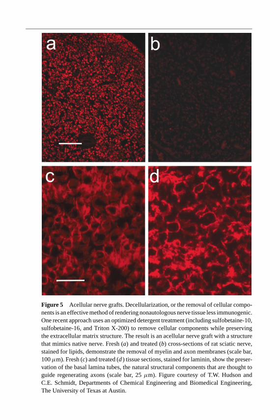
27 Jun 2003 20:51 AR AR191-11-COLOR.tex AR191-11-COLOR.SGM LaTeX2e(2002/01/18)P1: GCE
Figure 5 Acellular nerve grafts. Decellularization, or the removal of cellular compo-nents is an effective method of rendering nonautologous nerve tissue less immunogenic.One recent approach uses an optimized detergent treatment (including sulfobetaine-10,sulfobetaine-16, and Triton X-200) to remove cellular components while preservingthe extracellular matrix structure. The result is an acellular nerve graft with a structurethat mimics native nerve. Fresh (a) and treated (b) cross-sections of rat sciatic nerve,stained for lipids, demonstrate the removal of myelin and axon membranes (scale bar,100µm). Fresh (c) and treated (d) tissue sections, stained for laminin, show the preser-vation of the basal lamina tubes, the natural structural components that are thought toguide regenerating axons (scale bar, 25µm). Figure courtesy of T.W. Hudson andC.E. Schmidt, Departments of Chemical Engineering and Biomedical Engineering,The University of Texas at Austin.
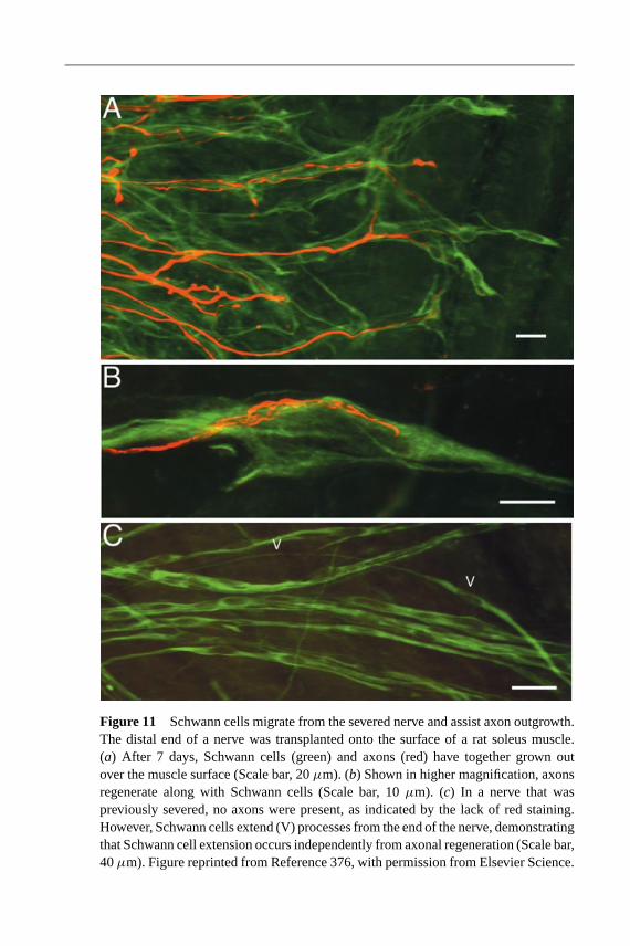
27 Jun 2003 20:51 AR AR191-11-COLOR.tex AR191-11-COLOR.SGM LaTeX2e(2002/01/18)P1: GCE
Figure 11 Schwann cells migrate from the severed nerve and assist axon outgrowth.The distal end of a nerve was transplanted onto the surface of a rat soleus muscle.(a) After 7 days, Schwann cells (green) and axons (red) have together grown outover the muscle surface (Scale bar, 20µm). (b) Shown in higher magnification, axonsregenerate along with Schwann cells (Scale bar, 10µm). (c) In a nerve that waspreviously severed, no axons were present, as indicated by the lack of red staining.However, Schwann cells extend (V) processes from the end of the nerve, demonstratingthat Schwann cell extension occurs independently from axonal regeneration (Scale bar,40µm). Figure reprinted from Reference 376, with permission from Elsevier Science.
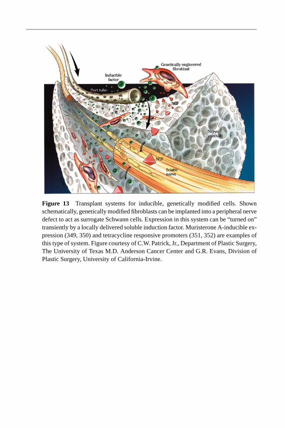
27 Jun 2003 20:51 AR AR191-11-COLOR.tex AR191-11-COLOR.SGM LaTeX2e(2002/01/18)P1: GCE
Figure 13 Transplant systems for inducible, genetically modified cells. Shownschematically, genetically modified fibroblasts can be implanted into a peripheral nervedefect to act as surrogate Schwann cells. Expression in this system can be “turned on”transiently by a locally delivered soluble induction factor. Muristerone A-inducible ex-pression (349, 350) and tetracycline responsive promoters (351, 352) are examples ofthis type of system. Figure courtesy of C.W. Patrick, Jr., Department of Plastic Surgery,The University of Texas M.D. Anderson Cancer Center and G.R. Evans, Division ofPlastic Surgery, University of California-Irvine.

27 Jun 2003 20:51 AR AR191-11-COLOR.tex AR191-11-COLOR.SGM LaTeX2e(2002/01/18)P1: GCE
Figure 14 Magnetically aligned hydrogels. Magnetically aligned fibrin and collagengels promote directed axonal outgrowth in vitro and in vivo compared to fibrin andcollagen gels with isotropic (i.e., randomly oriented) fibers (92, 104). Fibrin hydrogels,made with fluorescently labeled fibrinogen, were imaged with confocal laser scanningmicroscopy. (a) Isotropic fibrin gels show no directional alignment, whereas (b) fibringels exposed to a 9.4 Tesla magnetic field were unidirectionally aligned. (c) As seenunder bright field microscopy, neurite outgrowth from dorsal root ganglia is highlydirectional in magnetically aligned fibrin. Oriented neurite outgrowth from dorsal rootganglia also occurs in (d) aligned collagen gels, compared to (e) isotropic collagengels; in (d) and (e), neurites are stained green, Schwann cells are stained red andcolocalized neurites and Schwann cells appear yellow. Reprinted from Reference 104and 92, with permission from Elsevier Science.

27 Jun 2003 20:51 AR AR191-11-COLOR.tex AR191-11-COLOR.SGM LaTeX2e(2002/01/18)P1: GCE
Figure 16 Ridged nerve guidance channels. The nerve guidance channels in (a) and(b) were fabricated using MicroFab Technologies Inc., Tissue Engineering ink-jet print-ing station. The image in (a) illustrates a bifurcated nerve guidance channel containingcircumferential support ridges. Each segment is 1.8 mm in diameter and consists ofa proprietary polycaprolactone and PLA polymer solution. As shown, each segmenthas external support ribs that allow the conduit to resist in vivo compression withoutadding excess bulk or mass transfer resistance. The guidance channel in (b) illustratesa single channel with support ribs. For this case, a red fluorescent dye molecule wasincorporated into the channel in a gradient (the gradient is increasing from left to right).This illustrates the flexibility of this system to incorporate biomolecules into the guid-ance channels. Thus, using MicroFab’s ink-jet printing capabilities, substrates can becustom designed to meet degradation rate, growth factor elution rate, and degree ofcompression resistance according to the needs of the application. Images courtesy ofD.S. Silva and D. Hayes, MicroFab Technologies, Inc., Plano, Texas.