Embed Size (px)
Citation preview
1
Neural Stem Cells from Mammalian Brain: Isolation Protocols and Maintenance Conditions
Jorge Oliver-De la Cruz and Angel Ayuso-Sacido Regenerative Medicine Program, Centro de Investigación Príncipe Felipe,
REIG and Ciberned Spain
1. Introduction
Traditionally, the adult brain has been considered a quiescent organ, lacking the production
of new cells, or more exactly, new mature and functional neurons. This dogma has been
widely refused in the last decades with the discovery of proliferative cells with stem cell
properties in the adult brain.
First evidences come from the demonstration of neurogenesis in non-mammal vertebrates
such as birds or lizards (as reviewed in Garcia-Verdugo et al., 2002). Neurogenesis was also
confirmed to occur in adult mammals, like mice and rats, and, finally, in primates and
humans (for a complete revision see Gil-Perotín et al., 2009). Though the process of
neurogenesis in the adult is primarily confined to the subventricular zone (SVZ) and the
subgranular zone (SGZ) of the dentate gyrus, glial progenitors exist in other brain regions.
These widespread glial progenitors remain quiescent and do not generate mature glial cells,
but, in certain situations such as traumatic injury, they may act as true stem cells (Belachew
et al., 2003; Rivers et al., 2008).
The terminology of stem cell, progenitor cell and precursor cell has been adapted from
others tissues. Basically, a bona fide neural stem cell (NSC) must meet all these three features:
capacity of self-renewal, capacity to differentiate into the three neural lineages (neuron,
astrocyte and oligodendrocyte) and, finally, the ability to regenerate neural tissue. When
cells show a limited self-renewal and are already committed toward a specific fate, they are
classified as progenitor cells, while the term “precursor” represents intermediate stages.
Neural stem/progenitor cells (NSPc) primary cultures provide the best in vitro model to
study proliferation and differentiation signaling pathways, a difficult issue to address in
vivo. Additionally, these cells might be used in future replacement cell therapies, thus
motivating the development of protocols aimed to isolate and expand these cells in vitro.
These protocols display significant variations among them, and the introduction of new
technologies has increased drastically their number. The differences in the protocols have
rendered different results in terms of stem cell subpopulations, differentiation potential and
the amount of cells. The last is especially relevant in the case of human samples because of
their low availability.
www.intechopen.com
Neural Stem Cells and Therapy 4
Therefore, the aim of this chapter is to recapitulate some of these technical differences that
could induce variances in the final results. We have analyzed the main isolation protocols
from the two canonical neurogenic zones in the adult (subventricular zone and
hippocampus), described for both animal models (mouse and rat) and human.
2. Neural stem/progenitor cell isolation
The NSPc isolation procedures follow common steps including tissue dissection, digestion
and cell enrichment. However, comparing the different protocols found in the bibliography,
it is notable the presence of significant differences between them even when they are
consecutive works from the same group. The introduction of new technologies has also
increased drastically the number and variety of protocols. Additionally, some tissues like
normal human brain are particularly difficult to manage due to their low availability, which
requires improvements in the protocol to include modifications that increase the rate of
isolated cells. Interestingly, the diversity of isolation procedures results in the obtaining of
different stem/progenitor cell subpopulations with distinct differentiation potential, and
might be also responsible for the, sometimes contradictory, results observed in the literature.
Although the development of standard protocols would be the best option to assure that
results can be easily compared, in the practice, this is almost impossible. Different groups
have generated independently alternative procedures for the isolation, dissociation and
enrichment of NSPc. Furthermore, the animal model, the specific location of the brain
sample, or even the characteristics of the experiment have requirements that would make
unmanageable the use of universal procedures. Usually, the same group employs very
similar strategies to isolate cells from different samples, independently of their
developmental stage or animal/human origin. Nevertheless, it will be interesting to
establish flexible guidelines to indicate what can be modified from the standard procedures
and how to do so.
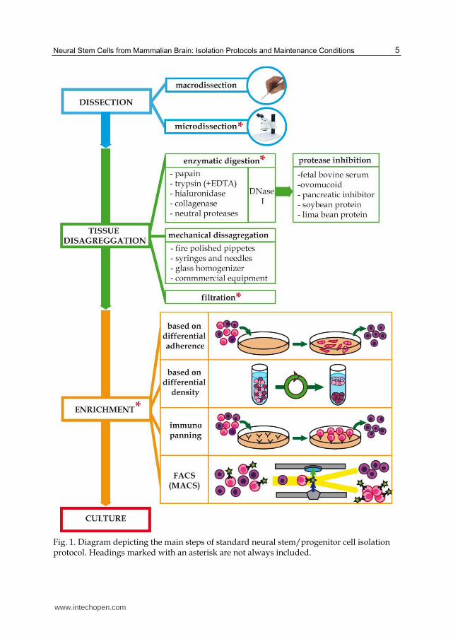
The basic scheme followed by NSPc isolation protocols is reflected in figure 1, and we will
discuss the specific methodology associated with every step in the following headings
2.1 Tissue dissection methods
The origin of the tissue influences the type of isolated cells as well as their proliferation and
differentiation capacity. A number of profound differences have been reported between
brain samples from different species (mouse, rat of human) or from different stages of
development within a given specie (Gritti et al., 2009; Svendsen et al., 1997). However, the
accurate dissection of specific regions of the brain has become more relevant as the
knowledge on the NSPc biology and location increases. In fact, regardless of the animal
model, one of the main factors that might determine the final results is the specific location
of the brain tissue from where NSPc are isolated.
Different regions of the brain have been used as a primary source of NSPc and,
consequently, discrepancies in the isolated cells have been reported. In this sense, analyzing
the distinct approaches for the tissue dissection might be useful to contextualize such a
controversy.
www.intechopen.com
Neural Stem Cells from Mammalian Brain: Isolation Protocols and Maintenance Conditions 5
Fig. 1. Diagram depicting the main steps of standard neural stem/progenitor cell isolation protocol. Headings marked with an asterisk are not always included.
www.intechopen.com
Neural Stem Cells and Therapy 6
We might consider three different levels of dissection according to the amount and location of tissue, ranging from large unselected brain tissue to microdissection. In a first level, a number of works start from whole brain (e.g. Von Visger et al., 1994) or large areas that include heterogeneous regions (e.g. whole human temporal lobe, Kirschenbaum et al., 1994). In these cases, the results can be highly variable, because of the different types of progenitors coming from distinct locations and giving rise to an artefactual impression of cell heterogeneity. An intermediate step of complexity is found in those works that use tissue from specific areas, but without the exclusion of contiguous tissues, i.e. macrodissection. In this regard, some authors reported the presence of multipotent stem cells from different regions of the adult parenchyma that differ from canonical neurogenic zones (SVZ and SGZ), e.g. from striatum (Reynolds & Weiss, 1992). However, these cells might arise from the cross-contamination of adjacent neurogenic regions (Lois & Alvarez-Buylla, 1993). Likewise, as will be discussed later, the existence of real neural stem cells in adult dentate gyrus of hippocampus has become a controversial subject. Some authors claim that there are true stem cells from this zone. However, others state that these isolated cells should be considered progenitors because of their low proliferation in vitro and their doubtful multipotentiality. The main argument of these authors is the lack of fine dissection, and the inclusion of neural stem cells from other adjacent tissues, like SVZ. Therefore, considering the current knowledge on NPSc niches location, an exhaustive microdissection is essential to take out the region of interest in a reliable way before starting the isolation procedures. Then, it is highly recommendable the use of thin slices of tissue for the accurate microdissection of different compartments under a dissecting microscope (e.g. Seaberg & van der Kooy, 2002).
Tissue dissection is particularly challenging in the case of human surgical samples, where orientation and anatomical organization is usually altered after surgery, making difficult the recognition of particular zones and, consequently, a good dissection. Alternatively, some authors have demonstrated the isolation of viable cells from postmortem tissue, especially in the case of human samples (e.g. Schwartz et al., 2001). While these procedures might be the only way to access some type of tissues, there might be some logistical inconveniences, the main one being that collection of tissue and cell isolation protocols need to be performed within few hours, because the number of NSPc decreases with time (Leonard et al., 2009; Xu et al., 2003), especially when samples are exposed to environmental temperature instead of 4ºC (Laywell et al., 1999).
2.2 Tissue digestion methods
2.2.1 Enzymatic dissociation
NSPc are surrounded by a highly structured extracellular matrix mainly composed by lecticans, hyaluronic acid, tenascin-C and tenascin-R (Rutka et al., 1988). These molecules interact among them and with membrane molecules on cell surfaces, and can regulate part of their behavior.
Therefore, one of the most successful strategies for removing NSPc from the rest of the tissue implies the use of proteases to degrade this matrix.
The first step, to prepare the tissue for enzymatic digestion, involves the mincing into small pieces (less than 1 mm3) in order to provide more degradable surface for the action of
www.intechopen.com
Neural Stem Cells from Mammalian Brain: Isolation Protocols and Maintenance Conditions 7
proteases. In this sense, the use of two different enzymes stands over the rest in the literature: trypsin (examples of its applications in different samples and developmental stages can be read at Kirschenbaum et al., 1994; Kukekov et al., 1997, Reynolds et al., 1992; Reynolds&Weiss, 1992; Svendsen et al., 1998) and papain (Babu et al., 2007; Roy et al., 2000a; Wang et al., 2000; Windrem et al., 2004). Trypsin is the most employed one, and is often combined with ethylenediaminetetraacetic acid (EDTA), a Ca2+ chelating agent that weakens intercellular unions. Regarding the concentration and the incubation time, it is not always possible to compare between different protocols as the enzyme units are not always specified and the incubation time ranges from 10 to 90 minutes. Additionally, other enzymes can be found in the bibliography such as hialuronidase (e.g. Gritti et al., 1995; Weiss et al., 1996), collagenase (e.g. Uchida et al., 2000), and neutral protease (dispase) (e.g. Babu et al., 2007), alone or in combination with others.
Generally, the use of proteases is linked to the utilization of Desoxiribonuclease I (DNase I),
usually from bovine origin, in order to eliminate the DNA mucus originated by cell lysis,
which could hinder cell survival and further experiments.
In any case, the employment of enzyme specific buffers (with adjusted pH and containing
activators) is necessary to allow the action of these enzymes. In some cases, antibiotic/
antimitotic is added to the digestion solution to prevent contamination. At this stage, some
authors also include kynurenic acid in order to reduce glutamate excitotoxicity through
NMDA receptor channels (e.g. Reynolds&Weiss, 1992). Afterward, the use of protease
inhibitors is necessary to stop enzymatic reaction. Papain is usually neutralized with fetal
bovine serum, whereas in the case of trypsin, the most employed method includes
ovomucoid, although there are commercially available soy, lima bean, and basic pancreatic
protein -based inhibitors.
The criterion for the choice of one or another enzyme is not clear, and frequently it has more
to do with the previous experience and skills of the group. Nevertheless, as a general rule,
embryo and early fetal samples require less amount of enzyme due to its laxity. For this
reason, some protocols reduce protease concentrations and/or exposure time (e.g. Svendsen
et al., 1998) or even recommend the use of mechanical disaggregation techniques alone (e.g.
Ciccolini&Svendsen, 1998; Reynolds&Weiss, 1996).
The enzymatic digestion is a critical step because it affects directly to the NSPc survival rate. In
this sense, some studies have been done to compare cell survival after dissociation with
different protease. Maric et al., 1998, used murine embryonic tissue to evaluate the efficacy of
papain, trypsin, and collagenase treatment, or mechanical disaggregation alone. The results
indicate that papain dissociation is optimal, achieving the maximum reproducible cell
recovery and viability. On the contrary, trypsin, collagenase, and mechanical dissociations
resulted on suboptimal and highly variable yields. Another study, carried out by Panchision et
al., 2007 also compared the results obtained for mouse embryonic stem cell isolation when
using papain, TrypleTM (a commercial analog of trypsin), or collagenase/neutral protease
commercial cocktails (AccutaseTM and Liberase-1TM). Data also confirmed that mechanic
dissociation induced more variability, cell death and more number of aggregates. However,
TrypleTM and papain produced more quantity of DNA mucus (but not an increased cell death)
and a lower adherence to culture plate after planting. They conclude that the best results were
obtained with papain, independently of the exposure time to the enzyme.
www.intechopen.com
Neural Stem Cells and Therapy 8
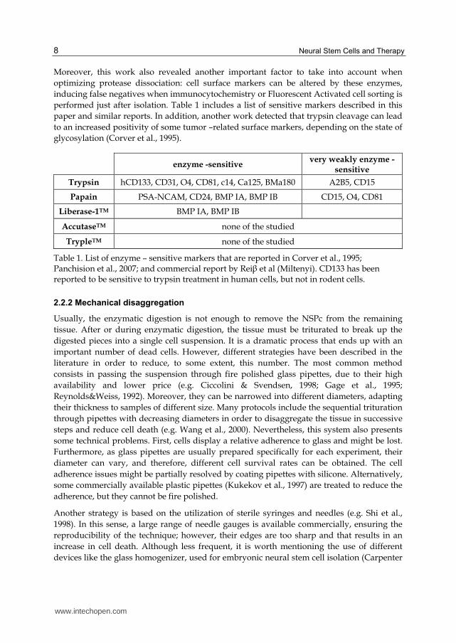
Moreover, this work also revealed another important factor to take into account when
optimizing protease dissociation: cell surface markers can be altered by these enzymes,
inducing false negatives when immunocytochemistry or Fluorescent Activated cell sorting is
performed just after isolation. Table 1 includes a list of sensitive markers described in this
paper and similar reports. In addition, another work detected that trypsin cleavage can lead
to an increased positivity of some tumor –related surface markers, depending on the state of
glycosylation (Corver et al., 1995).
enzyme -sensitive very weakly enzyme -
sensitive
Trypsin hCD133, CD31, O4, CD81, c14, Ca125, BMa180 A2B5, CD15
Papain PSA-NCAM, CD24, BMP IA, BMP IB CD15, O4, CD81
Liberase-1TM BMP IA, BMP IB
AccutaseTM none of the studied
TrypleTM none of the studied
Table 1. List of enzyme – sensitive markers that are reported in Corver et al., 1995; Panchision et al., 2007; and commercial report by Rei┚ et al (Miltenyi). CD133 has been reported to be sensitive to trypsin treatment in human cells, but not in rodent cells.
2.2.2 Mechanical disaggregation
Usually, the enzymatic digestion is not enough to remove the NSPc from the remaining
tissue. After or during enzymatic digestion, the tissue must be triturated to break up the
digested pieces into a single cell suspension. It is a dramatic process that ends up with an
important number of dead cells. However, different strategies have been described in the
literature in order to reduce, to some extent, this number. The most common method
consists in passing the suspension through fire polished glass pipettes, due to their high
availability and lower price (e.g. Ciccolini & Svendsen, 1998; Gage et al., 1995;
Reynolds&Weiss, 1992). Moreover, they can be narrowed into different diameters, adapting
their thickness to samples of different size. Many protocols include the sequential trituration
through pipettes with decreasing diameters in order to disaggregate the tissue in successive
steps and reduce cell death (e.g. Wang et al., 2000). Nevertheless, this system also presents
some technical problems. First, cells display a relative adherence to glass and might be lost.
Furthermore, as glass pipettes are usually prepared specifically for each experiment, their
diameter can vary, and therefore, different cell survival rates can be obtained. The cell
adherence issues might be partially resolved by coating pipettes with silicone. Alternatively,
some commercially available plastic pipettes (Kukekov et al., 1997) are treated to reduce the
adherence, but they cannot be fire polished.
Another strategy is based on the utilization of sterile syringes and needles (e.g. Shi et al.,
1998). In this sense, a large range of needle gauges is available commercially, ensuring the
reproducibility of the technique; however, their edges are too sharp and that results in an
increase in cell death. Although less frequent, it is worth mentioning the use of different
devices like the glass homogenizer, used for embryonic neural stem cell isolation (Carpenter
www.intechopen.com
Neural Stem Cells from Mammalian Brain: Isolation Protocols and Maintenance Conditions 9
et al., 1999), and some commercial equipment that appeared in the last years, promising a
higher efficiency via the automation of the isolation procedure (Rei┚ et al (Miltenyi)).
2.2.3 Filters utility
Some groups, after enzymatic digestion and mechanical disaggregation, include a filtering
step to remove the debris from the cell suspension. This additional step might eliminate
undissociated tissue pieces as well as avoid the presence of necrotic particles in the final
pellet that would potentially induce cell death. However, it also reduces the final number of
viable cells trapped into the filter. In any case, the use of filters usually requires a DNase I
treatment, to remove the mucus that can difficult the filtering, and it is strongly
recommended the dilution of cell suspension in a considerable volume of medium.
Regarding the type and size of the filters, some authors describe the use of cell strainers,
whereas others prefer sterile gauze (e.g. Kukekov et al., 1997). The mesh size also differs
among protocols (40 um (Wang et al., 2000), 70 um (Rietze et al., 2001), 100 um, etc), and
should be chosen in accordance with the efficiency of preceding methodology.
2.3 Neural stem/progenitor cells enrichment procedures
The initial protocols for NSPc isolation were designed with the only purpose of isolating
and culturing these cells to study their biology in vitro. However, as the knowledge on the
biology and differentiation potential of NSPc increased, it was evident that cell cultures
comprised a number of different subpopulations with different degree of stemness.
Consistently with this reality, many authors have recently included separation steps into
their NSPc isolation protocols. This separation is usually based on the NSPc phenotypic
characteristics closely related to their stem cell features.
In this sense, the first works on NSPc isolation and culture described a selection based on
their capacity to proliferate in the chosen medium and growth factors. Obviously, it was not
enough to discriminate heterogeneity. Consequently, many technical approaches have been
developed since then, for the enrichment of a specific subpopulation. This way, the
biological significance behind the molecule chosen to enrich for a specific type of cell and
the technology used for the procedure become an important step determining the
differentiation potential of the final cell culture. The current techniques for the separation
and enrichment of NSPc are described below.
2.3.1 Methods based on differential adherent properties of cells
One of the first methodologies for the enrichment of particular subpopulations was based
on the differential attachment of cells to the culture plate due to their particular adhesion
molecule patterns. By optimizing some parameters like substrates and time in culture it is
possible to distinguish between different types of cells. Astroglial cells show the biggest
adherence, even in untreated culture plate, whereas oligodendrocytes can be easily detached
through the agitation on a rotary shaker at slow revolutions (200-300 rpm) for 12-20 h. This
procedure has demonstrated to be useful, easy and affordable. As a consequence, it has been
common in the purification of specific cell types like oligodendrocytes (McCarthy & de
Vellis, 1980; Chen et al., 2007b).
www.intechopen.com
Neural Stem Cells and Therapy 10
Taking advantage of these properties, Lim&Alvarez-Buylla, 1999, reported the isolation of 4
cell fractions using serial streaming of medium or PBS over the surface of poly-D-lysine
treated plates, and a final step with trypsin. The first fraction (or fraction 1), which contains
the less adherent cells, was enriched in PSA-NCAM and Tuj1 (identified as migrating
neuroblasts). On the contrary, cells from the most adherent fraction (fraction 4) were GFAP+
and show characteristics of neural stem cells (type B/C according to the model of SVZ
organization (Fig.2). However, it is important to mention that this procedure does not allow
the obtaining of high purity cultures.
2.3.2 Differential gradient centrifugation
Another group of technical approaches for NSPc enrichment is based on fractionating cell
populations according to their buoyant density. Previously, the cells are dissolved in specific
solvents that, after centrifugation, generate a density gradient. The cells distribute in this
gradient and can be collected separately. The gradient might be formed by using different
types of reagents, being Percoll the most widely used (e.g. Palmer et al., 1999; K. Chen et al.,
2007a). It consists of colloidal silica particles coated with a layer of polyvinylpyrrolidone (PVP)
that can be used to form solution densities between 1.00 and 1.20 g/ml. A combination of
Percoll gradients can be generated in order to separate more subpopulations. Using a
discontinuous density gradient, Maric et al., 1998 reported the isolation of 20 different bands
and the delimitation of density bands can be facilitated by commercial color-coded density
marker beads. While its application has become very common because of its low interaction
with cells and low toxicity, it is restricted to research as it may contain variable quantities of
endotoxin (PVP). Alternatively, density gradients can be also generated using sucrose
solutions (Johansson et al., 1999) and Bovine Serum Albumin (Ericsson, 1977).
2.3.3 Immunopanning
Initial immunopanning applications were essentially directed to eliminate specific cell subpopulations by antibody union and complement-mediated lysis (e.g. Gard&Pfeiffer, 1993). Nevertheless, the present acceptation of the immunopanning technic comprises the purification of a cell population by exploiting their differential binding to the culture dishes previously coated with a cell-surface antibody. Cells expressing this surface antigen are retained on the dish and are thereby separated from the remaining cell population. It has been especially applied to the isolation of oligodendrocyte progenitor cells, using A2B5 or O4 (Barres et al., 1992; Wu et al., 2009; Mayer-Proschel, 2001) as molecular surface markers, but it can also be adapted to segregate immature neurons (PSA-NCAM) (Ben-Hur et al., 1998; Schmandt et al., 2005). Although the use of immunopanning has become less popular with the introduction of Fluorescence-activated cell sorting (FACS) technology, some authors had reported that immunopanning provides a higher survival (Mayer-Proschel, 2001).
2.3.4 Fluorescence activated cell sorting (FACS)
The main improvement in terms of separation and enrichment of specific NSPc comes with
the introduction of the FACS technology. As a specialized form of flow citometry, it
provides a method for sorting heterogeneous cells based upon the specific union of a
fluorophore-labeled antibody to a cell surface maker. In addition to antibodies, other
www.intechopen.com
Neural Stem Cells from Mammalian Brain: Isolation Protocols and Maintenance Conditions 11
molecules like lectins can be used to recognize the glycosylation state of some membrane
epitopes (as reviewed in Kitada et al., 2011).
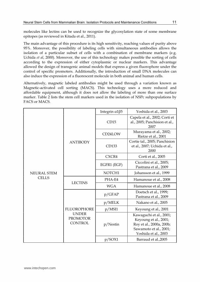
The main advantage of this procedure is its high sensitivity, reaching values of purity above 95%. Moreover, the possibility of labeling cells with simultaneous antibodies allows the isolation of a particular subset of cells with a combination of membrane markers (e.g. Uchida et al, 2000). Moreover, the use of this technology makes possible the sorting of cells according to the expression of either cytoplasmic or nuclear markers. This advantage allowed the design of transgenic animal models that express a given fluorophore under the control of specific promoters. Additionally, the introduction of small DNA molecules can also induce the expression of a fluorescent molecule in both animal and human cells.
Alternatively, magnetic labeled antibodies might be used through a variation known as Magnetic-activated cell sorting (MACS). This technology uses a more reduced and affordable equipment, although it does not allow the labeling of more than one surface marker. Table 2 lists the stem cell markers used in the isolation of NSPc subpopulations by FACS or MACS.
NEURAL STEM CELLS
ANTIBODY
Integrin ┙1┚5 Yoshida et al., 2003
CD15 Capela et al., 2002; Corti et al., 2005; Panchision et al.,
2007
CD24LOW Murayama et al., 2002;
Rietze et al., 2001
CD133 Cortie tal., 2005; Panchision
et al., 2007; Uchida et al., 2000
CXCR4 Corti et al., 2005
EGFR1 (EGF) Ciccolini et al., 2005; Pastrana et al., 2009
NOTCH1 Johansson et al., 1999
LECTINS PHA-E4 Hamanoue et al., 2008
WGA Hamanoue et al., 2008
FLUOROPHORE UNDER
PROMOTOR CONTROL
p/GFAP Doetsch et al., 1999; Pastrana et al., 2009
p/MELK Nakano et al., 2005
p/MSI1 Keyoung et al., 2001
p/Nestin
Kawaguchi et al., 2001; Keyoung et al., 2001;
Roy et al., 2000a, 200b; Sawamoto et al., 2001;
Yoshida et al., 2003
p/SOX1 Barraud et al.,2005
www.intechopen.com
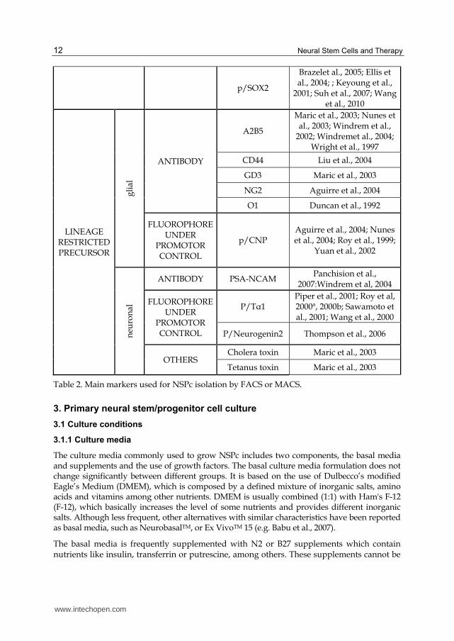
Neural Stem Cells and Therapy 12
p/SOX2
Brazelet al., 2005; Ellis et al., 2004; ; Keyoung et al.,
2001; Suh et al., 2007; Wang et al., 2010
LINEAGE RESTRICTED PRECURSOR
gli
al
ANTIBODY
A2B5
Maric et al., 2003; Nunes et al., 2003; Windrem et al.,
2002; Windremet al., 2004; Wright et al., 1997
CD44 Liu et al., 2004
GD3 Maric et al., 2003
NG2 Aguirre et al., 2004
O1 Duncan et al., 1992
FLUOROPHORE UNDER
PROMOTOR CONTROL
p/CNP Aguirre et al., 2004; Nunes et al., 2004; Roy et al., 1999;
Yuan et al., 2002
neu
ron
al
ANTIBODY PSA-NCAM Panchision et al.,
2007:Windrem et al, 2004
FLUOROPHORE UNDER
PROMOTOR CONTROL
P/T┙1 Piper et al., 2001; Roy et al, 2000ª, 2000b; Sawamoto et al., 2001; Wang et al., 2000
P/Neurogenin2 Thompson et al., 2006
OTHERS Cholera toxin Maric et al., 2003
Tetanus toxin Maric et al., 2003
Table 2. Main markers used for NSPc isolation by FACS or MACS.
3. Primary neural stem/progenitor cell culture
3.1 Culture conditions
3.1.1 Culture media
The culture media commonly used to grow NSPc includes two components, the basal media and supplements and the use of growth factors. The basal culture media formulation does not change significantly between different groups. It is based on the use of Dulbecco’s modified Eagle’s Medium (DMEM), which is composed by a defined mixture of inorganic salts, amino acids and vitamins among other nutrients. DMEM is usually combined (1:1) with Ham's F-12 (F-12), which basically increases the level of some nutrients and provides different inorganic salts. Although less frequent, other alternatives with similar characteristics have been reported as basal media, such as NeurobasalTM, or Ex VivoTM 15 (e.g. Babu et al., 2007).
The basal media is frequently supplemented with N2 or B27 supplements which contain nutrients like insulin, transferrin or putrescine, among others. These supplements cannot be
www.intechopen.com
Neural Stem Cells from Mammalian Brain: Isolation Protocols and Maintenance Conditions 13
added to basal formulation until they are used because of their short life at 4ºC. Although both of them might be used, even in combination, they have different properties that may influence cell culture behavior. B27 has a more complex composition than N2 supplement and only enhances cell survival during the period immediately following isolation (Svendsen et al., 1995), while N2 offers the same results, at a lower price. Babu et al. (2007) concluded that monolayer cells maintained with N2 supplement generated more neurons after differentiation, whereas B27 supplement promoted proliferation.
3.1.2 Serum and growth factors
Although basal media and supplements are quite similar in most cases, the most important
issue in terms of culture media is the use of either specific growth factors or serum. The first
works on NSPc isolation and maintenance described the use of serum in their culture media.
However, as the knowledge on NSPc biology increased, researchers found that the use of
serum, generally fetal bovine serum (FBS), had several disadvantages. As a complex
solution of undefined composition that can vary drastically among batches, the use of serum
does not contribute to improve our knowledge about trophic signals requirements.
Additionally, it is not a physiological condition, since neural stem cells are not exposed
directly to serum in vivo. Finally, serum includes a combination of different growth factors
that are able to maintain stem cell phenotype and also induce differentiation. All these
reasons made the authors substitute serum for a specific combination of purified growth
factors. The utilization of two main growth factors stands out from the rest: fibroblast
growth factor 2 (FGF-2, also called basic FGF or bFGF) and epidermal growth factor (EGF),
alone or in combination. Moreover, FGF-2 must be used in combination with heparin, which
mediates the binding of the growth factor to its receptor (Yayon et al., 1991).
Initial works (Reynolds, 1992; Reynolds & Weiss, 1992) described the isolation of an EGF-responsive neural stem cell population from striata/lateral ventricle, although some authors reported that similar cell cultures could be also maintained with FGF (Gritti et al., 1995; Vescovi et al., 1993). Similarly, some works also found a synergic effect of both EGF and FGF in proliferation, but only at low cell densities (Svendsen, 1997; Tropepe, 1999). Finally, a series of studies (Martens et al., 2000; Tropepe et al., 1999; Ciccolini, 2001; Maric et al., 2003) demonstrated that FGF- responsive cells arise earlier at development, and then give rise to both EGF/FGF- responsive cells. Moreover, it was revealed that the acquisition of EGF responsiveness is promoted by FGF in vitro (Ciccolini & Svendsen, 1998). First isolations could be explained with the discovery of a small autocrine/paracrine FGF production by neural stem cells, allowing the survival of FGF-2 dependent cells without FGF until the acquisition of EGF responsiveness (Maric et al., 2003).
Other growth factors that have been reported to support cell culture are Transforming
growth factor alpha (TGF-┙) (Reynolds et al., 1992), Leukemia inhibitory factor (LIF) and its
equivalent Ciliary neurotrophic factor (CNTF) (Carpenter et al., 1999), or Brain-derived
neurotrophic factor (BDNF), although its capacity to enhance later neuronal production and
survival has been questioned (Kirschenbaum & Goldman, 1995; Ahmed et al., 1995;
Reynolds & Weiss, 1996).
Platelet-derived growth factor alpha (PDGF┙) is frequently used in the maintenance media for oligodendrocyte progenitor cells. The signaling pathway through the PDGF┙/PDGFR┙
www.intechopen.com
Neural Stem Cells and Therapy 14
has different effects depending on the stage of differentiation of these progenitors: it provides signals favoring proliferation and migration in murine and human oligodendrocyte progenitors (Wilson et al., 2003; Calver et al., 1998), whereas later in development is related with cell survival (Gogate et al, 1994). Similarly, FGF promotes proliferation and blocks differentiation of oligodendrocyte progenitors, in part through the modulation of PDGFR┙ receptors expression (McKinnon et al., 1990).
Finally, some works have attempted to co-culture NSPc in the presence of other supportive cells like astrocytes (Richards et al., 1992; Lim et al., 1999), that seem to favor the NSPc growth by physical contact, or endothelial cells, that also enhance cell proliferation via VEGF production (Sun et al., 2010).
3.1.3 pH and oxygen levels
The metabolic processes undergone by the cells in culture give rise to acidic components that eventually are released to the media, thus decreasing the pH. This alteration, easily followed by the inclusion of a pH indicator like phenol red, has a direct influence in the behavior of the cells. Therefore, buffering agents are commonly added to medium formulation in order to control variations in the pH. In this sense, two main systems are routinely used in the elaboration of the media: sodium bicarbonate buffer, which is dependent on the CO2 concentration present in the incubator, and HEPES (4-(2-hydroxyethyl)-1-piperazineethanesulfonic acid), independent of atmospheric CO2. Although HEPES is better at maintaining physiological pH controls, the exposure of HEPES- containing media to light must be reduced as HEPES-containing media generates hydrogen peroxide when exposed to ambient light ({Zigler et al., 1985).
In contrast, the level of O2 tension remains to be optimized for NSPc cultures. The standard
conditions to culture NSPc had included atmospheric levels of O2 (21%), although
physiological levels are much lower (around 3%). Several studies have confirmed that NSPc
expansion under low level of O2 correlates with the expression of stemness markers and
higher survival rate both in vitro and in vivo after engraftment (reviewed in De Filippis &
Delia, 2011).
3.2 Monolayer versus neurosphere cultures
To maintain and propagate stem cell cultures different authors have published two
alternative methods of NSPc culture and expansion: as free-floating cell clusters
(neurospheres) or as adherent cultures forming a monolayer on the plate surface.
The neurosphere assay has been the most extended method to demonstrate the presence of
NSPc in culture (Reynolds et al., 1992) and it is still used with different modifications (Rietze&
Reynolds, 2006). Some authors claim that each neurosphere represents a microenvironment
that recapitulates neurogenic niche and allows survival of stem cells in vitro (Bez et al., 2003)
through direct cell-to-cell interaction. Nevertheless, a single neurosphere contains only a small
percentage of true stem cells, whereas the remaining cells are in different stages of
differentiation. Necrotic and apoptotic cells are also present (Lobo et al., 2003). Interestingly, it
has been reported that committed progenitors, like oligodendrocyte precursors, can generate
cell clusters similar to neurospheres (Chen Y. et al., 2007).
www.intechopen.com
Neural Stem Cells from Mammalian Brain: Isolation Protocols and Maintenance Conditions 15
This culture method has also several technical disadvantages. First, when neurospheres
become larger, the diffusion of nutrients and growth factors through the neurosphere is
compromised (Svendsen et al., 1997b), which makes difficult the interpretation of some
experimental results. Second, packed neurospheres do not allow the tracking of individual
cells, which also hinders studies relating to differentiation processes. Finally, recent
publications demonstrate that neurospheres are not static particles originated from a single
cell and isolated from the rest of the neurospheres and cells (Rietze, 2006). On the contrary,
they are dynamic structures within the culture, were cells are exchangeable from one to
another sphere. This effect, may be circumvented by either using a limiting dilution analysis
to obtain a single cell in each well or using semisolid cultures by adding methylcellulose
(Gritti et al., 1999; Kukekov et al., 1997) or collagen (Neural Colony-Forming Cell Assay
(Louis et al., 2008).
By contrast, monolayer cultures obviate some of these restrictions. They can be used to
study the properties of stem cells at individual cell level, although it does not allow cell
interaction during differentiation. Moreover, cells are exposed homogeneously to growth
factors and serum, with the consequent reduction in cell heterogeneity.
In all, there is not a prevalent method over the other. It has not been demonstrated a total
equivalence between both type of cultures and the two methods have advantages and
limitations that researchers should take into consideration in the experimental design. The
formation of neurospheres may be promoted by following several strategies, being the most
common one the use of nonadherent surfaces like poly-2-hydroxyethyl methacrylate
(Kukekov et al, 1999). Furthermore, it has been also reported the addition of
mercaptoethanol to avoid cell attachment (Kukekov et al., 1997). However, not all attempts
to transform an adherent culture into neurospheres have been successful (Walton et al.,
2006). Alternatively, cell attachment may be induced by coating the plate surface with
charged molecules such as poly-l-ornithine, poly-d-lysine or laminin.
3.3 Cell passaging
Before cells become totally confluent, it is necessary to subculture them after disaggregation
of cell clusters into single cell suspensions. Regardless of the type of culture, monolayer or
suspension, passaging should be performed before cells achieve their maximum confluence
(monolayer) or cell cluster become necrotic (neurospheres) in order to avoid senescence
associated with prolonged high cell density. The methodology employed for the
disaggregation step depends on the cell type.
Adherent cells are usually detached from the surface of the culture vessel by enzymatic
means. Trypsin, alone or in combination with EDTA, has been the most used protease (e.g.
Palmer et al., 1997); but in the last years it has been substituted in current protocols by
TrypleTM, since this commercial product is free of animal- and human- derived components,
less damaging to cells, and does not require the use of inhibitors.
In the case of neurospheres, cell disaggregation is performed by using mechanical
procedures which involve triturating spheres with fire polished pipettes. However, this is an
aggressive method that renders high levels of cell death. Enzymatic digestion can be also
www.intechopen.com
Neural Stem Cells and Therapy 16
used before triturating, however, this may alter the experimental results if FACS assays are
conducted right after disaggregation.
An alternative method was reported by Svendsen & ter Borg, 1998 for passaging
neurospheres isolated from human fetal tissue. Briefly, neurospheres were cut into 4 pieces
instead of standard trituration into single cell suspension. According to their data, this
sectioning method reduces cellular trauma and preserves cell interaction, allowing NSPc to
proliferate more replication rounds in vitro.
3.4 Cryopreservation
Cryopreservation allows the maintenance of NSPc in a suspension mode awaiting for
future experiments and saving expensive culture reagents. Considering the low number
of cells obtained from each sample, especially in human tissue, increasing the survival
ratio after long-term preservation of NSPc becomes a major concern. The main
cryopreservation protocols employ dimethylsulfoxyde (DMSO) diluted at 10-20% in
culture media to avoid ice crystallization, accompanied by a slow cooling step in
isopropanol recipients. Although less popular, glycerol can be used instead of DMSO.
Cellular viability can be improved adding animal serum to freezing medium, but it can
potentially introduce contaminants, and induce differentiation. In any case,
cryopreservation must follow some general rules to ensure the successful preservation of
cells. It must be performed during the logarithmic growth phase and high cell density in
each ampoule seems to facilitate cell recuperation. Smaller neurospheres survive better
than larger, so triturating cells until getting a suspension of small neurospheres improves
cell survival.
Recently, a new alternative preservation method, named vitrification, has been adapted for
NSPc (Tan et al., 2007). In brief, cells are sequentially submerged in a series of freezing
solutions with increasing concentrations of cryoprotectant (ethylene glycol and sucrose),
and finally transferred into borosilicate glass capillaries, snap-frozen and stored in liquid
nitrogen. The results showed that vitrification offered the best combination of cell viability,
multipotency, and preservation of structural integrity of neurospheres.
3.5 Differentiation
After isolation of proliferating cells, it is necessary to confirm the stemness characteristics of the cells, that is, the multipotent and self-renewal capacities. In this sense, cells with lower self-renewal or with potential to generate just one type of cell should be considered as progenitor cells. To evaluate the differentiation capacity, cells are exposed to differentiation signals coming from animal serum or chemically defined compounds.
The use of serum has the same problems highlighted above. Nevertheless, this is still the
standard methodology, because the specific signals inducing NSPc differentiation into a
specific lineage remains largely unknown. Cells maintained in defined medium tend to
differentiate when exposed to serum in a variable concentration (from 1% up to 10%)(e.g.
Ciccolini & Svendsen, 1998; Palmer et al., 1999; Roy et al., 2000; Wanget al., 2000), although a
preference towards astroglial differentiation has been reported (Palmer et al., 1995). The use
www.intechopen.com
Neural Stem Cells from Mammalian Brain: Isolation Protocols and Maintenance Conditions 17
of serum is usually accompanied by the addition of molecules such as Poly-L-ornithine,
laminin or matrigel to promote adhesion to substrate, which seem to enhance differentiation
of neurospheres cultures (Ciccolini & Svendsen, 1998; Reynolds & Weiss, 1996; Tropepe et
al., 1999). In some cases, the removal of growth factors in conjunction with an adherent
substrate has been also used to differentiate NSPc (Gritti et al., 1996).
Alternatively, media previously exposed to other cell cultures (conditioned medium) may
be used to induce differentiation. Probably the most employed one is B104 conditioned
medium, which is exposed to a neuroblastoma cell line and induces oligodendroglial
differentiation (Young & Levinson, 1997).
Few authors have conducted NSPc differentiation assays by using growth factor cocktails in
the absence of serum. Uchida et al., 2000 reported that a combination of BDNF and glial-
derived growth factor (GDNF) was enough to differentiate CD133+ cells from human fetal
tissue. Ling et al., 1998 reported a more specific differentiation protocol, proving that the
combination of Interlekine-1b, Intelkeulin-11 and GDNF promoted the appearance of
dopaminergic neurons (tyrosine hydroxylase -positive cells).
Furthermore, a number of chemical signals have been also reported to stimulate the
differentiation toward a particular neural lineage.
In the case of neuronal maturation, BDNF, retinoic acid, Neurotrophin (NT3), and Sonic
Hedgehog (SHH) have been associated to an enhanced neural obtaining (Babu et al., 2007;
Bull & Bartlett, 2005; Dutton et al., 1999; Roy et al., 2000a, 2000b).
Oligodendroglial differentiation can be also enhanced using PDGFa, which promotes their
survival (Gogate et al., 1994) in collaboration with NT3 and Triiodothyronine (T3), factors
necessary for the correct development of oligodendrocytes and the expression of myelin
proteins (Billon et al., 2002; Park et al., 2001).
4. Isolation from neurogenic zones
Neural stem cells seem to reside within specific niches of the adult brain. These regions are located in the subventricular zone of the lateral ventricles and the subgranular zone in the hippocampus. The origin of NSPc in these two areas has been the focus of intense debates in the literature and the isolation procedures of such cells from these specific locations need special attention.
Since the discovery of adult neural stem cells, the isolation procedures have been modified
along with the increased knowledge of NSPc biology. Initially, these cells were supposed to
be scattered within the brain parenchyma. However, soon after it was restricted to the SVZ,
although the individual cell identity is still a source of division among researchers due to the
lack of a specific marker to label neural stem cells. The nature and origin of the neural stem
cell in the SGZ of the hippocampus has been also a subject of an intense debate, questioning
whether they could be considered true neural stem cells or committed progenitors.
Additionally, other types of neural progenitors like oligodendrocyte progenitor cells (OPCs)
seem to be dispersed through the white matter, and their isolation procedures and
characterization have become recently relevant in the context of demyelinating diseases.
www.intechopen.com
Neural Stem Cells and Therapy 18
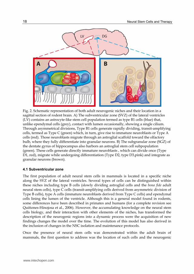
Fig. 2. Schematic representation of both adult neurogenic niches and their location in a sagittal section of rodent brain. A) The subventricular zone (SVZ) of the lateral ventricles (LV) contains an astrocyte-like stem cell population termed as type B1 cells (blue) that, unlike ependymal cells (grey), contact with lumen occasionally, showing a single cilium. Through asymmetrical divisions, Type B1 cells generate rapidly dividing, transit-amplifying cells, termed as Type C (green) which, in turn, give rise to immature neuroblasts or Type A cells (red). Those neuroblasts migrate through an astroglial scaffold toward the olfactory bulb, where they fully differentiate into granular neurons. B) The subgranular zone (SGZ) of the dentate gyrus of hippocampus also harbors an astroglial stem cell subpopulation (green). These cells generate directly immature neuroblasts , which can divide once (Type D1, red), migrate while undergoing differentiation (Type D2, type D3,pink) and integrate as granular neurons (brown).
4.1 Subventricular zone
The first population of adult neural stem cells in mammals is located in a specific niche along the SVZ of the lateral ventricles. Several types of cells can be distinguished within these niches including type B cells (slowly dividing astroglial cells and the bona fide adult neural stem cells), type C cells (transit-amplifying cells derived from asymmetric division of Type B cells), type A cells (immature neuroblasts derived from Type C cells) and ependymal cells lining the lumen of the ventricle. Although this is a general model found in rodents, some differences have been described in primates and humans (for a complete revision see Quiñones-Hinojosa et al., 2006). However, the accumulating knowledge on the neural stem cells biology, and their interaction with other elements of the niches, has transformed the description of the neurogenic regions into a dynamic process were the acquisition of new findings changes the model over the time. The evolution of this model has also derived in the inclusion of changes in the NSC isolation and maintenance protocols.
Once the presence of neural stem cells was demonstrated within the adult brain of
mammals, the first question to address was the location of such cells and the neurogenic
www.intechopen.com
Neural Stem Cells from Mammalian Brain: Isolation Protocols and Maintenance Conditions 19
region. Early studies reported the isolation of NSC from the striatal tissue of both rat (Gritti
et al., 1995; Reynolds & Weiss, 1992;) and mouse (Richards et al., 1992). Nevertheless, the
striatum is a relatively large region that is not consistent with data about NSC biology.
Afterwards the investigations in this field confirmed that only SVZ tissue was able to
generate cell cultures with stem cell properties, whether via explants (Kirschenbaum et al.,
1994; Kirschenbaum & Goldman, 1995; Lois & Alvarez-Buylla, 1993) or neurospheres
cultures (Morshead et al., 1994) were used. Subsequent articles which described the isolation
of neural stem cells from striatum specified the inclusion of the SVZ region within the
dissected tissue (Weiss et al., 1996) and confirmed that the results of neural stem cell
isolation exclusively from SVZ were identical to those obtained from extensive anatomical
regions containing the SVZ (Kukekov et al., 1997). These initial works emphasized the
relevance of a fine orientated dissection for a successful NSC isolation protocol.
Nonetheless, the SVZ comprises a heterogeneous population, and those early reports did
not reveal the cellular identity of the NSC. Probably, the first work addressing the NSC
identity in vitro, was performed by Johansson et al., 1999. Marking ventricular cells with DiI
(a lipophilic membrane stain that diffuses laterally to stain the entire cell), they concluded
that NSC were actually ciliated ependymal cells. By contrast, Doetsch et al. (1999) reported
that only GFAP expressing cells (marked via adenovirus which allowed the expression of
the green fluorescent protein (GFP) under the control of glial fibrillary acidic protein (GFAP)
promoter) give rise to neurospheres culture. Moreover, it was previously described that
those astrocyte-like cells occasionally contacted the ventricle and displayed a single cilium,
suggesting that DiI isolated cultures could be originated from them instead of ependymal
cells (a more complete model can be consulted at Mirzadeh et al. (2008)). Following previous
findings, two new studies described that both types of cells were able to proliferate in vitro,
but only SVZ astrocytes generated neurospheres with self-renewal and mutipotential
capacity (Chiasson et al., 1999; Laywell et al., 2000). These findings also marked the need for
an identification and selection step in the NSPc isolation protocol. With the introduction of
FACS technology, two phenotypical features of NSC supported the astrocyte-like theory, the
isolation of a CD15+ population with stem cell characteristics (Capela & Temple, 2002), a
carbohydrate only expressed in astrocytes, and the identification of NSC as CD24low (Rietze
et al, 2001), whereas ependymal cells are CD24+. Using an opposite strategy to deplete adult
GFAP+ cells, two independent studies demonstrated that the ablation of adult astrocytes
resulted in the loss of multipotent neurosphere formation (Morshead et al, 2003; Imura et al.,
2003). However, with the controversy surrounding the identity of NSCs, two later studies
reported the isolation of CD133+ cells from adult brain, as previously reported by Uchida et
al. (2000) for fetal tissue. Nevertheless, opposite results were obtained from each one. The
first one (Corti et al., 2007) concluded that CD133 stained a small number of cells underlying
the ependymal layer, and the sorting of those CD133+ cells leads to the isolation of a NSC
population. Interestingly, Mirzadeh et al. (2008) found that 29% of the apical processes of B1
cells were positive for CD133. By contrast, Coskun et al. (2008) found that both CD133+
ependymal cells and NSC originate from ependymal cells. In any case, it will be necessary to
find new markers in order to improve the identification and selection of either the real adult
neural stem cells and the different range of progenitor cells. This will allow the study of
their specific biological features and maybe modulate their behavior in vivo.
www.intechopen.com
Neural Stem Cells and Therapy 20
4.2 Subgranular zone
In the adult hippocampus, NSCs are located in the SGZ, a cellular layer found between the granule cell layer and the hilus, in the dentate gyrus. Similarly to SVZ, NSCs have been identified as astrocyte-like cells, with cell bodies located in the SGZ and vertical processes extended through the molecular layer. However, unlike what happens in the SVZ, these astrocytic cells generate an earlier immature neuroblast that divides only once and expresses neuronal markers (PSA-NCAM and doublecourtin) (Type D1 cells). Those cells migrate short distances within the granule cell layer while undergoing morphological changes (type D2 and Type D3 cells) until they reach a final position and differentiate into mature granular neurons.
The first data from adult hippocampal cell cultures were published by Palmer et al., 1995. They isolated a monolayer culture from adult female rat hippocampus. These cells were capable of proliferating in serum free media supplemented with FGF. In Gage et al., 1995, they were also able to derivate a FGF-2 -dependent adherent culture that differentiates into mature neurons when engrafted into adult rat brain. Short after, in vitro differentiation was attempted by Palmer et al., 1997. Using several combinations of growth factors, they demonstrated the multipotency of those progenitor cells, and the enhancement of neuronal maturation when BDNF was added (similarly to NT3 and retinoic acid results), whereas serum addition promoted astroglial differentiation.
As described previously for initial reports on SVZ, those studies also isolated cells with similar features from non-neurogenic zones, including the septum of striatum. However a few years later was published the first work that isolated specific hippocampal neural stem cells from adult human samples (Kukekov et al., 1999). They observed the presence of neurospheres when cultured in non-adherent conditions by using mercaptoethanol, similarly to previous studies carried out with SVZ samples (Kukekov et al., 1997). Following the enrichment step based on the expression of GFP under the control of an specific promoter (P/T┙1:hGFP and E/nestin:EGFP), described previously to identify NSC from the SVZ, Roy et al., 2000b, isolated, for the first time, neural stem cells from human hippocampal samples. However, these results did not reproduce in mice and later works criticized the gross microdissection done in these publications. Afterwards, Seaberg & van der Kooy, 2002 tried to generate neurospheres from microdissected mouse dentate gyrus. They were unable to generate neurospheres capable of self-renewal, and multipotency was also compromised. Nevertheless, hippocampus obtained with gross dissection was able to generate neurospheres, suggesting that previous results could be explained if the dissection procedure included contaminating cells from the ventricular layer next to the hippocampus, or even white matter progenitors. They also assayed different culture conditions and dissection techniques, including dentate gyrus microdissection, and were unable to obtain proliferating neurospheres. After a number of publications showing controversial results in this regard Babu et al., 2007, using dentate gyrus microdissection from p/Nestin:EGFP transgenic mouse, were able to obtain monolayer cultures with self-renewal (up to 66 passages) and multipotency characteristics. Moreover, they observed spheres-like colonies when performing a modified neurosphere assay using semisolid medium. Additionally, after trying different media and supplements they concluded that although B27 and EGF promoted a slightly higher proliferation, N2 supplement and bFGF maintained cells differentiate better into mature neurons. Moreover, they reconfirmed that BDNF, NT3, SHH promoted neuronal differentiation, while LIF and Bone morphogenetic protein 2 (BMP-2) promoted glial differentiation
www.intechopen.com
Neural Stem Cells from Mammalian Brain: Isolation Protocols and Maintenance Conditions 21
More recent works found that isolated neural progenitor/stem cells display paracrinal production of BMP, and the addition of noggin to culture media favors the formation of multipotential and self-renewal neurospheres (Bonaguidi et al., 2005; Bonaguidi et al., 2008).
The differences, in terms of culture media requirements, between NSC isolated from the SVZ and those from the SGZ might be due to their behavior in vivo. NSPc from lateral ventricle are prepared to migrate a larger distance to the olfactory lobes, while new neurons produced from dentate gyrus integrate nearby the stem cell niche and, therefore, are not prepared to maintain their stemness capacity in the absence of the niche signals.
5. Conclusions
Cell isolation and culture provides a powerful tool for the study of neural stem and
progenitor cells. Although in vitro analysis has several limitations, and results cannot be
directly extrapolated to the in vivo behavior of the isolated cells, it allows the analysis of
their features and potential capacities in a controlled environment that can be modified and
monitored more accurately.
Every step of the isolation procedure is likely to be optimized. Any protocol amendment
should be tested and not considered trivial, as it can have a high impact on the cell
population obtained. Consequently, isolation methods should be planned according to
further experimental applications and not based on the routine practices of each research
group, especially in the case of adaptation of protocols used previously for tissues collected
from different species.
Moreover, it should be considered that the final purpose of most experiments is to improve
our knowledge about stem cells and their clinical applications. For this reason, steps in the
protocol which include reagents with undefined composition or with the possibility of
introducing contaminants, such as serum, must be redesigned, because it is the only way to
understand the chemical signals underlying the biological behavior of neural stem or
progenitor cells.
6. Acknowledgements
We are especially grateful to Mario Soriano Navarro and Josefa Carrión Navarro for their
assistance. Jorge Oliver De La Cruz is a recipient of a Predoctoral Fellowship from The FPU
program (AP2008/02823), Ministerio de Educacion y Ciencia, Spain. This work was
supported in part by grants from Alicia Koplowitz Foundation (JMGC, AAS, JOC) The Gent
x Gent Foundation (AAS) and Fondo de Investigaciones Sanitarias (FIS) del Instituto de
Salud Carlos III (PI10/01069)(AAS).
7. References
Aguirre, A. A., Chittajallu, R., Belachew, S., et al. (2004). NG2-expressing cells in the subventricular zone are type C-like cells and contribute to interneuron generation in the postnatal hippocampus. J Cell Biol, Vol. 165, No. 4 (May 2004), pp. (575-589), ISSN 0021-9525
www.intechopen.com
Neural Stem Cells and Therapy 22
Ahmed, S., Reynolds, B. A.&Weiss, S. (1995). BDNF enhances the differentiation but not the survival of CNS stem cell-derived neuronal precursors. J Neurosci, Vol. 15, No. 8 (August 1995), pp. (5765-5778), ISSN 0270-6474
Babu, H., Cheung, G., Kettenmann, H., et al. (2007). Enriched monolayer precursor cell cultures from micro-dissected adult mouse dentate gyrus yield functional granule cell-like neurons. PLoS One, Vol. 2, No. 4, (2007), pp. (e388), ISSN 1932-6203
Barres, B. A., Hart, I. K., Coles, H. S., et al. (1992). Cell death and control of cell survival in the oligodendrocyte lineage. Cell, Vol. 70, No. 1, (July 1992), pp. (31-46), ISSN 0092-8674
Belachew, S., Chittajallu, R., Aguirre, A. A., et al. (2003). Postnatal NG2 proteoglycan-expressing progenitor cells are intrinsically multipotent and generate functional neurons. J Cell Biol, Vol. 161, No. 1, (April 2003), pp. (169-186), ISSN 0021-9525
Ben-Hur, T., Rogister, B., Murray, K., et al. (1998). Growth and fate of PSA-NCAM+ precursors of the postnatal brain. J Neurosci, Vol. 18, No. 15 (August 1998), pp. (5777-5788), ISSN 0270-6474
Bez, A., Corsini, E., Curti, D., et al. (2003). Neurosphere and neurosphere-forming cells: morphological and ultrastructural characterization. Brain Res, Vol. 993, No. 1-2, (December 2003), pp. (18-29), ISSN 0006-8993
Billon, N., Jolicoeur, C., Tokumoto, Y., et al. (2002). Normal timing of oligodendrocyte development depends on thyroid hormone receptor alpha 1 (TRalpha1). EMBO J, Vol. 21, No. 23, (December 2002), pp. (6452-6460), ISSN 0261-4189
Bonaguidi, M. A., McGuire, T., Hu, M., et al. (2005). LIF and BMP signaling generate separate and discrete types of GFAP-expressing cells. Development, Vol. 132, No. 24, (December 2005), pp. (5503-5514), ISSN 0950-1991
Bonaguidi, M. A., Peng, C. Y., McGuire, T., et al. (2008). Noggin expands neural stem cells in the adult hippocampus. J Neurosci, Vol. 28, No. 37, (September 2008), pp. (9194-9204), ISSN 1529-2401
Brazel, C. Y., Limke, T. L., Osborne, J. K., et al. (2005). Sox2 expression defines a heterogeneous population of neurosphere-forming cells in the adult murine brain. Aging Cell, Vol. 4, No. 4, (August 2005), pp. (197-207), ISSN 1474-9718
Bull, N. D.&Bartlett, P. F. (2005). The adult mouse hippocampal progenitor is neurogenic but not a stem cell. J Neurosci, Vol. 25, No. 47 (November 2005), pp. (10815-10821), ISSN 1529-2401
Calver, A. R., Hall, A. C., Yu, W. P., et al. (1998). Oligodendrocyte population dynamics and the role of PDGF in vivo. Neuron, Vol. 20, No. 5, (May 1998), pp. (869-882), ISSN 0896-6273
Capela, A.&Temple, S. (2002). LeX/ssea-1 is expressed by adult mouse CNS stem cells, identifying them as nonependymal. Neuron, Vol. 35, No. 5, (August 2002), pp. (865-875), ISSN 0896-6273
Carpenter, M. K., Cui, X., Hu, Z. Y., et al. (1999). In vitro expansion of a multipotent population of human neural progenitor cells. Exp Neurol, Vol. 158, No. 2, (August 1999), pp. (265-278), ISSN 0014-4886
Ciccolini, F.&Svendsen, C. N. (1998). Fibroblast growth factor 2 (FGF-2) promotes acquisition of epidermal growth factor (EGF) responsiveness in mouse striatal precursor cells: identification of neural precursors responding to both EGF and FGF-2. J Neurosci, Vol. 18, No. 19, (October 1998), pp. (7869-7880), ISSN 0270-6474
www.intechopen.com
Neural Stem Cells from Mammalian Brain: Isolation Protocols and Maintenance Conditions 23
Ciccolini, F. (2001). Identification of two distinct types of multipotent neural precursors that appear sequentially during CNS development. Mol Cell Neurosci, Vol. 17, No. 5, (May 2001), pp. (895-907), ISSN 1044-7431
Ciccolini, F., Mandl, C., Holzl-Wenig, G., et al. (2005). Prospective isolation of late development multipotent precursors whose migration is promoted by EGFR. Dev Biol, Vol. 284, No. 1, (August 2005), pp. (112-125), ISSN 0012-1606
Corti, S., Locatelli, F., Papadimitriou, D., et al. (2005). Multipotentiality, homing properties, and pyramidal neurogenesis of CNS-derived LeX(ssea-1)+/CXCR4+ stem cells. FASEB J, Vol. 19, No. 13, (November 2005), pp. (1860-1862), ISSN 1530-6860
Corti, S., Nizzardo, M., Nardini, M., et al. (2007). Isolation and characterization of murine neural stem/progenitor cells based on Prominin-1 expression. Exp Neurol, Vol. 205, No. 2, (June 2007), pp. (547-562), ISSN 0014-4886
Corver, W. E., Cornelisse, C. J., Hermans, J., et al. (1995). Limited loss of nine tumor-associated surface antigenic determinants after tryptic cell dissociation. Cytometry, Vol. 19, No. 3, (March 1995), pp. (267-272), ISSN 0196-4763
Coskun, V., Wu, H., Blanchi, B., et al. (2008). CD133+ neural stem cells in the ependyma of mammalian postnatal forebrain. Proc Natl Acad Sci U S A, Vol. 105, No. 3, (January 22 2008), pp. (1026-1031), ISSN 1091-6490
Chen, K., Hughes, S. M.&Connor, B. (2007). Neural progenitor cells derived from the adult rat subventricular zone: characterization and transplantation. Cell Transplant, Vol. 16, No. 8, (2007), pp. (799-810), ISSN 0963-6897
Chen, Y., Balasubramaniyan, V., Peng, J., et al. (2007). Isolation and culture of rat and mouse oligodendrocyte precursor cells. Nat Protoc, Vol. 2, No. 5 (2007), pp. (1044-1051), ISSN 1750-2799
Chiasson, B. J., Tropepe, V., Morshead, C. M., et al. (1999). Adult mammalian forebrain ependymal and subependymal cells demonstrate proliferative potential, but only subependymal cells have neural stem cell characteristics. J Neurosci, Vol. 19, No. 11, (June 1999), pp. (4462-4471), ISSN 1529-2401
De Filippis, L.&Delia, D. (2011). Hypoxia in the regulation of neural stem cells. Cell Mol Life Sci, Vol. 68, No. 17, (September 2011), pp. (2831-2844), ISSN 1420-9071
Doetsch, F., Caille, I., Lim, D. A., et al. (1999). Subventricular zone astrocytes are neural stem cells in the adult mammalian brain. Cell, Vol. 97, No. 6, (June 1999), pp. (703-716), ISSN 0092-8674
Duncan, I. D., Paino, C., Archer, D. R., et al. (1992). Functional capacities of transplanted cell-sorted adult oligodendrocytes. Dev Neurosci, Vol. 14, No. 2 (1992), pp. (114-122), ISSN 0378-5866
Dutton, R., Yamada, T., Turnley, A., et al. (1999). Sonic hedgehog promotes neuronal differentiation of murine spinal cord precursors and collaborates with neurotrophin 3 to induce Islet-1. J Neurosci, Vol. 19, No. 7, (April 1 1999), pp. (2601-2608), ISSN 0270-6474
Ellis, P., Fagan, B. M., Magness, S. T., et al. (2004). SOX2, a persistent marker for multipotential neural stem cells derived from embryonic stem cells, the embryo or the adult. Dev Neurosci, Vol. 26, No. 2-4, (March-August 2004), pp. (148-165),ISSN 0378-5866
Ericsson, R. J. (1977). Isolation and storage of progressively motile human sperm. Andrologia, Vol. 9, No. 1, (January-March 1977), pp. (111-114),ISSN 0303-4569
www.intechopen.com
Neural Stem Cells and Therapy 24
Gage, F. H., Coates, P. W., Palmer, T. D., et al. (1995). Survival and differentiation of adult neuronal progenitor cells transplanted to the adult brain. Proc Natl Acad Sci U S A, Vol. 92, No. 25, (December 1995), pp. (11879-11883), ISSN 0027-8424
Garcia-Verdugo, J. M., Ferron, S., Flames, N., et al. (2002). The proliferative ventricular zone in adult vertebrates: a comparative study using reptiles, birds, and mammals. Brain Res Bull, Vol. 57, No. 6, (April 2002), pp. (765-775) ,ISSN 0361-9230
Gard, A. L.&Pfeiffer, S. E. (1993). Glial cell mitogens bFGF and PDGF differentially regulate development of O4+GalC- oligodendrocyte progenitors. Dev Biol, Vol. 159, No. 2, (October 1993), pp. (618-630), ISSN 0012-1606
Gil-Perotin, S., Alvarez-Buylla, A.&Garcia-Verdugo, J. M. (2009). Identification and characterization of neural progenitor cells in the adult mammalian brain. Springer, ISBN 3540887180, Berlin, Germany
Gogate, N., Verma, L., Zhou, J. M., et al. (1994). Plasticity in the adult human oligodendrocyte lineage. J Neurosci, Vol. 14, No. 8, (August 1994), pp. (4571-4587), ISSN 0270-6474
Gritti, A., Cova, L., Parati, E. A., et al. (1995). Basic fibroblast growth factor supports the proliferation of epidermal growth factor-generated neuronal precursor cells of the adult mouse CNS. Neurosci Lett, Vol. 185, No. 3, (February 1995), pp. (151-154), ISSN 0304-3940
Gritti, A., Parati, E. A., Cova, L., et al. (1996). Multipotential stem cells from the adult mouse brain proliferate and self-renew in response to basic fibroblast growth factor. J Neurosci, Vol. 16, No. 3, (February 1996), pp. (1091-1100), ISSN 0270-6474
Gritti, A., Frolichsthal-Schoeller, P., Galli, R., et al. (1999). Epidermal and fibroblast growth factors behave as mitogenic regulators for a single multipotent stem cell-like population from the subventricular region of the adult mouse forebrain. J Neurosci, Vol. 19, No. 9, (May 1999), pp. (3287-3297), ISSN 0270-6474
Gritti, A., Dal Molin, M., Foroni, C., et al. (2009). Effects of developmental age, brain region, and time in culture on long-term proliferation and multipotency of neural stem cell populations. J Comp Neurol, Vol. 517, No. 3 (November 2009), pp. (333-349), ISSN 1096-9861
Hamanoue, M., Sato, K.&Takamatsu, K. (2008). Lectin panning method: the prospective isolation of mouse neural progenitor cells by the attachment of cell surface N-glycans to Phaseolus vulgaris erythroagglutinating lectin-coated dishes. Neuroscience, Vol. 157, No. 4, (December 2008), pp. (762-771), ISSN 0306-4522
Imura, T., Kornblum, H. I.&Sofroniew, M. V. (2003). The predominant neural stem cell isolated from postnatal and adult forebrain but not early embryonic forebrain expresses GFAP. J Neurosci, Vol. 23, No. 7, (April 2003), pp. (2824-2832), ISSN 1529-2401
Johansson, C. B., Momma, S., Clarke, D. L., et al. (1999). Identification of a neural stem cell in the adult mammalian central nervous system. Cell, Vol. 96, No. 1, (January 1999), pp. (25-34), ISSN 0092-8674
Kawaguchi, A., Miyata, T., Sawamoto, K., et al. (2001). Nestin-EGFP transgenic mice: visualization of the self-renewal and multipotency of CNS stem cells. Mol Cell Neurosci, Vol. 17, No. 2, (February 2001), pp. (259-273), ISSN 1044-7431
www.intechopen.com
Neural Stem Cells from Mammalian Brain: Isolation Protocols and Maintenance Conditions 25
Keyoung, H. M., Roy, N. S., Benraiss, A., et al. (2001). High-yield selection and extraction of two promoter-defined phenotypes of neural stem cells from the fetal human brain. Nat Biotechnol, Vol. 19, No. 9, (September 2001), pp. (843-850), ISSN 1087-0156
Kirschenbaum, B., Nedergaard, M., Preuss, A., et al. (1994). In vitro neuronal production and differentiation by precursor cells derived from the adult human forebrain. Cereb Cortex, Vol. 4, No. 6, (November-December 1994), pp. (576-589),ISSN 1047-3211
Kirschenbaum, B.&Goldman, S. A. (1995). Brain-derived neurotrophic factor promotes the survival of neurons arising from the adult rat forebrain subependymal zone. Proc Natl Acad Sci U S A, Vol. 92, No. 1 (January 1995), pp. (210-214), ISSN 0027-8424
Kitada, M., Kuroda, Y.&Dezawa, M. (2011). Lectins as a tool for detecting neural stem/progenitor cells in the adult mouse brain. Anat Rec (Hoboken), Vol. 294, No. 2, (February 2011), pp. (305-321), ISSN 1932-8494
Kukekov, V. G., Laywell, E. D., Thomas, L. B., et al. (1997). A nestin-negative precursor cell from the adult mouse brain gives rise to neurons and glia. Glia, Vol. 21, No. 4, (December 1997), pp. (399-407), ISSN 0894-1491
Kukekov, V. G., Laywell, E. D., Suslov, O., et al. (1999). Multipotent stem/progenitor cells with similar properties arise from two neurogenic regions of adult human brain. Exp Neurol, Vol. 156, No. 2, (April 1999), pp. (333-344), ISSN 0014-4886
Laywell, E. D., Kukekov, V. G.&Steindler, D. A. (1999). Multipotent neurospheres can be derived from forebrain subependymal zone and spinal cord of adult mice after protracted postmortem intervals. Exp Neurol, Vol. 156, No. 2, (April 1999), pp. (430-433), ISSN 0014-4886
Laywell, E. D., Rakic, P., Kukekov, V. G., et al. (2000). Identification of a multipotent astrocytic stem cell in the immature and adult mouse brain. Proc Natl Acad Sci U S A, Vol. 97, No. 25, (December 2000), pp. (13883-13888), ISSN 0027-8424
Leonard, B. W., Mastroeni, D., Grover, A., et al. (2009). Subventricular zone neural progenitors from rapid brain autopsies of elderly subjects with and without neurodegenerative disease. J Comp Neurol, Vol. 515, No. 3, (July2009), pp. (269-294), ISSN 1096-9861
Lim, D. A.&Alvarez-Buylla, A. (1999). Interaction between astrocytes and adult subventricular zone precursors stimulates neurogenesis. Proc Natl Acad Sci U S A, Vol. 96, No. 13, (June 1999), pp. (7526-7531), ISSN 0027-8424
Ling, Z. D., Potter, E. D., Lipton, J. W., et al. (1998). Differentiation of mesencephalic progenitor cells into dopaminergic neurons by cytokines. Exp Neurol, Vol. 149, No. 2 (February 1998), pp. (411-423), ISSN 0014-4886
Liu, Y., Han, S. S., Wu, Y., et al. (2004). CD44 expression identifies astrocyte-restricted precursor cells. Dev Biol, Vol. 276, No. 1, (December 2004), pp. (31-46), ISSN 0012-1606
Lobo, M. V., Alonso, F. J., Redondo, C., et al. (2003). Cellular characterization of epidermal growth factor-expanded free-floating neurospheres. J Histochem Cytochem, Vol. 51, No. 1, (January 2003), pp. (89-103), ISSN 0022-1554
Lois, C.&Alvarez-Buylla, A. (1993). Proliferating subventricular zone cells in the adult mammalian forebrain can differentiate into neurons and glia. Proc Natl Acad Sci U S A, Vol. 90, No. 5 (March1993), pp. (2074-2077), ISSN 0027-8424
www.intechopen.com
Neural Stem Cells and Therapy 26
Louis, S. A.&Reynolds, B. A. (2005). Generation and differentiation of neurospheres from murine embryonic day 14 central nervous system tissue. Methods Mol Biol, Vol. 290, (2005) , pp. (265-280), ISSN 1064-3745
Louis, S. A., Rietze, R. L., Deleyrolle, L., et al. (2008). Enumeration of neural stem and progenitor cells in the neural colony-forming cell assay. Stem Cells, Vol. 26, No. 4, (April 2008), pp. (988-996), ISSN 1549-4918
Maric, D., Maric, I.&Barker, J. L. (1998). Buoyant density gradient fractionation and flow cytometric analysis of embryonic rat cortical neurons and progenitor cells. Methods, Vol. 16, No. 3, (November 1998), pp. (247-259), ISSN 1046-2023
Maric, D., Maric, I., Chang, Y. H., et al. (2003). Prospective cell sorting of embryonic rat neural stem cells and neuronal and glial progenitors reveals selective effects of basic fibroblast growth factor and epidermal growth factor on self-renewal and differentiation. J Neurosci, Vol. 23, No. 1, (January 2003), pp. (240-251), ISSN 1529-2401
Martens, D. J., Tropepe, V.&van Der Kooy, D. (2000). Separate proliferation kinetics of fibroblast growth factor-responsive and epidermal growth factor-responsive neural stem cells within the embryonic forebrain germinal zone. J Neurosci, Vol. 20, No. 3, (February 2000), pp. (1085-1095),ISSN 1529-2401
Mayer-Proschel, M. (2001). Isolation and generation of oligodendrocytes by immunopanning. Curr Protoc Neurosci, Vol. Chapter 3, No. (May 2001), pp. (Unit 3 13),ISSN 1934-8576
McCarthy, K. D.&de Vellis, J. (1980). Preparation of separate astroglial and oligodendroglial cell cultures from rat cerebral tissue. J Cell Biol, Vol. 85, No. 3, (June 1980), pp. (890-902), ISSN 0021-9525
McKinnon, R. D., Matsui, T., Dubois-Dalcq, M., et al. (1990). FGF modulates the PDGF-driven pathway of oligodendrocyte development. Neuron, Vol. 5, No. 5, (November 1990), pp. (603-614), ISSN 0896-6273
Mirzadeh, Z., Merkle, F. T., Soriano-Navarro, M., et al. (2008). Neural stem cells confer unique pinwheel architecture to the ventricular surface in neurogenic regions of the adult brain. Cell Stem Cell, Vol. 3, No. 3, (September 2008), pp. (265-278),ISSN 1875-9777
Morshead, C. M., Reynolds, B. A., Craig, C. G., et al. (1994). Neural stem cells in the adult mammalian forebrain: a relatively quiescent subpopulation of subependymal cells. Neuron, Vol. 13, No. 5, (November 1994), pp. (1071-1082), ISSN 0896-6273
Morshead, C. M., Garcia, A. D., Sofroniew, M. V., et al. (2003). The ablation of glial fibrillary acidic protein-positive cells from the adult central nervous system results in the loss of forebrain neural stem cells but not retinal stem cells. Eur J Neurosci, Vol. 18, No. 1, (July 2003), pp. (76-84), ISSN 0953-816X
Murayama, A., Matsuzaki, Y., Kawaguchi, A., et al. (2002). Flow cytometric analysis of neural stem cells in the developing and adult mouse brain. J Neurosci Res, Vol. 69, No. 6, (September 2002), pp. (837-847), ISSN 0360-4012
Nakano, I., Paucar, A. A., Bajpai, R., et al. (2005). Maternal embryonic leucine zipper kinase (MELK) regulates multipotent neural progenitor proliferation. J Cell Biol, Vol. 170, No. 3, (August 2005), pp. (413-427), ISSN 0021-9525
www.intechopen.com
Neural Stem Cells from Mammalian Brain: Isolation Protocols and Maintenance Conditions 27
Nunes, M. C., Roy, N. S., Keyoung, H. M., et al. (2003). Identification and isolation of multipotential neural progenitor cells from the subcortical white matter of the adult human brain. Nat Med, Vol. 9, No. 4, (April 2003), pp. (439-447), ISSN 1078-8956
Palmer, T. D., Ray, J.&Gage, F. H. (1995). FGF-2-responsive neuronal progenitors reside in proliferative and quiescent regions of the adult rodent brain. Mol Cell Neurosci, Vol. 6, No. 5, (October 1995), pp. (474-486), ISSN 1044-7431
Palmer, T. D., Takahashi, J.&Gage, F. H. (1997). The adult rat hippocampus contains primordial neural stem cells. Mol Cell Neurosci, Vol. 8, No. 6, (1997), pp. (389-404), ISSN 1044-7431
Palmer, T. D., Markakis, E. A., Willhoite, A. R., et al. (1999). Fibroblast growth factor-2 activates a latent neurogenic program in neural stem cells from diverse regions of the adult CNS. J Neurosci, Vol. 19, No. 19, (October 1999), pp. (8487-8497), ISSN 1529-2401
Panchision, D. M., Chen, H. L., Pistollato, F., et al. (2007). Optimized flow cytometric analysis of central nervous system tissue reveals novel functional relationships among cells expressing CD133, CD15, and CD24. Stem Cells, Vol. 25, No. 6 (June 2007), pp. (1560-1570), ISSN 1066-5099
Park, S. K., Solomon, D.&Vartanian, T. (2001). Growth factor control of CNS myelination. Dev Neurosci, Vol. 23, No. 4-5, (2001), pp. (327-337), ISSN 0378-5866
Pastrana, E., Cheng, L. C.&Doetsch, F. (2009). Simultaneous prospective purification of adult subventricular zone neural stem cells and their progeny. Proc Natl Acad Sci U S A, Vol. 106, No. 15, (April 2009), pp. (6387-6392), ISSN 1091-6490
Piper, D. R., Mujtaba, T., Keyoung, H., et al. (2001). Identification and characterization of neuronal precursors and their progeny from human fetal tissue. J Neurosci Res, Vol. 66, No. 3, (November 2001), pp. (356-368), ISSN 0360-4012
Quinones-Hinojosa, A., Sanai, N., Soriano-Navarro, M., et al. (2006). Cellular composition and cytoarchitecture of the adult human subventricular zone: a niche of neural stem cells. J Comp Neurol, Vol. 494, No. 3, (January 2006), pp. (415-434), ISSN 0021-9967
Reynolds, B. A., Tetzlaff, W.&Weiss, S. (1992). A multipotent EGF-responsive striatal embryonic progenitor cell produces neurons and astrocytes. J Neurosci, Vol. 12, No. 11, (November 1992), pp. (4565-4574), ISSN 0270-6474
Reynolds, B. A.&Weiss, S. (1992). Generation of neurons and astrocytes from isolated cells of the adult mammalian central nervous system. Science, Vol. 255, No. 5052, (March 1992), pp. (1707-1710), ISSN 0036-8075
Reynolds, B. A.&Weiss, S. (1996). Clonal and population analyses demonstrate that an EGF-responsive mammalian embryonic CNS precursor is a stem cell. Dev Biol, Vol. 175, No. 1, (April 1996), pp. (1-13),ISSN 0012-1606
Richards, L. J., Kilpatrick, T. J.&Bartlett, P. F. (1992). De novo generation of neuronal cells from the adult mouse brain. Proc Natl Acad Sci U S A, Vol. 89, No. 18, (September 1992), pp. (8591-8595), ISSN 0027-8424
Rietze, R. L., Valcanis, H., Brooker, G. F., et al. (2001). Purification of a pluripotent neural stem cell from the adult mouse brain. Nature, Vol. 412, No. 6848, (August 2001), pp. (736-739),ISSN 0028-0836
Rietze, R. L.&Reynolds, B. A. (2006). Neural stem cell isolation and characterization. Methods Enzymol, Vol. 419, (2006), pp. (3-23), ISSN 0076-6879
www.intechopen.com
Neural Stem Cells and Therapy 28
Rivers, L. E., Young, K. M., Rizzi, M., et al. (2008). PDGFRA/NG2 glia generate myelinating oligodendrocytes and piriform projection neurons in adult mice. Nat Neurosci, Vol. 11, No. 12, (December 2008), pp. (1392-1401), ISSN 1546-1726
Roy, N. S., Wang, S., Harrison-Restelli, C., et al. (1999). Identification, isolation, and promoter-defined separation of mitotic oligodendrocyte progenitor cells from the adult human subcortical white matter. J Neurosci, Vol. 19, No. 22, (November1999), pp. (9986-9995), ISSN 1529-2401
Roy, N. S., Benraiss, A., Wang, S., et al. (2000a). Promoter-targeted selection and isolation of neural progenitor cells from the adult human ventricular zone. J Neurosci Res, Vol. 59, No. 3, (February 2000), pp. (321-331), ISSN 0360-4012
Roy, N. S., Wang, S., Jiang, L., et al. (2000b). In vitro neurogenesis by progenitor cells isolated from the adult human hippocampus. Nat Med, Vol. 6, No. 3, (March 2000), pp. (271-277), ISSN 1078-8956
Rutka, J. T., Apodaca, G., Stern, R., et al. (1988). The extracellular matrix of the central and peripheral nervous systems: structure and function. J Neurosurg, Vol. 69, No. 2 (August 1988), pp. (155-170), ISSN 0022-3085
Sawamoto, K., Yamamoto, A., Kawaguchi, A., et al. (2001). Direct isolation of committed neuronal progenitor cells from transgenic mice coexpressing spectrally distinct fluorescent proteins regulated by stage-specific neural promoters. J Neurosci Res, Vol. 65, No. 3, (August 2001), pp. (220-227), ISSN 0360-4012
Schmandt, T., Meents, E., Gossrau, G., et al. (2005). High-purity lineage selection of embryonic stem cell-derived neurons. Stem Cells Dev, Vol. 14, No. 1 (February 2005), pp. (55-64), ISSN 1547-3287
Schwartz, P. H., Bryant, P. J., Fuja, T. J., et al. (2003). Isolation and characterization of neural progenitor cells from post-mortem human cortex. J Neurosci Res, Vol. 74, No. 6, (December 2003), pp. (838-851), ISSN 0360-4012
Seaberg, R. M.&van der Kooy, D. (2002). Adult rodent neurogenic regions: the ventricular subependyma contains neural stem cells, but the dentate gyrus contains restricted progenitors. J Neurosci, Vol. 22, No. 5, (March 2002), pp. (1784-1793), ISSN 1529-2401
Shi, J., Marinovich, A.&Barres, B. A. (1998). Purification and characterization of adult oligodendrocyte precursor cells from the rat optic nerve. J Neurosci, Vol. 18, No. 12, (June 15 1998), pp. (4627-4636), ISSN 0270-6474
Suh, H., Consiglio, A., Ray, J., et al. (2007). In vivo fate analysis reveals the multipotent and self-renewal capacities of Sox2+ neural stem cells in the adult hippocampus. Cell Stem Cell, Vol. 1, No. 5, (November 2007), pp. (515-528), ISSN 1934-5909
Sun, J., Zhou, W., Ma, D., et al. (2010). Endothelial cells promote neural stem cell proliferation and differentiation associated with VEGF activated Notch and Pten signaling. Dev Dyn, Vol. 239, No. 9, (September 2010), pp. (2345-2353), ISSN 1097-0177
Svendsen, C. N., Fawcett, J. W., Bentlage, C., et al. (1995). Increased survival of rat EGF-generated CNS precursor cells using B27 supplemented medium. Exp Brain Res, Vol. 102, No. 3, (1995), pp. (407-414), ISSN 0014-4819
Svendsen, C. N., Caldwell, M. A., Shen, J., et al. (1997a). Long-term survival of human central nervous system progenitor cells transplanted into a rat model of Parkinson's disease. Exp Neurol, Vol. 148, No. 1, (November 1997), pp. (135-146),I SSN 0014-4886
www.intechopen.com
Neural Stem Cells from Mammalian Brain: Isolation Protocols and Maintenance Conditions 29
Svendsen, C. N., Skepper, J., Rosser, A. E., et al. (1997b). Restricted growth potential of rat neural precursors as compared to mouse. Brain Res Dev Brain Res, Vol. 99, No. 2, (April 1997), pp. (253-258), ISSN 0165-3806
Svendsen, C. N., ter Borg, M. G., Armstrong, R. J., et al. (1998). A new method for the rapid and long term growth of human neural precursor cells. J Neurosci Methods, Vol. 85, No. 2, (December 1998), pp. (141-152), ISSN 0165-0270
Tan, F. C., Lee, K. H., Gouk, S. S., et al. (2007). Optimization of cryopreservation of stem cells cultured as neurospheres: comparison between vitrification, slow-cooling and rapid cooling freezing protocols. Cryo Letters, Vol. 28, No. 6, (November-December 2007), pp. (445-460), ISSN 0143-2044
Thompson, L. H., Andersson, E., Jensen, J. B., et al. (2006). Neurogenin2 identifies a transplantable dopamine neuron precursor in the developing ventral mesencephalon. Exp Neurol, Vol. 198, No. 1, (March 2006), pp. (183-198), ISSN 0014-4886
Tropepe, V., Sibilia, M., Ciruna, B. G., et al. (1999). Distinct neural stem cells proliferate in response to EGF and FGF in the developing mouse telencephalon. Dev Biol, Vol. 208, No. 1, (April 1999), pp. (166-188), ISSN 0012-1606
Uchida, N., Buck, D. W., He, D., et al. (2000). Direct isolation of human central nervous system stem cells. Proc Natl Acad Sci U S A, Vol. 97, No. 26, (December2000), pp. (14720-14725), ISSN 0027-8424
Vescovi, A. L., Reynolds, B. A., Fraser, D. D., et al. (1993). bFGF regulates the proliferative fate of unipotent (neuronal) and bipotent (neuronal/astroglial) EGF-generated CNS progenitor cells. Neuron, Vol. 11, No. 5, (November 1993), pp. (951-966), ISSN 0896-6273
Von Visger, J. R., Yeon, D. S., Oh, T. H., et al. (1994). Differentiation and maturation of astrocytes derived from neuroepithelial progenitor cells in culture. Exp Neurol, Vol. 128, No. 1, (July 1994), pp. (34-40), ISSN 0014-4886
Walton, N. M., Sutter, B. M., Chen, H. X., et al. (2006). Derivation and large-scale expansion of multipotent astroglial neural progenitors from adult human brain. Development, Vol. 133, No. 18, (September 2006), pp. (3671-3681), ISSN 0950-1991
Wang, S., Roy, N. S., Benraiss, A., et al. (2000). Promoter-based isolation and fluorescence-activated sorting of mitotic neuronal progenitor cells from the adult mammalian ependymal/subependymal zone. Dev Neurosci, Vol. 22, No. 1-2, (2000), pp. (167-176), ISSN 0378-5866
Wang, S., Chandler-Militello, D., Lu, G., et al. (2010). Prospective identification, isolation, and profiling of a telomerase-expressing subpopulation of human neural stem cells, using sox2 enhancer-directed fluorescence-activated cell sorting. J Neurosci, Vol. 30, No. 44, (November 2010), pp. (14635-14648), ISSN 1529-2401
Weiss, S., Dunne, C., Hewson, J., et al. (1996). Multipotent CNS stem cells are present in the adult mammalian spinal cord and ventricular neuroaxis. J Neurosci, Vol. 16, No. 23, (December 1 1996), pp. (7599-7609), ISSN 0270-6474
Wilson, H. C., Onischke, C.&Raine, C. S. (2003). Human oligodendrocyte precursor cells in vitro: phenotypic analysis and differential response to growth factors. Glia, Vol. 44, No. 2, (November 2003), pp. (153-165), ISSN 0894-1491
Windrem, M. S., Roy, N. S., Wang, J., et al. (2002). Progenitor cells derived from the adult human subcortical white matter disperse and differentiate as oligodendrocytes
www.intechopen.com
Neural Stem Cells and Therapy 30
within demyelinated lesions of the rat brain. J Neurosci Res, Vol. 69, No. 6, (September 2002), pp. (966-975), ISSN 0360-4012
Windrem, M. S., Nunes, M. C., Rashbaum, W. K., et al. (2004). Fetal and adult human oligodendrocyte progenitor cell isolates myelinate the congenitally dysmyelinated brain. Nat Med, Vol. 10, No. 1, (January 2004), pp. (93-97), ISSN 1078-8956
Wright, A. P., Fitzgerald, J. J.&Colello, R. J. (1997). Rapid purification of glial cells using immunomagnetic separation. J Neurosci Methods, Vol. 74, No. 1, (June 6 1997), pp. (37-44), ISSN 0165-0270
Wu, C., Chang, A., Smith, M. C., et al. (2009). Beta4 tubulin identifies a primitive cell source for oligodendrocytes in the mammalian brain. J Neurosci, Vol. 29, No. 24, (June 2009), pp. (7649-7657),ISSN 1529-2401
Xu, Y., Kimura, K., Matsumoto, N., et al. (2003). Isolation of neural stem cells from the forebrain of deceased early postnatal and adult rats with protracted post-mortem intervals. J Neurosci Res, Vol. 74, No. 4, (2003), pp. (533-540), ISSN 1097-4547
Yayon, A., Klagsbrun, M., Esko, J. D., et al. (1991). Cell surface, heparin-like molecules are required for binding of basic fibroblast growth factor to its high affinity receptor. Cell, Vol. 64, No. 4, (February 1991), pp. (841-848), ISSN 0092-8674
Yoshida, N., Hishiyama, S., Yamaguchi, M., et al. (2003). Decrease in expression of alpha 5 beta 1 integrin during neuronal differentiation of cortical progenitor cells. Exp Cell Res, Vol. 287, No. 2, (July 2003), pp. (262-271), ISSN 0014-4827
Young, G. M.&Levison, S. W. (1997). An improved method for propagating oligodendrocyte progenitors in vitro. J Neurosci Methods, Vol. 77, No. 2, (December 1997), pp. (163-168),ISSN 0165-0270
Yuan, X., Chittajallu, R., Belachew, S., et al. (2002). Expression of the green fluorescent protein in the oligodendrocyte lineage: a transgenic mouse for developmental and physiological studies. J Neurosci Res, Vol. 70, No. 4, (November 2002), pp. (529-545), ISSN 0360-4012
Zigler, J. S., Jr., Lepe-Zuniga, J. L., Vistica, B., et al. (1985). Analysis of the cytotoxic effects of light-exposed HEPES-containing culture medium. In Vitro Cell Dev Biol, Vol. 21, No. 5, (May 1985), pp. (282-287), ISSN 0883-8364
www.intechopen.com
Neural Stem Cells and TherapyEdited by Dr. Tao Sun
ISBN 978-953-307-958-5Hard cover, 440 pagesPublisher InTechPublished online 15, February, 2012Published in print edition February, 2012
InTech EuropeUniversity Campus STeP Ri Slavka Krautzeka 83/A 51000 Rijeka, Croatia Phone: +385 (51) 770 447 Fax: +385 (51) 686 166www.intechopen.com
InTech ChinaUnit 405, Office Block, Hotel Equatorial Shanghai No.65, Yan An Road (West), Shanghai, 200040, China
Phone: +86-21-62489820 Fax: +86-21-62489821
This book is a collective work of international experts in the neural stem cell field. The book incorporates thecharacterization of embryonic and adult neural stem cells in both invertebrates and vertebrates. It highlightsthe history and the most advanced discoveries in neural stem cells, and summarizes the mechanisms ofneural stem cell development. In particular, this book provides strategies and discusses the challenges ofutilizing neural stem cells for therapy of neurological disorders and brain and spinal cord injuries. It is suitablefor general readers, students, doctors and researchers who are interested in understanding the principles ofand new discoveries in neural stem cells and therapy.
How to referenceIn order to correctly reference this scholarly work, feel free to copy and paste the following:
Jorge Oliver-De la Cruz and Angel Ayuso-Sacido (2012). Neural Stem Cells from Mammalian Brain: IsolationProtocols and Maintenance Conditions, Neural Stem Cells and Therapy, Dr. Tao Sun (Ed.), ISBN: 978-953-307-958-5, InTech, Available from: http://www.intechopen.com/books/neural-stem-cells-and-therapy/neural-stem-cells-from-mammalian-brain-isolation-protocols-and-maintenance-conditions

© 2012 The Author(s). Licensee IntechOpen. This is an open access articledistributed under the terms of the Creative Commons Attribution 3.0License, which permits unrestricted use, distribution, and reproduction inany medium, provided the original work is properly cited.