Embed Size (px)
Citation preview
www.elsevier.com/locate/ynimg
NeuroImage 21 (2004) 1320–1336
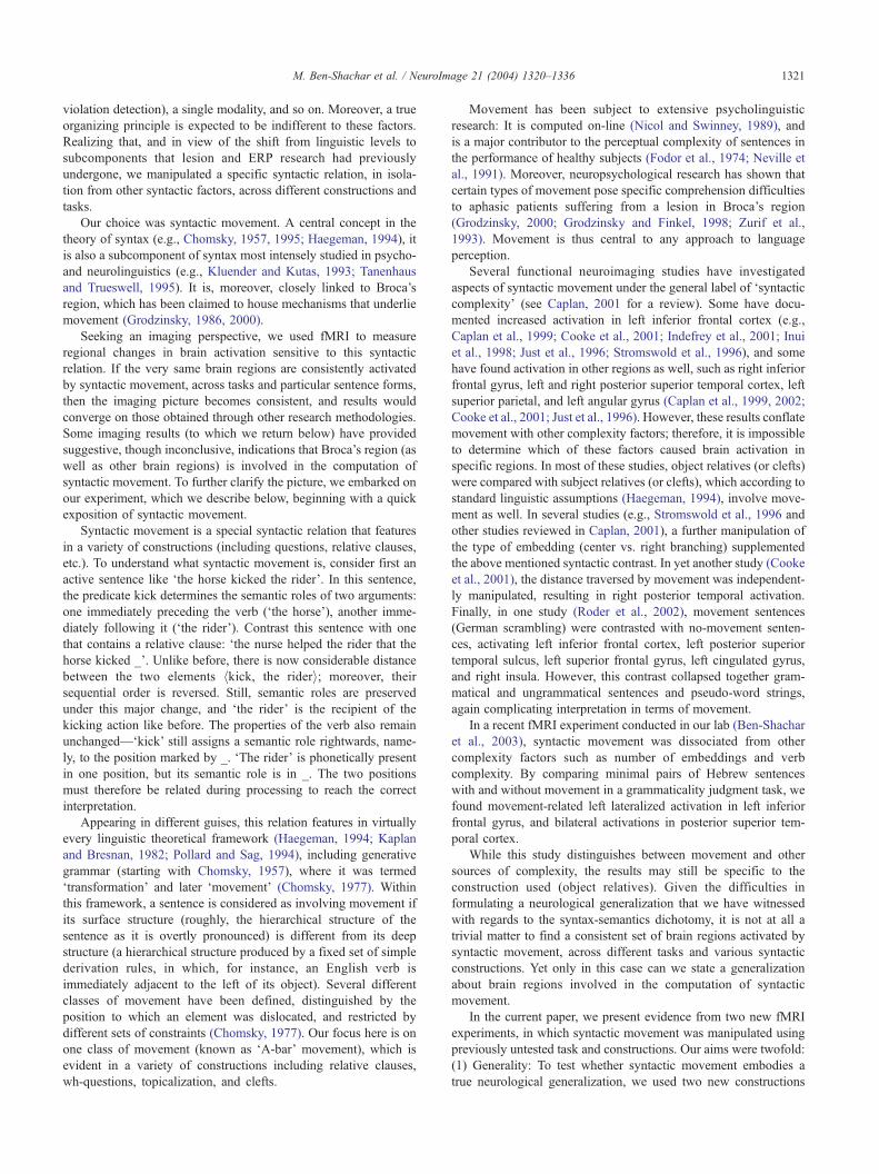
Neural correlates of syntactic movement: converging evidence from
two fMRI experiments
Michal Ben-Shachar,a,* Dafna Palti,a,b and Yosef Grodzinskya,c
aDepartment of Psychology, Tel Aviv University, Tel Aviv 69978, IsraelbWohl Institute for Advanced Imaging, Sourasky Medical Center, Tel Aviv, IsraelcDepartment of Linguistics, McGill University, Montreal, Quebec, Canada H3A-1A7
Received 10 June 2003; revised 6 November 2003; accepted 21 November 2003
This paper studies neural processes of sentence comprehension,
focusing on a specific syntactic operation—syntactic movement. We
describe two fMRI experiments that manipulate this particular
syntactic component. The sentences in each of the experiments are
different, yet the structural contrast in both is syntactically identical,
comparing movement and no-movement sentences. Two distinct
Hebrew constructions, topicalization and wh-questions, were presented
in an auditory comprehension task and compared to carefully matched
baseline sentences. We show that both contrasts, presented in an
auditory comprehension task, yield comparable activations in a
consistent set of brain regions, including left inferior frontal gyrus
(IFG), left ventral precentral sulcus (vPCS), and bilateral posterior
superior temporal sulcus (pSTS). Furthermore, we show that these
regions are not sensitive to two other syntactic contrasts. The results,
considered in the context of previous imaging and lesion studies,
suggest that the processing of syntactic movement involves a consistent
set of brain regions, regardless of the superficial properties of the
sentences at issue, and irrespective of task.
D 2004 Elsevier Inc. All rights reserved.
Keywords: fMRI; Syntactic movement; Topicalization
Introduction
This papers investigates the neural substrate of syntactic pro-
cessing—a focus of much research in current cognitive neurosci-
ence. There is dense lesion-based body of data about it, a host of
ERP studies, and more recently, a growing number of neuroimaging
studies. The lesion literature suggests that—contrary to traditional
views (e.g., Zurif, 1980)—gross distinctions between linguistic
levels (e.g., syntax, semantics) do not correspond to cerebral loci
(Broca’s or Wernicke’s region, respectively) in any obvious way
(Grodzinsky, 2000). The language regions of the brain seem rather
to be making finer functional distinctions. In particular, there are
1053-8119/$ - see front matter D 2004 Elsevier Inc. All rights reserved.
doi:10.1016/j.neuroimage.2003.11.027
* Corresponding author. Department of Psychology, Jordan Hall
Building 420, Stanford University, Stanford, CA, 94305-2130.
Fax: +1-360-483-1936.
E-mail address: [email protected] (M. Ben-Shachar).
Available online on ScienceDirect (www.sciencedirect.com.)
certain components of syntax that appear to be localizable (e.g.,
Grodzinsky, 1986, 1995; Neville et al., 1991; Stromswold et al.,
1996; Zurif et al., 1993), and they will be the focus of this paper.
Within the imaging literature, important series of studies have
aligned with the more traditional view: With few exceptions (to
which we will return below), most studies have concentrated on the
cerebral substrate of ‘syntax’ as compared to ‘semantics’. A survey
of these returns mixed, somewhat inconsistent results: In some
studies, syntactic conditions have activated both Broca’s and
Wernicke’s regions (Dapretto and Bookheimer, 1999; Embick et
al., 2000; Friederici et al., 2000; Keller et al., 2001; Luke et al.,
2002; Roder et al., 2002). In other studies, syntactic conditions
activated Wernicke’s but not Broca’s region (Kuperberg et al.,
2000; Vanderberghe et al., 2002). In one study, syntax activated
Broca’s but not Wernicke’s region; but then Broca’s region was
also activated by the semantic condition (Kang et al., 1999); in
another study, Broca’s region was activated more by syntax then by
semantics (Dapretto and Bookheimer, 1999); but this pattern was
not found in other studies directly comparing syntax with seman-
tics (Kuperberg et al., 2000, 2003; Luke et al., 2002; Newman et
al., 2001; Ni et al., 2000).
When syntax and semantics were crossed, an interaction
between the two was found in several regions, including Broca’s
region (Keller et al., 2001; Roder et al., 2002), left cingulate (Roder
et al., 2002), left posterior middle frontal gyrus, and left inferior
parietal cortex (Keller et al., 2001).
Finally, regions beyond those traditionally known to neuropsy-
chology as language areas also appear to be involved in syntactic
processing, including the right homologue of Broca’s region
(Embick et al., 2000; Friederici et al., 2000; Luke et al., 2002;
Moro et al., 2001; Ni et al., 2000 (Exp. 1)) and the right homologue
of Wernicke’s region (Friederici et al., 2000; Kuperberg et al.,
2000; Luke et al., 2002; Ni et al., 2000). In a recent study
(Kuperberg et al., 2003), Broca’s and Wernicke’s regions were
activated by conceptual and not by syntactic violations, the latter
activating bilateral inferior parietal lobule, bilateral parieto occip-
ital cortex, right middle frontal and precentral gyri, and other
regions in the right hemisphere.
Little anatomical consistency is found, then, when all syntactic
processing is lumped together and contrasted with semantics. This
is so even when the analysis is restricted to a single task (e.g.,
M. Ben-Shachar et al. / NeuroImage 21 (2004) 1320–1336 1321
violation detection), a single modality, and so on. Moreover, a true
organizing principle is expected to be indifferent to these factors.
Realizing that, and in view of the shift from linguistic levels to
subcomponents that lesion and ERP research had previously
undergone, we manipulated a specific syntactic relation, in isola-
tion from other syntactic factors, across different constructions and
tasks.
Our choice was syntactic movement. A central concept in the
theory of syntax (e.g., Chomsky, 1957, 1995; Haegeman, 1994), it
is also a subcomponent of syntax most intensely studied in psycho-
and neurolinguistics (e.g., Kluender and Kutas, 1993; Tanenhaus
and Trueswell, 1995). It is, moreover, closely linked to Broca’s
region, which has been claimed to house mechanisms that underlie
movement (Grodzinsky, 1986, 2000).
Seeking an imaging perspective, we used fMRI to measure
regional changes in brain activation sensitive to this syntactic
relation. If the very same brain regions are consistently activated
by syntactic movement, across tasks and particular sentence forms,
then the imaging picture becomes consistent, and results would
converge on those obtained through other research methodologies.
Some imaging results (to which we return below) have provided
suggestive, though inconclusive, indications that Broca’s region (as
well as other brain regions) is involved in the computation of
syntactic movement. To further clarify the picture, we embarked on
our experiment, which we describe below, beginning with a quick
exposition of syntactic movement.
Syntactic movement is a special syntactic relation that features
in a variety of constructions (including questions, relative clauses,
etc.). To understand what syntactic movement is, consider first an
active sentence like ‘the horse kicked the rider’. In this sentence,
the predicate kick determines the semantic roles of two arguments:
one immediately preceding the verb (‘the horse’), another imme-
diately following it (‘the rider’). Contrast this sentence with one
that contains a relative clause: ‘the nurse helped the rider that the
horse kicked _’. Unlike before, there is now considerable distance
between the two elements hkick, the rideri; moreover, their
sequential order is reversed. Still, semantic roles are preserved
under this major change, and ‘the rider’ is the recipient of the
kicking action like before. The properties of the verb also remain
unchanged—‘kick’ still assigns a semantic role rightwards, name-
ly, to the position marked by _. ‘The rider’ is phonetically present
in one position, but its semantic role is in _. The two positions
must therefore be related during processing to reach the correct
interpretation.
Appearing in different guises, this relation features in virtually
every linguistic theoretical framework (Haegeman, 1994; Kaplan
and Bresnan, 1982; Pollard and Sag, 1994), including generative
grammar (starting with Chomsky, 1957), where it was termed
‘transformation’ and later ‘movement’ (Chomsky, 1977). Within
this framework, a sentence is considered as involving movement if
its surface structure (roughly, the hierarchical structure of the
sentence as it is overtly pronounced) is different from its deep
structure (a hierarchical structure produced by a fixed set of simple
derivation rules, in which, for instance, an English verb is
immediately adjacent to the left of its object). Several different
classes of movement have been defined, distinguished by the
position to which an element was dislocated, and restricted by
different sets of constraints (Chomsky, 1977). Our focus here is on
one class of movement (known as ‘A-bar’ movement), which is
evident in a variety of constructions including relative clauses,
wh-questions, topicalization, and clefts.
Movement has been subject to extensive psycholinguistic
research: It is computed on-line (Nicol and Swinney, 1989), and
is a major contributor to the perceptual complexity of sentences in
the performance of healthy subjects (Fodor et al., 1974; Neville et
al., 1991). Moreover, neuropsychological research has shown that
certain types of movement pose specific comprehension difficulties
to aphasic patients suffering from a lesion in Broca’s region
(Grodzinsky, 2000; Grodzinsky and Finkel, 1998; Zurif et al.,
1993). Movement is thus central to any approach to language
perception.
Several functional neuroimaging studies have investigated
aspects of syntactic movement under the general label of ‘syntactic
complexity’ (see Caplan, 2001 for a review). Some have docu-
mented increased activation in left inferior frontal cortex (e.g.,
Caplan et al., 1999; Cooke et al., 2001; Indefrey et al., 2001; Inui
et al., 1998; Just et al., 1996; Stromswold et al., 1996), and some
have found activation in other regions as well, such as right inferior
frontal gyrus, left and right posterior superior temporal cortex, left
superior parietal, and left angular gyrus (Caplan et al., 1999, 2002;
Cooke et al., 2001; Just et al., 1996). However, these results conflate
movement with other complexity factors; therefore, it is impossible
to determine which of these factors caused brain activation in
specific regions. In most of these studies, object relatives (or clefts)
were compared with subject relatives (or clefts), which according to
standard linguistic assumptions (Haegeman, 1994), involve move-
ment as well. In several studies (e.g., Stromswold et al., 1996 and
other studies reviewed in Caplan, 2001), a further manipulation of
the type of embedding (center vs. right branching) supplemented
the above mentioned syntactic contrast. In yet another study (Cooke
et al., 2001), the distance traversed by movement was independent-
ly manipulated, resulting in right posterior temporal activation.
Finally, in one study (Roder et al., 2002), movement sentences
(German scrambling) were contrasted with no-movement senten-
ces, activating left inferior frontal cortex, left posterior superior
temporal sulcus, left superior frontal gyrus, left cingulated gyrus,
and right insula. However, this contrast collapsed together gram-
matical and ungrammatical sentences and pseudo-word strings,
again complicating interpretation in terms of movement.
In a recent fMRI experiment conducted in our lab (Ben-Shachar
et al., 2003), syntactic movement was dissociated from other
complexity factors such as number of embeddings and verb
complexity. By comparing minimal pairs of Hebrew sentences
with and without movement in a grammaticality judgment task, we
found movement-related left lateralized activation in left inferior
frontal gyrus, and bilateral activations in posterior superior tem-
poral cortex.
While this study distinguishes between movement and other
sources of complexity, the results may still be specific to the
construction used (object relatives). Given the difficulties in
formulating a neurological generalization that we have witnessed
with regards to the syntax-semantics dichotomy, it is not at all a
trivial matter to find a consistent set of brain regions activated by
syntactic movement, across different tasks and various syntactic
constructions. Yet only in this case can we state a generalization
about brain regions involved in the computation of syntactic
movement.
In the current paper, we present evidence from two new fMRI
experiments, in which syntactic movement was manipulated using
previously untested task and constructions. Our aims were twofold:
(1) Generality: To test whether syntactic movement embodies a
true neurological generalization, we used two new constructions
M. Ben-Shachar et al. / NeuroImage 21 (2004) 1320–13361322
that involve movement: topicalization and wh-questions, and
presented them in an original comprehension paradigm. (2)
Restrictedness: To distinguish between movement and other syn-
tactic effects, we included within each experiment an additional
syntactic contrast. In Experiment 1, the effect of topicalization was
compared with a change in the order of the objects. In Experiment
2, the effect of wh-questions was contrasted with the effect of
object versus subject questions. As we will show, our movement-
sensitive regions were insensitive to these syntactic contrasts.
Thus, our movement effects can be related to movement in
particular rather than to syntax in general.
3 It could be argued that using both conditions A and B as our baseline
for topicalization could diminish our effect, if indeed any of them involves
movement, and if this type of movement is processed by the same brain
Experiment 1
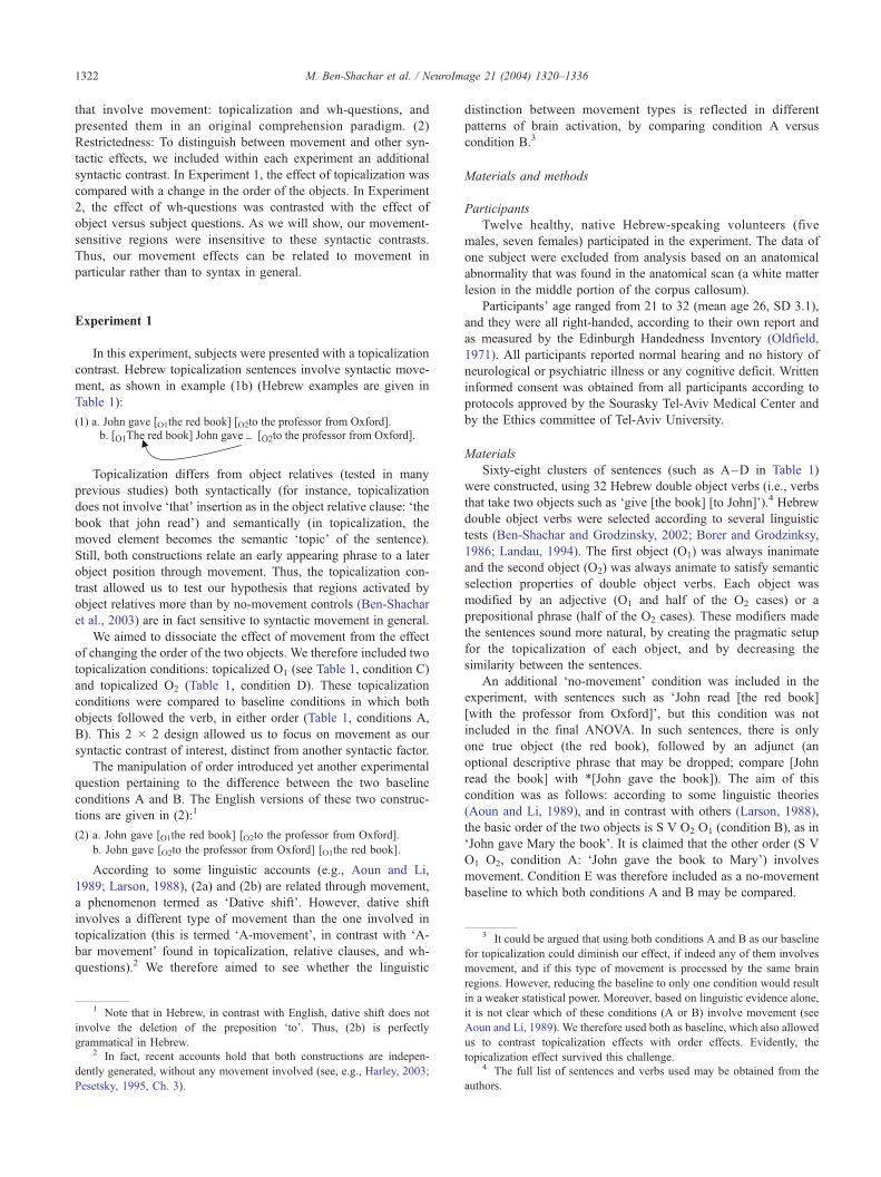
In this experiment, subjects were presented with a topicalization
contrast. Hebrew topicalization sentences involve syntactic move-
ment, as shown in example (1b) (Hebrew examples are given in
Table 1):
(1) a. John gave [O1the red book] [O2to the professor from Oxford].
Topicalization differs from object relatives (tested in many
previous studies) both syntactically (for instance, topicalization
does not involve ‘that’ insertion as in the object relative clause: ‘the
book that john read’) and semantically (in topicalization, the
moved element becomes the semantic ‘topic’ of the sentence).
Still, both constructions relate an early appearing phrase to a later
object position through movement. Thus, the topicalization con-
trast allowed us to test our hypothesis that regions activated by
object relatives more than by no-movement controls (Ben-Shachar
et al., 2003) are in fact sensitive to syntactic movement in general.
We aimed to dissociate the effect of movement from the effect
of changing the order of the two objects. We therefore included two
topicalization conditions: topicalized O1 (see Table 1, condition C)
and topicalized O2 (Table 1, condition D). These topicalization
conditions were compared to baseline conditions in which both
objects followed the verb, in either order (Table 1, conditions A,
B). This 2 � 2 design allowed us to focus on movement as our
syntactic contrast of interest, distinct from another syntactic factor.
The manipulation of order introduced yet another experimental
question pertaining to the difference between the two baseline
conditions A and B. The English versions of these two construc-
tions are given in (2):1
(2) a. John gave [O1the red book] [O2to the professor from Oxford].
b. John gave [O2to the professor from Oxford] [O1the red book].
According to some linguistic accounts (e.g., Aoun and Li,
1989; Larson, 1988), (2a) and (2b) are related through movement,
a phenomenon termed as ‘Dative shift’. However, dative shift
involves a different type of movement than the one involved in
topicalization (this is termed ‘A-movement’, in contrast with ‘A-
bar movement’ found in topicalization, relative clauses, and wh-
questions).2 We therefore aimed to see whether the linguistic
1 Note that in Hebrew, in contrast with English, dative shift does not
involve the deletion of the preposition ‘to’. Thus, (2b) is perfectly
grammatical in Hebrew.2 In fact, recent accounts hold that both constructions are indepen-
dently generated, without any movement involved (see, e.g., Harley, 2003;
Pesetsky, 1995, Ch. 3).
distinction between movement types is reflected in different
patterns of brain activation, by comparing condition A versus
condition B.3
Materials and methods
Participants
Twelve healthy, native Hebrew-speaking volunteers (five
males, seven females) participated in the experiment. The data of
one subject were excluded from analysis based on an anatomical
abnormality that was found in the anatomical scan (a white matter
lesion in the middle portion of the corpus callosum).
Participants’ age ranged from 21 to 32 (mean age 26, SD 3.1),
and they were all right-handed, according to their own report and
as measured by the Edinburgh Handedness Inventory (Oldfield,
1971). All participants reported normal hearing and no history of
neurological or psychiatric illness or any cognitive deficit. Written
informed consent was obtained from all participants according to
protocols approved by the Sourasky Tel-Aviv Medical Center and
by the Ethics committee of Tel-Aviv University.
Materials
Sixty-eight clusters of sentences (such as A–D in Table 1)
were constructed, using 32 Hebrew double object verbs (i.e., verbs
that take two objects such as ‘give [the book] [to John]’).4 Hebrew
double object verbs were selected according to several linguistic
tests (Ben-Shachar and Grodzinsky, 2002; Borer and Grodzinksy,
1986; Landau, 1994). The first object (O1) was always inanimate
and the second object (O2) was always animate to satisfy semantic
selection properties of double object verbs. Each object was
modified by an adjective (O1 and half of the O2 cases) or a
prepositional phrase (half of the O2 cases). These modifiers made
the sentences sound more natural, by creating the pragmatic setup
for the topicalization of each object, and by decreasing the
similarity between the sentences.
An additional ‘no-movement’ condition was included in the
experiment, with sentences such as ‘John read [the red book]
[with the professor from Oxford]’, but this condition was not
included in the final ANOVA. In such sentences, there is only
one true object (the red book), followed by an adjunct (an
optional descriptive phrase that may be dropped; compare [John
read the book] with *[John gave the book]). The aim of this
condition was as follows: according to some linguistic theories
(Aoun and Li, 1989), and in contrast with others (Larson, 1988),
the basic order of the two objects is S V O2 O1 (condition B), as in
‘John gave Mary the book’. It is claimed that the other order (S V
O1 O2, condition A: ‘John gave the book to Mary’) involves
movement. Condition E was therefore included as a no-movement
baseline to which both conditions A and B may be compared.
regions. However, reducing the baseline to only one condition would result
in a weaker statistical power. Moreover, based on linguistic evidence alone,
it is not clear which of these conditions (A or B) involve movement (see
Aoun and Li, 1989). We therefore used both as baseline, which also allowed
us to contrast topicalization effects with order effects. Evidently, the
topicalization effect survived this challenge.4 The full list of sentences and verbs used may be obtained from the
authors.
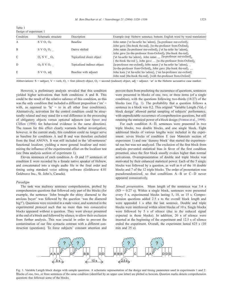
Table 1
Design of experiment 1
Condition Schematic structure Description Example (top: Hebrew sentence; bottom: English word by word translation)
A S V O1 O2 Baseline John natan [‘et ha-sefer ha-’adom]1 [la-professor me-oxford]2John gave [the-book the-red]1 [to-the-professor from-Oxford]2
B S V O2 O1 _ Dative shifted John natan [la-professor me-oxford]2 [‘et ha-sefer ha-’adom]1John gave [to-the-professor from-Oxford]2 [the-book the-red]1
C O1 S V _ O2 Topicalized direct object [‘et ha-sefer ha-’adom]1 John natan __ [la-professor me-oxford]2
[ the-book the-red ]1 John gave __ [to-the-professor from-Oxford]2D O2 S V O1 _ Topicalized indirect object [la-professor me-oxford]2 John natan [‘et ha-sefer ha-’adom]1 _
[to-the-professor from-Oxford]2 John gave [the-book the-red]1 __
E S V O1 adj Baseline with adjunct John kara [‘et ha-sefer ha-’adom]1 [‘im ha-professor me-oxford]
John read [the-book the-red]1 [with the-professor from-Oxford]
Abbreviations: S = subject, V = verb, O1 = first (direct) object, O2 = second (indirect) object, adj = adjunct. ‘et’ is the Hebrew accusative case marker.
M. Ben-Shachar et al. / NeuroImage 21 (2004) 1320–1336 1323
However, a preliminary analysis revealed that this condition
yielded higher activations than both conditions A and B. This
could be the result of the relative salience of this condition, as this
was the only condition that included a different preposition (‘im’ =
with, as opposed to ‘le’ = to in all other four conditions).
Alternatively, activation for the control condition could be struc-
turally related and may stand for a real difference in the processing
of obligatory objects versus optional adjuncts (see Speer and
Clifton (1998) for behavioral evidence in the same direction).
The reason for this effect clearly warrants further investigation;
however, in the current study, this condition could no longer serve
as baseline for conditions A and B and was therefore excluded
from the final ANOVA. It was still included in the ‘all-sentences’
functional localizer, yielding a more general localizer and mini-
mizing the influence of the experimental effect on the localizer test
(see Data analysis section of experiment 1).
Eleven sentences of each condition A–D and 17 sentences of
condition E were recorded by a female native speaker of Hebrew,
and concatenated into a single audio file in the final order and
timing using standard voice editing software (Goldwave 4.01
Goldwave Inc., St. John’s, Canada).
Paradigm
The task was auditory sentence comprehension, probed by
comprehension questions that followed only part of the blocks (for
example, the sentence ‘John brought the shiny diamond to the
anxious buyer’ was followed by the question ‘was the diamond
big?’). Questions were recorded in a male voice, and scattered in the
experimental protocol such that no more than two consecutive
blocks appeared without a question. They were always presented
at the end of a block and followed by silence, to allow their exclusion
from further analysis. This was crucial in order to prevent the
contamination of our fine syntactic contrast with a different con-
struction (questions). To force subjects’ constant attention and
Fig. 1. Variable Length block design with sample questions. A schematic represe
Blocks of one, two, or three sentences of the same condition (identified by an upp
questions that followed some of the blocks.
prevent them from predicting the occurrence of questions, sentences
were presented in blocks of one, two, or three items (of a single
condition), with the questions following two-thirds (18/27) of the
blocks (see Fig. 1). The probability that a question follows a
sentence in a block was 0.2. This original ‘Variable Length (VaL-)
block design’ allowed partial sampling of subjects’ performance,
with unpredictable occurrence of comprehension questions, but still
retaining the statistical power of a block design (Friston et al., 1999).
For each condition A–D, sentences were presented in two
triple blocks, two double blocks, and one single block. Eight
additional blocks of various lengths were included in the exper-
iment: seven blocks of condition E (see Materials section of
experiment 1) and one ‘dummy block’ that started the experimen-
tal run but was not analyzed. The exclusion of the first block from
analysis prevented statistical bias in favor of the first condition
presented, since the first block usually evokes higher than normal
activations. Overrepresentation of double and triple blocks was
motivated by their enhanced statistical power. Each of the 5 single
blocks was followed by a question, as well as 6 of the 10 double
blocks and 7 of the 12 triple blocks. The order of presentation was
pseudorandomized, so that conditions A–B or C–D never
appeared consecutively.
Stimuli presentation. Mean length of the sentences was 3.4 s
(SD = 0.27 s). Within a single block, sentences were presented
every 5 s, experimental blocks lasting 5, 10, or 15 s. Compre-
hension questions added 2.5 s to the overall block length and
were appended 1 s after the last sentence. Double and triple
blocks were interleaved within silent blocks of 10 s. Single blocks
were followed by 5 s of silence (due to the reduced signal
expected in those blocks). In addition, 20 s of silence were
inserted at the beginning of the experiment and 12.5 s of silence
ended the experiment. Overall, the experiment lasted 625 s (10
min and 25 s).
ntation of the design and timing parameters used in experiments 1 and 2.
er case letter) are plotted as boxcars. Question marks denote comprehension
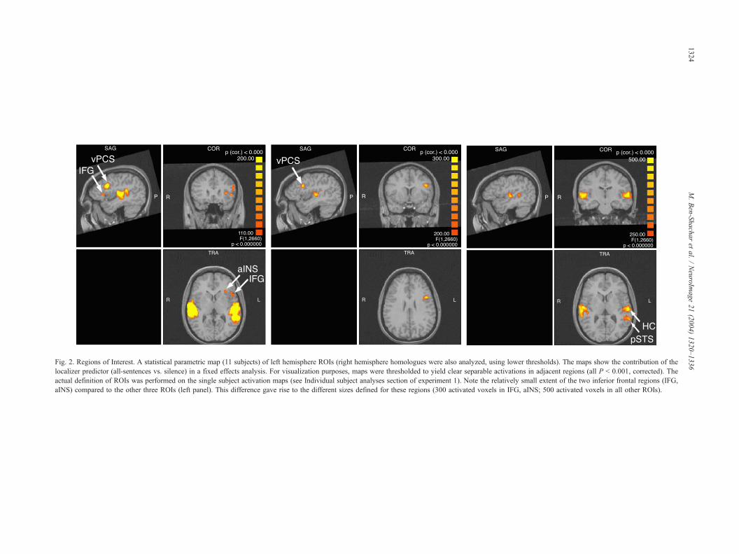
Fig. 2. Regions of Interest. A statistical parametric map (11 subjects) of left hemisphere ROIs (right hemisphere homologues were also analyzed, using lower thresholds). The maps show the contribution of the
localizer predictor (all-sentences vs. silence) in a fixed effects analysis. For visualization purposes, maps were thresholded to yield clear separable activations in adjacent regions (all P < 0.001, corrected). The
actual definition of ROIs was performed on the single subject activation maps (see Individual subject analyses section of experiment 1). Note the relatively small extent of the two inferior frontal regions (IFG,
aINS) compared to the other three ROIs (left panel). This difference gave rise to the different sizes defined for these regions (300 activated voxels in IFG, aINS; 500 activated voxels in all other ROIs).
M.Ben-Shacharet
al./NeuroIm
age21(2004)1320–1336
1324
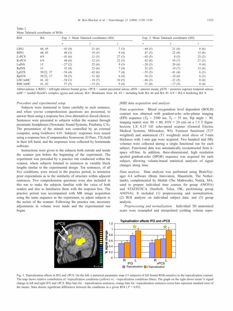
Table 2
Mean Talairach coordinates of ROIs
ROI BA Exp. 1: Mean Talairach coordinates (SD) Exp. 2: Mean Talairach coordinates (SD)
x y z x y z
LIFG 44, 45 �43 (4) 21 (6) 7 (3) �44 (5) 21 (4) 8 (6)
RIFG 44, 45 48 (5) 19 (5) 9 (4) 47 (5) 22 (9) 12 (8)
LvPCS 6/9 �41 (6) 11 (5) 27 (3) �45 (5) 8 (5) 25 (2)
RvPCS 6/9 44 (6) 12 (5) 32 (5) 42 (8) 10 (7) 27 (3)
LaINS 13 �27 (2) 22 (4) 9 (3) �28 (3) 20 (6) 9 (4)
RaINS 13 32 (4) 23 (6) 7 (4) 32 (2) 19 (7) 12 (6)
LpSTS 39/22, 37 �56 (4) �42 (6) 7 (4) �55 (5) �41 (4) 6 (3)
RpSTS 39/22, 37 58 (5) �31 (8) 6 (4) 56 (5) �34 (6) 6 (3)
LHC/mHC 41, 42 �54 (3) �18 (7) 10 (5) �46 (3) �21 (5) 8 (4)
RHC/mHC 41, 42 57 (5) �15 (5) 9 (4) 51 (4) �17 (5) 8 (4)
Abbreviations: L/RIFG = left/right inferior frontal gyrus; vPCS = ventral precentral sulcus; aINS = anterior insula; pSTS = posterior superior temporal sulcus;
mHC = medial Heschl’s complex (gyrus and sulcus). BA= Brodmann Area: 44, 45 = including both BA 44 and BA 45. 6/9 = BA 6 bordering BA 9.
M. Ben-Shachar et al. / NeuroImage 21 (2004) 1320–1336 1325
Procedure and experimental setup
Subjects were instructed to listen carefully to each sentence,
and when yes/no comprehension questions are presented, to
answer them using a response box (two alternatives forced choice).
Sentences were presented to subjects within the scanner through
pneumatic headphones (Newmatic Sound Systems, Petaluma, CA).
The presentation of the stimuli was controlled by an external
computer, using Goldwave 4.01. Subjects’ responses were issued
using a response box (Compumedics Neuroscan, El Paso, TX) held
in their left hand, and the responses were collected by homemade
software.
Instructions were given to the subjects both outside and inside
the scanner just before the beginning of the experiment. The
experiment was preceded by a practice run conducted within the
scanner, where subjects listened to sentences in variable block
lengths similar to the experimental design. Ten sentences, of all
five conditions, were mixed in the practice period, to minimize
prior expectations as to the similarity of structure within adjacent
sentences. Two comprehension questions were also included in
this run to make the subjects familiar with the voice of both
readers and also to familiarize them with the response box. The
practice period was accompanied with MR image acquisition
using the same sequence as the experiment, to adjust subjects to
the noises of the scanner. Following the practice run, necessary
adjustments in volume were made and the experimental run
began.
Fig. 3. Topicalization effects in IFG and vPCS. On the left, a statistical parametric
The map shows relative contribution of +topicalization conditions (yellow) vs. �to
change in left and right IFG and vPCS. Blue bars for �topicalization sentences, ora
the mean). Stars denote significant differences between the conditions in a given
fMRI data acquisition and analysis
Data acquisition. Blood oxygenation level dependent (BOLD)
contrast was obtained with gradient-echo echo-planar imaging
(EPI) sequence (TR = 2500 ms, TE = 55 ms, flip angle = 90,
imaging matrix size: 80 � 80, FOV = 24 cm) on a 1.5 T Signa-
horizon LX 8.25 GE echo-speed scanner (General Electric
Medical Systems, Milwaukee, WI). Fourteen functional (T2*
weighted) and anatomical (T1 weighted) axial slices of 5-mm
thickness with 1-mm gap were acquired. Two hundred and fifty
volumes were collected during a single functional run for each
subject. Functional data was automatically reconstructed from k-
space off-line. In addition, three-dimensional, high resolution
spoiled gradient-echo (SPGR) sequence was acquired for each
subject, allowing volume-based statistical analyses of signal
changes along time.
Data analysis. Data analysis was performed using BrainVoy-
ager 4.4 software (Brain Innovation, Maastricht, The Nether-
lands), complemented by Matlab (The Mathworks, Natick, MA;
used to prepare individual time courses for group ANOVA)
and STATISTICA (StatSoft, Tulsa, OK; performing group
ANOVA). It included (1) preprocessing and normalization;
(2) ROI analysis on individual subject data; and (3) group
analysis.
Preprocessing and normalization. Individual 3D anatomical
scans were resampled and interpolated yielding volume repre-
map (11 subjects) of left frontal ROIs sensitive to the topicalization contrast.
picalization conditions (blue). The graph on the right shows mean % signal
nge bars for +topicalization sentences (error bars represent standard error of
ROI ( P < 0.05).
5 Naturally, by using a functional localizer we may be missing relevant
brain regions that are involved in movement but are not activated by our
localizer. In particular, we are prone to miss brain regions that are activated
in rest as well as in sentence comprehension (we thank a NI reviewer for
pointing this out). Unfortunately, this issue may not be resolved in the
current experiment.6 This definition proved highly robust: the same shifts also maximized
two other related measures reflecting amount of ‘localizer’ activation: (a)
One-sample t test over percent signal changes in activation blocks; (b) The
integral of the average activation function.
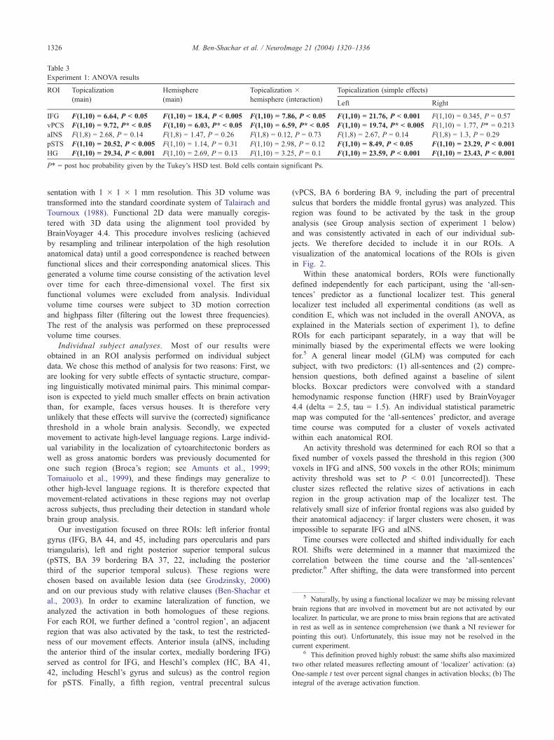
Table 3
Experiment 1: ANOVA results
ROI Topicalization Hemisphere Topicalization � Topicalization (simple effects)
(main) (main) hemisphere (interaction)Left Right
IFG F(1,10) = 6.64, P < 0.05 F(1,10) = 18.4, P < 0.005 F(1,10) = 7.86, P < 0.05 F(1,10) = 21.76, P < 0.001 F(1,10) = 0.345, P = 0.57
vPCS F(1,10) = 9.72, P* < 0.05 F(1,10) = 6.03, P* < 0.05 F(1,10) = 6.59, P* < 0.05 F(1,10) = 19.74, P* < 0.005 F(1,10) = 1.77, P* = 0.213
aINS F(1,8) = 2.68, P = 0.14 F(1,8) = 1.47, P = 0.26 F(1,8) = 0.12, P = 0.73 F(1,8) = 2.67, P = 0.14 F(1,8) = 1.3, P = 0.29
pSTS F(1,10) = 20.52, P < 0.005 F(1,10) = 1.14, P = 0.31 F(1,10) = 2.98, P = 0.12 F(1,10) = 8.49, P < 0.05 F(1,10) = 23.29, P < 0.001
HG F(1,10) = 29.34, P < 0.001 F(1,10) = 2.69, P = 0.13 F(1,10) = 3.25, P = 0.1 F(1,10) = 23.59, P < 0.001 F(1,10) = 23.43, P < 0.001
P* = post hoc probability given by the Tukey’s HSD test. Bold cells contain significant Ps.
M. Ben-Shachar et al. / NeuroImage 21 (2004) 1320–13361326
sentation with 1 � 1 � 1 mm resolution. This 3D volume was
transformed into the standard coordinate system of Talairach and
Tournoux (1988). Functional 2D data were manually coregis-
tered with 3D data using the alignment tool provided by
BrainVoyager 4.4. This procedure involves reslicing (achieved
by resampling and trilinear interpolation of the high resolution
anatomical data) until a good correspondence is reached between
functional slices and their corresponding anatomical slices. This
generated a volume time course consisting of the activation level
over time for each three-dimensional voxel. The first six
functional volumes were excluded from analysis. Individual
volume time courses were subject to 3D motion correction
and highpass filter (filtering out the lowest three frequencies).
The rest of the analysis was performed on these preprocessed
volume time courses.
Individual subject analyses. Most of our results were
obtained in an ROI analysis performed on individual subject
data. We chose this method of analysis for two reasons: First, we
are looking for very subtle effects of syntactic structure, compar-
ing linguistically motivated minimal pairs. This minimal compar-
ison is expected to yield much smaller effects on brain activation
than, for example, faces versus houses. It is therefore very
unlikely that these effects will survive the (corrected) significance
threshold in a whole brain analysis. Secondly, we expected
movement to activate high-level language regions. Large individ-
ual variability in the localization of cytoarchitectonic borders as
well as gross anatomic borders was previously documented for
one such region (Broca’s region; see Amunts et al., 1999;
Tomaiuolo et al., 1999), and these findings may generalize to
other high-level language regions. It is therefore expected that
movement-related activations in these regions may not overlap
across subjects, thus precluding their detection in standard whole
brain group analysis.
Our investigation focused on three ROIs: left inferior frontal
gyrus (IFG, BA 44, and 45, including pars opercularis and pars
triangularis), left and right posterior superior temporal sulcus
(pSTS, BA 39 bordering BA 37, 22, including the posterior
third of the superior temporal sulcus). These regions were
chosen based on available lesion data (see Grodzinsky, 2000)
and on our previous study with relative clauses (Ben-Shachar et
al., 2003). In order to examine lateralization of function, we
analyzed the activation in both homologues of these regions.
For each ROI, we further defined a ‘control region’, an adjacent
region that was also activated by the task, to test the restricted-
ness of our movement effects. Anterior insula (aINS, including
the anterior third of the insular cortex, medially bordering IFG)
served as control for IFG, and Heschl’s complex (HC, BA 41,
42, including Heschl’s gyrus and sulcus) as the control region
for pSTS. Finally, a fifth region, ventral precentral sulcus
(vPCS, BA 6 bordering BA 9, including the part of precentral
sulcus that borders the middle frontal gyrus) was analyzed. This
region was found to be activated by the task in the group
analysis (see Group analysis section of experiment 1 below)
and was consistently activated in each of our individual sub-
jects. We therefore decided to include it in our ROIs. A
visualization of the anatomical locations of the ROIs is given
in Fig. 2.
Within these anatomical borders, ROIs were functionally
defined independently for each participant, using the ‘all-sen-
tences’ predictor as a functional localizer test. This general
localizer test included all experimental conditions (as well as
condition E, which was not included in the overall ANOVA, as
explained in the Materials section of experiment 1), to define
ROIs for each participant separately, in a way that will be
minimally biased by the experimental effects we were looking
for.5 A general linear model (GLM) was computed for each
subject, with two predictors: (1) all-sentences and (2) compre-
hension questions, both defined against a baseline of silent
blocks. Boxcar predictors were convolved with a standard
hemodynamic response function (HRF) used by BrainVoyager
4.4 (delta = 2.5, tau = 1.5). An individual statistical parametric
map was computed for the ‘all-sentences’ predictor, and average
time course was computed for a cluster of voxels activated
within each anatomical ROI.
An activity threshold was determined for each ROI so that a
fixed number of voxels passed the threshold in this region (300
voxels in IFG and aINS, 500 voxels in the other ROIs; minimum
activity threshold was set to P < 0.01 [uncorrected]). These
cluster sizes reflected the relative sizes of activations in each
region in the group activation map of the localizer test. The
relatively small size of inferior frontal regions was also guided by
their anatomical adjacency: if larger clusters were chosen, it was
impossible to separate IFG and aINS.
Time courses were collected and shifted individually for each
ROI. Shifts were determined in a manner that maximized the
correlation between the time course and the ‘all-sentences’
predictor.6 After shifting, the data were transformed into percent
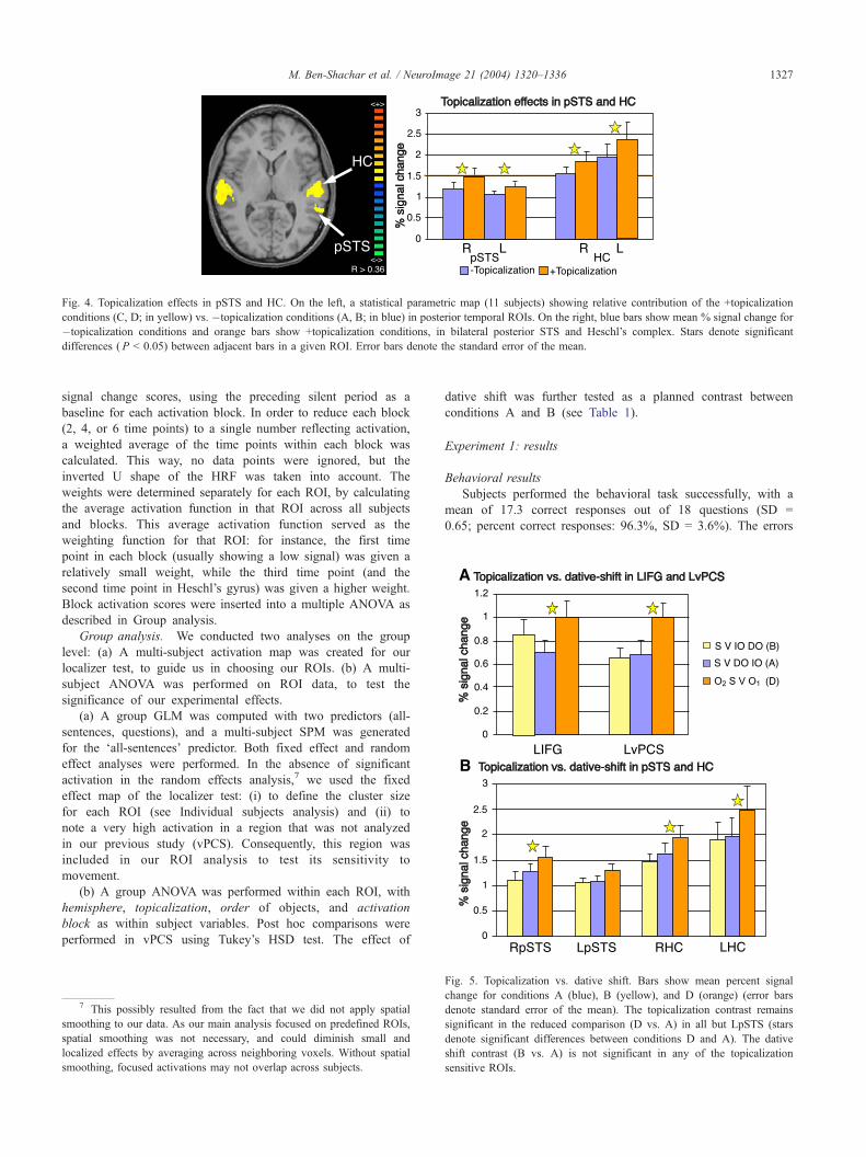
Fig. 4. Topicalization effects in pSTS and HC. On the left, a statistical parametric map (11 subjects) showing relative contribution of the +topicalization
conditions (C, D; in yellow) vs. �topicalization conditions (A, B; in blue) in posterior temporal ROIs. On the right, blue bars show mean % signal change for
�topicalization conditions and orange bars show +topicalization conditions, in bilateral posterior STS and Heschl’s complex. Stars denote significant
differences ( P < 0.05) between adjacent bars in a given ROI. Error bars denote the standard error of the mean.
M. Ben-Shachar et al. / NeuroImage 21 (2004) 1320–1336 1327
signal change scores, using the preceding silent period as a
baseline for each activation block. In order to reduce each block
(2, 4, or 6 time points) to a single number reflecting activation,
a weighted average of the time points within each block was
calculated. This way, no data points were ignored, but the
inverted U shape of the HRF was taken into account. The
weights were determined separately for each ROI, by calculating
the average activation function in that ROI across all subjects
and blocks. This average activation function served as the
weighting function for that ROI: for instance, the first time
point in each block (usually showing a low signal) was given a
relatively small weight, while the third time point (and the
second time point in Heschl’s gyrus) was given a higher weight.
Block activation scores were inserted into a multiple ANOVA as
described in Group analysis.
Group analysis. We conducted two analyses on the group
level: (a) A multi-subject activation map was created for our
localizer test, to guide us in choosing our ROIs. (b) A multi-
subject ANOVA was performed on ROI data, to test the
significance of our experimental effects.
(a) A group GLM was computed with two predictors (all-
sentences, questions), and a multi-subject SPM was generated
for the ‘all-sentences’ predictor. Both fixed effect and random
effect analyses were performed. In the absence of significant
activation in the random effects analysis,7 we used the fixed
effect map of the localizer test: (i) to define the cluster size
for each ROI (see Individual subjects analysis) and (ii) to
note a very high activation in a region that was not analyzed
in our previous study (vPCS). Consequently, this region was
included in our ROI analysis to test its sensitivity to
movement.
(b) A group ANOVA was performed within each ROI, with
hemisphere, topicalization, order of objects, and activation
block as within subject variables. Post hoc comparisons were
performed in vPCS using Tukey’s HSD test. The effect of
7 This possibly resulted from the fact that we did not apply spatial
smoothing to our data. As our main analysis focused on predefined ROIs,
spatial smoothing was not necessary, and could diminish small and
localized effects by averaging across neighboring voxels. Without spatial
smoothing, focused activations may not overlap across subjects.
dative shift was further tested as a planned contrast between
conditions A and B (see Table 1).
Experiment 1: results
Behavioral results
Subjects performed the behavioral task successfully, with a
mean of 17.3 correct responses out of 18 questions (SD =
0.65; percent correct responses: 96.3%, SD = 3.6%). The errors
Fig. 5. Topicalization vs. dative shift. Bars show mean percent signal
change for conditions A (blue), B (yellow), and D (orange) (error bars
denote standard error of the mean). The topicalization contrast remains
significant in the reduced comparison (D vs. A) in all but LpSTS (stars
denote significant differences between conditions D and A). The dative
shift contrast (B vs. A) is not significant in any of the topicalization
sensitive ROIs.
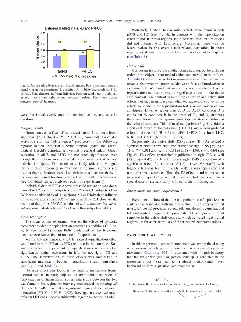
Fig. 6. Dative shift effects in right frontal regions. Bars show mean percent
signal change for experiment 1, condition A (in blue) and condition B (in
yellow). Stars denote significant difference between conditions in both right
anterior insula and right ventral precentral sulcus. Error bars denote
standard error of the mean.
M. Ben-Shachar et al. / NeuroImage 21 (2004) 1320–13361328
were distributed evenly and did not involve any one specific
question.
Imaging results
Group analysis: a fixed effect analysis on all 11 subjects found
significant [F(11,2640) = 25; P < 0.001, corrected] task-related
activation (for the ‘all-sentences’ predictor) in the following
regions: bilateral posterior superior temporal gyrus and sulcus,
bilateral Heschl’s complex, left ventral precentral sulcus. Group
activation in aINS and LIFG did not reach significance, even
though these regions were activated by the localizer test in most
individual subjects. This result most likely reflects low signal
levels in these regions (also reflected in the smaller cluster size
used in their definition), as well as high inter-subject variability in
the exact anatomical location of the activation within these regions
(see Individual subject analyses section of experiment 1).
Individual data in ROIs: Above threshold activation was docu-
mented in IFG in 10/11 subjects and in aINS in 9/11 subjects. Other
ROIs were activated by all 11 subjects. Mean Talairach coordinates
of the activations in each ROI are given in Table 2. Below are the
results of the group ANOVA conducted with topicalization, hemi-
sphere, order of objects, and block as within subject variables.
Movement effects
The focus of this experiment was on the effects of syntactic
movement evident in topicalization sentences (conditions C, D vs.
A, B; see Table 1) within ROIs predefined by the functional
localizer (see Materials and methods of experiment 1).
Within anterior regions, a left lateralized topicalization effect
was found in both IFG and vPCS (post hoc in the latter; see Data
analysis section of experiment 1): topicalization sentences evoked
significantly higher activations in left, but not right, IFG and
vPCS. The lateralization of these effects was manifested in
significant interactions between topicalization and hemisphere
(see Fig. 3 and Table 3).
No such effect was found in the anterior insula, our frontal
‘control region’ medially adjacent to IFG: neither an effect of
topicalization or hemisphere, nor an interaction between the two
was found in this region. An inter-regional analysis comparing left
IFG and left aINS yielded a significant region � topicalization
interaction [F(1,8) = 5.36, P < 0.05], showing that the topicalization
effect in LIFGwas indeed significantly larger than the one in LaINS.
Posteriorly, bilateral topicalization effects were found in both
pSTS and HC (see Fig. 4). In contrast with the topicalization
effect found in frontal regions, the posterior topicalization effects
did not interact with hemisphere. Moreover, there was no
lateralization in the overall task-related activation in these
regions, as shown in a nonsignificant main effect of hemisphere
(see Table 3).
Dative shift
Our design involved yet another contrast, given by the different
order of the objects in no-topicalization sentences (condition B vs.
A, Table 1), which may reflect movement of one object across the
other, a phenomenon known as ‘dative shift’ (see Introduction to
experiment 1). We found that none of the regions activated by the
topicalization contrast showed a significant effect for the dative
shift contrast. The contrast between topicalization and dative shift
effects persisted in most regions when we equated the power of the
effects by reducing the topicalization test to a comparison of two
conditions (D vs. A, rather than C, D vs. A, B; condition D is
equivalent to condition B in the order of O1 and O2 and was
therefore chosen as the representative topicalization condition in
the reduced contrast). This reduced comparison (Fig. 5) yielded a
significant effect of topicalization (D > A) and a nonsignificant
effect of dative shift (B > A) in LIFG, LvPCS (post hoc), LHC,
RHC, and RpSTS (but not in LpSTS).
Surprisingly, the dative shift (DS) contrast (B > A) yielded a
significant effect in two right frontal regions: right aINS [F(1,8) =
11.8, P < 0.01] and right vPCS [F(1,10) = 4.99, P = 0.049] (see
Fig. 6). This effect approached significance in right IFG as well
[F(1,10) = 4.47, P = 0.061]. Interestingly, RaINS also showed a
significant effect of linear order [F(1,8) = 16.84, P < 0.005], with
higher activations for the [O2, O1] order, across topicalized and
non-topicalized sentences. Thus, the DS effect found in this region
may not be specifically related to dative shift, but could be a
special case of the sensitivity to linear order in this region.
Intermediate summary—experiment 1
Experiment 1 showed that the comprehension of topicalization
sentences is associated with brain activation in left inferior frontal
gyrus, left ventral precentral sulcus, bilateral Heschl’s complex, and
bilateral posterior superior temporal sulci. These regions were not
sensitive to the dative shift contrast, which activated right frontal
regions—right anterior insula and right ventral precentral sulcus.
Experiment 2: wh-questions
In this experiment, syntactic movement was manipulated using
wh-questions, which are considered a classic case of syntactic
movement (Chomsky, 1973). It is assumed within linguistic theory
that the wh-phrase (such as [which tourist]) is generated in the
canonical position (e.g., subject or object position) and moves
backward to form a question (see example 3).
M. Ben-Shachar et al. / NeuroImage 21 (2004) 1320–1336 1329
Thus, wh-questions and topicalization sentences may be viewed
as instances of a single generalization, syntactic movement, even
though they differ in many other aspects (e.g., prosodic, semantic). If
the regions activated by topicalization indeed respond to syntactic
movement, they are expected to be activated by wh-questions as
well.
We presented Hebrew embedded questions of three types:
subject and object wh-questions (see Table 4, conditions B and
C), and yes/no questions as in (4) (Table 4, condition A):
(4) yes/no Q: the waiter asked if [the tourist ordered salad for lunch].
Note that the embedded yes/no question in (4) forms a
declarative sentence, so there is actually no movement involved.
Moreover, given that all question types were embedded within
declarative sentences, there was no difference in the type of
response triggered by each of these question conditions. Thus,
our movement contrast compared embedded wh-questions (subject
and object) with embedded yes/no questions.
Finally, we also compared between subject and object wh-
questions. Though both involve movement according to standard
linguistic theory (Haegeman, 1994), note that the subject wh-
questions we tested (Table 4, condition B) lack two main features
of object movement: the order of the subject, verb and object does
not change, and there are no words separating the wh-phrase from
its original (subject) position. By contrasting these two types of
questions, we aimed to examine whether this distinction is
reflected in the activation of movement-sensitive regions.
Materials and methods
Participants
Ten healthy volunteers (three males, seven females) participated
in experiment 2. Four of them took part in experiment 1 as well,
but this experiment was run in separate sessions, a year after
experiment 1 took place. Participants’ age ranged from 21 to 30
(mean age, 25.9; standard deviation, 3.1). The selection criteria and
protocol were the same as in experiment 1.
Materials
Sixty clusters of sentences were constructed (see examples in
Table 4). We used embedded questions because they allow a
straightforward comprehension task (such as the one used in
experiment 1), and yield a clean comparison with no-movement
questions. Simple wh-phrases (e.g., ‘which tourist’ rather than
‘which fat tourist’) were used in all conditions. The NP in the
embedded clause (the embedded object in condition A–B, the
embedded subject in condition C) was modified by a single
adjective.8
In sentence construction, five Hebrew verbs that take embed-
ded questions as their complements were used: sha’al (asked),
badak (checked), berer (found out), shaxax (forgot), hit’anyen
(was interested to know). Each verb repeated three times in all
conditions. All verbs and embedded questions were in past tense.
The referential nouns (waiter, tourist) were not repeated through-
out the experiment—only one version of each cluster was pre-
sented in the experiment. For each condition, 15 sentences of
8 The adjectives in condition C were shorter to compensate for the
extra syllables introduced by the determiners (‘ha-’ = the) in the embedded
subject.
different clusters were chosen, such that the mean length of the
sentences in each condition was identical (eight words, average
length = 21.4 syllables).
Sentences were recorded by a female native speaker of
Hebrew, and processed as in experiment 1. Thirteen comprehen-
sion questions were composed, referring either to the adjective,
the verb, the embedded subject, or the object. A couple of
representative sentence–question pairs are given in (5):(5) a. The boxer asked if the athlete received an honorable prize in the
ceremony.
Question: did the boxer receive a prize?
b. The artist checked which dealer purchased plastic paint in Sweden
Question: did the dealer buy oil paint?
c. The banker found out which stocks the heavy investors bought in the
stockmarket.
Question: did the banker find out about the stocks?
Questions were recorded in a male voice and interleaved in the
experimental protocol as in experiment 1.
Paradigm
The task and experimental paradigm were the same as in
experiment 1.
For each condition, 15 sentences were presented in 2 triple
blocks, 4 double blocks, and 1 single block. The overrepresentation
of double blocks was motivated by the relatively high signal
documented in experiment 1 for these blocks, and by timing
considerations. A dummy block of two sentences was used as in
experiment 1. Comprehension questions followed 13 out of 22
blocks.
Stimuli presentation: Mean length of the sentences was 3.6 s
(SD = 0.2). Within each block, sentences were presented every 5 s.
Blocks were separated by silent blocks of 12.5 s each. Compre-
hension questions added 5 s to the overall block length, appended 1
s after the last sentence. In addition, 30 s of silence were inserted at
the beginning of the experiment and 17.5 s of silence ended the
experiment.
Overall, the experiment lasted 610 s (10 min and 10 s).
Procedure, experimental setup, data acquisition
The same as in experiment 1.
Data analysis
Data analysis procedures were identical to those used in
experiment 1, except for the following sections.
Localizer test. The use of a functional localizer test was
adopted in experiment 2 as well. However, in this case, we
did not use the all-sentences predictor as a localizer test. This is
because two of our three conditions included wh-questions,
which could have biased the localizer in favor of our contrast
of interest (movement vs. no-movement). We therefore defined a
localizer test that included only two of our experimental con-
ditions—yes/no questions and subject questions (Table 4, con-
ditions A, B). Subject questions were chosen since they do not
involve a filler-gap distance, which could bias the localizer in yet
another direction.
The localizer predictor was constructed as a boxcar with zeros
in all silent blocks, and 1 s in blocks of conditions A and B.
Separate predictors were defined for condition C, the dummy
block, and comprehension questions. As in experiment 1, all
predictors were convolved with a standard HRF model (with
delta = 2.5, tau = 1.25).
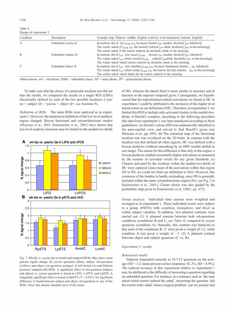
Table 4
Design of experiment 2
Condition Description Example (top: Hebrew; middle: English word by word translation; bottom: English)
A Embedded yes/no Q ha-meltzar sha’al ‘im [EMB [NP ha-tayar] hizmin [NP mashke alcoholi] [PP baboker]]
The waiter asked if [EMB [NP the-tourist] ordered [NP drink alcoholic] [PP in-the-morning]]
The waiter asked if the tourist ordered an alcoholic drink in the morning
B Embedded subject Q ha-meltzar sha’al [wh ‘eize tayar] [EMB _ hizmin [NP mashke alcoholi] [PP baboker]]
The waiter asked [wh which tourist] [EMB _ ordered [NPdrink alcoholic] [PP in-the-morning]]
The waiter asked which tourist ordered an alcoholic drink in the morning
C Embedded object Q ha-meltzar sha’al [wh ‘eize mashke] [EMB [NP ha-tayar hashamen] hizmin _ [PP baboker]]
The waiter asked [wh which drink] [EMB [NP the-tourist the-fat] ordered _ [PP in-the-morning]]
The waiter asked which drink the fat tourist ordered in the morning
Abbreviations: wh = wh-phrase, EMB = embedded clause, NP = noun phrase, PP = prepositional phrase.
M. Ben-Shachar et al. / NeuroImage 21 (2004) 1320–13361330
To make sure that the choice of a particular localizer test did not
bias the results, we compared the results in a single ROI (LIFG)
functionally defined by each of the two possible localizers (<yes/
no + subject Q>, <yes/no + object Q>; see footnote 9).
Definition of ROIs. The same ROIs were analyzed as in experi-
ment 1. However, the anatomical definition of the low-level auditory
region changed. Recent functional and cytoarchitectonic studies
(Morosan et al., 2001; Rademacher et al., 2001) have shown that
low level auditory functions may be limited to the medial two-thirds
Fig. 7. Wh-Qs vs. yes/no Qs in frontal and temporal ROIs. Bars show mean
percent signal change for yes/no questions (blue), subject wh-questions
(yellow), and object wh-questions (orange), in left frontal (A) and bilateral
posterior temporal (B) ROIs. A significant effect of wh-questions (subject
and object) vs. yes/no questions is found in LIFG, LvPCS, and LpSTS. A
marginally significant effect is found in RpSTS ( P = 0.051). No significant
difference is found between subject and object wh-questions in any of the
ROIs. Error bars denote standard error of the mean.
of HG, whereas the lateral third is more similar in structure and in
function to the superior temporal gyrus. Consequently, we hypoth-
esized that the topicalization-related activations we found in HC in
experiment 1 could be attributed to the inclusion of this higher level
lateral cortex in our definition of HC. Therefore, in experiment 2, we
limited this ROI to include only activated clusters in the medial two-
thirds of Heschl’s complex, according to the following procedure
(the data from experiment 1 was later reanalyzed according to these
guidelines): (a) Heschl’s sulcus (HS) was anatomically identified in
the para-sagittal view, and served to find Heschl’s gyrus (see
Morosan et al., pp. 695). (b) The statistical map of the functional
localizer test was overlayed on the 3D brain. In contrast with the
localizer test that defined all other regions, HC was defined with a
boxcar predictor (without smoothing by an HRF model) shifted in
one image. The reason for this difference is that only in this region, a
boxcar predictor yielded consistently higher activations as measured
by the number of activated voxels for any given threshold. (c)
Clusters activated by the localizer within the medial two-thirds of
HC were captured (since most of the activations within this region
fell in HS, we could not limit our definition to HG). However, this
extension of the borders is hardly misleading, since HS is generally
included within the same cytoarchitectonic region (Te1; see Fig. 2 in
Rademacher et al., 2001). Cluster choice was also guided by the
probability map given in Rademacher et al. (2001; pp. 675).
Group analysis. Individual time courses were weighted and
averaged as in experiment 1. These individual scores were subject
to a group ANOVA with condition, hemisphere, and block as
within subject variables. In addition, two planned contrasts were
carried out: (1) A planned contrast between both wh-questions
conditions (conditions B and C; see Table 4) compared to yes/no
questions (condition A). Naturally, this contrast was balanced so
that each of the conditions B–C were given a weight of 1/2, while
condition A was given a weight of �1. (2) A planned contrast
between object and subject questions (C vs. B).
Experiment 2: results
Behavioral results
Subjects responded correctly to 10.7/13 questions on the aver-
age (SD = 1.2; mean percent correct responses: 82.3%, SD = 8.9%).
The reduced accuracy in this experiment relative to experiment 1
may be attributed to the difficulty of answering a question regarding
an embedded question. For instance, in a sentence such as ‘the man
asked which tourist ordered the salad’, answering the question ‘did
the tourist order salad’ raises a logical problem: can we assume that
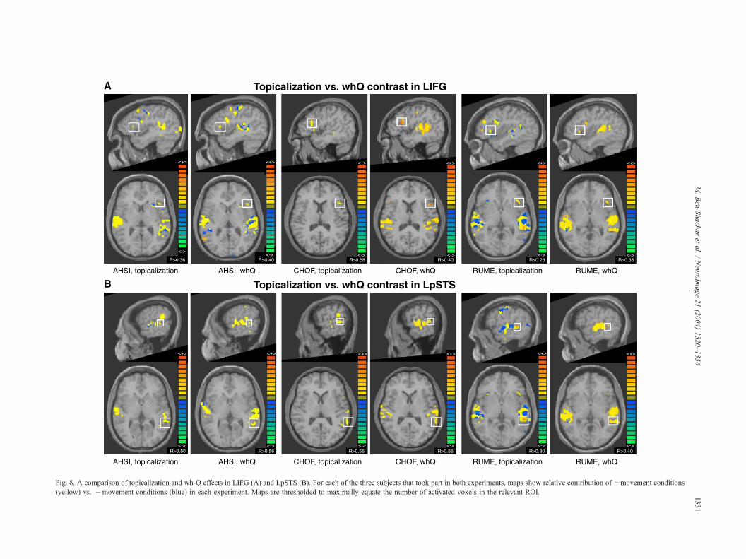
Fig. 8. A comparison of topicalization and wh-Q effects in LIFG (A) and LpSTS (B). For each of the three subjects that took part in both experiments, maps show relative contribution of +movement conditions
(yellow) vs. �movement conditions (blue) in each experiment. Maps are thresholded to maximally equate the number of activated voxels in the relevant ROI.
M.Ben-Shacharet
al./NeuroIm
age21(2004)1320–1336
1331
M. Ben-Shachar et al. / NeuroImage 21 (2004) 1320–13361332
the tourist indeed ordered salad simply by the fact that someone
asked about it? Such logical conflicts probably contributed to the
lower levels of performance in this experiment.
Imaging results
Group analysis. A fixed effects analysis on all 10 subjects found
significant [F(1,2366) = 31; P < 0.001, corrected] task-related
activation (for the functional localizer) in the following regions:
bilateral posterior superior temporal gyrus and sulcus, bilateral
Heschl’s complex, left IFG, bilateral vPCS, bilateral aINS.
Individual data in ROIs. Above threshold activation was docu-
mented in IFG in 9/10 subjects, and in aINS in 8/10 subjects. Other
ROIs were activated by all 10 subjects. Mean Talairach coordinates
of the activations in each ROI are given in Table 2. Below are the
results of a group ANOVA and planned comparisons (see Data
analysis section of experiment 2).
Movement effect
The focus of this experiment was on the effects of syntactic
movement evident in wh-questions [conditions (B, C) vs. (A)]
within ROIs predefined by the functional localizer (see Materials
and methods of experiment 2).
Within anterior regions, wh-questions yielded significantly
stronger activations than yes/no questions in both left IFG
[F(1,8) = 9.85, P < 0.015] and left vPCS [F(1,9) = 6.62, P <
0.05] (see Fig. 7A). This effect was neither significant in the right
homologues of these regions nor in bilateral aINS [right IFG:
F(1,8) = 1.32, P = 0.284; right vPCS: F(1,9) = 2.33, P = 0.16; left
aINS: F(1,7) = 3.42, P = 0.11; right aINS: F(1,7) = 3.16, P = 0.12).
No main effect of hemisphere was found in either of these regions
(IFG: F(1,8) = 1.22, P = 0.3; vPCS: F(1,9) = 0.24, P = 0.63; aINS:
F(1,7) = 0.98, P = 0.36).9
In posterior regions, a significant effect of wh-questions relative
to yes/no questions was found in left pSTS [F(1,9) = 10.7, P <
0.01] and a marginally significant effect was found in right pSTS
[F(1,9) = 5.06, P = 0.051] (see Fig. 7B).
No such effect was found in mHC [left: F(1,9) = 1.71, P = 0.22;
right: F(1,9) = 2.5, P = 0.15]. No main effect of hemisphere was
found in posterior regions (pSTS: F(1,9) = 2.29, P = 0.16; HC:
F(1,9) = 0.04, P = 0.85).
Reanalysis of mHC data from experiment 1
In view of the above findings in mHC, we hypothesized that
limiting the definition of this region to the medial two thirds of
Heschl’s complex would eliminate the movement effect that we
found in this region in experiment 1. We therefore conducted a
reanalysis of HC data from experiment 1, following the same
anatomical guidelines that where employed in experiment 2 (see
Definition of ROIs section of experiment 2). We found a nonsig-
nificant effect of topicalization in mHC [main effect: F(1,10) = 3.38,
P= 0.1; left: F(1,10) = 1.18, P= 0.3; right: F(1,10) = 4.13, P= 0.07].
9 The activations in LIFG were also captured with a different localizer
test (based on object Qs and yes/no Qs). The results showed a similar effect
of wh-questions compared to yes/no questions [F(1,8) = 10.27, P < 0.015].
The individual time courses captured with each localizer were highly
correlated (mean correlation coefficient r = 0.98, SD = 0.04). In general,
individual GLM maps produced by both localizers showed considerable
overlap. Thus, it is hardly likely that the results were systematically
influenced by the choice of one localizer over the other.
Object versus subject questions
Object questions did not yield higher activation than subject
questions in any of the ROIs (P > 0.1; see Fig. 7). To further test
this effect, we compared condition C (object questions) with
conditions B and A together (subject and yes/no questions). Here
too, there were no significant effects in any of the ROIs analyzed
(P > 0.1).10
Results summary—wh-questions
Wh-questions compared to yes/no questions yielded a stronger
fMRI signal in left IFG, left vPCS and left pSTS, and a marginally
significant effect in right pSTS. Other regions, including right IFG
and vPCS, bilateral anterior insula and bilateral HC, were not
sensitive to the experimental contrast. Object wh-questions did not
show significantly stronger activation than subject wh-questions in
any of the ROIs.
A comparison between the two experiments
Fig. 8 compares the activation maps acquired for the wh-Q
contrast and for the topicalization contrast in three subjects who
performed both experiments. Activation is compared in two ROIs:
LIFG and LpSTS. This individual comparison shows very similar
voxels activated by these two different contrasts in each individual,
regardless of the different syntactic constructions used. The figure
also demonstrates between-subject variability in the exact anatom-
ical focus of activation within ROIs.
General discussion
We have shown a consistent pattern of activation for two cases
of syntactic movement: topicalization-sentences and embedded
wh-questions. Both activated left inferior frontal gyrus, left ventral
precentral sulcus, and bilateral posterior superior temporal sulcus.
These regions were sensitive neither to the subject object contrast
(experiment 2) nor to the dative shift contrast (experiment 1),
which in turn activated right frontal regions. Other task-related
regions, such as anterior insula and medial Heschl’s gyrus, were
not sensitive to the movement contrasts.
A neurolinguistic generalization
The fMRI activations associated with topicalization and wh-
questions are consistent with those found in yet another case of
syntactic movement—object relatives tested earlier in our lab (Ben-
Shachar et al., 2003). These three experiments, though differing in
task, materials, and design, all manipulated syntactic movement. In
all three, left inferior frontal gyrus was activated, as well as bilateral
posterior superior temporal cortex. In all three, the left inferior
activation was dissociable from the left anterior insular cortex, and
10 It could be argued that this null effect (object vs. subject wh-
questions) reflects low statistical power. However, note that in both LIFG
and LpSTS, the other two simple comparisons (subject wh-Qs vs. yes/no
Qs, object wh-Qs vs. yes/no Qs) were significant [LIFG: F(1,8) = 7.26 and
5.51, respectively, P < 0.05; LpSTS: F(1,9) = 10.64 and 8.69, respectively,
P < 0.05]. Thus, the design seems powerful enough to detect simple effects
between single conditions.
M. Ben-Shachar et al. / NeuroImage 21 (2004) 1320–1336 1333
the bilateral posterior temporal activations were dissociable from
medial Heschl’s complex, both representing neighboring regions
activated by the task but indifferent to the linguistic contrast. Taken
together, the combined results of these studies suggest that syntactic
movement constitutes a neurally relevant linguistic generalization,
processed by this consistent set of brain regions.
Our results converge on a previous ERP study (Kluender and
Kutas, 1993) that compared English object wh-questions (‘what
have you forgotten . . .’) with yes/no questions (‘have you forgotten. . .’). The authors report: ‘ANOVAs restricted to individual electro-des thus showed main effects of sentence type only for Broca’s
area [.. P < 0.001; right hemisphere homologue of Broca’s ..
nonsignificant], left temporal regions (T5) [..; P < 0.001], and right
temporal regions (T6) [..; P < 0.001]’. (pp. 199). These results,
while lacking the anatomical precision provided by fMRI, supply
converging evidence to our conclusion that cuts across imaging
technologies, syntactic constructions, task and language.
With regard to lesion studies, the results are in agreement with
many findings demonstrating agrammatic (Broca’s) aphasics’
selective comprehension difficulties in various constructions in-
volving movement (reviewed in Grodzinsky, 2000). Our results
supply finer anatomical characterization of the regions involved
in the computation of movement, distinguishing, for example,
between Left IFG and Left anterior insula with respect to their
sensitivity to movement. However, based solely on our fMRI
results, we cannot determine which of the activated brain regions
is indeed critical for processing movement sentences. Results
from lesion studies make a strong case for the critical role of
Broca’s region in this respect.
As for Wernicke’s region, the evidence from lesion studies is
mixed (Grodzinsky and Finkel, 1998; Swinney et al., 1996). Our
findings, showing bilateral activations in a part of Wernicke’s
region (pSTS, see Figs. 4 and 7B), suggest that a more homoge-
neous deficit in the processing of movement may show up in
patients with bilateral posterior temporal damage. Further behav-
ioral studies with patients may also clarify the different roles
sustained by each of these homologues in the processing of
movement sentences.
The exact division of labor between the regions activated by
movement cannot be specified based on this study alone. One
interesting interpretation comes from the study of control process-
es in memory, where it was suggested that frontal regions are
engaged in selecting the appropriate representation while exclud-
ing context inappropriate ones (Buckner, 2003; Thompson-Schill
et al., 1997). These anterior regions maintain interactions with
posterior regions in temporal and parietal cortex that may serve as
storage sites (Buckner, 2003). In this context, activation in left
frontal regions could be related to the reactivation of the moved
element in an appropriate sentential position, whereas posterior
temporal activations could reflect maintenance of the moved
element in memory.
Task-related and construction-specific activation
Some differences should be noted between the current experi-
ments and our previous study of object relatives. First, the
activation of the left vPCS (see Figs. 3 and 7A) was not recorded
previously for the movement contrast. In fact, this region was not
analyzed in our previous study since it was not activated by the
functional localizer (grammaticality judgment on neutral senten-
ces) in all subjects. This suggests that the activation of this region
may be task-related, and given the appropriate activating task
(comprehension), its sensitivity to movement is evident.
The activations we found in LvPCS relate to two separate
lines of research. Within the imaging literature concerning mem-
ory, LvPCS (termed as pLIPC, anterior portion) showed relative
activation in phonologically related encoding tasks (Gold and
Buckner, 2002; Poldrack et al., 1999), as well as in semantic
tasks on single words (Wagner et al., 1998). In these studies, too,
LvPCS usually coactivated with LIFG (termed aLIPC). In our
movement sentences, LvPCS may have been involved in search-
ing for a semantically appropriate element to be linked, while
LIFG was performing a more syntactically guided search for this
element. This hypothesis can be tested by manipulating syntactic
and semantic plausibility of association orthogonally during
functional imaging.
Secondly, LvPCS activation fell within the caudal ventral pre-
motor cortex (see Picard and Strick, 2001; Rizzolatti et al., 2002).
There is some preliminary evidence to suggest that this region may
correspond to monkey area F4 (Rizzolatti et al., 2002), but the
functional homology between monkey F4 and human ventral
premotor is controversial (Grezes and Decety, 2001; Picard and
Strick, 2001). It is hard to see at this point how the activation
documented for our fine syntactic contrasts in the left vPCS could
be related to the motor planning functions attributed to monkey F4
(cf. Rizzolatti et al., 2002). However, attempts have been made to
relate monkey ‘mirror neurons’ in the adjacent F5 to language
functions in human BA 44 (Rizzolatti and Arbib, 1998). In the
future, this link between high motor functions in the monkey and
specific language functions in human may be further pursued in
caudal ventral premotor.
Another difference between the current study and our previous
study of object relatives pertains to the activation of HC by the
topicalization contrast (Fig. 4). This region was not sensitive to
movement in our previous study, and was not expected to show up
here due to its known lower level functions. One possible reason
for its activation by topicalization is stress changes that take place
in topicalized sentences (reflecting focus changes, see Introduction
to experiment 1).
Another possible explanation for the topicalization effect in
HC is that its anatomical delineation was not fine enough. Indeed,
when this region was carefully defined using better anatomical
guidelines published recently by Rademacher et al. (2001), there
was no movement-related activation found there for both wh-
questions and topicalization (see the reanalysis of HC data in
Experiment 2: results section). Thus, we suggest that the defini-
tion of HC in experiment 1 included parts that are functionally
related to STG, a higher level auditory region that might be
involved in movement analysis or in stress changes characteristic
of topicalization. Better functional discrimination is needed be-
tween these two regions to address this issue more precisely.
Linguistic distinctions in movement-sensitive regions
Having shown that several regions are activated by syntactic
movement, it is no less important to ask to which syntactic
contrasts these regions are not sensitive. An important result of
the current study is that regions activated by topicalization did
not show a comparable effect for dative shift (Fig. 5), which
involves a different class of movement (if any, see footnote 2).
Furthermore, regions activated by embedded wh-questions were
not sensitive to the subject–object contrast (Fig. 7). These results
M. Ben-Shachar et al. / NeuroImage 21 (2004) 1320–13361334
underline the selectivity of these regions and show that within the
syntactic realm, their activation cannot be attributed to just any
deviation from the canonical word order to which the listener
expects. The current evidence thus allows us to restrict our
neurolinguistic generalization to (A-bar) syntactic movement
rather than to syntax as a whole.
Dative shift effects
In contrast with topicalization and wh-questions, the dative shift
contrast activated right frontal regions. This effect is important for
two reasons: first, it shows that our manipulation was strong enough
to trigger activation in some part of the brain, and therefore supports
a true distinction between dative shift and topicalization in left
inferior frontal and bilateral posterior superior temporal regions.
Secondly, it supports a dichotomous distinction between movement
types, as opposed to a parametric measure of the amount of
movement. A parametric view would predict increased activation
in movement-sensitive regions that correlates with increased
amount of movement (no-movement < dative shift < topicalization).
Instead, we found that A-bar movement contrasts and the dative
shift contrast split, giving rise to different patterns of brain activa-
tion. This is in agreement with a linguistically based approach that
views each of these phenomena as belonging to different classes of
movement (Larson, 1988).
The activation of right frontal regions in itself is hard to
interpret, as little is known about these regions from neuro-
imaging and lesion studies. Still, it generates an interesting
prediction regarding aphasic patients’ ability to comprehend such
sentences. In particular, we would expect Broca’s and Wernicke’s
aphasics to process sentences with dative shift correctly, and right
hemisphere frontal patients to show a different pattern of behav-
ior. These predictions are not easily tested, however, due to the
irreversible nature of the semantic roles of the two objects,
among other things (but see Caplan and Futter, 1986 for one
such attempt). Further evidence from English and Hebrew speak-
ing aphasics with well-localized lesions may shed more light on
this issue.
Subject versus object questions
The fact that the subject–object contrast in wh-questions did
not turn out significant in any of the analyzed ROIs contrasts
with neuroimaging findings in English relative clauses and clefts
(reviewed in Caplan, 2001). These studies showed higher activa-
tion in Broca’s region (and sometimes in other regions as well,
e.g., Just et al., 1996) for object relatives and clefts compared to
subject relatives and clefts. The reasons for this contradiction
remain unclear. Note, however, that subject –object contrasts
failed to yield significant activations in these ROIs (and partic-
ularly in Broca’s region) in several other studies as well,
including Fiebach et al. (2001; contrasting subject vs. object
wh-questions), Indefrey et al. (2001; contrasting all-subject vs.
subject/object relative clauses), Cooke et al. (2001), and Caplan
et al. (2002; contrasting subject and object relatives). Some
possible reasons for this variability are given in Caplan et al.
(2002, pp. 36–37).
We suggest that both subject and object movement constructions
are processed in movement-sensitive regions. This could only be
seen if an appropriate no-movement baseline is used. Further
contrasts between object and subject movement may stem from
the difference in word order and/or in the distance between the
moved element and its original position (see Cooke et al., 2001, for
a clean manipulation of this variable). These may activate a partially
overlapping group of brain regions, as some of the movement-
sensitive regions are in fact involved in holding the moved element
in working memory, and therefore would work harder when the
distance is greater. The critical distance required for these effects is
likely to be influenced by the specific language and the task used
(see Baddeley, 1997, Chap. 4). Since our study was not oriented
toward this question, it is very likely that our contrast between
object and subject questions did not involve the required distance to
result in a significant effect.
Our interpretation is further corroborated by psycholinguistic
and lesion data showing that (a) healthy subjects show movement-
related (so-called ‘gap-filling’) effects in both object and subject
relative clauses (Balogh et al., 1998; Nicol and Swinney, 1989);
and (b) Broca’s agrammatic aphasics (Swinney et al., 1996; Zurif et
al., 1993) fail to show this effect in both subject and object relative
clauses. These findings suggest that Broca’s region is involved in
processing syntactic movement in both subject and object relative
clauses.
Finally, other types of movements were characterized within
linguistic theory, such as A-movement (as in passive construc-
tions) and head-movement (as in: ‘are you listening?’). A
neuropsychological dissociation has been documented between
‘head-movement’ and other types of movement (Grodzinsky and
Finkel, 1998), but not between A-movement and A-bar-move-
ment (evident in relative clauses, clefts, topicalization, and wh-
questions). fMRI may be more sensitive to this latter distinction,
since it usually finds activations in a wider set of brain regions
that contribute to a given cognitive process, and since it allows
the detection of gradual changes in activity. For instance, we may
witness different patterns of fMRI activation for A- versus A-bar
movement, or a relative difference in the amount of activation
triggered by these types of movement in a specific brain region.
Future findings of this kind will better our understanding of the
relation between fine linguistic distinctions and language process-
ing in the brain.
Acknowledgments
The study was supported by a grant from the Israel–U.S.A. Bi-
national Science Foundation (BSF 1997-0451).
We thank Talma Hendler and Gal Chechik for their help in all
stages of this work, and Galia Avidan for her help in recording the
sentences.
References
Amunts, K., Schleicher, A., Burgel, U., Mohlberg, H., Uylings, H.B.M.,
Zilles, K., 1999. Broca’s region revisited: cytoarchitecture and intersub-
ject variability. Journal of Comparative Neurology 412, 319–341.
Aoun, J., Li, A., 1989. Scope and constituency. Linguistic Inquiry 20,
141–172.
Baddeley, A., 1997. Human Memory: Theory and Practice. Psychology
Press, Hove, pp. 49–69.
Balogh, J., Zurif, E., Prather, P., Swinney, D., Finkel, L., 1998. Gap-filling
and end-of-sentence effects in real-time language processing: implica-
tions for modeling sentence comprehension in aphasia. Brain and Lan-
guage 61, 169–182.
Ben-Shachar, M., Grodzinsky, Y., 2002. On the derivation of Hebrew double
M. Ben-Shachar et al. / NeuroImage 21 (2004) 1320–1336 1335
objects—A functional imaging investigation. Poster Presented in the
North Eastern Linguistic Society Meeting (NELS 33), Boston, MA.
Ben-Shachar, M., Hendler, T., Kahn, I., Ben-Bashat, D., Grodzinsky, Y.,
2003. The neural reality of syntactic transformations—Evidence from
fMRI. Psychological Science 14, 433–440.
Borer, H., Grodzinksy, Y., 1986. Syntactic cliticization and lexical clitici-
zation: the case of Hebrew dative clitics. In: Borer, H. (Ed.), The Syntax
of Pronominal Clitics. Syntax and Semantics, vol. 19. Academic Press,
New York, pp. 175–217.
Buckner, R.L., 2003. Functional-anatomic correlates of control processes in
memory. Journal of Neuroscience 23, 3999–4004.
Caplan, D., 2001. Functional neuroimaging studies of syntactic processing.
Journal of Psycholinguistic Research 30, 297–320.
Caplan, D., Futter, C., 1986. Assignment of thematic roles by an agram-
matic aphasic patient. Brain and Language 27, 117–135.
Caplan, D., Alpert, N., Waters, G., 1999. PET studies of sentence process-
ing with auditory sentence presentation. NeuroImage 9, 343–351.
Caplan, D., Vijayan, S., Kuperberg, G., West, C., Waters, G., Greve, D.,
Dale, A.M., 2002. Vascular responses to syntactic processing: event-
related fMRI study of relative clauses. Human Brain Mapping 15,
26–38.
Chomsky, N., 1957. Syntactic Structures. Mouton, The Hague.
Chomsky, N., 1973. Conditions on transformations. In: Anderson, S.,
Kiparsky, P. (Eds.), A Festschrift for Morris Halle. Holt, Rinehart and
Winston, New York, pp. 232–286.
Chomsky, N., 1977. On wh-movement. In: Culicover, P., Wasow, T.,
Akmajian, A. (Eds.), Formal Syntax. Academic Press, San Diego,
pp. 71–132.
Chomsky, N., 1995. The Minimalist Program. MIT Press, Cambridge.
Cooke, A., Zurif, E.B., DeVita, C., Alsop, D., Koenig, P., Detre, J., Gee,
J.C., Pinango, M., Balogh, J., Grossman, M., 2001. Neural basis for
sentence comprehension: grammatical and short term memory compo-
nents. Human Brain Mapping 15, 80–94.
Dapretto, M., Bookheimer, S.Y., 1999. Form and content: dissociating
syntax and semantics in sentence comprehension. Neuron 24, 427–432.
Embick, D., Marantz, A., Miyashita, Y., O’Neil, W., Sakai, K.L., 2000.
A syntactic specialization for Broca’s area. Proceedings of the Na-
tional Academy of Sciences of the United States of America 97,
6150–6154.
Fiebach, C.J., Schlesewsky, M., Friederici, A.D., 2001. Syntactic working
memory and the establishment of filler-gap dependencies: insights from
ERPs and fMRI. Journal of Psycholinguistic Research 30, 321–338.
Fodor, J.A., Bever, T., Garrett, M.F., 1974. The Psychology of Language: An
Introduction to Psycholinguistics and Generative Grammar. McGraw-
Hill, New York.
Friederici, A.D., Meyer, M., von Cramon, D.Y., 2000. Auditory language
comprehension: an event-related fMRI study on the processing of syn-
tactic and lexical information. Brain and Language 74, 289–300.
Friston, K.J., Zarahn, E., Josephs, O., Henson, R.N., Dale, A.M., 1999.
Stochastic designs in event-related fMRI. NeuroImage 10, 607–619.
Gold, B.T., Buckner, R.L., 2002. Common prefrontal regions co-activate
with dissociable posterior regions during controlled semantic and pho-
nological tasks. Neuron 35, 803–812.
Grezes, J., Decety, J., 2001. Functional anatomy of execution, mental
simulation, observation and verb generation of actions: a meta analysis.
Human Brain Mapping 12, 1–19.
Grodzinsky, Y., 1986. Language deficits and the theory of syntax. Brain
and Language 27, 135–159.
Grodzinsky, Y., 1995. A restrictive theory of trace deletion in agramma-
tism. Brain and Language 51, 26–51.
Grodzinsky, Y., 2000. The neurology of syntax: language use without
Broca’s area. Behavioral and Brain Sciences 23, 1–21.
Grodzinsky, Y., Finkel, L., 1998. The neurology of empty categories:
aphasics’ failure to detect ungrammaticality. Journal of Cognitive Neu-
roscience 10, 281–292.
Haegeman, L., 1994. Introduction to Government and Binding Theory,
Third ed. Blackwell, Oxford.
Harley, H., 2003. Possession and the double object construction. Yearbook
of Linguistic Variation 2, 29–68.
Indefrey, P., Hagoort, P., Herzog, H., Seitz, J., Brown, C.M., 2001. Syn-
tactic processing in left prefrontal cortex is independent of lexical mean-
ing. NeuroImage 14, 546–555.
Inui, T., Otsu, Y., Tanaka, S., Okada, T., Nishizawa, S., Konishi, J., 1998. A
functional MRI analysis of comprehension processes of Japanese sen-
tences. NeuroReport 9, 3325–3328.
Just, M.A., Carpenter, P.A., Keller, T.A., Eddy, W.F., Thulborn, K.R., 1996.
Brain activation modulated by sentence comprehension. Science 274,
114–116.
Kang, A.M., Constable, R.T., Gore, J.C., Avrutin, S., 1999. An event-
related fMRI study of implicit phrase-level syntactic and semantic pro-
cessing. NeuroImage 10, 555–561.
Kaplan, R.M., Bresnan, J., 1982. Lexical functional grammar: a formal
system for grammatical representation. In: Bresnan, J. (Ed.), The Men-
tal Representation of Grammatical Relations. MIT Press, Cambridge,
pp. 173–281.
Keller, T.A., Carpenter, P.A., Just, M.A., 2001. The neural bases of sen-
tence comprehension: a fMRI examination of syntactic and lexical pro-
cessing. Cerebral Cortex 11, 223–237.
Kluender, R., Kutas, M., 1993. Bridging the gap: evidence from ERPs on
the processing of unbounded dependencies. Journal of Cognitive Neu-
roscience 5, 196–214.
Kuperberg, G.R., McGuire, P.K., Bullmore, E.T., Brammer, M.J., Rabe-
Hesketh, S., Wright, I.C., Lythgoe, D.J., Williams, S.C.R., David,
A.S., 2000. Common and distinct neural substrate for pragmatic, se-
mantic, and syntactic processing of spoken sentences: an fMRI study.
Journal of Cognitive Neuroscience 12, 321–341.
Kuperberg, G.R., Holcomb, P.J., Sitnikova, T., Greve, D., Dale, A.M.,
Caplan, D., 2003. Distinct patterns of neural modulation during the
processing of conceptual and syntactic anomalies. Journal of Cognitive
Neuroscience 15, 272–293.
Landau, I., 1994. Dative shift and extended VP-shells. MA Thesis, Tel-
Aviv University.
Larson, R.K., 1988. On the double object construction. Linguistic Inquiry
19, 335–391.
Luke, K.-K., Liu, H.-L., Wai, Y.-Y., Wan, Y.-L., Tan, L.H., 2002. Func-
tional anatomy of syntactic and semantic processing in language com-
prehension. Human Brain Mapping 16, 133–145.
Moro, A., Tettamanti, M., Perani, D., Donati, C., Cappa, S.F., Fazio, F.,
2001. Syntax and the brain: disentangling grammar by selective anoma-
lies. NeuroImage 13, 110–118.
Morosan, P., Rademacher, J., Schleicher, A., Amunts, K., Schormann, T.,
Zilles, K., 2001. Human primary auditory cortex: cytoarchitectonic sub-
divisions and mapping into a spatial reference system. NeuroImage 13,
684–701.
Neville, H.J., Nicol, J.L., Barss, A., Forster, K.I., Garrett, M.F., 1991. Syn-
tactically based sentence processing classes: evidence from event-related
brain potentials. Journal of Cognitive Neuroscience 3, 151–165.
Newman, A.J., Pancheva, R., Ozawa, K., Neville, H.J., Ullman, M.T.,
2001. An event-related fMRI study of syntactic and semantic violations.
Journal of Psycholinguistic Research 30, 339–364.
Ni, W., Constable, R.T., Mencl, W.E., Pugh, K.R., Fulbright, R.K.,
Shaywitz, S.E., Shaywitz, B.A., Gore, J.C., Shankweiler, D., 2000.
An event-related neuroimaging study distinguishing form and con-
tent in sentence processing. Journal of Cognitive Neuroscience 12,
120–133.
Nicol, J., Swinney, D., 1989. The role of structure in coreference assign-
ment during sentence comprehension. Journal of Psycholinguistic Re-
search 18, 5–19.
Oldfield, R.C., 1971. The assessment and analysis of handedness: the
Edinburgh inventory. Neuropsychologia 9, 97–113.
Pesetsky, D., 1995. Zero Syntax: Experiencers and Cascades. MIT Press,
Cambridge, MA.
Picard, N., Strick, P.L., 2001. Imaging the premotor areas. Current Opinion
in Neurobiology 11, 663–672.
M. Ben-Shachar et al. / NeuroImage 21 (2004) 1320–13361336
Poldrack, R.A., Wagner, A.D., Prull, M.W., Desmond, J.E., Glover, G.H.,
Gabrieli, J.D., 1999. Functional specialization for semantic and phono-
logical processing in the left inferior prefrontal cortex. NeuroImage 10,
15–35.
Pollard, C., Sag, I.A., 1994. Head Driven Phrase Structure Grammar. Uni-
versity of Chicago Press, Chicago.
Rademacher, J., Morosan, P., Schormann, T., Schleicher, A., Werner, C.,
Freund, H.-J., Zilles, K., 2001. Probabilistic mapping and volume
measurement of human primary auditory cortex. NeuroImage 13,
669–683.
Rizzolatti, G., Arbib, M.A., 1998. Language within our grasp. Trends in
Neurosciences 21, 188–194.
Rizzolatti, G., Fogassi, L., Gallese, V., 2002. Motor and cognitive functions
of the ventral premotor cortex. Current Opinion in Neurobiology 12,
149–154.
Roder, B., Stock, O., Neville, H., Bien, S., Rosler, F., 2002. Brain activa-
tion modulated by the comprehension of normal and pseudo-word sen-
tences of different processing demands: a functional magnetic
resonance imaging study. NeuroImage 15, 1003–1014.
Speer, S.R., Clifton Jr., C., 1998. Plausibility and argument structure in
sentence comprehension. Memory and Cognition 26, 965–978.
Stromswold, K., Caplan, D., Alpert, N., Rauch, S., 1996. Localization of
syntactic comprehension by positron emission tomography. Brain and
Language 52, 452–473.
Swinney, D., Zurif, E., Prather, P., Love, T., 1996. Neurobiological distri-
bution of processing resources underlying language comprehension.
Journal of Cognitive Neuroscience 8, 174–184.
Talairach, J., Tournoux, P., 1988. Coplanar Stereotaxic Atlas of the Human
Brain. Thieme, Stuttgart.
Tanenhaus, M.K., Trueswell, J.C., 1995. Sentence comprehension. In:
Miller, J.L., Eimas, P.D. (Eds.), Handbook of Perception and Cognition.
Speech Language and Communication, vol. 11. Academic Press, New
York, pp. 217–262.
Thompson-Schill, S., D’Esposito, M., Aguirre, G.K., Farah, M.J., 1997.
Role of left inferior prefrontal cortex in retrieval of semantic knowl-
edge: a reevaluation. Proceedings of the National Academy of Sciences
of the United States of America 94, 14792–14797.
Tomaiuolo, F., MacDonald, J.D., Caramanos, Z., Posner, G., Chiavaras,
M., Evans, A.C., Petrides, M., 1999. Morphology, morphometry
and probability mapping of the pars opercularis of the inferior
frontal gyrus: an in vivo MRI analysis. European Journal of Neu-
roscience 11, 3033–3046.
Vanderberghe, R., Nobre, A.C., Price, C.J., 2002. The response of left
temporal cortex to sentences. Journal of Cognitive Neuroscience 14,
550–560.
Wagner, A.D., Schacter, D.L., Rotte, M., Koutstaal, W., Maril, A., Dale, A.,
Rosen, B.R., Buckner, R.L., 1998. Building memories: remembering
and forgetting of verbal experiences as predicted by brain activity.
Science 281, 1188–1191.
Zurif, E.B., 1980. Language mechanisms: a neuropsychological perspec-
tive. American Scientist 68, 305–311.
Zurif, E., Swinney, D., Prather, P., Solomon, J., Bushell, C., 1993. An on-
line analysis of syntactic processing in Broca’s and Wernicke’s aphasia.
Brain and Language 45, 448–464.