Embed Size (px)
Citation preview
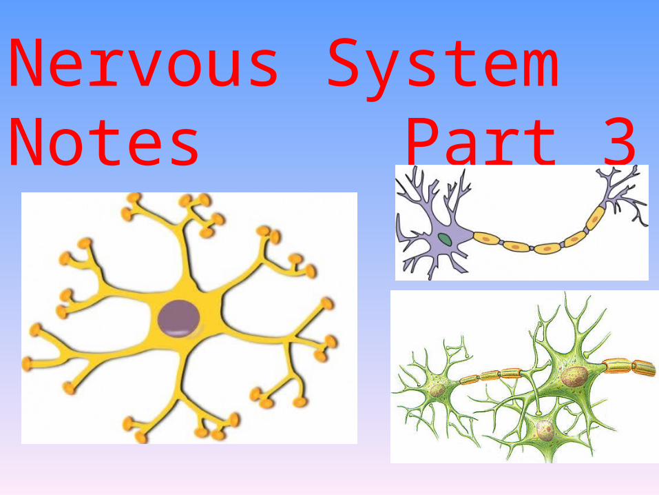
Nervous System Notes Part 3
EVEN MORE INTERESTING NERVOUS SYSTEM FACTSThe human brain alone consists of about 100 billion neurons. If all these neurons were to be lined up, it would form a 600 mile long line.
At any given point in time, only four percent of the cells in the brain are active, the rest are kept in reserve.
Copyright © 2009 Pearson Education, Inc., publishing as Benjamin Cummings
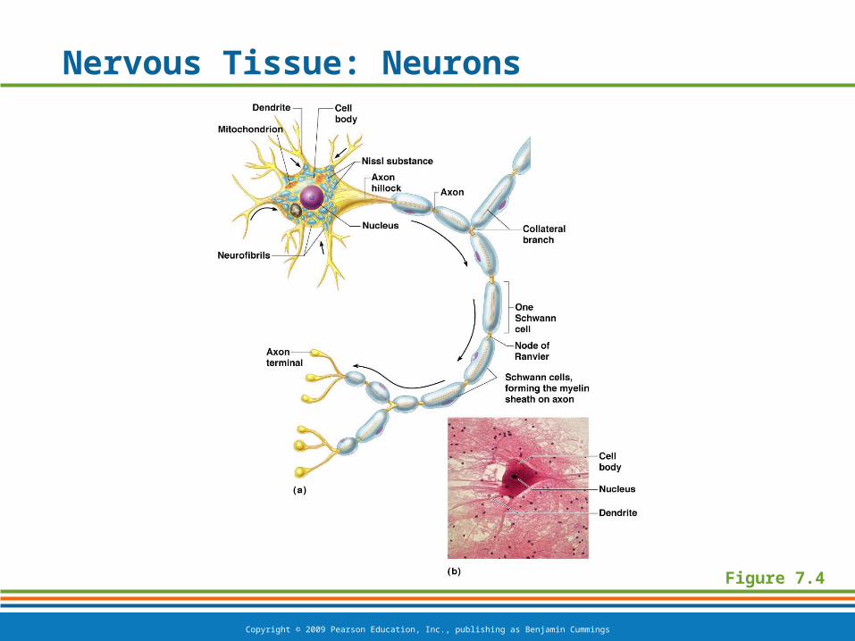
Nervous Tissue: Neurons
Figure 7.4
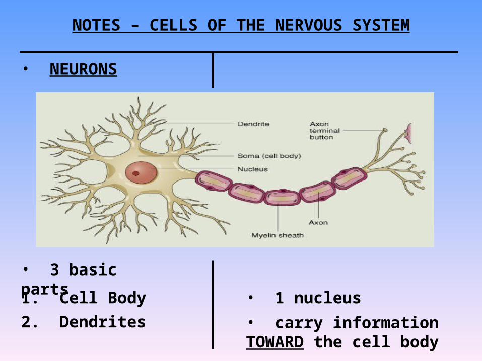
NOTES – CELLS OF THE NERVOUS SYSTEM
• NEURONS
• 3 basic parts1. Cell Body • 1 nucleus
2. Dendrites • carry information TOWARD the cell body
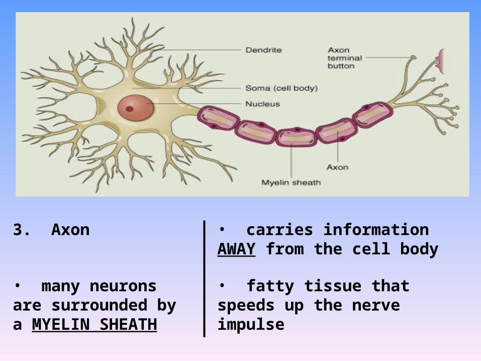
3. Axon • carries information AWAY from the cell body
• many neurons are surrounded by a MYELIN SHEATH
• fatty tissue that speeds up the nerve impulse
Copyright © 2009 Pearson Education, Inc., publishing as Benjamin Cummings
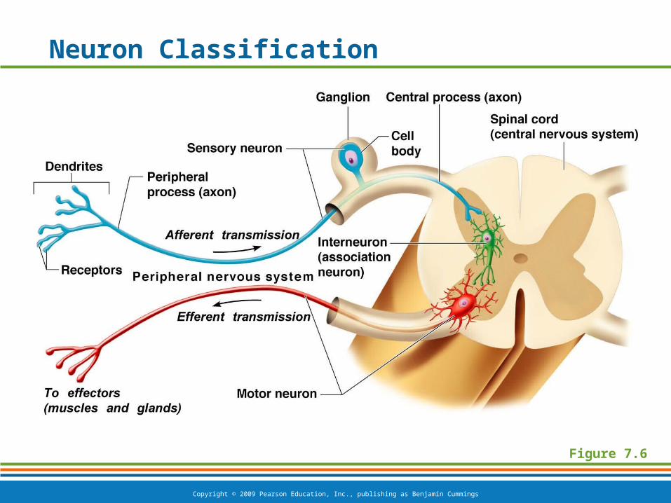
Neuron Classification
Figure 7.6
Copyright © 2009 Pearson Education, Inc., publishing as Benjamin Cummings
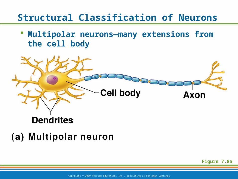
Figure 7.8a
Structural Classification of Neurons
Multipolar neurons—many extensions from the cell body
Copyright © 2009 Pearson Education, Inc., publishing as Benjamin Cummings
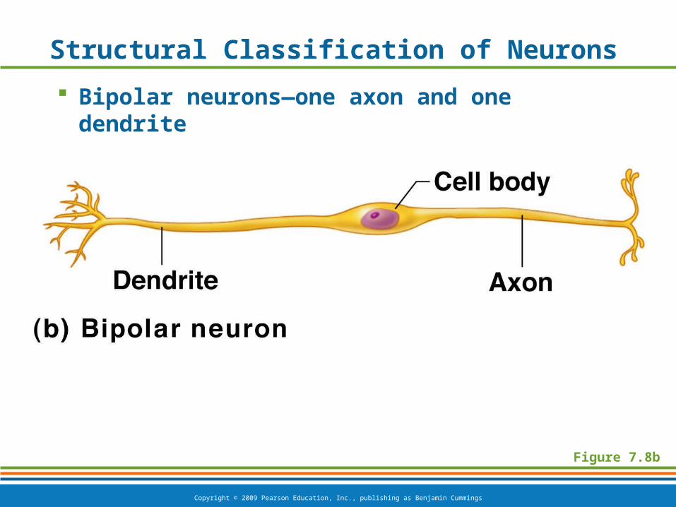
Structural Classification of Neurons
Bipolar neurons—one axon and one dendrite
Figure 7.8b
Copyright © 2009 Pearson Education, Inc., publishing as Benjamin Cummings
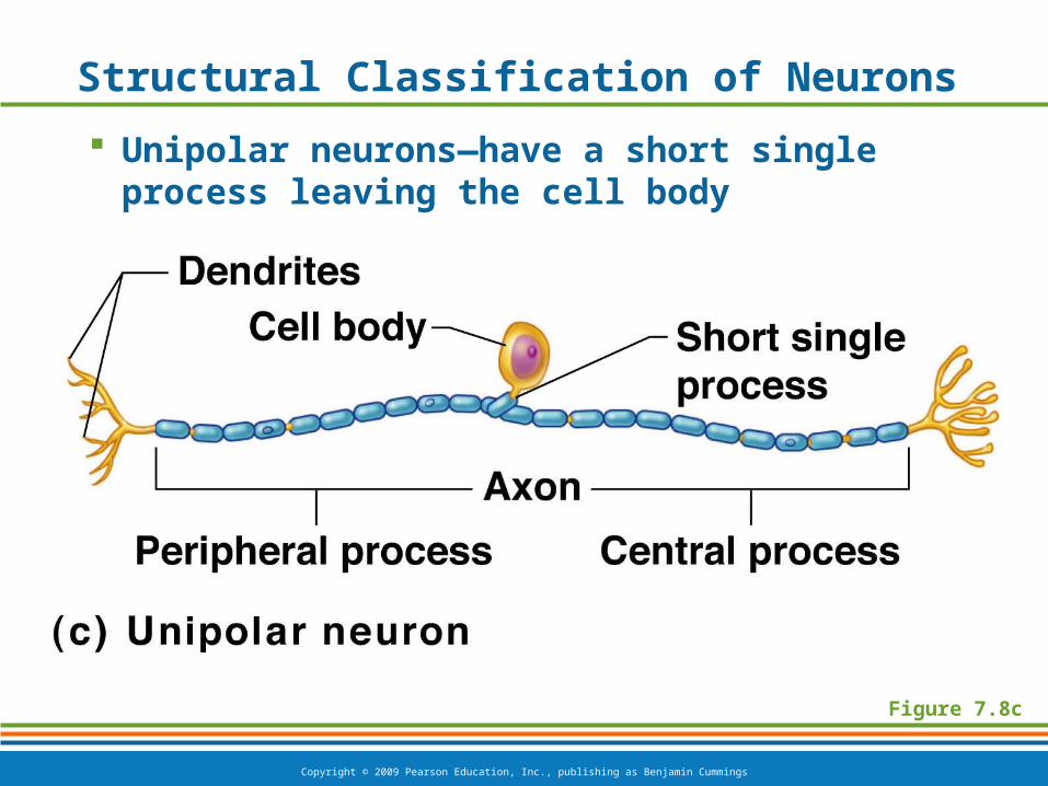
Structural Classification of Neurons
Unipolar neurons—have a short single process leaving the cell body
Figure 7.8c
Copyright © 2009 Pearson Education, Inc., publishing as Benjamin Cummings
Nervous Tissue: Support Cells
Support cells in the CNS are grouped together as “neuroglia”
Function: to support, insulate, and protect neurons
Copyright © 2009 Pearson Education, Inc., publishing as Benjamin Cummings
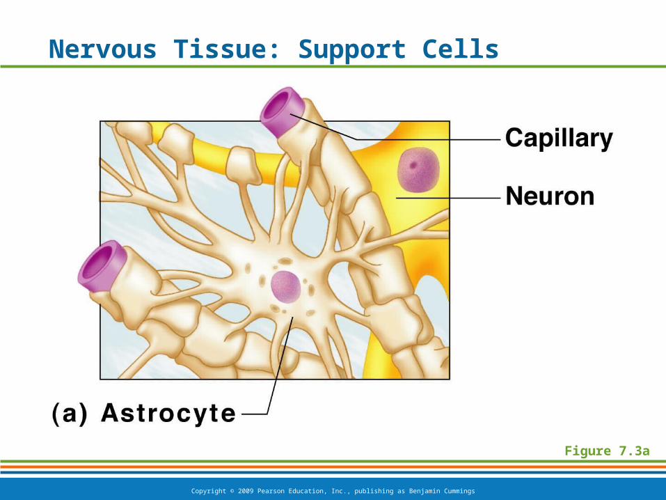
Nervous Tissue: Support Cells
Astrocytes
Abundant, star-shaped cells
Brace neurons
Form barrier between capillaries and neurons
Control the chemical environment of the brain
Copyright © 2009 Pearson Education, Inc., publishing as Benjamin Cummings
Nervous Tissue: Support Cells
Figure 7.3a

Copyright © 2009 Pearson Education, Inc., publishing as Benjamin Cummings
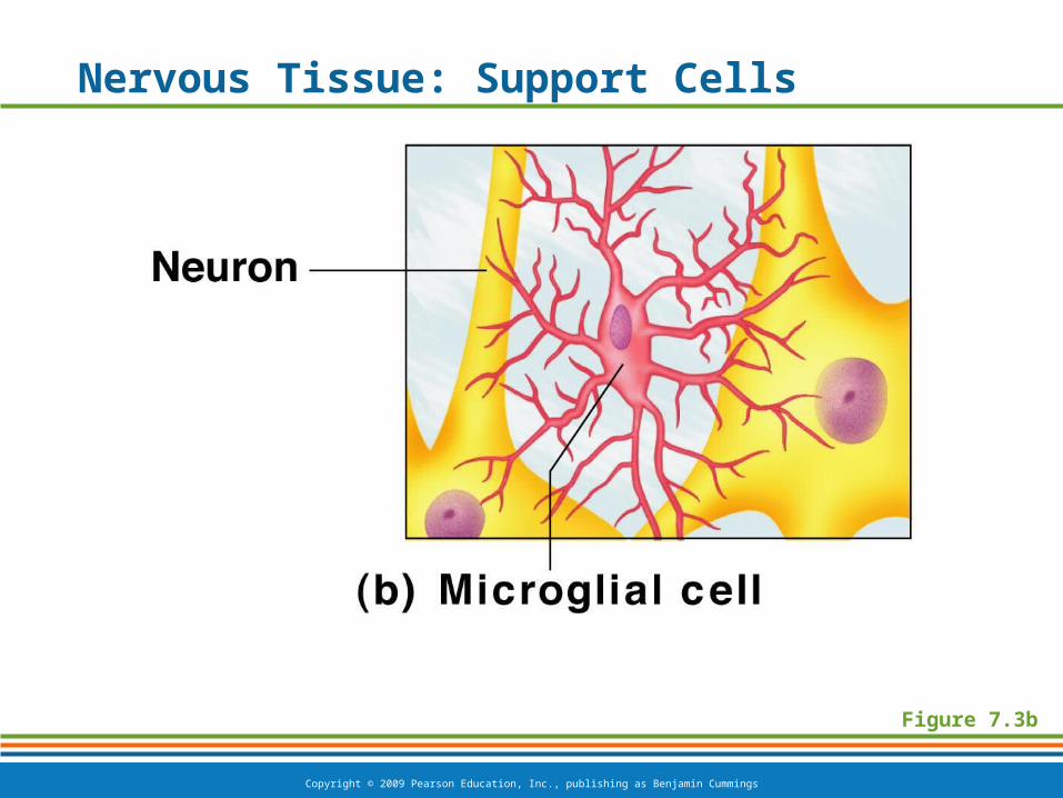
Nervous Tissue: Support Cells
Microglia
Spiderlike phagocytes
Dispose of debris
Copyright © 2009 Pearson Education, Inc., publishing as Benjamin Cummings
Nervous Tissue: Support Cells
Figure 7.3b
Copyright © 2009 Pearson Education, Inc., publishing as Benjamin Cummings
Nervous Tissue: Support Cells
Ependymal cells
Line cavities of the brain and spinal cord
Circulate cerebrospinal fluid

Copyright © 2009 Pearson Education, Inc., publishing as Benjamin Cummings
Nervous Tissue: Support Cells
Figure 7.3c
Copyright © 2009 Pearson Education, Inc., publishing as Benjamin Cummings
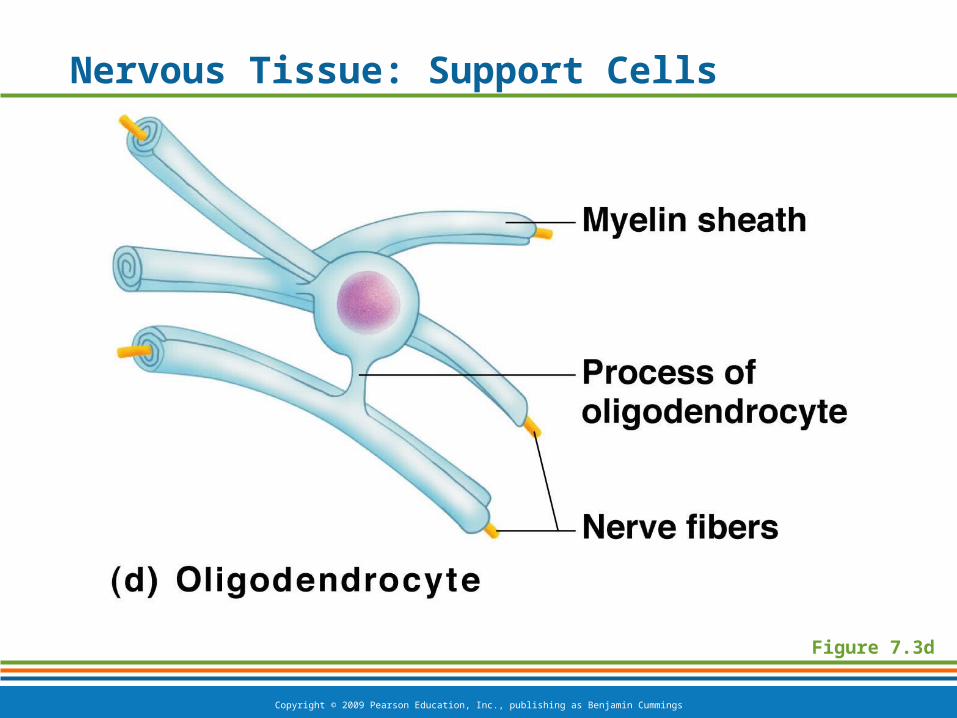
Nervous Tissue: Support Cells
Oligodendrocytes
Wrap around nerve fibers in the central nervous system
Produce myelin sheaths
Copyright © 2009 Pearson Education, Inc., publishing as Benjamin Cummings
Nervous Tissue: Support Cells
Figure 7.3d
Copyright © 2009 Pearson Education, Inc., publishing as Benjamin Cummings
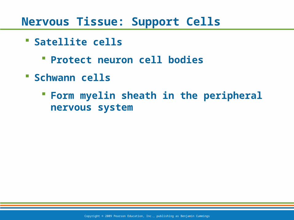
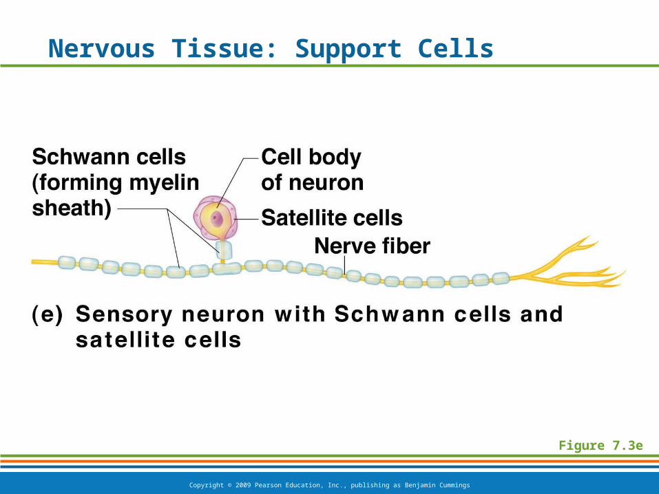
Nervous Tissue: Support Cells
Satellite cells
Protect neuron cell bodies
Schwann cells
Form myelin sheath in the peripheral nervous system
Copyright © 2009 Pearson Education, Inc., publishing as Benjamin Cummings
Nervous Tissue: Support Cells
Figure 7.3e
Copyright © 2009 Pearson Education, Inc., publishing as Benjamin Cummings
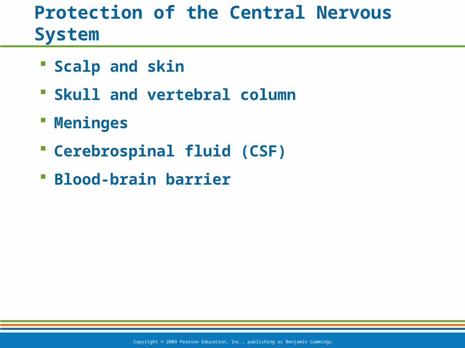
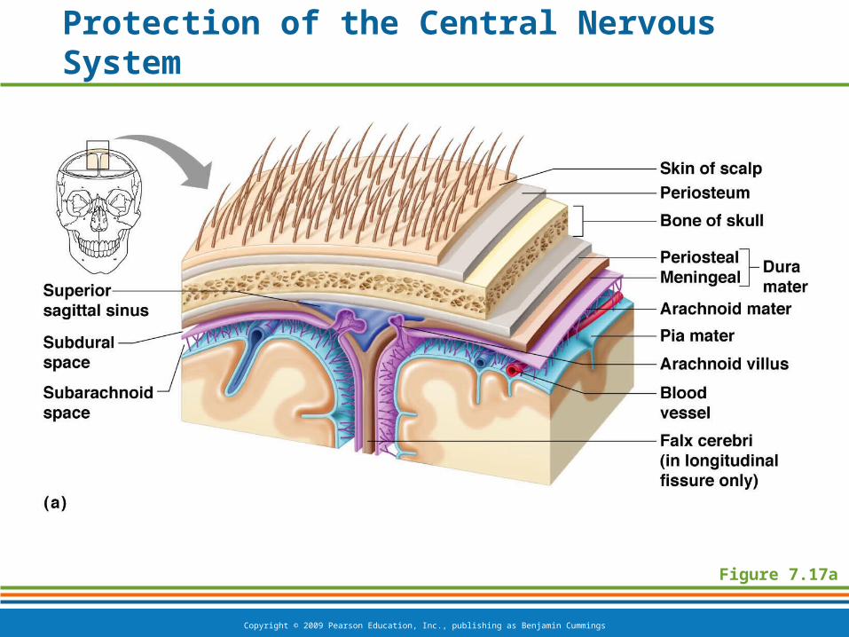
Protection of the Central Nervous System
Scalp and skin
Skull and vertebral column
Meninges
Cerebrospinal fluid (CSF)
Blood-brain barrier
Copyright © 2009 Pearson Education, Inc., publishing as Benjamin Cummings
Protection of the Central Nervous System
Figure 7.17a
Copyright © 2009 Pearson Education, Inc., publishing as Benjamin Cummings
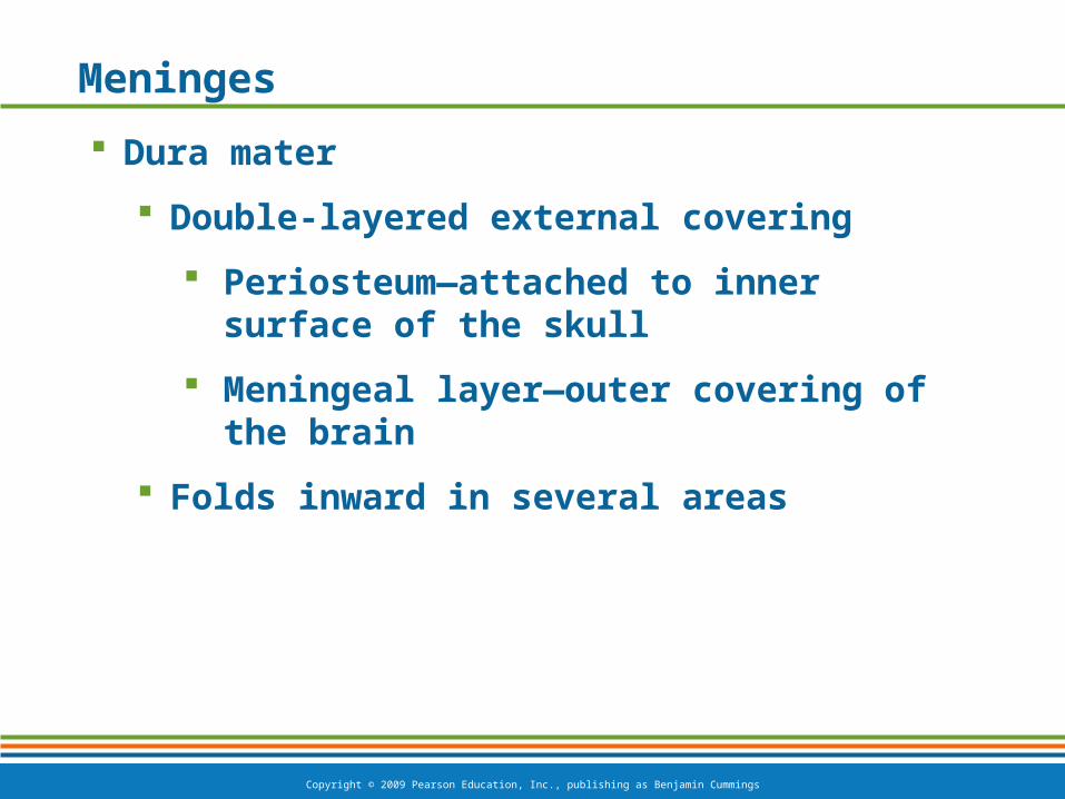
Meninges
Dura mater
Double-layered external covering
Periosteum—attached to inner surface of the skull
Meningeal layer—outer covering of the brain
Folds inward in several areas
Copyright © 2009 Pearson Education, Inc., publishing as Benjamin Cummings
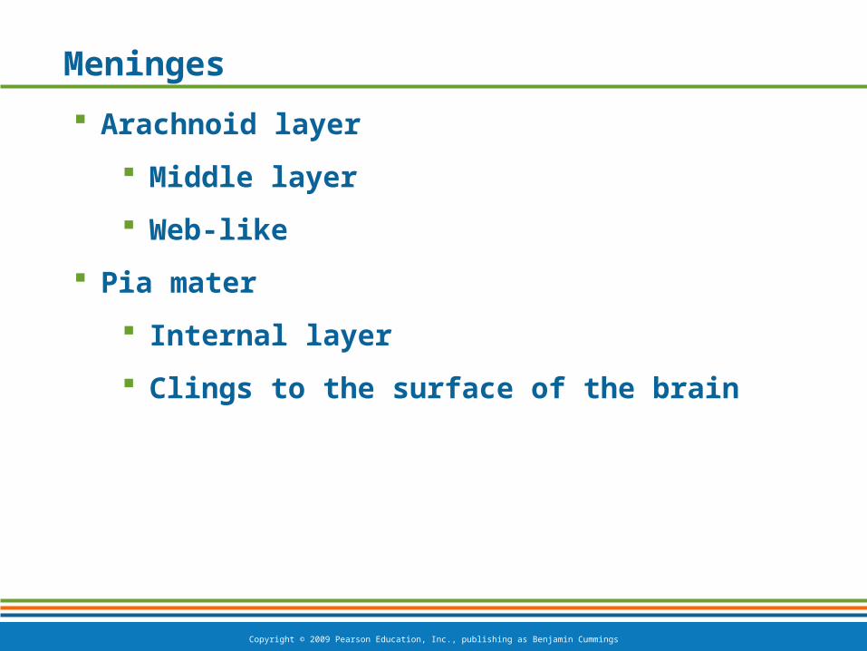
Meninges
Arachnoid layer
Middle layer
Web-like
Pia mater
Internal layer
Clings to the surface of the brain

Copyright © 2009 Pearson Education, Inc., publishing as Benjamin Cummings
Meninges
Figure 7.17b
Copyright © 2009 Pearson Education, Inc., publishing as Benjamin Cummings
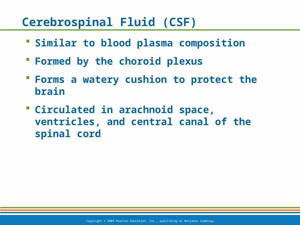
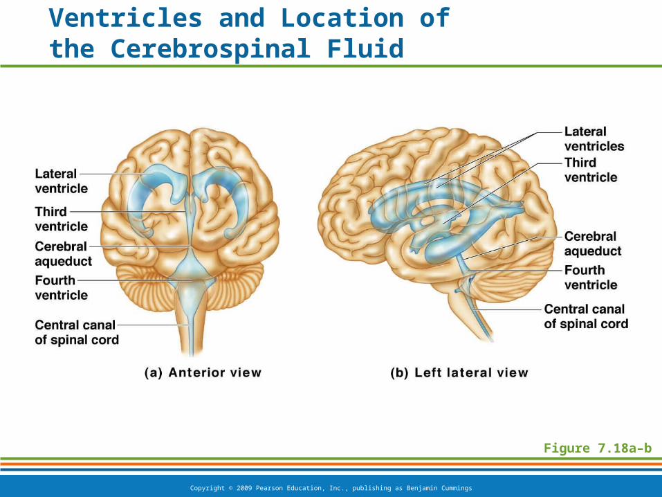
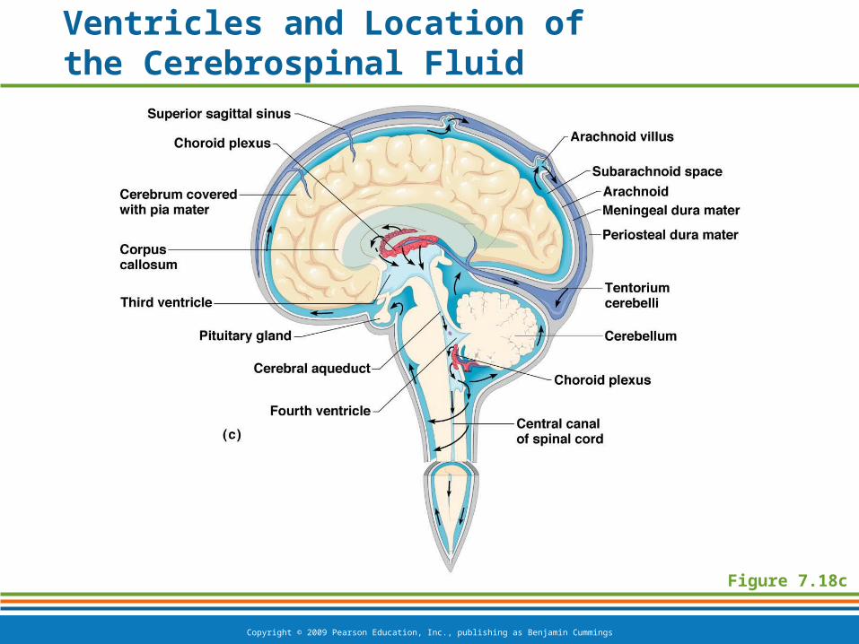
Cerebrospinal Fluid (CSF)
Similar to blood plasma composition
Formed by the choroid plexus
Forms a watery cushion to protect the brain
Circulated in arachnoid space, ventricles, and central canal of the spinal cord
Copyright © 2009 Pearson Education, Inc., publishing as Benjamin Cummings
Figure 7.18a–b
Ventricles and Location of the Cerebrospinal Fluid
Copyright © 2009 Pearson Education, Inc., publishing as Benjamin Cummings
Ventricles and Location of the Cerebrospinal Fluid
Figure 7.18c