Embed Size (px)
Citation preview

Nervous system
Chapters 48-49


Nervous system organization
CNS (central nervous system) Information processing
Brain Spinal cord

Nervous system organization
PNS (peripheral nervous system) Carry info to & from CNS Sensory neurons: Carry impulses to the CNS Motor neurons: Carry impulses from the CNS to effectors
(muscles or glands)

Nervous system organization
Interneurons: (association neurons) Located in brain & spinal column Higher functions or more complex
reflexes Learning & memory

Fig. 48-3
Sensor
Sensory input
Integration
Effector
Motor output
Peripheral nervoussystem (PNS)
Central nervoussystem (CNS)

Neuron structure
Cell body Contains nucleus & organelles Dendrites Branched, receives signals Axon Single, send signals Axon hillock: where signals are generated


Neuron structure
Synapse Site of communication between cells Presynaptic Transmitting neuron Postsynaptic Receiving cell Neurotransmitters Chemical messengers

Neuron structure
Glia “glue” Supporting cells Supply nutrients Remove wastes Guiding axon migration Immune functions
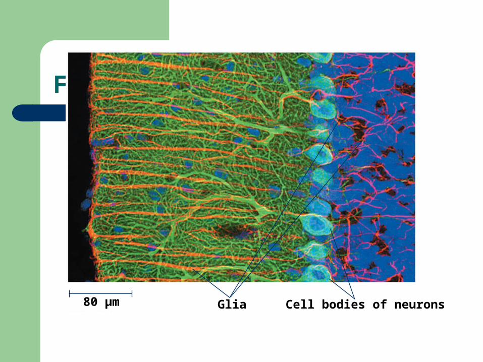
Figure 48.3
80 µm Glia Cell bodies of neurons

Membrane potential
Electrical charge across membrane of cell Cytoplasm is negative compared to
extracellular fluid Unequal distribution of anions & cations Either side of the membrane Ranges from –50 to –200 millivolts (mV)

Figure 48.6Key
Na+
K+
Sodium-potassiumpump
Potassiumchannel
Sodiumchannel INSIDE OF CELL
OUTSIDE OF CELL
Resting potential
OUTSIDECELL
[K+]5 mM
[Na+]150 mM
[Cl–]120 mM
INSIDECELL
[K+]140 mM
[Na+]15 mM
[Cl–]10 mM
[A–]100 mM
(a)


Technique
Microelectrode
Voltagerecorder
Referenceelectrode

Resting Potential

Resting membrane potential
Neurons are not stimulated, not transmitting signals
1. Fixed anions Proteins, carbohydrates & nucleic acids More abundant inside 2. Sodium/potassium pump
– 2K+ into cell/3Na+ out of cell 3. Ion leak channels Allows K+ to move out more than Na+ to move in Nerve cells –50 to –70 mV


Action potentials
Signals in the nervous system Sudden change in membrane voltage Change in membrane permeability to
ions Due to stimuli

Action Potential

Action potential
Ligand-gated (chemical) channels: Change shape when chemicals bind to
them Neurotransmitters or hormones Voltage-gated ion channels: Open when change in membrane potential Axons


Action potentials
Depolarization: Membrane potential less negative More positive ions flow in Na+1
Hyperpolarization: Membrane potential more negative Negative ions flow in (Cl-1) Positive ions flow out (K+1 or Na+1)

Action potentials
Threshold: Level of depolarization Produces an action potential All or none -55mV

Action potential
Nerve impulse Threshold Na & K voltage-gated ion channels opened First Na opens flows into cytoplasm (down concentration gradient) Potassium opens flows out Depolarizes the cell
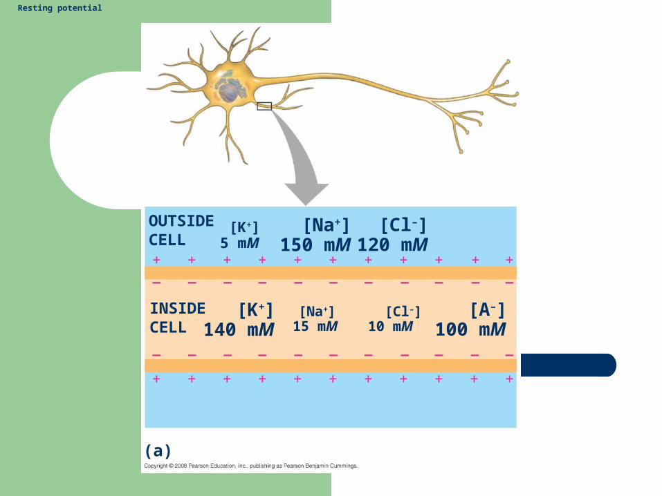
Action potential
Cl flows into cell Hyperpolarizes Na channels close K channels remain open a little longer Overshoot (hyperpolarize) Resting potential obtained Occurs in 1-2 milliseconds along axons

Action potential

Action potential

Action potential
Axon
Plasmamembrane
Cytosol
Actionpotential
Na+
Actionpotential
Na+
K+
K+
ActionpotentialK+
K+
Na+

Action potential
Strong depolarizing stimulus
+50
Mem
bra
ne
po
ten
tia
l (m
V)
–50 Threshold
Restingpotential
–1000 2 3 4
Time (msec)
(c) Action potential
1 5
0
Actionpotential
6


Action potential
Do not loose amplitude Greater speed of conduction Greater diameter of axon Myelinated Nodes of Ranvier Interruptions of myelin sheaths

Action potential
Saltatory impulse: Jump from one node to another

Saltatory impulse

Action potential
2 types of neuroglia Produce myelin sheaths Multiple layers of membrane around axon Insulation Schwann cells PNS Oligodendrocytes CNS

Figure 48.13
Axon Myelin sheathNodes ofRanvier
Schwanncell
SchwanncellNucleus ofSchwann cell
Axon
Layers of myelin
Node of Ranvier
0.1 µm

Synapses
2 types of synapses 1. Electrical Gap-junctions Membrane potentials change quickly 2. Chemical Neurotransmitters Most vertebrates


Synapses
Synaptic cleft: Space between pre & postsynaptic cell Synaptic vesicles: Located at end of axon Contain neurotransmitters

Synapses
Impulse down axon Causes rapid influx of Ca ions Synaptic vesicles to bind plasma
membrane Releases neurotransmitters by exocytosis Neurotransmitters bind postsynaptic
receptor proteins Response depends on neurotransmitters


Synapse

Types of neurotransmitters
Acetylcholine Amino acids
– Glutamate– Glycine– GABA (gamma-aminobutyric acid)
Biogenic amines– Epinephrine (adrenaline)– Dopamine– Norepinephrine– Serotonin
Gases– NO
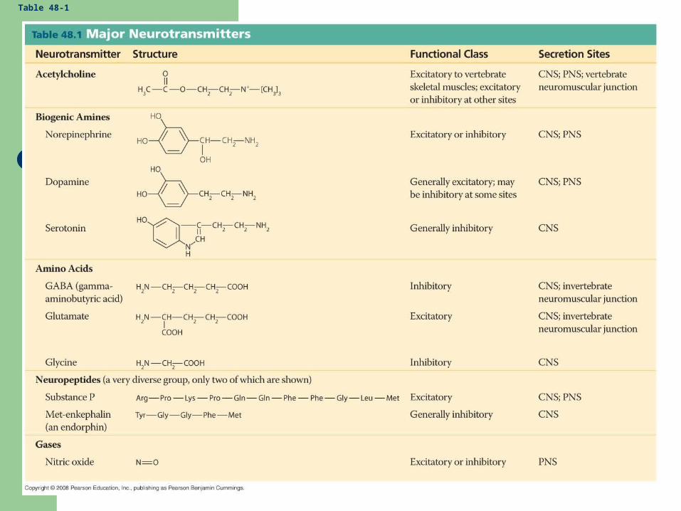
Table 48-1

Acetylcholine (ACh)
First discovered Synapse between motor neuron & a
muscle fiber Neuromuscular junction Binds postsynaptic membrane Causes ion channels to open Stimulates muscle contraction

Acetylcholine
Acetylcholinesterase (AChE) Enzyme located on postsynaptic membrane Enzyme cleaves ACh to be inactive Muscle relaxes Nerve gas & insecticide parathion Inhibitors of AChE Causes spastic paralysis Respiratory muscles causes death

Acetylcholine
Other synapses Usually between neurons Postsynaptic membrane is on dendrites
or cell body of another neuron Myasthenia gravis Alzheimer’s

Acetylcholine
Nicotine Affinity for Ach receptors Botulism Prevents pre-synaptic release of Ach BOTOX

EPSPs Excitatory postsynaptic potentials Towards threshold IPSPs Inhibitory Postsynaptic Potential Away threshold

Glutamate
Excitatory in CNS Normal amounts stimulate Excessive amounts show neuro
degeneration Huntington’s chorea

GABA and glycine
Inhibitory in CNS Neural control of body movements Other brain functions Valium (diazepam) sedative Increases GABA to bind receptor sites Increases GABA’s effectiveness

Biogenic amines
Epinephrine (adrenaline), norepinephrine & dopamine
Derived from tyrosine (aa) Dopamine Controls body movements (CNS, PNS) Excitatory Tremors, Parkinson disease Decrease in neurons releasing dopamine

Biogenic amines
Serotonin derived from tryptophan (aa) Inhibitory (CNS) Sleep, mood, attention and learning Decreased serotonin causes depression Prozac blocks uptake after release LSD binds receptors for serotonin

Gas
Nitric oxide (NO) Not stored Generated from arginine when needed PNS Smooth muscle relaxation
Neuropeptides
Polypeptides released by axons at synapses Substance P CNS, affects perception of pain Endorphins/Enkephalins Released in CNS Block perception of pain Opiates: morphine & heroin Similar in structure to neurotransmitters Bind receptor sites (pain-reducing)

Fig. 49-2
(e) Insect (arthropod)
Segmentalganglia
Ventralnerve cord
Brain
(a) Hydra (cnidarian)
Nerve net
Nervering
Radialnerve
(b) Sea star (echinoderm)
Anteriornerve ring
Longitudinalnerve cords
(f) Chiton (mollusc) (g) Squid (mollusc)
Ganglia
Brain
Ganglia
(c) Planarian (flatworm)
Nervecords
Transversenerve
Brain
EyespotBrain
(d) Leech (annelid)
Segmentalganglia
Ventralnervecord
Brain
Spinalcord(dorsalnervecord)
Sensoryganglia
(h) Salamander (vertebrate)

Fig. 49-4
Peripheral nervoussystem (PNS)
Cranialnerves
Brain
Central nervoussystem (CNS)
GangliaoutsideCNS
Spinalnerves
Spinal cord

Vertebrate Nervous System
CSF Cerebral spinal fluid Bathes brain, protects, provides nutrients Meninges Connective tissues that surround the
brain

CSF
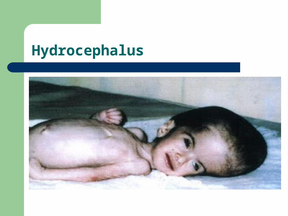
Hydrocephalus

Meninges

NS
White matter Myelinated axons Gray matter Unmyelinated axons Cell bodies

Spinal cord
Inner zone: Gray matter Cell bodies of interneurons, motor neurons &
neuralgia Outer zone: White matter Dorsal columns are sensory neurons Ventral columns are motor neurons Relay messages

Spinal cord
Reflexes Sensory neuron to motor neuron Spinal column Quick response Knee jerk

Reflexes

PNS
Cranial nerves Extend from brain Affect head, neck regions Spinal nerves Originate in spinal cord Extend to areas below head

PNS
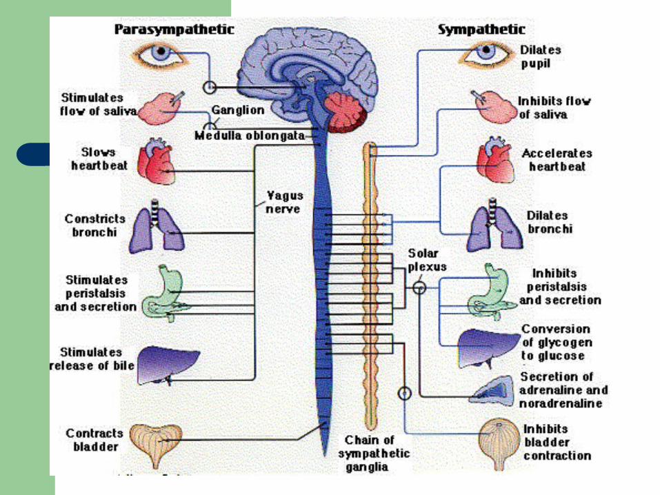
Afferent neurons(Sensory neurons) Towards brain Efferent neurons (Motor neurons) Away from brain Somatic motor neurons Stimulate skeletal muscles Autonomic motor neurons Regulate smooth & cardiac muscle, & glands Sympathetic/parasympathetic

PNS
Sympathetic Originate in the thoracic or lumbar regions Epinephrine or norepinephrine Parasympathetic Originate in the brain or sacral region Acetylcholine


Glia CNS
Astrocytes Support, increase blood flow, NT Oligodendrocytes Myelination Ependymal cell Line ventricles, CSF flow Microglial Defend against microorganisms

glia
Oligodendrocyte
Microglialcell
Schwann cells
Ependy-malcell
Neuron Astrocyte
CNS PNS
Capillary
(a) Glia in vertebrates
VENTRICLE

Brain
3 divisions in vertebrates (embryo) Hindbrain Cerebellum, medulla oblongata, pons Midbrain Forebrain Cerebrum, thalamus, hypothalamus,
basal ganglia, limbic system

Brain
Hindbrain Involuntary activities Coordinates motor activities Forebrain: Processing of olfactory input, regulation of
sleep, learning, and complex processing Midbrain: coordinates routing of sensory input
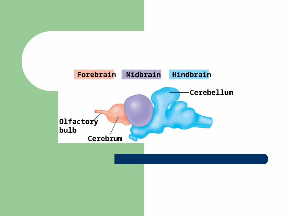
Olfactorybulb
Cerebrum
Cerebellum
Forebrain Midbrain Hindbrain
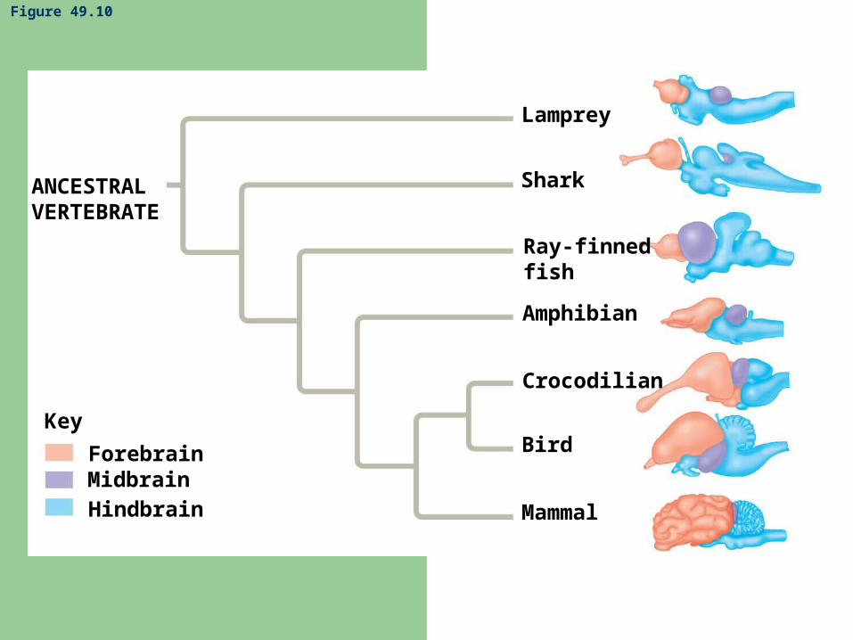
Figure 49.10
Lamprey
Shark
Ray-finnedfish
Amphibian
Crocodilian
Bird
Mammal
ANCESTRALVERTEBRATE
Key
ForebrainMidbrainHindbrain

Embryonic brain regions Brain structures in child and adult
Telencephalon
Diencephalon
Mesencephalon
Metencephalon
Myelencephalon Medulla oblongata (part of brainstem)
Pons (part of brainstem), cerebellum
Midbrain (part of brainstem)
Diencephalon (thalamus, hypothalamus, epithalamus)
Cerebrum (includes cerebral cortex, basal nuclei)
Cerebrum DiencephalonMesencephalon
Metencephalon
MyelencephalonDiencephalon
TelencephalonSpinalcord
ChildEmbryo at 5 weeksEmbryo at 1 month
Forebrain
Midbrain
Hindbrain
Hindbrain
MidbrainPons
Medullaoblongata
Cerebellum
Spinal cord
Bra
ins
tem
Midbrain
Forebrain

Brain

Brain

Cerebrum
Divided right & left cerebral hemispheres Connected by corpus callosum (band of
axons) Each hemisphere Cerebral cortex Internal white matter Basal nuclei (neurons in the white matter)

Fig. 49-13
Corpuscallosum
Thalamus
Left cerebralhemisphere
Right cerebralhemisphere
Cerebralcortex
Basalnuclei


Cerebrum
Divided further into four lobes Occipital lobe: vision Parietal lobe: body sensations, spatial
and visual perceptions Frontal: thought processing, behavior Temporal: hearing, understanding
language

Cerebrum

Cerebral cortex
Gray matter Outside of cerebrum Gyri: folds of nerves cells Sulcus: grooves or crease Functional areas in the cortex Sensory, motor or associative

Cerebral cortex
Sensory information comes to cortex Via the thalamus Primary sensory areas in different lobes Processed in association areas Motor command

Fig. 49-15
Speech
Occipital lobe
Vision
Temporal lobe
Frontal lobeParietal lobe
Somatosensoryassociationarea
Frontalassociationarea
Visualassociationarea
Reading
Taste
Hearing
Auditoryassociationarea
Speech
Smell
Mo
tor
cort
exS
omat
osen
sory
cor
tex

Motor cortex (control ofskeletal muscles) Somatosensory
cortex(sense of touch)
Sensory associationcortex (integrationof sensory information)
Frontal lobe
Temporal lobe
Parietal lobe
Occipital lobe
Prefrontal cortex(decision making,planning)
Broca’s area(forming speech)
Auditory cortex(hearing)
Cerebellum
Visualassociationcortex (combiningimages and objectrecognition)
Visual cortex(processing visualstimuli and patternrecognition)
Wernicke’s area(comprehendinglanguage)

Thalamus
Controls sensory information Visual, auditory & somatosensory
information Relays information to lobes of cortex
Basal Ganglia (nuclei)
Located in white matter of cerebrum Receives sensory information Receives motor commands from cortex
and cerebellum Participates in body movements

Limbic system
Located deep in the cerebrum Deals with emotions

Fig. 49-18
ThalamusHypothalamus
Prefrontalcortex
Olfactorybulb
Amygdala Hippocampus

Cerebellum
Coordination Balance and posture Hand-eye coordination

Hypothalamus
Controls visceral activities Regulates body temperature Hunger, thirst Emotional states Regulates the pituitary gland Regulates many endocrine glands

Brainstem
Medulla oblongata Controls various visceral activities Breathing, pulse, BP, swallowing Connects spinal cord to brain Pons Connects cerebellum & cerebrum to brain Nerves to eyes and face
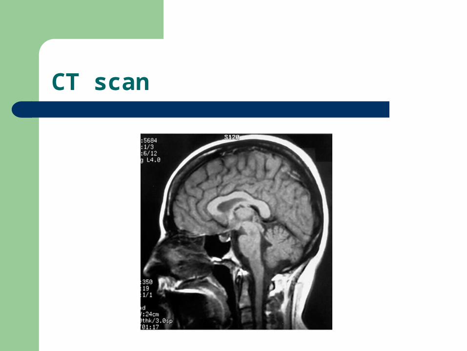
CT scan

MRI

PET scan

Phineas Gage