Embed Size (px)
Citation preview

NERVOUS SYSTEM AND NERVOUS SYSTEM AND NERVOUS TISSUENERVOUS TISSUE
Anatomy, activity and memoryAnatomy, activity and memory

Nervous System – Ch 11Nervous System – Ch 11
General scheme, fig General scheme, fig 11.111.1– Receives information Receives information
from receptors– from receptors– sensory inputsensory input..
– Interprets and Interprets and develops a plan of develops a plan of action – action – integrationintegration..
– Sends plan of action to Sends plan of action to effector(s) – effector(s) – motor motor outputoutput..
Organization of the Nervous Organization of the Nervous SystemSystem
Central Nervous System (CNS) – spinal Central Nervous System (CNS) – spinal cord and brain; interpretation & integration, cord and brain; interpretation & integration, fig 11.2.fig 11.2.
Peripheral Nervous System (PNS) – links Peripheral Nervous System (PNS) – links the rest of the body to the CNS.the rest of the body to the CNS.– 12 pair of 12 pair of cranial nervescranial nerves and 31 pairs of and 31 pairs of spinal spinal
nervesnerves..

Organization of the Nervous Organization of the Nervous SystemSystem
PNS divisionsPNS divisions– Sensory division (afferent) – relays Sensory division (afferent) – relays
information from receptors to CNSinformation from receptors to CNS
– Motor division (efferent) - relays information Motor division (efferent) - relays information from CNS to effectors.from CNS to effectors.

Organization of the Nervous Organization of the Nervous SystemSystem

HistologyHistology
80% cells; neurons and neuroglia 80% cells; neurons and neuroglia (supporting cells). 10X as many neuroglia (supporting cells). 10X as many neuroglia as neurons.as neurons.
Neuroglia of the CNS, 4 types (fig 11.3)Neuroglia of the CNS, 4 types (fig 11.3)– AstrocytesAstrocytes – –
– MicrogliaMicroglia – –

HistologyHistology
Neuroglia of the CNS cont’dNeuroglia of the CNS cont’d– EpendymalEpendymal – –
– OligodendrocytesOligodendrocytes – – – **Brain tumors**Brain tumors
Neuroglia of the PNS, 2 typesNeuroglia of the PNS, 2 types– Satellite cells - ?functionSatellite cells - ?function– Schwann cells – form myelin sheathsSchwann cells – form myelin sheaths


HistologyHistology
Neurons (fig 11.4)Neurons (fig 11.4)– Features: very irritable, long lived, amitotic Features: very irritable, long lived, amitotic
(except in the hippocampus?), high metabolic (except in the hippocampus?), high metabolic rate.rate.
– Cell bodyCell body (soma): large single nucleus with a (soma): large single nucleus with a prominent nucleolus, Nissl bodies (rough ER) prominent nucleolus, Nissl bodies (rough ER) and Golgi apparatus, + other organelles. and Golgi apparatus, + other organelles. Biosynthetic center. Primarily located in CNSBiosynthetic center. Primarily located in CNS

HistologyHistology
Neurons cont’dNeurons cont’d– ProcessesProcesses
DendritesDendrites – usually branched, input or receptive – usually branched, input or receptive regions; carry impulses TOWARD the cell body.regions; carry impulses TOWARD the cell body.AxonsAxons – usually one per cell, arise from – usually one per cell, arise from axon axon hillockhillock, short to very long, terminal branches. , short to very long, terminal branches. Output regions, carry impulses AWAY from the cell Output regions, carry impulses AWAY from the cell body.body.Nutrients and repair parts (proteins, etc) travel Nutrients and repair parts (proteins, etc) travel from cell body to axon and back again.from cell body to axon and back again.**Viruses and bacteria.**Viruses and bacteria.

HistologyHistology
Neuron (fig 11.5)Neuron (fig 11.5)– Myelin sheaths: protein/lipid sheet wrapped Myelin sheaths: protein/lipid sheet wrapped
around axons only, outer layer is neurilemma. around axons only, outer layer is neurilemma. Discontinuous - spaces are Nodes of Discontinuous - spaces are Nodes of Ranvier.Ranvier.
Functions: electrically insulate axon and speed up Functions: electrically insulate axon and speed up transmission of electrical impulses.transmission of electrical impulses.
– Gray matter – unmyelinated axons and cell Gray matter – unmyelinated axons and cell bodiesbodies
– White matter – myelinated axonsWhite matter – myelinated axons


Neurophysiology - ReviewNeurophysiology - Review
Membrane Ion Membrane Ion Channels (fig 11.6)Channels (fig 11.6)
Neurophysiology - ReviewNeurophysiology - Review
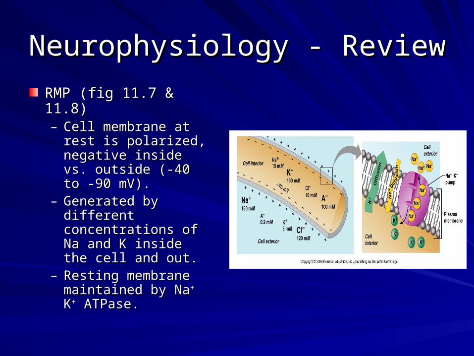
RMP (fig 11.7 & 11.8)RMP (fig 11.7 & 11.8)– Cell membrane at rest Cell membrane at rest
is polarized, negative is polarized, negative inside vs. outside (-40 inside vs. outside (-40 to -90 mV). to -90 mV).
– Generated by different Generated by different concentrations of Na concentrations of Na and K inside the cell and K inside the cell and out.and out.
– Resting membrane Resting membrane maintained by Namaintained by Na++ K K++ ATPase.ATPase.

SynapsesSynapses
The nervous system is like telegraph or The nervous system is like telegraph or “old” telephone lines.“old” telephone lines.
Synapse = connection points among the Synapse = connection points among the lines (neurons).lines (neurons).– ElectricalElectrical– ChemicalChemical
There may be 100’s or 1000’s of synapses There may be 100’s or 1000’s of synapses on a single neuron.on a single neuron.

Chemical Synapse ModelChemical Synapse Model

Chemical Synapses in Nervous Chemical Synapses in Nervous SystemSystem
Types of chemical Types of chemical synapses based on synapses based on position:position:– AxodendriticAxodendritic– AxosomaticAxosomatic– Axoaxonic, etcAxoaxonic, etc
**Presynaptic neuron **Presynaptic neuron vs Postsynaptic vs Postsynaptic neuronneuron

NeurophysiologyNeurophysiology
Membrane potentials that act as signals, Membrane potentials that act as signals, fig 11.9fig 11.9– DepolarizationDepolarization: decreases membrane : decreases membrane
potential (less negative).potential (less negative).– HyperpolarizationHyperpolarization: increases membrane : increases membrane
potential (more negative).potential (more negative).– Graded potentialsGraded potentials – – locallocal changes in changes in
membrane potential. Short lived, magnitude membrane potential. Short lived, magnitude varies with stimulus strength.varies with stimulus strength.

NeurophysiologyNeurophysiology
Membrane potentials that act as signalsMembrane potentials that act as signals– Graded potentials cont’dGraded potentials cont’d
EPSP – EPSP –
IPSP – IPSP –
Function: control of unintentional or unnecessary Function: control of unintentional or unnecessary impulses to and from your brain.impulses to and from your brain.


NeurophysiologyNeurophysiology
Membrane potentials that act as signalsMembrane potentials that act as signals– Graded potentials Graded potentials
SummationSummation – combination of these graded, local – combination of these graded, local potentials at the axon hillock can bring about an potentials at the axon hillock can bring about an action potential or inhibit the generation of the action potential or inhibit the generation of the action potential.action potential.
– SpatialSpatial: stimulation by many neurons at one time.: stimulation by many neurons at one time.– TemporalTemporal: increased numbers of impulses per minute.: increased numbers of impulses per minute.

Summation of Graded PotentialsSummation of Graded Potentials

NeurophysiologyNeurophysiology
Membrane potentials that act as signalsMembrane potentials that act as signals– Action Potential – rapid and large change in Action Potential – rapid and large change in
membrane potential, i.e. from -50 to +30 mV. membrane potential, i.e. from -50 to +30 mV. Occurs only in axons = nerve impulse.Occurs only in axons = nerve impulse.
– Action Potential and ion channels – review???Action Potential and ion channels – review???


Memory – Ch 12Memory – Ch 12
Basis: Most neurons are amitotic but new Basis: Most neurons are amitotic but new synapses form continually and existing synapses form continually and existing synapses can be “trained” to work more synapses can be “trained” to work more efficiently.efficiently.– Repeated EPSPs at the synapses of the same Repeated EPSPs at the synapses of the same
neuronal pathway causes physical changes called neuronal pathway causes physical changes called LONG TERM POTENTIATION.LONG TERM POTENTIATION.
– Receptor proteins are altered to respond more quickly Receptor proteins are altered to respond more quickly or more dramatically and generate an AP.or more dramatically and generate an AP.
– Studying – reading is input; recall or output = write it, Studying – reading is input; recall or output = write it, say it, explain it, draw it, act it out, etc.say it, explain it, draw it, act it out, etc.

Memory - StagesMemory - Stages
Short-term (STM) – working memory (RAM on Short-term (STM) – working memory (RAM on the computer).the computer).– Limited to 7-8 “chunks” of information.Limited to 7-8 “chunks” of information.– May be forgotten immediately.May be forgotten immediately.
Long term (LTM) is like information on the hard Long term (LTM) is like information on the hard disk on the computer). disk on the computer). – LimitlessLimitless– Conversion of STM to LTM is affected by emotional Conversion of STM to LTM is affected by emotional
state, rehearsal and association but can be automatic.state, rehearsal and association but can be automatic.
MemoryMemory
Categories: Categories: – DeclarativeDeclarative (facts) (facts)– Non-declarativeNon-declarative: procedural (skills), motor, : procedural (skills), motor,
emotional memories. Come from practice and emotional memories. Come from practice and experience.experience.
– Involve different pathways in brain???Involve different pathways in brain???
Storage in different areas of the brain – Storage in different areas of the brain – association areas near sensory/motor association areas near sensory/motor areas concerned with that particular cue.areas concerned with that particular cue.

Figure 12.23
Proposed Memory CircuitsProposed Memory Circuits
Declarative Procedural

Memory Memory
Mechanisms - what has been observed:Mechanisms - what has been observed:– Increased mRNA synthesisIncreased mRNA synthesis– Changes in dendritesChanges in dendrites– ““New” protein at synapses involved in LTMNew” protein at synapses involved in LTM– Increased number and size of presynaptic Increased number and size of presynaptic
terminals and neurotransmitter releasedterminals and neurotransmitter released– ““New” neurons in the hippocampus.New” neurons in the hippocampus.