Embed Size (px)
Citation preview
1
Nervous System
.IOverview.IIHistology
.IIIElectrical Signals.IVSignal Transmission at Synapses
.VNeurotransmitters.VINeural Circuits
.VIIRepairs.VIIIPathology
2
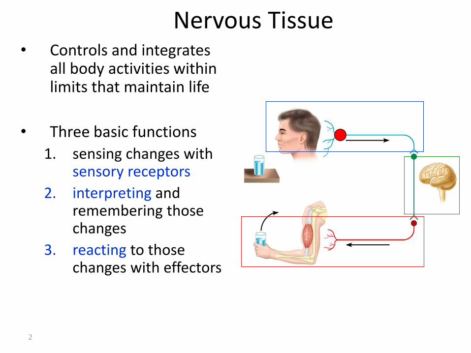
Nervous Tissue• Controls and integrates
all body activities within limits that maintain life
• Three basic functions
1. sensing changes with sensory receptors
2. interpreting and remembering those changes
3. reacting to those changes with effectors
3
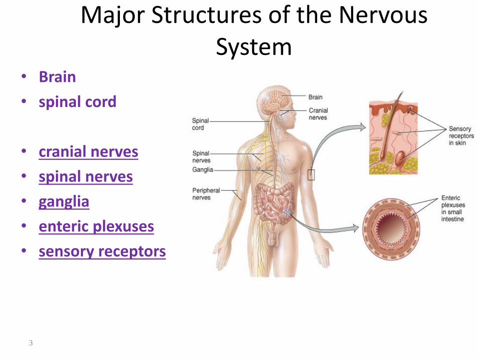
Major Structures of the Nervous System
• Brain
• spinal cord
• cranial nerves
• spinal nerves
• ganglia
• enteric plexuses
• sensory receptors
5
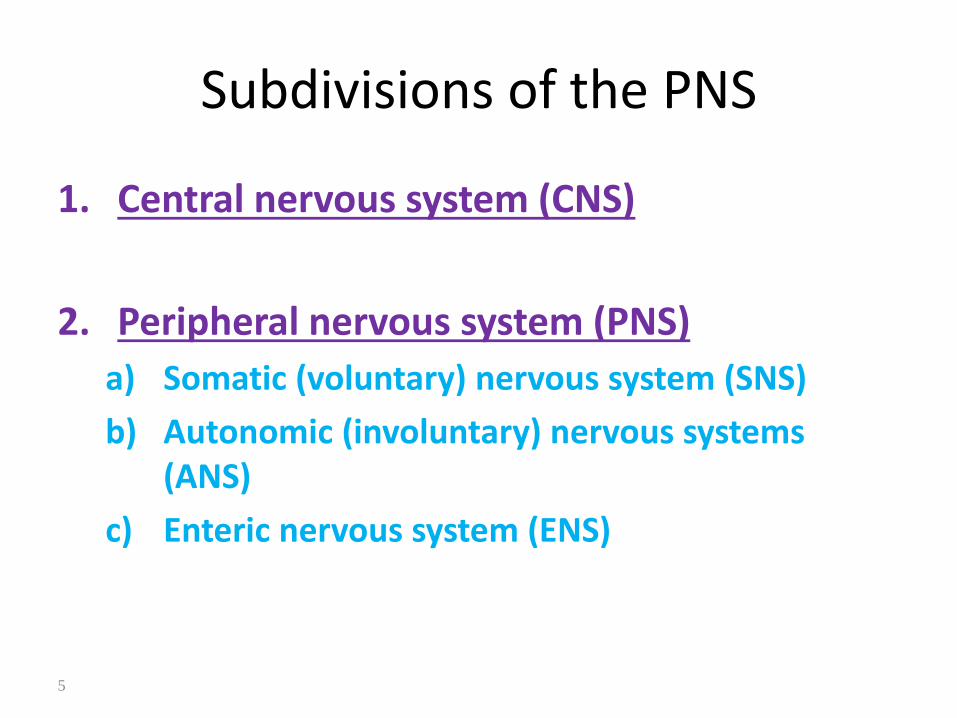
Subdivisions of the PNS
1. Central nervous system (CNS)
2. Peripheral nervous system (PNS)
a) Somatic (voluntary) nervous system (SNS)
b) Autonomic (involuntary) nervous systems (ANS)
c) Enteric nervous system (ENS)

6
OrganizationIntegration MotorSensory
SNS(Sensory)
ANS(Sensory)
Brain
Spinal cord
SNS(Motor)
ANS(Motor)
ENS(Sensory)

7
Neurons• Functional unit of nervous
system
1. Cell body
a) Nissl bodies (rER)
b) Neurofilaments
c) Microtubules
d) Lipofuscin pigment clumps
2. Cell processes a) Dendrites b) Axons

Electron MicroscopyLight-Stained Purkinje Neurons

11
Gray and White Matter• White matter = myelinated processes (white in color)
• Gray matter = nerve cell bodies, dendrites, axon terminals, bundles of unmyelinated axons and neuroglia (gray color)

12
Dendrites• Conducts Impulses
Towards The Cell Body
• Typically short, highly branched & unmyelinated
• Surfaces specialized for contact with other neurons
• Contains neurofibrils & Nissl bodies (rER)
impulse
13
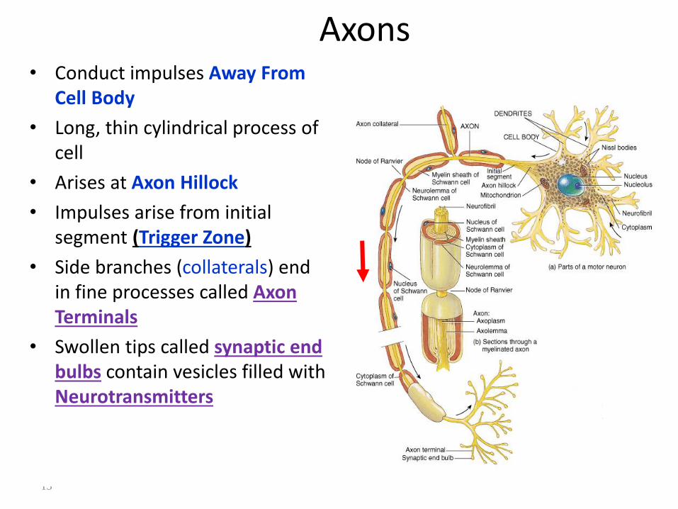
Axons• Conduct impulses Away From
Cell Body
• Long, thin cylindrical process of cell
• Arises at Axon Hillock
• Impulses arise from initial segment (Trigger Zone)
• Side branches (collaterals) end in fine processes called Axon Terminals
• Swollen tips called synaptic end bulbs contain vesicles filled with Neurotransmitters
15
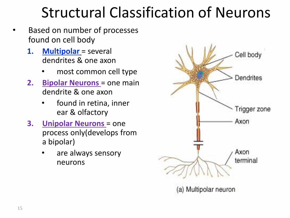
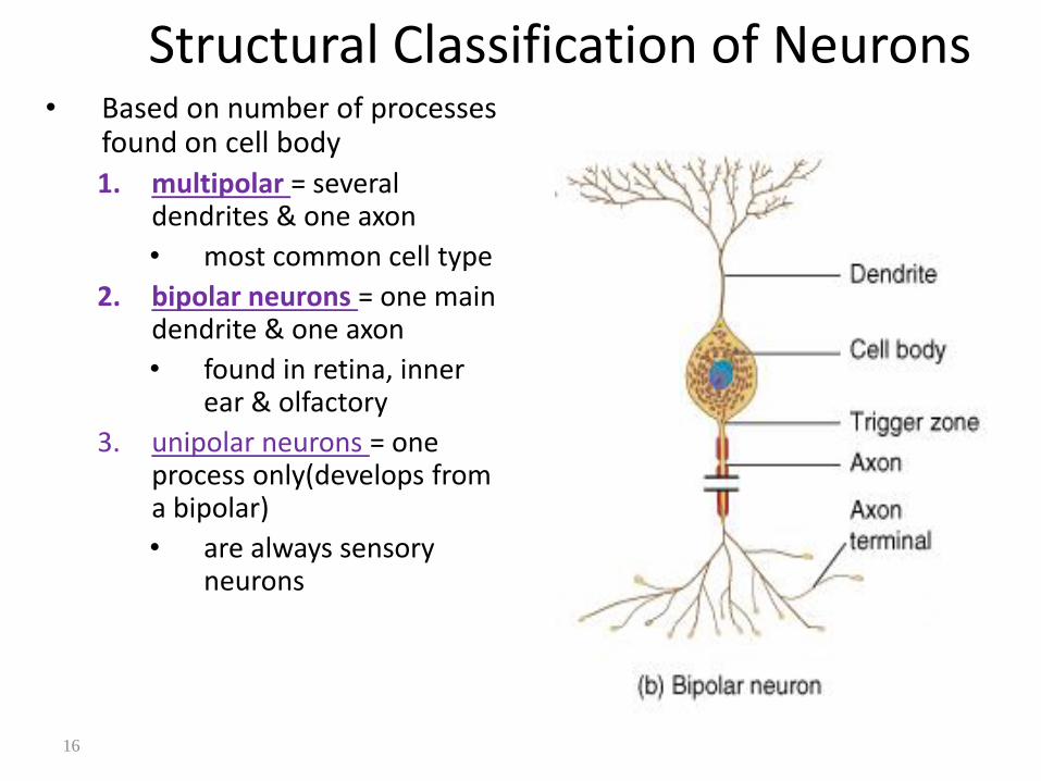
Structural Classification of Neurons• Based on number of processes
found on cell body
1. Multipolar = several dendrites & one axon
• most common cell type
2. Bipolar Neurons = one main dendrite & one axon
• found in retina, inner ear & olfactory
3. Unipolar Neurons = one process only(develops from a bipolar)
• are always sensory neurons
16
Structural Classification of Neurons• Based on number of processes
found on cell body
1. multipolar = several dendrites & one axon
• most common cell type
2. bipolar neurons = one main dendrite & one axon
• found in retina, inner ear & olfactory
3. unipolar neurons = one process only(develops from a bipolar)
• are always sensory neurons
17
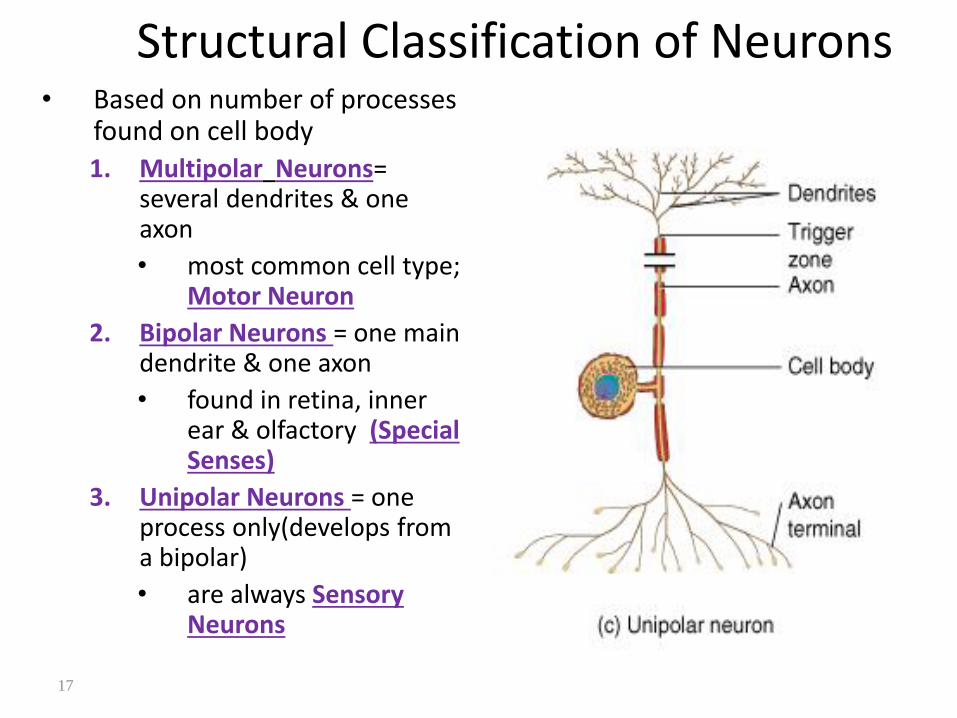
Structural Classification of Neurons• Based on number of processes
found on cell body
1. Multipolar Neurons= several dendrites & one axon
• most common cell type; Motor Neuron
2. Bipolar Neurons = one main dendrite & one axon
• found in retina, inner ear & olfactory (Special Senses)
3. Unipolar Neurons = one process only(develops from a bipolar)
• are always Sensory Neurons
18
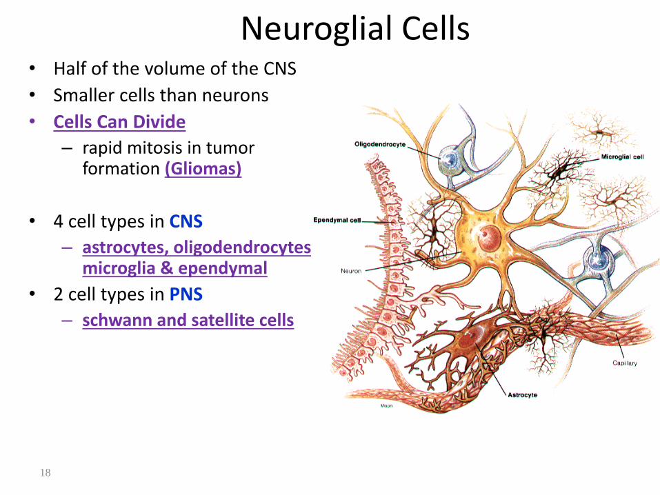
Neuroglial Cells• Half of the volume of the CNS
• Smaller cells than neurons
• Cells Can Divide
– rapid mitosis in tumor formation (Gliomas)
• 4 cell types in CNS
– astrocytes, oligodendrocytes, microglia & ependymal
• 2 cell types in PNS
– schwann and satellite cells
19
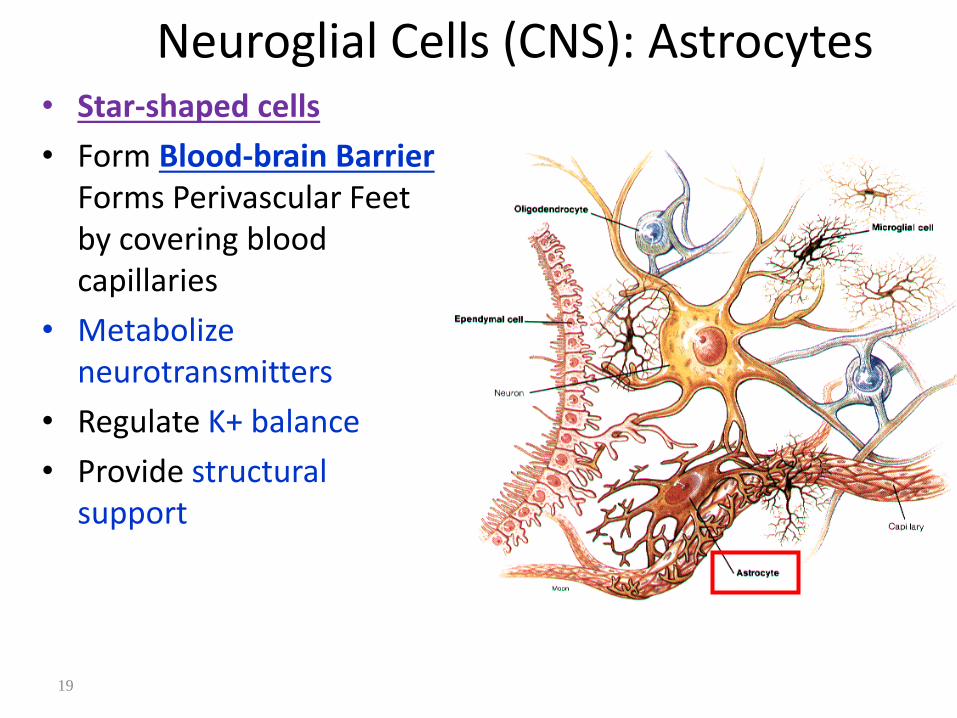
Neuroglial Cells (CNS): Astrocytes• Star-shaped cells
• Form Blood-brain BarrierForms Perivascular Feet by covering blood capillaries
• Metabolize neurotransmitters
• Regulate K+ balance
• Provide structural support

20
Neuroglial Cells (CNS): Oligodendrocytes
• Most common glial cell type
• Each forms Myelin Sheatharound more than one axons in CNS
• Analogous to Schwann cells of PNS

21
Neuroglial Cells (CNS): Microglia• Small cells found near
blood vessels
• Phagocytic Cell –
• Derived macrophages & monocytes

22
Neuroglial Cells (CNS): Ependymalcells
• Form epithelial membrane lining cerebral cavities &
central canal
• Produce Cerebrospinal Fluid (CSF)
23
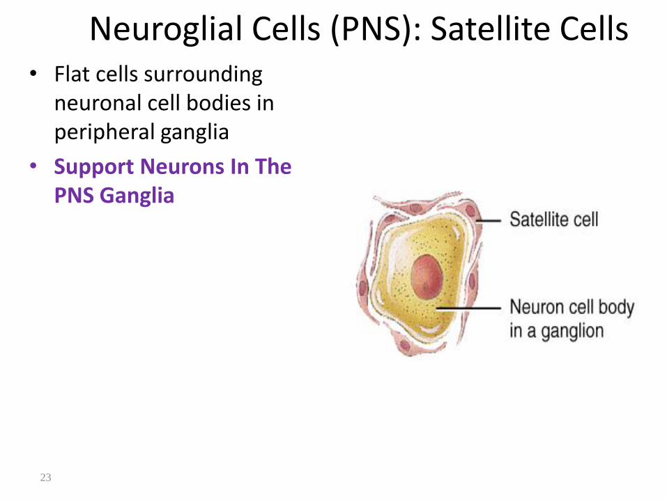
Neuroglial Cells (PNS): Satellite Cells• Flat cells surrounding
neuronal cell bodies in peripheral ganglia
• Support Neurons In The PNS Ganglia
24
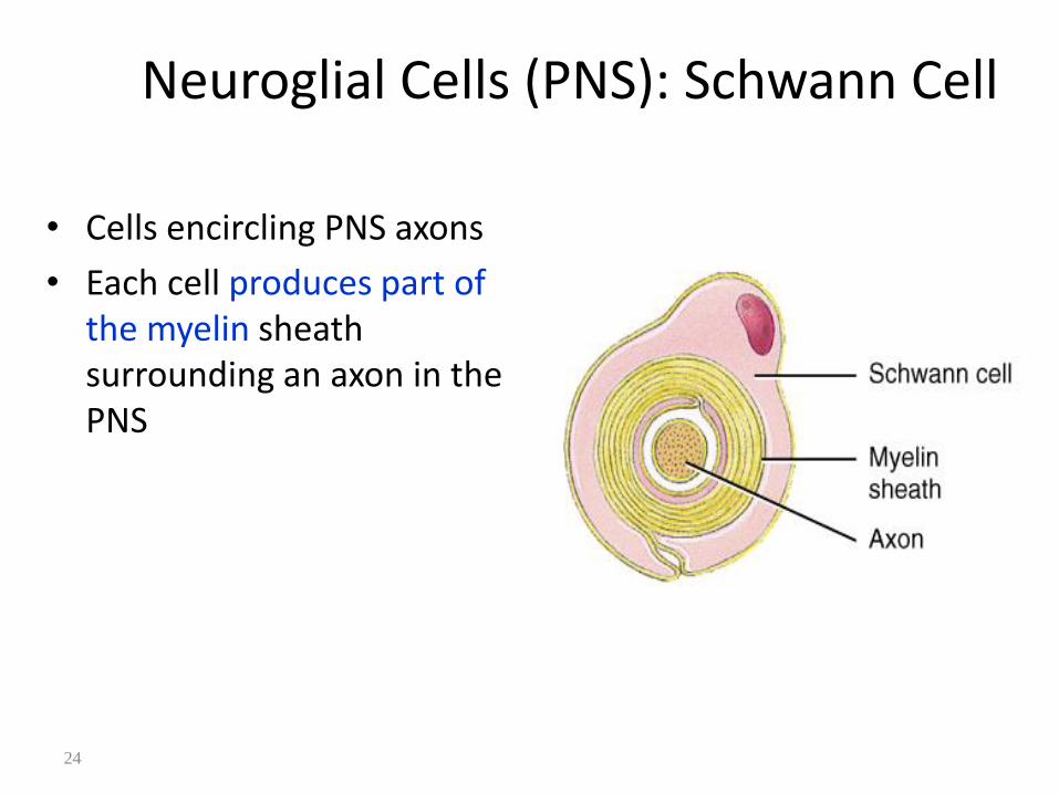
Neuroglial Cells (PNS): Schwann Cell
• Cells encircling PNS axons
• Each cell produces part of the myelin sheath surrounding an axon in the PNS
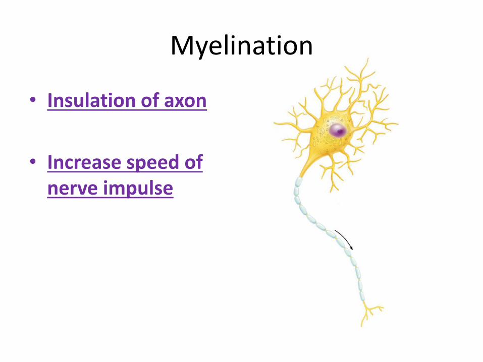
Myelination
• Insulation of axon
• Increase speed of nerve impulse

Myelination: PNS• All axons surrounded by a
lipid & protein covering (Myelin Sheath) produced by Schwann cells
• Neurilemma is fused layers of membranes of Schwann cell
– gaps called nodes of Ranvier
• Myelinated fibers
• Unmyelinated fibers
Node of Ranvier

27
Myelination: CNS• Oligodendrocytes
myelinate axons in the CNS
• Broad, flat cell processes wrap about CNS axons. One Oligo Can MyelinateUpto 50 Nerves.
• Schwan Cell: PNS
• Many Schwan Cells Myelinate One Peripheral Nerve
• End of one SchwanSegment Node of Ranvier
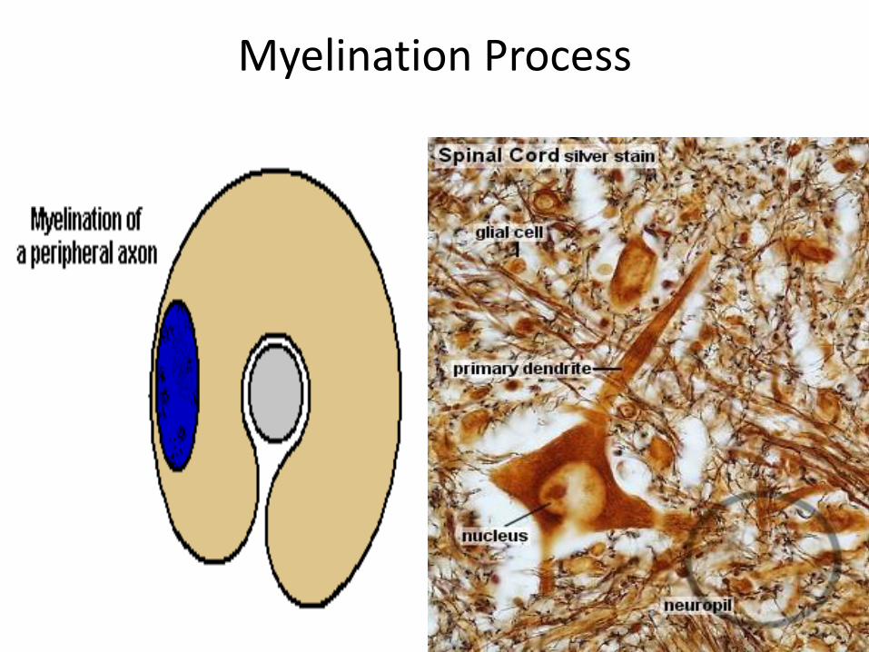
Myelination Process

29
Electrical Signals in Neurons
• Neurons are electrically excitable due to the voltagedifference across their membrane
• Communicate with 2 types of electric signals
1. action potentials that can travel long distances
2. graded potentials that are local membrane changes only
• In living cells, a flow of ions occurs Through Ion Channels In The Cell Membrane
31
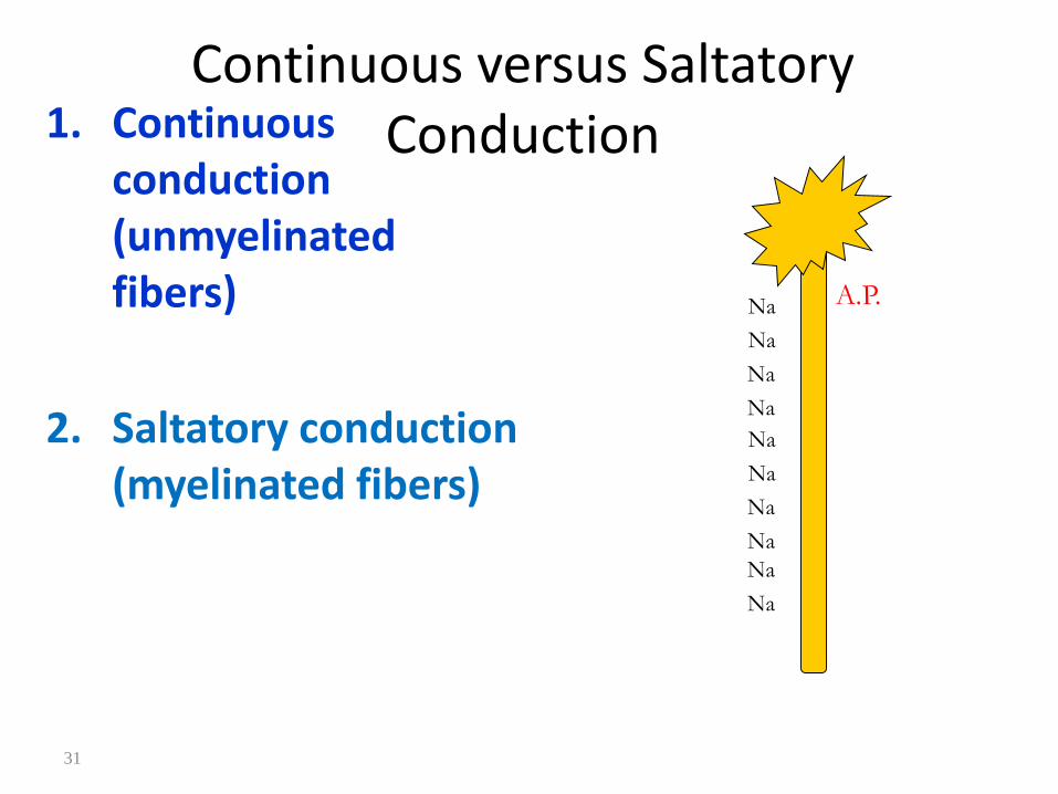
Continuous versus Saltatory Conduction1. Continuous
conduction (unmyelinatedfibers)
2. Saltatory conduction (myelinated fibers)
A.P.Na
Na
Na
Na
Na
Na
Na
Na
Na
Na
32
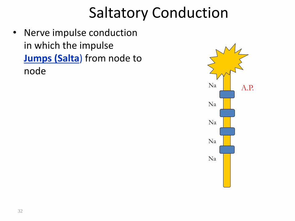
Saltatory Conduction• Nerve impulse conduction
in which the impulse Jumps (Salta) from node to node
A.P.Na
Na
Na
Na
Na
33
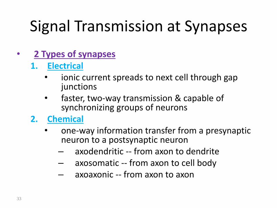
Signal Transmission at Synapses
• 2 Types of synapses1. Electrical
• ionic current spreads to next cell through gap junctions
• faster, two-way transmission & capable of synchronizing groups of neurons
2. Chemical• one-way information transfer from a presynaptic
neuron to a postsynaptic neuron– axodendritic -- from axon to dendrite– axosomatic -- from axon to cell body– axoaxonic -- from axon to axon

Synapses
Spinal Ganglia
Cell bodies Lie on posterior Nerve Root of Spinal Cord.
• Have Sensory Neurons (Unipolar).
• Widely Apart
• Sympathetic Ganglia
• Multipolar Type: Hence Many Dendrites Hence Widely Apart.