Embed Size (px)
Citation preview
Veterinary Parasitology 97 (2001) 153–157
Short communication
Neospora caninum infection associated withstillbirths in captive antelopes
(Tragelaphus imberbis)
M. Peters a,∗, P. Wohlsein a, A. Knieriem b, G. Schares c
a Department of Pathology, School of Veterinary Medicine, Bünteweg 17, D-30559 Hannover, Germanyb Zoological Garden Hannover, Adenauerallee 3, D-30175 Hannover, Germany
c Institute for Epidemiological Diagnostics, Federal Research Centre for Virus Diseases of Animals,Seestraße 55, D-16868 Wusterhausen, Germany
Received 18 July 2000; accepted 13 February 2001
Abstract
Neosporosis was diagnosed in full-term stillborn twin calves of captive antelopes (Tragelaphusimberbis) in a German zoo. In both calves a multifocal non-suppurative encephalitis was present andinfection with Neospora (N.) caninum was confirmed by foetal serology and PCR. N. caninum-DNAwas detected in brain, heart, lung, liver and spleen of a third full-term stillborn calf. Histopathologicallesions were absent in this calf and antibodies against N. caninum were not detected in pleural fluid.Immunohistochemistry failed to demonstrate N. caninum antigen in all three calves. © 2001 ElsevierScience B.V. All rights reserved.
Keywords: Neospora caninum; Stillbirth; Antelope
1. Introduction
Neospora (N.) caninum is an apicomplexan parasite closely related to Toxoplasma (T.)gondii. The protozoan causes neuromuscular disease in dogs and has been recognised as aneconomically important cause of bovine abortion, stillbirth and neonatal mortality (Dubeyand Lindsay, 1996). Furthermore, naturally occurring abortions and stillbirths associatedwith N. caninum infection were reported in sheep, goats, a horse, and a deer (Dubey, 1999).Dogs are definitive hosts for the parasite, shedding oocysts in their faeces after oral infection
∗ Corresponding author. Tel.: +49-511-9538664; fax: 49-511-9538675.E-mail address: [email protected] (M. Peters).
0304-4017/01/$ – see front matter © 2001 Elsevier Science B.V. All rights reserved.PII: S0 3 0 4 -4 0 17 (01 )00401 -0
154 M. Peters et al. / Veterinary Parasitology 97 (2001) 153–157
with tissue cyst-containing brain of N. caninum-infected mice (McAllister et al., 1998;Lindsay et al., 1999). In intermediate hosts, the only proven route of natural transmissionis congenital infection. Nevertheless, food and drinking water contaminated by N. caninumoocysts are in discussion to be a source of infection for intermediate hosts (Lindsay et al.,1999; McAllister et al., 1996). In this paper, we report N. caninum infection associated withstillbirths in captive antelopes (Tragelaphus imberbis) in a German zoo.
2. Material and methods
Two full-term, stillborn female twin antelope calves (Tragelaphus imberberis) (calves 1and 2) from the Zoological Garden of Hannover were submitted for post-mortem exami-nation in November 1999. Another full-term stillborn male calf (calf 3) was submitted inFebruary 2000. At the time of stillbirth the dam of the twin calves (dam 1) was six years old,the dam of the single calf (dam 2) four-years-old. Both were born in captivity, not related,and members of a herd of six adult Tragelaphus imberberis housed together with Antidorcasmarsupilians and Giraffa camelopardalis in a paddock. No abortion had occurred in anygroup of animals in the pen, but dam 2 had a further history of a stillbirth in November1998. Complete necropsies of the three stillbirths were done. Abomasal contents and liverswere examined using standard bacteriologic methods. Direct fluorescent antibody tests forBovine Herpes Virus 1 (BHV 1) and Bovine Virus Diarrhea (BVD)-Virus were done withspecific conjugates on frozen tissue sections. Native samples of brain, lung, liver, heartand spleen of calves 2 and 3 were removed for N. caninum- and T. gondii-PCR. For N.caninum-PCR the primers Np6+ and Np21+ (Müller et al., 1996), and for T. gondii-PCRthe primers oligos 1 and 4 (Burg et al., 1989) were used. Pleural fluids of all three calves andpre- and post-stillbirth serum samples of dam 1 (52 days before and 165 days after stillbirth,respectively) were examined for antibodies against N. caninum by an indirect immunfluo-rescent antibody test (IFAT) using an anti bovine IgG (H and L) fluorescein isothiocyanate(FITC) conjugate (samples were serially (two-fold) diluted from 1:2 to 1:1024) and im-munoblotting (IB) as described by Schares et al. (1998). For IB pleural fluids were diluted1:2 and sera 1:100. The recognition of two immunodominant antigens was regarded as pos-itive. Unfortunately, a serum sample of dam 2 was not available. Tissue samples of brain,heart, lung, liver, spleen, kidney, eyes, and muscle were fixed in 10% buffered formalinand processed using routine techniques. Tissue sections were cut at 5 �m and stained withhematoxylin and eosin. Deparaffinised sections were stained immunohistochemically witha rabbit antiserum developed against N. caninum (NC-1) tachyzoites as described earlier(Peters et al., 2000).
3. Results
Gross lesions were not detected in any of the three stillborn calves. Histologically, bothtwin calves (calves 1 and 2) revealed a moderate meningoencephalitis with multiple gran-ulomatous foci, gliosis and focal mild perivascular cuffing with lymphocytes and histio-cytes in the neuroparenchyma and leptomeninx. In the heart of calf 1, mild lymphocytic
M. Peters et al. / Veterinary Parasitology 97 (2001) 153–157 155
Table 1Results of serological examination of pleural fluids of the three stillborn calves, and of pre- and post-stillbirthserum samples of dam 1 for N. caninum antibodies, and results of N. caninum-PCR on tissue samples of calves 2and 3a
Animal N. caninum-Serology N. caninum-PCR
(IFAT) IB CNS Lung Heart Liver Spleen
Calf 1 1:16 + N.D. N.D. N.D. N.D. N.D.Calf 2 1:32 + + + − − −Dam 1 (52 d.a.s.) 1:128 + N.D. N.D. N.D. N.D. N.D.(165 d.p.s.) 1:128 + N.D. N.D. N.D. N.D. N.D.Calf 3 <1:2 − + + + + +
a N.D. = not done; IFAT = indirect immunofluorescent antibody test; IB = immunoblotting; d.a.s. = daysante-stillbirth; d.p.s. = days post-stillbirth.
myocarditis with focal mineralisation of myofibers was observed. In the livers mild peripor-tal infiltrations with lymphocytes and histiocytes were present. In all other tissues examined,inflammatory or degenerative lesions were absent. Inflammatory lesions were not detectedhistologically in any of the tissues of calf 3. N. caninum antigens were not detected in tissues
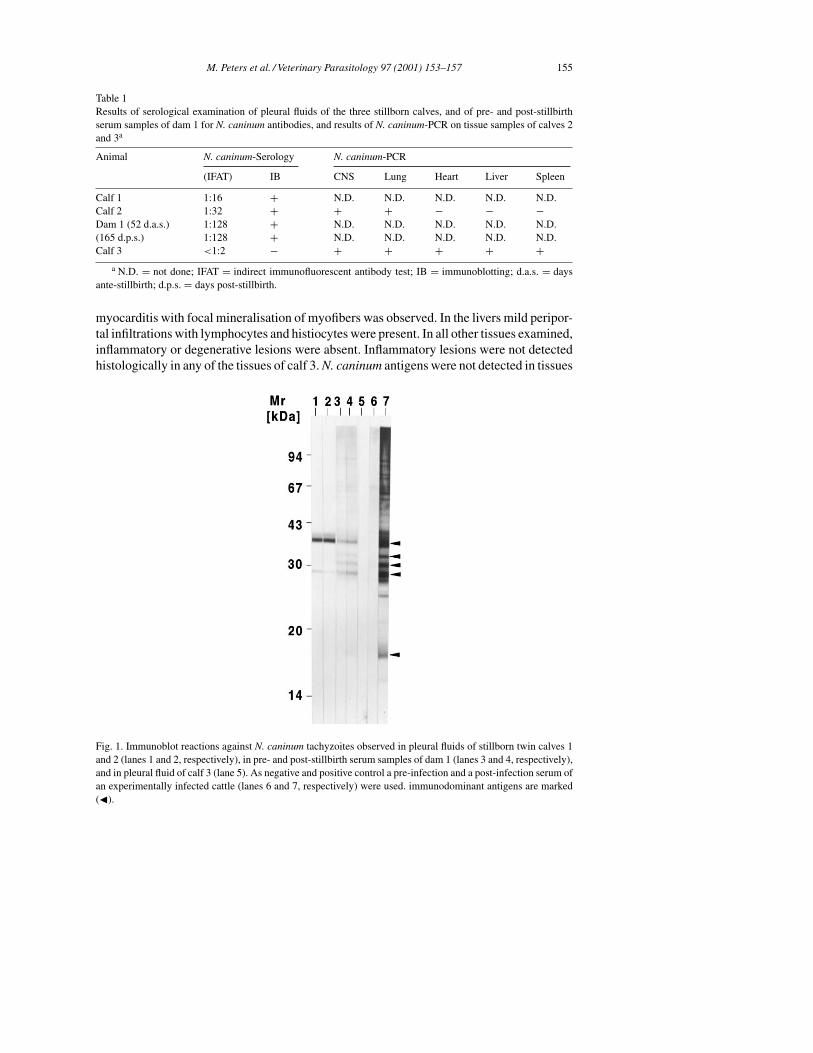
Fig. 1. Immunoblot reactions against N. caninum tachyzoites observed in pleural fluids of stillborn twin calves 1and 2 (lanes 1 and 2, respectively), in pre- and post-stillbirth serum samples of dam 1 (lanes 3 and 4, respectively),and in pleural fluid of calf 3 (lane 5). As negative and positive control a pre-infection and a post-infection serum ofan experimentally infected cattle (lanes 6 and 7, respectively) were used. immunodominant antigens are marked(�).
156 M. Peters et al. / Veterinary Parasitology 97 (2001) 153–157
of the three calves by immunohistochemistry. The results of serological examination of thepleural fluids of the calves and sera of dam 1, as well as the results of N. caninum-PCR ondifferent tissues of the calves are shown in Table 1. Immunoblot results are shown in Fig. 1.
T. gondii-DNA was not detected in tissues of calves 2 and 3 by PCR. In all three cases,bacterial cultures revealed a moderate growth of bacterial contaminants. BHV 1 and BVDantigens were not detected in tissues of the stillbirths.
4. Discussion
N. caninum infection was diagnosed in three stillborn calves of captive antelopes (Trage-laphus imberberis). This is the second report on congenital neosporosis in zoo animals. In1996, Dubey et al., reported transplacental neosporosis ending in stillbirth in a deer (Cervuseldi siamesis) from the Paris zoo.
The histopathological lesions of the stillborn twin calves are comparable with bovinefetal neosporosis (Thilsted and Dubey, 1989; Anderson et al., 1991; Wouda et al., 1997).Although, N. caninum antigen could not be detected in any of the tissue samples of thetwin calves by immunohistochemistry, infection with N. caninum was confirmed by foetalserology and PCR. Whether N. caninum infection of the twin calves was responsible for thestillbirths remains unclear. Twin pregnancies are rare in Tragelaphus imberbis (Cillié, 1987)and it is conceivable that they are a risk factor for abortion or stillbirth. In calf 3, inflam-matory histopathological lesions were absent. N. caninum antigens could not be detectedby immunohistochemistry. Nevertheless, N. caninum DNA was demonstrated by PCR inall tissue samples. Interestingly, calf 3 was seronegative for N. caninum. Possible explana-tions might be transplacental infection early in pregnancy before immunocompetence wasachieved, immunosuppression of the fetus, or recent infection of the fetus shortly beforestillbirth. The lack of histopathological lesions in this full-term calf indicates that verticalN. caninum infection in Tragelaphus imberberis can lead to birth of infected, but clinicallyhealthy calves, as it is well known for cattle. In cattle, only a small percentage of congen-itally infected calves develop clinical neosporosis (Paré et al., 1996; Schares et al., 1998).Unfortunately, a serum sample of dam 2 and serum samples of maternal ancestors of bothdams were not available. Therefore, possible vertical propagation of N. caninum infectionthrough different generations could not be followed. On the other hand, infection of theantelopes via contaminated food or drinking water cannot be ruled out, especially becausevisitors of the Zoological Garden of Hannover are allowed to take their dog with them.
References
Anderson, M.L., Blanchard, P.C., Barr, B.C., Dubey, J.P., Hoffman, R.L., Conrad, P.A., 1991. Neospora-likeprotozoan infection as a major cause of abortion in California dairy cattle. J. Am. Vet. Med. Assoc. 198,241–244.
Burg, J.L., Grover, C.M., Pouletty, P., Boothroyd, J.C., 1989. Direct and sensitive detection of a pathogenicprotozoan, Toxoplasma gondii, by polymerase chain reaction. J. Clin. Microbiol. 27, 1787–1792.
Cillié, B., 1987. Mammals of Southern Africa. Frandsen Publishers, Sandton.Dubey, J.P., 1999. Recent advances in Neospora and neosporosis. Vet. Parasitol. 67, 1–59.Dubey, J.P., Lindsay, D.S., 1996. A review of Neospora caninum and neosporosis. Vet. Parasitol. 67, 1–59.
M. Peters et al. / Veterinary Parasitology 97 (2001) 153–157 157
Dubey, J.P., Rigoulet, J., Lagourette, P., George, C., Longeart, L., LeNet, J.L., 1996. Fatal transplacental neosporosisin a deer (Cervus eldi siamensis). J. Parasitol. 82, 338–339.
Lindsay, D.S., Dubey, J.P., Duncan, R.B., 1999. Confirmation that the dog is a definitive host for Neospora caninum.Vet. Parasitol. 82, 327–333.
Lindsay, D.S., Dubey, J.P., McAllister, M., 1999. Neospora caninum and the potential for parasite transmission.J. Comp. Cont. Ed. 21, 317–321.
McAllister, M.M., Dubey, J.P., Lindsay, D.S., Jolly, W.R., Wills, R.A., McGuire, A.M., 1998. Dogs are definitivehosts of Neospora caninum. Int. J. Parasitol. 28, 1473–1478.
McAllister, M.M., Huffman, E.M., Hietala, S.K., Conrad, P.A., Anderson, M.L., Salman, M.D., 1996. Evidencesuggesting a point source exposure in an outbreak of bovine abortion due to neosporosis. J. Vet. Diagn. Invest.8, 355–357.
Müller, N., Zimmermann, V., Hentrich, B., Gottstein, B., 1996. Diagnosis of Neospora caninum and Toxoplasmagondii infection by PCR and DNA hybridisation immunoassay. J. Clin. Microbiol. 34, 2850–2852.
Paré, J., Thurmond, M.C., Hietala, S.K., 1996. Congenital Neospora caninum infection in dairy cattle andassociated calfhoud mortality. Can. J. Vet. Res. 60, 133–139.
Peters, M., Wagner, F., Schares, G., 2000. Canine neosporosis: clinical and pathological findings and first isolationof Neospora caninum in Germany. Parasitol. Res. 86, 1–7.
Schares, G., Peters, M., Wurm, R., Bärwald, A., Conraths, F.J., 1998. The efficiency of vertical transmission ofNeospora caninum in dairy cattle analysed by serological techniques. Vet. Parasitol. 80, 87–98.
Thilsted, J.P., Dubey, J.P., 1989. Neosporosis-like abortions in a herd of dairy cattle. J. Vet. Diagn. Invest. 1,205–209.
Wouda, W., Moen, A.R., Visser, I.J.R., van Knapen, F., 1997. Bovine fetal neosporosis: a comparison of epizooticand sporadic abortion cases and different age classes with regard to lesion severity and immunohistochemicalidentification of organisms in brain, heart, and liver. J. Vet. Diagn. Invest. 9, 180–185.