Embed Size (px)
DESCRIPTION
Neonatal Suctioning Guidelines
Citation preview
1
ERNBG Guideline – Suction February 2006 Review due: February 2006
Eastern Regional Neonatal Benchmarking Group
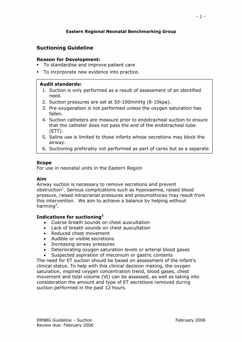
Suctioning Guideline
Reason for Development: § To standardise and improve patient care
§ To incorporate new evidence into practice.
Scope For use in neonatal units in the Eastern Region
Aim Airway suction is necessary to remove secretions and prevent obstruction 1 . Serious complications such as hypoxaemia, raised blood pressure, raised intracranial pressures and pneumothorax may result from this intervention. We aim to achieve a balance by helping without harming 2 .
Indications for suctioning 3
• Coarse breath sounds on chest auscultation • Lack of breath sounds on chest auscultation • Reduced chest movement • Audible or visible secretions • Increasing airway pressures • Deteriorating oxygen saturation levels or arterial blood gases • Suspected aspiration of meconium or gastric contents
The need for ET suction should be based on assessment of the infant's clinical status. To help with this clinical decision making, the oxygen saturation, inspired oxygen concentration trend, blood gases, chest movement and tidal volume (Vt) can be assessed, as well as taking into consideration the amount and type of ET secretions removed during suction performed in the past 12 hours.
Audit standards: 1. Suction is only performed as a result of assessment of an identified need.
2. Suction pressures are set at 50100mmHg (810kpa). 3. Preoxygenation is not performed unless the oxygen saturation has fallen.
4. Suction catheters are measure prior to endotracheal suction to ensure that the catheter does not pass the end of the endotracheal tube (ETT).
5. Saline use is limited to those infants whose secretions may block the airway.
6. Suctioning preferably not performed as part of cares but as a separate procedure or at the end of cares.
2
ERNBG Guideline – Suction February 2006 Review due: February 2006
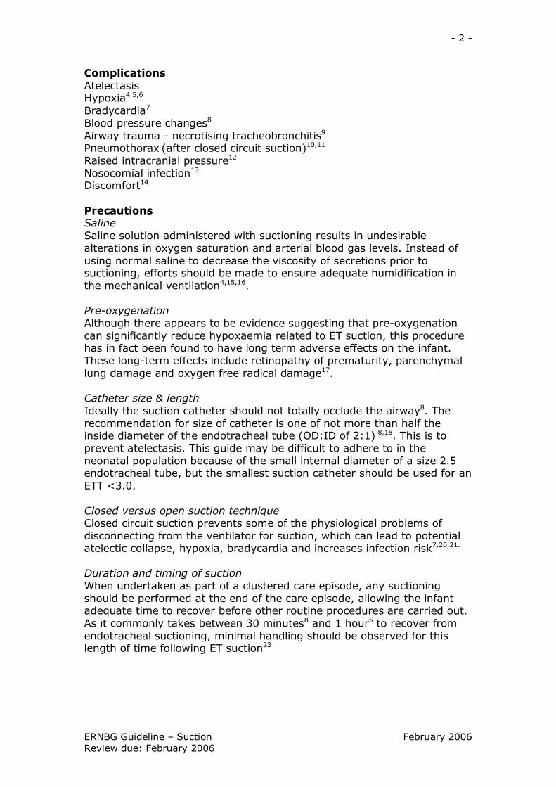
Complications Atelectasis Hypoxia 4,5,6
Bradycardia 7
Blood pressure changes 8
Airway trauma necrotising tracheobronchitis 9
Pneumothorax (after closed circuit suction) 10,11
Raised intracranial pressure 12
Nosocomial infection 13
Discomfort 14
Precautions Saline Saline solution administered with suctioning results in undesirable alterations in oxygen saturation and arterial blood gas levels. Instead of using normal saline to decrease the viscosity of secretions prior to suctioning, efforts should be made to ensure adequate humidification in the mechanical ventilation 4,15,16 .
Preoxygenation Although there appears to be evidence suggesting that preoxygenation can significantly reduce hypoxaemia related to ET suction, this procedure has in fact been found to have long term adverse effects on the infant. These longterm effects include retinopathy of prematurity, parenchymal lung damage and oxygen free radical damage 17 .
Catheter size & length Ideally the suction catheter should not totally occlude the airway 8 . The recommendation for size of catheter is one of not more than half the inside diameter of the endotracheal tube (OD:ID of 2:1) 8,18 . This is to prevent atelectasis. This guide may be difficult to adhere to in the neonatal population because of the small internal diameter of a size 2.5 endotracheal tube, but the smallest suction catheter should be used for an ETT <3.0.
Closed versus open suction technique Closed circuit suction prevents some of the physiological problems of disconnecting from the ventilator for suction, which can lead to potential atelectic collapse, hypoxia, bradycardia and increases infection risk 7,20,21.
Duration and timing of suction When undertaken as part of a clustered care episode, any suctioning should be performed at the end of the care episode, allowing the infant adequate time to recover before other routine procedures are carried out. As it commonly takes between 30 minutes 8 and 1 hour 5 to recover from endotracheal suctioning, minimal handling should be observed for this length of time following ET suction 23
3
ERNBG Guideline – Suction February 2006 Review due: February 2006
Endotracheal Suction
Equipment § Stethoscope § Gloves (for protection of the wearer) § Suction catheters § e.g. Paediatric Yankeur for meconium
6 fg for a 2.5mm endotracheal tube 8 fg for a 3.0mm – 4.00mm endotracheal tube 6fg – 10fg (for oral or nasal secretions depending on the thickness of secretions and the size and gestation of the baby).
§ Suction connecting tube § Suction bottle connected to suction apparatus – pressures set 50 100mmHg (810kpa) 24
§ Saline if required § Rubbish bag § Sterile water (to rinse suction tubes after the procedure) § Another person to comfort and contain the baby § Documentation
Preparation: 1. Undertake an assessment of need – § Have the physiological parameters changed? § Is the chest moving? § What was the result of the most recent blood gas? § Auscultate the chest – are the breath sounds noisy, is air entry equal? § Has the tidal volume or minute volume decreased? § Has the oxygen requirement increased? § What were the secretions like on the last suction event? § When was the last suction performed? § Has the infant recently been handled?
2. If the parents are present explain the reason for suction and the procedure
3. Wash hands
4. Prepare equipment as above
5. If using saline instillation prior to suctioning draw up 0.25 0.5 ml [0.25 =<2kg infant 0.5 mls = >2kg infant ] 25,4 into 2 ml syringe, taking care not to touch key parts, and replace syringe back into paper packet
6. Attach suction catheter to suction tubing, leaving catheter in the protective packaging to prevent contamination
7. Check the vacuum pressure ensuring that the maximum negative pressure does not exceed 50100mmHg (810kpa) 24
8. Determine length of endotracheal tube (including any dead space and length of the blue ETT hub). This is the length you will need to pass
4
ERNBG Guideline – Suction February 2006 Review due: February 2006
the suction catheter to ensure that the catheter reaches the end of the ETT tip 26,27 . This can be achieved by observing the markings of the suction catheter or by measuring the catheter against a precut tape measure which may be stuck to the inside of the incubator
9. Observe presuctioning saturation, apex beat and blood pressure (if monitored) and ensure the infant is well oxygenated prior to procedure without "preoxygenating" 20 .
Emergency suction For emergency suction of the ETT, step 9 of the preparation procedure may not be achievable. It may be necessary to increase the inspired oxygen concentration in response to desaturation, but in some cases (such as blocked ETT) waiting until the oxygen saturation is >90% is not practical. Suction in this situation should be performed to prevent further desaturation or bradycardia.
10.Auscultate the chest prior to suctioning to have baseline information on which to compare post suctioning auscultation
11.Position the head in the midline if possible to reduce changes to cerebral blood flow.
12.Where possible organize another person/parent to help with the procedure to allow the infant to be contained during the suctioning as it has been shown to aid recovery.
Procedure:
1. Wash hands thoroughly and dry, apply alcohol handrub and allow to dry
Saline if required 31,32,33,34
• If there are indications for using saline, instil this into the ETT • Reconnect the ventilator for a minimum of 5 breaths to ensure that the saline has thoroughly moistened the ETT
2. Put on two unsterile gloves to the dominant hand. Double gloving means that the top glove can be removed to contain the catheter after the suctioning is complete
3. Withdraw suction catheter from protective packaging, holding in hand with sterile glove
4. Silence ventilator alarm with non sterile hand
5. Detach ETT from ventilation tubing with nondominant hand
6. Steady the ET tube with the nondominant hand and insert the suction catheter down the ETT to the predetermined length with the gloved hand. The catheter should not advance beyond the end of the ETT 9 .
5
ERNBG Guideline – Suction February 2006 Review due: February 2006
7. Apply suction by occluding the finger tip control with either a finger or thumb
8. Withdraw catheter, whilst applying suction. It should take no longer than 4 5 seconds to completely withdraw catheter. The whole process should not exceed 1015 seconds 19.
9. Reattach ventilation tubing to the ETT.
10. Assess tolerance of the procedure by observing oxygen saturation, colour, heart rate and activity.
11. Adjust the FiO2 to stabilise the oxygen saturations of the baby and reset to baseline requirements when the baby reaches presuction apex and oxygen saturation levels
12. Auscultate the chest and repeat suction as necessary
13. If further suction is needed, repeat the procedure from step 11 using new suction catheter each time. Usually 12 attempts are sufficient to clear ET secretions
14. Observe colour, quantity and type of secretions in the suction catheter
15. Continue containment if the infant has not tolerated the procedure well.
Following procedure:
1. Remove the glove from the dominant hand by inverting it over the used catheter
2. Dispose of waste in yellow clinical waste bin
3. Use Chlorhexidine solution/water to clean through suction tubing and leave suction switched on in incubator but away from infant
4. Wash hands and dry thoroughly
5. Record on ITU chart including amount, type and colour of secretions. If fresh blood is obtained, report to medical staff immediately
6. Document tolerance and effectiveness of the procedure in the care plan
Collecting a specimen:
Additional equipment needed Mucus trap Ampoule of sterile water
6
ERNBG Guideline – Suction February 2006 Review due: February 2006
Procedure • If a specimen is required, a mucus trap should be attached between the suction tubing and the suction catheter.
• ET suction should then be performed in the same way as detailed in the guideline.
• Following suction the sterile water should be suctioned through the catheter into the mucus trap before it is sealed.
Additional Information:
HFOV & suctioning The procedure for ET suction is exactly the same for infants receiving HFOV or conventional ventilation. If using the sensor medics ventilator, the alarm should be silenced during disconnection from the ventilator and the start/stop button should be depressed to recommence ventilation once the ETT is reconnected to the ventilator tubing. After instilling the saline, it may be necessary to oscillate for a few seconds, as there is no great pressure shift to get the saline into the ETT. In some situations the mean airway pressure may need to be briefly increased by 12cm to rerecruit alveoli and stabilise ventilation, but care must be taken to reduce back to initial settings within a specific time frame (discuss on ward round with consultant) to avoid any over distension of already damaged lungs.
Oro/nasopharyngeal suction
Preparation: 1. As with the preparation for endotracheal suctioning there should be an assessment of need prior to undertaking suction.
2. If the parents are present explain the reason for suction and the procedure
3. Wash hands
4. Prepare equipment needed for nasopharyngeal suction and check that equipment is working.
5. Attach suction catheter to suction tubing, leaving catheter in the protective packaging to prevent contamination
6. Check the vacuum pressure ensuring that the maximum negative pressure does not exceed 50100mmHg (810kpa).
7. Observe presuctioning saturation, apex beat and blood pressure (if monitored) and ensure the infant is well oxygenated prior to procedure without "preoxygenating"
7
ERNBG Guideline – Suction February 2006 Review due: February 2006
8. Where possible organize another person/parent to help with the procedure to allow the infant to be contained during the suctioning as it has been shown to aid recovery.
Procedure: 1. Wash hands thoroughly and dry, apply alcohol handrub and allow to dry
2. Put on sterile gloves to protect the hands from any secretions and to keep the catheter clean prior to insertion.
Nasal suction 3. Measure the catheter from the mouth to the suprasternal notch to estimate the length required for insertion 33 .
4. It may be necessary to lubricate the tip of the catheter in some saline/water to prevent trauma to the lining of the nose.
5. Introduce the catheter gently into the nostril and ease it to the back of the pharynx approximately 48cm 2 .
6. Apply suction and gently withdraw the catheter not taking more than 10 seconds. Suction should not be applied whilst inserting the catheter as this causes mucosal irritation, damage and can potentially lead to hypoxia.
Oral suction 7. Gently insert the catheter into the mouth in an upward and backward direction, if the infant has a gag reflex he/she may cough
8. If the infant does not have a gag reflex, measure the catheter from the mouth to the suprasternal notch to estimate the length required for insertion and then insert the catheter as above
9. Apply suction and gently withdraw the catheter not taking more than 10 seconds. Suction should not be applied whilst inserting the catheter as this causes mucosal irritation, damage and can potentially lead to hypoxia.
8
ERNBG Guideline – Suction February 2006 Review due: February 2006
Tracheostomy suction
1. Assess the need for suction excessive coughing, cyanosis, tachypnoea, bradycardia and apnoea or excessive secretions
2. The suction equipment should be preset at 50100 mmHg (or 8 10kpa) to minimise the risk of trauma and atelectasis.
3. Measure the depth to which the suction tube needs to be passed against an identical tracheostomy tube 34 .
4. Wash hands, dry and apply alcohol hand rub to reduce the risk of infection
5. Open the suction catheter and attach to the tubing, leaving the rest of the catheter in the packet to keep the catheter as clean as possible
6. Put on two nonsterile gloves on your dominant hand to minimise the risk of infection. The gloves should be nonpowdered to prevent the introduction of powder into the airway.
7. Instil saline if needed due to the presence of thick or copious secretions. Always use a plastic ampoule (glass ampoules should never be used as there is the risk that glass fragments could be introduced with the saline) 0.250.5ml is sufficient to loosen secretions 35 .
8. Insert the catheter into the tracheostomy tube. DO NOT pass catheter beyond end of tracheostomy tube (check the length by measuring against another tube).
9. To minimise irritation of the mucous membranes apply suction to the side port only as the catheter is gently removed. Do not rotate the catheter as it is withdrawn.
10. Any pass of the suction catheter should not take longer than 10 seconds.
11. Reassess the infant to determine whether further suctioning is necessary, ensuring infant has recovery time between each pass. Use a new sterile catheter on each occasion.
12. Observe for recovery of oxygenation, heart rate, respiration altering FiO2 if necessary.
13. Disconnect and dispose of the catheter. Clear the suction tubing with aqueous Hibisol. Attach a new catheter ready for the next use.
14. Record the suctioning event on the ITU chart, indicating the amount, colour and consistency. Secretions are likely to be blood stained in the first 24 hours.
9
ERNBG Guideline – Suction February 2006 Review due: February 2006
Signs that suctioning has been effective § Reduced work of breathing § Reduced respiratory rate in the unventilated infant § Increased oxygen saturation § Chest movement improves § Mv/Vt improves § Apnoea & bradycardia events lessen § Visible evidence of secretion removal § Absence of audible/visible secretions in the upper airway
Others guidelines used in the development of this guideline § Queens Medical Centre. Tracheal Suction Physiotherapy Practice Guidelines
§ Royal Free (1999) Guidelines for Tracheal Suction
References 1. Levene, Tudehope and Thearle (2000) Essentials of neonatal medicine 3 rd Ed. p 123 Blackwell Science Ltd. Oxford and London. [III]
2. Czarnecki ML, Kariac CL. (1999) Infant basalpharyngeal suctioning; is it beneficial? Pediatric Nursing. March/April 25(2):1936,218. [III]
3. Day T, Wainwright SP, WilsonBarnett J. (2001) An evaluation of a teaching intervention to improve the practice of endotracheal suctioning in intensive care units. Journal of Clinical Nursing. September;10(5):682696. [IIb]
4. Kinloch D (2000) Installation of normal saline during endotracheal suctioning: effects on mixed venous oxygen saturation. American Journal of Critical Care.
5. January;9(1):789. [III]
6. Evans JC. (1991) Incidence of hypoxemia associated with caregiving in premature infants. Neonatal Network. September;10(2):1724.[III]
7. Evans JC. (1992) Reducing the hypoxemia, bradycardia and apnea associated with suctioning in low birth weight infants. Journal of Perinatology. June;12(2):13742. [III]
8. Tan AM, Gomez JM, Mathews J, Williams M, Paratz J, Rajadurai VS. (2005) Closed versus partially ventilated endotracheal suction in extremely preterm neonates: physiological consequences. Intensive Critical Care Nursing. August;21(4):23442. Epub. [Ib]
9. Simbruner G, Coradello H, Fodor M, Havelec L, Lubec G, Pollak A (1981) Effect of tracheal suction on oxygenation, circulation and lung mechanics in newborn infants. Archives of Disease in Childhood. 56 (5):326330 [Ib]
10. Kleiber C, Krutzfeld N, Rose EF. (1988) Acute histologic changes in the tracheobronchial tree associated with different suction catheter insertion techniques. Heart Lung. January;17(1):1014. [IIa]
10
ERNBG Guideline – Suction February 2006 Review due: February 2006
11. Thakur A, Buchmiller T, Atkinson J. (2000) Bronchial perforation after closedtube endotracheal suction. Journal of Pediatric Surgery. September;35(9):13535. [III]
12. GarciaAparico L, Castanon M, Tarrado X, Rodriguez L, Iriondo M, Morales L. (2002) Bronchial complication of a closedtube endotracheal suction catheter. Journal of Pediatric Surgery. October;37(10):14834. [III]
13. Durand M, Sangha B, Hoppenbrouwers T, Hodgman JE. (1989) Cardiopulmonary and intracranial pressure changes related to endotracheal suctioning in preterm infants. Critical Care Medicine. June;17(6):50610. [III]
14. Fiorentini A (1992) Potential hazards of tracheobronchial suctioning. Intensive Critical Care Nursing. December;8(4):21726. [IV]
15. Anand KJS, Barton RA, McIntosh N, Lagercrantz H, Pelausa E, Young TE, Vasa R. (1999) Analgesia and sedation in preterm neonates who require ventilatory support: results from the NOPAIN trial. Archives of Pediatric and Adolescent Medicine. April;153(4):3318. [Ib]
16. Akgul S, Akyolcu N. (2002) Effects of normal saline on endotracheal suctioning. Journal of Clinical Nursing. November;11(6):826830. [III]
17. Ridling DA, Martin LD, Bratton SL. (2003) Endotracheal suctioning with or without installation of isotonic sodium chloride in critically ill children. American Journal of Critical Care. May;12(3):212219. [Ib]
18. Pritchard M, Flenady V, Woodgate P (2000) Preoxygenation for tracheal suctioning intubated, ventilated newborn infants. The Cochrane Library [Ia]
19. Smith C (1995) Endotracheal suctioning in children: an overview of the principle problems. Journal of Neonatal and Paediatric Critical Care. 1(3):4650. Cited in: Wallace JL (1998) Suctioning a two edged sword: reducing the theorypractice gap. Journal of Neonatal Nursing.4(6):12,1417. [IV]
20. Mosca FA, Colnaghi M, Lattanzio M, Bray M, Pugiliese S, Fumagalli M. (1997) Closed versus open endotracheal suctioning in preterm infants: effects on cerebral oxygenation and blood volume. Biology of the Neonate. 72(1):914.
21. Cordero L, Sananes M, Ayers LW. (2000) Comparison of a closed (Trach Care MAC) with an open endotracheal suction system in small premature infants. Journal of Perinatology. AprilMay;20(3):1516. [Ib]
22. Woodgate PG, Flenady V. (2001) Tracheal suction without disconnection in intubated ventilated neonates. Cochrane Database of Systematic Reviews. (2):CD003065. [Ia]
11
ERNBG Guideline – Suction February 2006 Review due: February 2006
23. Hodge D (1991) Endotracheal suctioning and the infant: A nursing care protocol to decrease complications. Neonatal Network. 9 (5):715 [IV]
24. Bernert G et al. (1997) The effect of behavioural states on cerebral oxygenation during endotracheal suctioning of preterm babies. Neuropediatrics. April;28(2):1115. [IIb]
25. Howard F. (1994) Endotracheal suctioning and the neonate. Pediatric Nursing.6(7):1417. Cited in: Wallace JL (1998) Suctioning a two edged sword: reducing the theorypractice gap. Journal of Neonatal Nursing.4(6):12,1417. [IV]
26. Cordero L, Sananes M, Ayers LW (2001) A comparison of two airway suctioning frequencies in mechanically ventilated, very low birth weight infants. Respiratory Care. 46 (8):783788 [IIa]
27. Spence K, Gillies D, Waterworth L. (2005) Deep versus shallow suction of endotracheal tubes in ventilated neonates and young infants. Cochrane Database of Systematic Reviews.2:CD003309. [1a]
28. Youngmee A, Yonghoon J. (2003) The effects of the shallow and the deep endotracheal suctioning on oxygen saturation and heart rate in highrisk infants. International Journal of Nursing Studies.40:97104. [IIa]
29. Shorten D, Byrne P, Jones R (1991) Infant responses to saline instillations and endotracheal suctioning. Journal of Obstetric, Gynecological and Neonatal Nursing. 20 (6): 464 469 [IIa]
30. Blackwood B (1999) Normal saline instillation with endotracheal suctioning: primum non nocere (first do no harm). Journal of Advanced Nursing. 29 (4):928934 [IV]
31. Raymond SJ (1995) Normal saline instillation before suctioning: Helpful or harmful? A review of the literature. The American Journal of Critical Care. 4(4):267271 [IV]
32. Beeram MR, Dhanireddy R (1992) Effects of saline instillation during tracheal suction on lung mechanics in newborn infants. Journal of Perinatology 12 (2):120123 [IIb]
33. Kay J. (2000) Ch. 36 Suctioning. In: Huband S, Trigg E. Practices in Children’s Nursing. Chirchill Livingstone. Edinburgh. [IV]
34. Runton N. (1992) Suctioning artificial airways in children: appropriate technique. Pediatric Nursing. April;18(2):115118. [IV]
35. Butler A. (2000) Tracheostomy Care. In: Huband S, Trigg E. Practices in Children’s Nursing. Chirchill Livingstone. Edinburgh. [IV]