Embed Size (px)
Citation preview
Neonatal Resuscitation
A Life-Saving Guide for Newborns
Contents
Neonatal Resuscitation: An Overview ..............................................................................................................................................2
Transitioning from Fetus to Neonate ...............................................................................................................................................2
Normal Transition: A Birth ............................................................................................................................................................2
Problems with Transition .............................................................................................................................................................3
Predicting the Need for Resuscitation ..........................................................................................................................................3
Neonatal Assessment .......................................................................................................................................................................4
The Apgar Score ............................................................................................................................................................................4
The Apgar Score Is Not Used to Direct Resuscitation ...............................................................................................................5
Assessing the Need for Neonatal Resuscitation ...........................................................................................................................5
Staying with the Mother ...........................................................................................................................................................6
Further Evaluation ....................................................................................................................................................................6
Pulse Oximetry .............................................................................................................................................................................6
Neonatal Resuscitation Techniques .................................................................................................................................................7
Supplemental Oxygen...................................................................................................................................................................8
Positive Pressure Ventilation ........................................................................................................................................................8
Positive Pressure Ventilation with Bag-Mask Devices ..............................................................................................................8
Effective Positive Pressure Ventilation .....................................................................................................................................9
Laryngeal Mask Airway .................................................................................................................................................................9
Intubation .................................................................................................................................................................................. 10
Chest Compressions .................................................................................................................................................................. 11
Epinephrine ............................................................................................................................................................................... 11
Volume Expansion ..................................................................................................................................................................... 11
Resuscitation Tools ........................................................................................................................................................................ 12
Factors That May Complicate Resuscitation ................................................................................................................................. 12
Airway Obstructions .................................................................................................................................................................. 12
Choanal Atresia...................................................................................................................................................................... 12
Robin Syndrome .................................................................................................................................................................... 12
Pulmonary Complications .............................................................................................................................................................1
Impaired Respiratory Drive ..........................................................................................................................................................1
Cardiac Abnormalities ..................................................................................................................................................................1
Post-Resuscitation Care ....................................................................................................................................................................2
Resuscitating Preterm Babies ...........................................................................................................................................................3
Ethical Considerations ......................................................................................................................................................................4
References ........................................................................................................................................................................................5
Neonatal Resuscitation: An Overview
Nine out of ten neonates successfully transition from fetus to newborn. Unfortunately, the remaining 10% of neonates require
assistance from medical providers. Less than 10% of these newborn babies (approximately 1% overall) will require neonatal
resuscitation. On the other hand, almost one-quarter of neonatal deaths occur because of birth asphyxia, according to the
medical journal The Lancet (Lawn, Cousens, & Zupan, 2005). Therefore, practitioners who are adept at neonatal resuscitation
can save the lives of newborn babies.
Transitioning from Fetus to Neonate
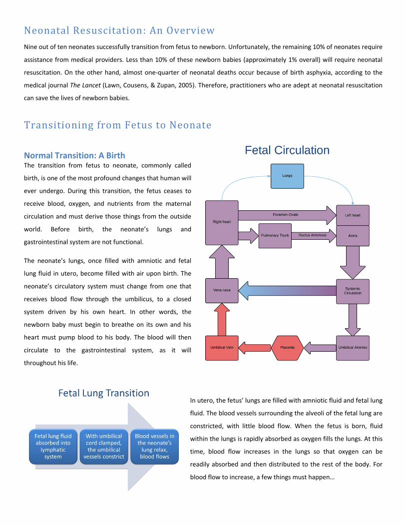
Normal Transition: A Birth The transition from fetus to neonate, commonly called
birth, is one of the most profound changes that human will
ever undergo. During this transition, the fetus ceases to
receive blood, oxygen, and nutrients from the maternal
circulation and must derive those things from the outside
world. Before birth, the neonate’s lungs and
gastrointestinal system are not functional.
The neonate’s lungs, once filled with amniotic and fetal
lung fluid in utero, become filled with air upon birth. The
neonate’s circulatory system must change from one that
receives blood flow through the umbilicus, to a closed
system driven by his own heart. In other words, the
newborn baby must begin to breathe on its own and his
heart must pump blood to his body. The blood will then
circulate to the gastrointestinal system, as it will
throughout his life.
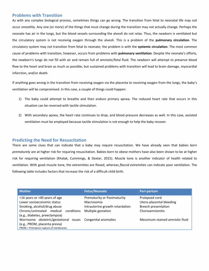
In utero, the fetus’ lungs are filled with amniotic fluid and fetal lung
fluid. The blood vessels surrounding the alveoli of the fetal lung are
constricted, with little blood flow. When the fetus is born, fluid
within the lungs is rapidly absorbed as oxygen fills the lungs. At this
time, blood flow increases in the lungs so that oxygen can be
readily absorbed and then distributed to the rest of the body. For
blood flow to increase, a few things must happen…
Problems with Transition As with any complex biological process, sometimes things can go wrong. The transition from fetal to neonatal life may not
occur smoothly. Any one (or more) of the things that must change during the transition may not actually change. Perhaps the
neonate has air in the lungs, but the blood vessels surrounding the alveoli do not relax. Thus, the newborn is ventilated but
the circulatory system is not receiving oxygen through the alveoli. This is a problem of the pulmonary circulation. The
circulatory system may not transition from fetal to neonate; the problem is with the systemic circulation. The most common
cause of problems with transition, however, occurs from problems with pulmonary ventilation. Despite the neonate's efforts,
the newborn's lungs do not fill with air and remain full of amniotic/fetal fluid. The newborn will attempt to preserve blood
flow to the heart and brain as much as possible, but sustained problems with transition will lead to brain damage, myocardial
infarction, and/or death.
If anything goes wrong in the transition from receiving oxygen via the placenta to receiving oxygen from the lungs, the baby’s
ventilation will be compromised. In this case, a couple of things could happen:
1) The baby could attempt to breathe and then endure primary apnea. The reduced heart rate that occurs in this
situation can be reversed with tactile stimulation.
2) With secondary apnea, the heart rate continues to drop, and blood pressure decreases as well. In this case, assisted
ventilation must be employed because tactile stimulation is not enough to help the baby recover.
Predicting the Need for Resuscitation There are some clues that can indicate that a baby may require resuscitation. We have already seen that babies born
prematurely are at higher risk for requiring resuscitation. Babies born to obese mothers have also been shown to be at higher
risk for requiring ventilation (Khalak, Cummings, & Dexter, 2015). Muscle tone is another indicator of health related to
ventilation. With good muscle tone, the extremities are flexed, whereas flaccid extremities can indicate poor ventilation. The
following table includes factors that increase the risk of a difficult child birth.
Mother Fetus/Neonate Peri-partum
<16 years or >40 years of age Prematurity or Postmaturity Prolapsed cord Lower socioeconomic status Macrosomia Utero-placental bleeding Smoking, alcohol/drug abuse Intrauterine growth retardation Breech presentation Chronic/untreated medical conditions (e.g., diabetes, preeclampsia)
Multiple gestation Chorioamnionitis
Worrisome obstetric/gestational issues (e.g., PROM, placenta previa)
Congenital anomalies Meconium-stained amniotic fluid
PROM = Premature rupture of membranes
Neonatal Assessment
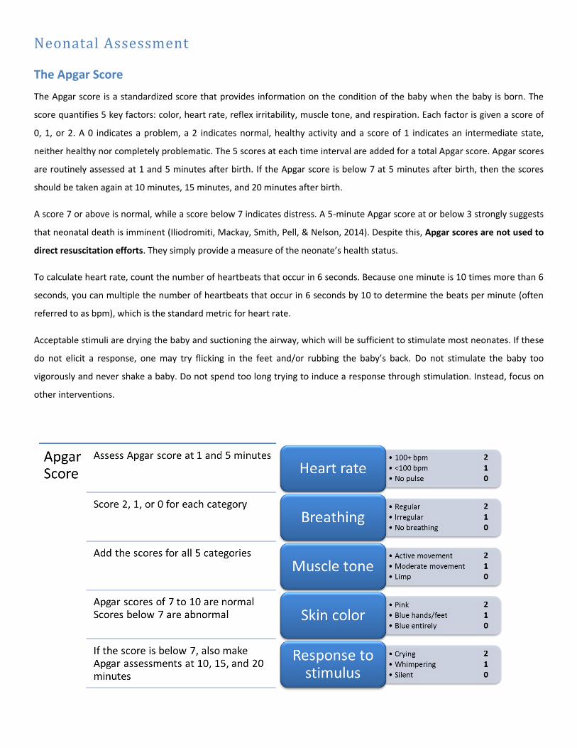
The Apgar Score
The Apgar score is a standardized score that provides information on the condition of the baby when the baby is born. The
score quantifies 5 key factors: color, heart rate, reflex irritability, muscle tone, and respiration. Each factor is given a score of
0, 1, or 2. A 0 indicates a problem, a 2 indicates normal, healthy activity and a score of 1 indicates an intermediate state,
neither healthy nor completely problematic. The 5 scores at each time interval are added for a total Apgar score. Apgar scores
are routinely assessed at 1 and 5 minutes after birth. If the Apgar score is below 7 at 5 minutes after birth, then the scores
should be taken again at 10 minutes, 15 minutes, and 20 minutes after birth.
A score 7 or above is normal, while a score below 7 indicates distress. A 5-minute Apgar score at or below 3 strongly suggests
that neonatal death is imminent (Iliodromiti, Mackay, Smith, Pell, & Nelson, 2014). Despite this, Apgar scores are not used to
direct resuscitation efforts. They simply provide a measure of the neonate’s health status.
To calculate heart rate, count the number of heartbeats that occur in 6 seconds. Because one minute is 10 times more than 6
seconds, you can multiple the number of heartbeats that occur in 6 seconds by 10 to determine the beats per minute (often
referred to as bpm), which is the standard metric for heart rate.
Acceptable stimuli are drying the baby and suctioning the airway, which will be sufficient to stimulate most neonates. If these
do not elicit a response, one may try flicking in the feet and/or rubbing the baby’s back. Do not stimulate the baby too
vigorously and never shake a baby. Do not spend too long trying to induce a response through stimulation. Instead, focus on
other interventions.
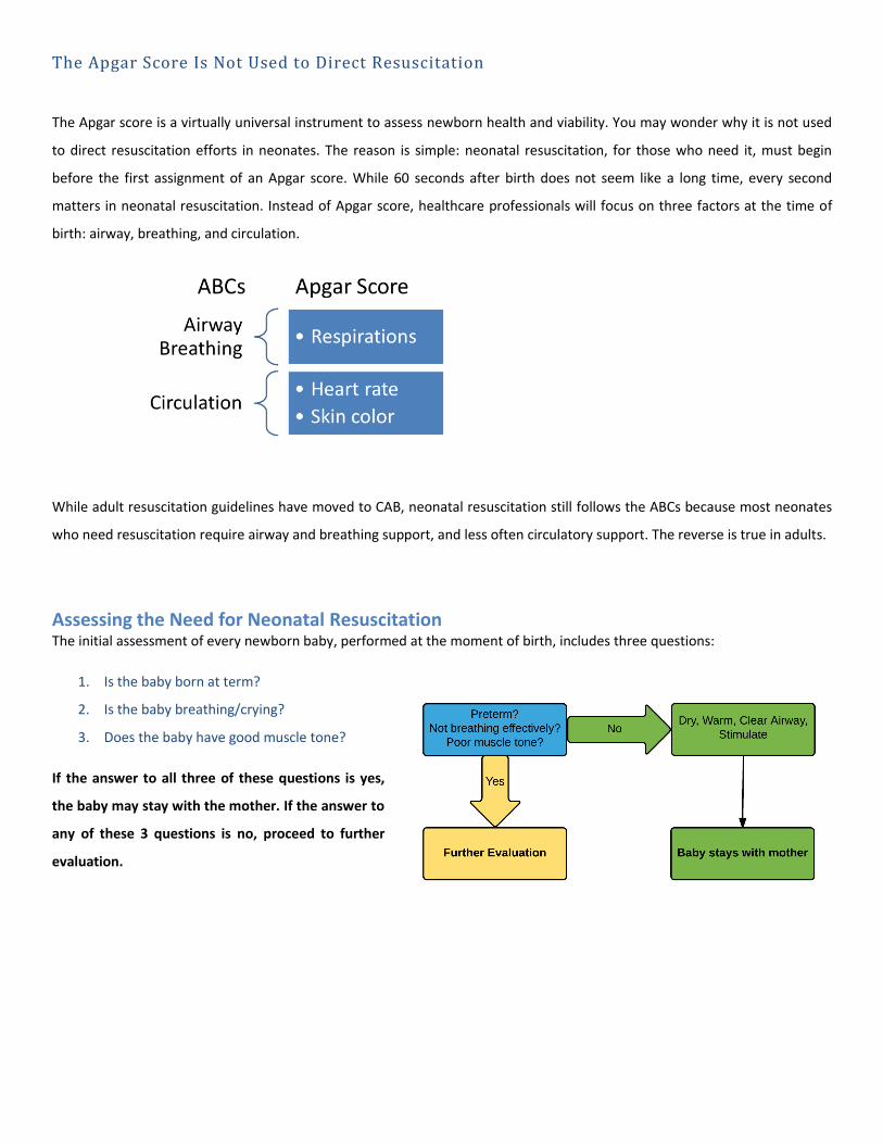
The Apgar Score Is Not Used to Direct Resuscitation
The Apgar score is a virtually universal instrument to assess newborn health and viability. You may wonder why it is not used
to direct resuscitation efforts in neonates. The reason is simple: neonatal resuscitation, for those who need it, must begin
before the first assignment of an Apgar score. While 60 seconds after birth does not seem like a long time, every second
matters in neonatal resuscitation. Instead of Apgar score, healthcare professionals will focus on three factors at the time of
birth: airway, breathing, and circulation.
While adult resuscitation guidelines have moved to CAB, neonatal resuscitation still follows the ABCs because most neonates
who need resuscitation require airway and breathing support, and less often circulatory support. The reverse is true in adults.
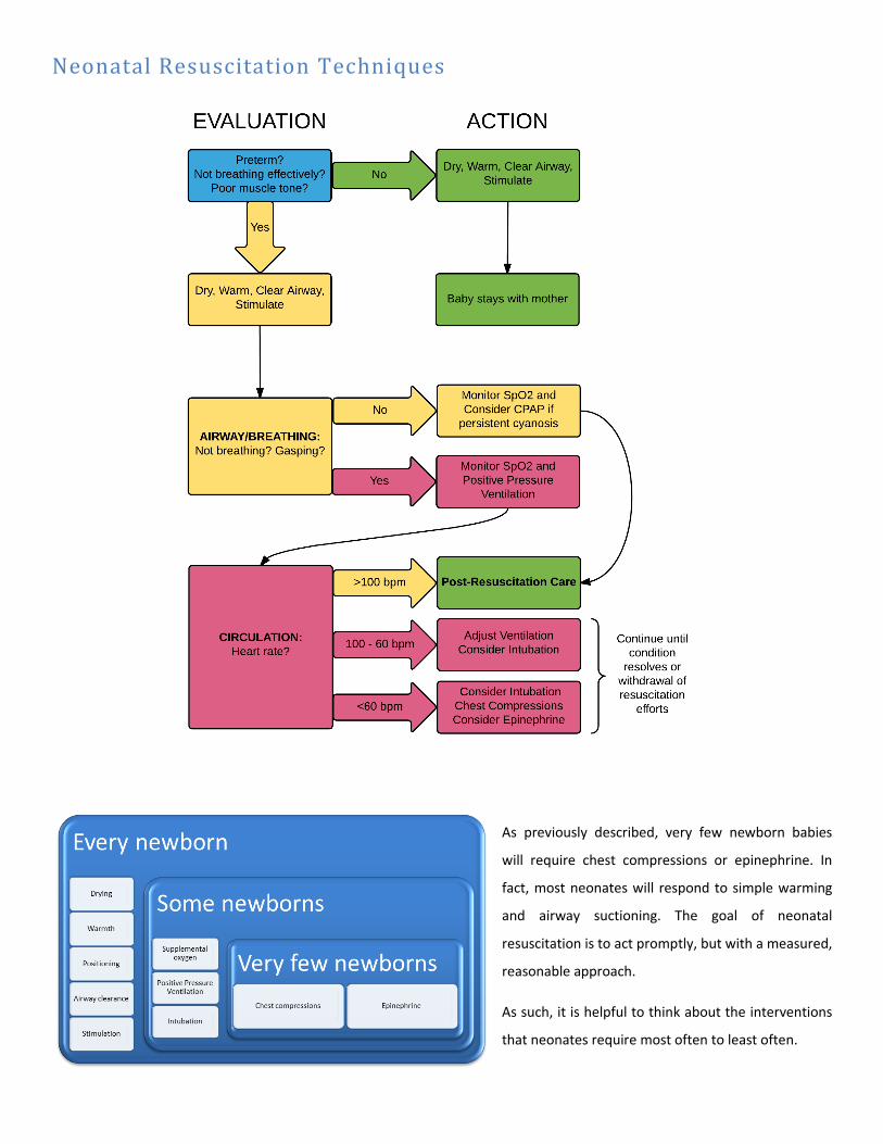
Assessing the Need for Neonatal Resuscitation The initial assessment of every newborn baby, performed at the moment of birth, includes three questions:
1. Is the baby born at term?
2. Is the baby breathing/crying?
3. Does the baby have good muscle tone?
If the answer to all three of these questions is yes,
the baby may stay with the mother. If the answer to
any of these 3 questions is no, proceed to further
evaluation.
Staying with the Mother Staying with the mother does not necessarily mean placing the baby and the mother's arms immediately. Every neonate
requires a few steps before initial mother child bonding can occur.
1. Warm the baby – To reduce any further heat loss, dry the baby and remove any wet linens. The baby may be put under a radiant warmer to reduce heat loss, but not be put under towels or blankets. Monitor the baby’s temperature to ensure that the baby does not overheat.
2. Open the baby’s airway – Put the baby in the “sniffing” position on its back or side, and be careful not to extend the neck too much or too little.
3. Clear the baby’s airway – How precisely you clear the airway depends on whether the baby’s skin has meconium on it, as well as the baby’s activity level.
a. If meconium is present – clear the baby’s mouth and nose and dry the baby, stimulate it, and reposition it.
b. If meconium is absent – check to see if the baby is vigorous, meaning that the baby has a heart rate over 100 bpm, good muscle tone, and is making respiratory efforts.
i. If the baby is vigorous – behave as if meconium were present
ii. If the baby is not vigorous - suction the baby’s mouth and trachea gently
Further Evaluation The first step in “further evaluation” is the same 3 steps as above:
1. Warm the baby
2. Open the baby’s airway
3. Clear the baby’s airway
All babies should be immediately assessed for airway patency and proper ventilation. Once a baby is born, a vigorous cry
usually means that a baby is breathing, but breathing can also be observed by watching a baby’s chest. If the baby is not
breathing, he or she will need resuscitation.
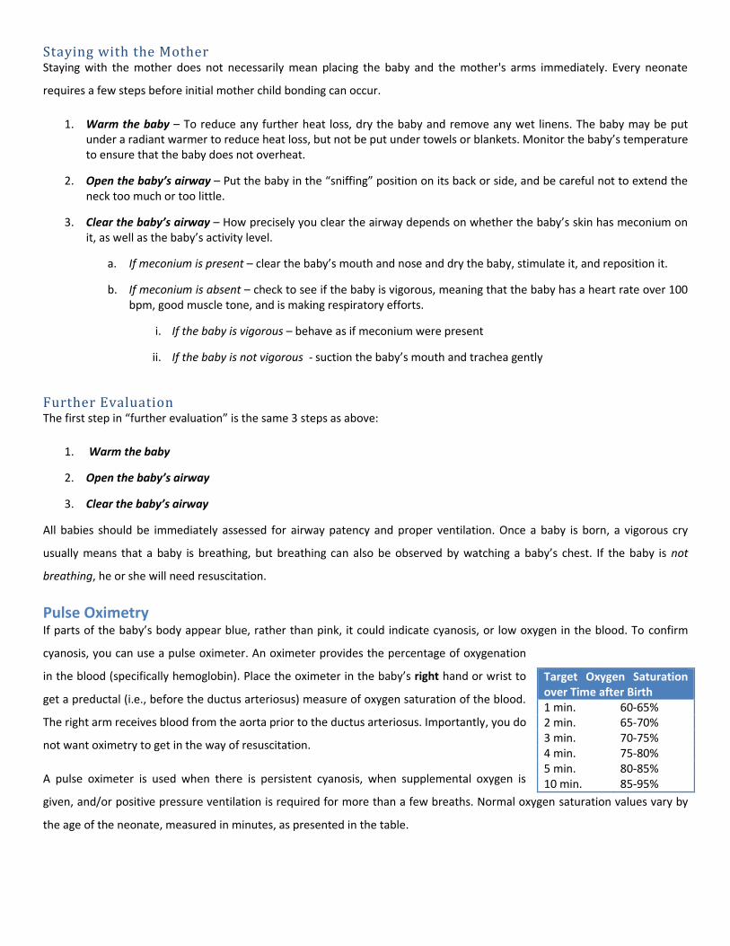
Pulse Oximetry If parts of the baby’s body appear blue, rather than pink, it could indicate cyanosis, or low oxygen in the blood. To confirm
cyanosis, you can use a pulse oximeter. An oximeter provides the percentage of oxygenation
in the blood (specifically hemoglobin). Place the oximeter in the baby’s right hand or wrist to
get a preductal (i.e., before the ductus arteriosus) measure of oxygen saturation of the blood.
The right arm receives blood from the aorta prior to the ductus arteriosus. Importantly, you do
not want oximetry to get in the way of resuscitation.
A pulse oximeter is used when there is persistent cyanosis, when supplemental oxygen is
given, and/or positive pressure ventilation is required for more than a few breaths. Normal oxygen saturation values vary by
the age of the neonate, measured in minutes, as presented in the table.
Target Oxygen Saturation over Time after Birth 1 min. 60-65% 2 min. 65-70% 3 min. 70-75% 4 min. 75-80% 5 min. 80-85% 10 min. 85-95%
Neonatal Resuscitation Techniques
As previously described, very few newborn babies
will require chest compressions or epinephrine. In
fact, most neonates will respond to simple warming
and airway suctioning. The goal of neonatal
resuscitation is to act promptly, but with a measured,
reasonable approach.
As such, it is helpful to think about the interventions
that neonates require most often to least often.
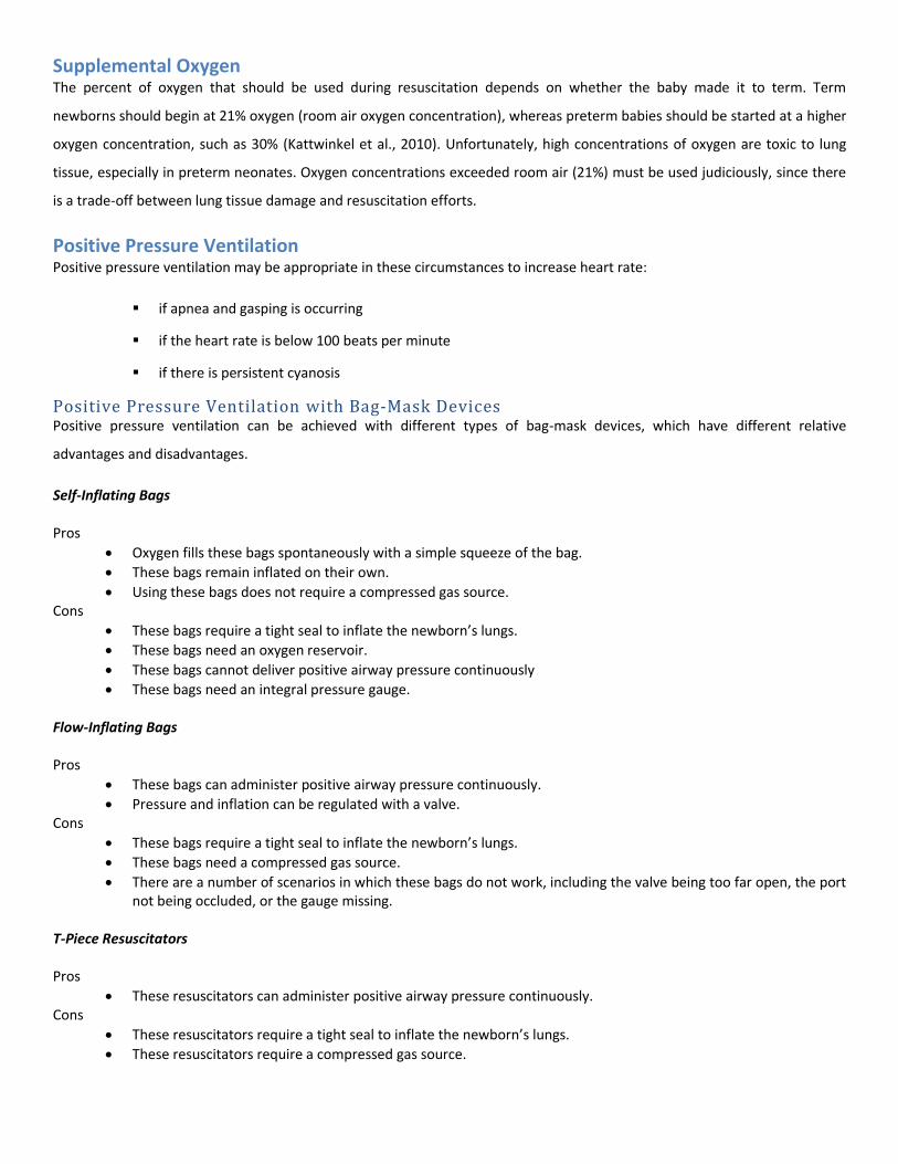
Supplemental Oxygen The percent of oxygen that should be used during resuscitation depends on whether the baby made it to term. Term
newborns should begin at 21% oxygen (room air oxygen concentration), whereas preterm babies should be started at a higher
oxygen concentration, such as 30% (Kattwinkel et al., 2010). Unfortunately, high concentrations of oxygen are toxic to lung
tissue, especially in preterm neonates. Oxygen concentrations exceeded room air (21%) must be used judiciously, since there
is a trade-off between lung tissue damage and resuscitation efforts.
Positive Pressure Ventilation Positive pressure ventilation may be appropriate in these circumstances to increase heart rate:
if apnea and gasping is occurring
if the heart rate is below 100 beats per minute
if there is persistent cyanosis
Positive Pressure Ventilation with Bag-Mask Devices Positive pressure ventilation can be achieved with different types of bag-mask devices, which have different relative
advantages and disadvantages.
Self-Inflating Bags Pros
Oxygen fills these bags spontaneously with a simple squeeze of the bag.
These bags remain inflated on their own.
Using these bags does not require a compressed gas source. Cons
These bags require a tight seal to inflate the newborn’s lungs.
These bags need an oxygen reservoir.
These bags cannot deliver positive airway pressure continuously
These bags need an integral pressure gauge. Flow-Inflating Bags Pros
These bags can administer positive airway pressure continuously.
Pressure and inflation can be regulated with a valve. Cons
These bags require a tight seal to inflate the newborn’s lungs.
These bags need a compressed gas source.
There are a number of scenarios in which these bags do not work, including the valve being too far open, the port not being occluded, or the gauge missing.
T-Piece Resuscitators Pros
These resuscitators can administer positive airway pressure continuously. Cons
These resuscitators require a tight seal to inflate the newborn’s lungs.
These resuscitators require a compressed gas source.
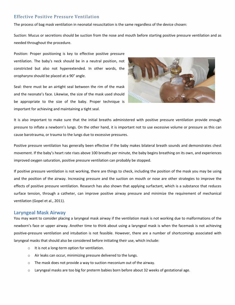
Effective Positive Pressure Ventilation
The process of bag mask ventilation in neonatal resuscitation is the same regardless of the device chosen:
Suction: Mucus or secretions should be suction from the nose and mouth before starting positive pressure ventilation and as
needed throughout the procedure.
Position: Proper positioning is key to effective positive pressure
ventilation. The baby's neck should be in a neutral position, not
constricted but also not hyperextended. In other words, the
oropharynx should be placed at a 90° angle.
Seal: there must be an airtight seal between the rim of the mask
and the neonate’s face. Likewise, the size of the mask used should
be appropriate to the size of the baby. Proper technique is
important for achieving and maintaining a tight seal.
It is also important to make sure that the initial breaths administered with positive pressure ventilation provide enough
pressure to inflate a newborn’s lungs. On the other hand, it is important not to use excessive volume or pressure as this can
cause barotrauma, or trauma to the lungs due to excessive pressures.
Positive pressure ventilation has generally been effective if the baby makes bilateral breath sounds and demonstrates chest
movement. If the baby’s heart rate rises above 100 breaths per minute, the baby begins breathing on its own, and experiences
improved oxygen saturation, positive pressure ventilation can probably be stopped.
If positive pressure ventilation is not working, there are things to check, including the position of the mask you may be using
and the position of the airway. Increasing pressure and the suction on mouth or nose are other strategies to improve the
effects of positive pressure ventilation. Research has also shown that applying surfactant, which is a substance that reduces
surface tension, through a catheter, can improve positive airway pressure and minimize the requirement of mechanical
ventilation (Gopel et al., 2011).
Laryngeal Mask Airway You may want to consider placing a laryngeal mask airway if the ventilation mask is not working due to malformations of the
newborn’s face or upper airway. Another time to think about using a laryngeal mask is when the facemask is not achieving
positive-pressure ventilation and intubation is not feasible. However, there are a number of shortcomings associated with
laryngeal masks that should also be considered before initiating their use, which include:
o It is not a long-term option for ventilation.
o Air leaks can occur, minimizing pressure delivered to the lungs.
o The mask does not provide a way to suction meconium out of the airway.
o Laryngeal masks are too big for preterm babies born before about 32 weeks of gestational age.
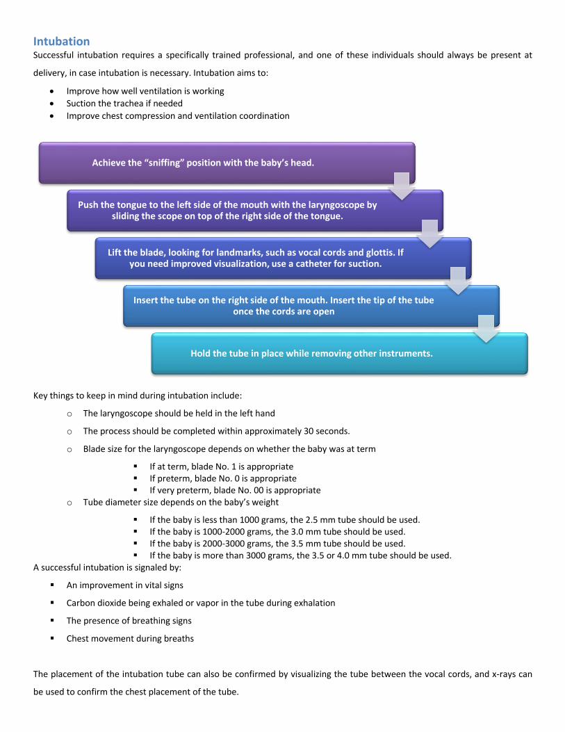
Intubation Successful intubation requires a specifically trained professional, and one of these individuals should always be present at
delivery, in case intubation is necessary. Intubation aims to:
Improve how well ventilation is working
Suction the trachea if needed
Improve chest compression and ventilation coordination
Key things to keep in mind during intubation include:
o The laryngoscope should be held in the left hand
o The process should be completed within approximately 30 seconds.
o Blade size for the laryngoscope depends on whether the baby was at term
If at term, blade No. 1 is appropriate If preterm, blade No. 0 is appropriate If very preterm, blade No. 00 is appropriate
o Tube diameter size depends on the baby’s weight
If the baby is less than 1000 grams, the 2.5 mm tube should be used. If the baby is 1000-2000 grams, the 3.0 mm tube should be used. If the baby is 2000-3000 grams, the 3.5 mm tube should be used. If the baby is more than 3000 grams, the 3.5 or 4.0 mm tube should be used.
A successful intubation is signaled by:
An improvement in vital signs
Carbon dioxide being exhaled or vapor in the tube during exhalation
The presence of breathing signs
Chest movement during breaths
The placement of the intubation tube can also be confirmed by visualizing the tube between the vocal cords, and x-rays can
be used to confirm the chest placement of the tube.
Achieve the “sniffing” position with the baby’s head.
Push the tongue to the left side of the mouth with the laryngoscope by sliding the scope on top of the right side of the tongue.
Lift the blade, looking for landmarks, such as vocal cords and glottis. If you need improved visualization, use a catheter for suction.
Insert the tube on the right side of the mouth. Insert the tip of the tube once the cords are open
Hold the tube in place while removing other instruments.
Chest Compressions Chest compressions are not often required during the resuscitation of newborns; however, if a baby’s heart rate has not risen
above 60 beats per minute after 30 seconds of positive-pressure ventilation, chest compressions should be administered.
Chest compressions increase the pressure within the thoracic cavity by compressing the heart against the spine, thereby
reducing the volume within that space. The effect is that blood should circulate to important organs of the body.
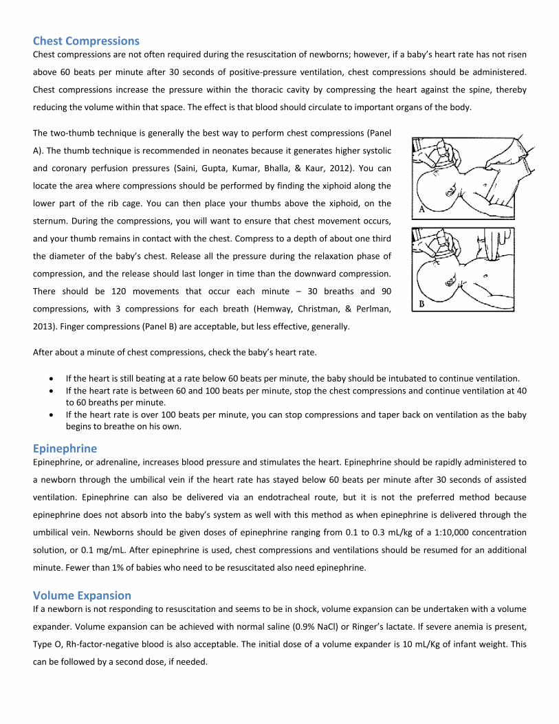
The two-thumb technique is generally the best way to perform chest compressions (Panel
A). The thumb technique is recommended in neonates because it generates higher systolic
and coronary perfusion pressures (Saini, Gupta, Kumar, Bhalla, & Kaur, 2012). You can
locate the area where compressions should be performed by finding the xiphoid along the
lower part of the rib cage. You can then place your thumbs above the xiphoid, on the
sternum. During the compressions, you will want to ensure that chest movement occurs,
and your thumb remains in contact with the chest. Compress to a depth of about one third
the diameter of the baby’s chest. Release all the pressure during the relaxation phase of
compression, and the release should last longer in time than the downward compression.
There should be 120 movements that occur each minute – 30 breaths and 90
compressions, with 3 compressions for each breath (Hemway, Christman, & Perlman,
2013). Finger compressions (Panel B) are acceptable, but less effective, generally.
After about a minute of chest compressions, check the baby’s heart rate.
If the heart is still beating at a rate below 60 beats per minute, the baby should be intubated to continue ventilation.
If the heart rate is between 60 and 100 beats per minute, stop the chest compressions and continue ventilation at 40 to 60 breaths per minute.
If the heart rate is over 100 beats per minute, you can stop compressions and taper back on ventilation as the baby begins to breathe on his own.
Epinephrine Epinephrine, or adrenaline, increases blood pressure and stimulates the heart. Epinephrine should be rapidly administered to
a newborn through the umbilical vein if the heart rate has stayed below 60 beats per minute after 30 seconds of assisted
ventilation. Epinephrine can also be delivered via an endotracheal route, but it is not the preferred method because
epinephrine does not absorb into the baby’s system as well with this method as when epinephrine is delivered through the
umbilical vein. Newborns should be given doses of epinephrine ranging from 0.1 to 0.3 mL/kg of a 1:10,000 concentration
solution, or 0.1 mg/mL. After epinephrine is used, chest compressions and ventilations should be resumed for an additional
minute. Fewer than 1% of babies who need to be resuscitated also need epinephrine.
Volume Expansion If a newborn is not responding to resuscitation and seems to be in shock, volume expansion can be undertaken with a volume
expander. Volume expansion can be achieved with normal saline (0.9% NaCl) or Ringer’s lactate. If severe anemia is present,
Type O, Rh-factor-negative blood is also acceptable. The initial dose of a volume expander is 10 mL/Kg of infant weight. This
can be followed by a second dose, if needed.
Resuscitation Tools There are a number of things that should be prepared ahead of every single birth, to ensure efficient resuscitation procedures
are implemented if resuscitation is required. The things to prepare include:
o A stethoscope
o A laryngoscope with blades
o A bulb syringe
o A meconium aspirator
o A suction catheter
o A preheat warmer
o Warming blankets or towels
o Warming pad
o A free-flow oxygen device
o An air-oxygen blender
o A pulse oximeter
o A pulse oximeter probe
o A positive-pressure ventilation device
o A feeding tube
o Endotracheal tubes
o Stylets
o A laryngeal mask
o Epinephrine
o Saline
o Documentation materials
o A transport incubator
o Plastic bags or wraps
Factors That May Complicate Resuscitation
Airway Obstructions If resuscitation does not seem to be working, there are some special considerations that should be assessed. In many cases,
complication relates to a constricted or blocked airway such as laryngeal webs, cystic hygroma, or congenital goiter. Practically
speaking, the airway obstruction is usually in the nasal pharynx (e.g., choanal atresia) or the oral pharynx (e.g., Robin
syndrome).
Choanal Atresia Babies do not normally breathe through their mouths unless they are crying. In a way, they can be considered obligate nose
breathers. In the case of choanal atresia, however, the nasal airway is not fully patent (open). This means that the baby can
only breathe effectively through crying or with assistance. One clue to the existence of choanal atresia is the presence of
meconium or mucus is in the nasal airway. A suction catheter gently applied through the nares into the posterior pharynx can
test for this condition. If the catheter cannot pass so that it is visible in the oral pharynx, you can assume that choanal atresia
exists and an oral airway will be necessary.
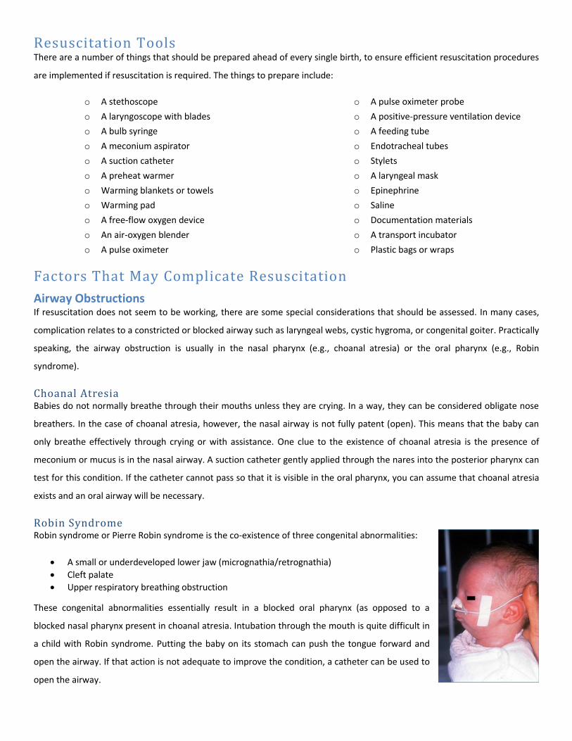
Robin Syndrome Robin syndrome or Pierre Robin syndrome is the co-existence of three congenital abnormalities:
A small or underdeveloped lower jaw (micrognathia/retrognathia)
Cleft palate
Upper respiratory breathing obstruction
These congenital abnormalities essentially result in a blocked oral pharynx (as opposed to a
blocked nasal pharynx present in choanal atresia. Intubation through the mouth is quite difficult in
a child with Robin syndrome. Putting the baby on its stomach can push the tongue forward and
open the airway. If that action is not adequate to improve the condition, a catheter can be used to
open the airway.
Pulmonary Complications The neonate, and especially the premature infant, can develop one or more problems in the lungs that complicate neonatal
resuscitation. In the very premature infant, the lungs either cannot support respiration and oxygenation or can only do so
marginally. Artificial surfactant can help considerably in these cases by reducing surface tension in the alveoli and reducing
pressures required to ventilate the lungs.
Another form of lung malformation is pulmonary hypoplasia. In pulmonary hypoplasia (which is more common in fetuses
exposed to insufficient amounts of amniotic fluid during gestation), the lungs have simply not formed during fetal
development. Less severe cases of pulmonary hypoplasia can be effectively treated with long-term intensive care, but children
with severe cases of pulmonary hypoplasia often do not survive the neonatal period.
Some of the more common causes of impaired lung function can be reversed with timely bedside or surgical procedures,
assuming they are detected in the early neonatal period. For example, many babies who require neonatal resuscitation are
born with a pneumothorax or develop one during resuscitation (particularly ventilation). In pneumothorax is the presence of
air in the pleural space, between the chest wall and the outside of the lungs. A pneumothorax causes substantial respiratory
distress and is diagnosed through trans illumination of the chest cavity, the absence of lung sounds of one of the chest, or a
portable chest x-ray if needed. A pneumothorax can be treated with needle thoracostomy where the placement of a catheter
to evacuate the air in the pleural space.
Pleural effusions and congenital diaphragmatic hernias are rare, but potentially treatable causes of poor lung function in the
neonate. A pleural effusion is treated in much the same way as a pneumothorax, releasing fluid instead of air. A baby with
congenital diaphragmatic hernia is usually diagnosed by ultrasound prior to delivery. However in women who have not had
routine prenatal screenings, the hernia may go undiagnosed until delivery. The baby can be stabilized with separate tubes in
the trachea and stomach until pediatric surgery can repair the hernia.
Impaired Respiratory Drive Women who received opioid analgesics during delivery or women who are actively intoxicated with illicit opioids may deliver
infants with substantial levels of opioids in their systems. In these cases, the problem with respiration is not an impaired
airway or a pulmonary problem, but the drive to breathe is depressed. When this occurs, the baby can be ventilated until the
opioids had been metabolized. Naloxone, an opioid antagonist, should be avoided in babies of women with opioid abuse
problems or on methadone treatment because the drug can cause withdrawal seizures in the neonate.
Cardiac Abnormalities Several types of congenital heart malformation can interfere with circulation, but few of them manifest in the newly born
infant. Providers may consider a congenital heart problem after ventilation has proved fruitless. This requires specialist
diagnostic and management skills that are outside the purview of neonatal resuscitation.
Post-Resuscitation Care Once the newborn has been successfully resuscitated, the baby is moved to post-resuscitation care. Post-resuscitation care is
considered separately because infants who have required resuscitation are at risk of developing complications from the
resuscitation and during the period after resuscitation (Frazier & Werthammer, 2007). As such, neonates who require
resuscitation are usually moved to the neonatal intensive care unit for close monitoring.
Blood pressure: Hypotension is the most likely cardiovascular result of resuscitation. Monitoring heart rate and blood pressure
are the best ways to determine if hypotension is an issue for newborns who have been resuscitation. Volume replacement
and inotrope administration are relevant interventions in the case of hypotension.
Electrolytes: Hyponatremia and hypocalcemia are common in recently resuscitated newborns. Electrolyte abnormalities are
diagnosed/monitored with chemistry panels. Standard treatment is to reverse deficits with intravenous supplementation.
Metabolic acidosis: Poor cardiac output and/or hypoxemia can cause a buildup of lactic acid, which leads to metabolic
acidosis. When possible, acidosis (acidemia) should be treated with increased ventilation (drawing off carbon dioxide from the
lungs) Sodium bicarbonate can be given in cases of extreme or persistent metabolic acidosis, but it should be used with
extreme caution since it is caustic, irritates blood vessels, and can actually decrease pH in cells.
Blood glucose: Hypoglycemia is a concern in the post-resuscitation period. Even in a healthy newborn, plasma glucose
concentration will fall during the first two hours after delivery, usually to a value as low as (but usually not below) 40 mg/dL
(2.2 mmol/L). Within 4 to 6 hours of birth, blood glucose should stabilize between 45 and 80 mg/dL (2.5 and 4.4 mmol/L)
(Cornblath et al., 2000). In general, newborns within the first 48 hours of birth should be able to maintain circulating blood
glucose above 50 mg/dL (2.8 mmol/L) (Stanley et al., 2015).
Central nervous system function: Seizures, apnea, and other neurological issues can result from resuscitation. Therapeutic
hypothermia and anticonvulsants are potential interventions for brain disturbances resulting from resuscitation.
Pulmonary function: A number of lung complications can arise because of resuscitation. These complications include
pulmonary hypertension, meconium aspiration syndrome, pneumonia, pneumothorax, transient tachypnea, and surfactant
deficiency (especially in premature infants). Maintaining proper oxygenation and ventilation, delaying feedings, using
antibiotics, taking x-rays, and using surfactant therapy are all interventions that can help with specific lung complications.
Feeding difficulties: Providers should look for ileus, gastrointestinal bleeding, or poor suckling/swallowing. Delaying feedings
and providing intravenous fluids and parenteral nutrition are potential ways to intervene with these issues.
Renal function: Acute tubular necrosis is the most common kidney complication resulting from resuscitation. This condition
can be identified by monitoring urine output and serum electrolytes.
Infection and blood cell counts: Complete blood cell counts (CBCs) can be used to diagnose anemia (low red blood cell count),
thrombocytopenia (low platelet count), and infection (elevated white blood cell count, usually with elevated body
temperature). Pneumonia is the likely culprit of infection in the neonate, either from aspiration that occurred during
resuscitation or from infection that is present congenitally. Provider should also be aware of the possibility of sepsis.
Resuscitating Preterm Babies Preterm babies are at increased risk for requiring resuscitation. There are a number of reason for this group’s vulnerability,
including that they lose heat quickly, they are quite vulnerable to the changes in oxygen levels, their vital organs, such as the
brain and lungs are immature, they are more susceptible to infection, and they have a lower blood volume, which makes
blood loss more problematic for them.
When preparing for a preterm birth, those responsible for resuscitation should compile extra resources and personnel. Having
additional equipment for warming the baby is important, as is a compressed air source, an oxygen blender, and a pulse
oximeter. These extra tools will be useful for the following reasons:
o Because preterm babies lose heat quickly, a number of different mechanisms to reduce heat loss should be
employed. The room temperature should be increased, and a radiant warmer should be preheated. Having a
warming pad, a polyethylene wrap, and a warmed transport incubator are other strategies to consider for
preterm babies.
o Because preterm babies are more susceptible to changes in oxygen concentration, increasing their oxygen levels
needs to occur at a slower rate than would occur with normal babies of term. The oximeter and blender can
therefore be used to achieve optimal oxygen saturations during right after resuscitation.
The actual process of ventilation for preterm babies should follow the same protocol as positive-pressure ventilation for term
babies.
There are a number of precautions you can take during the resuscitation of preterm babies to reduce the chances that the
baby endures brain injury. These precautions including avoiding the Trendelenburg position, high airway pressures, as well as
intravenous fluid that enters too rapidly or has high ionic concentrations. Generally treating the baby with care and gently
altering ventilation can also reduce the risk of brain injury.
Post-resuscitation care is the same as it is for less premature neonates; however, assessments and treatments should be more
frequent and every maneuver should be done even more gently in the extremely premature neonate (e.g., ventilation,
feedings, IV infusions)
Ethical Considerations
There are no specific legal or ethical guidelines regarding when to attempt resuscitation or when to cease resuscitation
protocols. These decisions are largely determined collaboratively by healthcare professionals and the family of the patient.
The parents are generally deferred to for decisions regarding the health of the baby. Accordingly, research has shown that the
majority of healthcare professionals initiate resuscitation or refrain from resuscitation according to the wishes of the parents,
though 98% of healthcare professionals have reported initiating resuscitation when the parents are unsure of which option to
pursue (Peerzada, Richardson, & Burns, 2004). However, after 10 minutes of no heart rate, the discontinuation of
resuscitation efforts should be seriously considered.
When dealing with neonates who do not survive, it is important to remember the following:
The ethical issues involved with resuscitation are the same for any human of any age—neonates are no different even
though they have only been alive for a very short period.
Some neonates will be born with problems that are not compatible with life such as incomplete gestation, ultra-low
birth weight, and/or congenital abnormalities.
In some cases, resuscitation may only be able to prolong life temporarily, prolong suffering, or result in a viable infant
with massive, permanent disabilities. It may be acceptable, in these cases, to withhold resuscitation efforts. Specific
examples of cases in which it is appropriate to withhold resuscitation include:
o Anencephaly
o Lethal genetic abnormality
o Marked, ongoing disability, usually a case that would not be considered a “meaningful” life
o Gestational age of less than 23 weeks
o Birth weight of less than 400 g (fetal weight estimates may be wrong by ± 20%)
When possible, parents of fetuses at very high risk should be engaged in end of life discussions before delivery.
Providers should consider and respect the wishes of parents who have been fully informed. On the other hand,
providers have a legal obligation to provide care if, in their expert opinion, the neonate has a reasonable chance of
surviving and acceptable risk of morbidity, even if this is against the parents’ wishes.
Difficult or borderline cases should include discussions with physicians, nurses, social workers, and medical ethicists,
though parental wishes should be strongly considered.
Words matter. The loss of a neonate should be treated like the loss of any other child—a somber, important moment.
Be empathic and clear with parents of the deceased. Avoid phrases such as
o “You are young. You can always have another baby.”
o “At least you did not know this baby yet.”
o “It was for the best.”
References
AAP/AHA. (2011). Neonatal Resuscitation. 6th Edition. Cornblath, M., Hawdon, J. M., Williams, A. F., Aynsley-Green, A., Ward-Platt, M. P., Schwartz, R., & Kalhan, S. C. (2000). Controversies
regarding definition of neonatal hypoglycemia: suggested operational thresholds. Pediatrics, 105(5), 1141-1145. Frazier, M. D., & Werthammer, J. (2007). Post-resuscitation complications in term neonates. J Perinatol, 27(2), 82-84. doi:
10.1038/sj.jp.7211644 Gopel, W., Kribs, A., Ziegler, A., Laux, R., Hoehn, T., Wieg, C., . . . Herting, E. (2011). Avoidance of mechanical ventilation by surfactant
treatment of spontaneously breathing preterm infants (AMV): an open-label, randomised, controlled trial. Lancet, 378(9803), 1627-1634. doi: 10.1016/s0140-6736(11)60986-0
Hemway, R. J., Christman, C., & Perlman, J. (2013). The 3:1 is superior to a 15:2 ratio in a newborn manikin model in terms of quality of chest compressions and number of ventilations. Arch Dis Child Fetal Neonatal Ed, 98(1), F42-45. doi: 10.1136/archdischild-2011-301334
Iliodromiti, S., Mackay, D. F., Smith, G. C., Pell, J. P., & Nelson, S. M. (2014). Apgar score and the risk of cause-specific infant mortality: a population-based cohort study. Lancet, 384(9956), 1749-1755. doi: 10.1016/s0140-6736(14)61135-1
Kattwinkel, J., Perlman, J. M., Aziz, K., Colby, C., Fairchild, K., Gallagher, J., . . . Zaichkin, J. (2010). Part 15: neonatal resuscitation: 2010 American Heart Association Guidelines for Cardiopulmonary Resuscitation and Emergency Cardiovascular Care. Circulation, 122(18 Suppl 3), S909-919. doi: 10.1161/circulationaha.110.971119
Khalak, R., Cummings, J., & Dexter, S. (2015). Maternal obesity: significance on the preterm neonate. Int J Obes (Lond). doi: 10.1038/ijo.2015.107
Lawn, J. E., Cousens, S., & Zupan, J. (2005). 4 million neonatal deaths: when? Where? Why? Lancet, 365(9462), 891-900. doi: 10.1016/s0140-6736(05)71048-5
Peerzada, J. M., Richardson, D. K., & Burns, J. P. (2004). Delivery room decision-making at the threshold of viability. J Pediatr, 145(4), 492-498. doi: 10.1016/j.jpeds.2004.06.018
Saini, S. S., Gupta, N., Kumar, P., Bhalla, A. K., & Kaur, H. (2012). A comparison of two-fingers technique and two-thumbs encircling hands technique of chest compression in neonates. J Perinatol, 32(9), 690-694. doi: 10.1038/jp.2011.167
Stanley, C. A., Rozance, P. J., Thornton, P. S., De Leon, D. D., Harris, D., Haymond, M. W., . . . Wolfsdorf, J. I. (2015). Re-evaluating "transitional neonatal hypoglycemia": mechanism and implications for management. J Pediatr, 166(6), 1520-1525 e1521. doi: 10.1016/j.jpeds.2015.02.045
This Neonatal Resuscitation Course complies with the sixth edition of the Neonatal Resuscitation textbook by the American Heart Association and the American
Academy of Pediatrics (AAP/AHA, 2011). Other sources used to compile this guide are listed in the References section. Cover image is attributed to ceejayoz.
Copyright 2015. All rights reserved.


![Acute Kidney Injury in Asphyxiated neonates admitted into a ......with perinatal asphyxia. Perinatal Asphyxia ranks as the second most important cause of neonatal death[1]. Major risk](https://img.pdfslide.us/doc/110x75/61110be6b93f5b0fcd11cc91/acute-kidney-injury-in-asphyxiated-neonates-admitted-into-a-with-perinatal.jpg)