Embed Size (px)
Citation preview

September 2009
INTERGROWTH-21st
International Fetal and Newborn Growth Standards for the 21st Century
The International Fetal and Newborn Growth Consortium
BASIC NEONATAL CARE MANUAL
2
Please read this manual carefully and refer to it throughout the study
if any clarification is needed
This Operations Manual was produced by the INTERGROWTH-21st Neonatal Group, based on
the 1st Meeting of the Neonatal Group, Oxford, July 2009. This document reflects the
consensus reached by members of the group regarding the definitions of neonatal
morbidities and the minimum standards of care to be provided by all centres taking part in
the INTERGROWTH-21st study.
INTERGROWTH-21st is a large project involving health institutions from eight geographically
diverse countries. It is therefore essential that all participating institutions follow a
standardized neonatal care protocol.

3
Table of Contents
Credits ........................................................................................................................................................... 4
Abbreviations ................................................................................................................................................ 5
Definitions of Neonatal Morbidities ..................................................................................................... 6
Transient Tachypnea of Newborn (TTN) ................................................................................................... 7
Respiratory Distress Syndrome (RDS) ........................................................................................................ 7
Bronchopulmonary Dysplasia (BPD) ......................................................................................................... 8
Meconium Aspiration Syndrome ............................................................................................................... 8
Retinopathy of Prematurity (ROP) ............................................................................................................ 8
Periventricular Leukomalacia .................................................................................................................... 9
Apnea of Prematurity ................................................................................................................................ 9
Hypoxic Ischemic Encephalopathy (HIE) ................................................................................................. 10
Birth Asphyxia ......................................................................................................................................... 10
Intraventricular Hemorrhage (IVH) ......................................................................................................... 11
Necrotizing Enterocolitis (NEC) ............................................................................................................... 12
Polycythemia ........................................................................................................................................... 12
Anemia requiring transfusion ................................................................................................................. 13
Acute Bilirubin Encephalopathy .............................................................................................................. 14
Chronic Bilirubin Encephalopathy ........................................................................................................... 14
Hypotension in Neonates ........................................................................................................................ 14
Hypoglycemia .......................................................................................................................................... 14
Inborn Error of Metabolism .................................................................................................................... 15
Treatment and Management Recommendations for Neonatal Morbidities ............................... 17
Administration of Antenatal Steroids to Women at Risk of Preterm Delivery ........................................ 18
Feeding the Preterm Infant ..................................................................................................................... 19
Thermoregulation in Preterm Newborn Babies ...................................................................................... 21
Administration of Surfactant to Preterm Infants with or at Risk for RDS ............................................... 25
Use of Nasal CPAP for Respiratory Distress Syndrome (RDS) .................................................................. 26
Screening for Intraventricular Hemorrhage (IVH) and Periventricular Leukomalacia (PVL) ................... 27
Periventricular Leukomalacia (PVL) ........................................................................................................ 28
Screening for Retinopathy of Prematurity (ROP) .................................................................................... 29
Hypoxic Ischemic Encephalopathy (HIE) ................................................................................................. 34
Neonatal Hyperbilirubinemia .................................................................................................................. 39
Hypotension in Neonates ........................................................................................................................ 43
Patent Ductus Arteriosus (PDA) in Preterm Neonates: (<34 weeks gestation) ....................................... 45
Sepsis ....................................................................................................................................................... 48

4
Credits
This manual was prepared by members of the INTERGROWTH-21st Neonatal Group and reflects the
general consensus reached during the Neonatal Group Meeting, Oxford, 6-7th July 2009, regarding the
minimum standards of neonatal care to be provided by all INTERGROWTH-21st centres.
The following people made important contributions to this final version, for which we thank them:
Zulfiqar Bhutta – Group Leader (Husein Laljee Dewraj Professor and Chairman, Department of
Paediatrics and Child Health, Aga Khan University Medical Centre, Karachi, Pakistan)
Anila Haroon – (Senior Instructor, Department of Pediatrics and Child Health, Aga Khan University
Medical Centre, Karachi, Pakistan)
Francesca Giuliani – (Medical doctor, Neonatal Unit, Department of Pediatrics of Turin University,
Regina Margherita, S. Anna Hospital, Turin, Italy)
Maneesh Batra – (Assistant Professor of Pediatrics, Division of Neonatology, University of Washington
School of Medicine, Seattle, USA)
Enrico Bertino – (Professor, Neonatal Unit, Department of Pediatrics of Turin University, Regina
Margherita, S. Anna Hospital, Turin, Italy)
Vinod Paul – (Professor, Division of Neonatology, All India Institute of Medical Sciences, New Delhi,
India)
Kenny McCormick – (Consultant & Honorary Senior Lecturer, Neonatal Unit, John Radcliffe Hospital,
Oxford, UK)
Elaine Albernaz – (Pediatrician, Pediatric Intensive Care Unit, Univeridade Catolica de Pelotas, Pelotas,
Brazil)
Bashir Bhat – (Neonatologist, Department of Pediatrics, SCBU, Khoula Hospital, Muscat, Oman)
Roseline Ochieng – (Program Director, Department of Pediatrics and Child Health, Aga Khan University
Hospital, Nairobi, Kenya)
Vikram Rajan – (Director; Neonatal Unit, Rajan Newborn Care Unit (P) Ltd; Honorary Asst. Professor
Neonatology, Indra Gandhi Medical College, Nagpur, India)
Pang Ruyan – (Principal Investigator, Beijing Obstetrics and Gynaecology Hospital,Maternal and Child
Health Center,Capital Medical University, Beijing, China)
Hannah Knight – (Research Associate, INTERGROWTH-21ST Project, Nuffield Department of Obstetrics
and Gynaecology, University of Oxford, Oxford, UK)

5
Abbreviations
LBW Low birth weight infants, defined as <2500g at birth
VLBW Very low birth weight infants, defined as <1500g at birth
ELBW Extremely low birth weight infants, defined as <1000g at birth

6
Definitions of Neonatal Morbidities to be used
for the INTERGROWTH-21st Study
7
Transient Tachypnea of Newborn (TTN)
TTN is a parenchymal lung disorder characterized by pulmonary edema resulting from delayed
resorption and clearance of fetal alveolar fluid.
The onset of TTN is usually at the time of birth and within two hours after delivery with tachypnea being
the most prominent clinical feature. Characteristic findings on chest radiograph support the diagnosis
and include increased lung volumes, and prominent vascular markings, with fluid in the interlobar
fissure. In order to make the diagnosis, other conditions (such as pneumonia, respiratory distress
syndrome, pneumothorax, etc) must be ruled out.
Symptoms of TTN usually last for 12 to 24 hours, but may persist as long as 72 hours in severe cases.
Infants rarely require supplemental oxygen, but if required they usually respond to oxygen therapy.
When oxygen is needed, usually concentrations less than 40 percent are sufficient to achieve adequate
oxygenation.
REFERENCE
Guglani, L et al. “Transient Tachypnea of the Newborn.” Pediatr. Rev. 2008; 29;e59-e65
Respiratory Distress Syndrome (RDS)
An infant is determined to have respiratory distress syndrome if each of the following is true:
Requires O2 at 6 hours of life continuing to age 24 hours
Demonstrates clinical features within age 24 hours
Has need for respiratory support to age 24 hours, AND
Has an abnormal chest x-ray within age 24 hours consistent with surfactant deficiency OR
Has received surfactant therapy within the first 24 hours of life
REFERENCE
Fanaroff AA, Stoll BJ, Wright LL, et al; NICHD Neonatal Research Network. Trends in neonatal morbidity and mortality for very low birth weight infants. Am J Obstet Gynecol 2007; 196:147.e1-147.e8

8
Bronchopulmonary Dysplasia (BPD)
BPD defined as 1) Chronic supplemental oxygen needs for >28 days (28 days oxygen need based BPD)
OR
2) Chronic supplemental oxygen needs at either PMA of 36 weeks or discharge from hospital whichever
come first (36 weeks Oxygen need based BPD)
REFERENCE
Pascal M. Lavoie, Chandra Pham, Kerry L .Jang. Heritabilty of Bronchopulmonary Dysplasia, defined
according to consensus statement of National Institute of Health.Pediatrics.2008; 122:479-485.
Meconium Aspiration Syndrome
Meconium Aspiration Syndrome (MAS) is defined as respiratory distress in an infant born through
meconium stained amniotic fluid (MSAF), whose symptoms cannot be otherwise explained. This
disorder may be life threatening complicated by respiratory failure, pulmonary air leaks and persistent
pulmonary hypertension.
REFERENCE
Fanaroff AA. Meconium aspiration syndrome: historical aspects. J Perinatol.2008; 28:3-7
Retinopathy of Prematurity (ROP)
ROP a developmental vascular retinopathy occurs only in the incompletely vascularized retina of
premature infants, leading to a wide range of outcomes from normal vision to blindness. For a diagnosis
of ROP to be documented we need a confirmed diagnosis by an ophthalmologist in the notes according
to the staging criteria below;
Staging of ROP:
Stage1: Demarcation line separating the avascular retina anteriorly from the vascularized retina
posteriorly, with abnormal branching of small vessels immediately posterior.

9
Stage 2: Intraretinal ridge; the demarcation line has increased in volume, but proliferative tissue remains
intraretinal.
Stage 3: Ridge with extraretinal fibrovascular proliferation.
Stage 4: Partial retinal detachment
Stage 5: Total retinal detachment.
REFERENCES
International Committee for the classification of Retinopathy of Prematurity “The international
classification of Retinopathy of Prematurity Revisited” Arch Ophtalmol 2005;123:991-999.
M.Subhani, Adriann Coombs, Pamela Weber, Corina Gerontis. Screening guidelines for Retinopathy of
Prematurity: The needs for revision in Extremely Low Birth Weight Infants.Pediatrics.2001; 107:656-659
Periventricular Leukomalacia
Damage to the deep white matter (WM) in the centrum semiovale is the main characteristic feature of
PVL. The damage may vary from punctuate areas of hemorrhage & necrosis to more extensive injuries
including cystic changes, scarring, hypomyelination / demyelination, and even hemorrhagic infarction of
the white matter.
REFERENCE
De Vries LS, Eken P, Dubowitz LMS. The spectrum of leukomalacia using cranial ultrasound. Behav Brain
Res 1992;49:1-6
Apnea of Prematurity
Clinically significant apnea in infants is defined as breathing pauses that last for > 20 seconds or for > 10
seconds if associated with bradycardia (e.g. < 80 beats per minute) or oxygen desaturation (e.g. O2
saturation of < 80-85 % ).
REFERNECE
Finer N, Higgins R, Kattwinkel J, Martin RJ. Summary Proceedings From the Apnea-of-Prematurity Group.
Pediatrics 2006;117;S47-S51.

10
Hypoxic Ischemic Encephalopathy (HIE)
Hypoxic Ischemic Encephalopathy (HIE) of the newborn is “a clinically defined syndrome of disturbed
neurological function in the earliest days of life in the term infant, manifested by difficulty with initiating
and maintaining respiration, depression of tone and reflexes, sub normal level of consciousness and
often seizures.
REFERENCE
Nelson KB, Leviton A. How much of neonatal encephalopathy is due to birth asphyxia? Am J Dis Child
1991
Birth Asphyxia
Infant satisfying at least one of the following criteria:
Apgar score of 5 or less at 10 minutes
Mechanical ventilation or resuscitation at 10 minutes
Cord pH < 7.1, or an arterial pH < 7.1 or base deficit of 12 or more within 60 minutes of birth
Evidence of encephalopathy according to Sarnat staging (Sarnat 1976; Finer 1981): Stage 1 (Mild): hyperalertness, hyper-reflexia, dilated pupils, tachycardia, absence of seizures.
Stage 2 (Moderate): lethargy, hyper-reflexia, miosis, bradycardia, seizures, hypotonia with weak
suck and Moro.
Stage 3 (Severe): stupor, flaccidity, small to midposition pupils which react poorly to light,
decreased stretch reflexes, hypothermia and absent Moro.
REFERENCE
Sarnat H, Sarnat M. Neonatal encephalopathy following fetal distress. A clinical and
electroencephalographic study. Arch Neurol 1976 vol. 33 (10) pp. 696-705
Finer N, Tommy P. Controlled evaluation of muscle relaxation in the ventilated neonate. Pediatrics 1981
vol. 67 (5) pp. 641-6
Jacobs SE, Hunt R, Tarnow-Mordi WO, Inder TE, Davis PG. Cooling for newborns with hypoxic ischaemic
encephalopathy. Cochrane Database Syst Rev. 2007 Oct 17;(4):CD003311.

11
Postnatal Infection (Sepsis)
Neonatal sepsis is a clinical syndrome of systemic illness accompanied by bacteremia occurring in the
first month of life.
Late onset sepsis defined as 1 or more positive blood cultures obtained after 3 days of age from infants
with clinical features of sepsis
Since culture positive sepsis is relatively rare, a physician documented episode of sepsis would suffice.
REFERENCES
Infectious disease: In Gomella TL, Cunningham MD( eds): a LANGE Clinical manual. Neonatology:
Management, procedures, on Call Problems Diseases and Drugs.5th ed. McGraw Hill, 2004.p434-440.
M Gary Karlowickz, E Stephen Buescher Fulminant Late Onset Sepsis in a intensive neonatal care unit,
1987-1997, and the impact of avoiding empiric vancomycin therapy. Pediatrics.2000; 106:1387-1390
Intraventricular Hemorrhage (IVH)
A diagnosis of IVH should be based on a documentation of IVH based on Ultrasonographic findings
conducted by a qualified ultrasonographer/ultrasonologist.
Intraventricular hemorrhage is graded by the classification of Papile et al on ultrasonographic
examination as follows:
Grade1: Blood in the periventricular germinal matrix regions or germinal matrix hemorrhage.
Grade2: Blood within the lateral ventricular system without ventricular dilatation.
Grade3: Blood acutely distends the lateral ventricles.
Grade4: Blood within ventricular system and parenchyma
REFERENCE
Papile LA, Burstein J, Burstein R, Koffler H. Incidence and evolution of subependymal and
intraventricular hemorrhage: a study of infants with birth weights less than 1,500 gm. J Pediatr
1978;92(4):529-34.

12
Necrotizing Enterocolitis (NEC)
A diagnosis and staging of Necrotizing enterocolitis (NEC) should be based on a clinical documentation
by treating clinician based on the following criteria:
Stage1: Suspected
*History of perinatal stress
*Systemic signs of ill health: temperature instability, lethargy, apnea
*Gastrointestinal manifestations: poor feeding, increased volume of gastric aspirates, vomiting,
mild abdominal distension, faecal occult blood (no fissure)
Stage2: Confirmed
*Any of the features of stage 1 plus:
*persistent occult, or gross gastrointestinal bleeding, marked abdominal distension
*abdominal radiograph: intestinal distension, bowel wall oedema, unchanging bowel loops,
pneumatosis intestinalis, portal vein gas.
Stage3: Advanced
*Any of features of stages 1 or 2 plus:
*Deterioration in vital signs, evidence of shock or severs sepsis, or marked gastrointestinal
hemorrhage
*Abdominal radiograph shows any of features of stage 2 plus pneumoperitoneum
REFERENCE
Gastrointestinal disorder: In Roberton’s R Text book of Neonatology (3rded). Churchill Livingstone. 1999,
p752.
Polycythemia

13
Polycythemia in term infant is the presence of a venous hematocrit more than 65% or a venous
hemoglobin concentration in excess of 22 gm/dl.
REFERENCE
Phibbs RH:Neonatal polycythemia. In Rudolph AB (ed): Pediatrics, 16th ed. Newyork: Appleton Century
Crofts, 1997.
Anemia requiring transfusion
There is no consensus on definition of Anemia of Prematurity.
Shown below is the criteria for transfusion taken from US and Canadian collaborative study. Patients are
transfused in a volume of 15ml/kg, administered over 2-3 hours.
Table 1 adapted from Donato et al. Pediatrics. 2000;105(5):1066-72.
REFERENCE
Donato H, Vain N, Rendo P, Vivas N, Prudent L, Larguía M, Digregorio J, Vecchiarelli C, Valverde R, García
C, Subotovsky P, Solana C, Gorenstein A. Effect of early versus late administration of human

14
recombinant erythropoietin on transfusion requirements in premature infants: results of a randomized,
placebo-controlled, multicenter trial. Pediatrics. 2000;105(5):1066-72.
Acute Bilirubin Encephalopathy
A clinical syndrome in the presence of severe hyperbilirubinemia, of lethargy, hypotonia, and poor suck,
which may progress to hypertonia (with opisthotonus and retrocollis) with a high-pitched cry and fever
and eventually to seizures and coma.
Chronic Bilirubin Encephalopathy
The clinical sequelae of acute encephalopathy with athetoid cerebral palsy with or without seizures,
developmental delay, hearing deficit, occlumotor disturnances, dental dysplasia and mental deficiency.
REFERENCE
Guidelines for detection, management and prevention of hyperbilirubinemia in term and late preternm
newborn infants (35 or more week’s gestation).Canadian Pediatric Society. Pediatr Child Health.2007;
12:1-12
Hypotension in Neonates
Hypotension is a blood pressure (B.P) >2 standard deviations below normal for age. For infants who are
<30 weeks gestation, a mnemonic that is helpful in remembering BP is that the mean BP should be at
least the same number as gestational age. For example, a 23 week infant should have a mean BP of 23
mmHg.
REFERENCE
Hypotension and shock in Gomella TL, Cunningham MD (eds): a LANGE clinical manual, Neonatology: 5th
ed. McGraw Hill 2004
Hypoglycemia

15
A normal range for neonatal hypoglycemia has not been properly defined, and there is controversy over
safe blood glucose concentration. The World Health Organization designates a blood glucose
“operational threshold”<2.6 mmol/L or 46.8 mg/dl as requiring treatment and make no distinction
between preterm and term infants.
REFERENCE
Division of Child Health and Development and Maternal and Newborn Health/Safe Motherhood,
Hypoglycemia of the Newborn. Review of the literature. World Health Organization.Geneva.1997.1-55
Inborn Error of Metabolism
Inborn errors of metabolism comprise a large class of genetic diseases involving disorders of
metabolism. The majority are due to defects of single genes that code for enzymes that facilitate
conversion of various substances (substrate) into others (products). In most of the disorders, problems
arise due to accumulation of substances which are toxic or interfere with normal function, or to the
effects of reduced ability to synthesize essential compounds. Inborn errors of metabolism are now often
referred to as congenital metabolic diseases or inherited metabolic diseases, and these terms are
considered synonymous.
REFERENCE
Charles Scriver, Beaudet A.L, Valle D, Sly, WS, Vogeistein, B Kinzler. K.W. The online Metabolic and
molecular bases of inherited disease. Newyork: McGraw Hill 2001.

16

17
Treatment and Management
Recommendations for Significant Neonatal
Problems

18
Administration of Antenatal Steroids to Women at Risk of Preterm Delivery
Single course of corticosteroids for all pregnant women between 24-34 weeks gestation who are at
risk of preterm delivery within 7 days
o Betamethasone 12mg IM x2doses, 24 hours apart, OR
o Dexamethasone 6mg IM x4doses, 12 hours apart
Single course of corticosteroids should be administered to all women with PROM < 32 weeks
gestation
Repeat courses (“rescue”) should not be used routinely, but may be considered
REFERENCES
ACOG. “Antenatal Corticosteroid Therapy for Fetal Maturation.” ACOG Committee Opinion No. 402,
March 2008.
Sweet et al. “European consensus guidelines on the management of neonatal respiratory distress
syndrome.” Journal of Perinatal Medicine. Volume 35, Issue 3, Pages 175–186, June 2007
Garite TJ et al. “Impact of a ‘rescue course’ of antenatal corticosteroids: a multicenter randomized
placebo-controlled trial. Am J Obstet Gynecol 2009; 200:248.e1-248.e9
19
Feeding the Preterm Infant
The goal of this guideline is to promote exclusive breast feeding of the preterm infant at the time of hospital discharge
What to feed in order of preference:
Mothers own milk from the breast
Mothers expressed breast milk
Donor Human Milk if available
Preterm formula if < 32 weeks gestation
Feed volumes:
Start at around 80ml/kg/day.
Increase fluids by 10-20 ml/kg/day to maximum of around 160 -180ml/kg/day by the end of the first week of life.
Human Milk Supplementation:
Vitamin D 400 IU per day if <1500g
Phosphorus & calcium – some evidence that reduce metabolic bone disease in <1500g
Iron 2mg/kg/d – start by 8 weeks
Multi-component fortifiers – associated with short term increases in weight gain, linear and head
growth. There is no evidence of long-term benefits or adverse effects.
Duration of exclusive breast feeding:
6 months in low birth weight infants accompanied with iron supplementation
How to feed:
Orogastric better than nasogastric tube especially if infant has increased work of breathing
No strong evidence for continuous over bolus feeds
Babies should be encouraged to suck at the breast once sucking behaviour is observed

20
Feed progression:
Most babies > 32 weeks tolerate maintenance enteral feeds on day 1
Babies <32 weeks. Introduce small amounts of trophic feeds (5-10ml/kg/d) on first day of life unless
illness severity prevents this.
Babies >32 weeks are likely to tolerate faster increases in volume better than smaller less mature babies.
Table 2. Recommended daily enteral nutrients for pre-term infants >1000g at birth
REFERENCE
Adapted from WHO Technical review: Optimal feeding of low-birth-weight infants, by K. Edmond and R
Bahl (2006)
Nutrient Birth to 7 days Period after birth Stabilization to Term
Energy, kJ/kg(kcal/kg) 292-334 (70-80) 438-563 (105-135)
Protein, g/kg 1.0-3.0 3.0-3.6
Fat, g/kg 0.5-3.6 4.5-6.8
Carbohydrate, g/kg 5.0-20.0 7.5-15.5
21
Thermoregulation in Preterm Newborn Babies
At Delivery
The delivery room or operating theatre is always too cold for a VLBW baby. The baby should be thoroughly dried, wrapped, resuscitated and removed to a warmer environment, as soon as possible. Wrapping the baby in a polyethylene film straightaway without prior drying, until after transfer to a warmer or an incubator is an effective way of avoiding hypothermia in the immediate newborn period
Problems Encountered in Premature newborns
• NICU admission temperatures remain sub optimal despite intervention with polyethylene wrap. • Polyethylene wrap alone is insufficient to maintain an adequate temp if the delivery suite
environment is <25°C Therefore maintaining adequate thermoregulation for the preterm infant in the immediate newborn period is dependent on 1. Adequate environmental temperature in delivery suite >25°C
2. Use of transwarmers for infants < 29 weeks or where a prolonged resuscitation is required
3. Occlusive plastic wrapping at delivery for all infants <32 weeks
There is no evidence that one type of plastic wrap is superior to another
Utilize sterile plastic bags
Unwrap and remove paper insert
Insert baby directly into bag at delivery or immediately on arrival at resuscitaire. NB ensure that the infant’s head is not inside the bag.
Baby should not be dried prior to plastic covering
Baby can be placed into plastic bag by obstetrician at section as the bag is sterile.
Cling film wrap may be used if sterile plastic bag is not available
Access through plastic can easily be made for saturation probes or umbilical access
4. Hats and swaddling
5. Maintaining temperature stability during stabilization in NICU
Leave in occlusive wrap during initial practical procedures
Transwarmers should be transferred with the babies
Limit access during line insertion to port holes – sides should not be lowered.
Incubator Care A closed incubator provides a baby with high ambient temperature while allowing attendants to work at
a lower and comfortable temperature. Air mode is considered more satisfactory for nursing most
newborn babies. Temperature settings depend on whether the baby is clothed or naked, on the weight
and the postnatal age of the baby. Following values are recommended (Hey, 1975).

22
Table 3: Average incubator air temperature needed to provide a suitable thermal environment for
naked, healthy babies
Birth weight Environmental temperature
(Kg) 35° C 34° c 33° C 32° C
1.0-1.5 For 10 days After 10 days After 3 weeks After 5 weeks
1.5-2.0 For 10 days After 10 days After 4 weeks
2.0-2.5 For 2 days After 2 days After 3 weeks
> 2.5 For 2 days After 2 days
In a single-walled incubator, the environmental temperature needs to be increased by
1° C for every 7° C difference between room and incubator temperature
VLBW infants (< 1 kg) need higher air temperature and a humidified incubator in the
first week.
Radiant Warmer: Table 4: Suggested abdominal skin temperature settings for infants under radiant warmers or Servo- mode incubators (Rutter, 2008) ______________________________________________________________________
Weight (Kg) Abdominal skin temperature (°C)
_______________________________________________________________________
< 1.0 36.9
1.0-1.5 36.7
1.5-2.0 36.5
2.0-2.5 36.3
> 2.5 36.0
______________________________________________________________________

23
Provision of incubator humidity in premature babies Evidence and current practice guidelines support the use of humidity in caring for extremely preterm infants (Marshall, 1997; Harpin and Rutter, 1985). However, the infant’s needs for humidity reduce substantially after 7-14 days.
• Humidity should be commenced in all infants born at < 31 weeks gestation. • Humidity should start at 85% (>85% results in rainout and temperature instability). • Humidity should be reduced with respect to gestation and temperature stability. The gestation specific parameters are outlined below. • Humidity should be discontinued in all infants when a level of 40% has been demonstrated to be compatible with thermal stability.
Infants of 28-30 weeks gestation
• If temperature remains stable at 24 hours, start to reduce humidity by 5% daily Infants of <28 weeks gestation
• Maintain humidity of 85% for first 7 days and then decrease by 5% daily as temperature allows.
Humidity systems
• The water used in the humidity drawer should be sterile water. • The water in the drawer should never be topped up - a complete exchange of water is
necessary if refilling the drawer. • The drawer should be washed (tap water) and dried thoroughly with hand towels whenever
the water is being changed (this should reduce the risk of a bacterial film build up) Incubator cleaning
• The interior of the incubator should be wiped down daily when in use and kept free of visible particulate matter.
• Incubators should be changed in all but the most unstable babies at weekly intervals. • All signs of physical contamination should be removed. • Incubators and parts should be dried thoroughly with hand towels after washing. They should
not be switched on to run until dry as this will encourage colonization. • Incubator parts should not be soaked in detergent solutions but should be cleaned as above.
Weaning to Crib:
When the infant’s temperature is maintained within a normal range in low incubator temperatures (i.e.
<340
C) and heater output of less than 50%, then thermal aides can be slowly weaned. Remove them one
at a time and assess effectiveness over 6 – 8 hours before making further changes.
What is needed: Overall approval of process, serious concerns (if any), supportive suggestions,
agreement that physician order is not required after implementation of the project.

24
REFERENCES
Hey E.N. Thermal Neutrality. British Medical Bulletin. 1975, 31: 69-74
Nicholas Rutter. Temperature control and disorders. Robertson’s Textbook of Neonatology. 4th Ed. Elsevier Churchill Livingstone, 2008, p 274
Marshall A. Humidifying the environment for the premature infant: maintenance of a thermo-neutral environment. Journal of Neonatal Nursing 1997; January: 32-36. Harpin V. and Rutter N. Humidity of incubators. Arch Dis Child .1985; 60: 219-224. Gelhar D.K., Miserendino C.A., O’Sullivan P.L., Vessey J.A. 1994. J Obstet Gynecol Neonatal Nurs. 23: 341-4 Medoff Cooper B. (1994). Transition of the preterm infant to an open crib (clinical issues). J Obstet Gynecol Neonatal Nurs. 23: 329-335. Meier P.P (1994). Transition of the preterm infant to an open crib: process of the project group. J Obstet Gynecol Neonatal Nurs. 23: 321-6. 8. New K. Flenady V. & Davies M.W. (2006) Transfer of preterm infants from incubator to open cot at lower versus higher body weight. Cochrane Lib, 1: (CD004214).
25
Administration of Surfactant to Preterm Infants with or at Risk for RDS
Surfactant should be given to infants with RDS as soon as possible after intubation irrespective of
antenatal steroid exposure, or gestational age
Prophylactic (<30min of life) surfactant replacement should be considered for extremely preterm
infants at his risk of RDS especially if they have not been exposed to antenatal steroids
REFERENCES
William A. Engle and the Committee on Fetus and Newborn. “Surfactant-Replacement Therapy for
Respiratory Distress in the Preterm and Term Neonate.” Pediatrics 2008; 121;419-432
Sweet et al. “European consensus guidelines on the management of neonatal respiratory distress
syndrome.” Journal of Perinatal Medicine. Volume 35, Issue 3, Pages 175–186, June 2007

26
Use of Nasal CPAP for Respiratory Distress Syndrome (RDS)
a. When possible, infants < 27 weeks gestation should be intubated and given prophylactic surfactant
in the delivery room or as soon as possible after intubation
b. For babies ≥ 27 weeks, use nCPAP for respiratory distress if:
1. Low risk for significant RDS and/or mild RDS on x-ray
2. Infants with a diagnosis of RDS should receive surfactant if they require intubation and
supplemental oxygen for respiratory failure
c. Initial settings: 4-5 (max 7-8) cm H2O, FiO2 0.4-0.5, flow 6-8 lpm
d. Monitoring:
1. Clinical: respiratory rate, work of breathing, color/O2 saturation, perfusion, blood pressure,
chest expansion and pneumothorax on CXR, presence of nasal trauma
2. Blood gas monitoring when available
3. Centers employing nCPAP should be able to diagnose and manage air leak syndrome
e. When using nCPAP, if FiO2 > 0.6, or more than one apneic episode per hour requiring stimulation,
or significant work of breathing, the baby should be intubated and mechanical ventilation along with
surfactant should be provided.
f. Extubation to CPAP:
1. After intubation and administration of surfactant, preterm infants with RDS, regardless of
birth weight, can be extubated and stabilized on CPAP if the infant is active, exhibits
spontaneous respiratory effort, and is not in respiratory failure (FiO2 < 50%, PCO2 < 60 mm
Hg on low ventilator settings).
g. Weaning from CPAP: Wean to CPAP of 5 and discontinue when infant is stable with FiO2 < 0.30, and
recommence CPAP if apnea or FiO2 increases.
REFERENCES
NHS Trust. “Use of Nasal CPAP in Respiratory Distress Syndrome in Newborn Guidelines.” March 2009
Davis and Morley. “Non-invasive respiratory support: an alternative to mechanical ventilation in preterm
infants.” In The Newborn Lung: Questions and Controversies. Chapter 16 pp 361-376.
Welty et al. “Treatment and complications of respiratory distress syndrome in preterm infants.” Up To
Date, January 2009

27
Screening for Intraventricular Hemorrhage (IVH) and Periventricular Leukomalacia
(PVL)
In 2001, a committee of prominent neonatologists, neurologists, perinatal epidemiologists, and neonatal radiologists was appointed to review the literature and make recommendations concerning the routine neuro-imaging of the at-risk neonate. Their findings were published as a practice parameter that was published in Neurology (2002); 58:1726-38. Dr. Papile participated on this committee, and summarized in their recommendations:
Only infants < 30 weeks gestation should undergo routine screening for IVH or PVL.
The initial brain ultrasound exam should not be done until 7-14 days (unless indicated for
clinical reasons).
A repeat cranial sonography should be done at 36-40 weeks (or just prior to discharge) to
screen for periventricular leukomalacia (PVL).
If the initial screening brain ultrasound (at 7-14 days) is normal or shows a grade 1 or grade
2 IVH, no further screening ultrasound exams need to be done prior to the exam at the time
of discharge.
REFERENCES
Ment LR, Bada HS, Barnes P, Grant PE, Hirtz D, Papile LA, Pinto-Martin J, Rivkin M, Slovis TL. Practice
parameter: neuroimaging of the neonate: report of the Quality Standards Subcommittee of the
American Academy of Neurology and the Practice Committee of the Child Neurology Society. Neurology.
2002 Jun 25;58(12):1726-38.
28
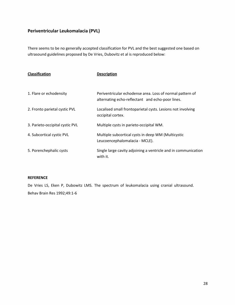
Periventricular Leukomalacia (PVL)
There seems to be no generally accepted classification for PVL and the best suggested one based on
ultrasound guidelines proposed by De Vries, Dubovitz et al is reproduced below:
Classification Description
1. Flare or echodensity Periventricular echodense area. Loss of normal pattern of
alternating echo-reflectant and echo-poor lines.
2. Fronto parietal cystic PVL Localised small frontoparietal cysts. Lesions not involving
occipital cortex.
3. Parieto-occipital cystic PVL Multiple cysts in parieto-occipital WM.
4. Subcortical cystic PVL Multiple subcortical cysts in deep WM (Multicystic
Leucoencephalomalacia - MCLE).
5. Porenchephalic cysts Single large cavity adjoining a ventricle and in communication
with it.
REFERENCE
De Vries LS, Eken P, Dubowitz LMS. The spectrum of leukomalacia using cranial ultrasound.
Behav Brain Res 1992;49:1-6

29
Screening for Retinopathy of Prematurity (ROP)
Infants with birth weight of <1500 gm or gestational age < 32 weeks and selected infants with a birth
weight of 1500-2000gm or gestational age > 32 weeks with an unstable clinical course, including those
require cardio respiratory support, are candidates for screening for ROP.
1) The following table provides the schedule for detecting ROP potentially damaging to retina with 99%
confidence.
Table 5. Timing of First Eye Examination Based on Gestational Age at Birth:
Gestational Age at Birth, wk
Age at Initial Examination, wk
Postmenstrual Chronologic
22a 31 9
23a 31 8
24 31 7
25 31 6
26 31 5
27 31 4
28 32 4
29 33 4
30 34 4
31b 35 4
32b 36 4
Shown is a schedule for detecting pre-threshold ROP with 99% confidence, usually well before any required treatment.
This guideline should be considered tentative rather than evidence-based for infants with a gestational age of 22 to 23 weeks because of the small number of survivors in these gestational-age categories.

30
2) Follow up examination should be recommended by the examining ophthalmologist on the basis of retinal findings classified according to the international classification. (See Figure 1)
Figure 1 (adapted from American Academy of Pediatrics. Screening examination for Preterm Infants for
Retinopathy of Prematurity.Pediatrics.2006; 117:572-576).
Scheme of retina of the right and left eyes showing zone borders and clock hours used to describe the
location and extent of retinopathy of prematurity. Diagrammatic representation of the potential total
area of the premature retina, with zone I (the most posterior) symmetrically surrounding the optic nerve
head (the earliest to develop). A larger retinal area is present temporally (laterally) than nasally
(medially) (zone III). Only zones I and II are present nasally. The retinal changes discussed in
recommendation 4 are usually recorded on a diagram such as this.
1 Week or less follow up
Stage 1 or 2 ROP: Zone I
Stage 3 ROP: Zone II
1-2 Week follow up
Immature vascularization: Zone I –no ROP
Stage 2 ROP: zone II
Regressing ROP: Zone I
2 Week follow up

31
Stage 1 ROP: zone II
Regressing ROP: Zone II
2-3 Week follow up
Immature vasularization: Zone II—no ROP
Stage 1 or 2 ROP: Zone III
Regressing ROP: Zone III
One examination is sufficient only if it is unequivocally shows the retina to be fully vascularized in each eye.
REFERENCE
American Academy of Pediatrics. Screening examination for Preterm Infants for Retinopathy of Prematurity.Pediatrics.2006; 117:572-576.

32
Apnea of Prematurity:
It usually occur in infants who are <34 weeks gestation, weight<1800gm, and have no identifiable cause
of the apnea of prematurity
1. General measure: include tactile stimulation, correct anemia, maintain normal body temperature,
correct electrolyte imbalance if present.
2. Look for IVH, signs or symptoms of Sepsis, PDA, NEC, and gastro-oesophageal reflux, and treat it
accordingly.
3. Specific treatment: Theophylline or caffeine: The choice of drugs depends on availability and
neonatologist’s preference.
Aminophylline:
Dose for apnea of prematurity: 5-6 mg/kg PO or I/V (infused over 15-30 minutes) as a loading dose,
followed by a maintenance dose of 2 mg/ kg q 12 h starting 12 hr after the loading dose.
Adverse effects: hyperglycemia, diuresis, dehydration, feeding intolerance, tachycardia, tachycardia
arrthymias , hyperreflexia, jitteriness and seizures.
Therapeutic level for apnea: 6-11 mg/ml
Caffeine:
Loading dose: 20mg of caffeine citrate (10 mg of caffeine base) I/V or P/O
Maintenance dose (caffeine base): 2.5-5 mg/kg/day as single dose
Adverse effects: Nausea, vomiting, gastric irritation, agitation, tachycardia and diuresis. Symptoms of
over dosage include arrhythmias and tonic clonic seizures
Therapeutic level: 5-25 mcg/ml
a. Therapy can usually be discontinued by postconceptional age (usually 37 weeks), depending on the
weight of the infant (usually 1800-2000g), or if the infant has been apnea free for 7 days. More
immature infants will require treatment longer.
b. If the above mentioned drug trial fails, use continuous positive air way pressure(CPAP) via nasal
prongs at a rate of 2-4 cm H2O and continue administration of drug. An alternative to nasal CPAP is to
use high flow nasal cannula at 1-2 L /min.
c) If apnea persists, consider doxapram (not commonly used). Doxapram may be efficacious when
theophylline and caffeine have failed.
Dosage: 0.5-1.5mg/kg/hr by continuous I/V infusion, decrease the dose when control of apnea is
achieved.

33
d) Mechanical Ventilation: Should be continued if apnea and bradycardia cannot be controlled by drug
therapy or nasal CPAP. Low pressures are used to prevent apnea.
REFERENCE
Apnea and Bradycardia: In Gomella TL, Cunninngham MD (eds): a LANGE Clinical Manual, Neonatology:
5th ed. McGraw Hill 2004.210

34
Hypoxic Ischemic Encephalopathy (HIE)
Table 6. 3 clinical Stages of encephalopathy described are:
Stage 1
1. Duration<24 hrs with hyperalertness 2. Uninhibited Moro and stretch reflexes 3. Sympathetic effects 4. Normal electroencephalogram
Stage2
1. Obtundation 2. Hypotonia 3. Decreased spontaneous movement with or without seizures
Stage3
1. Stupor 2. Flaccidity 3. Seizures 4. Suppressed brain stem and autonomic functions 5. EEG may be isopotential or have infrequent periodic discharges
1) Stage 3 or persistence of stage 2 for more than seven days or failure of the EEG to revert to
normal is associated with Neurodevelopmental impairment or death.
2) Full-term infants who develop long-term neurologic sequelae from intrapartum.
3) Asphyxia may not have low Apgar scores but will demonstrate neurological dysfunction within
48 hours.(2)
Postnatal Management:
Management of every asphyxiated baby is individualized. Below is the broad guideline for management
of the asphyxiated baby.
Systemic management: The corner stone of management following hypoxia ischemia is supportive care
and the careful maintenance of systemic homeostasis(3)

35
Figure 2. Summary of initial management of asphyxiated neonates
Figure Adapted from: Aggarwal R, Deorari AK, Paul VK. Post-resuscitation management of asphyxiated neonates. Indian J Pediatr. 2001 Dec;68(12):1149-53.
Transfer the baby to NICU/ special care unit:
A baby who fails to initiate and sustain respiration at birth is at risk of hypoxic brain injury and needs
regular monitoring. Infants with moderate hypoxia (APGARS 4-6 at one minute of age) may be
transferred to the mother. However, these infants should also be monitored frequently in the first 48-72
hours for features suggestive of HIE. Infants with severe asphyxia (Apgar score 0-3 at 1 minute or need

36
for prolonged bag and mask ventilation >5 minute) should be transferred to NICU for observation and
treatment.
Thermoregulation:
Place the baby under radiant warmer after drying the baby. Maintain normal body temperature of the
baby.
Monitoring of vital signs:
Immediate clinical assessment should be made by recording respiration, heart rate, blood pressure,
capillary refill time, temperature and oxygen saturation. Urine output monitoring should be done.
Start intravenous fluids:
Give two third of the maintenance fluid due to the possibility of syndrome of inappropriate secretion of
anti-diuretic hormone (SIADH).
Check blood sugar, hematocrit and blood gas (if needed).
Consider infusion of Volume Expanders:
Volume expanders: Use normal saline, Ringer lactate or if needed blood product to maintain
intravascular volume.
If capillary refill is >3 seconds or if there is metabolic acidosis, volume expansion with normal saline (or
Ringer lactate) 10mg/kg should be instituted, may repeat if necessary
Subsequent management (4)
Fluid and electrolytes:
Judicious fluid management is needed and fluid overload should be avoided.
Give 10% Dextrose/water at 40-60ml/kg/day on 1st day of life and increase fluid and electrolytes
according to day of life, serum level and neurologic condition of baby.
Maintain normal calcium level: Calcium should be provided in a maintenance dose of 4 ml/kg/day (10%
calcium gluconate) for 1-2 days. This can be given in 1:1 dilution under cardiac monitoring.
Treat metabolic acidosis:
If metabolic acidosis is severe or persistent then sodium bicarbonate may be used but caution is advised as rapid infusion increases serum osmolality and alkalisation may decrease cerebral blood flow.
Avoid hypoglycemia and hyperglycemia:

37
Maintain adequate perfusion:
Global ischemia may affect the myocardium and cardiovascular support may be needed to improve
perfusion. The markers for normal perfusion are normal blood pressure, capillary refill time of <3
seconds, normal urine output and absence of metabolic acidosis. The blood pressure should be
maintained to the upper normal range. This can be achieved with the judicious use of volume expanders
and vasopressor agents.
Vasopressors: Dopamine Is the drug of first choice in the treatment of neonatal hypotension.
Dosage: 4-20 mcg/kg/min
Route: Intravenously
Adverse effects: may cause ectopic heart beats, tachycardia, hypotension, hypertension and excessive
diuresis
Dobutamine: If dopamine fails to improve BP, dobutamine is recommended as second line drug. In
neonates it is usually given together with dopamine infusion.
Dosage: 2-10mcg/kg/min by continuous infusion. Maximum: 20mcg/kg/min
Route: Intravenously
Adverse effects: dose related. Ectopic heart beats, increased heart rate, and blood pressure elevations
Renal failure management:
Manage renal failure if urine output is <0.5ml/kg/hr after 48 hours of life.
Respiratory support may be necessary for babies with Hypoxic Ischemic Encephalopathy.
Prevent cerebral edema and avoid fluid overload:
Restrict maintenance fluids (by up to 70%) if cerebral oedema is suspected on the basis of neurologic
signs. Observe for hypertension and SIADH (hyponatremia, volume overload and decreased urine
output). Manage SIADH with restriction of maintenance fluids.
Treat seizures:
Metabolic disturbances should be excluded before initiating anticonvulsant therapy.
Phenobarbitone: Is the initial drug of choice. Initially a 20mg/kg loading dose of phenobarbitone is used
(watch breathing if not ventilated). If no effect a further 10-20 mg/kg loading may be given, followed by
a maintenance dose of 3-5 mg/kg PO or IV every 12 hours, started 12-24 hours after the loading dose.

38
Phenytoin is usually added when seizures are not controlled by phenobarbitone. The loading dose is 15-
20mg/kg I/V followed by maintenance dose of 4-8 mg/kg/day.
Clonazepam may be useful as a second line 100-200 microgram/kg loading followed by an 100
microgram/kg/day.
Gastrointestinal Care:
Keep NPO. Pass Orogastric tube and attach it with bag on gravity to decompress gut. Look for signs of
NEC. If the patient is clinically stable, start feeding gradually according to feeding guideline.
Hematologic Care:
If there is thrombocytopenia and coagulopahy (prolonged PT, APTT) correct it accordingly by platelet
and plasma administration.
Nutrition:
Asphyxiated infants are at risk of developing necrotizing enterocolitis. They are at risk also of aspiration
if there is pharyngeal incoordination. There may be intolerance to feed because of reduced small
intestinal motility, therefore enteral feeding should be commenced cautiously.
Brain or Total Body Cooling:
There are increasing data to support cooling theraphy in the treatment of neonatal hypoxic ischaemic
encephalopathy. Such treatment will be guided by local or national protocols.
REFERENCES
1. Nelson KB, Leviton A. How much of neonatal encephalopathy is due to birth asphyxia? Am J Dis Child. 1991;145(11):1325-31.
2. Sarnat HB, Sarnat MS. Neonatal encephalopathy following fetal distress. A clinic and encephalographic study. Arch Neurol 1976;33:696-705.
3. Aggarwal R, Deorari AK, Paul VK. Post-resuscitation management of asphyxiated neonates. Indian J Pediatr. 2001 Dec;68(12):1149-53.
4. Birth Asphyxia: In Gomella TL,Cunninngham MD (eds):a LANGE Clinical Manual, Neonatology:
Management of Perinatal asphyxia.5th ed. McGraw Hill 2004
5. Denis V. Azzopardi, Brenda Strohm, A. David Edwards, Leigh Dyet, Henry L. Halliday, Edmund
Juszczak, Olga Kapellou, Malcolm Levene, Neil Marlow, Emma Porter, Marianne Thoresen, Andrew
Whitelaw and Peter Brocklehurst for the TOBY Study Group. Moderate Hypothermia to Treat
Perinatal Asphyxial Encephalopathy. N Engl J Med 361;14:1349-1358
6. Jacobs SE, Hunt R, Tarnow-Mordi WO, Inder TE, Davis PG. Cooling for newborns with hypoxic
ischaemic encephalopathy. Cochrane Database Syst Rev. 2007 Oct 17;(4):CD003311.

39
Neonatal Hyperbilirubinemia
Guideline for Neonatal Hyperbilirubinemia
Hyperbilirubinemia is very common and usually benign in the term newborn infant and the late preterm
infant at 35 to 36 completed weeks’ gestation. Critical hyperbilirubinemia is uncommon but has the
potential for causing long-term neurological impairment.
Clinical assessment: Clinicians should ensure that all infants are routinely monitored for the
development of jaundice. Clinically jaundice is assessed by blanching the skin with digital pressure
revealing the underlying color of the skin and subcutaneous tissue. The assessment of jaundice should
be performed in a well lit room, or preferably in day light. Jaundice is seen first in the face and progress
caudally to the trunk and extremities1.
Laboratory measurements: measurement of total serum bilirubin (TSB) and/or total cutaneous bilirubin
(TcB) should be performed in every newborn who develops jaundice in the first 24 hours and repeating
of TSB will depend on the first bilirubin value, the age of infant and the evolution of jaundice2.
When indicated, infants should be treated with phototherapy or exchange transfusion: recommendation
for treatment is given in fig 3 and 4.
Acute bilirubin encephalopathy: Immediate exchange transfusion is recommended in any infant who is
jaundiced and manifests the signs of moderate to advanced encephalopathy (hypotonia, arching,
retrocollis, opisthotonus, fever, high pitched cry) even if the TSB is falling.
Fig 3. Guidelines for phototherapy in hospitalized infants of 35 or more weeks’ gestation 1

40
Fig 4. Guidelines for exchange transfusion in infants 35 or more weeks’ gestation1
Hyperbilirubinemia in preterm and low birth weight babies:
Basic pathophysiology of jaundice is the same in term and preterm babies but lower gestation babies
are at higher risk of developing hyperbilirubinemia and require close surveillance and monitoring. TSB
for intervention also vary in term, near term, and preterm neonates <35 weeks period of gestation.
Clinicians should ensure that all premature infants are routinely monitored for the development of
jaundice. TSB should be measured at 24 hour of age with follow up estimation every 12-24 hours until
the level stabilizes. TSB should be repeated within 12- 24 hours of stopping phototherapy or sooner if
clinical jaundice reappears.
The American Academy of Pediatrics recommends guideline for neonates >35 weeks of gestation, the
management of preterm infants are still a grey zone, and most centers have individual treatment
guidelines. The following tables provide guidelines for intervention in LBW and/or preterm babies3.
Guidelines for the use of phototherapy and exchange transfusion in low birth weight infants based on
birth weight 3
Table 7.

41
*Consider initiating treatment at these levels. Range allows discretion based on clinical conditions or other
circumstances. Note that bilirubin levels refer to total serum bilirubin concentrations. Direct reacting or conjugated
bilirubin levels should not be subtracted from the total.
Used at these levels and in therapeutic doses, phototherapy should, with few exceptions, eliminate the need for
exchange transfusions.
Levels for exchange transfusion assume that bilirubin continues to rise or remains at these levels despite
intensive phototherapy.
Guidelines for use of phototherapy and exchange transfusion in preterm infants based on gestational
age 3
Guidelines for initiating phototherapy and exchange transfusion in ELBW infants 3
Bilirubine conversion values:
>8.0 mg/dl >136.8 µmol/L
>10.0 mg/dl > 171 µmol/L
>13.0 mg/dl > 222.3 µmol/L
>15.0 mg/dl > 256.5 µmol/L
Table 8.
Table 9.

42
REFERENCES
1. Ron Keren, Xianqun Luan, Susan Friedman, Stephanie Saddlemire, Avital Cnaan, Vinod K. Bhutani. A
Comparison of Alternative Risk-Assessment Strategies for Predicting Significant Neonatal
Hyperbilirubinemia in Term and Near-Term Infants. Pediatrics 2008;121(1): e171-e179.
2. American Academy of Pediatrics. Clinical practice guideline. Management of hyperbilirubinemia in
the newborn infants 35 or more weeks of gestation. Pediatrics.2004:114:297-316.
3. M J Maisels, J F Watchko. Treatment of jaundice in low birthweight infants. Arch Dis Child Fetal
Neonatal Ed 2003;88:F459–F463.

43
Hypotension in Neonates
Table 10. Blood pressure ranges 1
Assessment for the cause of hypotension: Assess the baby to find out the cause of hypotension. The
basic decision is whether the infant requires volume replacement or administration of inotropic agents.
History: A careful history should be obtained to rule out birth asphyxia, blood loss (antepartum or
postpartum), drug infusion, birth trauma (adrenal hemorrhage or liver injury), and cardiac problem.
Physical examination: a careful physical examination will often reveal which organ system is involved
Chest X-ray: A small heart is seen in volume depletion; a large heart is seen in cardiac disease.
Management:
Empirical volume expansion with crystalloid (normal saline) (10ml/kg intravenously over 30 minutes)
should be started if unsure of the cause of hypotension
If there is a response, continue volume expansion (Volume administration is less effective in the
immediate postnatal period, and its extensive use is associated with significant untoward effects,
especially in preterm infants)
Provide respiratory support: as needed
Correct metabolic acidosis: If there is no response,
An inotropic agent should be started:
Dopamine: See Section on HIE (page 36)
Dobutamine: See Section on HIE (page 36)

44
Refractory hypotension:
During the course of their disease, some of the sickest newborns become unresponsive to volume and
pressor administration; this is called as refractory hypotension.
Definition: Refractory hypotension is defined as mean arterial pressure <than the gestational age
despite a total inotrope dose of 20 microgram/kg/minute (2). In these cases, stress dose of
hydrocortisone 1mg/kg 8 hourly for 5 days rapidly up regulates cardiovascular adrenergic receptor and
decreases the requirement for pressor support in the critically ill newborn with volume and pressor
resistance hypotension.(3)
REFERENCES
1. Hypotension and shock: In Gomella TL, Cunninngham MD (eds): a LANGE Clinical Manual,
Neonatology: 5th ed. McGraw Hill 2004
2. Seri I, Evan J. Controversies in the Diagnosis and Management of Hypotension in the Newborn
Infant. Curr Opin Pediatr.2001; 13:116-123
3. Baker CF, Barks JD, Engmann C, Vazquez DM, Neal CR. Hydrocortisone Administration for the
Treatment of Refractory Hypotension in the Critically ill Newborns. J Perinatol.2008; 28:412-419.

45
Patent Ductus Arteriosus (PDA) in Preterm Neonates: (<34 weeks gestation)
Preterm infants have a high incidence of persistent PDA, about 40-60%. A clinically symptomatic PDA
has been associated with significant morbidity in this high risk population.
Clinically significant PDA: PDA diagnosed on the basis of continuous or systolic mumur heard best in
the left subclavicular are associated with hyperactive precordium and a wide pulse pressure or bounding
pulses.
Heart failure is assessed on the basis of one or more of the following clinical findings: tachycardia, gallop
rythhm, hepatomegaly, cardiomegaly, deteriorating respiratory status, and radiologic evidence of
increased pulmonary vascularity or pleural fluid.
Echocardiography: PDA is echocardiographically significant when ductal size > 1.5 mm or the left atrial
to aortic root ratio >1.4.
Management of PDA in newborn <34 weeks gestation:
1) The following should be tried initially:
a) Fluid restriction: Fluid should be restricted to 2/3 rd of maintenance fluid.
b) Furosemide: 1 mg/kg intavenousely twice a day
c) Digioxin (if there are signs and symptoms of cardiac failure)
If no improvement:
Then consider:
2) Non steroidal anti inflammatory drugs: Indomethacin or Ibuprufen
Randomized controlled trials have demonstrated similar efficacy in ductus closure with these two agents
in infants born at up to 34 weeks of gestation (1)
1) Indomethacin:
Dose: 0.2mg/Kg/day, enterally or parenterally, 3 doses. Second and third doses are given 12 and 36 hour
after the first dose. The second and third doses are 0.1mg/kg/dose if the infant is <7 days old and the
infant is <1250 g birth weight. If the infant is either> days old or >1250 g, then the second and third dose
are also 0.2mg/kg. (2)
46
Monitor: urine output, serum creatinine, and platelet count.
Measure serum creatinine and platelets before each dose, hold the drug if serum creatinine is >2 mg/dl,
platelet count is <60,000 or urine output is <1.0ml/kg/day, or if there is evidence of NEC or bleeding
disorders.(3)
Feeding should be given cautiously and monitor for aspirates, abdominal distension, and other signs and
symptoms of necrotizing enterocolitis.
After a course of indomethacin, assess the baby clinically, and if possible by echocardiography. If there
is still a significant PDA consider a second course of indomethacin. In 20-30% of cases, the ductus will
reopen after the first course of indomethacin. A significant proportion of these cases will benefit from a
second course of indomethacin.
Side effects: Renal dysfunction, gastrointestinal hemorrhage, necrotizing enterocolitis, intraventricular
hemorrhage, platelet dysfunction.
2) Ibuprufen: Oral Ibuprufen (as shown in various studies) is as effective as indomethacin in ductal
closure (4)
Dose: 10mg/kg followed by 2 additional doses of 5mg/kg on consecutive days
Clinical improvement after medical treatment: Murmur abolished or decreased in intensity, if on
ventilator parameters decreased. Signs and symptoms of heart failure improved.
Surgical Ligation: Failure of ductus to close after one or two courses of drug therapy along with
persistence of heart failure and ventilator dependence.
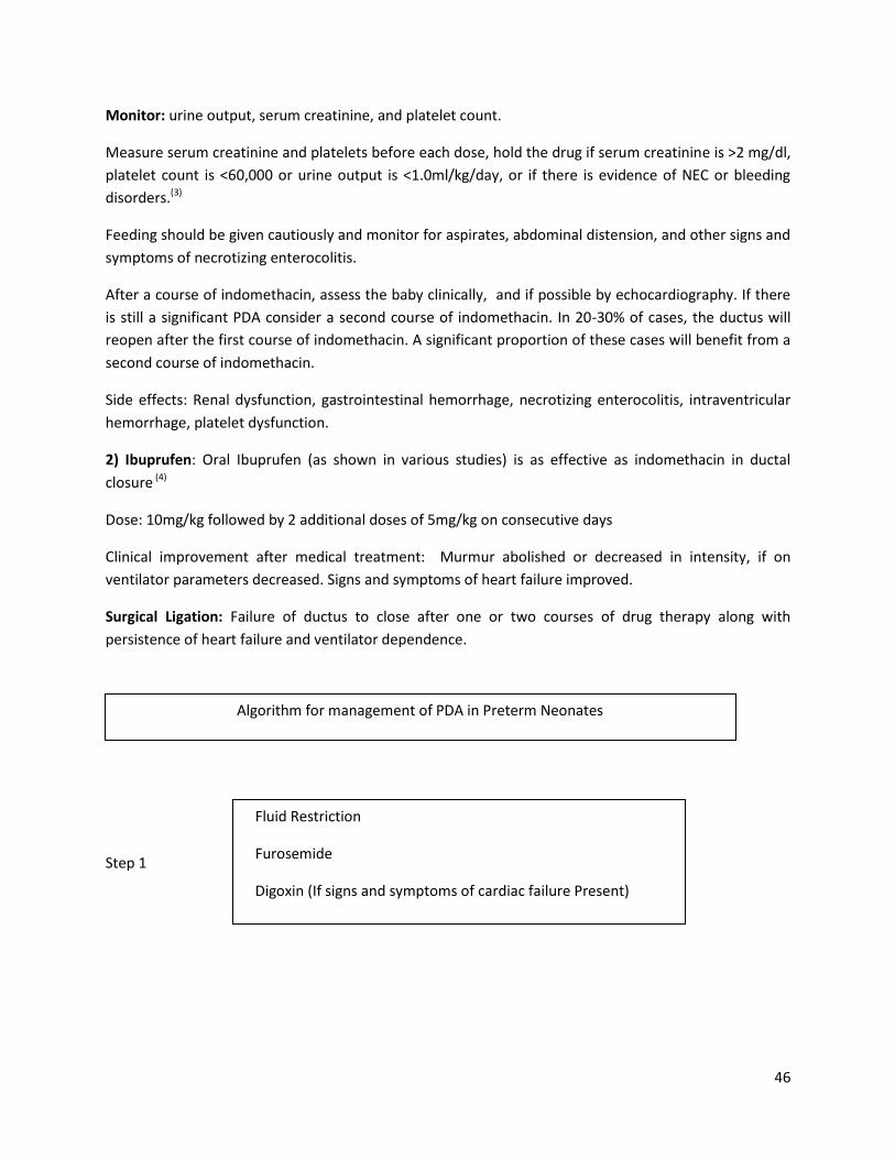
Step 1 STEP1
Algorithm for management of PDA in Preterm Neonates
Fluid Restriction
Furosemide
Digoxin (If signs and symptoms of cardiac failure Present)

47
Step 2
Step 3
REFERENCES
1. Bart Van Overmeire, Ingrid Follens, Suzanne Hartmann. Treatment of PDA with Ibuprufen. Arch Dis
Childhood.1997; 76:179-184.
2. PDA: In Gomella TL, Cunninngham MD (eds): a LANGE Clinical Manual, Neonatology: Patent ductus
arteriosus.5th ed. McGraw Hill 2004.
3. Ruth M, Yanagi, Allen Wilson, Edgar A, Newfeld, Kalim U.Aziz. Indonethacin Treatment for
Symptomatic Patent ductus Arteriosus. Adouble Blind Control Study.1981; 67:647-652.
4. Eli Heyman, Iris Morag, David Batash, Rimona Keidar, Shaul Baram. Closure of PDA with Oral
Ibuprufen Suspension in Premature Newborns. A pilot study.Pediatrics.2003; 112:354.
NSAIDs: Indomethacin: 0.2mg/kg followed by 0.1mg/kg if
<1250gm or >7days old, the dose is same if weight>1250gm or
>7 days old given at, 8 and 36 hours
Or
Ibuprufen: 10mg/kg at 0hour then at 5mg/kg at 24hr and 48hr
Consider surgical ligation if medical treatment fails along with
persistence heart failure or ventilator dependency

48
Sepsis
Criteria for suspecting sepsis:
The signs and symptoms of neonatal sepsis often are nonspecific. Most, but not all, infants with sepsis
have respiratory signs, including cyanosis or apnoea. Other signs such as feeding difficulties or lethargy
are nonspecific and may be subtle or insidious. Thus, a high index of suspicion is required to identify and
evaluate at-risk infants.
REFERENCE
William McGuire, Linda Clerihew, Peter W Fowlie. Infection in the preterm infant. BMJ 2004;329:1277-1280
Investigations:
Blood culture
Take into consideration urine culture and cerebral spinal fluid culture also
Specific Management:
After blood culture, first line antibiotic therapy should be started without delay. Local guidelines for
antibiotics should exist, taking into account the prevalence and sensitivities of organisms within each
unit. Consider changing antibiotics if there is no clinical improvement or according to culture-sensitivity
report.

49
Prevention
-Spacing
-Avoid overcrowding
- Good handwashing before and after touching the baby
- Application of alcohol gel
Supportive management:
-Provide thermoneutral environment
-Provide supplemental O2 if required, Ventilatory support may be required in some cases.
-Monitor vital signs, peripheral perfusion and urine output.
-Monitor renal function to avoid aminoglycoside toxicity.
-Fluid resuscitation. If there is poor peripheral perfusion (capillary refill>3 sec, give normal saline
10ml/kg IV bolus. Continue IV fluids. IV bolus can be repeated if necessary.
-Treat convulsions if present.
-Give blood products as indicated.
-Monitor and correct electrolyte imbalance if present.
-Blood sugar monitoring
REFERENCE
Edwards MS: Postnatal Bacterial Infections. In Fanaroff AA, Martin J: Neonatal Perinatal Medicine:
Diseases of the Fetus and Infant, 8th ed.Mosby.2006.P791-804.