Embed Size (px)
Citation preview
Neonatal Emergencies
and Transport

Relative Anatomy
and Physiology
Physiology of
Thermoregulation • Neonate at significant risk of
hypothermia
– Ratio of neonatal body surface area to
volume is four times that of an adult
– Neonate has less adipose tissue than adult
– Thermogenesis in neonate only one and a
half as high as adult
– Muscle tone is immature in neonate
– Neonate cannot shiver effectively enough
to generate heat
Heat Loss in the Neonate
• Results from:
– Evaporation
• Most of heat loss, especially in moments immediately
after birth
– Convection
• Depends on birthing environment
• When care providers are comfortable in the room, it is
too cold for the neonate
– Conduction
– Radiation
• Room’s ambient temperature should be as close to core
temperature as possible

Heat Loss in the Neonate
Glucose Requirements
• Newborns at significant risk of acute hypoglycemia due to: – Poor glucose stores
– Inability to stimulate the immature neonatal liver to release glucose
– Increased metabolism that uses large quantities of available glucose
• Assess neonatal glucose levels within 1 to 2 hours after birth
– Reassess every 30 minutes to 1 hour thereafter until glucose levels are normal
• Neonate blood glucose levels (BGLs) should be maintained above 70–80 mg/dL
Signs and Symptoms
of Hypoglycemia
• Twitching, seizure activity, eye rolling
• Muscular hypotonia (limpness)
• High-pitched cry
• Respiratory apnea, irregular respirations
Management of
Hypoglycemia • Administer 10 percent dextrose as
needed at 80ml/kg/day
Airway Anatomy and
Physiology
• Unique differences between neonatal and
adult airway anatomy and physiology
– Neonatal tongue larger compared to the
oropharynx
– Little room for airway edema
– Increased likelihood of airway obstruction in
depressed neonate
– Neonatal trachea more pliable, narrow
– Airway obstruction from:
• Hyperextension, hyperflexion kinking
• Edema
Airway Anatomy and
Physiology
– Neonatal epiglottis is large and more U-
shaped or oblong, floppy from incomplete
cartilaginous support
– Use of straight versus curved blade during
laryngoscopy
– Neonatal larynx more cephalad, anterior
– Level of first or second cervical vertebrae
– Harder to achieve single plane view
needed for optimal orotracheal intubation
conditions
Pulmonary Anatomy and
Physiology
• Many differences in neonatal pulmonary anatomy and physiology compared to the adult
• Bones in neonatal thoracic cavity not fully calcified – Flexible
• Neonatal ribs are more horizontal than they are rounded – Little leverage to increase the anterior and
posterior diameter of the chest
– Inability to provide the degree of lift needed to increase the volume of the chest cavity upon inspiration
Pulmonary Anatomy and
Physiology
• Poorly developed accessory muscles – Cause diaphragmatic breathing
• Neonatal sternum very pliable – Contributes to inability to create a strong negative
intrathoracic pressure
– Inhibits efficiency of inspiratory effort
Pulmonary Anatomy and
Physiology
• Neonates have diminished pulmonary reserve capacity – Heart larger, ribs and sternum fail to adequately support the
lungs
– Less space for lung expansion compared to adults
– More rapid development of hypoxemia and hypercapnia
• Neonates are primarily abdominal breathers – Rely heavily on diaphragmatic motion to breathe
– Overcrowding of the neonatal abdominal cavity a significant problem
– Negatively affects the neonate’s compensatory ventilation mechanisms
– Limits diaphragmatic excursion secondary to increased abdominal pressure
Pulmonary Anatomy and
Physiology
• Neonates consume twice the oxygen of
adults
– Lower pulmonary reserve capacity coupled
with a higher metabolic demand for oxygen
predisposes the neonate to hypoxemia
Cardiovascular Anatomy
and Physiology
• Several differences between adult and
neonatal cardiovascular systems
• While still in utero, the fetus receives its
oxygen through the placenta
– Disturbances to alveolar ventilation and
gas exchange following birth must be dealt
with immediately
Cardiovascular Anatomy
and Physiology
Cardiovascular Anatomy
and Physiology
• Neonatal heart can usually only
increase rate to improve cardiac output
– Cannot increase contractile force
– Cardiac output drastically reduced with
bradycardia
Cardiovascular Anatomy
and Physiology
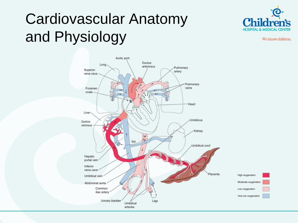
• Most of physiologic change that occurs with
the shift from intrauterine to extrauterine life
occurs in the first few minutes after delivery
– Clamping of umbilical cord moves circulation from
placenta to pulmonary system
– Interruption of low-resistance, placental blood flow
from the umbilical cord increases systemic
vascular resistance (SVR)
– Increased SVR closes the ductus venosus
– Closure of ductus venosus causes renal perfusion
Cardiovascular Anatomy
and Physiology
• Neonate’s first breaths expands the lungs
– Lung expansion reduces pulmonary vascular resistance
– Reduced pulmonary vascular resistance: • Increases pulmonary blood flow
• Reduces pulmonary artery pressures – Left side of heart assumes higher pressures than right
• Closes the foramen ovale
• Closes the ductus arteriosis – Occurs in first hours to weeks after birth
General Pathophysiology:
Pulmonary
• Assessment of respiratory distress
– Etiology of respiratory compromise may not be readily identifiable
• First goal is to replace any lost function of the airway or breathing components
• Once airway or breathing insult is corrected, can identify potential causes of the hemodynamic and/or respiratory compromise
– Goals in managing respiratory compromise in the critical care environment are to:
» Identify a set of causes and
» Treat the patient based on the most likely etiology
Respiratory Distress, Failure,
and Arrest
• Must use precise terms when describing
respiratory distress, respiratory failure, and
respiratory arrest
– Distinction between the three dictates the
management of the acutely ill neonate
– Respiratory distress
• Maintains the ability to compensate
– Respiratory failure
• Has exhausted compensatory mechanisms
– Respiratory arrest
• Patient is apneic
Persistent Pulmonary
Hypertension
of the Newborn
• Clinical syndrome in which pulmonary
vascular resistance is elevated in the
presence of changes in pulmonary
vessel reactivity
– Results in sustained fetal circulation
– Ductus arteriosus and foramen ovale
remain open
Persistent Pulmonary
Hypertension
of the Newborn
• Commonly associated with severe hypoxia, meconium aspiration syndrome, and congenital diaphragmatic hernia
• Clinical presentation mirrors many of the signs and symptoms of congenital heart diseases
– May be difficult to assess in the aeromedical or ground transport environment
Persistent Pulmonary
Hypertension
of the Newborn • Management
– Maintain oxygenation
– Give nitric oxide
• Promotes pulmonary vascular dilation
• Keeps pulmonary perfusion pressures closer to
normal
• Closes vascular structures
– Use adenosine, magnesium sulfate as
pulmonary vasodilators
Meconium Aspiration
Syndrome
• Meconium expelled prematurely in 10 to 15
percent of all deliveries
– Only 2 to 10 percent will aspirate meconium into
lower airways
• Meconium aspiration can obstruct airway
and/or may contribute to inactivation of
alveolar surfactant
• No known prevention strategies
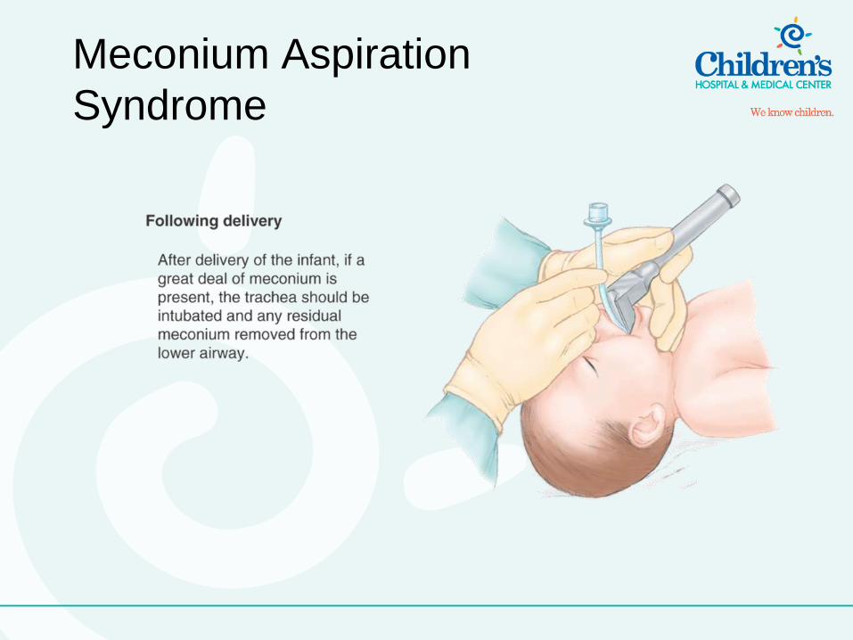
– Nasopharyngeal and endotracheal suctioning
before delivery of the thoracic cavity may limit
meconium aspiration into the lower airways
Meconium Aspiration
Syndrome
Transient Tachypnea of the
Newborn (TTN)
• Also known as “wet lung” or “Type II
Respiratory Distress Syndrome”
• Self-limiting process
– Auto-resolves within 48–72 hours from birth
– Caused by delayed clearing of fluids in the lungs
• Management
– Ensure adequate oxygenation
– Give antibiotic therapy until sepsis, pneumonia
ruled out
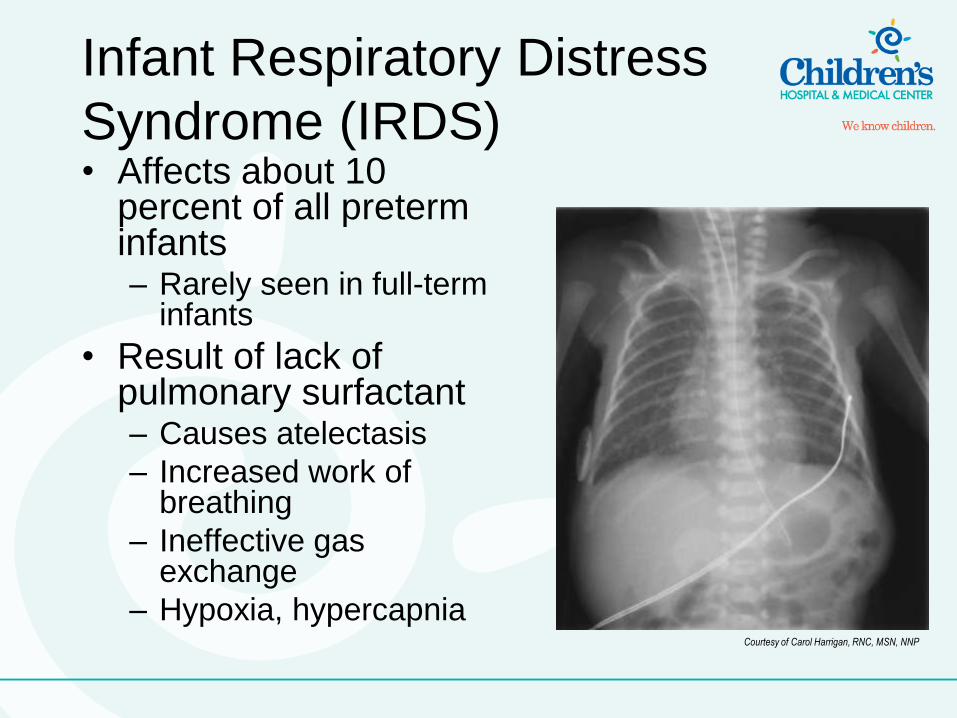
Infant Respiratory Distress
Syndrome (IRDS) • Affects about 10
percent of all preterm infants – Rarely seen in full-term
infants
• Result of lack of pulmonary surfactant – Causes atelectasis
– Increased work of breathing
– Ineffective gas exchange
– Hypoxia, hypercapnia Courtesy of Carol Harrigan, RNC, MSN, NNP
Infant Respiratory Distress
Syndrome (IRDS) • Signs and symptoms include:
– Tachypnea, shortness of breath
– Accessory muscle use, sternal retractions, grunting, nasal flaring
– Respiratory arrest from muscle fatigue, hypoxemia, and acidosis
• Management – Ensure adequate ventilation and oxygenation
– Administer exogenous surfactant
Congenital Diaphragmatic
Hernia
• Complication in which the bowel protrudes
into the thoracic cavity through an interruption
of the diaphragm
– Usually the result of congenital abnormality
– 85 percent of all congenital diaphragmatic hernias
occur on left side
– Mortality rate between 40 and 60 percent
• Herniated abdominal contents prevent full
lung expansion in the affected hemithorax
– Pulmonary compromise ensues
Congenital Diaphragmatic
Hernia • Signs and symptoms
– Respiratory distress
– Unequal lung sounds
– Scaphoid shaped abdomen
• Management – Ensure adequate ventilation and oxygenation
– Insert NG tube
– Conduct gastric decompression
– Repair surgically (definitive treatment)
– General pathophysiology, cardiovascular
Congenital Heart Disease
Overview • Incidence of congenital heart disease in the
United States is approximately 8 per 1,000
live births
– About 40,000 neonates born each year with a
heart defect
• Many congenital heart defects are subclinical
• Defects can cause:
– Abnormalities in volumes and/or pressures in the atria or
ventricles
– Mixing of venous and arterial blood
– Inadequate cardiac output and poor systemic perfusion
• Neonate can have multiple defects at once
Left-to-Right Shunt Defects
• Condition in which oxygenated blood
shifts from left to right side of the heart
• Defect is considered acyanotic
– Higher pressures on left side of heart
prevent unoxygenated blood from right
side from entering the aorta and systemic
circulation
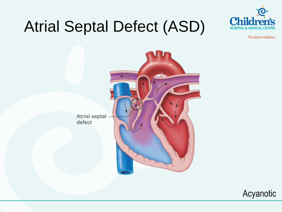
Atrial Septal Defect (ASD)
• Commonly the result of foramen ovale
nonclosure
– “Patent” foramen ovale
– Oxygenated blood from pulmonary vein
enters left atria
– Higher left atrial pressure compared to
right produces volume shift to right side
– Eventually causes right atrial and
ventricular enlargement
Acyanotic
Atrial Septal Defect (ASD)
• Signs and symptoms
– Commonly subclinical
– Clinical significance related to size of
defect
– Rarely, congestive heart failure might
develop
• Management
– Give supportive care
– Repair surgically (definitive treatment)
Acyanotic
Atrial Septal Defect (ASD)
Acyanotic
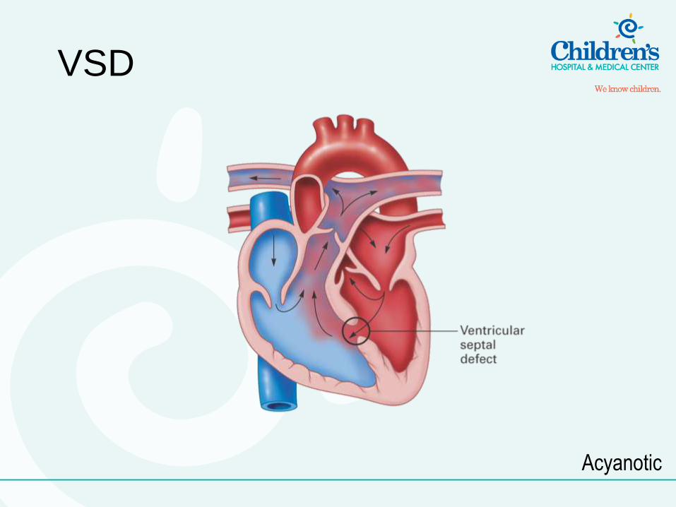
Ventricular Septal Defect
(VSD)
• Defect in ventricular septum allows
blood flow between ventricles
– Can cause:
• Left-to-right shunting of blood
• Pulmonary hypertension
• Changes in pulmonary vascular bed
– Size of defect determines clinical
significance
Acyanotic
Small VSD
• Produces a small, left-to-right shunt
• Little pulmonary vascular congestion,
chamber enlargement
• More difficult to diagnose
Acyanotic
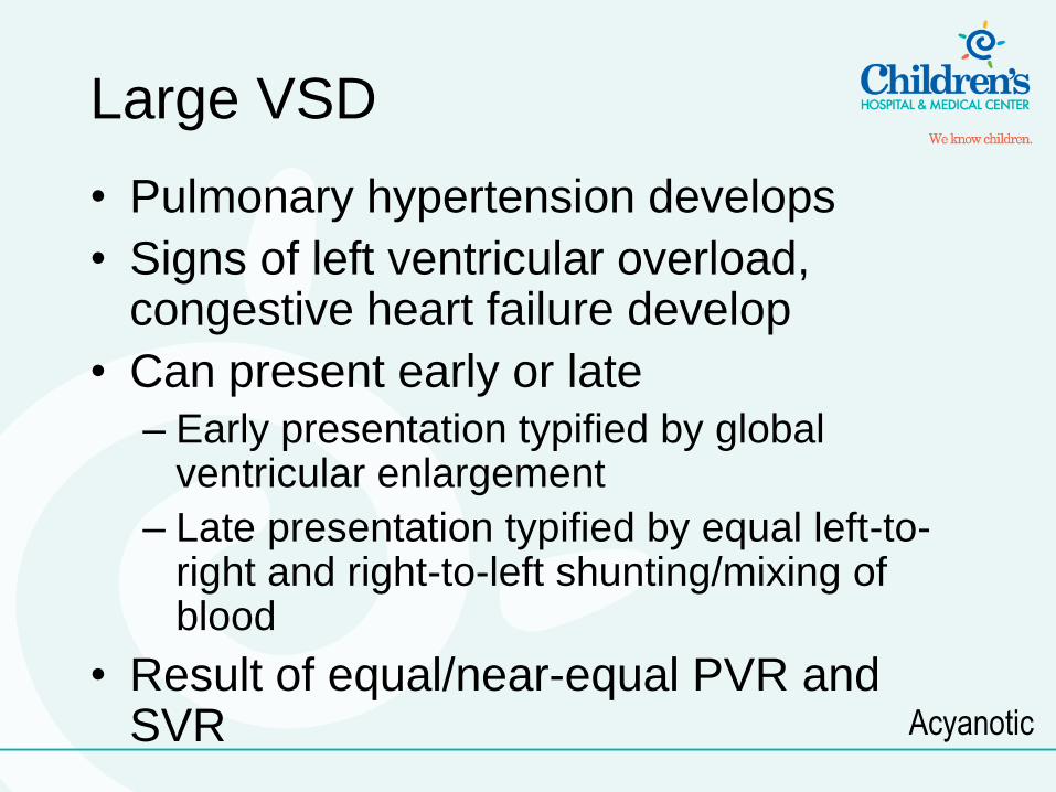
Large VSD
• Pulmonary hypertension develops
• Signs of left ventricular overload, congestive heart failure develop
• Can present early or late
– Early presentation typified by global ventricular enlargement
– Late presentation typified by equal left-to-right and right-to-left shunting/mixing of blood
• Result of equal/near-equal PVR and SVR Acyanotic
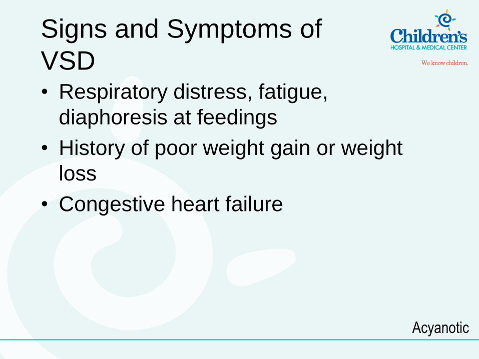
Signs and Symptoms of
VSD • Respiratory distress, fatigue,
diaphoresis at feedings
• History of poor weight gain or weight
loss
• Congestive heart failure
Acyanotic
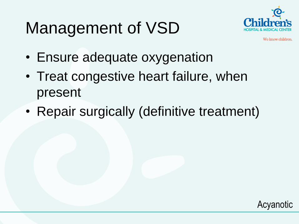
Management of VSD
• Ensure adequate oxygenation
• Treat congestive heart failure, when
present
• Repair surgically (definitive treatment)
Acyanotic
VSD
Acyanotic
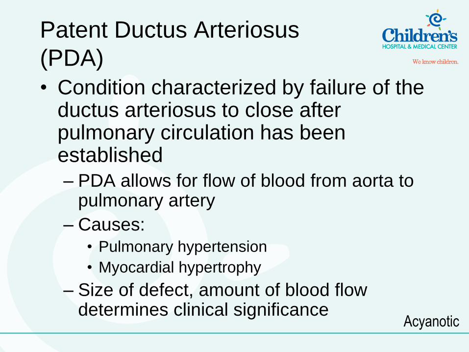
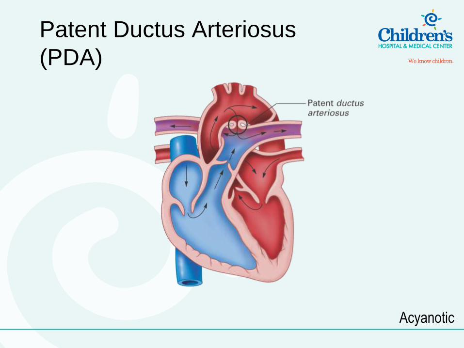
Patent Ductus Arteriosus
(PDA)
• Condition characterized by failure of the ductus arteriosus to close after pulmonary circulation has been established
– PDA allows for flow of blood from aorta to pulmonary artery
– Causes: • Pulmonary hypertension
• Myocardial hypertrophy
– Size of defect, amount of blood flow determines clinical significance
Acyanotic
Patent Ductus Arteriosus
(PDA)
• Signs and symptoms
– Difficulty breathing, tachypnea, tachycardia
– Bounding pulses, widening pulse
pressures, fatigue at feedings
• Management
– Give supportive care
– Administer aIdomethacin
– Use prostaglandin inhibitor
Acyanotic
Patent Ductus Arteriosus
(PDA)
Acyanotic
Obstructive Defects
• Overview
– Complete or partial blockage of blood flow
commonly caused by a structural deformity
– Signs and symptoms are secondary to the
cardiovascular structures involved
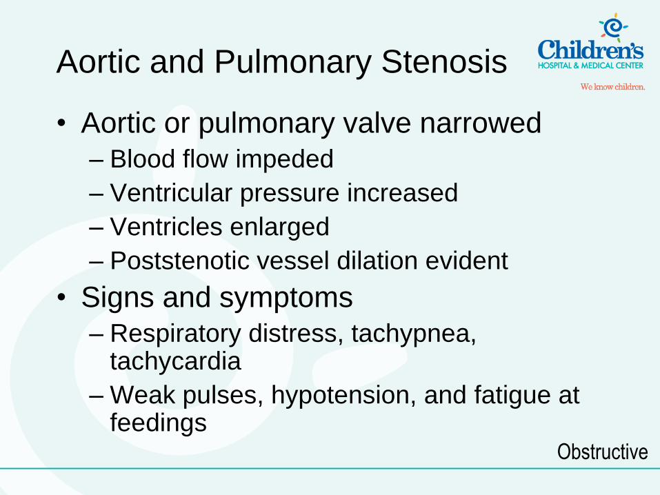
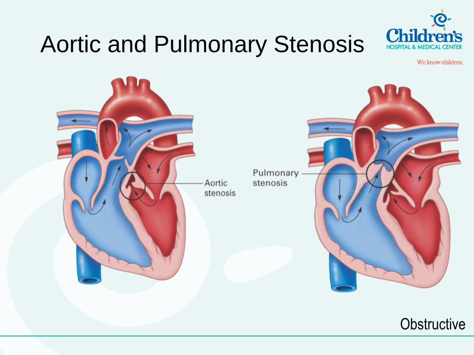
Aortic and Pulmonary Stenosis
• Aortic or pulmonary valve narrowed
– Blood flow impeded
– Ventricular pressure increased
– Ventricles enlarged
– Poststenotic vessel dilation evident
• Signs and symptoms
– Respiratory distress, tachypnea, tachycardia
– Weak pulses, hypotension, and fatigue at feedings
Obstructive
Aortic and Pulmonary Stenosis
• Management
– Give supportive care
– Conduct oxygenation
– Proceed with pharmacologic management
– Undertake balloon
angioplasty/valvuloplasty
Obstructive
Aortic and Pulmonary Stenosis
Obstructive
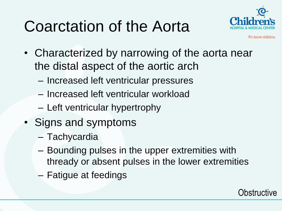
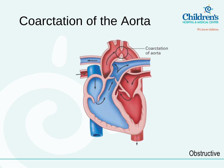
Coarctation of the Aorta
• Characterized by narrowing of the aorta near
the distal aspect of the aortic arch
– Increased left ventricular pressures
– Increased left ventricular workload
– Left ventricular hypertrophy
• Signs and symptoms
– Tachycardia
– Bounding pulses in the upper extremities with
thready or absent pulses in the lower extremities
– Fatigue at feedings
Obstructive
Coarctation of the Aorta
• Management – Give supportive care
– Administer prostaglandin
– Treat congestive heart failure, when present
– Complete balloon angioplasty/surgical resection (definitive treatment)
Obstructive
Coarctation of the Aorta
Obstructive
Cyanotic Defects
• Characterized by poor pulmonary blood flow resulting from one or more of the following:
– Difficulty in pumping blood out the right side of the heart
– Greater pressure gradient from right to left side of the heart that shunts blood to left side
• Returns unoxygenated blood to the left side
– Blockage of pulmonary blood flow or structural deformity
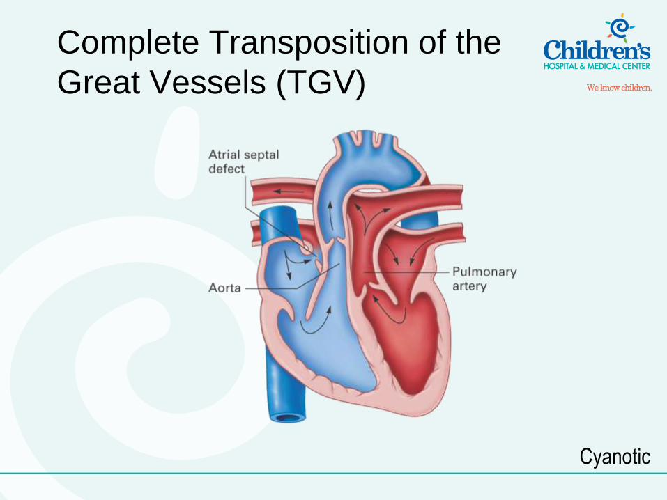
Complete Transposition of the
Great Vessels (TGV)
• Characterized by abnormal positioning
of the aorta and pulmonary arteries
– Pulmonary artery leaves the left ventricle
– Aorta leaves the right ventricle
– Creates parallel circulations
Cyanotic
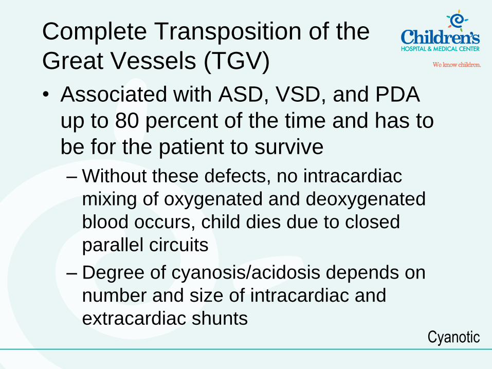
Complete Transposition of the
Great Vessels (TGV)
• Associated with ASD, VSD, and PDA
up to 80 percent of the time and has to
be for the patient to survive
– Without these defects, no intracardiac
mixing of oxygenated and deoxygenated
blood occurs, child dies due to closed
parallel circuits
– Degree of cyanosis/acidosis depends on
number and size of intracardiac and
extracardiac shunts Cyanotic
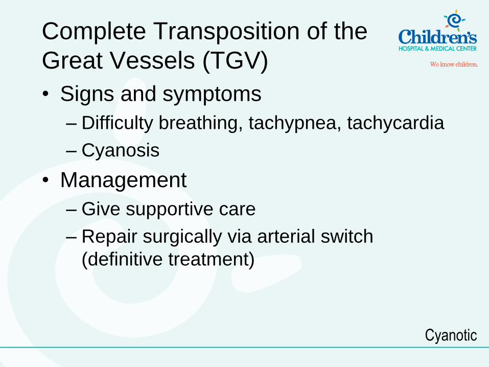
Complete Transposition of the
Great Vessels (TGV)
• Signs and symptoms
– Difficulty breathing, tachypnea, tachycardia
– Cyanosis
• Management
– Give supportive care
– Repair surgically via arterial switch
(definitive treatment)
Cyanotic
Complete Transposition of the
Great Vessels (TGV)
Cyanotic
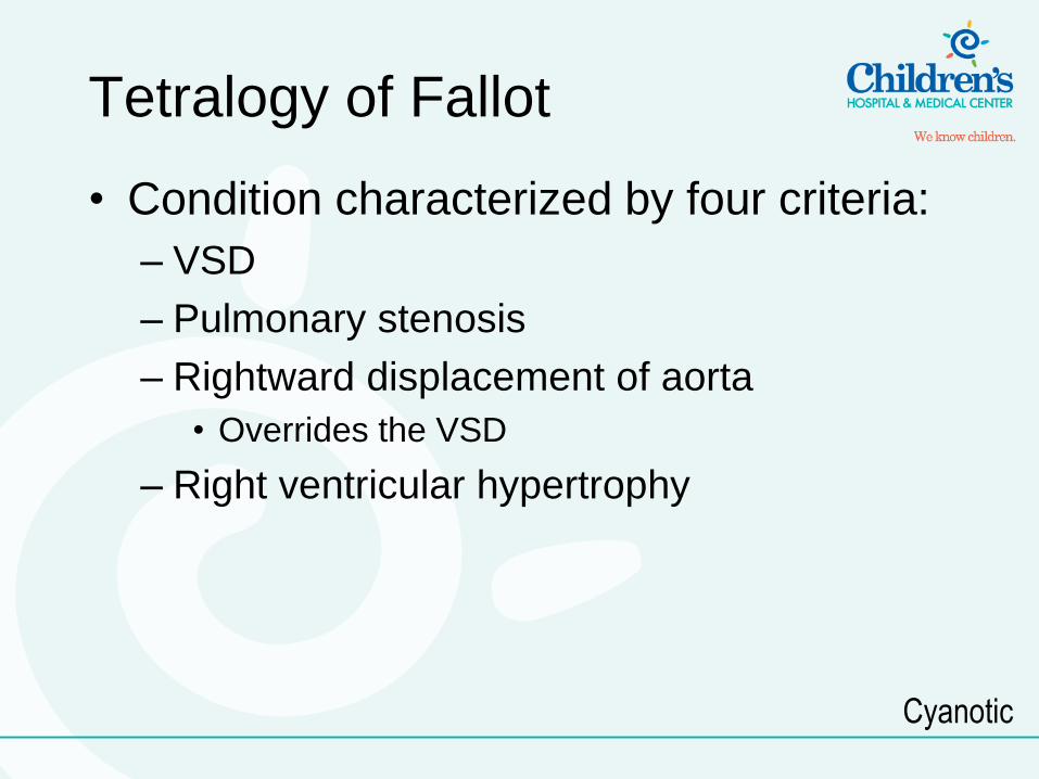
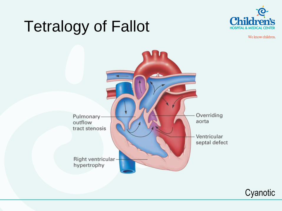
Tetralogy of Fallot
• Condition characterized by four criteria:
– VSD
– Pulmonary stenosis
– Rightward displacement of aorta
• Overrides the VSD
– Right ventricular hypertrophy
Cyanotic
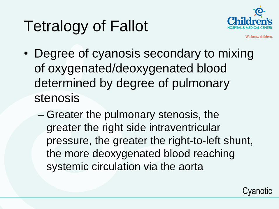
Tetralogy of Fallot
• Degree of cyanosis secondary to mixing
of oxygenated/deoxygenated blood
determined by degree of pulmonary
stenosis
– Greater the pulmonary stenosis, the
greater the right side intraventricular
pressure, the greater the right-to-left shunt,
the more deoxygenated blood reaching
systemic circulation via the aorta
Cyanotic
Tetralogy of Fallot
• Signs and symptoms
– Tachypnea, tachycardia
– Fatigue at feedings
• Management
– Give supportive care
– Ensure adequate oxygenation
– Administer prostaglandin
Cyanotic
Tetralogy of Fallot
Cyanotic
Transport Guidelines for
Congenital Heart Defects
• Ensure patent airway
• Ensure adequate ventilation,
oxygenation
• Treat congestive heart failure
• Correct circulatory compromise
– Conduct fluid volume resuscitation
– Administer vasopressors
• Keep patient warm
General Pathophysiology:
Other Neonatal
Emergencies
Necrotizing Enterocolitis (NEC)
• Most common serious abdominal
emergency in neonates that requires
emergency surgical intervention
– Acute inflammation of the large intestine
leading to necrosis of the intestinal mucosa
– Risk factors include insult to intestinal
mucosa and bacterial growth
– Causative agent has not been identified
– Risk of sepsis secondary to bowel
perforation
Necrotizing Enterocolitis (NEC)
• Signs and symptoms
– Abdominal distention
– Decreased or absent bowel sounds
– Vomiting
– Bloody diarrhea
– Lethargy
– Poor feeding habits
– Depressed core body temperature

Necrotizing Enterocolitis (NEC)
• Management
– Give supportive care
– Keep the patient NPO
– Insert NG tube and conduct gastric
decompression
– Maintain acid-base and electrolyte balance
– Maintain IV fluids
– Administer antibiotic therapy
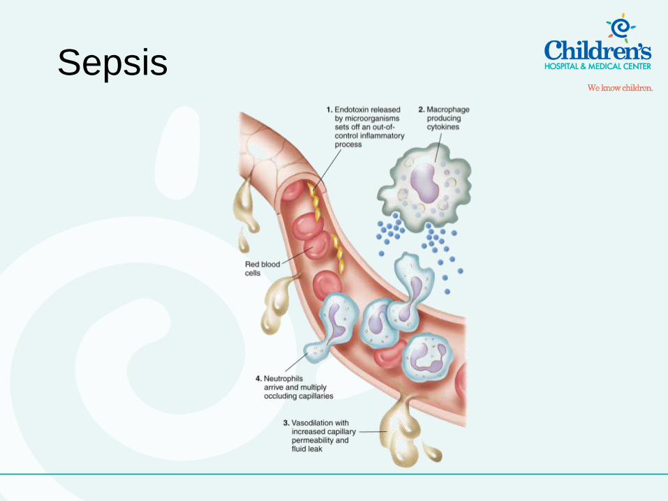
Sepsis
• Life-threatening infection of the bloodstream resulting in systemic toxicity – Often subtle in neonate and may be difficult to
distinguish from a noninfectious pathology
– Maternal gastrointestinal or genital infections are most common etiology
– Primary site of infection may often be difficult to identify
– Shock may develop
– Result of vasodilation secondary to release of bacterial endotoxins
– Distributive shock
Sepsis
• Signs and symptoms – Hypothermia
– Respiratory distress
– Pulmonary hypertension
– Hypoxemia
– Severe hypoperfusion
– Disseminated intravascular coagulation (DIC)
Sepsis
Sepsis
• Management – Give supportive care
– Ensure cardiovascular support
– Administer antibiotic therapy
General Neonatal
Assessment
Findings/Considerations

Skin Color
• Cyanosis commonly found
– Insignificant when neonate is crying
• Jaundice
– Result of high serum bilirubin levels
– Usually resolves without intervention
• When needed, treat with fluorescent light
• Blood transfusion needed when fluorescent
light treatment fails
Vital Signs
• Neonatal vital signs variable, deviate
from “norm”
• Access to reference material advisable
– Prdiatric Broselow tape
• In addition to respiratory rate, blood
pressure, heart rate, consider blood
glucose level a vital sign in neonate
– 70–80 mg/dL considered nonhypoglycemic
General Neonatal
Considerations
Airway
• Should be secured and maintained as soon
as possible
– RSI less common in adults but should be used
when needed
• Accidental extubation most frequent
respiratory complication
– Sedation
– NMBAs
– C-collars
– Lateral immobilization
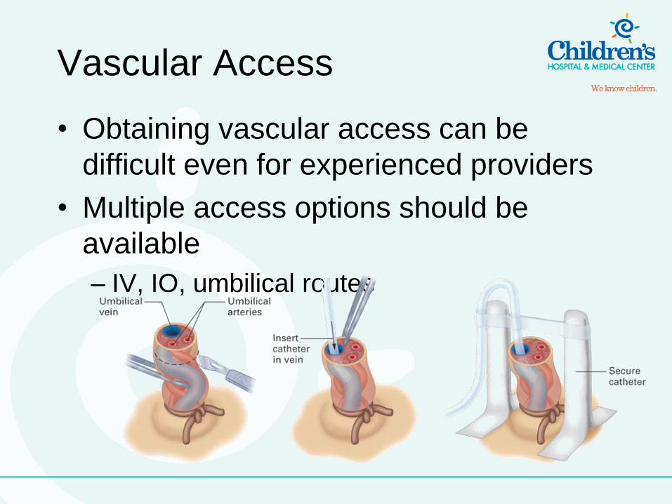
Vascular Access
• Obtaining vascular access can be
difficult even for experienced providers
• Multiple access options should be
available
– IV, IO, umbilical routes
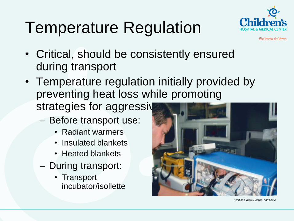
Temperature Regulation
• Critical, should be consistently ensured during transport
• Temperature regulation initially provided by preventing heat loss while promoting strategies for aggressive warming – Before transport use:
• Radiant warmers
• Insulated blankets
• Heated blankets
– During transport: • Transport
incubator/isollette
Scott and White Hospital and Clinic
Hypoglycemia
• Hypoglycemia should be managed
aggressively
– Use 10 percent dextrose and water
• Infuse D10W at 80cc/kg/day
– D25W, D50W administration
contraindicated
– Can cause significant increases in plasma
osmolarity
– Hypernatremia
• Cerebral edema
Summary
• Common denominator for unexpected deaths
in neonates is hypoxia
– Via infectious diseases, congenital heart disease,
pulmonary compromise, other etiologies
– Neonates can compensate until they are
extremely hypoxic
• High index of suspicion needed to identify developing
hypoxia before decompensation
– Airway and ventilation highest priority
• Neonates with high metabolism, high oxygen
consumption

Summary
• Manage CHD after addressing airway, breathing, and pulmonary function – Transport care for the CHD patient is primarily
supportive
– May require significant intervention
• Ability to diagnose specific defects not top concern – Critical care practitioner should know how various
defects affect normal perfusion
– Care provider is responsible for staying abreast of common neonatal emergencies and their current standards of care


![Neonatal Thermoregulation - University of · PDF fileNeonatal Thermoregulation Julia Petty. ... A care study. Journal of Neonatal Nursing. ... 5 Thermoregulation [Compatibility Mode]](https://img.pdfslide.us/doc/110x75/5aafe83f7f8b9a6b308de3c0/neonatal-thermoregulation-university-of-thermoregulation-julia-petty-a-care.jpg)