Embed Size (px)
Citation preview

Cancer Letters 306 (2011) 205–213
Contents lists available at ScienceDirect
Cancer Letters
journal homepage: www.elsevier .com/locate /canlet
Neo-angiogenesis and the premalignant micro-circulatory augmentationof early colon carcinogenesis
Ashish K. Tiwari a, Susan E. Crawford b, Andrew Radosevich c, Ramesh K. Wali a, Yolanda Stypula c,Dhananjay P. Kunte a, Nikhil Mutyal c, Sarah Ruderman c, Andrew Gomes c, Mona L. Cornwell b,Mart De La Cruz a, Jeffrey Brasky a, Tina P. Gibson a, Vadim Backman c, Hemant K. Roy a,⇑a Department of Medicine, NorthShore University HealthSystem, Evanston, IL, United Statesb Department of Pathology, NorthShore University HealthSystem, Evanston, IL, United Statesc Biomedical Engineering Department, Northwestern University, Evanston, IL, United States
a r t i c l e i n f o
Article history:Received 3 November 2010Received in revised form 7 March 2011Accepted 11 March 2011
Keywords:Colon carcinogenesisColon cancerNeo-angiogenesis
0304-3835/$ - see front matter � 2011 Elsevier Ireldoi:10.1016/j.canlet.2011.03.008
⇑ Corresponding author. Address: Department of MUniversity HealthSystem, University of Chicago Priicine, United States. Tel.: +1 847 570 3115; fax: +1
E-mail address: [email protected] (H.K. R
a b s t r a c t
Spectroscopic techniques have demonstrated that in the microscopically normal mucosa,there is an increase in mucosal micro-circulation in patients harboring neoplasia elsewherein the colon (i.e. marker of field carcinogenesis). However, the physiological and molecularbasis of this early increase in blood supply (EIBS) has not been elucidated. We, therefore,investigated the microvessel density (MVD) and angiogenic gene expression in the prema-lignant colonic mucosa from the well-validated azoxymethane (AOM)-treated rat experi-mental model of colon carcinogenesis.
Fisher 344 rats were treated with AOM (15 mg/kg i.p.) or saline and euthanized 14 weekslater (a time-point that precedes carcinoma development). Colon sections were studied forMVD via immunohistochemical assessment for CD31 and location was compared withoptical assessment of mucosal hemoglobin with low-coherence enhanced backscatteringspectroscopy (LEBS). Finally, we performed a pilot real-time PCR angiogenesis microarray(84 genes) from the microscopically normal colonic mucosa of AOM and age-matched sal-ine treated rats. AOM treatment increased MVD in both the mucosa and submucosa of therats (125% increase in mucosa; p < 0.007, and 96% increase in submucosa; p < 0.02) but theincrease was most pronounced at the cryptal base consistent with the LEBS data showingmaximal hemoglobin augmentation at 200–225 lm depth. Microarray analysis showedstriking dysregulation of angiogenic and anti-angiogenic factors. We demonstrate, for thefirst time, that neo-angiogenesis occurs in the microscopically normal colonic mucosaand was accentuated at the bottom of the crypt. This finding has potential implicationsas a biomarker for risk-stratification and target for chemoprevention.
� 2011 Elsevier Ireland Ltd. All rights reserved.
1. Introduction
Colorectal carcinogenesis is characterized by sequentialprogression through various morphological stages (aber-rant crypt foci, small adenoma, large adenoma, carci-
and Ltd. All rights reserved.
edicine, NorthShoretzker School of Med-847 733 5041.oy).
noma-in situ, invasive cancer). These are orchestrated bya well established series of mutational/epigenetic eventsinitiated by loss of either adenomatous polyposis coli(APC) tumor suppressor gene or DNA mismatch repair(e.g. hMLH1 or hMSH2) function [1]. It has been estimatedthat for CRC to develop approximately 15 signaling path-ways are required to be altered [2]. Given that the colono-cyte is relatively short-lived (3–7 days before becomingshed into the fecal stream), it is becoming increasinglyclear that dysregulation of apoptosis and proliferation are

206 A.K. Tiwari et al. / Cancer Letters 306 (2011) 205–213
prerequisites for the formation of dysplastic lesions. Thesealterations in cell growth/death occur throughout the co-lon reflecting the diffuse ‘‘field of injury’’ as a consequenceof endogenous (e.g. genetic, diabetes) and exogenous (diet,smoking, etc.) risk factors [3,4]. Thus, neoplastic transfor-mation in the colon epitomizes the field carcinogenesisconcept. This has numerous well recognized clinical impli-cations such as an elevated risk for both synchronous andmetachronous lesions [5]. In this regard, current guidelinesmandate that patients with a distal adenoma (i.e. detectedon flexible sigmoidoscopy) require full colonic evaluation(colonoscopy). Furthermore, since patients with one ade-noma are at higher risk of developing future lesions, theircolonoscopic interval is usually shortened (e.g. 3 years ifan advanced adenoma is detected versus 10 years for noadenoma detection) [5].
Given the important clinical ramifications, there is anemerging interest in accurately identifying and elucidatingthe biological nature of colonic field carcinogenesis. For in-stance, epigenetic, genomic, proteomic and micro-archi-tectural biomarkers have been demonstrated to bealtered in the microscopically normal mucosa during fieldcarcinogenesis [6–9]. On a cellular note, it has long beenrecognized that the mucosa is hyperproliferative in pa-tients who harbor neoplasia elsewhere in their colon[10]. Indeed, the proliferative indices from rectal biopsieshave been shown to correlate with proximal neoplasia[11]. The corollary to this is that the hyperproliferative mu-cosa would be expected to be hypermetabolic. The geneexpression consequences of the ‘‘relative hypoxia’’ havebeen recently demonstrated through a series of elegantmicroarray studies using APC mutations [12]. As wouldbe predicted, the gene expression profile suggested a rela-tive hypoxia in the APC mutated mouse model.
Our group was the first to confirm existence of the phe-nomena of colonic early increase in blood supply (EIBS)using an optical technology, four dimensional elastic lightscattering fingerprinting (4D-ELF) [13–15]. This noveltechnique allows highly accurate depth selective quantifi-cation of the microvascular blood supply. We demon-strated that in the well-validated model of coloncarcinogenesis, the azoxymethane (AOM)-treated rat.Importantly, the alterations in microvascular blood flowwas demonstrated in microscopically normal mucosa at apremalignant time-point (2–15 weeks after AOM treat-ment) and the magnitude mirrored risk of future develop-ment of colonic neoplasia [13]. These were replicated inthe MIN mouse, a genetic model of intestinal tumorigene-sis [13]. We then demonstrated EIBS clinically using devel-oped an endoscopically-compatible 4D-ELF fiber-opticprobe in patients undergoing colonoscopy. Importantly,EIBS was detectable in the visually normal rectum in pa-tients harboring advanced adenomas elsewhere in the co-lon [14], thus suggesting applications as a minimally-intrusive risk-stratification.
While the potential clinical significance of EIBS is clear,the biological underpinnings behind EIBS have been largelyunexplored. It is logical to postulate angiogenesis may be amajor factor in EIBS given its well-established role in coloncarcinogenesis. Indeed, suppression of angiogenesis is amainstay of CRC therapy. However, to our knowledge, no
previous studies have evaluated angiogenesis at pre-malig-nant stages (histologically-normal) mucosa where the phe-nomenon of EIBS is apparent. We, therefore, wanted toassess angiogenesis in the microscopically normal mucosaas the mechanisms of EIBS. We used the AOM-treated ratmodel because of its well-validated nature, defined time-frame for carcinogenesis (adenomas start developing in20 weeks and carcinomas require 35–40 weeks) and thefact that it recapitulates many of the genetic and epige-netic features of human field carcinogenesis [13,15–17].
2. Materials and methods
2.1. Animal studies
All animal procedures were reviewed and approved bythe Institutional Animal Care and Use Committee forNorthShore University HealthSystem. Twenty-four Fisher344 rats (150–200 g; Harlan, Indianapolis, IN) were treatedwith either 2 weekly injections (i.p.) of 15 mg/kg AOM(Midwest Research Institute, Kansas City, MO) or saline.The rats were kept on a standard AIN76a diet. Rats wereeuthanized after 14 weeks of second AOM injection, colonswere removed, rinsed with 1 mM dithiothreitol in normalsaline and presence of adenoma was ruled out. For theanalysis, the distal colon was utilized given our previousdata that in the AOM-treated rat model, EIBS (and also fu-ture neoplasia) was most marked in this region of thecolon.
2.2. Mucosal RNA isolation
Briefly, after euthanizing rats (both AOM/saline trea-ted), the colons were isolated, washed and opened longitu-dinally. The distal colonic mucosa was inspected to assureno neoplastic lesions and then isolated through gentlescraping with a microscope slide as previously described[18]. Total RNA was extracted using TRI Reagent (Sigma)and stored at �80 �C.
2.3. Spectroscopic analysis of Hb content at different depths ofthe colonic wall
In order to study the depths at which blood content isaltered in field carcinogenesis we employed a depth re-solved optical technique known as low-coherence en-hanced backscattering spectroscopy (LEBS) [19–21]. TheLEBS instrumentation has been described in full detail inother publications [22,23]. In brief, the LEBS system em-ploys spatial coherence gating to interrogate the opticalscattering and absorption spectrum at different depthswithin a tissue specimen. Using of an algorithm based onBeer’s law, the absorption spectrum can be used to quan-tify the hemoglobin concentration at each depth withinthe colonic wall. Due to the ex vivo nature of the study,hemoglobin was rapidly deoxygenated and thus it wasnot possible to accurately determine contributions be-tween oxy-hemoglobin and deoxy-hemoglobin. Therefore,we present all data as total hemoglobin. LEBS measure-ments were obtained from 9 AOM-treated and 9 saline

A.K. Tiwari et al. / Cancer Letters 306 (2011) 205–213 207
control animals at 10 distinct sites on fresh distal colonicsegments.
2.4. Immunohistochemical staining
Formalin fixed, paraffin embedded distal colonic seg-ments from 14 weeks post AOM-treated rats and theirage matched controls were subjected to immunohisto-chemical analysis as described previously [18,24]. Briefly,5 lm paraffin-embedded sections was mounted on super-frost + slides (Vector Laboratories, Burlingame, CA), heatedat 60 �C for 1 h and then deparaffinized with two 5 minwashes of xylene followed by a series of graded ethanolwashes. Antigen retrieval for CD31 and Col18a1 wereaccomplished by pressure microwaving (NordicWare, Min-neapolis, MN) in antigen unmasking reagent (Vector Labo-ratories) at high power setting for 9 min. Endogenousperoxidase activity was quenched by a 5 min wash in 3%hydrogen peroxide while the non-specific binding wasblocked by 30 min incubation with 5% horse serum atroom temperature. Sections were then incubated over-night with primary antibodies anti-CD31 (1:200) from CellSignaling Technology; anti-Col18a1 (1:100) from SantaCruz Biotechnology] followed by incubation with appropri-ate biotinylated secondary antibodies. After three 5 minwashings in PBS, the specimen was further developedusing Vectastatin Elite ABC kit (Vector Laboratories).
2.5. RT2 Profiler PCR array
Rat angiogenesis array was performed using RNA iso-lated from rat mucosal scrapings and RT2 Profiler PCR arraykit from SA Biosciences (Maryland, USA), which quantita-tively assessed a panel of 84 genes implicated in angiogen-esis. This was a pre-specified commercially available arraythat includes both pro- and anti-angiogenic factors knownto modulate different facets of angiogenesis either directlyor through intermediates. The 84 genes included in the ar-ray encompassed growth factors/receptors, adhesion mole-
Fig. 1.1. Increased microvessel density in the premalignant mucosa of the AOMtissue sections from rats euthanized after 14 weeks of second AOM injection and tof neoplasia. Examination of hematoxylin and eosin (H and E) stained slides noted(a) versus the age-matched saline treated controls (b). Of note, the microscopic exin the mucosa. (For interpretation of the references to color in this figure legend
cules, matrix proteins, proteases (along with theirinhibitors); cytokines/chemokines and transcription fac-tors (the complete list of the genes is provided in the Supple-mentary material for this article). RT2 First strand kit fromSA Biosciences was used for cDNA synthesis and RT2 SY-BER� Green qPCR Master Mix and compatible BioRad�
CFX-384 Cycler and 384-well array plates (96 � 4 format)were used for the gene expression profiling. The assaywas performed as per manufacturer’s directions. Theexpression normalization was done with five constitutivelyexpressed genes (Rplp1, Hprt1, Rpl13a, Ldha and Actb).
2.6. Statistical analysis
Micro-vessel density (MVD) was quantified by countingmicrovessel number in five randomly selected fields look-ing at mucosal and submucosal regions under 40� in distalcolon tissue sections from AOM and saline treated Fischer344 rats. The pathologist (S.C.) was blinded to the treat-ment group. The comparison of MVD in saline and AOM-treated rats was performed by averaging out the numbersof micro-vessels in 5 random fields in each colonic sectionfrom AOM and saline treated rats and p value was calcu-lated by students t-test. Staining intensity for Col18a1 pro-tein expression in tissue sections was measured on a five-point intensity scale (0, none; 1, equivocal; 2, low; 3, mod-erate; 4 and 5; high) by a pathologist blinded to the treat-ment group and standard averaging and t-test wasperformed for statistical analysis. Microarray analysis wasperformed by using array analysis web portal tool availableat manufacturer’s (SA Biosciences) website and fold changeabove 1.5 and below 0.67 was considered significant.
3. Results
3.1. Angiogenesis occurs at pre-adenoma stage and is predominantlypericryptal in location
To assess angiogenesis in the premalignant mucosa, we utilized bothH and E sections (Fig. 1.1) and those where endothelial cells were high-lighted with CD31 immunostaining (Fig. 1.2) from paraffinized sections
-treated rat: These studies were performed on paraffin embedded colonheir age-matched saline treated rats. Necropsies confirmed lack of evidencea profound increase in microvessels (green arrows) in the AOM-treated rat
amination of the slides did not reveal any evidence of dysplastic alterations, the reader is referred to the web version of this article.)
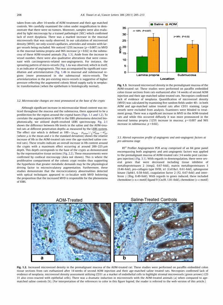
Fig. 1.3. Increased microvessel density in the premalignant mucosa of the
208 A.K. Tiwari et al. / Cancer Letters 306 (2011) 205–213
taken from rats after 14 weeks of AOM treatment and their age matchedcontrols. We carefully examined the colon under magnification to dem-onstrate that there were no tumors. Moreover, samples were also evalu-ated by light microscopy by a trained pathologist (SEC) which confirmedlack of overt dysplasia. There was a marked increase in the mucosalmicrovessels that was easily observed. In our calculation of microvesseldensity (MVD), we only scored capillaries, arterioles and venules with lar-ger vessels being excluded. We noticed 125% increase (p < 0.007) in MVDin the mucosal lamina propria and 96% increase (p < 0.02) in the submu-cosa of these AOM-treated animals (Fig. 1.3). Aside from the increase invessel number, there were also qualitative alterations that were conso-nant with carcinogenesis-related neo-angiogenesis. For instance, thesprouting pattern of micro-vessels (Fig. 1.4a was observed, which in itself,is an indicator of angiogenesis. This was accompanied by significant vaso-dilation and arteriolarization (Fig. 1.4b in mucosal and submucosal re-gions (more pronounced in the submucosal micro-vessels. Thearteriolarization in the pre-existing micro-vessels is suggestive of higherpressure reflecting the augmented colonic blood supply early in neoplas-tic transformation (when the epithelium is histologically normal).
AOM-treated rat: These studies were performed on paraffin embeddedcolon tissue sections from rats euthanized after 14 weeks of second AOMinjection and their age-matched saline treated rats. Necropsies confirmedlack of evidence of neoplasia. Quantification of microvessel density(MVD) was calculated by examining five random fields under 40� in bothAOM and age-matched saline treated rats after CD31 staining. Largevessels were excluded from analysis. Examiners were blinded to treat-ment group, There was a significant increase in MVD in the AOM-treatedrats and while this occurred diffusely it was more pronounced in themucosal lamina propria (125% increase in mucosa; p < 0.007 and 96%increase in submucosa; p < 0.02).
3.2. Microvascular changes are most pronounced at the base of the crypts
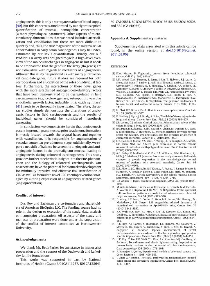
Although significant increase in microvascular blood content was no-ticed throughout the mucosa and the submucosa, there appeared to be apredilection for the region around the cryptal bases (Figs. 1.1 and 1.2). Tocorrelate the augmentation in MVD to the EIBS phenomena detected bio-photonically, we utilized depth-resolved LEBS spectroscopy. Fig. 2.1shows the difference between Hb levels in the saline and the AOM-trea-ted rats at different penetration depths as measured by the LEBS system.The effect size which is defined as 100 � ðlAOM � lSalineÞ=
ffiffiffiffiffiffiffiffiffiffiffiffiffiffiffiffiffiffiffiffiffiffiffiffiffir2
AOM þ r2Sal
q,
(where l is the mean and r is the standard deviation) shows the relativeincrease of Hb in the AOM-treated rats over the age-matched saline con-trol rats). These results indicate an overall increase in Hb content aroundthe crypts with a maximum effect occurring at around 200–225 lmdepth. This depth corresponds to the base of the crypts as demonstratedby the representative tissue sections (Fig. 2.2). These measurements wereconfirmed by confocal microscopy (data not shown). This is where theproliferative compartment of the colonic crypt resides thus supportingthe hypothesis that greater metabolic demands may be the physiologicaldriving factor to microcirculatory augmentation. Furthermore, thesestudies demonstrate that the microcirculatory abnormalities detectedwith optical techniques appeared to co-localize with MVD bolsteringthe proposition that the increased MVD is responsible for the phenomenaof EIBS.
Fig. 1.2. Increased microvessel density in the premalignant mucosa of the AOMtissue sections from rats euthanized after 14 weeks of second AOM injectionevidence of neoplasia. microvessel density assessment utilizing CD31 as a marke31 also cross-reacted with epithelial cells). There was a dramatic induction inmatched saline controls (b). (For interpretation of the references to color in this
3.3. Altered expression profile of angiogenic and anti-angiogenic factors atpre-adenoma stage
RT2 Profiler Angiogenesis PCR array comprised of an 84 gene panelencompassing both angiogenic and anti-angiogenic factors was appliedto the premalignant mucosa of AOM-treated rats (14 weeks post carcino-gen injection) (Fig. 3.1). With regards to downregulation, there were sev-eral genes that were decreased including tissue inhibitor ofmetalloproteinases 2 (timp2, 0.67-fold), matrix metalloproteinases 3(0.46-fold), pro-collagen type XVIII, a1 (Col18a1, 0.61-fold), sphingosinekinase (Sphk1, 0.58-fold), coagulation factor 2 (F2, 0.67-fold) and inter-feron c (Ifng, 0.49-fold). With regards to genes induced, these includedchemokine (c-x-c motif) ligand 9 (Cxcl9, 1.81-fold), chemokine (c–c motif)
-treated rat: These studies were performed on paraffin embedded colonand their age-matched saline treated rats. Necropsies confirmed lack ofr of endothelial cells to highlight stromal microvessels (green arrows) (CDmicrovessels with the AOM-treated animals (a) when compared to age-figure legend, the reader is referred to the web version of this article.)

Fig. 1.4. Increased microvessel density in the premalignant mucosa of the AOM-treated rat: These studies were performed on paraffin embedded colontissue sections from rats euthanized after 14 weeks of second AOM injection and their age-matched saline treated rats. Necropsies confirmed lack ofevidence of neoplasia. Qualitative alterations in microvessels: AOM-treatment resulted not simply in the doubling of MVD but also qualitative changes thatare hallmarks of neo-angiogenesis. These include the ‘‘sprouting’’ blood vessel pattern (a) and arteriolization (b).
Fig. 2.1. The depth of maximal mucosal hemoglobin concentrations co-localizes with the increase in MVD. Mucosal hemoglobin concentrationwas measured biophotonically with LEBS. The measures are the differ-ential between AOM-treated and age matched controls expressed as‘‘effect size’’ expressed as a percentage. As can be seen, while at all depthsthere was a positive effect size with AOM (more hemoglobin), themaximal differential appeared to be at �200–225 lm from tissue surface.As one probes deeper, there is a modest diminution in the AOM versussaline differences in hemoglobin.
Fig. 2.2. The depth of maximal mucosal hemoglobin concentrations co-localizes with the increase in MVD. In order to understand the histologicalcorrelates for this �200–225 lm optimal effect size in Hb we analyzed Hand E stained sections of the colonic crypts were imaged at 20�magnification using an Olympus BH-2 brightfield microscope equippedwith a SPOT camera. Scale bars were added using ImageJ software (NIH)to show various depths of the colonic mucosa. It appears that �200–225 lm from the tissue surface corresponds to the base of the cryptsconsistent with the hypothesis related to increased mucosal oxygendemand.
A.K. Tiwari et al. / Cancer Letters 306 (2011) 205–213 209
ligand 2 (Ccl2 1.75-fold) and connective tissue growth factor (Ctgf, 1.50-fold). In general there was a shift in balance with increased angiogenicand decreased anti-angiogenic factors. However, there were some excep-tions underscoring the complexity/cross-talk in these angiogenic regula-tory pathways. The expression profile also suggested of pathologicalangiogenesis as seen with tumors and not with the angiogenesis associ-ated with physiological processes [25]. An intriguing case in point isCol18a1 is a strong anti-angiogenic factor associated with tumor angio-genesis but does not appear to have a role during the angiogenesisassociated with wound healing or reproduction [26]. We corroboratedthe microarray findings with immunohistochemical detection of Col18a1(Fig. 3.2a).
4. Discussion
We report, herein, for the first time the occurrence ofangiogenesis in the premalignant mucosa of the colon dur-ing colorectal carcinogenesis prior to adenoma formation.Moreover, not only was the micro-vessel density increased,but there was evidence of vasodilation and arteriolariza-tion. All of these could contribute to the phenomena ofearly increase in blood supply (EIBS) that we have ob-served through the utilization of novel spectroscopic tech-niques [13]. Importantly, the LEBS data shows thephysiological measurement of micro-circulation co-local-ized with the anatomic findings of increased MVD support-ing the role of neo-angiogenesis in EIBS. We developed

Fig. 3.1. RT2 Profiler PCR array performed with the rat colon samplesfrom 14 weeks post AOM-treated rats and their age matched controlslooked at the expression of 84 genes implicated directly or indirectly inangiogenesis. The heat map (saline treated animals left, AOM-treatedanimals right) indicates significant alterations in levels of both angiogenicand anti-angiogenic factors consonant with the induction of neo-angiogenesis.
210 A.K. Tiwari et al. / Cancer Letters 306 (2011) 205–213
some potential mechanistic insights by demonstratingupregulation of a number of pro-angiogenic factors alongwith inhibition of several anti-angiogenic mediators dur-ing colorectal carcinogenesis.
It bears emphasis that these changes in the microvascu-lature occur in the premalignant (histologically-normal)mucosa. We chose 14 week post-AOM injection rats be-cause this is a pre-adenomatous time-point (in this modelit takes �20 weeks for adenomas to start developing and35–40 weeks for carcinomas) and recapitulate many ofthe genetic and epigenetic features of human field carcino-genesis [15,16]. We had previously shown that the14 week time-point manifested robust induction in bloodsupply and the changes correlated well with changes infield carcinogenesis in humans. Indeed, our clinical studies(both ex vivo and in situ) have shown an excellent correla-tion. From a teleological perspective, this increase inmicrovascular blood flow is needed to support the diffusemucosal hyperproliferation, which is a hallmark of fieldcarcinogenesis. It is intriguing that the maximal effect size(differences in concentration between AOM-treated andsaline treated rats) from both optical Hb concentrationand MVD was in the region of the bottom third of the cryptcorresponding to the proliferative compartment (the cellsin the middle third typically are not proliferating butundergoing differentiation while the top third are in theprocess of apoptosis) [22]. Thus, the base of the cryptswould be expected would to have a higher metabolic rate.In support of this concept is the demonstration that APCmutations induced alterations in a various metabolic genesin the microscopically-normal intestinal mucosa suggest-ing a reactive pattern to the relative hypoxia which wouldbe anticipated to lead to augment the micro-circulation[12]. From a neoplastic perspective, it is intriguing to notethat this area is also where the colonic stem resides, whichare the putative initiators of colon carcinogenesis and dys-regulated in field carcinogenesis [27].
While there has been evidence from other systems thatangiogenesis mediates the hyperplasia–dysplasia transi-tion [28], this is the first demonstration of increasedMVD in the microscopically normal colon at the premalig-nant (hyperproliferative) time-point. MVD is a well-estab-lished technique to assess angiogenesis in CRCs and hasprognostic significance. It is clear that MVD increases ear-lier at the small adenoma stage [29]. The role of MVD in theearliest lesion in colon carcinogenesis, the aberrant cryptfoci (ACF), is controversial. On one hand, Shiptz and col-leagues [30] noted a marked upregulation in MVD in ACFs,whereas a recent study by Cho et al. [31] using similarmethodology failed to confirm the marker. It is importantto note that the majority of these studies addressed MVDbetween the lesion (ACF and adenoma) versus the unin-volved mucosa. To our knowledge, this is the first studyto compare the pre-malignant microscopically normal mu-cosa of colon versus those without any carcinogenesis. Thisis, we believe, the most relevant comparison for under-standing the role of increased micro-circulation in fieldcarcinogenesis. Indeed, this has been our comparators forall studies to date demonstrating EIBS in rat models (com-paring AOM versus saline), mice models (comparing APCmutated versus wildtype animals) and humans (comparingpatients harboring neoplasia elsewhere in their colon ver-sus those who are neoplasia-free) [13]. In this regard, therehave been microarray studies showing that putativevascular modulating agents such as osteopontin and

Fig. 3.2. RT2 Profiler PCR array performed with the rat colon samples from 14 weeks post AOM-treated rats and their age matched controls looked at theexpression of 84 genes implicated directly or indirectly in angiogenesis. Col18a1 immunostaining. Col18a1 was noted to be downregulated in the AOM-treated rats by the real time microarray. To assess at a protein level, we performed immunohistochemical evaluation and noted that saline treated controlshad markedly higher expression than AOM-treated animals. This provides pilot corroboration of the array findings that anti-angiogenic factors decreasedand pro-angiogenic factors increased with AOM.
A.K. Tiwari et al. / Cancer Letters 306 (2011) 205–213 211
cyclooxygenase from the microscopically normal mucosaof patients with CRC were 10–20-fold upregulated com-pared to the colonic mucosa taken from patients withoutneoplasia [32].
With regards to molecular mechanisms, we performeda pilot study to demonstrate that numerous angiogenicregulators are modulated in the premalignant colonic mu-cosa. The overall expression pattern is consonant with themulti-step process of angiogenesis including loss of integ-rity of basement membranes of micro-vessels, endothelialcell proliferation, migration, vessel tube formation and sta-bilization. For example, loss of endogenous inhibitorCol18a1, an integral part of micro-vessels basement mem-branes leads to a wide array of effects ranging from endo-thelial cell proliferation (mediated through upregulation ofmany angiogenic stimulators and downregulation of inhib-itor) to extracellular matrix (ECM) remodeling (mediatedthrough the loss of blockage of MMP2, 3 and 19 activity[33]). Additionally, the altered expression of MMP3 andTIMP2 also points towards ongoing ECM remodeling dur-ing carcinogenesis. Interestingly, we observed alteredexpression of several secreted factors (both epithelial andstromal) like Ifng, Cxcl2, Cxcl9 and CTGF, which suggeststhe possibility of epithelial–stromal interaction and theireffects on microvasculature, especially endothelial cellsmigration as seen with tumor angiogenesis [34,35]. Thegradient nature of the effect of such secreted factors couldactually be responsible for the relatively higher increase inMVD locally near the cryptal bases. However, it needs to bementioned that this model, while teleologically compel-ling, is conjecture. In this case, these genes, in conjunctionwith factors already identified in AOM-induced EIBS suchas iNOS [36] and factors that have been shown to be mod-ulated in human field carcinogenesis (COX2, VEGF, osteo-pontin) [32] create a picture that is complex withinterplay between various factors, and requires much fur-ther investigation. Finally, it bears emphasis that the studywas conducted exclusively on premalignant mucosa so nodata is available on dysplastic tissue. However, the other
reports have supported the progressive modulation inangiogenic-modulatory factors (VEGF, COX2, etc.) duringthe transition from premalignant to frank colorectal can-cers [32]).
There are a number of potential implications of thiswork. Firstly, we have demonstrated that biophotonicdetection of increased microvascular blood content corre-lates with increased angiogenesis during carcinogenesis.Since this is a diffuse phenomenon, we have been able touse the rectum to predict lesions elsewhere in the colon.The clinical need is that only 5–6% of the currently recom-mended screening colonoscopy yields significant neoplasiaand thus imparts a cancer preventive benefit [23]. Thus,developing a minimally intrusive pre-screening techniquecould substantially undermine the need for colonoscopy.We have published in situ data from over 700 subjects thatconfirms the promise of this approach (for advanced ade-nomas the area under the receiver operator curve was�0.90) [14,37]. We envision simplifying our probes to theextent that they could be used at the primary care physi-cian’s office during annual rectal examination to effectivelyrisk-stratify the population for colorectal cancer. This workis important in further solidifying the biological underpin-nings for this novel pre-screening technique for clinicalapplications. Secondly, it may have implications for cancerprevention. This supports the emerging field of angiopre-vention—targeting vasculature [38,39]. And finally, thisprovides important insights into the early events in colo-rectal cancer biology.
On the other hand, it is important to acknowledge someof the limitations of this work. Firstly, the study is con-ducted solely in the AOM-treated rat model. However,AOM is a specific colorectal carcinogen and is widely ac-cepted model for colorectal field carcinogenesis studies[16,17]. Moreover, the observation that MVD and opticallyderived EIBS appeared intimately related in the AOM-trea-ted rat suggests that it may be reasonable to speculate thatMVD may also be noted in human colonic field carcinogen-esis. Secondly, while MVD is a standard indicator of
212 A.K. Tiwari et al. / Cancer Letters 306 (2011) 205–213
angiogenesis, this is only a surrogate marker of blood supply[40]. But this concern is ameliorated by our rigorous opticalquantification of mucosal hemoglobin concentration(a more physiological parameter). Other aspects of micro-circulatory abnormalities that we noted included arterioli-zation and vasodilation but these are more difficult toquantify and, thus, the true magnitude of the microvascularabnormalities in early colon carcinogenesis may be under-estimated by our MVD quantification. Thirdly, our RT2
Profiler PCR Array was designed to yield a high-level over-view of the molecular changes in angiogenesis but it needsto be emphasized that the genes in the array (84 genes) arenot exhaustive with regards to mediators of angiogenesis.Although this study has provided us with many putative no-vel candidate genes, future studies are required for bothcorroboration and elucidation of the roles of individual fac-tors. Furthermore, the interactions of these novel geneswith the more established angiogenic-modulatory factorsthat have been demonstrated to be dysregulated in fieldcarcinogenesis (e.g. cyclooxygenase, osteopontin, vascularendothelial growth factor, inducible nitric oxide synthase)[41] needs to be thoroughly investigated. Therefore, the ar-ray studies simply demonstrate the alterations of angio-genic factors in field carcinogenesis and the results ofindividual genes should be considered hypothesisgenerating.
In conclusion, we demonstrate herein that angiogenesisoccurs in premalignant mucosa prior to adenoma formation,is mostly located towards the cryptal bases and togetherwith vasodilation, it is responsible for augmentation ofvascular content at pre-adenoma stage. Additionally, we re-port a net shift of balance between the angiogenic and anti-angiogenic factors in the premalignant mucosa leading toangiogenesis during colorectal carcinogenesis. This studyprovides further mechanistic insights into the EIBS phenom-enon and the biology of colorectal carcinogenesis. Ourobservations have the potential for biomarker developmentfor minimally intrusive and effective risk stratification ofCRC as well as formulate novel CRC chemoprevention strat-egies by altering expression of angiogenesis related genes(angioprevention).
Conflict of interest
Drs. Roy and Backman are co-founders and sharehold-ers of American BioOptics LLC. The funding source had norole in the design or execution of the study, data analysisor manuscript preparation. All aspects of the study andmanuscript preparation were done under the supervisionof the conflict of interest committee at NorthwesternUniversity.
Acknowledgements
We thank Ms. Beth Parker for assistance in manuscriptpreparation and the support of the Duckworth and Lefkof-sky family foundations.
This works was supported in part by NationalInstitutes of Health (Grants U01CA111257, R01CA128641,
R01CA109861, R01CA118794, R01CA156186, 5R42CA130508,and 5R21CA140936).
Appendix A. Supplementary material
Supplementary data associated with this article can befound, in the online version, at doi:10.1016/j.canlet.2011.03.008.
References
[1] K.W. Kinzler, B. Vogelstein, Lessons from hereditary colorectalcancer, Cell 87 (1996) 159–170.
[2] L.D. Wood, D.W. Parsons, S. Jones, J. Lin, T. Sjoblom, R.J. Leary, D.Shen, S.M. Boca, T. Barber, J. Ptak, N. Silliman, S. Szabo, Z. Dezso, V.Ustyanksky, T. Nikolskaya, Y. Nikolsky, R. Karchin, P.A. Wilson, J.S.Kaminker, Z. Zhang, R. Croshaw, J. Willis, D. Dawson, M. Shipitsin, J.K.Willson, S. Sukumar, K. Polyak, B.H. Park, C.L. Pethiyagoda, P.V. Pant,D.G. Ballinger, A.B. Sparks, J. Hartigan, D.R. Smith, E. Suh, N.Papadopoulos, P. Buckhaults, S.D. Markowitz, G. Parmigiani, K.W.Kinzler, V.E. Velculescu, B. Vogelstein, The genomic landscapes ofhuman breast and colorectal cancers, Science 318 (2007) 1108–1113.
[3] H. Chai, R.E. Brown, Field effect in cancer-an update, Ann. Clin. Lab.Sci. 39 (2009) 331–337.
[4] K. Steiling, J. Ryan, J.S. Brody, A. Spira, The field of tissue injury in thelung and airway, Cancer Prev. Res. (Phila.) 1 (2008) 396–403.
[5] J.S. Levine, D.J. Ahnen, Clinical practice. Adenomatous polyps of thecolon, N. Engl. J. Med. 355 (2006) 2551–2557.
[6] B.C. Paun, D. Kukuruga, Z. Jin, Y. Mori, Y. Cheng, M. Duncan, S.A. Stass,E. Montgomery, D. Hutcheon, S.J. Meltzer, Relation between normalrectal methylation, smoking status, and the presence or absence ofcolorectal adenomas, Cancer 116 (2010) 4495–4501.
[7] C.Y. Hao, D.H. Moore, Y.S. Chiu, P. Wong, J.L. Bennington, A.P. Smith,L.C. Chen, N.M. Lee, Altered gene expression in normal colonicmucosa of individuals with polyps of the colon, Dis. Colon Rectum 48(2005) 2329–2335.
[8] A.C. Polley, F. Mulholland, C. Pin, E.A. Williams, D.M. Bradburn, S.J.Mills, J.C. Mathers, I.T. Johnson, Proteomic analysis reveals field-widechanges in protein expression in the morphologically normalmucosa of patients with colorectal neoplasia, Cancer Res. 66(2006) 6553–6562.
[9] D.S. Alberts, J.G. Einspahr, R.S. Krouse, A. Prasad, J. Ranger-Moore, P.Hamilton, A. Ismail, P. Lance, S. Goldschmid, L.M. Hess, M. Yozwiak,H.G. Bartels, P.H. Bartels, Karyometry of the colonic mucosa, CancerEpidemiol. Biomarkers Prev. 16 (2007) 2704–2716.
[10] D.J. Ahnen, T. Byers, Proliferation happens, JAMA 280 (1998) 1095–1096.
[11] M. Anti, G. Marra, F. Armelao, A. Percesepe, R. Ficarelli, G.M. Ricciuto,A. Valenti, G.L. Rapaccini, I. De Vitis, G. D’Agostino, Rectal epithelialcell proliferation patterns as predictors of adenomatous colorectalpolyp recurrence, Gut 34 (1993) 525–530.
[12] D. Wang, R.C. Pezo, G. Corner, C. Sison, M.L. Lesser, S.M. Shenoy, J.M.Mariadason, R.H. Singer, L.H. Augenlicht, Altered dynamics ofintestinal cell maturation in Apc1638N/+ mice, Cancer Res. 70(2010) 5348–5357.
[13] R.K. Wali, H.K. Roy, Y.L. Kim, Y. Liu, J.L. Koetsier, D.P. Kunte, M.J.Goldberg, V. Turzhitsky, V. Backman, Increased microvascular bloodcontent is an early event in colon carcinogenesis, Gut 54 (2005) 654–660.
[14] H.K. Roy, A.J. Gomes, S. Ruderman, L.K. Bianchi, M.J. Goldberg, V.Stoyneva, J.D. Rogers, V. Turzhitsky, Y. Kim, E. Yen, M. Jameel, A.Bogojevic, V. Backman, Optical measurement of rectalmicrovasculature as an adjunct to flexible sigmoidoscopy: gender-specific implications, Cancer Prev. Res. (Phila.) 3 (2010) 844–851.
[15] H.K. Roy, Y. Liu, R.K. Wali, Y.L. Kim, A.K. Kromine, M.J. Goldberg, V.Backman, Four-dimensional elastic light-scattering fingerprints aspreneoplastic markers in the rat model of colon carcinogenesis,Gastroenterology 126 (2004) 1071–1081.
[16] A. Banerjee, P. Quirke, Experimental models of colorectal cancer, Dis.Colon Rectum 41 (1998) 490–505.
[17] J. Chen, X.F. Huang, The signal pathways in azoxymethane-inducedcolon cancer and preventive implications, Cancer Biol. Ther. 8 (2009)1313–1317.
A.K. Tiwari et al. / Cancer Letters 306 (2011) 205–213 213
[18] D.P. Kunte, R.K. Wali, J.L. Koetsier, H.K. Roy, Antiproliferative effect ofsulindac in colonic neoplasia prevention: role of COOH-terminal Srckinase, Mol. Cancer Ther. 7 (2008) 1797–1806.
[19] Y.L. Kim, K. Liu, R.K. Wali, H.K. Roy, M.J. Goldberg, A.K. Kromine, K.Chen, V. Backman, Detection of the initial stages of colorectalcarcinogenesis using polarization light scattering spectroscopy withmultivariate statistical analysis, Lasers Surg. Med. 13 (2003) 13.
[20] Y.L. Kim, Y. Liu, V.M. Turzhitsky, H.K. Roy, R.K. Wali, V. Backman,Coherent backscattering spectroscopy, Opt. Lett. 29 (2004) 1906–1908.
[21] Y.L. Kim, Y. Liu, R.K. Wali, H.K. Roy, V. Backman, Low-coherentbackscattering spectroscopy for tissue characterization, Appl. Opt.44 (2005) 366–377.
[22] H.K. Roy, Y.L. Kim, Y. Liu, R.K. Wali, M.J. Goldberg, V. Turzhitsky, J.Horwitz, V. Backman, Risk stratification of colon carcinogenesisthrough enhanced backscattering spectroscopy analysis of theuninvolved colonic mucosa, Clin. Cancer Res. 12 (2006) 961–968.
[23] H.K. Roy, V. Turzhitsky, Y. Kim, M.J. Goldberg, P. Watson, J.D. Rogers,A.J. Gomes, A. Kromine, R.E. Brand, M. Jameel, A. Bogovejic, P.Pradhan, V. Backman, Association between rectal optical signaturesand colonic neoplasia: potential applications for screening, CancerRes. 69 (2009) 4476–4483.
[24] H.K. Roy, D.P. Kunte, J.L. Koetsier, J. Hart, Y.L. Kim, Y. Liu, M.Bissonnette, M. Goldberg, V. Backman, R.K. Wali, Chemopreventionof colon carcinogenesis by polyethylene glycol: suppression ofepithelial proliferation via modulation of SNAIL/beta-cateninsignaling, Mol. Cancer Ther. 5 (2006) 2060–2069.
[25] F. Rastinejad, P.J. Polverini, N.P. Bouck, Regulation of the activity of anew inhibitor of angiogenesis by a cancer suppressor gene, Cell 56(1989) 345–355.
[26] J. Folkman, Antiangiogenesis in cancer therapy – endostatin and itsmechanisms of action, Exp. Cell Res. 312 (2006) 594–607.
[27] N. Barker, R.A. Ridgway, J.H. van Es, M. van de Wetering, H. Begthel,M. van den Born, E. Danenberg, A.R. Clarke, O.J. Sansom, H. Clevers,Crypt stem cells as the cells-of-origin of intestinal cancer, Nature457 (2009) 608–611.
[28] J. Folkman, K. Watson, D. Ingber, D. Hanahan, Induction ofangiogenesis during the transition from hyperplasia to neoplasia,Nature 339 (1989) 58–61.
[29] T. Aotake, C.D. Lu, Y. Chiba, R. Muraoka, N. Tanigawa, Changes ofangiogenesis and tumor cell apoptosis during colorectalcarcinogenesis, Clin. Cancer Res. 5 (1999) 135–142.
[30] B. Shpitz, S. Gochberg, D. Neufeld, M. Grankin, G. Buklan, E. Klein, J.Bernheim, Angiogenic switch in earliest stages of human colonictumorigenesis, Anticancer Res. 23 (2003) 5153–5157.
[31] N.L. Cho, M. Redston, A.G. Zauber, A.M. Carothers, J. Hornick, A.Wilton, S. Sontag, N. Nishioka, F.M. Giardiello, J.R. Saltzman, C.Gostout, C.J. Eagle, E.T. Hawk, M.M. Bartagnolli, Aberrant crypt foci inthe adenoma prevention with celecoxib trial, Cancer Prev. Res. (PhilaPa) 1 (2008) 21–31.
[32] L. Chen, C. Hao, Y. Chiu, P. Wong, J. Melnick, M. Brotman, J. Moretto,F. Mendes, A. Smith, J. Bennington, D. Moore, N. Lee, Alteration ofgene expression in normal-appearing colon mucosa of APCmin miceand human cancer patients, Cancer Res. 64 (2004) 3694–3700.
[33] P. Nyberg, P. Heikkila, T. Sorsa, J. Luostarinen, R. Heljasvaara, U.H.Stenman, T. Pihlajaniemi, T. Salo, Endostatin inhibits human tonguecarcinoma cell invasion and intravasation and blocks the activationof matrix metalloprotease-2, -9, and -13, J. Biol. Chem. 278 (2003)22404–22411.
[34] P.A. Gerber, A. Hippe, B.A. Buhren, A. Muller, B. Homey, Chemokinesin tumor-associated angiogenesis, Biol. Chem. 390 (2009) 1213–1223.
[35] R.M. Strieter, P.J. Polverini, D.A. Arenberg, A. Walz, G. Opdenakker, J.Van Damme, S.L. Kunkel, Role of C-X-C chemokines as regulators ofangiogenesis in lung cancer, J. Leukocyte Biol. 57 (1995) 752–762.
[36] H.K. Roy, R.K. Wali, Y. Kim, Y. Liu, J. Hart, D.P. Kunte, J.L. Koetsier, M.J.Goldberg, V. Backman, Inducible nitric oxide synthase (iNOS)mediates the early increase of blood supply (EIBS) in coloncarcinogenesis, FEBS Lett. 581 (2007) 3857–3862.
[37] A.J. Gomes, H.K. Roy, V. Turzhitsky, Y. Kim, J.D. Rogers, S. Ruderman,V. Stoyneva, M.J. Goldberg, L.K. Bianchi, E. Yen, A. Kromine, M.Jameel, V. Backman, Rectal mucosal microvascular blood supplyincrease is associated with colonic neoplasia, Clin. Cancer Res. 15(2009) 3110–3117.
[38] S.R. Menakuru, N.J. Brown, C.A. Staton, M.W. Reed, Angiogenesis inpre-malignant conditions, Br. J. Cancer 99 (2008) 1961–1966.
[39] A. Albini, D.M. Noonan, N. Ferrari, Molecular pathways for cancerangioprevention, Clin. Cancer Res. 13 (2007) 4320–4325.
[40] B. Nico, V. Benagiano, D. Mangieri, N. Maruotti, A. Vacca, D. Ribatti,Evaluation of microvascular density in tumors: pro and contra,Histol. Histopathol. 23 (2008) 601–607.
[41] K.J. Murphy, K.R. Nielson, K.H. Albertine, Defining a molecularlynormal colon, J. Histochem. Cytochem. 49 (2001) 667–668.