Embed Size (px)
Citation preview
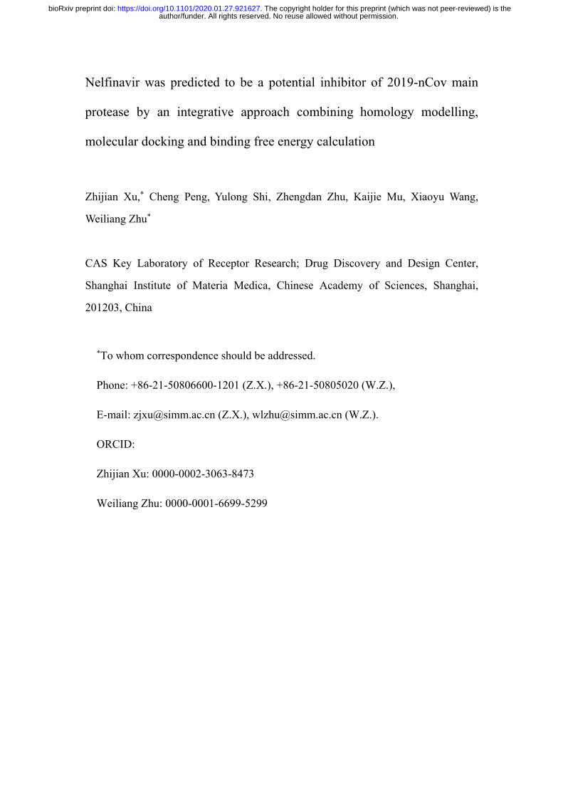
Nelfinavir was predicted to be a potential inhibitor of 2019-nCov main
protease by an integrative approach combining homology modelling,
molecular docking and binding free energy calculation
Zhijian Xu,* Cheng Peng, Yulong Shi, Zhengdan Zhu, Kaijie Mu, Xiaoyu Wang,
Weiliang Zhu*
CAS Key Laboratory of Receptor Research; Drug Discovery and Design Center,
Shanghai Institute of Materia Medica, Chinese Academy of Sciences, Shanghai,
201203, China
*To whom correspondence should be addressed.
Phone: +86-21-50806600-1201 (Z.X.), +86-21-50805020 (W.Z.),
E-mail: [email protected] (Z.X.), [email protected] (W.Z.).
ORCID:
Zhijian Xu: 0000-0002-3063-8473
Weiliang Zhu: 0000-0001-6699-5299
author/funder. All rights reserved. No reuse allowed without permission. The copyright holder for this preprint (which was not peer-reviewed) is the. https://doi.org/10.1101/2020.01.27.921627doi: bioRxiv preprint

Abstract
2019-nCov has caused more than 80 deaths as of 27 January 2020 in China, and
infection cases have been reported in more than 10 countries. However, there is no
approved drug to treat the disease. 2019-nCov Mpro is a potential drug target to combat
the virus. We built homology models based on SARS Mpro structures, and docked 1903
small molecule drugs to the models. Based on the docking score and the 3D similarity
of the binding mode to the known Mpro ligands, 4 drugs were selected for binding free
energy calculations. Both MM/GBSA and SIE methods voted for nelfinavir, with the
binding free energy of -24.69±0.52 kcal/mol and -9.42±0.04 kcal/mol, respectively.
Therefore, we suggested that nelfinavir might be a potential inhibitor against 2019-
nCov Mpro.
author/funder. All rights reserved. No reuse allowed without permission. The copyright holder for this preprint (which was not peer-reviewed) is the. https://doi.org/10.1101/2020.01.27.921627doi: bioRxiv preprint

1. Introduction
In December 2019, cluster of patients with pneumonia were reported in Wuhan,
Hubei Province, China.1, 2 Shortly, a new coronavirus, temporally named 2019-nCov,
was identified to be the cause of the disease, which is the seventh member of the family
betacoronavirus.1 More than 2,700 infection cases and 80 deaths were reported as of 27
January 2020 from China. In addition, infection cases have been reported from Thailand,
Australia, Malaysia, Singapore, France, Japan, South Korea, the United States, Vietnam,
Canada and Nepal, indicating that the disease is a potential threat to the global health.3
Sadly, the number is still growing rapidly and no drug has been approved to be effective.
Therefore, it is urgent to discover and develop drugs to cure the disease.
Based on its function, the main protease (Mpro) or chymotrypsin-like protease
(3CLpro)4 is suggested to be a potential drug target to combat 2019-nCov, which is
highly conservable among coronaviruses. Sequence alignment revealed that the Mpro of
2019-nCov shares 96% similarity with that of SARS (severe acute respiratory
syndrome) (Figure 1). Studies for identifying the inhibitors of 2019-nCov Mpro were
quickly performed for discovering and developing drugs against the disease. For
example, Hualiang Jiang and collaborators identified 30 drugs and compounds as the
Mpro inhibitors via protein modelling and virtual screening, which is a rapid progress in
the way to cope with the crisis. In addition, one of the 30 drugs/compounds, remdesivir,
was also suggested to be potential inhibitor against 2019-nCov by Liu et al,5 who also
suggested 3 other possible inhibitors.
author/funder. All rights reserved. No reuse allowed without permission. The copyright holder for this preprint (which was not peer-reviewed) is the. https://doi.org/10.1101/2020.01.27.921627doi: bioRxiv preprint
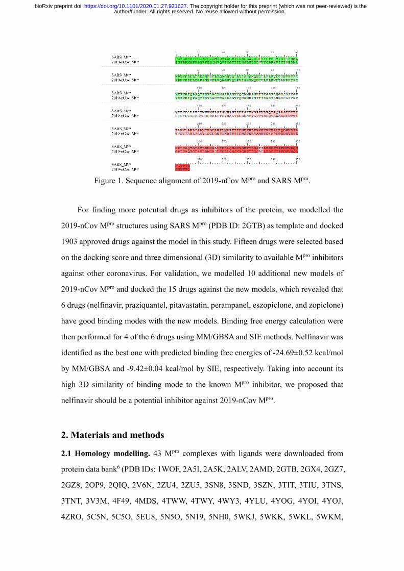
Figure 1. Sequence alignment of 2019-nCov Mpro and SARS Mpro.
For finding more potential drugs as inhibitors of the protein, we modelled the
2019-nCov Mpro structures using SARS Mpro (PDB ID: 2GTB) as template and docked
1903 approved drugs against the model in this study. Fifteen drugs were selected based
on the docking score and three dimensional (3D) similarity to available Mpro inhibitors
against other coronavirus. For validation, we modelled 10 additional new models of
2019-nCov Mpro and docked the 15 drugs against the new models, which revealed that
6 drugs (nelfinavir, praziquantel, pitavastatin, perampanel, eszopiclone, and zopiclone)
have good binding modes with the new models. Binding free energy calculation were
then performed for 4 of the 6 drugs using MM/GBSA and SIE methods. Nelfinavir was
identified as the best one with predicted binding free energies of -24.69±0.52 kcal/mol
by MM/GBSA and -9.42±0.04 kcal/mol by SIE, respectively. Taking into account its
high 3D similarity of binding mode to the known Mpro inhibitor, we proposed that
nelfinavir should be a potential inhibitor against 2019-nCov Mpro.
2. Materials and methods
2.1 Homology modelling. 43 Mpro complexes with ligands were downloaded from
protein data bank6 (PDB IDs: 1WOF, 2A5I, 2A5K, 2ALV, 2AMD, 2GTB, 2GX4, 2GZ7,
2GZ8, 2OP9, 2QIQ, 2V6N, 2ZU4, 2ZU5, 3SN8, 3SND, 3SZN, 3TIT, 3TIU, 3TNS,
3TNT, 3V3M, 4F49, 4MDS, 4TWW, 4TWY, 4WY3, 4YLU, 4YOG, 4YOI, 4YOJ,
4ZRO, 5C5N, 5C5O, 5EU8, 5N5O, 5N19, 5NH0, 5WKJ, 5WKK, 5WKL, 5WKM,
author/funder. All rights reserved. No reuse allowed without permission. The copyright holder for this preprint (which was not peer-reviewed) is the. https://doi.org/10.1101/2020.01.27.921627doi: bioRxiv preprint

6FV1) and aligned to 2GTB in PyMOL.7 11 complexes (PDB IDs: 2A5K, 2GTB, 2GX4,
3SND, 3TNS, 3V3M, 4F49, 4YLU, 5NH0, 5WKJ, 5WKM) were served as templates
to build 11 2019-nCov Mpro models in SWISS-MODEL server by “user template”
mode.8
2.2 Approved Drugs. 1905 approved small molecule drugs with 3D coordinates were
downloaded from DrugBank release version 5.1.5,9 while 1903 drugs could be
converted to pdbqt format by prepare_ligand4.py script in MGLToos version 1.5.6.10
2.3 Molecular Docking. 1903 approved drugs in pdbqt format were docked to 2019-
nCov Mpro model (template: 2GTB) by smina,11 which is a fork of AutoDock Vina12
with improving scoring and minimization. The hydrogens were added to 2019-nCov
Mpro model by pdb2pqr (--ff=amber --ffout=amber --chain --with-ph=7).13 Then the
model was converted to pdbqt format by prepare_receptor4.py script in MGLToos
version 1.5.6.10 The ligand in 2GTB was used to define the grid and the buffer space
was set to 6.0 Å (autobox_add). The random seed was explicitly set to 0 (seed). The
exhaustiveness of the global search was set to 32 (exhaustiveness) and at most 1 binding
mode was generated for each drug (num_modes). MolShaCS, which utilized Gaussian-
based description of molecular shape and charge distribution, was used to calculate the
3D similarities between approved drugs and available Mpro inhibitors.14
2.4 Molecular dynamics simulation. Each simulation system was immersed in a cubic
box of TIP3P water that was extended by 9 Å from the solute, with a rational number
of counter ions of Na+ or Cl- to neutralize the system. General Amber force field
(GAFF)15 and Amber ff03 force field16 were used to parameterize the ligand and protein,
respectively. 10,000 steps of minimization with constraints (10 kcal/mol/Å2) on heavy
atoms of complex, including 5,000 steps of steepest descent minimization and 5,000
steps of conjugate gradient minimization, was used to optimize each system. Then each
system was heated to 300 K within 0.2 ns followed by 0.1 ns equilibration in NPT
ensemble. Finally, 5 ns MD simulation on each system at 300 K was performed. The
minimization, heating and equilibrium are performed with sander program in Amber16.
The 5 ns production run was performed with pmemd.cuda.
2.5 Binding free energy calculation. Based on the 5 ns MD simulation trajectory, bind
author/funder. All rights reserved. No reuse allowed without permission. The copyright holder for this preprint (which was not peer-reviewed) is the. https://doi.org/10.1101/2020.01.27.921627doi: bioRxiv preprint

free energy (ΔG) was calculated with MM/GBSA17, 18 and SIE19 approaches. In the
MM/GBSA, the ΔG was calculated according to equation (1),
∆𝐺𝐺 = ∆𝐻𝐻 − 𝑇𝑇∆𝑆𝑆 = ∆𝐸𝐸𝑒𝑒𝑒𝑒𝑒𝑒 + ∆𝐸𝐸𝑉𝑉𝑉𝑉𝑉𝑉 + ∆𝐺𝐺𝑔𝑔𝑔𝑔 + ∆𝐺𝐺𝑛𝑛𝑛𝑛 − 𝑇𝑇∆𝑆𝑆 (1)
where ΔEele and ΔEVDW refer to electrostatic and van der Waals energy terms,
respectively. ΔGgb and ΔGnp refer to polar and non-polar solvation free energies,
respectively. Conformational entropy (TΔS) was calculated by nmode module in
Amber16. The dielectric constants for solvent and solute were set to 80.0 and 1.0,
respectively, and OBC solvation model (igb = 5 and PBradii = 5)20 was used in this
study. Other parameters are set to default values.
In the SIE, the ΔG was calculated based on equation (2),
∆𝐺𝐺𝑔𝑔𝑏𝑏𝑛𝑛𝑏𝑏 = 𝛼𝛼[𝐸𝐸𝐶𝐶(𝐷𝐷𝑏𝑏𝑛𝑛) + ∆𝐺𝐺𝑔𝑔𝑏𝑏𝑛𝑛𝑏𝑏𝑅𝑅 (𝜌𝜌,𝐷𝐷𝑏𝑏𝑛𝑛) + 𝐸𝐸𝑉𝑉𝑉𝑉𝑉𝑉 + 𝛾𝛾∆𝑀𝑀𝑆𝑆𝑀𝑀(𝜌𝜌)] + 𝐶𝐶 (2)
where EC and EVDW refer to the sum of intermolecular Coulomb and van der Waals
interaction energies, respectively. ΔGRbind and ΔMSA refer to the changes of reaction
field energy and molecular surface area upon ligand binding, respectively. Default
values of the global proportionality coefficient (α = 0.1048), the solute interior
dielectric constant (Din = 2.25), the van der Waals radii linear scaling coefficient (ρ =
1.1), the molecular surface area coefficient (γ = 0.0129 kcal/mol/Å2), and the constant
(C = -2.89 kcal/mol) are used in this study.
3. Results and discussion
3.1 Docking results of 1903 approved drugs against Mpro models. 1903 approved
small molecule drugs were docked to a Mpro homology model built by SWISS-MODEL
using 2GTB as template. There are 672 drugs with the docking score better than -7.0
kcal/mol. The binding modes are essential to the activities. If a compound shares a
similar binding mode to a known ligand, it is more likely to have a similar activity to
the ligand. Therefore, we calculated the 3D similarities of the binding mode of the
docked drugs to the available binders against Mpro. 39 binders in 44 complexes were
chosen as the references to calculate the 3D similarities (Table 1). 159 drugs have a 3D
similarity larger than 65.0% with at least one known Mpro binder.
author/funder. All rights reserved. No reuse allowed without permission. The copyright holder for this preprint (which was not peer-reviewed) is the. https://doi.org/10.1101/2020.01.27.921627doi: bioRxiv preprint
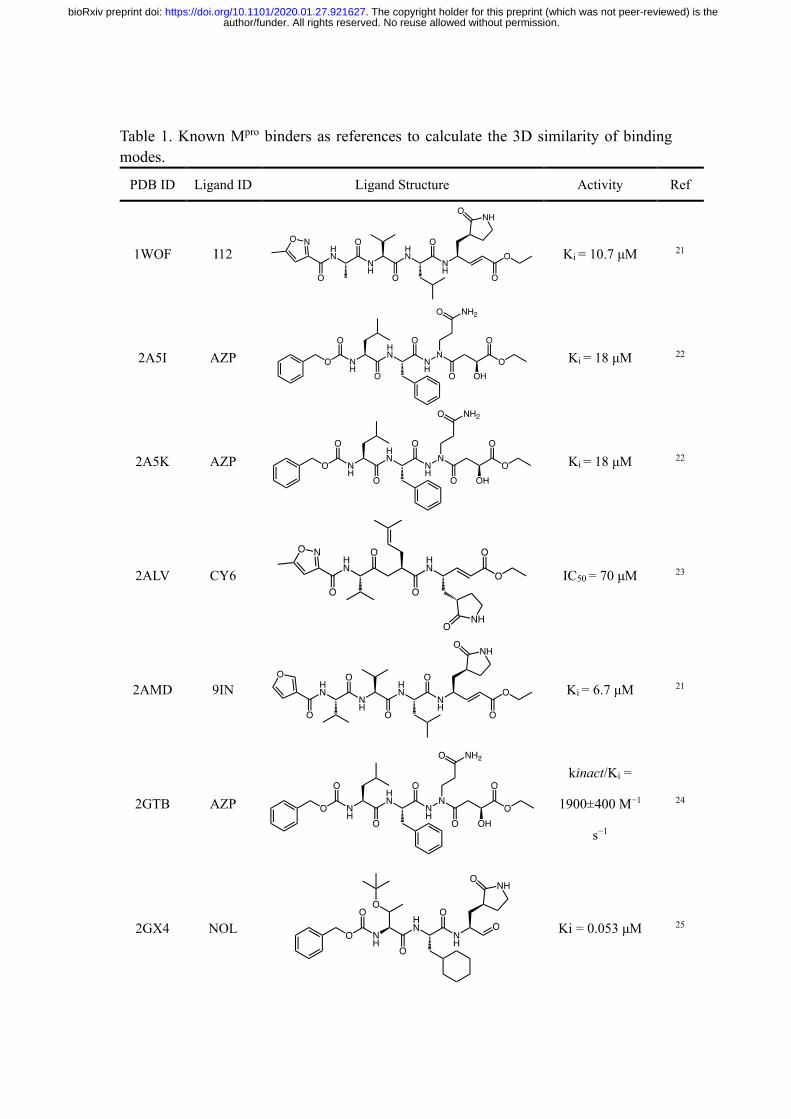
Table 1. Known Mpro binders as references to calculate the 3D similarity of binding modes.
PDB ID Ligand ID Ligand Structure Activity Ref
1WOF I12
Ki = 10.7 μM 21
2A5I AZP
Ki = 18 μM 22
2A5K AZP
Ki = 18 μM 22
2ALV CY6
IC50 = 70 μM 23
2AMD 9IN
Ki = 6.7 μM 21
2GTB AZP
kinact/Ki =
1900±400 M−1
s−1
24
2GX4 NOL
Ki = 0.053 μM 25
author/funder. All rights reserved. No reuse allowed without permission. The copyright holder for this preprint (which was not peer-reviewed) is the. https://doi.org/10.1101/2020.01.27.921627doi: bioRxiv preprint

2GZ7 D3F
IC50 = 0.3 μM 26
2GZ8 F3F
IC50 = 3 μM 26
2OP9 WR1
Ki = 2.2 μM 27
2QIQ CYV
IC50 = 80 μM 28
2V6N XP1
Ki = 1.38 μM 29
2ZU4 ZU3
Ki = 0.038 μM 30
2ZU5 ZU5
N
NN
NO
HO
OH
OH
OH
O
O H H
H
H
H
Ki = 0.099 μM 30
3SN8 S89 NN
OH
OH
OH
H
H
H
H
Ki = 2.24 μM 31
author/funder. All rights reserved. No reuse allowed without permission. The copyright holder for this preprint (which was not peer-reviewed) is the. https://doi.org/10.1101/2020.01.27.921627doi: bioRxiv preprint

3SND PRD_000
772 NH
N NNN
N
OH
HO
OHOH
OH
OHO
OH
OHOH
OH
H
HH
HH
H
Ki = 8.27±1.52
μM 31
3SZN G75
N
NN
O
HO
OH
OH
OO
H
H
H
NA
3TIT G81
N
NN
O
HO
OH
OH
OO
H
H
H
NA
3TIU G82 N
NN
N
O O
OH
OH
OH OHO
O
O
H
H H
H
NA
3TNS G83
N
NN
NO
O
HO
OH
OH
OHO
O
OH
H
H
H
NA
3TNT G85
N
NN
NO
HO
OH
OH
OHO
O
OH
H
H
H
NA
3V3M 0EN
N
NN
OH
O
OH
IC50 = 4.8 μM 32
author/funder. All rights reserved. No reuse allowed without permission. The copyright holder for this preprint (which was not peer-reviewed) is the. https://doi.org/10.1101/2020.01.27.921627doi: bioRxiv preprint

4F49 K36
N
NN
HO OH
OHOH
OHO
OO S
HH
HH
IC50 = 0.82 μM 33
4MDS 23H
N
N
N NN
NN
OH
O
OHH
IC50 = 6.2 μM 34
4TWW 3A7 Br
N
HN HN
N
O
O
H
H
H
H
IC50 = 63 μM 35
4TWY 3BL H
H
H H
OO
N
HN
NH
N
IC50 = 108 μM 35
4WY3 3X5
N
NH
HN
N
OO
HH
HH
IC50 = 240 μM 35
4YLU R30 NH
NN
NN
OO
S
IC50 > 100 μM 36
4YOG 4F5
O
HN
NO
NH
ON
N N
S
IC50 = 1.8 μM 37
author/funder. All rights reserved. No reuse allowed without permission. The copyright holder for this preprint (which was not peer-reviewed) is the. https://doi.org/10.1101/2020.01.27.921627doi: bioRxiv preprint

4YOI 4F4
O
NN
HN
O
NN
S
S
IC50 = 0.33 μM 37
4YOJ RFM
O
NN
HN
O
NN
S
IC50 = 0.41 μM 37
4ZRO PRD_002
174 O
O
NHO
NH
OHN
ONH
O
HO
HN
O
O
IC50 = 0.59 μM 38
5C5N SLH O
HN
N
O
NH
NHN
NA
5C5O SDJ O
HN
N
O
NH
NHN
NA
5EU8 PRD_002
214 HN
ONH
OHN
O
O
NH
NHO
O
O
NO
NA
5N5O 8O5
OH
NHO
NH
OHN
O
O
NH
NA
5N19 D03
OH
NHO
NH
OHN
O
O
NH
NA
5NH0 8X8 O
O
NH
HNO
OH
O
NH
NA
author/funder. All rights reserved. No reuse allowed without permission. The copyright holder for this preprint (which was not peer-reviewed) is the. https://doi.org/10.1101/2020.01.27.921627doi: bioRxiv preprint

5WKJ B1S HN
O
OO
NH
NHO
OH
SOHO
O
EC50 = 0.9 μM* 39
5WKK AW4 HN
O
OClO
NH
NHO
OH
SOHO
O
EC50 = 0.5 μM* 39
5WKL B3J
IC50 = 0.8 μM 39
5WKM N02
IC50 = 6.1 μM 39
6FV1 E8E HN
O
O
NH
NHO
OH
O
NH
NA
* Racemic activity; NA: Not Available.
After visualizing the docked complexes carefully, we selected 15 drugs (Table 2)
for further analysis. There are some differences between the conformations of the
protein in the 3D similarity reference and 2GTB model. Therefore, we modelled 10 new
homology models using the proteins in 3D similarity reference as templates and we re-
docked the 15 drugs to the 10 new homology models. 6 drugs (nelfinavir, pitavastatin,
perampanel, praziquantel, zopiclone, and eszopiclone) show good docking scores and
binding modes (Table 3). Because eszopiclone and zopiclone was used to treat insomnia
in a low dosage which may not be suitable to treat pneumonia, we carried out further
binding free energy calculation for the rest 4 drugs.
author/funder. All rights reserved. No reuse allowed without permission. The copyright holder for this preprint (which was not peer-reviewed) is the. https://doi.org/10.1101/2020.01.27.921627doi: bioRxiv preprint

Table 2. 15 drugs selected from 2GTB model.
DrugBank ID Docking Score against 2GTB model (kcal/mol)
3D Similarity Reference
3D Similarity
DB00328 -7.16 3V3M 71.3% DB13680 -7.13 3SND 70.8% DB01165 -7.31 3V3M 70.2% DB01198 -7.81 5NH0 68.9% DB08934 -8.65 2GX4 68.6% DB01165 -7.31 5WKM 68.3% DB08860 -8.06 2GTB 68.1% DB00402 -7.75 5NH0 67.8% DB08883 -8.17 5NH0 67.1% DB01288 -7.96 5WKM 67.1% DB08860 -8.06 2A5K 66.9% DB00972 -7.71 5NH0 66.7% DB00482 -8.46 4YLU 66.7% DB00220 -8.91 2GX4 66.4% DB01058 -7.90 3V3M 65.7% DB00328 -7.16 4F49 65.6% DB08934 -8.65 3TNS 65.5% DB00904 -7.45 5WKM 65.4% DB11951 -8.27 4F49 65.3% DB11951 -8.27 5WKJ 65.2%
Table 3. 6 drugs selected from 10 new homology models.
DrugBank ID Name Homology
Template Docking Score
(kcal/mol) 3D
Similarity
DB00220 nelfinavir 2GX4 -9.18 70.2%
DB08860 pitavastatin 2GTB -8.06 68.1%
DB08883 perampanel 5NH0 -8.63 66.9%
DB01058 praziquantel 3V3M -7.38 65.8%
DB01198 zopiclone 5NH0 -8.46 63.5%
DB00402 eszopiclone 5NH0 -8.46 63.5%
3.2 Binding Free Energy calculated by MM/GBSA and SIE. The 4 docked complex
structures, i.e., nelfinavir-2GX4, pitavastatin-2GTB, perampanel-5NH0 and
praziquantel-3V3M, were subjected to 5 ns molecular dynamics simulations using
author/funder. All rights reserved. No reuse allowed without permission. The copyright holder for this preprint (which was not peer-reviewed) is the. https://doi.org/10.1101/2020.01.27.921627doi: bioRxiv preprint

Amber 16. To provide insight into their binding mechanisms, the binding free energies
were calculated by MM/GBSA and SIE approaches. In results of the MM/GBSA
approach, the calculated binding free energies of nelfinavir-2GX4, pitavastatin-2GTB,
perampanel-5NH0, and praziquantel-3V3M, are -24.69±0.52, -12.70±0.38, -
14.98±0.34, and -6.51±0.21 kcal/mol, respectively (Table 4), which highlight
nelfinavir as the most active one. In pitavastatin-2GTB, perampanel-5NH0, and
praziquantel-3V3M, the van der Waals interaction (Evdw) makes a more significant
contribution than the electrostatic interaction (Eele) (Table 4), indicating that van der
Waals interaction is the main driving force for the 3 drugs binding. However, the Eele
interaction of nelfinavir-2GX4 is very strong, suggesting that the electrostatic
interaction also play an important role in the binding of nelfinavir. Furthermore, in
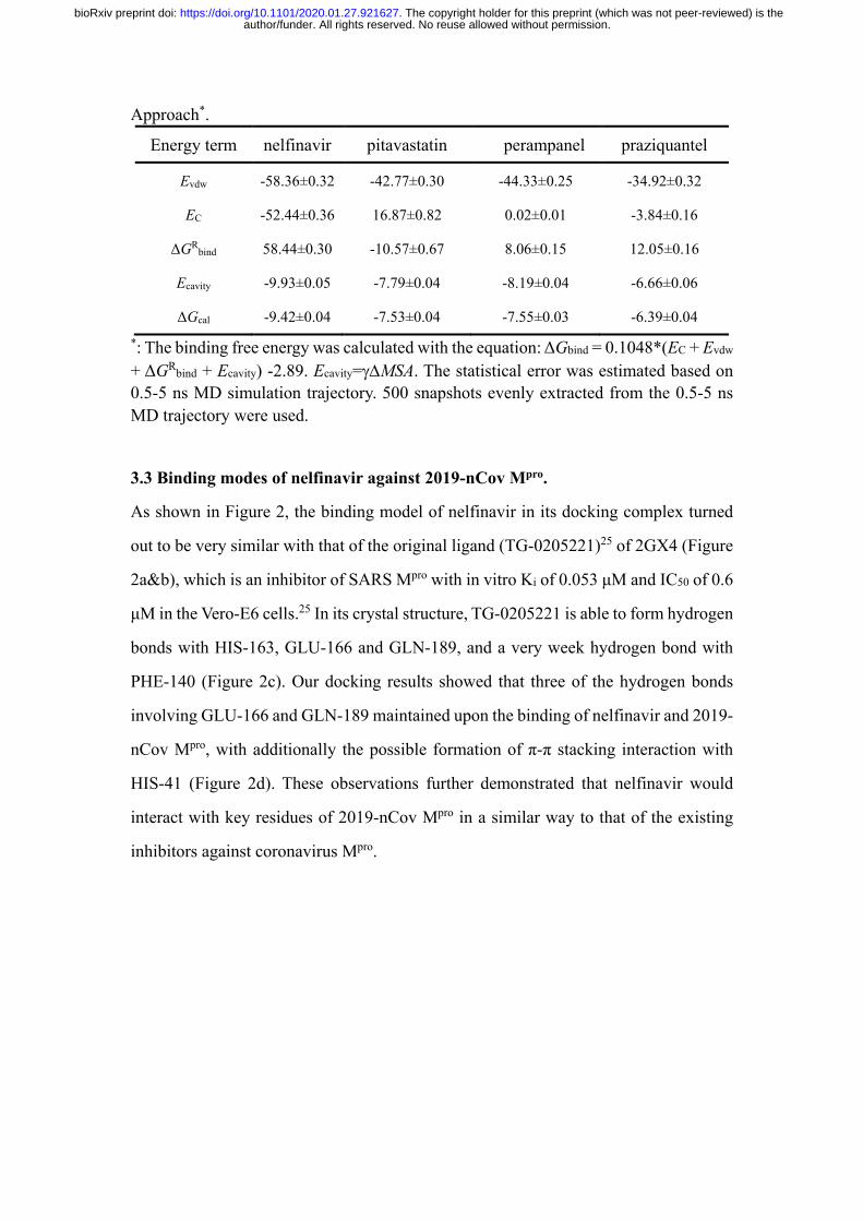
results of the SIE approach, the rank of the calculated binding free energies for the 4
docking complexes is consistent with that of the MM/GBSA approach (Table 5) that
the nelfinavir has the strongest binding free energy (-9.42±0.04 kcal/mol), indicating
the reliability of the current binding free energy analysis.
Table 4. Components of the Binding Free Energy (kcal/mol) Calculated by MM/GBSA Approach*.
Energy term nelfinavir pitavastatin perampanel praziquantel
Evdw -58.36±0.32 -42.77±0.30 -44.33±0.25 -34.92±0.32
Eele -117.95±0.81 37.95±0.86 0.05±0.03 -8.64±0.35
Ggb 134.49±0.83 -22.40±1.63 14.76±0.29 21.53±0.32
Gnp -6.69±0.03 -5.60±0.03 -5.43±0.03 -4.34±0.03
ΔH -48.50±0.36 -32.82±0.38 -34.96±0.27 -26.37±0.30
TΔS -23.81±0.67 -20.12±0.38 -19.98±0.42 -19.86±0.12
ΔGcal -24.69±0.52 -12.70±0.38 -14.98±0.34 -6.51±0.21 *: The statistical error was estimated based on 0.5-5 ns MD simulation trajectory. 500 snapshots evenly extracted from the 0.5-5 ns MD trajectory of complex were used for MM/GBSA calculations and 50 snapshots for the entropy term calculations. Table 5. Components of the Binding Free Energy (kcal/mol) Calculated by SIE
author/funder. All rights reserved. No reuse allowed without permission. The copyright holder for this preprint (which was not peer-reviewed) is the. https://doi.org/10.1101/2020.01.27.921627doi: bioRxiv preprint
Approach*.
Energy term nelfinavir pitavastatin perampanel praziquantel
Evdw -58.36±0.32 -42.77±0.30 -44.33±0.25 -34.92±0.32
EC -52.44±0.36 16.87±0.82 0.02±0.01 -3.84±0.16
ΔGRbind 58.44±0.30 -10.57±0.67 8.06±0.15 12.05±0.16
Ecavity -9.93±0.05 -7.79±0.04 -8.19±0.04 -6.66±0.06
ΔGcal -9.42±0.04 -7.53±0.04 -7.55±0.03 -6.39±0.04 *: The binding free energy was calculated with the equation: ΔGbind = 0.1048*(EC + Evdw
+ ΔGRbind + Ecavity) -2.89. Ecavity=γ∆MSA. The statistical error was estimated based on 0.5-5 ns MD simulation trajectory. 500 snapshots evenly extracted from the 0.5-5 ns MD trajectory were used.
3.3 Binding modes of nelfinavir against 2019-nCov Mpro.
As shown in Figure 2, the binding model of nelfinavir in its docking complex turned
out to be very similar with that of the original ligand (TG-0205221)25 of 2GX4 (Figure
2a&b), which is an inhibitor of SARS Mpro with in vitro Ki of 0.053 μM and IC50 of 0.6
μM in the Vero-E6 cells.25 In its crystal structure, TG-0205221 is able to form hydrogen
bonds with HIS-163, GLU-166 and GLN-189, and a very week hydrogen bond with
PHE-140 (Figure 2c). Our docking results showed that three of the hydrogen bonds
involving GLU-166 and GLN-189 maintained upon the binding of nelfinavir and 2019-
nCov Mpro, with additionally the possible formation of π-π stacking interaction with
HIS-41 (Figure 2d). These observations further demonstrated that nelfinavir would
interact with key residues of 2019-nCov Mpro in a similar way to that of the existing
inhibitors against coronavirus Mpro.
author/funder. All rights reserved. No reuse allowed without permission. The copyright holder for this preprint (which was not peer-reviewed) is the. https://doi.org/10.1101/2020.01.27.921627doi: bioRxiv preprint
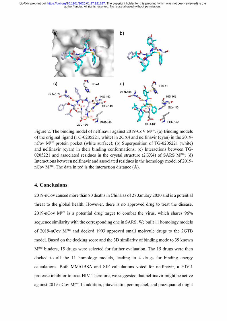
Figure 2. The binding model of nelfinavir against 2019-CoV Mpro. (a) Binding models of the original ligand (TG-0205221, white) in 2GX4 and nelfinavir (cyan) in the 2019-nCov Mpro protein pocket (white surface); (b) Superposition of TG-0205221 (white) and nelfinavir (cyan) in their binding conformations; (c) Interactions between TG-0205221 and associated residues in the crystal structure (2GX4) of SARS Mpro; (d) Interactions between nelfinavir and associated residues in the homology model of 2019-nCov Mpro. The data in red is the interaction distance (Å).
4. Conclusions
2019-nCov caused more than 80 deaths in China as of 27 January 2020 and is a potential
threat to the global health. However, there is no approved drug to treat the disease.
2019-nCov Mpro is a potential drug target to combat the virus, which shares 96%
sequence similarity with the corresponding one in SARS. We built 11 homology models
of 2019-nCov Mpro and docked 1903 approved small molecule drugs to the 2GTB
model. Based on the docking score and the 3D similarity of binding mode to 39 known
Mpro binders, 15 drugs were selected for further evaluation. The 15 drugs were then
docked to all the 11 homology models, leading to 4 drugs for binding energy
calculations. Both MM/GBSA and SIE calculations voted for nelfinavir, a HIV-1
protease inhibitor to treat HIV. Therefore, we suggested that nelfinavir might be active
against 2019-nCov Mpro. In addition, pitavastatin, perampanel, and praziquantel might
author/funder. All rights reserved. No reuse allowed without permission. The copyright holder for this preprint (which was not peer-reviewed) is the. https://doi.org/10.1101/2020.01.27.921627doi: bioRxiv preprint
also have moderate activities against 2019-nCov.
Acknowledgments
This work was supported by the National Key R&D Program of China
(2017YFB0202601). The simulations were partially run at TianHe 1 supercomputer in
Tianjin. We thank Prof. Tingjun Hou in Zhejiang University, China for helpful
discussions on the MM/GBSA calculations.
References 1. Zhu, N.; Zhang, D.; Wang, W.; Li, X.; Yang, B.; Song, J.; Zhao, X.; Huang, B.; Shi, W.; Lu, R.; Niu, P.; Zhan, F.; Ma, X.; Wang, D.; Xu, W.; Wu, G.; Gao, G. F.; Tan, W., A Novel Coronavirus from Patients with Pneumonia in China, 2019. The New England journal of medicine 2020. 2. Lu, H.; Stratton, C. W.; Tang, Y.-W., Outbreak of Pneumonia of Unknown Etiology in Wuhan China: the Mystery and the Miracle. J. Med. Virol. 2020. 3. Hui, D. S.; I Azhar, E.; Madani, T. A.; Ntoumi, F.; Kock, R.; Dar, O.; Ippolito, G.; McHugh, T. D.; Memish, Z. A.; Drosten, C.; Zumla, A.; Petersen, E., The continuing 2019-nCoV epidemic threat of novel coronaviruses to global health - The latest 2019 novel coronavirus outbreak in Wuhan, China. International journal of infectious diseases : IJID : official publication of the International Society for Infectious Diseases 2020, 91, 264-266. 4. Chen, Y.; Liu, Q.; Guo, D., Coronaviruses: genome structure, replication, and pathogenesis. J. Med. Virol. 2020. 5. Jared S., M.; Tyler, L.; Shiqing, X.; Wenshe, L., Learning from the Past: Possible Urgent Prevention and Treatment Options for Severe Acute Respiratory Infections Caused by 2019-nCoV. 2020. 6. Burley, S. K.; Berman, H. M.; Bhikadiya, C.; Bi, C.; Chen, L.; Di Costanzo, L.; Christie, C.; Dalenberg, K.; Duarte, J. M.; Dutta, S.; Feng, Z.; Ghosh, S.; Goodsell, D. S.; Green, R. K.; Guranović, V.; Guzenko, D.; Hudson, B. P.; Kalro, T.; Liang, Y.; Lowe, R.; Namkoong, H.; Peisach, E.; Periskova, I.; Prlić, A.; Randle, C.; Rose, A.; Rose, P.; Sala, R.; Sekharan, M.; Shao, C.; Tan, L.; Tao, Y.-P.; Valasatava, Y.; Voigt, M.; Westbrook, J.; Woo, J.; Yang, H.; Young, J.; Zhuravleva, M.; Zardecki, C., RCSB Protein Data Bank: biological macromolecular structures enabling research and education in fundamental biology, biomedicine, biotechnology and energy. Nucleic Acids Res. 2018, 47 (D1), D464-D474. 7. Schrodinger, LLC, The PyMOL Molecular Graphics System, Version 2.4. 2019. 8. Waterhouse, A.; Bertoni, M.; Bienert, S.; Studer, G.; Tauriello, G.; Gumienny, R.; Heer, F. T.; de Beer, T. A P.; Rempfer, C.; Bordoli, L.; Lepore,
author/funder. All rights reserved. No reuse allowed without permission. The copyright holder for this preprint (which was not peer-reviewed) is the. https://doi.org/10.1101/2020.01.27.921627doi: bioRxiv preprint

R.; Schwede, T., SWISS-MODEL: homology modelling of protein structures and complexes. Nucleic Acids Res. 2018, 46 (W1), W296-W303. 9. Wishart, D. S.; Feunang, Y. D.; Guo, A. C.; Lo, E. J.; Marcu, A.; Grant, J. R.; Sajed, T.; Johnson, D.; Li, C.; Sayeeda, Z.; Assempour, N.; Iynkkaran, I.; Liu, Y.; Maciejewski, A.; Gale, N.; Wilson, A.; Chin, L.; Cummings, R.; Le, D.; Pon, A.; Knox, C.; Wilson, M., DrugBank 5.0: a major update to the DrugBank database for 2018. Nucleic Acids Res. 2018, 46 (D1), D1074-D1082. 10. Morris, G. M.; Huey, R.; Lindstrom, W.; Sanner, M. F.; Belew, R. K.; Goodsell, D. S.; Olson, A. J., AutoDock4 and AutoDockTools4: Automated docking with selective receptor flexibility. J. Comput. Chem. 2009, 30 (16), 2785-2791. 11. Koes, D. R.; Baumgartner, M. P.; Camacho, C. J., Lessons Learned in Empirical Scoring with smina from the CSAR 2011 Benchmarking Exercise. J. Chem. Inf. Model. 2013, 53 (8), 1893-1904. 12. Trott, O.; Olson, A. J., AutoDock Vina: Improving the speed and accuracy of docking with a new scoring function, efficient optimization, and multithreading. J. Comput. Chem. 2010, 31 (2), 455-461. 13. Dolinsky, T. J.; Nielsen, J. E.; McCammon, J. A.; Baker, N. A., PDB2PQR: an automated pipeline for the setup of Poisson–Boltzmann electrostatics calculations. Nucleic Acids Res. 2004, 32 (suppl_2), W665-W667. 14. Vaz de Lima, L. A.; Nascimento, A. S., MolShaCS: a free and open source tool for ligand similarity identification based on Gaussian descriptors. Eur. J. Med. Chem. 2013, 59, 296-303. 15. Wang, J.; Wolf, R. M.; Caldwell, J. W.; Kollman, P. A.; Case, D. A., Development and testing of a general amber force field. J. Comput. Chem. 2004, 25 (9), 1157-74. 16. Duan, Y.; Wu, C.; Chowdhury, S.; Lee, M. C.; Xiong, G.; Zhang, W.; Yang, R.; Cieplak, P.; Luo, R.; Lee, T.; Caldwell, J.; Wang, J.; Kollman, P., A point-charge force field for molecular mechanics simulations of proteins based on condensed-phase quantum mechanical calculations. J. Comput. Chem. 2003, 24 (16), 1999-2012. 17. Kollman, P. A.; Massova, I.; Reyes, C.; Kuhn, B.; Huo, S.; Chong, L.; Lee, M.; Lee, T.; Duan, Y.; Wang, W.; Donini, O.; Cieplak, P.; Srinivasan, J.; Case, D. A.; Cheatham, T. E., 3rd, Calculating structures and free energies of complex molecules: combining molecular mechanics and continuum models. Acc. Chem. Res. 2000, 33 (12), 889-97. 18. Srinivasan, J.; Cheatham, T. E.; Cieplak, P.; Kollman, P. A.; Case, D. A., Continuum Solvent Studies of the Stability of DNA, RNA, and Phosphoramidate−DNA Helices. J. Am. Chem. Soc. 1998, 120 (37), 9401-9409. 19. Naïm, M.; Bhat, S.; Rankin, K. N.; Dennis, S.; Chowdhury, S. F.; Siddiqi, I.; Drabik, P.; Sulea, T.; Bayly, C. I.; Jakalian, A.; Purisima, E. O., Solvated Interaction Energy (SIE) for Scoring Protein−Ligand Binding Affinities. 1. Exploring the Parameter Space. J. Chem. Inf. Model. 2007, 47 (1), 122-133. 20. Onufriev, A.; Bashford, D.; Case, D. A., Exploring protein native states and large-scale conformational changes with a modified generalized born model. Proteins 2004,
author/funder. All rights reserved. No reuse allowed without permission. The copyright holder for this preprint (which was not peer-reviewed) is the. https://doi.org/10.1101/2020.01.27.921627doi: bioRxiv preprint

55 (2), 383-94. 21. Yang, H.; Xie, W.; Xue, X.; Yang, K.; Ma, J.; Liang, W.; Zhao, Q.; Zhou, Z.; Pei, D.; Ziebuhr, J.; Hilgenfeld, R.; Yuen, K. Y.; Wong, L.; Gao, G.; Chen, S.; Chen, Z.; Ma, D.; Bartlam, M.; Rao, Z., Design of wide-spectrum inhibitors targeting coronavirus main proteases. PLoS Biol 2005, 3 (10), e324. 22. Lee, T. W.; Cherney, M. M.; Huitema, C.; Liu, J.; James, K. E.; Powers, J. C.; Eltis, L. D.; James, M. N., Crystal structures of the main peptidase from the SARS coronavirus inhibited by a substrate-like aza-peptide epoxide. J Mol Biol 2005, 353 (5), 1137-51. 23. Ghosh, A. K.; Xi, K.; Ratia, K.; Santarsiero, B. D.; Fu, W.; Harcourt, B. H.; Rota, P. A.; Baker, S. C.; Johnson, M. E.; Mesecar, A. D., Design and synthesis of peptidomimetic severe acute respiratory syndrome chymotrypsin-like protease inhibitors. J Med Chem 2005, 48 (22), 6767-71. 24. Lee, T. W.; Cherney, M. M.; Liu, J.; James, K. E.; Powers, J. C.; Eltis, L. D.; James, M. N., Crystal structures reveal an induced-fit binding of a substrate-like Aza-peptide epoxide to SARS coronavirus main peptidase. J Mol Biol 2007, 366 (3), 916-32. 25. Yang, S.; Chen, S. J.; Hsu, M. F.; Wu, J. D.; Tseng, C. T.; Liu, Y. F.; Chen, H. C.; Kuo, C. W.; Wu, C. S.; Chang, L. W.; Chen, W. C.; Liao, S. Y.; Chang, T. Y.; Hung, H. H.; Shr, H. L.; Liu, C. Y.; Huang, Y. A.; Chang, L. Y.; Hsu, J. C.; Peters, C. J.; Wang, A. H.; Hsu, M. C., Synthesis, crystal structure, structure-activity relationships, and antiviral activity of a potent SARS coronavirus 3CL protease inhibitor. J. Med. Chem. 2006, 49 (16), 4971-80. 26. Lu, I. L.; Mahindroo, N.; Liang, P. H.; Peng, Y. H.; Kuo, C. J.; Tsai, K. C.; Hsieh, H. P.; Chao, Y. S.; Wu, S. Y., Structure-based drug design and structural biology study of novel nonpeptide inhibitors of severe acute respiratory syndrome coronavirus main protease. J Med Chem 2006, 49 (17), 5154-61. 27. Goetz, D. H.; Choe, Y.; Hansell, E.; Chen, Y. T.; McDowell, M.; Jonsson, C. B.; Roush, W. R.; McKerrow, J.; Craik, C. S., Substrate specificity profiling and identification of a new class of inhibitor for the major protease of the SARS coronavirus. Biochemistry 2007, 46 (30), 8744-52. 28. Ghosh, A. K.; Xi, K.; Grum-Tokars, V.; Xu, X.; Ratia, K.; Fu, W.; Houser, K. V.; Baker, S. C.; Johnson, M. E.; Mesecar, A. D., Structure-based design, synthesis, and biological evaluation of peptidomimetic SARS-CoV 3CLpro inhibitors. Bioorg Med Chem Lett 2007, 17 (21), 5876-80. 29. Verschueren, K. H.; Pumpor, K.; Anemuller, S.; Chen, S.; Mesters, J. R.; Hilgenfeld, R., A structural view of the inactivation of the SARS coronavirus main proteinase by benzotriazole esters. Chem Biol 2008, 15 (6), 597-606. 30. Lee, C. C.; Kuo, C. J.; Ko, T. P.; Hsu, M. F.; Tsui, Y. C.; Chang, S. C.; Yang, S.; Chen, S. J.; Chen, H. C.; Hsu, M. C.; Shih, S. R.; Liang, P. H.; Wang, A. H., Structural basis of inhibition specificities of 3C and 3C-like proteases by zinc-coordinating and peptidomimetic compounds. J Biol Chem 2009, 284 (12), 7646-55. 31. Zhu, L.; George, S.; Schmidt, M. F.; Al-Gharabli, S. I.; Rademann, J.; Hilgenfeld, R., Peptide aldehyde inhibitors challenge the substrate specificity of the
author/funder. All rights reserved. No reuse allowed without permission. The copyright holder for this preprint (which was not peer-reviewed) is the. https://doi.org/10.1101/2020.01.27.921627doi: bioRxiv preprint

SARS-coronavirus main protease. Antiviral Res 2011, 92 (2), 204-12. 32. Jacobs, J.; Grum-Tokars, V.; Zhou, Y.; Turlington, M.; Saldanha, S. A.; Chase, P.; Eggler, A.; Dawson, E. S.; Baez-Santos, Y. M.; Tomar, S.; Mielech, A. M.; Baker, S. C.; Lindsley, C. W.; Hodder, P.; Mesecar, A.; Stauffer, S. R., Discovery, synthesis, and structure-based optimization of a series of N-(tert-butyl)-2-(N-arylamido)-2-(pyridin-3-yl) acetamides (ML188) as potent noncovalent small molecule inhibitors of the severe acute respiratory syndrome coronavirus (SARS-CoV) 3CL protease. J Med Chem 2013, 56 (2), 534-46. 33. Kim, Y.; Lovell, S.; Tiew, K. C.; Mandadapu, S. R.; Alliston, K. R.; Battaile, K. P.; Groutas, W. C.; Chang, K. O., Broad-spectrum antivirals against 3C or 3C-like proteases of picornaviruses, noroviruses, and coronaviruses. J Virol 2012, 86 (21), 11754-62. 34. Turlington, M.; Chun, A.; Tomar, S.; Eggler, A.; Grum-Tokars, V.; Jacobs, J.; Daniels, J. S.; Dawson, E.; Saldanha, A.; Chase, P.; Baez-Santos, Y. M.; Lindsley, C. W.; Hodder, P.; Mesecar, A. D.; Stauffer, S. R., Discovery of N-(benzo[1,2,3]triazol-1-yl)-N-(benzyl)acetamido)phenyl) carboxamides as severe acute respiratory syndrome coronavirus (SARS-CoV) 3CLpro inhibitors: identification of ML300 and noncovalent nanomolar inhibitors with an induced-fit binding. Bioorg Med Chem Lett 2013, 23 (22), 6172-7. 35. Shimamoto, Y.; Hattori, Y.; Kobayashi, K.; Teruya, K.; Sanjoh, A.; Nakagawa, A.; Yamashita, E.; Akaji, K., Fused-ring structure of decahydroisoquinolin as a novel scaffold for SARS 3CL protease inhibitors. Bioorg Med Chem 2015, 23 (4), 876-90. 36. Tomar, S.; Johnston, M. L.; St John, S. E.; Osswald, H. L.; Nyalapatla, P. R.; Paul, L. N.; Ghosh, A. K.; Denison, M. R.; Mesecar, A. D., Ligand-induced Dimerization of Middle East Respiratory Syndrome (MERS) Coronavirus nsp5 Protease (3CLpro): IMPLICATIONS FOR nsp5 REGULATION AND THE DEVELOPMENT OF ANTIVIRALS. J Biol Chem 2015, 290 (32), 19403-22. 37. St John, S. E.; Tomar, S.; Stauffer, S. R.; Mesecar, A. D., Targeting zoonotic viruses: Structure-based inhibition of the 3C-like protease from bat coronavirus HKU4--The likely reservoir host to the human coronavirus that causes Middle East Respiratory Syndrome (MERS). Bioorg Med Chem 2015, 23 (17), 6036-48. 38. St John, S. E.; Therkelsen, M. D.; Nyalapatla, P. R.; Osswald, H. L.; Ghosh, A. K.; Mesecar, A. D., X-ray structure and inhibition of the feline infectious peritonitis virus 3C-like protease: Structural implications for drug design. Bioorg Med Chem Lett 2015, 25 (22), 5072-7. 39. Galasiti Kankanamalage, A. C.; Kim, Y.; Damalanka, V. C.; Rathnayake, A. D.; Fehr, A. R.; Mehzabeen, N.; Battaile, K. P.; Lovell, S.; Lushington, G. H.; Perlman, S.; Chang, K. O.; Groutas, W. C., Structure-guided design of potent and permeable inhibitors of MERS coronavirus 3CL protease that utilize a piperidine moiety as a novel design element. Eur J Med Chem 2018, 150, 334-346.
author/funder. All rights reserved. No reuse allowed without permission. The copyright holder for this preprint (which was not peer-reviewed) is the. https://doi.org/10.1101/2020.01.27.921627doi: bioRxiv preprint