Embed Size (px)
Citation preview
The Rockefeller University Press, 0021-9525/2001/01/325/10 $5.00The Journal of Cell Biology, Volume 152, Number 2, January 22, 2001 325–334http://www.jcb.org/cgi/content/full/152/2/325 325
Negative Regulation of Ros Receptor Tyrosine Kinase Signaling: An Epithelial Function of the SH2 Domain Protein Tyrosine Phosphatase SHP-1
Heike Keilhack,* Marit Müller,* Sylvia-Annette Böhmer,* Carsten Frank,* K. Michael Weidner,
i
** Walter Birchmeier,
i
Tanja Ligensa,** Alexander Berndt,
‡
Hartwig Kosmehl,
‡
Bernd Günther,
§
Thomas Müller,
¶
Carmen Birchmeier,
¶
and Frank D. Böhmer*
*Research Unit, Molecular Cell Biology,
‡
Institute of Pathology, and
§
Institute of Experimental Animal Investigation Friedrich-
Schiller-Universität, D-07747 Jena, Germany; Max-Delbrück-Centrum für Molekulare Medizin,
i
Department of Cell Biology
and
¶
Department of Medical Genetics, 13122 Berlin, Germany; **Roche-Pharma Research, D-82377 Penzberg, Germany
Abstract.
Male “viable motheaten” (
me
v
) mice, with anaturally occurring mutation in the gene of the SH2 do-main protein tyrosine phosphatase SHP-1, are sterile.Known defects in sperm maturation in these mice cor-relate with an impaired differentiation of the epididy-mis, which has similarities to the phenotype of micewith a targeted inactivation of the Ros receptor ty-rosine kinase. Ros and SHP-1 are coexpressed in epi-didymal epithelium, and elevated phosphorylation ofRos in the epididymis of
me
v
mice suggests that Ros sig-naling is under control of SHP-1 in vivo. Phosphory-lated Ros strongly and directly associates with SHP-1 inyeast two-hybrid, glutathione
S
-transferase pull-down,
and coimmunoprecipitation experiments. Strong bind-ing of SHP-1 to Ros is selective compared to six otherreceptor tyrosine kinases. The interaction is mediatedby the SHP-1 NH
2
-terminal SH2 domain and Ros phos-photyrosine 2267. Overexpression of SHP-1 results inRos dephosphorylation and effectively downregulatesRos-dependent proliferation and transformation. Wepropose that SHP-1 is an important downstream regu-lator of Ros signaling.
Key words: protein tyrosine phosphatase • regulation• receptor tyrosine kinase • epididymis • fertility
Introduction
The transmembrane tyrosine kinase Ros, encoded by the
protooncogene
c-ros
, is an “orphan” receptor with exclu-sive expression in specific epithelia (Sonnenberg et al.,1991; Sonnenberg-Riethmacher et al., 1996). The first dis-covered oncogenic variants of
c-ros
were found to encodeproteins with truncated extracellular domain, and theywere detected in a chick retrovirus and in human tumorcell lines (Neckameyer and Wang, 1985; Birchmeier et al.,1986). The oncogenic potential of the Ros tyrosine kinasehas also been demonstrated by ligand-dependent transfor-mation of NIH3T3 fibroblasts, which were stably trans-fected with a chimeric receptor consisting of the TrkA/
nerve growth factor (NGF)
1
receptor extracellular domainand the Ros transmembrane and cytoplasmic domains(Riethmacher et al., 1994). The physiological function of
Ros has been characterized in mice with a targeted muta-
tion of
c-ros
. Male Ros
2
/
2
mice exhibit defects in differen-tiation and regionalization of the epididymal epitheliumand, because of this defect, are sterile (Sonnenberg-Rieth-macher et al., 1996).
The SH2 domain protein tyrosine phosphatase (PTP)SHP-1 (Shen et al., 1991) is expressed in hematopoieticand, at lower levels, epithelial cells. In the latter cell type,SHP-1 expression is driven by a cell type–specific pro-moter, which leads to the generation of an epithelial-spe-cific SHP-1 variant (Banville et al., 1995). Multiple bindingpartners and substrates for SHP-1 have been identified inhematopoietic cells: SHP-1 negatively regulates the signal-ing of cytokine receptors, receptor tyrosine kinases, adhe-sion receptors, and immunoreceptors (for reviews seeFeng and Pawson, 1994; Frearson and Alexander, 1997;Neel and Tonks, 1997). “Motheaten” (
me
) or “viable
motheaten” (
me
v
) mice carry mutations in the SHP-1 gene,which lead to a complete loss or a 80–90% reduction ofSHP-1 activity, respectively (Green and Shultz, 1975;Shultz et al., 1984, 1993). Homozygous
me
and
me
v
miceexhibit multiple abnormalities, including immunodeficien-cies, increased proliferation of macrophage, neutrophil,
Address correspondence to Frank D. Böhmer, Research Unit, MolecularCell Biology, Drackendorfer Strasse 1, D-07747 Jena, Germany. Tel.: 49-36-41-30-44-60. Fax: 49-36-41-30-44-62. E-mail: [email protected]
1
Abbreviations used in this paper:
aa, amino acid; ATc, anhydrotetracy-cline; Erk, extracellular signal regulated kinase; GST, glutathione
S
-trans-ferase;
me
,
motheaten
; me
v
, viable motheaten; NGF, nerve growth factor;PDGF, platelet-derived growth factor; PTP, protein tyrosine phosphatase;RTK, receptor tyrosine kinase.
The Journal of Cell Biology, Volume 152, 2001 326
and erythrocyte progenitors (Shultz et al., 1997), and de-creased bone density, which is a result of elevated osteo-clast activity (Umeda et al., 1999). Cells isolated from
me
and
me
v
mice allowed the identification of SHP-1 targetproteins in hematopoietic cells (Klingmüller et al., 1995;Chen et al., 1996). In such cells, phosphorylation of the tar-get proteins is elevated, either constitutively or upon acti-vation of the appropriate signal transduction pathways.Homozygous
me
mice die before they attain puberty. Incontrast, homozygous
me
v
mice reach a mean age of 8 to 9wk, and male homozygous
me
v
mice are sterile. The de-fects in
me
v
/
me
v
mice leading to sterility are incompletelyunderstood. A reduced testosterone level in these mice isassociated with impaired spermatogenesis. Interestingly,testosterone treatment rescues spermatogenesis, but is notsufficient to allow for the production of fully fertile sperm(Shultz et al., 1984). This suggests that late stages of spermmaturation in the epididymis are impaired in
me
v
/
me
v
mice, as in Ros
2
/
2
mice. The proximal segment of the epi-didymis, where Ros and SHP-1 are coexpressed, is aber-rantly differentiated in
me
v
/
me
v
mice. Therefore, we spec-ulated that the defects in sperm maturation in Ros
2
/
2
and
me
v
/
me
v
mice might be related at the molecular level. IfRos and SHP-1 interact in a common signal transductionpathway, impairment of epididymal function might resultfrom inactivation of either gene. Indeed, our analysis re-vealed that SHP-1 strongly binds Ros and regulates Rossignaling in a negative manner. In
me
v
/
me
v
mice, Ros is hy-perphosphorylated, consistent with an aberrant signalingactivity in vivo. We propose that Ros is a target of SHP-1and that deregulated Ros activity in
me
v
/
me
v
mice contrib-utes to male sterility.
Materials and Methods
Reagents, DNAs, and Mice
NGF
b
was purchased from Biomol Feinchemikalien GmbH. Polyclonalanti-phosphotyrosine and monoclonal anti–SHP-1 antibodies were ob-tained from Transduction Laboratories, polyclonal anti–SHP-1 antibodiesand the corresponding blocking peptide from Santa Cruz Biotechnology,Inc., and anti-vinculin monoclonal antibodies from Upstate Biotechnol-ogy. Antibodies recognizing activated, phosphorylated extracellular signalregulated kinases (Erks, “phospho-p44/42 MAPK antibody”) and againstErk kinase (“pan-Erk antibody”) were from Cell Signaling Technology,Inc., and Transduction Laboratories, respectively. The polyclonal anti-Ros antibodies (directed against a COOH-terminal peptide of murineRos) have been described previously (Riethmacher et al., 1994). HumanSHP-1 cDNA was provided by Drs. A. Ullrich and R. Lammers (Max-Planck-Institute für Biochemie, Martinsried, Germany), and cDNAs formurine SHP-1 and human platelet-derived growth factor (PDGF)
b
recep-tor were obtained from Dr. M. Thomas (Washington University School ofMedicine, St. Louis, MO) and Drs. L. Claesson-Welsh and C.H. Heldin(Ludwig Institute for Cancer Research, Uppsala, Sweden), respectively.The chimerical TrkA-Ros (cloned in the eukaryotic expression vectorpEFBOS) has been described previously (Riethmacher et al., 1994; Sachset al., 1996). SHP-1 mutant
me
v
mice (C57aBL/6J-Hcph
me
v
[stock no.000811], heterozygous breeding pairs and homozygous males) and controlanimals (C57BL/6J) were purchased from the Jackson Laboratory. Geno-typing was performed by PCR according to a protocol provided by theJackson Laboratory.
Expression of SHP-1 and Characterization of Epididymal Tissue
Expression analysis of SHP-1 in murine epididymis was carried out usingin situ hybridization. Labeled antisense riboprobe (corresponding to nu-cleotides 164–1,349 in the murine SHP-1 cDNA; Matthews et al., 1992)and a corresponding sense control, were prepared by in vitro transcription
in the presence of [
35
S]CTP and [
35
S]UTP using T7 and T3 RNA poly-merases, respectively. In situ hybridization was carried out as describedpreviously (Sonnenberg et al., 1991). Expression of SHP-1 in human epi-didymis was analyzed by immunohistochemistry. The anti–SH-PTP1 anti-body C-19/sc-2872 (Santa Cruz Biotechnology, Inc.) was incubated with5-
m
m tissue cryosections overnight at 4
8
C. After washing, the sections weretreated with a mouse anti–rabbit antibody (1:400; Dako), subsequentlywith a rabbit anti–mouse immunoglobulin (1:70; Dako), and then with amouse APAAP (alkaline phosphatase monoclonal anti-alkaline phos-phatase) complex (Dako). Naphtol-AS-biphosphate (Sigma-Aldrich) andnew fuchsin (Merck) were used as substrate and developer, respectively.To inhibit endogenous tissue enzyme activity, the developing solution wassupplemented with 0.25 mM levamisole (Sigma-Aldrich). To evaluate thespecificity of immunostaining, the primary antibody was replaced by non-immune serum, or the incubation was done in the presence of 100
m
g/mlof the corresponding blocking peptide.
To monitor tyrosyl phosphorylation in
me
v
mice, the animals were chal-lenged with peroxovanadate (Ruff et al., 1997). For this, a pervanadate so-lution was prepared by mixing a 5 mM solution of Na
3
VO
4
with 30X H
2
O
2
to a final H
2
0
2
concentration of 50 mM, and incubation was for 15 min atroom temperature. This solution was injected intraperitoneally (10
m
l/gbody weight). After 10 min, the mice were killed; the epididymis, intes-tine, and stomach were prepared and preserved either for histological ex-amination or lysis. For lysis, the epididymis was shock frozen. The tissuewas lysed in lysis buffer (
z
1 ml/2 glands) with the aid of a Dounce ho-mogenizer. The lysates were centrifuged at 100,000
g
at 4
8
C for 30 min andpassed through a 0.22-
m
m filter. Lysate aliquots (
z
30
m
g of total protein)were used for SDS-PAGE and immunoblot analyses. Equal amounts oflysates (500
m
l, protein amounts equilibrated with lysis buffer) were usedfor anti-Ros immunoprecipitations (10
m
l anti-Ros antiserum/reaction).Paraffin sections of murine epididymis were stained by hematoxylin/
eosin.
Expression Constructs
The TrkA-Ros chimera was recloned from pEFBOS to pcDNA3 usingEcoRI. Point mutations were introduced into the TrkA-Ros cDNA usingan M13 mutagenesis kit (Bio-Rad Laboratories), according to the manu-facturer’s instructions. Mammalian expression constructs for SHP-1 andvarious derivatives have been described previously (Tenev et al., 1997;Keilhack et al., 1998), as were the bacterial glutathione
S
-transferase(GST)–fusion expression constructs of SHP-1, SHP-1 SH2 (tandem SH2domains), SHP-1 R32K, SHP-1 R138K (Tenev et al., 1997), and SHP-1
D
41 (Frank et al., 1999). The construct for the production of a GST fusionprotein of the isolated NH
2
-terminal SH2 domain was generated by the di-gestion of pGEX-5X-1 SH2 (see above) with EcoRI and SpeI. This frag-ment was cloned into a modified pGEX-5X-1 vector (with an additionalXbaI site from pBKS) opened with EcoRI and XbaI. The COOH-termi-nal SH2 domain was cloned by digestion of pGEX-5X-1 SH2 with SpeIand SmaI. The fragment was treated with Pfu to create blunt ends andcloned into pBKS, which was digested with SmaI. Subsequently the frag-ment with the proper orientation was excised using EcoRI and NotI frompBKS COOH-SH2 and ligated into pGEX-5X-1, which was opened withEcoRI and NotI. SHP-1 CS was subcloned from pBKS to pGEX-5X-1 us-ing EcoRI and NotI.
Yeast Two-Hybrid Assays
Constructs and the method for determining the interaction strength of apanel of receptor tyrosine kinase cytoplasmic domains with various mole-cules containing phosphotyrosine interaction domains were described ear-lier (Weidner et al., 1996; Bai et al., 1998; Tamura et al., 1999; Vayssiere etal., 2000). Interaction strength was assigned qualitatively by visual inspec-tion of yeast growth in the appropriate selection medium compared toother interaction partners as “strong,” “weak,” or “not detectable” (seeFig. 4 A and Table I). For quantitative
b
-galactosidase assays, cDNA frag-ments representing the entire cytoplasmic domain of human PDGF
b
re-ceptor (Claesson-Welsh et al., 1988; amino acid [aa] 558–1,106) and mu-rine c-Ros (Riethmacher et al., 1994; aa 1,880–2,339) were cloned into thepLexA vector (CLONTECH Laboratories, Inc.). cDNA sequences corre-sponding to the tandem SH2 domains and hinge domain of human SHP-1(aa 1–270) or SHP-2 (aa 1–271) were cloned into pB42AD (CLONTECHLaboratories, Inc.). Expression of all fusion proteins and autophosphory-lation activity of kinase constructs were verified by immunoblotting. Ex-pression constructs for the tested interaction partners were cotransformedinto
Saccharomyces cereviseae
strain EY48 (CLONTECH Laboratories,Inc.), and individual colonies were spotted multiply on agar plates with“induction medium” (synthetic dropout medium, Gal, Raf,
2
His,
2
Trp,
Keilhack et al.
Regulation of Ros by SHP-1
327
2
Ura) containing X-gal (5-bromo-4-chloro-3-indolyl-
b
-
D
-galactopyrano-side). Growth was allowed for several hours, then the plates were scannedwith a flat-bed scanner, and the mean intensity of color development wasdetermined by densitometric evaluation with the program NIH Image1.57. Signals in the presence of both interaction partners were correctedby subtraction of signals obtained with yeast transfected with expressionconstructs for the kinase baits and empty pB42AD.
Transient Transfection, Immunoprecipitation, and Dephosphorylation Assay
293 cells were transfected with expression constructs for TrkA-Ros orTrkA-Ros mutants, SHP-1, SHP-1 CS, or empty vector using calciumphosphate coprecipitation (Lammers et al., 1993). For coimmunoprecipi-tation, 10
m
g pRK5RS SHP-1 or SHP-1 C455S DNA was cotransfectedwith 10
m
g pcDNA3 TrkA-Ros DNA per 10-cm dish. For assessment ofdephosphorylation, 0.5
m
g pcDNA3 TrkA-Ros was cotransfected with 3.5
m
g pRK5RS SHP-1, pcDNA3 SHP-1 C455S, or empty vector in 6-wellplates. For titration of SHP-1 efficacy, variable amounts of pRK5RS SHP-1were used (see the legend to Fig. 5 E). After transfection, the cells werestimulated with 50 ng/ml NGF
b
or vehicle for 10 min and lysed in 700 (10-cm dish) or 200
m
l (6-well plate) lysis buffer (20 mM Hepes, pH 7.4, 150mM NaCl, 2 mM EDTA, 2 mM EGTA, 20
m
M zinc acetate, 50 mM NaF,10 mM NaPP, 1 mM Na
3
VO
4
, 1% Triton X-100, 1 mM PMSF, 1
m
g/mlpepstatin A, 2
m
g/ml aprotinin, 10
m
g/ml leupeptin). The lysates were clar-ified by centrifugation at 25,000
g
and 4
8
C for 20 min. For coimmunopre-cipitation, 10
m
l anti-Ros antiserum or 1
m
g monoclonal anti–SHP-1 anti-body were used. Immunoprecipitation was carried out as described, andassociated proteins were analyzed by SDS-PAGE and immunoblotting.For dephosphorylation assays, lysate aliquots were resolved by SDS-PAGE, and the tyrosyl phosphorylation or activation of endogenous Erkwas visualized by immunoblotting.
GST Pull-down Assays and Sequential Immuno and Affinity Precipitations
For pull-down assays using different fusion proteins of SHP-1 domains, 20
m
g pcDNA3 TrkA-Ros was transfected into 293 cells (10-cm dishes). Thecells were stimulated with 50 ng/ml NGF
b
for 10 min and lysed in 700
m
llysis buffer. GST pull-down assays were done, as described previously(Keilhack et al., 1998), using 20 pmol of each fusion protein. Sequentialimmuno- and affinity precipitations to monitor direct phosphotyrosine-mediated interactions were carried out as described previously (Keilhacket al., 1998). In brief, 20
m
g pcDNA3 TrkA-Ros or point mutants weretransfected into 293 cells (10-cm dishes). After transfection, the cells werelysed and TrkA-Ros was immunoprecipitated. The immunoprecipitateswere denatured and partially renatured by dilution with lysis buffer.These partially renatured lysates were applied to 2
m
g GST-SH2 immobi-lized on glutathione-Sepharose (Amersham Pharmacia Biotech). Boundproteins were visualized by SDS-PAGE and immunoblotting.
Establishment of Inducible SHP-1–expressing NIH3T3 TrkA-Ros Cells
NIH3T3 cells stably transfected with TrkA-Ros (using G418 resistance forselection) (Riethmacher et al., 1994) were supertransfected with SHP-1DNA in the tetracycline-regulated expression vector pNRTIS-21 (pNR-TIS SHP-1) (Tenev et al., 2000) and pSBC (with a hygromycin resistancegene) (Tenev et al., 2000) in a 5:1 ratio in 24-well plates using Superfect(QIAGEN), according to the manufacturer’s instructions. The cells wereselected with 100 ng/ml anhydrotetracycline (ATc), 200
m
g/ml hygromycinB, and 400
m
g/ml G418 until colonies formed. The cells were then ampli-fied and clones were generated using limited dilution. Clones that induc-ibly expressed SHP-1 upon ATc withdrawal were selected for furtheranalysis. Tyrosyl phosphorylation of TrkA-Ros in these cell clones wastested after the cells were grown for 5 d in the absence or presence of 100ng/ml ATc (six-well plates). Thereafter, cells were treated with 100 or 200ng/ml NGF
b
or vehicle and lysed in 200
m
l lysis buffer. TrkA-Ros was im-munoprecipitated and its tyrosyl phosphorylation was assessed using SDS-PAGE and immunoblotting. To monitor growth, the cell clones weregrown with or without 100 ng/ml ATc for 5 d. Thereafter, 5
3
10
4
cellswere seeded into six-well plates and kept for 8 d with or without 100 ng/mlATc and in the absence or presence of 50 ng/ml NGF
b
. The medium waschanged every second day. Subsequently, the cells were trypsinized andcounted. The results are represented as fold growth and compared to theseeded number of cells. For focus assays, cell monolayers were grown inthe absence or presence of 50 ng/ml NGF
b
for 12 d (the medium was
changed every second day). Subsequently, the monolayers were stainedwith crystal violet dye and photographed. Alternatively, a small numberof transfected cells were tested for their ability to form foci in a monolayerof wild-type NIH3T3 fibroblasts. 20 transfected cells were mixed with 10
6
wild-type cells and seeded in 6-cm dishes. The cells were kept for 14 d withor without 100 ng/ml ATc in the absence or presence of 50 ng/ml NGF
b
(the medium was changed every second day), and foci were counted afterstaining the plates.
Figure 1. Epididymal differentiation is impaired in homozygousmev mice. Paraffin sections of the epididymis of heterozygous,mev/mev, and Ros2/2 mice were stained with hematoxylin/eosin.Depicted sections represent the proximal segment of the epididy-mal caput at a magnification of 643. Bars, 30 mm.
The Journal of Cell Biology, Volume 152, 2001 328
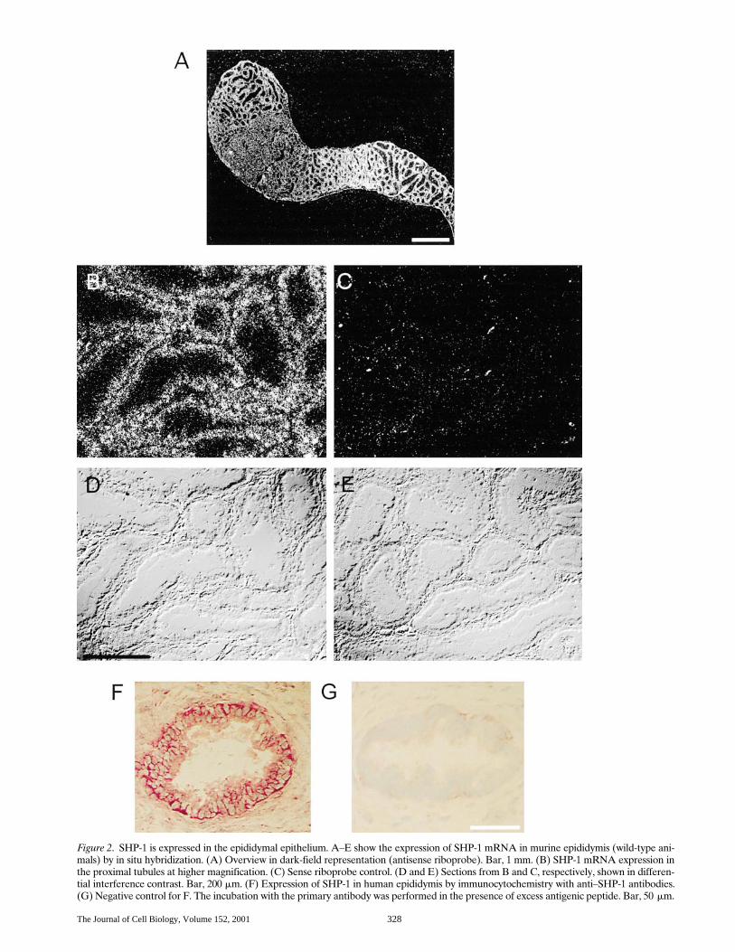
Figure 2. SHP-1 is expressed in the epididymal epithelium. A–E show the expression of SHP-1 mRNA in murine epididymis (wild-type ani-mals) by in situ hybridization. (A) Overview in dark-field representation (antisense riboprobe). Bar, 1 mm. (B) SHP-1 mRNA expression inthe proximal tubules at higher magnification. (C) Sense riboprobe control. (D and E) Sections from B and C, respectively, shown in differen-tial interference contrast. Bar, 200 mm. (F) Expression of SHP-1 in human epididymis by immunocytochemistry with anti–SHP-1 antibodies.(G) Negative control for F. The incubation with the primary antibody was performed in the presence of excess antigenic peptide. Bar, 50 mm.
Keilhack et al. Regulation of Ros by SHP-1 329
Results
Ros Is Hyperphosphorylated in the Epididymis ofmev Mice
To assess whether SHP-1 might be relevant for epididy-mal function, we histologically examined the epididymis ofmale mev/mev mice and compared them to heterozygous con-trols. Macroscopically, the epididymis of mev/mev mice wassmaller, though the overall shape appeared normal. Thissize reduction was proportional to the reduced body weightof the mev/mev mice. Histological sections revealed that thetall columnar epithelial cells, which form the tubules of theproximal segment in heterozygous animals, are replaced byconsiderably flatter epithelial cells in the epithelium of mev/mev mice (Fig. 1). In heterozygous animals, these epithelialcells are clearly structured. The nucleus is located above aclearly defined basal cytoplasmic zone. The cytoplasmiczone above the nucleus is broad and the apical surface isrough. In contrast, the nuclei in epithelial cells of mev/mev
are often located closer to the basal membrane, and the api-cal surfaces appear smooth (Fig. 1). The epithelium in themore distal segments of the epididymis exhibited little dif-ferences in mutant and control animals (not shown). Histo-logically, the epithelium of the proximal segment in mev/mev
mice has similarities to that of Ros2/2 mice (Fig. 1). We alsoanalyzed the epithelium of other organs. Epithelial cells inthe intestine of mev/mev mice were smaller than their coun-terparts in heterozygous animals, but appeared fully differ-entiated. No differences between mev/mev and heterozygousanimals were detectable in epithelia of stomach and pan-creas (not shown). In summary, the epididymal epitheliumof mev/mev mice exhibits signs of aberrant differentiation.This phenotype may be related to a loss of function of SHP-1in the epididymal epithelial cells, since SHP-1 is clearly ex-pressed in these cells (Fig. 2). In situ hybridization with anantisense RNA generated from a 1,184-bp fragment of mu-rine SHP-1 cDNA revealed expression of SHP-1 mRNA inthe epithelial cells surrounding the tubules of the initial epi-didymal segment (Fig. 2, A–E). More distal segments ex-hibit low level signals. Thus, SHP-1 expression mirrors theone of Ros in this part of the epididymis (Sonnenberg-Riethmacher et al., 1996). Strong SHP-1 expression is alsoseen in the corpus with highest levels in the most distal seg-ments. These distal epithelia are devoid of Ros (Sonnen-berg-Riethmacher et al., 1996). SHP-1 expression could alsobe detected by immunoblotting in the epididymis of mice(not shown); immunostaining failed with the available anti-bodies. However, in human epididymis, strong epithelialSHP-1 expression could be visualized by immunohisto-chemistry (Fig. 2, F and G). Thus, SHP-1 is expressed in ep-ithelial cells of the epididymis and exhibits an overlappingexpression domain with Ros.
The overlapping expression of SHP-1 and Ros promptedus to test whether impairment of SHP-1 activity in mev/mev
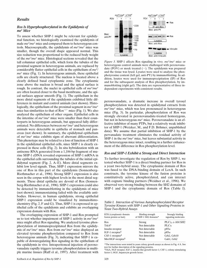
mice might affect Ros signaling. We analyzed tyrosine phos-phorylation of immunoprecipitated Ros from the epididy-mis of mev/mev mice. Ros from mev/mev mice displayed anelevated tyrosine phosphorylation compared to Ros fromheterozygous animals (Fig. 3), indicating that SHP-1 is ca-pable of downregulating Ros signaling in the epithelium ofthe epididymis in vivo. Intraperitoneal injection of peroxo-vanadate rapidly triggers tyrosine phosphorylation in multi-ple murine tissues (Ruff et al., 1997). After treatment with
peroxovanadate, a dramatic increase in overall tyrosylphosphorylation was detected in epididymal extracts frommev/mev mice, which was less pronounced in heterozygousmice (Fig. 3). In particular, phosphorylation of Ros wasstrongly elevated in peroxovanadate-treated homozygous,but not in heterozygous mev mice. Peroxovanadate is an ef-fective inhibitor of many PTPs, but a relatively weak inhibi-tor of SHP-1 (Wetzker, M., and F.D. Böhmer, unpublisheddata). We assume that partial inhibition of SHP-1 by thepervanadate treatment eliminates the residual activity ofSHP-1 in the mev/mev mice, but leaves substantial activity inthe heterozygous mice intact, resulting in a further enhance-ment of the difference in Ros phosphorylation level.
Ros and SHP-1 Exhibit a Strong and Direct Interaction
To further investigate the regulation of Ros by SHP-1, wetested whether SHP-1 is a direct binding partner for Ros ina yeast two-hybrid assay. The cytoplasmic domain of Roswas fused to the DNA-binding domain of LexA. In suchconstructs, the tyrosine kinase of the fusion proteins isconstitutively active, phosphorylated, and can interactwith cognate binding partners (Weidner et al., 1996). Weobserved very strong binding between the SH2 domains ofSHP-1 and the cytoplasmic domain of Ros (Table I).
Figure 3. SHP-1 affects Ros signaling in vivo. mev/mev mice orheterozygous control animals were challenged with peroxovana-date (POV) or mock treated (2). The epididymis was preparedand the tissue was lysed. Lysates were used to analyze the phos-photyrosine content (left gel, anti-PY) by immunoblotting. In ad-dition, lysates were used for immunoprecipitation (IP) of Rosand for the subsequent analysis of Ros phosphorylation, by im-munoblotting (right gel). The data are representative of three in-dependent experiments with consistent results.
Table I. Interaction of Various Autophosphorylated Receptor Tyrosine Kinases with SHP-1 and Other Signaling Proteins in Yeast Two-Hybrid Assays
RTK (cytoplasmic domainfusion protein as bait)
Binding strengthof SHP-1 SH2 domains*
Strongly bindingsignaling molecules
Ros Strong PLCg, c-Abl SH2TrkA§ Not detectable PLCg
Insulin receptor Not detectable p85a
IGF-1 receptor§ Not detectable p85a
CSF-1 receptor§ Not detectable Grb2, Grb10Met/HGF receptor§ Weak Gab1, Grb2
*The interactions were tested in yeast colony growth assays as shown in Fig. 4 A. Seetext for further description of the signaling proteins. §TrkA, NGF receptor; IGF-1, insulin-like growth factor-1; CSF-1, colony-stimulatingfactor-1; HGF, hepatocyte growth factor.
The Journal of Cell Biology, Volume 152, 2001 330
Other tested receptor tyrosine kinases (RTKs) exhibitedlittle or no detectable interaction with SHP-1 SH2 do-mains, but interacted strongly with other partners. Thus,the direct binding of SHP-1 to Ros is remarkably specificcompared to other tested receptor baits. In addition to theSH2 domains of SHP-1, phospholipase Cg, the SH2 do-
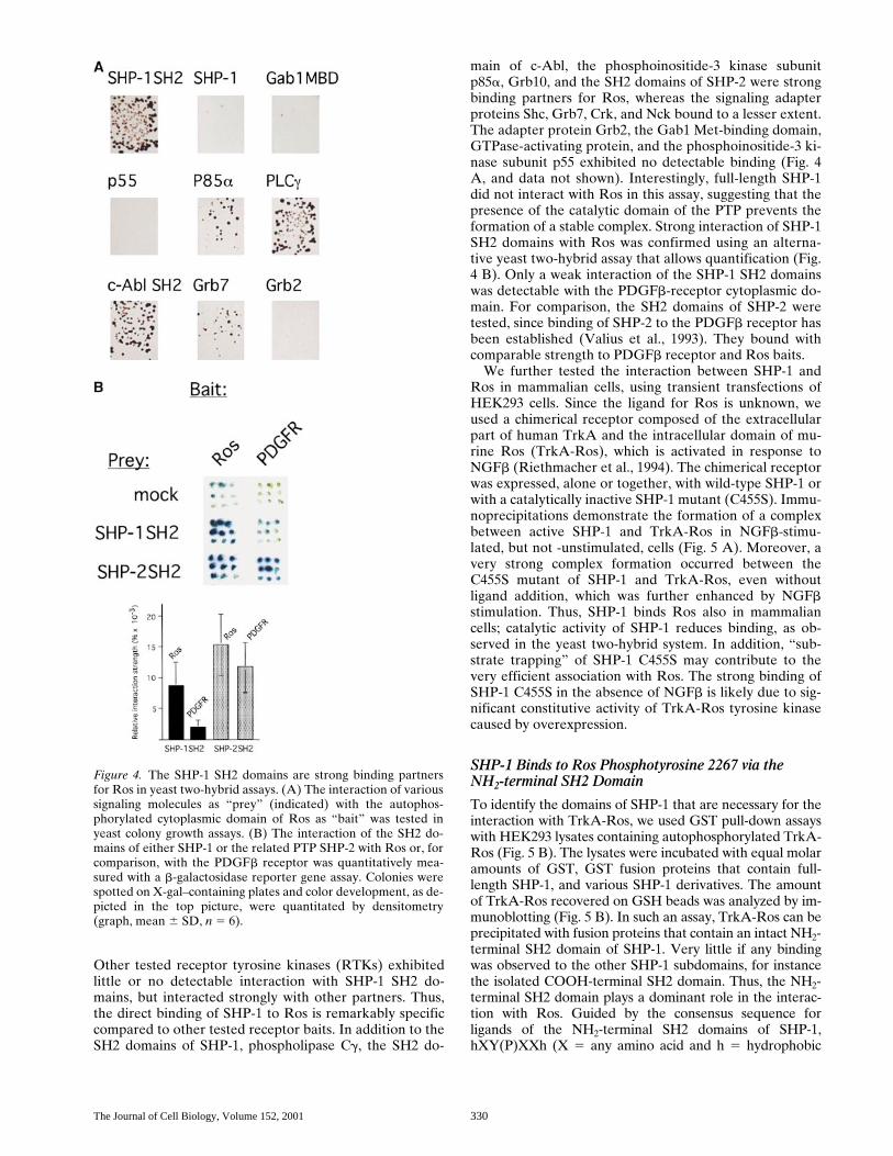
main of c-Abl, the phosphoinositide-3 kinase subunitp85a, Grb10, and the SH2 domains of SHP-2 were strongbinding partners for Ros, whereas the signaling adapterproteins Shc, Grb7, Crk, and Nck bound to a lesser extent.The adapter protein Grb2, the Gab1 Met-binding domain,GTPase-activating protein, and the phosphoinositide-3 ki-nase subunit p55 exhibited no detectable binding (Fig. 4A, and data not shown). Interestingly, full-length SHP-1did not interact with Ros in this assay, suggesting that thepresence of the catalytic domain of the PTP prevents theformation of a stable complex. Strong interaction of SHP-1SH2 domains with Ros was confirmed using an alterna-tive yeast two-hybrid assay that allows quantification (Fig.4 B). Only a weak interaction of the SHP-1 SH2 domainswas detectable with the PDGFb-receptor cytoplasmic do-main. For comparison, the SH2 domains of SHP-2 weretested, since binding of SHP-2 to the PDGFb receptor hasbeen established (Valius et al., 1993). They bound withcomparable strength to PDGFb receptor and Ros baits.
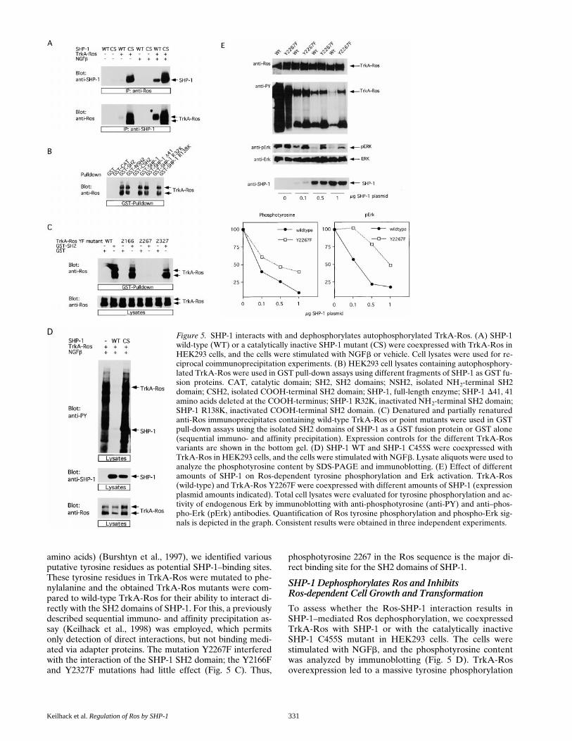
We further tested the interaction between SHP-1 andRos in mammalian cells, using transient transfections ofHEK293 cells. Since the ligand for Ros is unknown, weused a chimerical receptor composed of the extracellularpart of human TrkA and the intracellular domain of mu-rine Ros (TrkA-Ros), which is activated in response toNGFb (Riethmacher et al., 1994). The chimerical receptorwas expressed, alone or together, with wild-type SHP-1 orwith a catalytically inactive SHP-1 mutant (C455S). Immu-noprecipitations demonstrate the formation of a complexbetween active SHP-1 and TrkA-Ros in NGFb-stimu-lated, but not -unstimulated, cells (Fig. 5 A). Moreover, avery strong complex formation occurred between theC455S mutant of SHP-1 and TrkA-Ros, even withoutligand addition, which was further enhanced by NGFbstimulation. Thus, SHP-1 binds Ros also in mammaliancells; catalytic activity of SHP-1 reduces binding, as ob-served in the yeast two-hybrid system. In addition, “sub-strate trapping” of SHP-1 C455S may contribute to thevery efficient association with Ros. The strong binding ofSHP-1 C455S in the absence of NGFb is likely due to sig-nificant constitutive activity of TrkA-Ros tyrosine kinasecaused by overexpression.
SHP-1 Binds to Ros Phosphotyrosine 2267 via theNH2-terminal SH2 Domain
To identify the domains of SHP-1 that are necessary for theinteraction with TrkA-Ros, we used GST pull-down assayswith HEK293 lysates containing autophosphorylated TrkA-Ros (Fig. 5 B). The lysates were incubated with equal molaramounts of GST, GST fusion proteins that contain full-length SHP-1, and various SHP-1 derivatives. The amountof TrkA-Ros recovered on GSH beads was analyzed by im-munoblotting (Fig. 5 B). In such an assay, TrkA-Ros can beprecipitated with fusion proteins that contain an intact NH2-terminal SH2 domain of SHP-1. Very little if any bindingwas observed to the other SHP-1 subdomains, for instancethe isolated COOH-terminal SH2 domain. Thus, the NH2-terminal SH2 domain plays a dominant role in the interac-tion with Ros. Guided by the consensus sequence forligands of the NH2-terminal SH2 domains of SHP-1,hXY(P)XXh (X 5 any amino acid and h 5 hydrophobic
Figure 4. The SHP-1 SH2 domains are strong binding partnersfor Ros in yeast two-hybrid assays. (A) The interaction of varioussignaling molecules as “prey” (indicated) with the autophos-phorylated cytoplasmic domain of Ros as “bait” was tested inyeast colony growth assays. (B) The interaction of the SH2 do-mains of either SHP-1 or the related PTP SHP-2 with Ros or, forcomparison, with the PDGFb receptor was quantitatively mea-sured with a b-galactosidase reporter gene assay. Colonies werespotted on X-gal–containing plates and color development, as de-picted in the top picture, were quantitated by densitometry(graph, mean 6 SD, n 5 6).
Keilhack et al. Regulation of Ros by SHP-1 331
amino acids) (Burshtyn et al., 1997), we identified variousputative tyrosine residues as potential SHP-1–binding sites.These tyrosine residues in TrkA-Ros were mutated to phe-nylalanine and the obtained TrkA-Ros mutants were com-pared to wild-type TrkA-Ros for their ability to interact di-rectly with the SH2 domains of SHP-1. For this, a previouslydescribed sequential immuno- and affinity precipitation as-say (Keilhack et al., 1998) was employed, which permitsonly detection of direct interactions, but not binding medi-ated via adapter proteins. The mutation Y2267F interferedwith the interaction of the SHP-1 SH2 domain; the Y2166Fand Y2327F mutations had little effect (Fig. 5 C). Thus,
phosphotyrosine 2267 in the Ros sequence is the major di-rect binding site for the SH2 domains of SHP-1.
SHP-1 Dephosphorylates Ros and InhibitsRos-dependent Cell Growth and Transformation
To assess whether the Ros-SHP-1 interaction results inSHP-1–mediated Ros dephosphorylation, we coexpressedTrkA-Ros with SHP-1 or with the catalytically inactiveSHP-1 C455S mutant in HEK293 cells. The cells werestimulated with NGFb, and the phosphotyrosine contentwas analyzed by immunoblotting (Fig. 5 D). TrkA-Rosoverexpression led to a massive tyrosine phosphorylation
Figure 5. SHP-1 interacts with and dephosphorylates autophosphorylated TrkA-Ros. (A) SHP-1wild-type (WT) or a catalytically inactive SHP-1 mutant (CS) were coexpressed with TrkA-Ros inHEK293 cells, and the cells were stimulated with NGFb or vehicle. Cell lysates were used for re-ciprocal coimmunoprecipitation experiments. (B) HEK293 cell lysates containing autophosphory-lated TrkA-Ros were used in GST pull-down assays using different fragments of SHP-1 as GST fu-sion proteins. CAT, catalytic domain; SH2, SH2 domains; NSH2, isolated NH2-terminal SH2domain; CSH2, isolated COOH-terminal SH2 domain; SHP-1, full-length enzyme; SHP-1 D41, 41amino acids deleted at the COOH-terminus; SHP-1 R32K, inactivated NH2-terminal SH2 domain;SHP-1 R138K, inactivated COOH-terminal SH2 domain. (C) Denatured and partially renaturedanti-Ros immunoprecipitates containing wild-type TrkA-Ros or point mutants were used in GSTpull-down assays using the isolated SH2 domains of SHP-1 as a GST fusion protein or GST alone(sequential immuno- and affinity precipitation). Expression controls for the different TrkA-Rosvariants are shown in the bottom gel. (D) SHP-1 WT and SHP-1 C455S were coexpressed withTrkA-Ros in HEK293 cells, and the cells were stimulated with NGFb. Lysate aliquots were used toanalyze the phosphotyrosine content by SDS-PAGE and immunoblotting. (E) Effect of differentamounts of SHP-1 on Ros-dependent tyrosine phosphorylation and Erk activation. TrkA-Ros(wild-type) and TrkA-Ros Y2267F were coexpressed with different amounts of SHP-1 (expressionplasmid amounts indicated). Total cell lysates were evaluated for tyrosine phosphorylation and ac-tivity of endogenous Erk by immunoblotting with anti-phosphotyrosine (anti-PY) and anti–phos-pho-Erk (pErk) antibodies. Quantification of Ros tyrosine phosphorylation and phospho-Erk sig-nals is depicted in the graph. Consistent results were obtained in three independent experiments.
The Journal of Cell Biology, Volume 152, 2001 332
of TrkA-Ros and other proteins. The C455S mutant ofSHP-1 had virtually no effect, whereas SHP-1 expressionresulted in a complete disappearance of the phosphory-lated proteins. We reproducibly observed some reductionof the TrkA-Ros protein level upon SHP-1 transfection;this decrease in expression may contribute, but is by farnot sufficient to explain the reduced phosphorylation levelof TrkA-Ros. Thus, a very pronounced suppression ofRos-dependent tyrosine phosphorylation is mediated bythe catalytic activity of SHP-1. This effect could be ac-counted for by a direct dephosphorylation of TrkA-Rosand, subsequently, a decrease of its kinase activity andsubstrate binding and/or by the dephosphorylation ofTrkA-Ros substrates.
We compared the relative susceptibility of TrkA-Rossignaling activity with that of the TrkA-Ros Y2267F mu-tant. The Y2267F mutant exhibited a reduced autophos-phorylation and phosphorylation of cellular substrates,presumably resulting from loss of a major autophosphory-lation site (Fig. 5 E). However, the level of activation ofErk was similar in cells expressing either TrkA-Ros vari-ant. Consistent with the loss of direct SHP-1 binding,phosphorylation activity of the TrkA-Ros Y2267F mutantand TrkA-Ros Y2267F-dependent Erk phosphorylation
were less effectively downregulated by SHP-1 than in thecase of wild-type TrkA-Ros. This finding supports thefunctional importance of direct SHP-1 binding to Ros fornegative regulation of signaling activity. Still, signaling ofTrkA-Ros Y2267F is negatively affected by SHP-1, in par-ticular, at higher SHP-1 levels (Fig. 5 E), suggesting theadditional operation of an indirect Ros-SHP-1 interactionin intact cells.
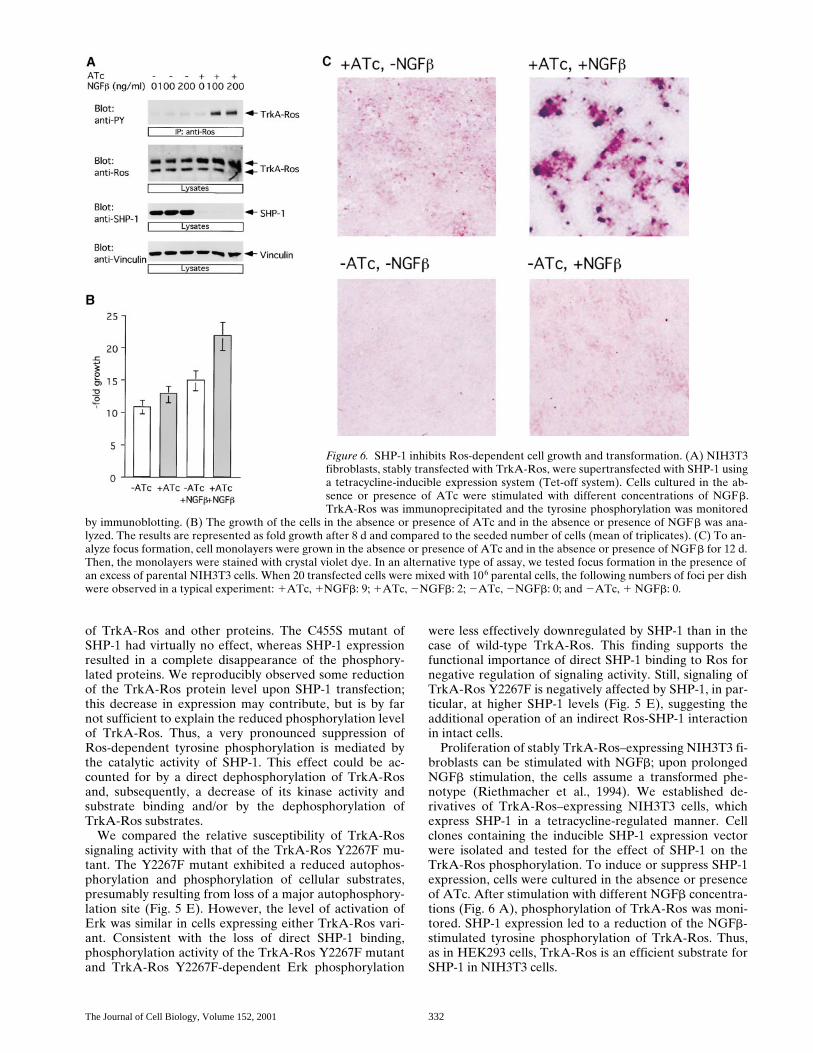
Proliferation of stably TrkA-Ros–expressing NIH3T3 fi-broblasts can be stimulated with NGFb; upon prolongedNGFb stimulation, the cells assume a transformed phe-notype (Riethmacher et al., 1994). We established de-rivatives of TrkA-Ros–expressing NIH3T3 cells, whichexpress SHP-1 in a tetracycline-regulated manner. Cellclones containing the inducible SHP-1 expression vectorwere isolated and tested for the effect of SHP-1 on theTrkA-Ros phosphorylation. To induce or suppress SHP-1expression, cells were cultured in the absence or presenceof ATc. After stimulation with different NGFb concentra-tions (Fig. 6 A), phosphorylation of TrkA-Ros was moni-tored. SHP-1 expression led to a reduction of the NGFb-stimulated tyrosine phosphorylation of TrkA-Ros. Thus,as in HEK293 cells, TrkA-Ros is an efficient substrate forSHP-1 in NIH3T3 cells.
Figure 6. SHP-1 inhibits Ros-dependent cell growth and transformation. (A) NIH3T3fibroblasts, stably transfected with TrkA-Ros, were supertransfected with SHP-1 usinga tetracycline-inducible expression system (Tet-off system). Cells cultured in the ab-sence or presence of ATc were stimulated with different concentrations of NGFb.TrkA-Ros was immunoprecipitated and the tyrosine phosphorylation was monitored
by immunoblotting. (B) The growth of the cells in the absence or presence of ATc and in the absence or presence of NGFb was ana-lyzed. The results are represented as fold growth after 8 d and compared to the seeded number of cells (mean of triplicates). (C) To an-alyze focus formation, cell monolayers were grown in the absence or presence of ATc and in the absence or presence of NGFb for 12 d.Then, the monolayers were stained with crystal violet dye. In an alternative type of assay, we tested focus formation in the presence ofan excess of parental NIH3T3 cells. When 20 transfected cells were mixed with 106 parental cells, the following numbers of foci per dishwere observed in a typical experiment: 1ATc, 1NGFb: 9; 1ATc, 2NGFb: 2; 2ATc, 2NGFb: 0; and 2ATc, 1 NGFb: 0.
Keilhack et al. Regulation of Ros by SHP-1 333
We further assessed whether the SHP-1–mediated de-crease of TrkA-Ros phosphorylation changes the growthbehavior of the NIH3T3 fibroblasts. SHP-1 expression wasinduced (2ATc) or suppressed (1ATc) in cells grown inthe absence or presence of NGFb in low serum. NGFbtreatment resulted in a stimulation of cell proliferation(Fig. 6 B). Expression of SHP-1 attenuated growth of thecells in the absence of NGFb, and strongly compromisedthe growth stimulation by NGFb. Thus, SHP-1 negativelyregulates the TrkA-Ros–mediated growth response. Whencultured with NGFb and in the presence of ATc (suppres-sion of SHP-1 expression), the cells spontaneously formedmultiple foci (Fig. 6 C). Induction of SHP-1 expression inNGFb-treated cells completely suppressed focus forma-tion. No foci were formed in the absence of NGFb, regard-less of whether the cells did or did not express SHP-1.Likewise, the ability of a small number of transfected cellsto form foci in a monolayer of untransfected NIH3T3 fi-broblasts (Riethmacher et al., 1994) was suppressed bySHP-1 expression, as explained in the legend to Fig. 6. Insummary, SHP-1 can suppress Ros-dependent phosphory-lation, cell proliferation, and transformation.
DiscussionThe signal transduction of receptor tyrosine kinases ismodulated by PTPs, however, the identity of relevant PTPmolecules is often not known. For the SH2 domain PTPSHP-1, a negative regulator of signal transduction, varioustargets in hematopoietic cells have been identified, includ-ing the RTK Kit/SCF receptor (Lorenz et al., 1996; Paul-son et al., 1996) and the CSF-1 receptor (Chen et al.,1996). Here, we present evidence for an epithelial target ofSHP-1: the “orphan” RTK Ros. We show that SHP-1 andRos are coexpressed in the proximal segment of the epi-didymal caput. In agreement with a functional interactionof Ros and SHP-1 in these cells, we observed hyperphos-phorylation of Ros in mev mice, whose SHP-1 activity isstrongly compromised. Experiments in different cellularmodels further support that SHP-1 is an effective negativeregulator of Ros phosphorylation and signaling.
The effects of SHP-1 appear to be mediated by a re-markably efficient and direct binding to the phosphory-lated Ros receptor. Direct binding is mediated by theNH2-terminal SH2 domain of SHP-1 and phosphotyrosine2267 in the COOH-terminal part of Ros. The Ros se-quence LNY2267MVL matches the known consensus forbinding the SHP-1 NH2-terminal SH2 domain and maymediate a particularly high affinity interaction. Alterna-tively, a very high stoichiometry of phosphorylation of thissite may explain the high efficiency of interaction. Phos-phorylated Ros is not only a strong binding partner forSHP-1, but also a very good substrate. Dephosphorylationof Ros appears to include the tyrosine that serves as abinding site of the SHP-1 SH2 domain, which abolishesbinding. This would explain why no interaction is observedbetween Ros and a catalytically active SHP-1 protein inyeast two-hybrid interaction experiments, though Ros andthe SH2 domain of SHP-1 interact strongly. Similarly, theamounts of detectable SHP-1–TrkA-Ros complex arehighly elevated when a catalytically inactive variant ofSHP-1 is used in coimmunoprecipitation and pull-downexperiments. Destruction of a SHP-1–binding site by SHP-
1–mediated dephosphorylation has been previously ob-served for the interleukin 3 receptor (Bone et al., 1997).Coexpression of SHP-1 with the TrkA-Ros chimera leadsnot only to dephosphorylation of TrkA-Ros, but also abol-ishes tyrosine phosphorylation of other substrates. Thiscould be correlated to dephosphorylation of Ros, whichwill lead to a decrease of the catalytic activity of its kinaseand a decrease of the capacity to recruit substrate proteins.Alternatively, or in addition, Ros substrates may also be-come directly dephosphorylated by SHP-1.
The hitherto best understood function of Ros is its rolein differentiation and regionalization of the epididymalepithelium of mice; the correct development of this epithe-lium is essential for male fertility. Deregulation of epididy-mal differentiation in Ros2/2 mice leads to sterility (Son-nenberg-Riethmacher et al., 1996). Sterility is due tosubtle defects in sperm maturation, which have not yetbeen characterized at the molecular level (Cooper, 1998;Yeung et al., 1998). It is possible that the phenotypic ab-normalities that we have observed in the epididymis ofmale mev/mev mice contribute to the sterility of these micein a similar manner. We observed histological aberrationsonly in the most proximal segment of the epididymis, i.e.,in the region where SHP-1 and Ros are coexpressed. Ourresults for the SHP-1/Ros interaction in vitro and the ob-servation that Ros is hyperphosphorylated in mev/mev
mice in vivo strongly suggest an elevated level of Ros sig-naling activity in these animals. It is possible that the histo-logical changes in the caput epididymis of mev/mev miceare the consequence of deregulated Ros activity. It is obvi-ous that the molecular consequences of Ros inactivationand elevated Ros signaling in epididymal cells will be verydifferent. Interestingly, the phenotypic abnormalities seenin the epididymis of Ros2/2 mice and mev/mev mice havesimilarities on the morphological level. A more detailedcharacterization of the functional defects in epididymaldifferentiation in mev/mev, as well as Ros2/2, mice will berequired to better define similarities and differences of thetwo phenotypes.
This work was supported by grants from Deutsche Forschungsgemein-schaft (Bo 1043/3-1 to F.D. Böhmer) and from the Max-Planck-Society (toF.D. Böhmer).
Submitted: 28 July 2000Revised: 14 November 2000Accepted: 28 November 2000
References
Bai, R.Y., T. Jahn, S. Schrem, G. Munzert, K.M. Weidner, J.Y. Wang, and J.Duyster. 1998. The SH2-containing adapter protein GRB10 interacts withBCR-ABL. Oncogene. 17:941–948.
Banville, D., R. Stocco, and S.H. Shen. 1995. Human protein tyrosine phos-phatase 1C (ptpn6) gene structure—alternate promoter usage and exonskipping generate multiple transcripts. Genomics. 27:165–173.
Birchmeier, C., D. Birnbaum, G. Waitches, O. Fasano, and M. Wigler. 1986.Characterization of an activated human ros gene. Mol. Cell. Biol. 6:3109–3116.
Bone, H., U. Dechert, F. Jirik, J.W. Schrader, and M.J. Welham. 1997. SHP1and SHP2 protein-tyrosine phosphatases associate with betac after interleu-kin-3-induced receptor tyrosine phosphorylation. Identification of potentialbinding sites and substrates. J. Biol. Chem. 272:14470–14476.
Burshtyn, D.N., W. Yang, T. Yi, and E.O. Long. 1997. A novel phosphotyrosinemotif with a critical amino acid at position 22 for the SH2 domain-mediatedactivation of the tyrosine phosphatase SHP-1. J. Biol. Chem. 272:13066–13072.
Chen, H.E., S. Chang, T. Trub, and B.G. Neel. 1996. Regulation of colony-stim-ulating factor-1 receptor signaling by the SH2 domain-containing tyrosinephosphatase SHPTP1. Mol. Cell. Biol. 16:3685–3697.
The Journal of Cell Biology, Volume 152, 2001 334
Claesson-Welsh, L., A. Eriksson, A. Moren, L. Severinsson, B. Ek, A. Östman,C. Betsholtz, and C.H. Heldin. 1988. cDNA cloning and expression of a hu-man platelet-derived growth factor (PDGF) receptor specific for B-chain-containing PDGF molecules. Mol. Cell. Biol. 8:3476–3486.
Cooper, T.G. 1998. Interactions between epididymal secretions and spermato-zoa. J. Reprod. Fertil. Suppl. 53:119–136.
Feng, G.S., and T. Pawson. 1994. Phosphotyrosine phosphatases with SH2 do-mains—regulators of signal transduction. Trends Genet. 10:54–58.
Frank, C., H. Keilhack, F. Opitz, O. Zschörnig, and F.D. Böhmer. 1999. Bind-ing of phosphatidic acid to the protein-tyrosine phosphatase SHP-1 as a ba-sis for activity modulation. Biochemistry. 38:11993–12002.
Frearson, J.A., and D.R. Alexander. 1997. The role of phosphotyrosine phos-phatases in hematopoietic-cell signal-transduction. Bioessays. 19:417–427.
Green, M.C., and L.D. Shultz. 1975. Motheaten, an immunodeficient mutant ofthe mouse. I. Genetics and pathology. J. Hered. 66:250–258.
Keilhack, H., T. Tenev, E. Nyakatura, J. Godovac-Zimmermann, L. Nielsen, K.Seedorf, and F.D. Böhmer. 1998. Phosphotyrosine 1173 mediates binding ofthe protein-tyrosine phosphatase SHP-1 to the epidermal growth factor re-ceptor and attenuation of receptor signaling. J. Biol. Chem. 273:24839–24846.
Klingmüller, U., U. Lorenz, L.C. Cantley, B.G. Neel, and H.F. Lodish. 1995.Specific recruitment of SH-PTP1 to the erythropoietin receptor causes inac-tivation of JAK2 and termination of proliferative signals. Cell. 80:729–738.
Lammers, R., B. Bossenmaier, D.E. Cool, N.K. Tonks, J. Schlessinger, E.H.Fischer, and A. Ullrich. 1993. Differential activities of protein tyrosine phos-phatases in intact cells. J. Biol. Chem. 268:22456–22462.
Lorenz, U., A.D. Bergemann, H.N. Steinberg, J.G. Flanagan, X. Li, S.J. Galli,and B.G. Neel. 1996. Genetic analysis reveals cell type-specific regulation ofreceptor tyrosine kinase c-Kit by the protein tyrosine phosphatase SHP1. J.Exp. Med. 184:1111–1126.
Matthews, R.J., D.B. Bowne, E. Flores, and M.L. Thomas. 1992. Characteriza-tion of hematopoietic intracellular protein tyrosine phosphatases: descrip-tion of a phosphatase containing an SH2 domain and another enriched inproline-, glutamic acid-, serine-, and threonine-rich sequences. Mol. Cell.Biol. 12:2396–2405.
Neckameyer, W.S., and L.H. Wang. 1985. Nucleotide sequence of avian sar-coma virus UR2 and comparison of its transforming gene with other mem-bers of the tyrosine protein kinase oncogene family. J. Virol. 53:879–884.
Neel, B.G., and N.K. Tonks. 1997. Protein tyrosine phosphatases in signaltransduction. Curr. Opin. Cell Biol. 9:193–204.
Paulson, R.F., S. Vesely, K.A. Siminovitch, and A. Bernstein. 1996. Signallingby the W/Kit receptor tyrosine kinase is negatively regulated in vivo by theprotein tyrosine phosphatase SHP1. Nat. Genet. 13:309–315.
Riethmacher, D., O. Langholz, S. Gödecke, M. Sachs, and C. Birchmeier. 1994.Biochemical and functional characterization of the murine ros protoonco-gene. Oncogene. 9:3617–3626.
Ruff, S.J., K. Chen, and S. Cohen. 1997. Peroxovanadate induces tyrosine phos-phorylation of multiple signaling proteins in mouse liver and kidney. J. Biol.Chem. 272:1263–1267.
Sachs, M., K.M. Weidner, V. Brinkmann, I. Walther, A. Obermeier, A. Ullrich,and W. Birchmeier. 1996. Motogenic and morphogenic activity of epithelialreceptor tyrosine kinases. J. Cell. Biol. 133:1095–1107.
Shen, S.H., L. Bastien, B.I. Posner, and P. Chretien. 1991. A protein-tyrosinephosphatase with sequence similarity to the SH2 domain of the protein-tyro-sine kinases. Nature. 352:736–739.
Shultz, L.D., D.R. Coman, C.L. Bailey, W.G. Beamer, and C.L. Sidman. 1984.“Viable motheaten,” a new allele at the motheaten locus. I. Pathology. Am.J. Pathol. 116:179–192.
Shultz, L.D., P.A. Schweitzer, T.V. Rajan, T. Yi, J.N. Ihle, R.J. Matthews, M.L.Thomas, and D.R. Beier. 1993. Mutations at the murine motheaten locus arewithin the hematopoietic cell protein-tyrosine phosphatase (Hcph) gene.Cell. 73:1445–1454.
Shultz, L.D., T.V. Rajan, and D.L. Greiner. 1997. Severe defects in immunityand hematopoiesis caused by SHP-1 protein-tyrosine-phosphatase defi-ciency. Trends Biotechnol. 15:302–307.
Sonnenberg, E., A. Godecke, B. Walter, F. Bladt, and C. Birchmeier. 1991.Transient and locally restricted expression of the ros1 protooncogene duringmouse development. EMBO (Eur. Mol. Biol. Organ.) J. 10:3693–3702.
Sonnenberg-Riethmacher, E., B. Walter, D. Riethmacher, S. Gödecke, and C.Birchmeier. 1996. The c-ros tyrosine kinase receptor controls regionalizationand differentiation of epithelial cells in the epididymis. Genes Dev. 10:1184–1193.
Tamura, T., A. Mancini, H. Joos, A. Koch, C. Hakim, J. Dumanski, K.M.Weidner, and H. Niemann. 1999. FMIP, a novel Fms-interacting protein, af-fects granulocyte/macrophage differentiation. Oncogene. 18:6488–6495.
Tenev, T., H. Keilhack, S. Tomic, B. Stoyanov, M. Stein-Erlach, R. Lammers,A.V. Krivtsov, A. Ullrich, and F.D. Böhmer. 1997. Both SH2 domains areinvolved in interaction of SHP-1 with the epidermal growth-factor receptorbut cannot confer receptor-directed activity to SHP-1/SHP-2 chimera. J.Biol. Chem. 272:5966–5973.
Tenev, T., S.A. Böhmer, R. Kaufmann, S. Frese, T. Bittorf, T. Beckers, andF.D. Böhmer. 2000. Perinuclear localization of the protein-tyrosine phos-phatase SHP-1 and inhibition of epidermal growth factor-stimulatedSTAT1/3 activation in A431 cells. Eur. J. Cell Biol. 79:1–11.
Umeda, S., W.G. Beamer, K. Takagi, M. Naito, S. Hayashi, H. Yonemitsu, T.L.Yi, and L.D. Shultz. 1999. Deficiency of SHP-1 protein-tyrosine phosphataseactivity results in heightened osteoclast function and decreased bone den-sity. Am. J. Pathol. 155:223–233.
Valius, M., C. Bazenet, and A. Kazlauskas. 1993. Tyrosine-1021 and tyrosine-1009 are phosphorylation sites in the carboxy terminus of the platelet-derived growth factor receptor-beta subunit and are required for binding ofphospholipase C gamma and a 64-kilodalton protein, respectively. Mol. Cell.Biol. 13:133–143.
Vayssiere, B., G. Zalcman, Y. Mahe, G. Mirey, T. Ligensa, K.M. Weidner, P.Chardin, and J. Camonis. 2000. Interaction of the Grb7 adapter protein withRnd1, a new member of the Rho family. FEBS Lett. 467:91–96.
Weidner, K.M., S. Di Cesare, M. Sachs, V. Brinkmann, J. Behrens, and W.Birchmeier. 1996. Interaction between Gab1 and the c-Met receptor ty-rosine kinase is responsible for epithelial morphogenesis. Nature. 384:173–176.
Yeung, C.H., E. Sonnenberg-Riethmacher, and T.G. Cooper. 1998. Receptortyrosine kinase c-ros knockout mice as a model for the study of epididymalregulation of sperm function. J. Reprod. Fertil. Suppl. 53:137–147.


![Department of Health and Human Services - GPO · 12.02.2015 · Department of Health and Human Services ... 2015 Jkt 235001 PO 00000 Frm 00001 Fmt 4717 Sfmt 4717 E: ... [CMS–4159–F2]](https://img.pdfslide.us/doc/110x75/5b63643d7f8b9af84b8bdd73/department-of-health-and-human-services-12022015-department-of-health.jpg)