Embed Size (px)
Citation preview

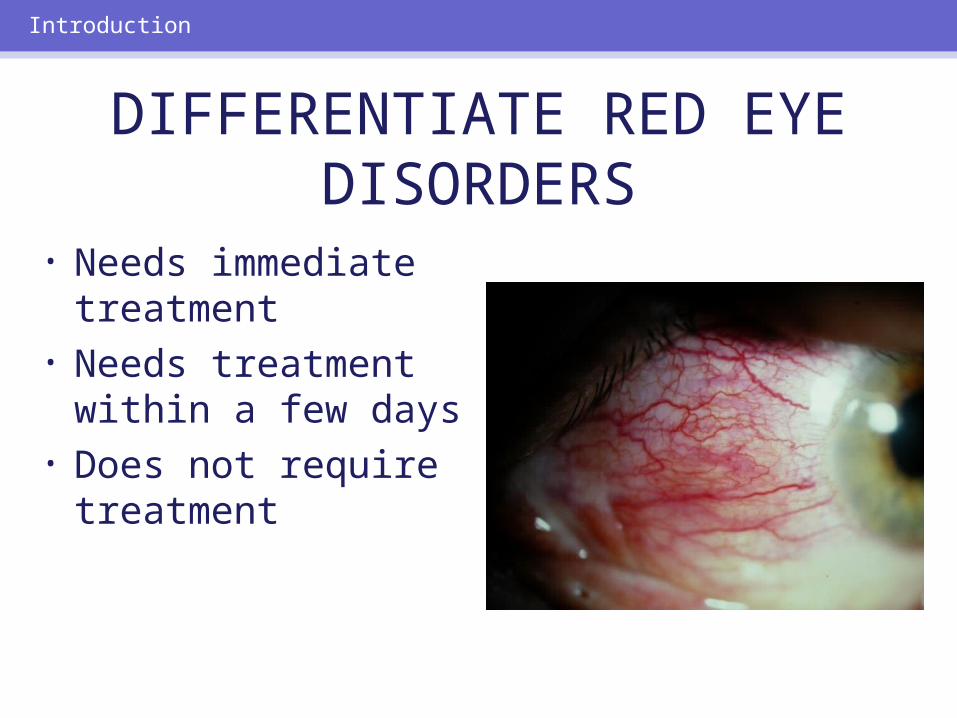
• Needs immediate treatment
• Needs treatment within a few days
• Does not require treatment
Introduction
DIFFERENTIATE RED EYE DISORDERS

SUBJECTIVE EYE COMPLAINTS
• Decreased vision • Pain• Redness
Characterize the complaint through history and exam.
Introduction

TYPES OF RED EYE DISORDERS
• Mechanical trauma• Chemical trauma• Inflammation/infection
Introduction

ETIOLOGIES OF RED EYE
1. Chemical injury2. Angle-closure glaucoma3. Ocular foreign body4. Corneal abrasion5. Uveitis6. Conjunctivitis7. Ocular surface disease8. Subconjunctival hemorrhage
Introduction

RED EYE: POSSIBLE CAUSES
• Trauma• Chemicals• Infection• Allergy• Systemic conditions
Evaluation

RED EYE: CAUSE AND EFFECT
Symptom CauseItching AllergyBurning Lid disorders, dry eyeForeign body sensation
Foreign body, corneal abrasion
Localized lid tenderness
Hordeolum, chalazion
Evaluation

RED EYE: CAUSE AND EFFECT (Continued)
Symptom CauseDeep, intense pain Corneal abrasions,
scleritis, iritis, acute glaucoma, sinusitis, etc.
Photophobia Corneal abrasions, iritis, acute glaucoma
Halo vision Corneal edema (acute glaucoma, uveitis)
Evaluation

Evaluation
Equipment needed to evaluate red eye

Refer red eye with vision loss to ophthalmologist for evaluation
Evaluation
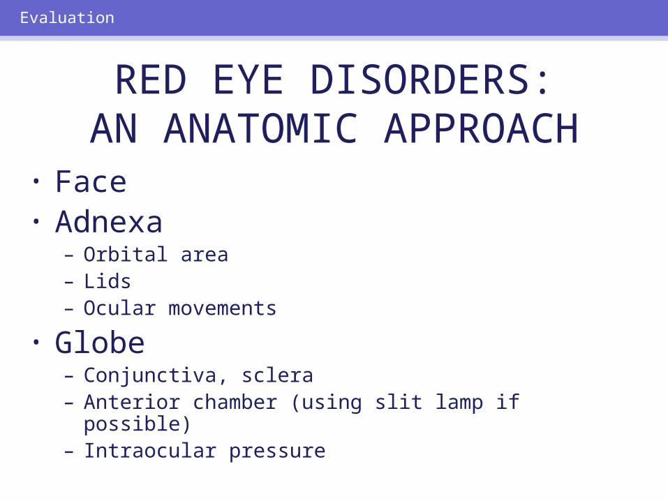
RED EYE DISORDERS:AN ANATOMIC APPROACH
• Face• Adnexa
– Orbital area – Lids– Ocular movements
• Globe– Conjunctiva, sclera– Anterior chamber (using slit lamp if possible)– Intraocular pressure
Evaluation

Disorders of the Ocular Adnexa

Disorders of the Ocular Adnexa
Hordeolum

Disorders of the Ocular Adnexa

Disorders of the Ocular Adnexa
Chalazion

HORDEOLUM/CHALAZION:TREATMENT
• Goal– To promote drainage
• Treatment– Acute/subacute: Warm-hot compresses, tid– Chronic: Refer to ophthalmologist
Disorders of the Ocular Adnexa

BLEPHARITIS
• Inflammation of lid margin• Associated with dry eyes• Seborrhea causes dried skin and wax
on base of lashes• May have Staphylococcal infection• Symptoms: lid burning, lash
mattering
Disorders of the Ocular Adnexa

Disorders of the Ocular Adnexa
Collarettes on eyelashes of patient with blepharitis
BLEPHARITIS: TREATMENT
• Lid and face hygiene– Warm compresses to loosen deposits on lid margin– Gentle scrubbing with nonirritating shampoo or scrub
pads
• Artificial tears to alleviate dry eye• Antibiotic or antibiotic-corticosteroid
ointment• Oral doxycycline 100 mg daily for
refractory cases
Disorders of the Ocular Adnexa
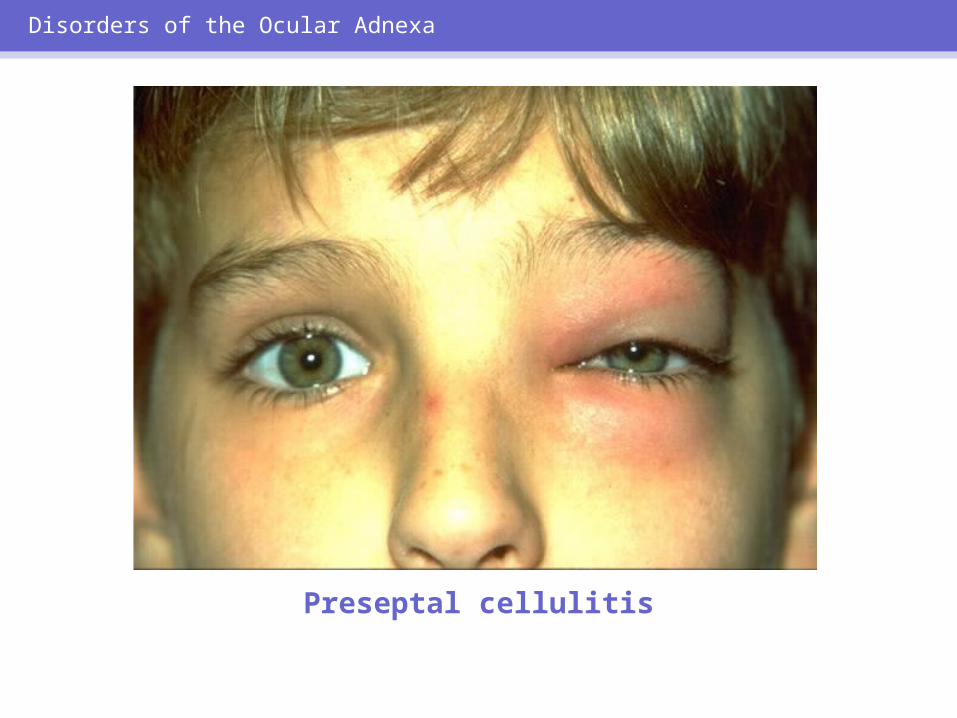
Disorders of the Ocular Adnexa
Preseptal cellulitis

Disorders of the Ocular Adnexa
Orbital cellulitis

• External signs: redness, swelling
• Motility impaired, painful
• ± Proptosis• Often fever and
leukocytosis• ± Optic nerve:
decreased vision, afferent pupillary defect, disc edema
Disorders of the Ocular Adnexa
ORBITAL CELLULITIS: SIGNS AND SYMPTOMS

ORBITAL CELLULITIS: MANAGEMENT
• Hospitalization• Ophthalmology consult• Eye consult• Blood culture• Orbital CT scan• ENT consult if pre-existing sinus
disease
Disorders of the Ocular Adnexa

ORBITAL CELLULITIS: TREATMENT
• IV antibiotics stat: Staphylococcus, Streptococcus, H. influenzae
• Surgical debridement if fungus, no improvement, or subperiosteal abscess
• Complications: cavernous sinus thrombosis, meningitis
Disorders of the Ocular Adnexa

Lacrimal System Disorders
Lacrimal system

Lacrimal System Disorders
Dacryocystitis

NASOLACRIMAL DUCTOBSTRUCTION: CONGENITAL
• Massage tear sac daily• Probing, irrigation, if chronic• Systemic antibiotics if infected
Lacrimal System Disorders

NASOLACRIMAL DUCTOBSTRUCTION: ACQUIRED
• Trauma a common cause• Systemic antibiotics if infected• Surgical procedure after one episode
of dacryocystitis (dacryocystorhinostomy) prn
Lacrimal System Disorders

Ocular Surface Disorders

Ocular Surface Disorders
Dilated conjunctival blood vessels

ADULT CONJUNCTIVITIS:MAJOR CAUSES
• Bacterial• Viral• Allergic
Ocular Surface Disorders

CONJUNCTIVITIS: DISCHARGE
Discharge CausePurulent BacterialClear Viral*Watery, with stringy; white mucus
Allergic**
Ocular Surface Disorders
* Preauricular lymphadenopathy signals viral infection** Itching often accompanies
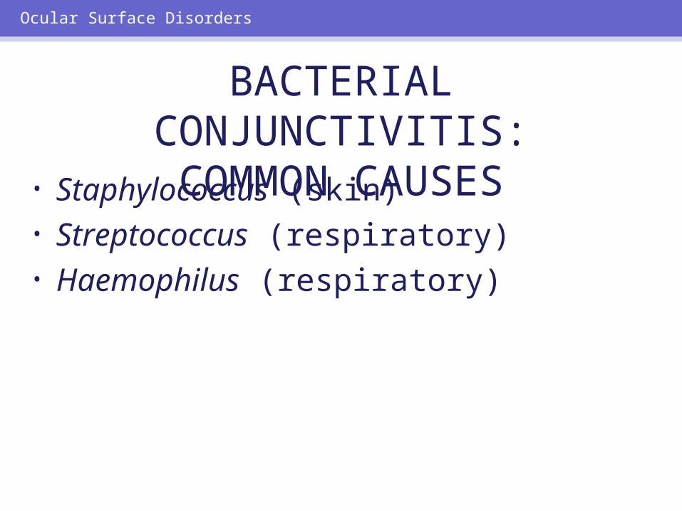
BACTERIAL CONJUNCTIVITIS:COMMON CAUSES
• Staphylococcus (skin)• Streptococcus (respiratory)• Haemophilus (respiratory)
Ocular Surface Disorders

BACTERIAL CONJUNCTIVITISTREATMENT
• Topical antibiotic: qid x 7 days (aminoglycoside, erythromycin, fluoroquinolone, sulfacetamide, or trimethoprim-polymyxin)
• Warm compresses• Refer if not markedly improved in 3
days
Ocular Surface Disorders

Ocular Surface Disorders
Copious purulent discharge: Suspect Neisseria gonorrhoeae.

Ocular Surface Disorders
Viral conjunctivitis
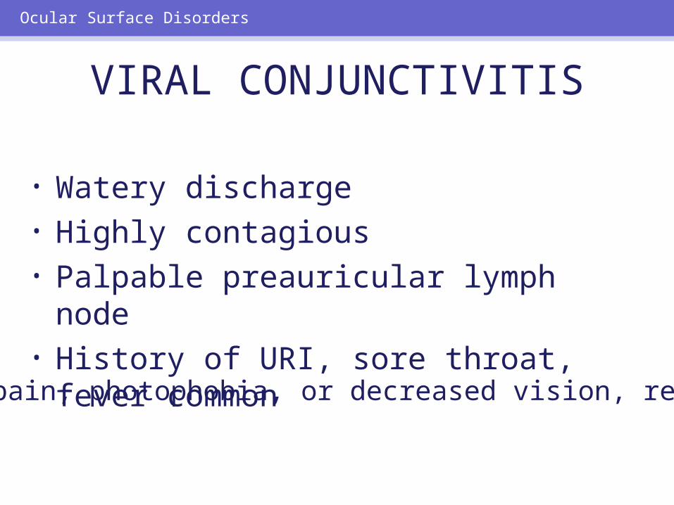
VIRAL CONJUNCTIVITIS
• Watery discharge• Highly contagious• Palpable preauricular lymph node• History of URI, sore throat, fever
common
Ocular Surface Disorders
If pain, photophobia, or decreased vision, refer.

Ocular Surface Disorders
Allergic conjunctivitis

ALLERGIC CONJUNCTIVITIS
• Associated conditions: hay fever, asthma, eczema
• Contact allergy: chemicals, cosmetics, pollen
• Treatment: topical antihistamine/decongestant drops
• Systemic antihistamines if necessary for systemic disease Refer refractory cases.
Ocular Surface Disorders

NEONATAL CONJUNCTIVITIS:CAUSES
• Bacteria (N. gonorrhoeae, 2–4 days) • Bacteria (Staphylococcus,
Streptococcus, 3–5 days)• Chlamydia (5–12 days)• Viruses (eg, herpes, from mother)
Ocular Surface Disorders

Ocular Surface Disorders
Neonatal gonococcal conjunctivitis

Ocular Surface Disorders
Neonatal chlamydial conjunctivitis

NEONATAL CHLAMYDIALCONJUNCTIVITIS: TREATMENT• Erythromycin ointment: qid x 4
weeks• Erythromycin po x 2–3 weeks
40–50 mg/kg/day ¸ 4
Ocular Surface Disorders

Ocular Surface Disorders
Subconjunctival hemorrhage

TEARS AND DRY EYES
• Tear functions:– Lubrication – Bacteriostatic and immunologic functions
• Dry eye (keratoconjunctivitis sicca) is a tear deficiency state
Ocular Surface Disorders

TEAR DEFICIENCY STATES:SYMPTOMS
• Burning• Foreign-body sensation• Paradoxical reflex tearing• Symptoms can be made worse by
reading, computer use, television, driving, lengthy air travel
Ocular Surface Disorders

TEAR DEFICIENCY STATES:ASSOCIATED CONDITIONS
• Aging• Rheumatoid arthritis• Stevens-Johnson syndrome• Chemical injuries• Ocular pemphigoid• Systemic medications
Ocular Surface Disorders

DRY EYES: TREATMENT
• Artificial tears, cyclosporine drops• Nonpreserved artificial tears• Lubricating ointment at bedtime• Punctal occlusion• Counseling about activities that make
dry eyes worse
Ocular Surface Disorders
Ocular Surface Disorders
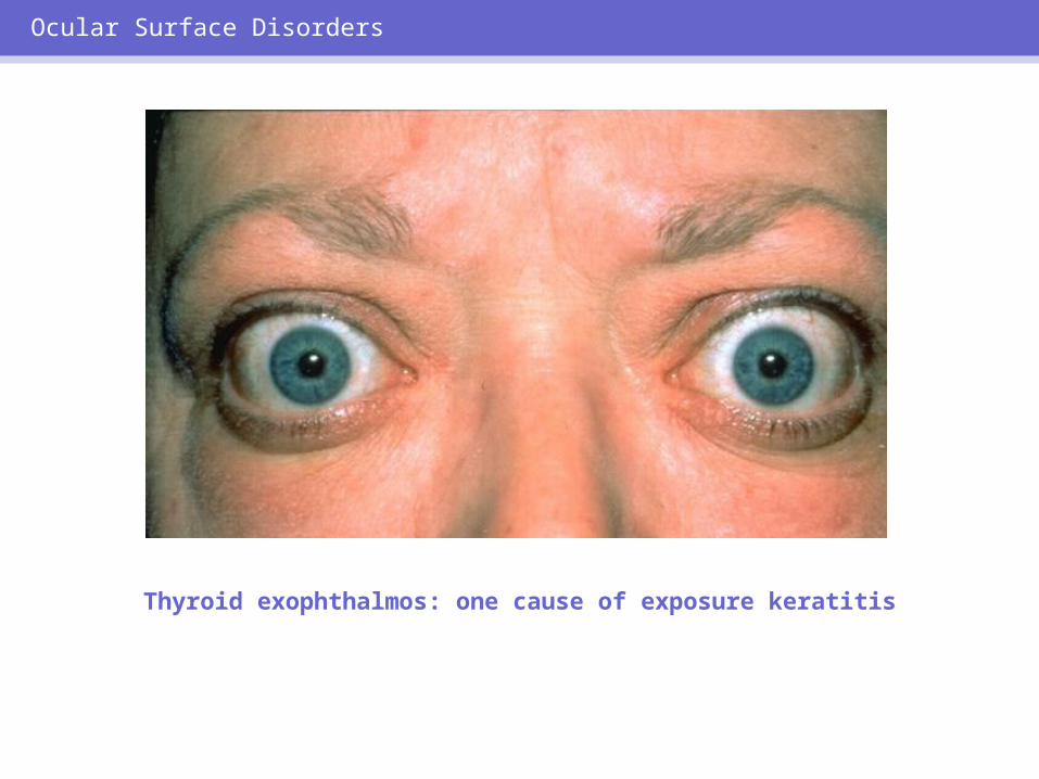
Thyroid exophthalmos: one cause of exposure keratitis
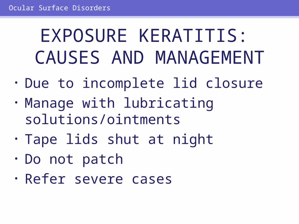
EXPOSURE KERATITIS: CAUSES AND MANAGEMENT
• Due to incomplete lid closure• Manage with lubricating
solutions/ointments• Tape lids shut at night• Do not patch• Refer severe cases
Ocular Surface Disorders

Ocular Surface Disorders
Pinguecula

Ocular Surface Disorders
Pterygium

INFLAMED PINGUECULAAND PTERYGIUM:
MANAGEMENT• Artificial tears• Counsel patients to avoid irritation• If documented growth or vision loss,
refer
Ocular Surface Disorders

Anterior Segment Disorders
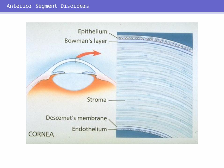
Anterior Segment Disorders

ACUTE CORNEAL DISORDERS:SYMPTOMS• Eye pain
– Foreign-body sensation– Deep and boring
• Photophobia• Blurred vision
Anterior Segment Disorders

Anterior Segment Disorders
Irregular corneal light reflex and central corneal opacity

Anterior Segment Disorders
Fluorescein dye strip applied to the conjunctiva

Anterior Segment Disorders
Corneal abrasion, stained with fluoresceinand viewed with cobalt blue light

CORNEAL ABRASION
• Signs and symptoms: redness, tearing, pain, photophobia, foreign-body sensation, blurred vision, small pupil
• Causes: injury, welder’s arc, contact lens overwear
Anterior Segment Disorders

CORNEAL ABRASION: MANAGEMENT
• Goals:– Promote rapid healing– Relieve pain– Prevent infections
• Treatment:– 1% cyclopentolate – Topical antibiotics
Drops (eg, fluoroquinolone, others) or ointment (eg, erythromycin, bacitracin/polymyxin)
– ± Pressure patch x 24–48 hours– ± Oral analgesics
Anterior Segment Disorders

Anterior Segment Disorders
Applying a pressure patch

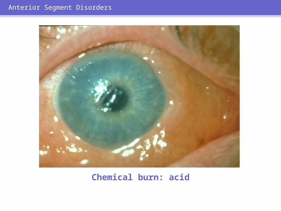
CHEMICAL INJURY
• A true ocular emergency• Requires immediate irrigation with
nearest source of water• Management depends on offending
agent
Anterior Segment Disorders
Anterior Segment Disorders
Chemical burn: acid

Anterior Segment Disorders
Chemical burn: alkali

Corneal ulcer Giant papillary conjunctivitis
Anterior Segment Disorders

INFECTIOUS KERATITIS
• Frequently result from mechanical trauma
• Can cause permanent scarring and decreased vision
• Early detection, aggressive therapy are vital
Anterior Segment Disorders

Anterior Segment Disorders
Bacterial infection of the cornea

Anterior Segment Disorders
Primary herpes simplex infection

Anterior Segment Disorders
Corneal herpes simplex dendrites, stained with fluorescein

RxTopical
Anesthetics
Anterior Segment Disorders

TOPICAL STEROIDS: SIDE EFFECTS
• Facilitate corneal penetration of herpes virus
• Elevate IOP (steroid-induced glaucoma)
• Cataract formation and progression• Potentiate fungal corneal ulcers
Anterior Segment Disorders

Anterior Segment Disorders
Hyphema

INFLAMMATORY CONDITIONS CAUSING A RED EYE:
• Episcleritis• Scleritis• Anterior uveitis (iritis)
Anterior Segment Disorders

Episcleritis Scleritis
Anterior Segment Disorders

Recognize and refer.
Anterior Segment Disorders
IRITIS
Signs and Symptoms
• Circumlimbal redness• Pain• Photophobia• Decreased vision• Miotic pupil
Rule Out• Systemic
inflammation• Trauma• Autoimmune
disease• Systemic infection

UVEITIS: SLIT LAMP FINDINGS
Anterior Segment Disorders
White cells in anterior
chamber
Hypopyon
Keratic precipitates

Anterior Segment Disorders

ACUTE GLAUCOMA:SIGNS AND SYMPTOMS
• Red eye• Severe pain in, around eye• Frontal headache• Blurred vision, halos seen around lights• Nausea, vomiting• Pupil fixed, mid-dilated, slightly larger than
contralateral side• Elevated IOP• Corneal haze
Anterior Segment Disorders

Anterior Segment Disorders
Acute angle-closure glaucoma

ACUTE GLAUCOMA: INITIAL TREATMENT
• Pilocarpine 2% drops q 15 min x 2• Timolol maleate 0.5%, 1 drop• Apraclonidine 0.5%, 1 drop• Acetazolamide 500 mg po or IV• IV mannitol 20% 300–500 cc
Anterior Segment Disorders

COMMON RED EYE DISORDERS:
TREATMENT INDICATED• Hordeolum• Chalazion• Blepharitis• Conjunctivitis• Subconjunctival hemorrhage• Dry eyes• Corneal abrasions (most)
Summary

VISION-THREATENING RED EYE SIGNS & SYMPTOMS: REFERRAL
INDICATED• Decreased vision• Ocular pain• Photophobia• Circumlimbal redness• Corneal edema• Corneal ulcers/ dendrites• Abnormal pupil• Elevated IOP
Summary

VISION-THREATENING RED EYE
DISORDERS: URGENT REFERRAL
• Orbital cellulitis• Scleritis• Chemical injury • Corneal infection• Hyphema• Iritis• Acute glaucoma
Summary

• Clinical expertise• Cooperation• Communication
Summary
MANAGING THE RED EYE: PCP AND OPHTHALMOLOGIST