Embed Size (px)
Citation preview

Joseph MAMAZZA Isabelle RAÎCHE
NEEDLESCOPIC FOREGUT SURGERY
®


NEEDLESCOPIC FOREGUT SURGERY
Joseph MAMAZZA, MDCM, FRCPS(C)Chair of General Surgery – University of Ottawa
MIS Fellowship Program Director Co-Director Bariatric Surgery Program – The Ottawa Hospital
Isabelle Raîche, MDMIS Fellow

Needlescopic Foregut Surgery4
Needlescopic Foregut SurgeryJoseph Mamazza, MDCM, FRCPS(C) Chair of General Surgery – University of Ottawa MIS Fellowship Program Director Co-Director Bariatric Surgery Program – The Ottawa HospitalIsabelle Raîche, MDMIS Fellow
Correspondence address of the author: Joseph Mamazza, M.D. Chair of General Surgery – University of Ottawa MIS Fellowship Program Director Co-Director Bariatric Surgery Program – The Ottawa Hospital Phone: 001-613-798-5555 extension 13151 Fax: 001-613-761-4124 E-mail: [email protected]
All rights reserved. 1st edition 2011 © 2015 GmbH P.O. Box, 78503 Tuttlingen, Germany Phone: +49 (0) 74 61/1 45 90 Fax: +49 (0) 74 61/708-529 E-mail: [email protected]
No part of this publication may be translated, reprinted or reproduced, trans-mitted in any form or by any means, electronic or mechanical, now known or hereafter invent ed, including photocopying and recording, or utilized in any information storage or retrieval system without the prior written permission of the copyright holder.
Editions in languages other than English and German are in preparation. For up-to-date information, please contact GmbH at the address shown above.
Design and Composing: GmbH, Germany
Printing and Binding: Straub Druck + Medien AG Max-Planck-Straße 17, 78713 Schramberg, Germany
07.15-0.3
ISBN 978-3-89756-543-2
Important notes:
Medical knowledge is ever changing. As new research and clinical experience broaden our knowledge, changes in treat ment and therapy may be required. The authors and editors of the material herein have consulted sources believed to be reliable in their efforts to provide information that is complete and in accord with the standards accept ed at the time of publication. However, in view of the possibili ty of human error by the authors, editors, or publisher, or changes in medical knowledge, neither the authors, editors, publisher, nor any other party who has been involved in the preparation of this booklet, warrants that the information contained herein is in every respect accurate or complete, and they are not responsible for any errors or omissions or for the results obtained from use of such information. The information contained within this booklet is intended for use by doctors and other health care professionals. This material is not intended for use as a basis for treatment decisions, and is not a substitute for professional consultation and/or use of peer-reviewed medical literature.
Some of the product names, patents, and re gistered designs referred to in this booklet are in fact registered trademarks or proprietary names even though specific reference to this fact is not always made in the text. Therefore, the appearance of a name without designation as proprietary is not to be construed as a representation by the publisher that it is in the public domain.
The use of this booklet as well as any implementation of the information contained within explicitly takes place at the reader’s own risk. No liability shall be accepted and no guarantee is given for the work neither from the publisher or the editor nor from the author or any other party who has been involved in the preparation of this work. This particularly applies to the content, the timeliness, the correctness, the completeness as well as to the quality. Printing errors and omissions cannot be completely excluded. The publisher as well as the author or other copyright holders of this work disclaim any liability, particularly for any damages arising out of or associated with the use of the medical procedures mentioned within this booklet.
Any legal claims or claims for damages are excluded.
In case any references are made in this booklet to any 3rd party publication(s) or links to any 3rd party websites are mentioned, it is made clear that neither the publisher nor the author or other copyright holders of this booklet endorse in any way the content of said publication(s) and/or web sites referred to or linked from this booklet and do not assume any form of liability for any factual inaccuracies or breaches of law which may occur therein. Thus, no liability shall be accepted for content within the 3rd party publication(s) or 3rd party websites and no guarantee is given for any other work or any other websites at all.

5Needlescopic Foregut Surgery
Contents
1.0 Needlescopic Surgery. . . . . . . . . . . . . . . . . . . . . . . . . . . . . . . . . . . . . . . . . . . . 61.1 Introduction . . . . . . . . . . . . . . . . . . . . . . . . . . . . . . . . . . . . . . . . . . . . . . . . 61.2 Comparison with Other Approaches . . . . . . . . . . . . . . . . . . . . . . . . . . . 7
2.0 Needlescopic Nissen Fundoplication . . . . . . . . . . . . . . . . . . . . . . . . . . . . . . . 82.1 Introduction . . . . . . . . . . . . . . . . . . . . . . . . . . . . . . . . . . . . . . . . . . . . . . . . 82.2 IndicationsforAnti-refluxSurgery . . . . . . . . . . . . . . . . . . . . . . . . . . . . . 82.3 Preoperative Evaluation for Nissen Fundoplication. . . . . . . . . . . . . . . 92.4 Principles of Nissen Fundoplication. . . . . . . . . . . . . . . . . . . . . . . . . . . . 102.5 Instrumentation for Needlescopic Nissen Fundoplication . . . . . . . . . 102.6 Technique of Needlescopic Nissen Fundoplication . . . . . . . . . . . . . . . 112.7 Cosmetic Results . . . . . . . . . . . . . . . . . . . . . . . . . . . . . . . . . . . . . . . . . . . 172.8 Results of Nissen Fundoplication . . . . . . . . . . . . . . . . . . . . . . . . . . . . . . 172.9 Results of Needlescopic Nissen Fundoplication . . . . . . . . . . . . . . . . . 182.10 Conclusion . . . . . . . . . . . . . . . . . . . . . . . . . . . . . . . . . . . . . . . . . . . . . . . . 18
3.0 Needlescopic Heller Myotomy. . . . . . . . . . . . . . . . . . . . . . . . . . . . . . . . . . . . . 183.1 Introduction . . . . . . . . . . . . . . . . . . . . . . . . . . . . . . . . . . . . . . . . . . . . . . . . 183.2 Epidemiology of Achalasia . . . . . . . . . . . . . . . . . . . . . . . . . . . . . . . . . . . 183.3 Etiology. . . . . . . . . . . . . . . . . . . . . . . . . . . . . . . . . . . . . . . . . . . . . . . . . . . . 193.4 Clinical Presentation. . . . . . . . . . . . . . . . . . . . . . . . . . . . . . . . . . . . . . . . . 193.5 Diagnosis . . . . . . . . . . . . . . . . . . . . . . . . . . . . . . . . . . . . . . . . . . . . . . . . . . 203.6 Non-Surgical Treatment. . . . . . . . . . . . . . . . . . . . . . . . . . . . . . . . . . . . . . 203.7 Surgical Treatment . . . . . . . . . . . . . . . . . . . . . . . . . . . . . . . . . . . . . . . . . . 213.8 Principles of Heller Myotomy . . . . . . . . . . . . . . . . . . . . . . . . . . . . . . . . . 213.9 Instrumentation for Needlescopic Heller Myotomy and
Dor Fundoplication . . . . . . . . . . . . . . . . . . . . . . . . . . . . . . . . . . . . . . . . . . 213.10TechniqueofNeedlescopicHellerMyotomy and
Dor Fundoplication . . . . . . . . . . . . . . . . . . . . . . . . . . . . . . . . . . . . . . . . . . 223.11 Appearance of Incisions at the End of the Procedure . . . . . . . . . . . . . 273.12 Intra-operative Esophageal Perforation . . . . . . . . . . . . . . . . . . . . . . . . 283.13 Long-term Cosmetic Result . . . . . . . . . . . . . . . . . . . . . . . . . . . . . . . . . . 283.14 Results of Heller Myotomy . . . . . . . . . . . . . . . . . . . . . . . . . . . . . . . . . . . 293.15 Special Consideration . . . . . . . . . . . . . . . . . . . . . . . . . . . . . . . . . . . . . . . 293.16 Conclusion . . . . . . . . . . . . . . . . . . . . . . . . . . . . . . . . . . . . . . . . . . . . . . . . . 29
References . . . . . . . . . . . . . . . . . . . . . . . . . . . . . . . . . . . . . . . . . . . . . . . . . . . . . . . . 30

Needlescopic Foregut Surgery6
1.0 Needlescopic Surgery
1.1 IntroductionSince the advent of laparoscopy, there has been a growing interest in reducing the size of the instrumentation used for surgical procedures. This has lead to the development of needlescopic surgery (laparoscopic surgery with instruments ≤ 3 mm of outer diameter). The possible benefits of those smaller instruments include: better cosmetic results with 66% of incisions not being visible in one study,1 a reduced risk of trocar site herniation, a potential shorter length of stay and a reduced need for postoperative analgesia.2 The potential risks of needlescopic surgery are related to the loss of tactile feedback and the increased risk of trauma to adjacent structures due to the smaller diameter of the instruments.
1 Relative trocar size. From right to left: 3.5 mm, 5 mm, 10 mm.
2 Relative size of instruments: 5 mm Maryland dissector vs. 3 mm needledriver.
3 Relative size of 3 mm needledriver and needle.
7Needlescopic Foregut Surgery
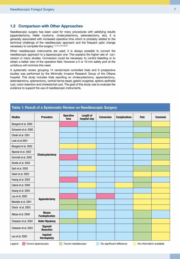
1.2 Comparison with Other ApproachesNeedlescopic surgery has been used for many procedures with satisfying results (appendectomy, Heller myotomy, cholecystectomy, adrenalectomy, etc). It is gene rally associated with increased operative time which is probably related to the technical challenge of the needlescopic approach and the frequent optic change necessary to complete the surgery.1, 3, 4, 5, 24, 30, 37
When needlescopic instruments are used, it is always possible to convert the needlescopic approach to a laparoscopic one. This explains the higher rate of con-version in many studies. Conversion could be necessary to control bleeding or to obtain a better view of the operative field. However a 5 or 10 mm safety port at the umbilicus will minimize this need.
A systematic review grouping 14 randomized controlled trials and 6 prospective studies was performed by the Minimally Invasive Research Group of the Ottawa hospital. This study includes trials reporting on cholecystectomy, appendectomy, adrenalectomy, splenectomy, ventral hernia repair, gastric surgeries, splenic epithelial cyst, colon resection and choledochal cyst. The goal of this study was to evaluate the evidence to support the use of needlescopic instruments.
Legend Favors laparoscopic Favors needlescopic No significant difference No information available
Studies Procedure Operative time
Length of hospital stay Conversion Complications Pain Cosmesis
Bissgard et al. 2000
Cholecystectomy
Schwenk et al. 2000
Cheah et al. 2001
Look et al.2001
Bissgard et al. 2002
Alponat et al. 2002
Schmidt et al. 2002
Ainslie et al. 2003
Sarli et al. 2003
Hsieh et al. 2003
Huang et al. 2003
Cabral et al. 2008
Huang et al. 2003
AppendectomyLau et al. 2002
Mostafa et al. 2001
Chock et al. 2003
Abbas et al. 2009 Nissen Fundoplication
Chaisson et al. 2002 Heller Myotomy
Chaisson et al. 2003 Sigmoid Resection
Lau et al. 2002 Inguinal Hernioplasty
Table 1: Result of a Systematic Review on Needlescopic Surgery

Needlescopic Foregut Surgery8
The conclusion of this review was that the current quality of evidence is poor to moderate and the studies likely underpowered to detect difference in complications and conversion rate. However, with the available data, needlescopic surgery seems to be a safe approach. It is possibly associated with better pain control and better cosmetic results.
One trial compared needlescopic surgery and single- incision laparoscopic cholecystectomy (SILS). This trial involving 70 patients showed a significant longer operative time for the SILS surgery. Rate of complications, postoperative pain and analgesic requirement were equivalent for both type of procedure. There was an advantage in cosmetic results for patient with SILS surgery associated with a shorter total wound length.6 The main advantage of needlescopic approach over single incision is that needlescopic does not require major change in the usual basic principles of laparoscopic surgery.
Nissen fundoplication and Heller myotomy are ideal procedures for needlescopic instruments. There is no specimen to extract, there is no heavy organ to manipulate with small instruments and there is no anastomosis to create with 12 mm endoscopic stapler.
2.0 Needlescopic Nissen Fundoplication
2.1 IntroductionGastro-esophageal reflux disease (GERD) is a frequent cause of complaints in developed countries. It is estimated that 20% of Western population experiences heartburn weekly.7 Many of these patients can be treated with simple intermittent medication but others may require lifelong medical therapy or may be only partially relieved by the medication. For these patients and for those who develop complications of GERD, surgery is a real and viable alternative. Since the first description of a laparoscopic Nissen fundoplication in 1991, it has become procedure of choice for GERD worldwide.8, 9
In this booklet we will review the indications of Nissen fundoplication and the outcomes from this surgery and we will explain the technical steps to perform a needlescopic fundoplication.
2.2 IndicationsforAnti-refluxSurgery
Patients with symptoms related to GERD not completely eliminated by optimized medical treatment.
Patients with symptoms controlled by medical treatment who do not desire lifelong medication.
Patients with complication of reflux: stricture, Barrett esophagus, vocal cord injury.

9Needlescopic Foregut Surgery
2.3 Preoperative Evaluation for Nissen FundoplicationBecause the symptoms of GERD are often non-specific, it is necessary to confirm the diagnosis with more objective testing before discussing the potential benefits of surgery with the patient. The evaluation should be used to assess the severity of GERD, to identify GERD as the cause of the symptoms and to select the appropriate procedure for a particular patient.
24h pH Monitoring
pH monitoring is the most reliable test to diagnosis GERD with sensitivity and a specificity of 96%.7 An esophageal pH < 4 reflects an episode of reflux of gastric juice into the esophagus. This test will also link the symptoms of the patient to the episodes of reflux. The DeMeester score has been developed to offer a global evaluation of the severity of the reflux by combining six components of reflux disease: the total time of pH < 4, the upright time with pH < 4, the supine time with pH < 4, the number of episodes of reflux, the number of episode ≥ 5 min and the longest episode. A score > 14.7 is considered as abnormal.
Endoscopy
Patients should be evaluated by endoscopy to search for esophagitis and other potential complications. The absence of esophagitis does not exclude the diagnosis of GERD since 40–50%,7, 10 of patient with confirmed GERD don’t present with esophagitis. There is also 10% of esophagitis that are not related to GERD. The esophagogastroduodenoscopy also permits to search for alternate diagnoses like gastritis, peptic ulcer disease and carcinoma that can present with non-specific symptoms similar to GERD. Endoscopy also affords a possibility to look for compli cations of reflux such as stricture and Barrett’s oesophagus. Finally, endoscopy allows the evaluation of the gastro-esophageal junction and the search for a paraesophageal hernia.
Manometry
Manometry is classically routinely performed before anti-reflux surgery. It has been used as a tool to select the appropriate procedure for patients with reflux. However, many trials have demonstrated that tailoring the surgery by using manometry does not offer any advantage in terms of surgical outcomes and patient satisfaction.8 A recent review on the role of manometry in GERD concludes that tailoring the surgery according to the results of manometry is not indicated and that the type of fundo plication used by surgeon is based more on the personal preference of the surgeon than evidence-base data.11 However, manometry remains a useful tool to exclude primary or secondary motility disorders. In a study from Akyuz et al, 11% of patients evaluated for reflux had a primary esophageal motility disorder (achalasia, uncoordinated contractions, nutcracker esophagus) and 2% had secondary motility disorder due to scleroderma.12

Needlescopic Foregut Surgery10
2.4 Principles of Nissen FundoplicationTo offer the best results to patients some general principles must be applied to the procedure:10
There must be 3 cm of intra-abdominal esophagus.
The fundus should be used for the fundoplication.
The wrap should be anchored on the esophagus.
The vagal nerves should be protected during the surgery.
The wrap should not be longer than 2–3 cm.
The crura should be closed with non-absorbable suture.
The fundoplication should be constructed over a large caliber bougie.
Needlescopic instruments allow the surgeon to perform these same critical steps as do regular laparoscopic instruments. Needlescopic surgery does not require changes in accepted laparoscopic surgical standards and preserves the laparo-scopic paradigm unlike single port surgery.
4 Instrument Set for Needlescopic Foregut Surgery.
1
2
4
57
96 83
2.5 Instrumentation for Needlescopic Nissen Fundoplication
1 5 mm and 3 mm scope2 Four 3.8 mm trocars and one 5 mm trocar3 One 3 mm curved scissor4 One 3 mm Maryland dissector 5 One 3 mm L-hook cautery 6 Three 3 mm atraumatic graspers7 One toothed 3 mm grasper with ratchet8 One 3 mm suction9 Two 3 mm needle drivers

11Needlescopic Foregut Surgery
5 Difference in clarity of vision between 5 mm (upper image) and 3 mm scope (lower image).
a b
6 Trocars sites. 7 Liver retraction.
2.6 TechniqueofNeedlescopic Nissen Fundoplication
The patient is placed in semi-lithotomy position on a bean bag.
A small incision on the skin of the umbilicus allows the insertion on a Veress needle. After the creation of a 15 mmHg pneumoperitoneum, a 5 mm trocar is inserted at the base of the umbilicus. The abdominal cavity is explored with the 5 mm scope. A 3 mm scope is used as a bridge to allow the insertion of hemostatic device such as hemoclip or harmonic scalpel and for the insertion of the needles via the 5 mm port. The surgery is mainly performed with the 5 mm scope which offers better visua li zation of the operative field (Figs. 5a, b).
Four 3.8 mm trocars are inserted. One in the subxiphoid region, one in the right upper quadrant and two in the left upper quadrant (Fig. 6).
A grasper with teeth is inserted in the subxiphoid trocar and tacked to the diaphragm to lift the left lobe of the liver. Alternatively a liver retractor may be used (Fig. 7).

Needlescopic Foregut Surgery12
8 Opening of pars flaccida. 9 Hemostasis with cautery and Maryland dissector.
The pars flaccida is then dissected and the lesser sac is entered using cautery. Vessels are controlled with a pinch burn technique (Figs. 8, 9).
The medial border of the right crus is identified and dissected, taking care not to damage the peritoneal lining of the crus. Traction on the esophagus helps this dissection (Figs. 10a, b).
10 Dissection of right crus.
a
b
Right crusRight crus
Esophagus
Esophagus

13Needlescopic Foregut Surgery
The retroesophageal space is dissected to the left crus. The posterior vagal trunk should be protected during this dissection (Figs. 11a, b).
The anterior dissection is completed by taking down the phrenoesophageal ligament anteriorly from right to left (Fig. 12)
The short gastric vessels are then controlled using a pinch burn cautery technique, or hemostatic clips or ultrasonic dissector inserted in the umbilical trocar. The mobilization of the fundus is completed to allow a floppy wrap (Figs. 13, 14).
12 Dissection of phrenoesophageal ligament.
Esophagus
13 Beginning of the short gastric dissection. 14 Control of the short gastric with pinch and burn technique.
11 Dissection of the retroesophageal space.
a
Right crusLeft crus
Esophagus
b
Right crus
Esophagus

Needlescopic Foregut Surgery14
The exposure of the left crus is completed from the left side (Fig. 15).
By the end of the dissection, there is at least 3 cm of intra-abdominal oesophagus and the hiatal structures are all well delineated (Fig. 16)
The hiatal reconstruction is done using non-absorbable braided suture on a straight needle. The straight needle is easily inserted via the 5 mm umbilical trocar. A figure of 8 stitch is placed using the 3 mm needledrivers (Figs. 17–19).
15 Left crus exposure.
Esophagus
Left crus
16 Retro-esophageal space after complete dissection.
Esophagus
Right crus
Left crus
17 Left crus stitch.
18 Right crus stitch. 19 Completed hiatoplasty.

15Needlescopic Foregut Surgery
The 360° wrap is then completed. The fundus is grasped and passed behind the esophagus and progressively brought on the right side using the great curvature as traction point (Figs. 20–22).
The wrap is anchored with one stitch between the stomach and the esophagus. Care is taken when tying this stitch not to pull anteriorly to avoid injury to the esophagus (Figs. 23, 24).
20 A grasper is passed under the esophagus. 21 The fundus is grasped.
22 The fundus is brought posteriorly to complete the wrap.
23 First stitch on the wrap. 24 Tying the first stitch.

Needlescopic Foregut Surgery16
Two other stitches are then placed to complete the wrap. A 50F bougie should be inserted in the esophagus without resistance at the end of the fundoplication (Fig. 25).
Finally the wrap is fixed to the right crus using another non-absorbable braided suture (Figs. 26–28).
The skin of the 5 mm umbilical trocar is closed with one subcuticular suture (Fig. 29).
25 Two last stitches on the wrap. 26 Fixation of the wrap to the crus.
27 Fixation completed.
28 Completed fundoplication. 29 Appearance of the incisions at the end of the procedure.

17Needlescopic Foregut Surgery
2.7 Cosmetic Results
30 Cosmetic results at 2–3 weeks. 31 Cosmetic results at 3 months.
2.8 Results of Nissen Fundoplication
Cost-effectiveness
Many studies were done to evaluate the benefits of Nissen fundoplication over medical therapy. The REFLUX trial concluded that the surgery was cost effective if the patient was not taking medication 5 years after the surgery.13 A Cochrane systematic review of 4 studies including 1232 patients demonstrated that GERD related quality of life is improved at 3 months and one year but that the cost of surgery was higher for the first year.14
Useofanti-refluxmedication
The use of medication after the fundoplication has been inconsistent among the studies. In the Cochrane systematic review, 12,5% of patients were taking over the counter medication one year after the fundoplication.14 With a follow-up of 5 years another group found that 15–21% of patients were on acid suppressing drugs after surgery versus 81–82% in the non-surgical group.15
Complications
The most frequent complications reported with Nissen fundoplication is dysphagia and bloating. 1–19% of dysphagia is reported in different studies.8,14 The post- operative dysphagia can usually be treated by dilatation of the fundoplication. Bloating is increased after anti-reflux surgery by 3%–5% and incapacity to belch affects 25% of the patients.8,16 0–15% of patient undergoing a Nissen fundoplication will need another surgery during the follow-up.14, 15 The most frequent indications for those surgeries include: dysphagia, recurrent reflux and intrathoracic herniation of the wrap.

Needlescopic Foregut Surgery18
2.9 Results of Needlescopic Nissen FundoplicationOne study compared needlescopic and laparoscopic Nissen fundoplication. There was no significant difference in estimated blood loss, operative time and intraope rative complications. The postoperative period was also similar between the groups in term of narcotics requirements, hospital stay and complications. At follow-up (average 61 days) there was no difference in dysphagia or bloating.17 There was a significant reduction in operative time after the first 4 cases 166 versus 120 min p: 0,03. The relatively short learning curve in this study was probably related to the previous experience of the participating surgeons in laparoscopy.
2.10 Conclusion As with other procedures, needlescopic Nissen fundoplication is safe in the properly selected patient. It should be used by surgeons with experience in advanced laparo scopy and in patients with adequate body habitus. The benefits in terms of cosmetic results with the needlescopic approach are not obtained at the cost of a major change in the well established surgical paradigm of Nissen fundoplication. This advantage will probably allow a better acceptance by the surgical community.
3.0 Needlescopic Heller Myotomy
3.1 IntroductionMinimally invasive surgery was initially used to treat achalasia in 1991. First, the myotomy was performed through a left thoracoscopic approach. However, this procedure was associated with a cumbersome anesthesia setting, a sub-optimal view of the gastro-esophageal junction and a high risk of post-operative reflux.18, 38 Laparoscopic Heller myotomy was developed to allow a safe and effective treatment of achalasia avoiding the thoracic approach and providing a better exposure of the gastro-esophageal junction. Myotomy combined with a fundoplication is now considered to be the treatment of choice of achalasia.
In this booklet we will briefly review the clinical features of achalasia and the result of Heller myotomy. We will then explain the operative steps necessary to perform a needlescopic Heller myotomy with a Dor anterior fundoplication.
3.2 Epidemiology of AchalasiaAchalasia is affecting 10 patients per 100 000 in United States. There is no gender preponderance.38 The incidence is increasing with advanced aged. There is a peak incidence between 25 and 60 years old.19

19Needlescopic Foregut Surgery
3.3 Etiology19
Primary achalasia: Degeneration of ganglion cells in the myenteric plexus
Secondary achalasia: Cancer (Most common cause of secondary achalasia) Mechanisms:
Direct invasion Paraneoplasic symptoms (secretion of antineuronal antibodies)
Anti-reflux surgery
Laparoscopic gastric banding
Post-vagotomy
Amyloidosis
Sarcoidosis
Sjögren’s Syndrome
Chagas disease Trypanosoma cruzi infection,
associated with megacolon, heart disease and neurologic disorders
Allgrove’s disease Achalasia Alacrima Autonomic disturbance ACTH insensivity
3.4 Clinical PresentationThe cardinal symptom of achalasia is dysphagia, present in > 90% of patients. Dysphagia is generally associated with both solid and liquid, a feature that differen tiates achalasia from mechanical abnormalities.18, 38
Regurgitation is present in 60–80% of patients. This can lead to aspiration and respiratory symptoms. Chest pain, heartburn and difficulty belching are also commonly found in those patients.
Heartburn can be caused by many factors: retention and fermentation of undigested food by bacteria, neuropathic pain, secondary and tertiary contraction of the esophagus, gastro-esophageal reflux. In a 2008 study looking at the presentation of achalasia, 45% of patients with achalasia had been treated for GERD before the diagnosis of achalasia was made.20
Weight loss rarely exceeds 5–10 kg. Elderly patients with a weight loss of more than 10 kg and symptoms for less than 6 months should raise the suspicion of a secondary achalasia related to esophageal cancer.18, 19, 38

Needlescopic Foregut Surgery20
32 Bird’s beak appearance on barium swallow.
3.5 Diagnosis
Barium study
Barium swallow is the recommended first test in patient with dysphagia. It defines the anatomy and allows for a safer endoscopy. The classic features of achalasia are: dilated esophagus with bird`s beak gastro-esophageal junction and absence of peristalsis. There is no frank correlation between the severity of the symptoms of the patient and the radiologic appearance. 64% of esophagogram suggest the diagnostic of achalasia.18, 38
Endoscopy
Upper endoscopy is recommended for all patients presenting with dysphagia. This exam is useful to rule out malignancy. Typically, esophagus may be dilated with retained food and an increased resistance at the gastro-esophageal junction. However, 40% of patients will have a normal upper endoscopy.18
Manometry
This test is the gold standard for the diagnosis of achalasia. Classic criteria include: absence of esophageal peristalsis in the body of esophagus, hypertensive lower esophageal sphincter (LES) (pressure > 45 mmHg), non-relaxing LES (residual pressure > 8 mmHg). LES is not hypertensive in up to 50% of patients.38
3.6 Non-Surgical Treatment
Medications
Many medications have been used to improve the symptoms of achalasia: nitrates, calcium channel blockers, nitric oxide donor. The efficacy of those is generally poor and no medical treatment is widely used for this disease.
Endoscopic Pneumatic Balloon Dilatation
Standard balloon dilatation and bougie dilatation are generally ineffective for treatment of achalasia. Pneumatic balloon dilatation has been used with good result in terms of initial symptoms improvement, relieving dysphagia in 55–70% of patients after one treatment and up to 90% of patients after multiple attempts. However it is associated with a higher rate of recurrence then surgery, with 25% of patients needing repeated dilatation.23 The main downside of this therapy is the risk of perforation that is estimated to be around 1–2% but can be as high as 12% in some series.21 Other complications associated with this treatment include GERD in up to 33% of patients and gastroesophageal bleeding from mucosal tear.23
EndoscopicBotulinumToxinInjection
This strategy is associated with good short term results but repeated injections are generally necessary. With repeated injections there is a potential 85% of relief of dysphagia at 2 years.22 In a randomized controlled trial, 66% of patients in the endoscopic botulinum injection group were symptomatic compared to 13.5% in the surgical group at 2 years follow-up.23 There is concerning evidence that myotomies performed after injections are more difficult. There is an initial resistance to this treatment in 26% of patients related to antibody formation.23 Endoscopic botulinum toxin injection is generally reserved for patients presenting with co-morbidities precluding a surgical treatment or endoscopic pneumatic dilatation (Ex: sigmoid esophagus). It can also be used as a test to confirm the diagnosis. A good response to botulinum toxin injection is predictive of a good clinical result after a myotomy.21

21Needlescopic Foregut Surgery
3.7 Surgical Treatment
Role of Surgical Myotomy
The surgical approach to achalasia has evolved since the introduction of Heller myotomy in 1913. The advent of laparoscopy has changed the role of the surgical myotomy in the treatment of achalasia. Before minimally invasive surgery, the left thoracotomy used for the myotomy and the complications related to this approach precluded widespread use of the surgery as a primary treatment. Since the introduction of laparoscopic Heller myotomy, surgical treatment is associated with low morbidity and a short hospital stay that render this approach more attractive. In a meta-regression analysis, the laparoscopic approach offered a better rate of symptoms improvements then the thoracoscopic approach (89 vs 78%) with a lower risk of post-operative reflux (28 vs 15%).23
The use of needlescopic instruments to perform this surgery may allow for possible improvements of these results in terms of pain control, length of stay and cosmetic results. A retrospective study including 29 patients was published in 2003. It was comparing standard laparoscopic Heller myotomy versus needlescopic Heller myotomy. The needlescopic approach demonstrated shorter duration of surgery and a shorter length of stay with similar analgesia requirements, complications and conversions.24
The surgical myotomy has been compared to endoscopic treatment. It is associated with the best long term results with 95% of dysphagia relief at 5 years versus 65% for dilatation.23 Even in the elderly population, the myotomy is associated with a smaller number of interventions and the best results in terms of long-term relief of symptoms.25
Necessity of Fundoplication
Without a fundoplication, Heller myotomy is associated with a 30–60% incidence of reflux. Post Heller myotomy GERD is hard to diagnose and the symptoms are not reliable.26 The addition of a fundoplication to the myotomy has been shown to reduce the incidence of reflux after the surgery without compromising relief of dysphagia.27 A partial fundoplication should be used as it has been demonstrated to diminish the risk of dysphagia in long term follow-up. Esophageal dilatation can be considered a contraindication to Nissen fundoplication after myotomy because it is associated with an unacceptable rate of postoperative dysphagia at 5 years.28
3.8 Principles of Heller Myotomy
Myotomy of both the longitudinal and the circular fibers.
Myotomy on 50% of the mucosa surface to prevent refusion of the fibers.
Myotomy extends for 7 cm from esophagus (5 cm) to the stomach (2 cm).
Fundoplication to prevent reflux and protect the exposed mucosa.
3.9 InstrumentationforNeedlescopicHellerMyotomy and Dor Fundoplication
The instrumentation is the same as for needlescopic Nissen fundoplication.
Needlescopic Foregut Surgery22
33 Trocars positioning. 34 Liver retraction.
35 Opening of the phrenoesophageal ligament.
36 Pinch and burn control of vessels.
a b
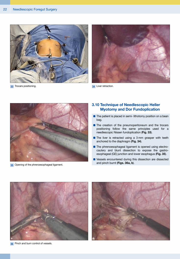
3.10 Technique of Needlescopic Heller Myotomy andDorFundoplication
The patient is placed in semi- lithotomy position on a bean bag.
The creation of the pneumoperitoneum and the trocars positioning follow the same principles used for a needlescopic Nissen fundoplication (Fig. 33).
The liver is retracted using a 3 mm grasper with teeth anchored to the diaphragm (Fig. 34).
The phrenoesophageal ligament is opened using electro - cautery and blunt dissection to expose the gastro- esophageal (GE) junction and lower esophagus (Fig. 35).
Vessels encountered during this dissection are dissected and pinch burnt (Figs. 36a, b).

23Needlescopic Foregut Surgery
37 Anterior vagus nerve.
Anterior vagus nerve
The anterior vagus nerve is exposed and protected (Fig. 37).
The dissection is carried upwards to expose 6-7 cm of esophagus into the lower mediastinum (Figs. 38–40).
38 Sharp dissection of the hiatus.
39 Blunt dissection of the hiatus. 40 Dissection completed.
Anterior hiatus
Esophagus
Esophagus

Needlescopic Foregut Surgery24
41 Measurement of the myotomy.
a b
A 7 cm long suture is used to measure the length of the myotomy (Figs. 41a, b).
The anterior surface of the esophagus is then scored using the electrocautery (Figs. 42a, b).
42 Scoring of the anterior esophagus.
a b

25Needlescopic Foregut Surgery
45 Myotomy on the gastric wall.
The longitudinal and circular muscles are gently teased off the esophagus on at least 50% of the circumference of the esophagus (Figs. 43, 44).
The myotomy is continued down to the gastric wall where the obliques fibers are encountered (Fig. 45).
The fundoplication is then started. First the mobility of the gastric fundus is assessed. It is not always necessary to mobilize the short gastric for a Dor fundoplication. If the short gastrics are to be controlled, a pinch burn technique is usually sufficient; larger vessels can be taken with 5 mm clips (Fig. 46).
43 Circular and longitudinal fibers.
Circular fibers
Longitudinal fibers
44 Esophageal myotomy completed.
Esophageal mucosa
46 Assessment of the mobility of the gastric fundus.
Right crus
Right edge of myotomy
Gastric fundus
Esophagus

Needlescopic Foregut Surgery26
47 First stitch on the fundus. 48 First stitch on the myotomy.
The fundoplication is done using non-resorbable braided suture stitches. First, the fundus is tacked to the left crural pillar incorporating esophageal musculature in the stitch (Figs. 47, 48).
Few others sutures are then used to fix the fundus to the left side of the myotomy (Fig. 49).
The fundus is fixed to the upper part of the hiatus (Figs. 50a, b).
49 Fundus fixed to the left side of the myotomy.
Left side of the myotomy
Fundus
Esophagus
50 Fixation of the fundus to the upper part of the hiatus.
a
EsophagusFundus
b
Hiatus

27Needlescopic Foregut Surgery
52 Completion of the fundoplication on the right side.51 Fixation of the fundoplication to the right crus.
Fundus
Right crus
Then the fundus is fixed to the right crus and the right side of the myotomy (Fig. 51).
Other stitches complete the fundoplication by anchoring the fundus to the right side of the myotomy until the myotomy is completely covered (Figs. 52, 53).
53 Completed fundoplication.
A Floppy Nissen fundoplication can also be used as an anti-reflux procedure. In this case, the posterior dissection is conducted as described previously. Other authors preferred the Toupet partial fundoplication. This technique results in a fundoplication that tends to keep the edges of the myotomy apart, providing the theoretical advantage of preventing the closure of the myotomy. The downside of this approach is the absence of coverage of the myotomy. It is not recommended to use this technique when a perforation has been repaired.
54 Incisions.
3.11 Appearance of Incisions at the End of the Procedure

Needlescopic Foregut Surgery28
3.12 Intra-operative Esophageal PerforationPrevious dilatation or botulinum toxin injection can make the myotomy more challenging because of tissue scarring.29 This scarring can fuse the plans, rendering perforation a more frequent occurrence. In a recently published review, a perforation occurred in 6.9% of myotomy and were repaired and the index surgery. Only 0.7% of patients developed clinical manifestation of the perforation. The rate of symptomatic perforation was found to be similar in the surgical group compared to the endoscopic dilatation group (0.7 vs 1.6%).23
If a perforation is suspected, it can be assessed using air insufflation or methylene blue through an orogastric tube. An intra-operative upper endoscopy is another option to evaluate the integrity of the mucosa. If a perforation occurs during the procedure, it can be fixed by interrupted stitches of 3-0 resorbable suture (Fig. 56). Simple or double layer repair are acceptable. The anterior fundoplication is then used as a serosal patch to reinforce the repair.
56 Closure of the perforation.55 Esophago-gastric perforation.
Perforation
57 3 months cosmetic results.
3.13 Long-term Cosmetic Result

29Needlescopic Foregut Surgery
3.14 Results of Heller Myotomy
Immediate Result 90% of patients can be discharged within 48 of the surgery. There is a 6% risk of perioperative complications and a 0.1% risk of mortality.23
Long-term Relief of Dysphagia A study on the long term effects of the myotomy found 80% of dysphagia impro vement at a mean follow-up of 6.4 years. Most of the failures were caused by fibrosis of the LES or by end stage esophageal dysfunction. The mean time to recurrence was 21.3 months. The patients with failed Heller myotomy were treated with either repeated myotomy or esophagectomy.30 Factors associated with good outcomes in this study include: elevated LES tone, short duration of symptoms, absence of sigmoid esophagus and absence of previous endoscopic treatment. Another option for patients experiencing symptoms recurrence is endoscopic pneumatic dilatation with a success rate as high as 97% in some series.22
Long-term Quality of Life Few study report very long term results of myotomy plus partial fundoplication. In a 2006 publication, Csendes reports the very long follow-up of 67 patients. More then 20 years after surgery, only 65% of patients reported excellent or good result. The main source of complaints of those patients was GERD causing 93% of failure. Only one patient had an incomplete myotomy.31
Long-term Complications The most frequent long term complication of Heller myotomy is GERD with an incidence of around 14% in meta-analysis.32 Incomplete myotomy with food fermentation is to be considered in the differential diagnosis of a patient presenting with heartburn after a myotomy. A 24h pH study is used as a diagnostic tool. GERD after myotomy must be aggressively treated because the impaired esophageal peristalsis of the esophagus increased the contact time of acid with the mucosa and also increased the risk of GERD complications. There is a poor correlation between the symptoms of heartburn and the presence of GERD on the pH study. Because of that some authors recommend a routine pH study after Heller myotomy.22
3.15 Special Consideration
Mega-esophagus Mega-esophagus or sigmoid esophagus is defined by an esophagus that is more than 6 cm wide. This condition is associated with a higher rate of re-intervention after a myotomy. However, less than 10% of patients with mega-esophagus need an esophagectomy as the treatment of their condition. It is recommended to proceed with a myotomy as the initial treatment. Patti et al published the results of Heller myotomy and Dor fundoplication in 19 patients with an esophagus > 6 cm. Eighty-nine percent of patients had excellent or good results after a mean follow-up of 22 months.33 A series of 4 cases including mega-esophagus up to 12 cm demonstrated the feasibility and the good outcomes of the myotomy.34 Patients who still have symptoms after the myotomy can be offered pneumatic dilatation with good results in up to 59% of patients. The option of esophagectomy should be reserved for patients who failed myotomy and dilatation. Esophagectomy is a morbid surgery with a mortality rate of 3% and a perioperative morbidity rate of 17%35 and it should be considered as a last resort option.
3.16 ConclusionHeller myotomy with Dor fundoplication is considered the preferred approach for treatment of achalasia. The principles of this surgery can be respected when performed with needlescopic instruments. Needlescopic Heller myotomy with Dor fundoplication is a safe and feasible option in well selected patients.

Needlescopic Foregut Surgery30
References1. GAGNER et al: Technical Aspects of Minimally Invasive Abdominal Surgery
Performed with Needlescopic Instruments, Surg Lap Endosc, 8(3) 171-179
2. MAMAZZA et al: Needlescopic Surgery, A logical evolution from conventional laparoscopic surgery, Surg Endosc, 2001, 15 :1208-1212
3. SAJID et al: Needlescopic versus laparoscopic cholecystectomy: a meta-analysis, Anz J Surg, 2009, 79, 437-442
4. INDERBIR et al: Needlescopic adrenalectomy- the initial series comparison with conventional laparoscopic adrenalectomy, Urology, 1998, 52, 180-186
5. CHIASSON et al: Needlescopic Heller Myotomy, Surg Laparosc Endosc Percutan Tech 2003, 13 2 67-70
6. LEE et al: Randomized clinical trial of single-incision laparoscopic cholecyctectomy versus minilaparoscopic cholecystectomy, Br J Surg, 2010, 97, 1007-1012
7. YEO et al: Shackelford’s Surgery of the Alimentary Tract, volume 1 sixth edition
8. STRATE et al: Laparoscopic fundoplication: Nissen versus Toupet two-year outcome of a prospective randomized study of 200 patients regarding preoperative esophageal motility, Sur Endosc, 2008, 22:21-30
9. FEIN et al: Is There a Role for Anything Other Than a Nissen’s Operation? J Gastrointest Surg, 2010, 14 (suppl 1) S67-S74
10. POULIN et al: Correcting Reflux Laparoscopically, Can J Gastroenterol, 1998, 12, 5, 327-332
11. FEIN et al: Is There a Role for Anything Other than a Nissen’s Operation?, J Gastrointest Surg 2010, 14 (Suppl 1) S67-S74
12. AKYUZ et al: Utility of esophageal manometry and pH-metry in gastroesophageal reflux disease before surgery, Tur J Gastroenterol, 2009, 20(4) 261-265
13. EPSTEIN et al: Laparoscopic fundoplication compared with medical management for gastro-oesophageal reflux disease : cost effectiveness study, The REFLUX trial group, BMJ 2009; 338:b2576\
14. WILEMAN et al: Medical versus surgical management for gastro-intestinal reflux disease (GORD) in adults, Cochrane Database Systematic Reviews, 2010, 3
15. BROEDERS et al: Long-term outcome of Nissen fundoplication in non-erosive and erosive gastro-oesopgageal reflux disease, Br J Surg, 2010, 97 845-852
16. PAPASAVAS et al: Effectiveness of lapaoscopic fundoplication in relieving the symptoms of gastroesophageal reflux disease (GERD) ans eliminating antireflux medical therapy. Surg Endosc 2003, 17 1200-1205
17. PACE et al: Needlescopic fundoplication, Surg Endosc, 2002, 16 578-580
18. PATTI et al: Esophageal Achalasia, Mastery of Endoscopic and Laparoscopic Surgery, Third edition, 2009, 130-135
19. WALZER et al: Achalasia, Gastroenterology Clin N Am, 2008; 37, 807-825

Needlescopic Foregut Surgery 31
20. FISICHELLA et al: Clinical, Radiological and Manometric profile in 145 patients with untreated achalasia. Word J Surg 2008; 32: 1974-1979
21. NUSSBAUM: Minimally Invasive treatment of Achalasia and Other Esophageal Dysmotility, Mastery of Surgery, 2007, 742-751
22. CHEATHAM et al: Current approach to the treatment of achalasia, curr gastroenterol rep, 2011; 13 219-225
23. CAMPOS et al: Endoscopic and Surgical Treatments for Achalasia, A systematic Review and Meta-Analysis, Ann of Surg, 2009, 249;1 45-57
24. CHIASSON et al: “Needlescopic” Heller Myotomy, Surg lap Endosc, 2003; 13,2, 67-70
25. CRAFT et al: Outcomes of Minimally Invasive Myotomy for the treatment of achalasia in the elderly, jSLS, 2010, 14: 342-347
26. PETERS: An antireflux procedure is critical to the long-term outcome of esophageal myotomy for achalasia, J Gastroint Surg, 2001; 5:1 17-20
27. BELLO et al: Evolution of the Minimally Invasive Treatment of Esophageal Achalasia, World j Surg. March 2011
28. REBECCHI et al: Randomized controlled trial of laparoscopic Heller myotomy plus Dor fundoplication versus Nissen fundoplication for achalasia – Long-term results, Ann surg,2008. 248, 6 1023-1029
29. PATTI et al: Effects of previous treatment on results of laparoscopic Heller myotomy for achalasia, Dig Dis Sci, 1999,44;11: 2270-2276
30. kILIC et al: Long-term outcomes of laparoscopic Heller myotomy for achalasia, Surgery 2009; 146: 826-833
31. CSENDES et al: Very late results of esophagomyotomy for patients with achalasia, clinical, endoscopic, histologic, manometric and acide reflux studies in 67 patients for a mean follow-up of 190 months, Ann Surg, 2006. 243;2 196-203
32. NOVAIS et al: 24-h pHmonitoring patterns and clinical response after achalasia treatment with pneumatic dilatation or laparoscopic Heller myotomy, Aliment Pharmacol Ther, 32, 1257-1265, 2010
33. PATTI et al: Laparoscopic Heller myotomy relieves dysphagia in achalasia when esophagus is dilated, Surg endorc, 1999, 13: 843-847
34. SCOTT et al: Results of laparoscopic Heller myotomy for extreme megaesophagus, an alternative to Esophagectomy, Surg Laparosc endosc Percutan Tech, 2009; 19 198-200
35. SWEET et al: The outcome of laparoscopic Heller myotomy for achalasia is not influenced by the degree of esophageal dilatation, J Gastrointest Surg, 2008, 12: 159-165
36. HOSONO et al: Minilaparoscopic vesus conventional laparoscopic cholecystectomy: a meta-analysis of randomized controlled trials, j Laparosc adv Surg Tech, 2007, 17,2, 191-199
37. FRANCIS et al: Achalasia: An Update on the Disease and its Treatment, Gastroenterology, 2010, 139, 369-374

Needlescopic Foregut Surgery32
HOPKINS® TelescopesDiameter 3.3 mm
533TVA
533 TVA Adaptor, autoclavable, permits telescope changing under sterile conditions
Diameter 5 mm
26046AA
26046BA HOPKINS® Forward-Oblique Telescope 30°, enlarged view, diameter 5 mm, length 29 cm, autoclavable, fiber optic light transmission incorporated, color code: red
26046AA HOPKINS® Straight Forward Telescope 0°, enlarged view, diameter 5 mm, length 29 cm, autoclavable, fiber optic light transmission incorporated, color code: green
26046FA HOPKINS® Telescope 45°, enlarged view, diameter 5 mm, length 29 cm, autoclavable, fiber optic light transmission incorporated, color code: black
26007AA HOPKINS® Straight Forward Telescope 0°, enlarged view, diameter 3.3 mm, length 25 cm, autoclavable, fiber optic light transmission incorporated, color code: green
26007BA HOPKINS® Forward-Oblique Telescope 30°, enlarged view, diameter 3.3 mm, length 25 cm, autoclavable, fiber optic light transmission incorporated, color code: red
26007AA

33Needlescopic Foregut Surgery
with connector for insufflation for use with instruments size 3 mm
Silicone Leaflet ValveCannula
Trocar only
TrocarsSize 3.5 mm
Trocar, with pyramidal tip including:
Cannula, with LUER-Lock connector
Trocar only Silicone Leaflet Valve
30114GL
30114G2
30114C30114L1
30114GKX
30114G3
30114KX30114L1
10 cmgreen-yellow
15 cmgreen-red
Trocar, with conical tip including: Cannula,
with LUER-Lock connector Trocar only Silicone Leaflet Valve
30114GZL
30114G2
30114ZL30114L1
30114GZX
30114G3
30114ZX30114L1
3.5 mmSize:Working length:Color code:
Trocar, with blunt tip including: Cannula,
with LUER-Lock connector Trocar only Silicone Leaflet Valve
30114GAL
30114G2
30114A30114L1
It is recommended to check the suitability of the product for the intended procedure prior to use.

Needlescopic Foregut Surgery34
TrocarsSize 3.5 mm
with connector for insufflation for use with instruments size 3 mm
Silicone Leaflet ValveCannula
Trocar only
Trocar, with pyramidal tip including:
Cannula, with LUER-Lock connector
Trocar only Silicone Leaflet Valve
30114GL
30114G2
30114C30114L1
30114GKX
30114G3
30114KX30114L1
10 cmgreen-yellow
15 cmgreen-red
Trocar, with conical tip including: Cannula,
with LUER-Lock connector Trocar only Silicone Leaflet Valve
30114GZL
30114G2
30114ZL30114L1
30114GZX
30114G3
30114ZX30114L1
3.5 mmSize:Working length:Color code:
Trocar, with blunt tip including: Cannula,
with LUER-Lock connector Trocar only Silicone Leaflet Valve
30114GAL
30114G2
30114A30114L1
30160GP
30160GP Trocar, with pyramidal tip, with LUER-Lock connector for insufflation, size 6 mm, working length 10.5 cm, color code: black for use with instruments size 5 mm,
including: Cannula Trocar only Silicone Leaflet Valve
Standard TrocarSize 6 mm

35Needlescopic Foregut Surgery
Dissecting and Grasping ForcepsCLICKLINE – rotating, dismantling, insulated, with connector pin for unipolar coagulation
unipolar
Size 3 mm
Operating instruments, length 36 cm, for use with trocars size 3.5 mm
Double Action Jaws
CLICKLINE KELLY Dissecting and Grasping Forceps, long
30310MLG 30352MLG 30353MLG 30356MLG 30321MLG 30325MLG 30327MLG30351MLG
CLICKLINE KELLY Dissecting and Grasping Forceps
Outer Sheath with
Working Insert
CLICKLINE Dissecting and Grasping Forceps, jaws right angled
30310MDG 30352MDG 30353MDG 30356MDG 30321MDG 30325MDG 30327MDG30351MDG
30310RG 30352RG 30353RG 30356RG 30321RG 30325RG 30327RG30351RG
Complete Instrument
LengthHandle
33125 331273315633152 33153 3312133151
36 cm
CLICKLINE Grasping Forceps, atraumatic, fenestrated
30353KG 30356KG30351KG 30352KG30310KG 30321KG 30325KG 30327KG
14
10
10
11

Needlescopic Foregut Surgery36
Size 3 mm
Operating instruments, length 36 cm, for use with trocars size 3.5 mm
Double Action Jaws
Complete Instrument
Outer Sheath with
Working Insert
CLICKLINE REDDICK-OLSEN Dissecting and Grasping Forceps, robust
30310ULG 30332ULG 30333ULG 30341ULG 30346ULG 30347ULG30361ULG 30341ULG
CLICKLINE Dissecting and Grasping Forceps, “tiger-jaws”, 2x 4 teeth
30310MGG 30332MGG 30333MGG 30341MGG 30346MGG 30347MGG30361MGG 30341MGG
CLICKLINE Grasping Forceps, atraumatic, fenestrated
30310AFG 30332AFG 30333AFG 30341AFG 30346AFG 30347AFG30361AFG 30341AFG
LengthHandle
33161 33131 33132 33133 33141 33146 33147
Single Action Jaws
CLICKLINE MATKOWITZ Grasping Forceps
30310 KWG 30332 KWG 30333 KWG 30341 KWG 30346 KWG 30347 KWG30361 KWG 30331 KWG
36 cm
Dissecting and Grasping ForcepsCLICKLINE – rotating, dismantling,without connector pin for unipolar coagulation
11
13
15
11
CLICKLINE Grasping Forceps, with fine atraumatic serration, fenestrated
30310ONG 30332ONG 30333ONG 30341ONG 30346ONG 30347ONG30361ONG 30331ONG
16

37Needlescopic Foregut Surgery
RoBi® Bipolar Rotating Instruments
bipolar
Operating instruments, length 36 cm, for use with trocars size 3.5 mm and 3.9 mm
Size 3.5 mm
Handle
RoBi® KELLY Grasping Forceps, Modell CLERMONT-FERRAND, with connector pin for bipolar coagulation, suitable for dissection, double-action jaws
38951MD38910MD
Complete Instrument
Outer Sheath
Length 36 cm
38151
Working Insert
12
RoBi® Grasping Forceps, Modell CLERMONT-FERRAND, with connector pin for bipolar coagulation, with fine atraumatic serration, fenestrated, double action jaws
38951ON38910ON
12
RoBi® Scissors Insert with Outer Sheath, METZENBAUM Scissors Insert with Outer Sheath, CLERMONT-FERRAND model, curved blades, double action jaws
38951MW38910MW
12

Needlescopic Foregut Surgery38
ScissorsCLICKLINE – rotating, dismantling,with connector pin for unipolar coagulation
unipolar
Size 3 mm
Operating instruments, length 36 cm, for use with trocars size 3.5 mm
Double Action Jaws
Complete InstrumentOuter Sheath with Working Insert
CLICKLINE Scissors, serrated, curved, conical
Handle
36 cm
33151 33121Length
30351MWG 30321MWG30310MWG 30325MWG 30327MWG
n
33125 33127
10
39Needlescopic Foregut Surgery
Coagulating and Dissecting Electrodeswithout suction channel, insulated sheath, with connector pin for unipolar coagulation
unipolar
Operating instruments, length 36 cm, for use with trocars size 3.5 mm
Size 3 mm
Special Features:## The hook electrode is suitable for resection, isolating structures as well as dissection and coagulation.
## The distal tip is semicircular: The thick outer curvature enables safe dissection.
## The thin inner curvature enables a controlled coagulation.
## The striated handle facilitates gripping.
Coagulating and Dissecting Electrode, L-shaped
Instrument
Length
36 cm
Instrument
Distal Tip
26870UFG
Coagulating and Dissecting Electrode, spatula-shaped, blunt26665UEL
CADIERE Coagulating and Dissecting Electrode, L-shaped, with cm-marking, distal tip tapered
Instrument
Length
36 cm
Instrument
Distal Tip
25775CL

Needlescopic Foregut Surgery40
Needle Holders, Palpation Probe
26167FNL KOH Ultramicro Needle Holder, jaws curved to left, with tungsten carbide inserts, straight handle, with disengageable ratchet, size 3 mm, length 36 cm
Size 3 mm
26167FKL KOH Ultramicro Needle Holder, jaws slightly curved to right, with tungsten carbide inserts, straight handle, with disengageable ratchet, size 3 mm, length 36 cm
Operating instruments, length 36 cm, for use with trocars size 3.5 mm
26167FN
26167TL
26167TL Palpation Probe, with cm-marking, size 3 mm, length 36 cm

41Needlescopic Foregut Surgery
Suction and Irrigation Tube
26167LHL Suction and Irrigation Tube, size 3 mm, length 36 cm, for use with Two-Way Stopcock 26167 H or modular handles for irrigation and suction
26167A Adaptor, for use with Handles 30805, 30810, 37112 A and 37113 A
26167H Two-Way Stopcock, for use with Suction and Irrigation Tubes 26167LH/LHS/LHL
26167LH
26167A
26167H
Size 3 mm
Operating instruments, length 36 cm, for use with trocars size 3.5 mm
Accessories for use with Suction and Irrigation Tube 26167LHL

Needlescopic Foregut Surgery42
Innovative Design## Dashboard: Complete overview with intuitive menu guidance
## Live menu: User-friendly and customizable## Intelligent icons: Graphic representation changes when settings of connected devices or the entire system are adjusted
## Automatic light source control## Side-by-side view: Parallel display of standard image and the Visualization mode
## Multiple source control: IMAGE1 S allows the simultaneous display, processing and documentation of image information from two connected image sources, e.g., for hybrid operations
Dashboard Live menu
Side-by-side view: Parallel display of standard image and Visualization mode
Intelligent icons
Economical and future-proof## Modular concept for flexible, rigid and 3D endoscopy as well as new technologies
## Forward and backward compatibility with video endoscopes and FULL HD camera heads
## Sustainable investment## Compatible with all light sources
IMAGE1 S Camera System
43Needlescopic Foregut Surgery
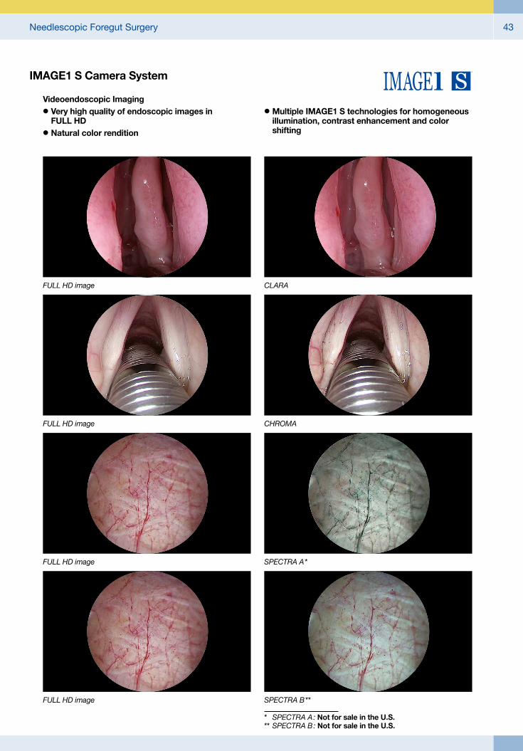
Videoendoscopic Imaging## Very high quality of endoscopic images in FULL HD
## Natural color rendition
## Multiple IMAGE1 S technologies for homogeneous illumination, contrast enhancement and color shifting
FULL HD image CHROMA
FULL HD image SPECTRA A *
FULL HD image
FULL HD image CLARA
SPECTRA B **
* SPECTRA A : Not for sale in the U.S.** SPECTRA B : Not for sale in the U.S.
IMAGE1 S Camera System

Needlescopic Foregut Surgery44
TC200EN* IMAGE1 S CONNECT, connect module, for use with up to 3 link modules, resolution 1920 x 1080 pixels, with integrated KARL STORZ-SCB and digital Image Processing Module, power supply 100 – 120 VAC/200 – 240 VAC, 50/60 Hz
including: Mains Cord, length 300 cm DVI-D Connecting Cable, length 300 cm SCB Connecting Cable, length 100 cm USB Flash Drive, 32 GB, USB silicone keyboard, with touchpad, US
* Available in the following languages: DE, ES, FR, IT, PT, RU
Specifications:
HD video outputs
Format signal outputs
LINK video inputs
USB interface SCB interface
- 2x DVI-D - 1x 3G-SDI
1920 x 1080p, 50/60 Hz
3x
4x USB, (2x front, 2x rear) 2x 6-pin mini-DIN
100 – 120 VAC/200 – 240 VAC
50/60 Hz
I, CF-Defib
305 x 54 x 320 mm
2.1 kg
Power supply
Power frequency
Protection class
Dimensions w x h x d
Weight
TC300 IMAGE1 S H3-LINK, link module, for use with IMAGE1 FULL HD three-chip camera heads, power supply 100 – 120 VAC/200 – 240 VAC, 50/60 Hz, for use with IMAGE1 S CONNECT TC200ENincluding:Mains Cord, length 300 cm
Link Cable, length 20 cm
For use with IMAGE1 S IMAGE1 S CONNECT Module TC200EN
IMAGE1 S Camera System
TC300 (H3-Link)
TH100, TH101, TH102, TH103, TH104, TH106 (fully compatible with IMAGE1 S) 22 2200 55-3, 22 2200 56-3, 22 2200 53-3, 22 2200 60-3, 22 2200 61-3, 22 2200 54-3, 22 2200 85-3 (compatible without IMAGE1 S technologies CLARA, CHROMA, SPECTRA*)
1x
100 – 120 VAC/200 – 240 VAC
50/60 Hz
I, CF-Defib
305 x 54 x 320 mm
1.86 kg
Camera System
Supported camera heads/video endoscopes
LINK video outputs
Power supply
Power frequency
Protection class
Dimensions w x h x d
Weight
Specifications:
TC200EN
TC300
* SPECTRA A : Not for sale in the U.S.** SPECTRA B : Not for sale in the U.S.
45Needlescopic Foregut Surgery
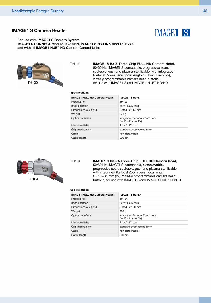
TH104
TH104 IMAGE1 S H3-ZA Three-Chip FULL HD Camera Head, 50/60 Hz, IMAGE1 S compatible, autoclavable, progressive scan, soakable, gas- and plasma-sterilizable, with integrated Parfocal Zoom Lens, focal length f = 15 – 31 mm (2x), 2 freely programmable camera head buttons, for use with IMAGE1 S and IMAGE 1 HUB™ HD/HD
IMAGE1 FULL HD Camera Heads
Product no.
Image sensor
Dimensions w x h x d
Weight
Optical interface
Min. sensitivity
Grip mechanism
Cable
Cable length
IMAGE1 S H3-ZA
TH104
3x 1/3" CCD chip
39 x 49 x 100 mm
299 g
integrated Parfocal Zoom Lens, f = 15 – 31 mm (2x)
F 1.4/1.17 Lux
standard eyepiece adaptor
non-detachable
300 cm
Specifications:
TH100 IMAGE1 S H3-Z Three-Chip FULL HD Camera Head, 50/60 Hz, IMAGE1 S compatible, progressive scan, soakable, gas- and plasma-sterilizable, with integrated Parfocal Zoom Lens, focal length f = 15 – 31 mm (2x), 2 freely programmable camera head buttons, for use with IMAGE1 S and IMAGE 1 HUB™ HD/HD
IMAGE1 FULL HD Camera Heads
Product no.
Image sensor
Dimensions w x h x d
Weight
Optical interface
Min. sensitivity
Grip mechanism
Cable
Cable length
IMAGE1 S H3-Z
TH100
3x 1/3" CCD chip
39 x 49 x 114 mm
270 g
integrated Parfocal Zoom Lens, f = 15 – 31 mm (2x)
F 1.4/1.17 Lux
standard eyepiece adaptor
non-detachable
300 cm
Specifications:
For use with IMAGE1 S Camera System IMAGE1 S CONNECT Module TC200EN, IMAGE1 S H3-LINK Module TC300 and with all IMAGE 1 HUB™ HD Camera Control Units
IMAGE1 S Camera Heads
TH100

Needlescopic Foregut Surgery46
9826NB
9826NB 26" FULL HD Monitor, wall-mounted with VESA 100 adaption, color systems PAL/NTSC, max. screen resolution 1920 x 1080, image fomat 16:9, power supply 100 – 240 VAC, 50/60 Hzincluding:External 24 VDC Power SupplyMains Cord
9619NB
9619NB 19" HD Monitor, color systems PAL/NTSC, max. screen resolution 1280 x 1024, image format 4:3, power supply 100 – 240 VAC, 50/60 Hz, wall-mounted with VESA 100 adaption,including:
External 24 VDC Power SupplyMains Cord
Monitors

47Needlescopic Foregut Surgery
Monitors
Optional accessories:9826SF Pedestal, for monitor 9826NB9626SF Pedestal, for monitor 9619NB
26"
9826NB
l
–
l
l
l
l
l
–
l
–
l
l
l
l
l
l
19"
9619NB
l
–
–
l
l
l
l
l
l
l
–
l
l
l
l
l
KARL STORZ HD and FULL HD Monitors
Wall-mounted with VESA 100 adaption
Inputs:
DVI-D
Fibre Optic
3G-SDI
RGBS (VGA)
S-Video
Composite/FBAS
Outputs:
DVI-D
S-Video
Composite/FBAS
RGBS (VGA)
3G-SDI
Signal Format Display:
4:3
5:4
16:9
Picture-in-Picture
PAL/NTSC compatible
19"
optional
9619NB
200 cd/m2 (typ)
178° vertical
0.29 mm
5 ms
700:1
100 mm VESA
7.6 kg
28 W
0 – 40°C
-20 – 60°C
max. 85%
469.5 x 416 x 75.5 mm
100 – 240 VAC
EN 60601-1, protection class IPX0
Specifications:
KARL STORZ HD and FULL HD Monitors
Desktop with pedestal
Product no.
Brightness
Max. viewing angle
Pixel distance
Reaction time
Contrast ratio
Mount
Weight
Rated power
Operating conditions
Storage
Rel. humidity
Dimensions w x h x d
Power supply
Certified to
26"
optional
9826NB
500 cd/m2 (typ)
178° vertical
0.3 mm
8 ms
1400:1
100 mm VESA
7.7 kg
72 W
5 – 35°C
-20 – 60°C
max. 85%
643 x 396 x 87 mm
100 – 240 VAC
EN 60601-1, UL 60601-1, MDD93/42/EEC, protection class IPX2

Needlescopic Foregut Surgery48
Accessories for Video Documentation
495 NA Fiber Optic Light Cable, with straight connector, diameter 3.5 mm, length 230 cm
Cold Light Fountain XENON 300 SCB
20 133101-1 Cold Light Fountain XENON 300 SCB
with built-in antifog air-pump, and integrated KARL STORZ Communication Bus System SCB power supply: 100 –125 VAC/220 –240 VAC, 50/60 Hz
including: Mains Cord SCB Connecting Cord, length 100 cm20133027 Spare Lamp Module XENON
with heat sink, 300 watt, 15 volt20133028 XENON Spare Lamp, only,
300 watt, 15 volt
20131501 Cold Light Fountain XENON NOVA® 175, power supply: 100 –125 VAC/220 –240 VAC, 50/60 Hz
including: Mains Cord20132026 XENON Spare Lamp,
175 watt, 15 volt
Cold Light Fountain XENON NOVA® 175

49Needlescopic Foregut Surgery
HAMOU ENDOMAT® with KARL STORZ SCBSuction and Irrigation System
26 3311 01-1 HAMOU® ENDOMAT® SCB, power supply 100 – 240 VAC, 50/60 Hz
including: Mains Cord 5x HYST Tubing Set*, for single use 5x LAP Tubing Set*, for single use SCB Connecting Cable, length 100 cm VACUsafe Promotion Pack Suction*, 2 l
Subject to the customer’s application-specific requirements additional accessories must be ordered separately.
*
ENDOFLATOR® 40 with KARL STORZ SCBwith High Flow Insufflation (40 l/min.)
UI400S1 ENDOFLATOR® 40 SCB including: ENDOFLATOR® 40 with KARL STORZ SCB
power supply 100 – 240 VAC, 50/60 Hz Mains Cord 6x Single-use insufflation tubing set with gas filter, sterile * Universal Wrench SCB Connecting Cable, length 100 cm
Please note: For fully utilizing maximum insufflation capacity of the ENDOFLATOR® 40 the use of KARL STORZ HiCap® Trocars is recommended. For additional information see catalog LAPARO SCOPY.
*

Needlescopic Foregut Surgery50
UG540 Monitor Swivel Arm, height and side adjustable, can be turned to the left or the right side, swivel range 180°, overhang 780 mm, overhang from centre 1170 mm, load capacity max. 15 kg, with monitor fixation VESA 5/100, for usage with equipment carts UGxxx
UG540
Equipment Cart
UG220
UG220 Equipment Cart wide, high, rides on 4 antistatic dual wheels equipped with locking brakes 3 shelves, mains switch on top cover, central beam with integrated electrical subdistributors with 12 sockets, holder for power supplies, potential earth connectors and cable winding on the outside,
Dimensions: Equipment cart: 830 x 1474 x 730 mm (w x h x d), shelf: 630 x 510 mm (w x d), caster diameter: 150 mm
inluding: Base module equipment cart, wide Cover equipment, equipment cart wide Beam package equipment, equipment cart high 3x Shelf, wide Drawer unit with lock, wide 2x Equipment rail, long Camera holder

51Needlescopic Foregut Surgery
Recommended Accessories for Equipment Cart
UG310 Isolation Transformer, 200 V – 240 V; 2000 VA with 3 special mains socket, expulsion fuses, 3 grounding plugs, dimensions: 330 x 90 x 495 mm (w x h x d), for usage with equipment carts UGxxx
UG310
UG410 Earth Leakage Monitor, 200 V – 240 V, for mounting at equipment cart, control panel dimensions: 44 x 80 x 29 mm (w x h x d), for usage with isolation transformer UG310
UG410
UG510 Monitor Holding Arm, height adjustable, inclinable, mountable on left or rigth, turning radius approx. 320°, overhang 530 mm, load capacity max. 15 kg, monitor fixation VESA 75/100, for usage with equipment carts UGxxx
UG 510

Needlescopic Foregut Surgery52
Notes:

with the compliments of
KARL STORZ — ENDOSKOPE