Embed Size (px)
Citation preview
NECROTIZING ENTEROCOLITIS*
CHARLES R. ROSENFELDD)epartment of Pediatrics
Albert Einstein College of MedicineNew York, N. Y.
I N I89I Genersich' reported the case of a 45-hour-old prematureinfant who had vomiting, cyanosis, and abdominal distention; death
occurred within 24 hours. At postmortem examination the infant wasfound to have inflammation of the ileum and an area of perforation.No cause could be determined. Subsequent observers have reportedsimilar cases in increasing numbers and have labeled the disease "ne-crotizing enterocolitis." This entity has been characterized by vomit-ing, abdominal distention, and bloody stools; roentgen studies have re-vealed evidence of pneumatosis intestinalis, pneumoperitoneum, or gasin the main portal vein. Up to the present time slightly more thanioo cases have been reported. Approximately 3o00/K of the patientshave survived. It is the purpose of this paper to present the case of achild who survived and to discuss the disease.
CASE REPORT
On September I4, i969 an infant weighing 1,2I9 gm. was born toa 27-year-old Negro primigravida after an uneventful 3o-week gesta-tion and delivery. During the first 24 hours the infant had frequentattacks of cyanosis, which were treated with 400'0 oxygen. At 27 hoursshe was transferred to the Neonatal Intensive Care Unit of the BronxMunicipal Hospital Center. On admission she was noted to be a pink,vigorous infant not in respiratory distress; there were occasional apneicepisodes, which responded to stimulation. Vital signs: pulse ii2/min.,respirations 48/min., temperature 36.40C.; weight I,200 gm. The phy-sical examination was unremarkable, except for the absence of ear cart-ilage and breast tissue; neurological findings were compatible with agestational age of 29 to 30 weeks. At the time of admission the follow-ing laboratory observations were made: hemoglobin 20.2 gm.00, hem-atocrit 59%, white-blood count 7,50oo/mm.3 with normal differential
*Presented at a meeting of the Section on Pediatrics, April 9, 1970.
Vol. 47, No. 2, February 1971
i6zC. R. ROSENFELD
uro,
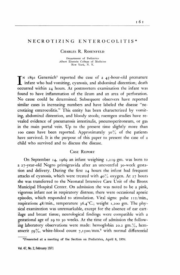
Fig. 1. Supine x ray of the abdomen at 52 hours of age; gas under the diaphragmand overlying the liver.
Bull. M.Y. Acad. Mod.
i 6 2 C. R. ROSENFELD
I L mwl. W%
NECROTIZING ENTEROCOLITIS
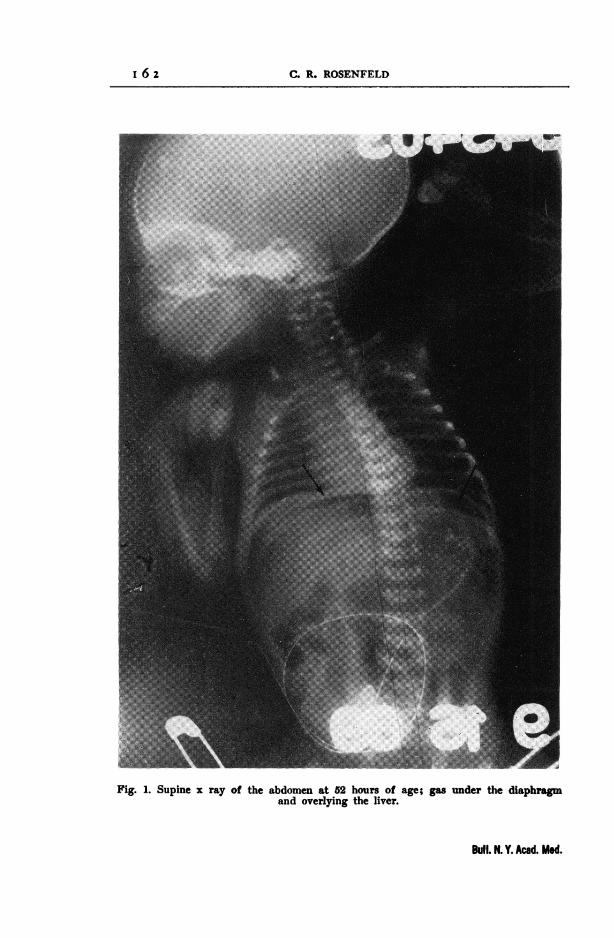
Fig. 2. X ray of the chest with catheter extending from the external jugular veininto the superior vena cava for intravenous hyperalimentation.
Vol. 47, No. 2, February 1971
I 6 3

TABLE I. COMPOSITION OF INFUSATE (SOLUTION No. 1)
Fibrin hydrolysate (5%)-dextrose (5%) * 375 cc.Dextrose (50%)* 200 cc.Vitamin C 500 mg.Berocia Ct 1 cc.Sodium heparin (1000u./ml.) 500 unitsPotassium chloride 10 meq.Sodium chloride 10 meq.
*Abbott Laboratories, N. Chicago, Ill.tRoche Laboratories, Nutley, N.J.
TABLE II. COMPOSITION OF INFUSATE AS SUGGESTED BY FILLER, ET AL.8(SOLUTION No. 2)
Constituent Amount (cc.)
Fibrin hydrolysate (5%) dextrose (5%)* 1,000.0Dextrose (50%) * 500.0Potassium chloride (2 mEq./ml.) 15.0Magnesium sulfate (10%) 1.25Calcium gluconate (10%) 15.0Phytadione (Agua Mephyton) 0.2 mg./ml.) 1.5Vitamin B,_, (100 m./cc.) 0.1Multivitamin infusion (MVI)t 1.5Sodium chloride (2.5 mEq./ml.) 6.0Sodium heparin (1,000 u./ml.) 1.5
*Abbott Laboratories, N. Chicago, Ill.tU.S. Vitamins and Pharmaceutical Corp., New York, N. Y.
and 25 nucleated RBC/ ioo WBC; blood type 0-positive, Coombstest negative, urine normal, blood sugar iio mg. %. Shortly after ad-mission a catheter was placed in an umbilical artery and an infusionof 5% dextrose and water was begun. The patient was then placedin 6o% oxygen; after 15 minutes arterial blood was drawn from thecatheter for studies. The results were: pH 7.35, PCo2 45 mm. Hg,Po2 240 mm. Hg, total C02 24.5 meq./l, bicarbonate 23.5 meq./l.,and a base deficit of 2.5 meq./l. The concentration of oxygen was thenreduced. Episodes of apnea continued, but became less frequent. At38 hours the infant was doing well and was placed in room air; half-strength Alacta feedings were started. At approximately 52 hours of
Bull. N. Y. Acad. Med.
I 64 C. R. ROSENFELD
NECROTIZING ENTEROCOLITIS
I0 10 20 30 40 50AGE IN DAYS
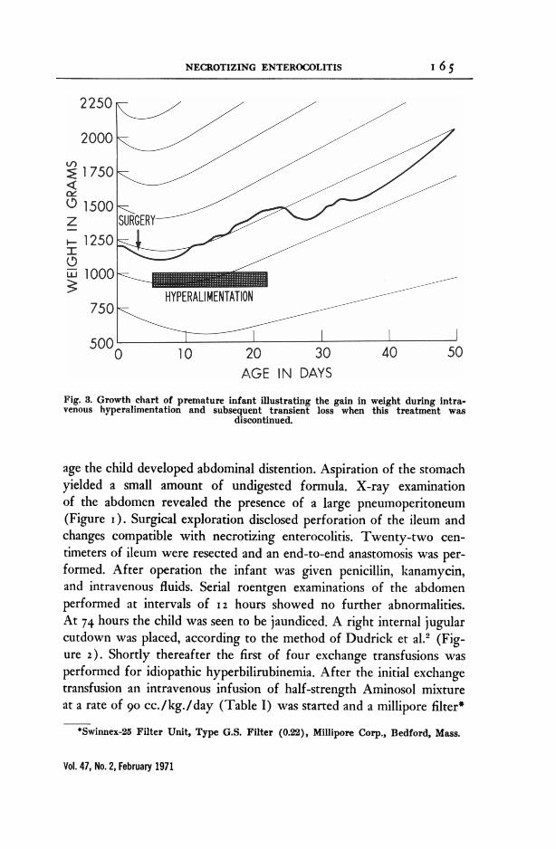
Fig. 3. Growth chart of premature infant illustrating the gain in weight during intra-venous hyperalimentation and subsequent transient loss when this treatment was
discontinued.
age the child developed abdominal distention. Aspiration of the stomachyielded a small amount of undigested formula. X-ray examinationof the abdomen revealed the presence of a large pneumoperitoneum(Figure i). Surgical exploration disclosed perforation of the ileum andchanges compatible with necrotizing enterocolitis. Twenty-two cen-timeters of ileum were resected and an end-to-end anastomosis was per-formed. After operation the infant was given penicillin, kanamycin,and intravenous fluids. Serial roentgen examinations of the abdomenperformed at intervals of 12 hours showed no further abnormalities.At 74 hours the child was seen to be jaundiced. A right internal jugularcutdown was placed, according to the method of Dudrick et al.2 (Fig-ure 2). Shortly thereafter the first of four exchange transfusions wasperformed for idiopathic hyperbilirubinemia. After the initial exchangetransfusion an intravenous infusion of half-strength Aminosol mixtureat a rate of go cc./kg./day (Table I) was started and a millipore filter*
*Swinnex-25 Filter Unit, Type G.S. Filter (0.22), Millipore Corp., Bedford, Mass.
Vol. 47, No. 2, February 1971
I 65
i6
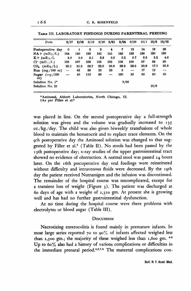
TABLE III. LABORATORY FINDINGS DURING PARENTERAL FEEDING
Date 9/17 9/18 9/19 9/20 9/21 9/24 9/29 10/1 10/6 10/12
Postoperative day 0 1 2 3 4 7 12 14 19 25NA+ (mE(1./L) 144 140 135 141 141 142 138 136 130 136K+ (mEj./L 5.9 5.0 3.1 3.3 6.0 5.2 5.7 5.6 5.2 4.6CI- (mE I /L) 109 107 103 105 102 108 109 97 98 90CO (mEq./L) 20.1 21.2 28.7 32.0 26.6 23.6 23.6 20.8 17.1 25.9Bun (mg./100 cc) - 43 39 26 23 9 - 17 12 -
Sugar (ing./100 40 110 85 - 130 85 90 50 65cc)
Solution No. 1* 9/26Solution No. 2t 10/6
*Aminosol, Abbott Laboratories, North Chicago, IlI.tAs per Filler et al.'
was placed in line. On the second postoperative day a full-strengthsolution was given and the volume was gradually increased to I35cc./kg./day. The child was also given biweekly transfusions of wholeblood to maintain the hematocrit and to replace trace elements. On the9th postoperative day the Aminosol solution was changed to that sug-gested by Filler et al.3 (Table II). No stools had been passed by theI3th postoperative day; x-ray studies of the upper gastrointestinal tractshowed no evidence of obstruction. A normal stool was passed 24 hourslater. On the i6th postoperative day oral feedings were reinstitutedwithout difficulty and intravenous fluids were decreased. By the i9thday the patient received Nutramigen and the infusion was discontinued.The remainder of the hospital course was uncomplicated, except fora transient loss of weight (Figure 3). The patient was discharged at6o days of age with a weight of 2,320 gin. At present she is growingwell and has had no further gastrointestinal dysfunction.
At no time during the hospital course were there problems withelectrolytes or blood sugar (Table 111).
DISCUSSION
Necrotizing enterocolitis is found mainly in premature infants. Inmost large series reported 70 to 90% of infants affected weighed lessthan 2,500 gm.; the majority of these weighed less than i,6oo gin. 4-8Up to 6o% also had a history of various complications or difficulties inthe immediate prenatal period.4'5'7'9 The maternal complications con-
Bull. N. Y. Acad. Med.
I 66 C. R. ROSENFELD
NECROTIZING ENTEROCOLITIS
sisted of either premature rupture of membranes, prenatal fevers, orinfected amniotic fluid, while the neonatal difficulties consisted of lowApgar scores, minor apneic episodes, cyanosis, idiopathic respiratorydistress syndrome, or exchange transfusion.
The infants seem to do well for the first two to five days despitethe problems previously mentioned. Then they experience the insidiousonset of vomiting associated with delayed gastric emptying. The epi-sodes of apnea tend to recur or increase in severity.4 Chest x rays aregenerally normal. The symptoms are followed by abdominal distentionand by an increase in the vomiting; the vomitus is now bile-stained.The stools are bloody and often loose.4'7'9 During this phase of theillness the findings revealed on x-ray examination of the abdomen arevariable. In most reported cases the subsequent course has been ful-minating. The infant appears lethargic, has increasing apneic episodes,becomes jaundiced, enters a state of shock resembling that seen withsepsis, and dies shortly thereafter despite all modes of treatment.
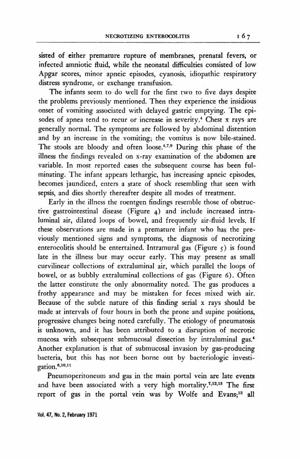
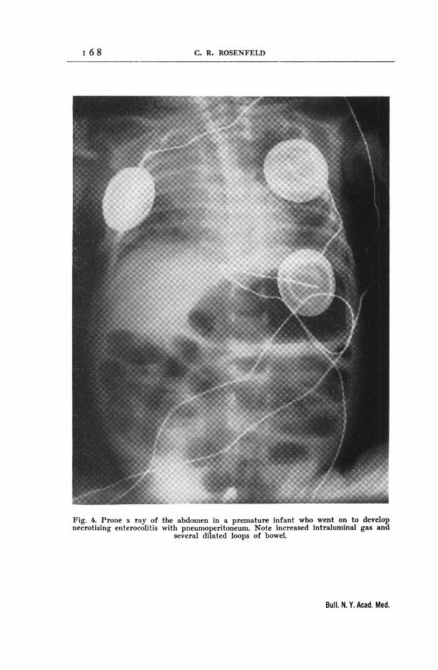
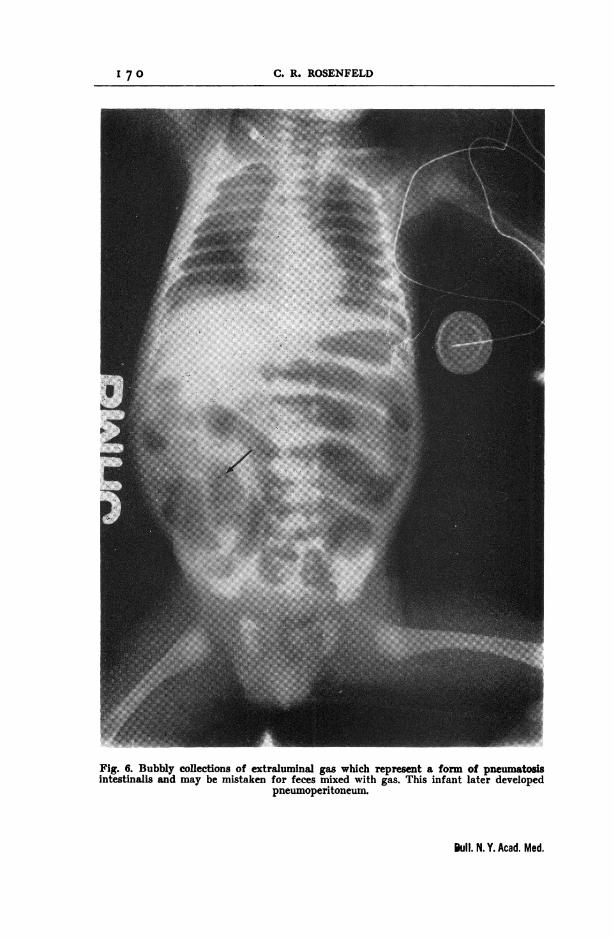
Early in the illness the roentgen findings resemble those of obstruc-tive gastrointestinal disease (Figure 4) and include increased intra-luminal air, dilated loops of bowel, and frequently air-fluid levels. Ifthese observations are made in a premature infant who has the pre-viously mentioned signs and symptoms, the diagnosis of necrotizingenterocolitis should be entertained. Intramural gas (Figure 5) is foundlate in the illness but may occur early. This may present as smallcurvilinear collections of extraluminal air, which parallel the loops ofbowel, or as bubbly extraluminal collections of gas (Figure 6). Oftenthe latter constitute the only abnormality noted. The gas produces afrothy appearance and may be mistaken for feces mixed with air.Because of the subtle nature of this finding serial x rays should bemade at intervals of four hours in both the prone and supine positions,progressive changes being noted carefully. The etiology of pneumatosisis unknown, and it has been attributed to a disruption of necroticmucosa with subsequent submucosal dissection by intraluminal gas.4Another explanation is that of submucosal invasion by gas-producingbacteria, but this has not been borne out by bacteriologic investi-gation'6'10'"
Pneumoperitoneum and gas in the main portal vein are late eventsand have been associated with a very high mortality.7'2"3 The firstreport of gas in the portal vein was by Wolfe and Evans;12 all
Vol. 47, No. 2, February 1971
I 6 7
i 68 C. R. ROSENFELD
Fig. 4. Prone x ray of the abdomen in a premature infant who went on to developnecrotizing enterocolitis with pneumoperitoneum. Note increased intraluminal gas and
several dilated loops of bowel.
Bull. N. Y. Acad. Med.
NECROTIZING ENTEROCOLITIS
Fig. 5. Pneumatosis intestinalis presenting as curvilinear collections of extraluminalgas in a premature infant who later developed pneumoperitoneum.
six of their patients died. The following year Miskin et al. reportedsix more cases; of the four survivors two had received medical therapyonly.13 Though the gravity of the situation is much increased wheneither pneumoperitoneum or gas in the portal vein is present, onemust remember that the disease is not always fatal, as our case shows.
At autopsy and in surgical specimens the microscopic findings havebeen nonspecific. The lesions have been found predominantly in thelower ileum, cecum, and the ascending colon. The early changes con-
Vol. 47, No. 2, February 1971
I 6 9
17 .R OEFL
P
Fig. 6. Bubbly collections of extraluminal gas which represent a form of pneumatosisintestinalis and may be mistaken for feces mixed with gas. This infant later developed
pneumoperitoneum.
Bull. N. Y. Acad. Med.
C. R. ROSENFELDI 7 0
NECROTIZING ENTEROCOLITIS
sist of broadening of the mucosal villi with dilation of venules andcapillaries, along with moderate infiltration by mononuclear cells. Inlater lesions the tissue is markedly friable; there is hemorrhage in thelamina propria, sloughing of mucosa, perforations, and an overlyingpseudomembrane.4'9 There is no evidence of vasculitis, thrombosedvessels, or aganglionosis, and no bacteria have been seen or culturedfrom lesions.
In the treatment of this disorder early recognition is crucial, andshould be followed by prompt initiation of gastrointestinal decom-pression. Antibiotic therapy should include coverage for enteric organ-isms. The usual supportive measures should be used. Surgical con-sultation should be obtained immediately because of the frequentlyrapid progression to perforation. Serial x-ray films of the abdomenshould be observed closely for extension of intramural gas or for othersigns of progression. If during the ensuing 24 to 48 hours the infant'scondition deteriorates or there are signs of radiologic progression, earlysurgical intervention is indicated and may lead to resection of variablelengths of bowel with ileostomy or colostomy. This course was fol-lowed by Stevenson et al.4 in 2I cases; 67% survival was obtained andmay be compared with the II% reported by Mizrahi5 in a series inwhich an operation was performed only when there was evidence ofperforation. In the former series nine out of I2 infants who requiredoperation lived; this represented 64% of the survivors. None of theinfants reported by Mizrahi who underwent operation survived.
In infants requiring operations antibiotics should be continuedfor at least seven to io days in the postoperative period, andserial roentgen examinations should be made for 24 to 48 hours in orderto determine further extension of the disease. In infants who have hadextensive disease that required large resections of bowel or in those inwhom the extent of the disease is questionable at the time of operation,hyperalimentation may be life-saving.2'3 A barium enema should beplanned a few weeks after recovery for those who survive after medi-cal therapy, since there have been reports of intestinal obstruction afterrecovery.11,14
ACKNOWLEDGMENTS
I express my appreciation to Dr. Keith Schneider, Division of Pedi-atric Surgery, Albert Einstein College of Medicine, who performed the
Vol. 47, No. 2, February 1971
I 7 I
I 7 2 C. R. ROSENFELD
operation and was closely involved in the care of the patient, and toDrs. Larry Gartner and Chester Edelman for their help in preparingthis paper.
R E F E R E N C E S
1. Genersich, A.: Bauchfellentzundung beimNeugeborenen in Folge von Perforationdes Illeum. Arch. Path. Anat. 126:485,1891.
2. Wilmore, D. W., Groff, D. B., Bishop,H. C. and Dudrick, S. J.: Total paren-teral nutrition in infants with catas-trophic gastrointestinal anomalies. J.Ped. Surg. 4:181, 1969.
3. Filler, R. M., Eraklis, A. J., Rubin,V. G. and Das, J. B.: Long-term totalparenteral nutrition in infants. NewEng. J. Med. 281:589, 1969.
4. Stevenson, J. K., Graham, C. B., Oliver,T. K. and Goldenberg, V. E.: Neonatalnecrotizing enterocolitis. Amer. J. 8Surg.148:260, 1969.
5. Mizrahi, A., Barlow, O., Berdon, W.,Blanc, W. A. and Silverman, W. A.:Necrotizing enterocolitis in prematureinfants. Pediatrics 66:697, 1965.
6. Hyde, G. A. and Santulli, T. V.: Idio-pathic perforation of the small intes-tine in the neonatal period. Pediatrics26:261, 1960.
7. Touloukian, R. J., Berdon, W. E.,Amoury, R. A. and Santulli, T. V.:Surgical experience with necrotizingenterocolitis in the infant. J. Ped. Surg.2:389, 1967.
8. Schmid, 0. and Quaiser, K.: tYber eine
besondere schwere verlaufende Formvon Enteritis beim Saugling. Oesterr.Kinderh. 8:144, 1953.
9. Berdon, W. E., Grossman, H., Baker,D. H., Mizrahi, A., Barlow, 0. andBlanc, W. A.: Necrotizing enterocolitisin the premature infant. Radiology83:879, 1964.
10. Waldhausen, J. A., Herendean, T. andKing, H.: Necrotizing colitis of thenewborn: Common cause of perforationof the colon. Surgery 54:365, 1963.
11. Rabinowitz, J. G., Wolfe, B. S., Feller,M. R. and Krasna, I.: Colonic changesfollowing necrotizing enterocolitis inthe newborn. Amer. J. Roentgen.103:359, 1968.
12. Wolfe, J. N. and Evans, W. A.: Gasin the portal vein of the liver in in-fants. Amer. J. Roentgen. 74:486, 1955.
13. Miskin, M. et al.: Gas in the intestinalwall and portal venous system in in-fants. Canad. Med. Assoc. J. 101:129,1969.
14. Krasna, I. H., Becker, J. M., Schneider,K. M., Beck, A. R. and Strauss, L.:Colonic stenosis following necrotizingenterocolitis of the newborn. Presentedat The American Academy of Pediat-rics (Chicago), 1969.
Bull. N. Y. Acad. Med.