Embed Size (px)
Citation preview
NCERT Solutions for Class 11 Biology Chapter 7 Structural Organisation in Animals
Q1. Answer in one word or one line
(i).Give the common name of Periplaneta americana.
Answer:
The common name of Periplaneta americana is cockroach.
Q1 . Answer in one word or one line
(ii) How many spermathecae are found in earthworm?
Answer:
Four pairs of spermathecae are found in the earthworm. These are found to be located
between the sixth and the ninth segments. The function of spermathecae is to receive
and store the spermatozoa during copulation.
Q1. Answer in one word or one line
(iii) What is the position of ovaries in cockroach?
Answer:
The pair of ovaries is located between 12th and 13th abdominal segments in the
cockroach.
Q1. Answer in one word or one line
(iv) How many segments are present in the abdomen of cockroach?
Aakas
h Ins
titute
Answer:
In total, 10 segments are present in the abdomen of cockroach whether male or female.
Q1. Answer in one word or one line
(v) Where do you find Malpighian tubules?
Answer:
Malpighian tubules are the major excretory organs of cockroaches. These form a part of
the alimentary canal.
Q2. Answer the following
(i) What is the function of nephridia?
Answer:
Nephridia are the excretory organs of earthworm. They are involved in the functions of
excretion and osmoregulation in the earthworm.
Q2. Answer the following
(ii) How many types of nephridia are found in earthworm based on their location?
Answer:
On the basis of location, nephridia can be of three types i.e. septal nephridia,
integumentary nephridia and pharyngeal nephridia. Aak
ash I
nstitu
te
1. Septal nephridia: These nephridia are found to be present on both sides of the
intersegmental septa behind the 15th segment. They open into the intestines.
2. Integumentary nephridia: These nephridia lie attached to the body wall from the third
segment to the last segment, which opens on the body surface.
3. Pharyngeal nephridia: These nephridia are found to be are present in fourth, fifth. and
sixth segments.
Solutions for NCERT class 11 biology chapter 7 structural organisation in animals:
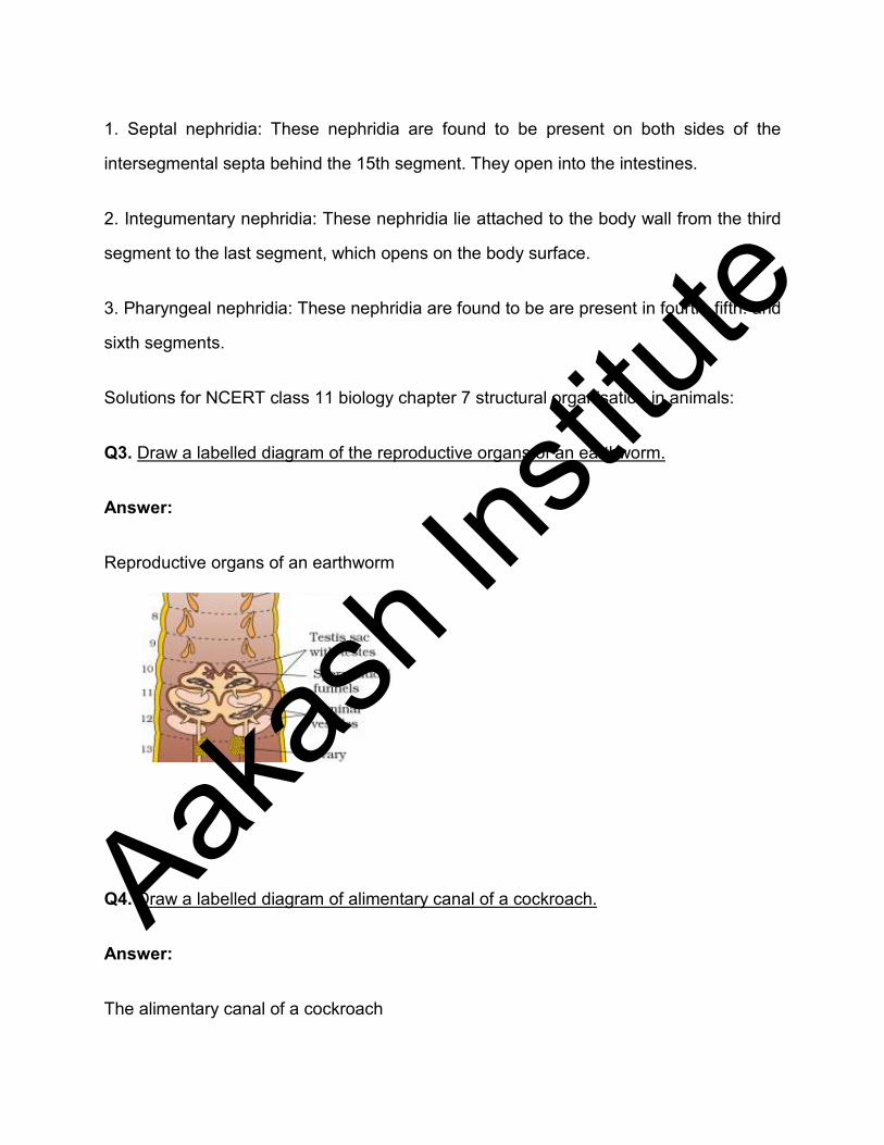
Q3. Draw a labelled diagram of the reproductive organs of an earthworm.
Answer:
Reproductive organs of an earthworm
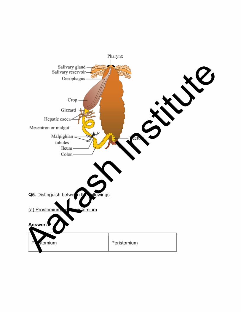
Q4. Draw a labelled diagram of alimentary canal of a cockroach.
Answer:
The alimentary canal of a cockroach
Aakas
h Ins
titute
Q5. Distinguish between the followings
(a) Prostomium and peristomium
Answer:
Prostomium Peristomium Aakas
h Ins
titute
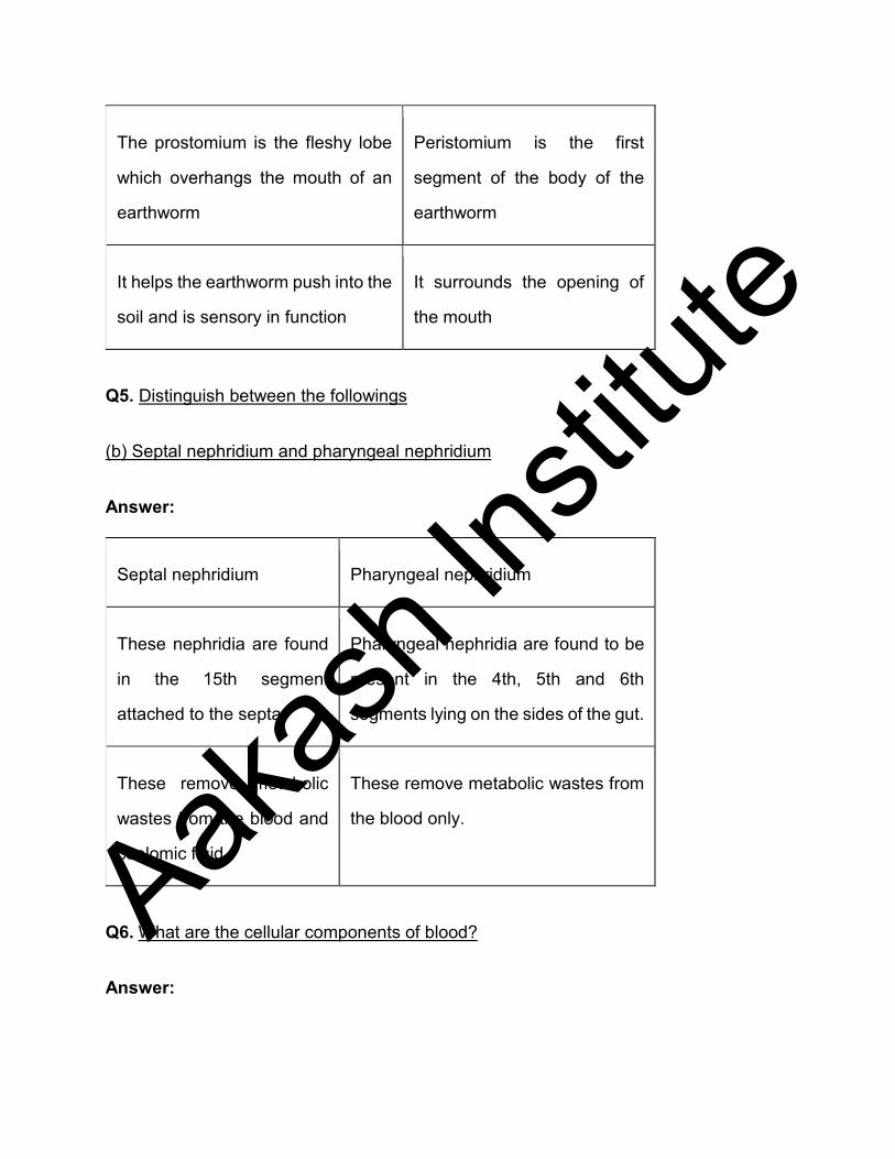
The prostomium is the fleshy lobe
which overhangs the mouth of an
earthworm
Peristomium is the first
segment of the body of the
earthworm
It helps the earthworm push into the
soil and is sensory in function
It surrounds the opening of
the mouth
Q5. Distinguish between the followings
(b) Septal nephridium and pharyngeal nephridium
Answer:
Septal nephridium Pharyngeal nephridium
These nephridia are found
in the 15th segment
attached to the septa.
Pharyngeal nephridia are found to be
present in the 4th, 5th and 6th
segments lying on the sides of the gut.
These remove metabolic
wastes from the blood and
coelomic fluid
These remove metabolic wastes from
the blood only.
Q6. What are the cellular components of blood?
Answer:
Aakas
h Ins
titute
Blood is the fluid connective tissue. It consists of blood plasma and cellular components.
The cellular components of blood are of following types.
1. Erythrocytes- These are red blood cells. Erythrocytes are red due to the presence of
haemoglobin. Erythrocytes are biconcave, coloured cells devoid of a nucleus. These
mainly help in transporting respiratory gases.
2. Leucocytes- These are white blood cells. There are 5 types of leucocytes i.e.
neutrophils, eosinophils, basophils, lymphocytes and monocytes. Leucocytes are mainly
involved in fighting infections and providing immunity.
3. Thrombocytes- These are blood platelets involved in the coagulation of blood.
Q7. What are the following and where do you find them in animal body.
(a) Chondriocyte
Answer:
Chondriocytes- These are cells of cartilages. They are found to be present in the small
cavities within the matrix secreted by them.
Q7. What are the following and where do you find them in animal body.
(b) Axon
Answer:
Axons- An axon is the long, slender projection found in the neuron. It helps in carrying
nerve impulses from the neuron body.
Q7. What are the following and where do you find them in animal body
Aakas
h Ins
titute
(c) Ciliated epithelium
Answer:
Ciliated epithelium- The ciliated epithelium consists of cells which bear fine, vibratile
cytoplasmic processes called cilia on its free surface. It is found in the inner lining of
bronchioles, urinary tubules of kidneys, nasal passage, oviducts and ventricles of the
brain.
NCERT solutions for class 11 biology chapter 7 structural organisation in animals:
Q8. Describe various types of epithelial tissues with the help of labelled diagrams.
Answer:
Epithelial tissue provides covering or lining for some parts of the body. It consists of a
layer of cells and a basement membrane. The cells of epithelium are compactly packed
without intercellular space.
(a) Simple epithelium : It is the single layer of cells which are in direct contact with the
basement membrane. Simple epithelium is further subdivided into the following types:
(i) Simple squamous epithelium: It consists of a single layer of flat cells with irregular
boundaries. Simple squamous epithelium is mostly found in the walls of the blood vessels
and in the lining of alveoli.
(ii) Simple cuboidal epithelium: It consists of a single layer of cube-like cells and is present
in regions where secretion and absorption of substances take place such as the proximal
convoluted tubule region of the nephron.
Aakas
h Ins
titute
(iii) Simple columnar epithelium: This epithelium is formed by a single layer of tall, slender
cells with their nuclei present at the base of the cells. These generally possess microvilli
on the free surfaces. Columnar epithelium forms the lining of the stomach and intestines.
The major function of simple columnar epithelium is secretion and absorption.
(iv) Ciliated epithelium: It consists of columnar or cuboidal cells with cilia on their free
surfaces. They are present in bronchioles and oviducts from where they direct mucus and
eggs in specific directions.
(v) Glandular epithelium: These are columnar or cuboidal cells involved in the secretion
of substances. Glands are of two types, unicellular glands (goblet cells of the alimentary
canal) and multicellular glands (salivary glands). They can be classified as exocrine
(ductless glands) and endocrine glands (duct glands) based on the method through which
they release enzymes.
(b) Compound epithelium : When the simple epithelium is consists of many layers of
cells, it is called compound epithelium. The compound epithelium is involved mainly in
the function of providing protection and has a limited role in secretion and absorption.
Examples of compound epithelium include the dry surface of the skin or moist inner lining
of the buccal cavity, pharynx, pancreatic ducts, and the inner lining of ducts of salivary
glands.
Aakas
h Ins
titute
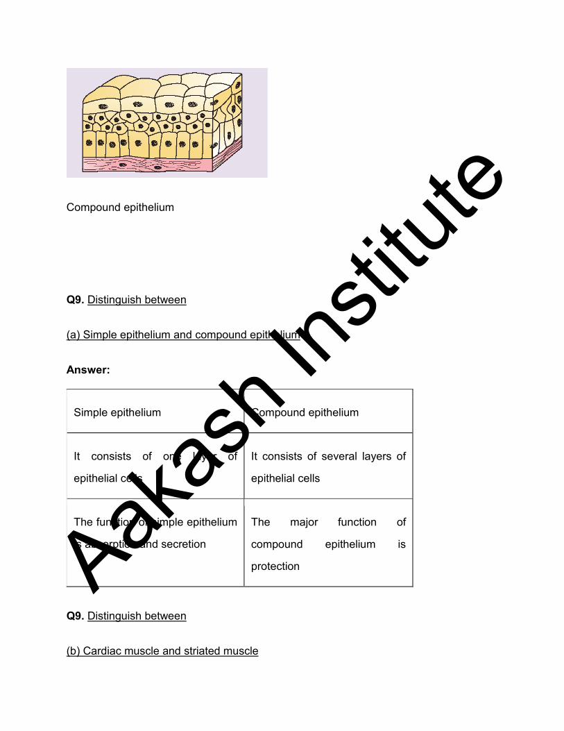
Compound epithelium
Q9. Distinguish between
(a) Simple epithelium and compound epithelium
Answer:
Simple epithelium Compound epithelium
It consists of one layer of
epithelial cells
It consists of several layers of
epithelial cells
The function of simple epithelium
is absorption and secretion
The major function of
compound epithelium is
protection
Q9. Distinguish between
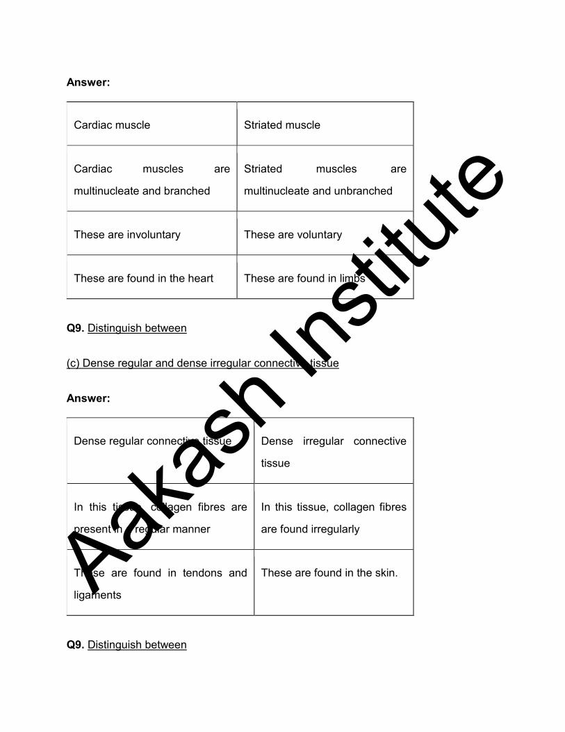
(b) Cardiac muscle and striated muscle
Aakas
h Ins
titute
Answer:
Cardiac muscle Striated muscle
Cardiac muscles are
multinucleate and branched
Striated muscles are
multinucleate and unbranched
These are involuntary These are voluntary
These are found in the heart These are found in limbs
Q9. Distinguish between
(c) Dense regular and dense irregular connective tissue
Answer:
Dense regular connective tissue Dense irregular connective
tissue
In this tissue, collagen fibres are
present in a regular manner
In this tissue, collagen fibres
are found irregularly
These are found in tendons and
ligaments
These are found in the skin.
Q9. Distinguish between
Aakas
h Ins
titute
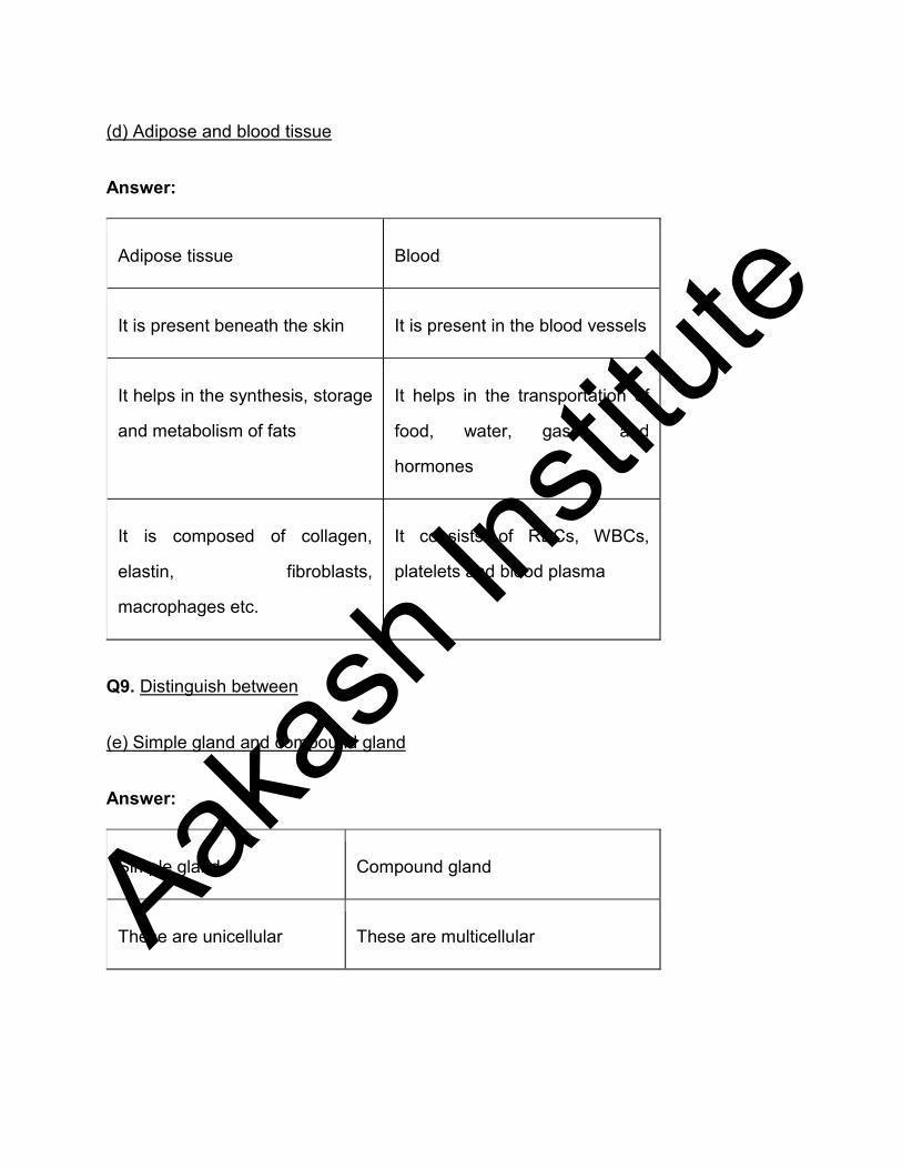
(d) Adipose and blood tissue
Answer:
Adipose tissue Blood
It is present beneath the skin It is present in the blood vessels
It helps in the synthesis, storage
and metabolism of fats
It helps in the transportation of
food, water, gases and
hormones
It is composed of collagen,
elastin, fibroblasts,
macrophages etc.
It consists of RBCs, WBCs,
platelets and blood plasma
Q9. Distinguish between
(e) Simple gland and compound gland
Answer:
Simple gland Compound gland
These are unicellular These are multicellular Aakas
h Ins
titute

These possess isolated
glandular cells
These type of glands are composed
of secretory cells
Q10. Mark the odd one in each series:
(a) Areolar tissue; blood; neuron; tendon
Answer:
Areolar tissue, blood and tendons are types of connective tissue whereas neuron is
nervous tissue. Thus, neuron is odd one out.
Q10. Mark the odd one in each series:
(b) RBC; WBC; platelets; cartilage
Answer:
RBS, WBC and platelets are cellular components of blood while cartilage is specialised
connective tissue. Thus, cartilage is the odd one out in the series.
Q10. Mark the odd one in each series:
(c) Exocrine; endocrine; salivary gland; ligament
Answer:
Exocrine, endocrine and salivary glands are simple glandular epithelium. However,
ligaments represent connective tissue, thus, the ligament is the odd one out in the series.
Q10. Mark the odd one in each series:
Aakas
h Ins
titute
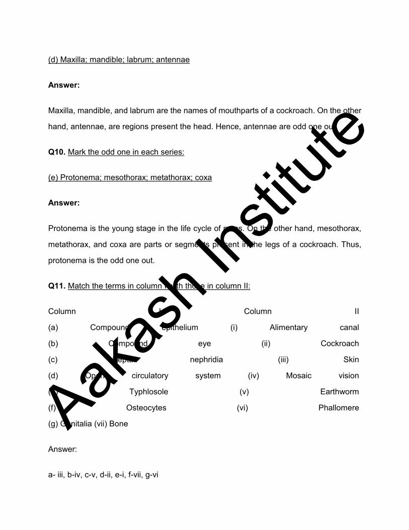
(d) Maxilla; mandible; labrum; antennae
Answer:
Maxilla, mandible, and labrum are the names of mouthparts of a cockroach. On the other
hand, antennae, are regions present the head. Hence, antennae are odd one out.
Q10. Mark the odd one in each series:
(e) Protonema; mesothorax; metathorax; coxa
Answer:
Protonema is the young stage in the life cycle of moss. On the other hand, mesothorax,
metathorax, and coxa are parts or segments present in the legs of a cockroach. Thus,
protonema is the odd one out.
Q11. Match the terms in column I with those in column II:
Column I Column II
(a) Compound epithelium (i) Alimentary canal
(b) Compound eye (ii) Cockroach
(c) Septal nephridia (iii) Skin
(d) Open circulatory system (iv) Mosaic vision
(e) Typhlosole (v) Earthworm
(f) Osteocytes (vi) Phallomere
(g) Genitalia (vii) Bone
Answer:
a- iii, b-iv, c-v, d-ii, e-i, f-vii, g-vi
Aakas
h Ins
titute
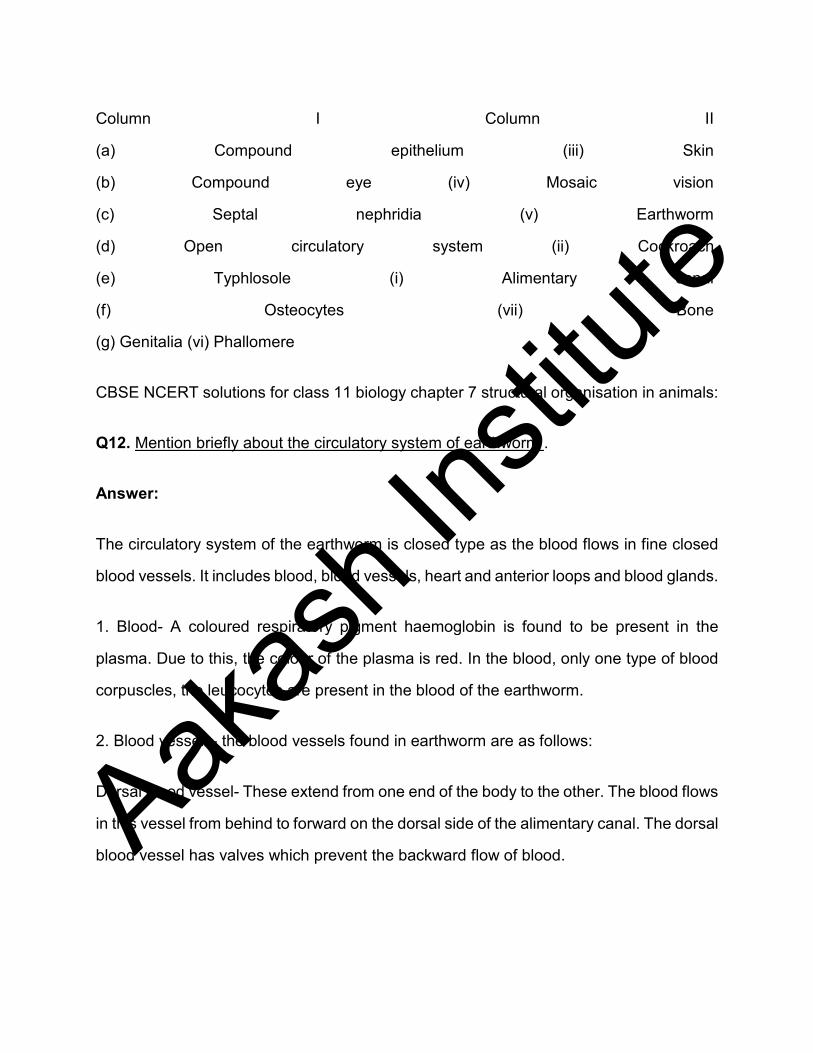
Column I Column II
(a) Compound epithelium (iii) Skin
(b) Compound eye (iv) Mosaic vision
(c) Septal nephridia (v) Earthworm
(d) Open circulatory system (ii) Cockroach
(e) Typhlosole (i) Alimentary canal
(f) Osteocytes (vii) Bone
(g) Genitalia (vi) Phallomere
CBSE NCERT solutions for class 11 biology chapter 7 structural organisation in animals:
Q12. Mention briefly about the circulatory system of earthworm .
Answer:
The circulatory system of the earthworm is closed type as the blood flows in fine closed
blood vessels. It includes blood, blood vessels, heart and anterior loops and blood glands.
1. Blood- A coloured respiratory pigment haemoglobin is found to be present in the
plasma. Due to this, the colour of the plasma is red. In the blood, only one type of blood
corpuscles, the leucocytes are present in the blood of the earthworm.
2. Blood vessels- the blood vessels found in earthworm are as follows:
Dorsal blood vessel- These extend from one end of the body to the other. The blood flows
in this vessel from behind to forward on the dorsal side of the alimentary canal. The dorsal
blood vessel has valves which prevent the backward flow of blood. Aakas
h Ins
titute

Ventral blood vessel- These extend from the one end to the other end of the body. It does
not have any valves and flow of the blood is from the anterior to the posterior end of the
body. It is the major distributing vessel.
Sub-neural blood vessel- It runs from the posterior end of the body up to the 14th segment
in front. It collects blood from the body wall and nerve cord. This blood is then sent to the
dorsal blood vessel through commissural vessels.
Lateral oesophageal vessel- These are the paired blood vessels lying one on either
ventrolateral side of the alimentary canal between the body wall and the alimentary canal
in the first 14th segment.
Supra-oesophageal blood vessel- It is a single vessel which lies on the dorsal side of the
alimentary canal between the 9th and 13th segment. It receives blood from the lateral
oesophageal through two pairs of anterior loops and pours into two pairs of latero-
oesophageal hearts present in the 12th and 13th segments.
Hearts and anterior loops- In earthworm, four pairs of tubular hearts which are provided
with valves. The anterior two pairs of the heart are known as the lateral heart and they lie
in the 7th and 9th segment. They receive blood from the dorsal blood vessel and convey
it to the ventral blood vessel. The posterior two pairs of hearts are situated in the 12th
and 13th segments. These carry blood from the dorsal blood vessel and supra
oesophageal vessel to ventral blood vessel. Two pairs of anterior loops are present in
10th and 11th segments.
3. Blood glands- These are situated in the 4th, 5th and 6th segments and produce blood
corpuscles and haemoglobin.
Q13. Draw a neat diagram of digestive system of frog.
Aakas
h Ins
titute
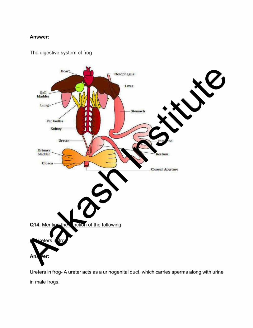
Answer:
The digestive system of frog
Q14. Mention the function of the following
(a)Ureters in frog
Answer:
Ureters in frog- A ureter acts as a urinogenital duct, which carries sperms along with urine
in male frogs.
Aakas
h Ins
titute
Q14. Mention the function of the following
(b) Malpighian tubule
Answer:
Malpighian tubules- These are the excretory organs in the earthworm.
Q14. Mention the function of the following
(c) Body wall in earthworm
Answer:
Body wall in earthworm- Body wall helps the earthworm in movement and burrowing.
Aakas
h Ins
titute