Embed Size (px)
Citation preview
NCCN Clinical Practice Guidelines in Oncology (NCCN Guidelines®)
Acute Myeloid Leukemia
Version 2.2016
Continue
NCCN.org
Version 2.2016, 06/29/16 © National Comprehensive Cancer Network, Inc. 2016, All rights reserved. The NCCN Guidelines® and this illustration may not be reproduced in any form without the express written permission of NCCN®.
NCCN Guidelines IndexAML Table of Contents
Discussion
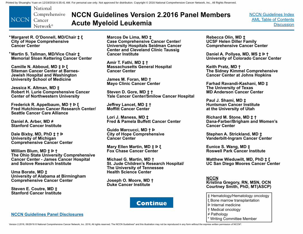
NCCN Guidelines Version 2.2016 Panel MembersAcute Myeloid Leukemia
*Margaret R. O’Donnell, MD/Chair ‡ ξCity of Hope Comprehensive Cancer Center
Martin S. Tallman, MD/Vice Chair ‡Memorial Sloan Kettering Cancer Center
Camille N. Abboud, MD ‡ Þ ξSiteman Cancer Center at Barnes- Jewish Hospital and Washington University School of Medicine
Jessica K. Altman, MD ‡Robert H. Lurie Comprehensive Cancer Center of Northwestern University
Frederick R. Appelbaum, MD † Þ ξFred Hutchinson Cancer Research Center/Seattle Cancer Care Alliance
Daniel A. Arber, MD ≠Stanford Cancer Institute
Dale Bixby, MD, PhD ‡ † ÞUniversity of Michigan Comprehensive Cancer Center
William Blum, MD ‡ Þ †The Ohio State University Comprehensive Cancer Center - James Cancer Hospital and Solove Research Institute
Uma Borate, MD ‡ University of Alabama at Birmingham Comprehensive Cancer Center
Steven E. Coutre, MD ‡Stanford Cancer Institute
Marcos De Lima, MD ‡Case Comprehensive Cancer Center/University Hospitals Seidman Cancer Center and Cleveland Clinic Taussig Cancer Institute
Amir T. Fathi, MD ‡ †Massachusetts General Hospital Cancer Center
James M. Foran, MD †Mayo Clinic Cancer Center
Steven D. Gore, MD ‡ † Yale Cancer Center/Smilow Cancer Hospital
Jeffrey Lancet, MD ‡ † Moffitt Cancer Center
Lori J. Maness, MD ‡Fred & Pamela Buffett Cancer Center
Guido Marcucci, MD † ÞCity of Hope Comprehensive Cancer Center
Mary Ellen Martin, MD ‡ Þ ξFox Chase Cancer Center
Michael G. Martin, MD †St. Jude Children’s Research Hospital/The University of Tennessee Health Science Center
Joseph O. Moore, MD †Duke Cancer Institute
Rebecca Olin, MD ‡UCSF Helen Diller FamilyComprehensive Cancer Center
Daniel A. Pollyea, MD, MS ‡ Þ †University of Colorado Cancer Center
Keith Pratz, MD †The Sidney Kimmel Comprehensive Cancer Center at Johns Hopkins
Farhad Ravandi-Kashani, MD ‡The University of Texas MD Anderson Cancer Center
Paul J. Shami, MD ‡Huntsman Cancer Institute at the University of Utah
Richard M. Stone, MD ‡ †Dana-Farber/Brigham and Women’s Cancer Center
Stephen A. Strickland, MD ‡Vanderbilt-Ingram Cancer Center
Eunice S. Wang, MD ‡Roswell Park Cancer Institute
Matthew Wieduwilt, MD, PhD ‡ ξUC San Diego Moores Cancer Center
NCCNKristina Gregory, RN, MSN, OCNCourtney Smith, PhD, MT(ASCP)
Continue
NCCN Guidelines Panel Disclosures
‡ Hematology/Hematology oncologyξ Bone marrow transplantationÞ Internal medicine† Medical oncology≠ Pathology* Writing Committee Member
*
Version 2.2016, 06/29/16 © National Comprehensive Cancer Network, Inc. 2016, All rights reserved. The NCCN Guidelines® and this illustration may not be reproduced in any form without the express written permission of NCCN®.
Printed by Shuanghu Yuan on 12/19/2016 6:35:41 AM. For personal use only. Not approved for distribution. Copyright © 2016 National Comprehensive Cancer Network, Inc., All Rights Reserved.
Clinical Trials: NCCN believes that the best management for any cancer patient is in a clinical trial. Participation in clinical trials is especially encouraged. To find clinical trials online at NCCN Member Institutions, click here:nccn.org/clinical_trials/physician.html.NCCN Categories of Evidence and Consensus: All recommendations are category 2A unless otherwise specified. See NCCN Categories of Evidence and Consensus.
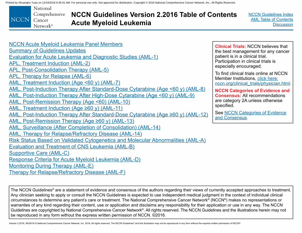
NCCN Acute Myeloid Leukemia Panel MembersSummary of Guidelines UpdatesEvaluation for Acute Leukemia and Diagnostic Studies (AML-1)APL, Treatment Induction (AML-2)APL, Post-Consolidation Therapy (AML-5)APL, Therapy for Relapse (AML-6)AML, Treatment Induction (Age <60 y) (AML-7)AML, Post-Induction Therapy After Standard-Dose Cytarabine (Age <60 y) (AML-8)AML, Post-Induction Therapy After High-Dose Cytarabine (Age <60 y) (AML-9)AML, Post-Remission Therapy (Age <60) (AML-10)AML, Treatment Induction (Age ≥60 y) (AML-11)AML, Post-Induction Therapy After Standard-Dose Cytarabine (Age ≥60 y) (AML-12)AML, Post-Remission Therapy (Age ≥60 y) (AML-13)AML, Surveillance (After Completion of Consolidation) (AML-14)AML, Therapy for Relapse/Refractory Disease (AML-14)Risk Status Based on Validated Cytogenetics and Molecular Abnormalities (AML-A)Evaluation and Treatment of CNS Leukemia (AML-B)Supportive Care (AML-C)Response Criteria for Acute Myeloid Leukemia (AML-D)Monitoring During Therapy (AML-E)Therapy for Relapse/Refractory Disease (AML-F)
The NCCN Guidelines® are a statement of evidence and consensus of the authors regarding their views of currently accepted approaches to treatment. Any clinician seeking to apply or consult the NCCN Guidelines is expected to use independent medical judgment in the context of individual clinical circumstances to determine any patient’s care or treatment. The National Comprehensive Cancer Network® (NCCN®) makes no representations or warranties of any kind regarding their content, use or application and disclaims any responsibility for their application or use in any way. The NCCN Guidelines are copyrighted by National Comprehensive Cancer Network®. All rights reserved. The NCCN Guidelines and the illustrations herein may not be reproduced in any form without the express written permission of NCCN. ©2016.
NCCN Guidelines Version 2.2016 Table of ContentsAcute Myeloid Leukemia
NCCN Guidelines IndexAML Table of Contents
Discussion
Version 2.2016, 06/29/16 © National Comprehensive Cancer Network, Inc. 2016, All rights reserved. The NCCN Guidelines® and this illustration may not be reproduced in any form without the express written permission of NCCN®.
Printed by Shuanghu Yuan on 12/19/2016 6:35:41 AM. For personal use only. Not approved for distribution. Copyright © 2016 National Comprehensive Cancer Network, Inc., All Rights Reserved.
NCCN Guidelines IndexAML Table of Contents
Discussion
UPDATES
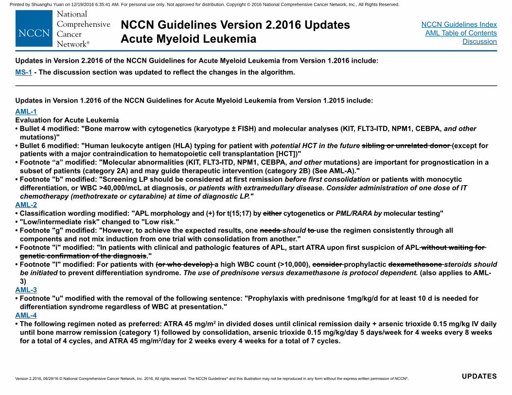
NCCN Guidelines Version 2.2016 UpdatesAcute Myeloid Leukemia
Updates in Version 1.2016 of the NCCN Guidelines for Acute Myeloid Leukemia from Version 1.2015 include:AML-1Evaluation for Acute Leukemia• Bullet 4 modified: "Bone marrow with cytogenetics (karyotype ± FISH) and molecular analyses (KIT, FLT3-ITD, NPM1, CEBPA, and other
mutations)"• Bullet 6 modified: "Human leukocyte antigen (HLA) typing for patient with potential HCT in the future sibling or unrelated donor (except for
patients with a major contraindication to hematopoietic cell transplantation [HCT])"• Footnote “a” modified: "Molecular abnormalities (KIT, FLT3-ITD, NPM1, CEBPA, and other mutations) are important for prognostication in a
subset of patients (category 2A) and may guide therapeutic intervention (category 2B) (See AML-A)."• Footnote "b" modified: "Screening LP should be considered at first remission before first consolidation or patients with monocytic
differentiation, or WBC >40,000/mcL at diagnosis, or patients with extramedullary disease. Consider administration of one dose of IT chemotherapy (methotrexate or cytarabine) at time of diagnostic LP."
AML-2• Classification wording modified: "APL morphology and (+) for t(15;17) by either cytogenetics or PML/RARA by molecular testing"• "Low/intermediate risk" changed to "Low risk."• Footnote "g" modified: "However, to achieve the expected results, one needs should to use the regimen consistently through all
components and not mix induction from one trial with consolidation from another."• Footnote "i" modified: "In patients with clinical and pathologic features of APL, start ATRA upon first suspicion of APL without waiting for
genetic confirmation of the diagnosis." • Footnote "l" modified: For patients with (or who develop) a high WBC count (>10,000), consider prophylactic dexamethasone steroids should
be initiated to prevent differentiation syndrome. The use of prednisone versus dexamethasone is protocol dependent. (also applies to AML-3)
AML-3• Footnote "u" modified with the removal of the following sentence: "Prophylaxis with prednisone 1mg/kg/d for at least 10 d is needed for
differentiation syndrome regardless of WBC at presentation."AML-4• The following regimen noted as preferred: ATRA 45 mg/m2 in divided doses until clinical remission daily + arsenic trioxide 0.15 mg/kg IV daily
until bone marrow remission (category 1) followed by consolidation, arsenic trioxide 0.15 mg/kg/day 5 days/week for 4 weeks every 8 weeks for a total of 4 cycles, and ATRA 45 mg/m2/day for 2 weeks every 4 weeks for a total of 7 cycles.
Version 2.2016, 06/29/16 © National Comprehensive Cancer Network, Inc. 2016, All rights reserved. The NCCN Guidelines® and this illustration may not be reproduced in any form without the express written permission of NCCN®.
Updates in Version 2.2016 of the NCCN Guidelines for Acute Myeloid Leukemia from Version 1.2016 include:MS-1 - The discussion section was updated to reflect the changes in the algorithm.
Printed by Shuanghu Yuan on 12/19/2016 6:35:41 AM. For personal use only. Not approved for distribution. Copyright © 2016 National Comprehensive Cancer Network, Inc., All Rights Reserved.
NCCN Guidelines IndexAML Table of Contents
Discussion
UPDATES
NCCN Guidelines Version 2.2016 UpdatesAcute Myeloid Leukemia
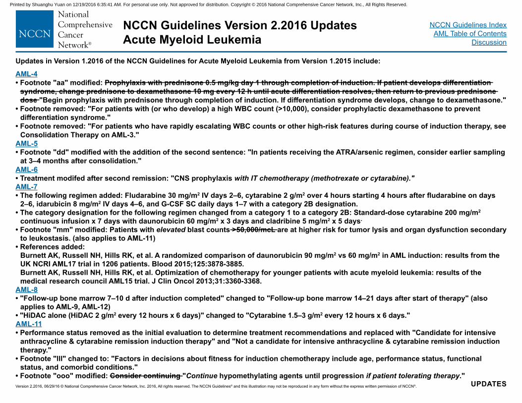
Updates in Version 1.2016 of the NCCN Guidelines for Acute Myeloid Leukemia from Version 1.2015 include:
AML-4• Footnote "aa" modified: Prophylaxis with prednisone 0.5 mg/kg day 1 through completion of induction. If patient develops differentiation
syndrome, change prednisone to dexamethasone 10 mg every 12 h until acute differentiation resolves, then return to previous prednisone dose "Begin prophylaxis with prednisone through completion of induction. If differentiation syndrome develops, change to dexamethasone."
• Footnote removed: "For patients with (or who develop) a high WBC count (>10,000), consider prophylactic dexamethasone to prevent differentiation syndrome."
• Footnote removed: "For patients who have rapidly escalating WBC counts or other high-risk features during course of induction therapy, see Consolidation Therapy on AML-3."
AML-5• Footnote "dd" modified with the addition of the second sentence: "In patients receiving the ATRA/arsenic regimen, consider earlier sampling
at 3–4 months after consolidation."AML-6• Treatment modifed after second remission: "CNS prophylaxis with IT chemotherapy (methotrexate or cytarabine)."AML-7• The following regimen added: Fludarabine 30 mg/m2 IV days 2–6, cytarabine 2 g/m2 over 4 hours starting 4 hours after fludarabine on days
2–6, idarubicin 8 mg/m2 IV days 4–6, and G-CSF SC daily days 1–7 with a category 2B designation.• The category designation for the following regimen changed from a category 1 to a category 2B: Standard-dose cytarabine 200 mg/m2
continuous infusion x 7 days with daunorubicin 60 mg/m2 x 3 days and cladribine 5 mg/m2 x 5 days.
• Footnote "mm" modified: Patients with elevated blast counts >50,000/mcL are at higher risk for tumor lysis and organ dysfunction secondary to leukostasis. (also applies to AML-11)
• References added: Burnett AK, Russell NH, Hills RK, et al. A randomized comparison of daunorubicin 90 mg/m2 vs 60 mg/m2 in AML induction: results from the UK NCRI AML17 trial in 1206 patients. Blood 2015;125:3878-3885. Burnett AK, Russell NH, Hills RK, et al. Optimization of chemotherapy for younger patients with acute myeloid leukemia: results of the medical research council AML15 trial. J Clin Oncol 2013;31:3360-3368.
AML-8• "Follow-up bone marrow 7–10 d after induction completed" changed to "Follow-up bone marrow 14–21 days after start of therapy" (also
applies to AML-9, AML-12)• "HiDAC alone (HiDAC 2 g/m2 every 12 hours x 6 days)" changed to "Cytarabine 1.5–3 g/m2 every 12 hours x 6 days."AML-11• Performance status removed as the initial evaluation to determine treatment recommendations and replaced with "Candidate for intensive
anthracycline & cytarabine remission induction therapy" and "Not a candidate for intensive anthracycline & cytarabine remission induction therapy."
• Footnote "lll" changed to: "Factors in decisions about fitness for induction chemotherapy include age, performance status, functional status, and comorbid conditions."
• Footnote "ooo" modified: Consider continuing "Continue hypomethylating agents until progression if patient tolerating therapy." Version 2.2016, 06/29/16 © National Comprehensive Cancer Network, Inc. 2016, All rights reserved. The NCCN Guidelines® and this illustration may not be reproduced in any form without the express written permission of NCCN®.
Printed by Shuanghu Yuan on 12/19/2016 6:35:41 AM. For personal use only. Not approved for distribution. Copyright © 2016 National Comprehensive Cancer Network, Inc., All Rights Reserved.
NCCN Guidelines IndexAML Table of Contents
Discussion
NCCN Guidelines Version 2.2016 UpdatesAcute Myeloid Leukemia
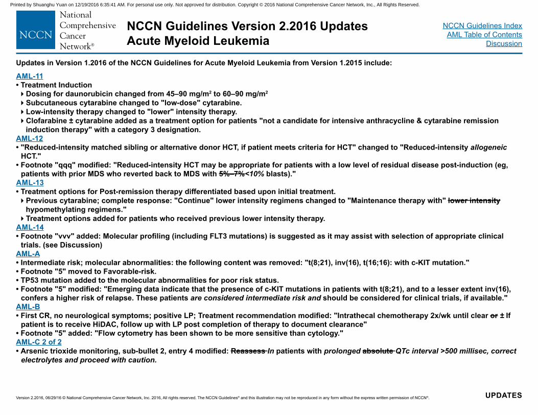
Updates in Version 1.2016 of the NCCN Guidelines for Acute Myeloid Leukemia from Version 1.2015 include:
AML-11• Treatment Induction�Dosing for daunorubicin changed from 45–90 mg/m2 to 60–90 mg/m2
�Subcutaneous cytarabine changed to "low-dose" cytarabine.�Low-intensity therapy changed to "lower" intensity therapy.�Clofarabine ± cytarabine added as a treatment option for patients "not a candidate for intensive anthracycline & cytarabine remission
induction therapy" with a category 3 designation.AML-12• "Reduced-intensity matched sibling or alternative donor HCT, if patient meets criteria for HCT" changed to "Reduced-intensity allogeneic
HCT."• Footnote "qqq" modified: "Reduced-intensity HCT may be appropriate for patients with a low level of residual disease post-induction (eg,
patients with prior MDS who reverted back to MDS with 5%–7%<10% blasts)."AML-13• Treatment options for Post-remission therapy differentiated based upon initial treatment.�Previous cytarabine; complete response: "Continue" lower intensity regimens changed to "Maintenance therapy with" lower intensity
hypomethylating regimens."�Treatment options added for patients who received previous lower intensity therapy.
AML-14• Footnote "vvv" added: Molecular profiling (including FLT3 mutations) is suggested as it may assist with selection of appropriate clinical
trials. (see Discussion)AML-A• Intermediate risk; molecular abnormalities: the following content was removed: "t(8;21), inv(16), t(16;16): with c-KIT mutation."• Footnote "5" moved to Favorable-risk.• TP53 mutation added to the molecular abnormalities for poor risk status.• Footnote "5" modified: "Emerging data indicate that the presence of c-KIT mutations in patients with t(8;21), and to a lesser extent inv(16),
confers a higher risk of relapse. These patients are considered intermediate risk and should be considered for clinical trials, if available." AML-B• First CR, no neurological symptoms; positive LP; Treatment recommendation modified: "Intrathecal chemotherapy 2x/wk until clear or ± If
patient is to receive HiDAC, follow up with LP post completion of therapy to document clearance" • Footnote "5" added: "Flow cytometry has been shown to be more sensitive than cytology."AML-C 2 of 2• Arsenic trioxide monitoring, sub-bullet 2, entry 4 modified: Reassess In patients with prolonged absolute QTc interval >500 millisec, correct
electrolytes and proceed with caution.
UPDATESVersion 2.2016, 06/29/16 © National Comprehensive Cancer Network, Inc. 2016, All rights reserved. The NCCN Guidelines® and this illustration may not be reproduced in any form without the express written permission of NCCN®.
Printed by Shuanghu Yuan on 12/19/2016 6:35:41 AM. For personal use only. Not approved for distribution. Copyright © 2016 National Comprehensive Cancer Network, Inc., All Rights Reserved.
NCCN Guidelines Version 2.2016 Acute Myeloid Leukemia
NCCN Guidelines IndexAML Table of Contents
Discussion
Note: All recommendations are category 2A unless otherwise indicated.Clinical Trials: NCCN believes that the best management of any cancer patient is in a clinical trial. Participation in clinical trials is especially encouraged.
Version 2.2016, 06/29/16 © National Comprehensive Cancer Network, Inc. 2016, All rights reserved. The NCCN Guidelines® and this illustration may not be reproduced in any form without the express written permission of NCCN®. AML-1
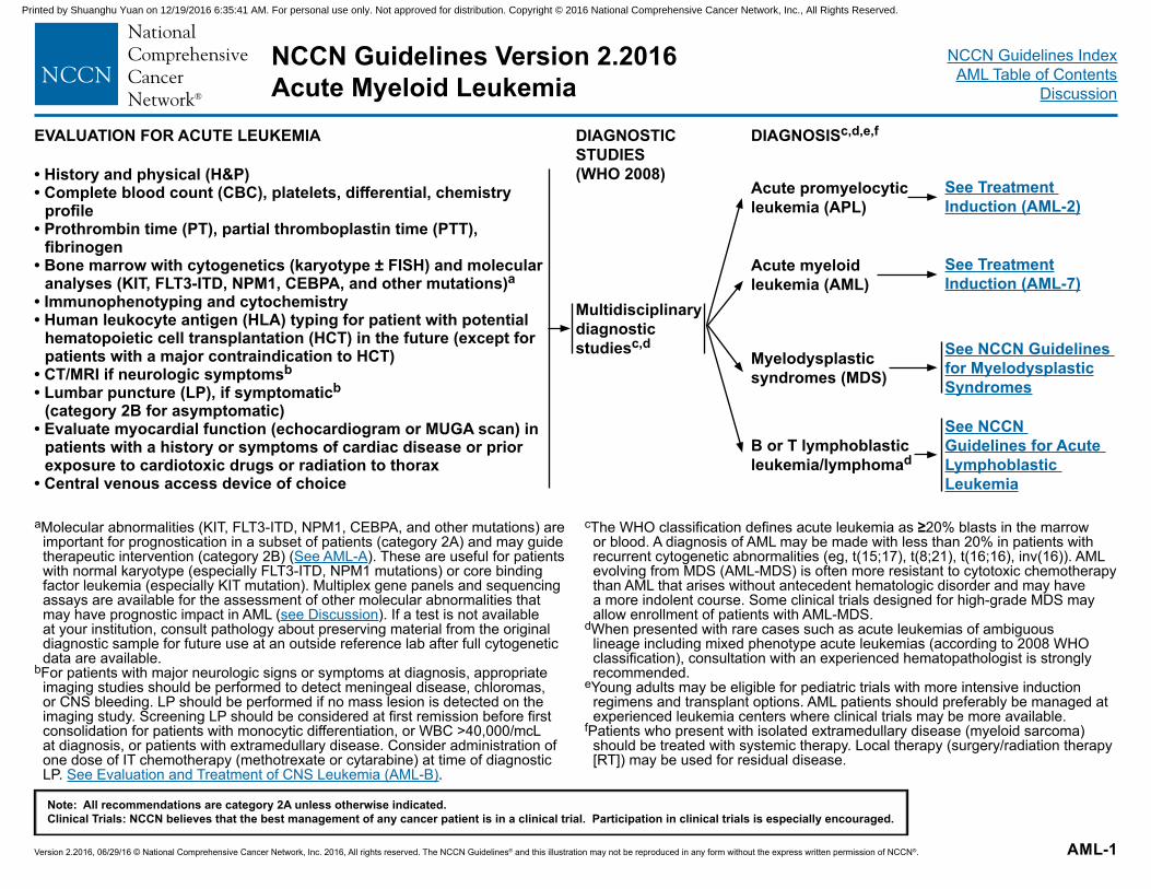
EVALUATION FOR ACUTE LEUKEMIA DIAGNOSTIC STUDIES (WHO 2008)
DIAGNOSISc,d,e,f
• History and physical (H&P)• Complete blood count (CBC), platelets, differential, chemistry
profile• Prothrombin time (PT), partial thromboplastin time (PTT),
fibrinogen• Bone marrow with cytogenetics (karyotype ± FISH) and molecular
analyses (KIT, FLT3-ITD, NPM1, CEBPA, and other mutations)a• Immunophenotyping and cytochemistry• Human leukocyte antigen (HLA) typing for patient with potential
hematopoietic cell transplantation (HCT) in the future (except for patients with a major contraindication to HCT)
• CT/MRI if neurologic symptomsb
• Lumbar puncture (LP), if symptomaticb (category 2B for asymptomatic)
• Evaluate myocardial function (echocardiogram or MUGA scan) in patients with a history or symptoms of cardiac disease or prior exposure to cardiotoxic drugs or radiation to thorax
• Central venous access device of choice
Multidisciplinary diagnostic studiesc,d
Acute promyelocytic leukemia (APL)
Acute myeloid leukemia (AML)
Myelodysplastic syndromes (MDS)
B or T lymphoblastic leukemia/lymphomad
See Treatment Induction (AML-2)
See Treatment Induction (AML-7)
See NCCN Guidelines for Myelodysplastic Syndromes
See NCCN Guidelines for Acute Lymphoblastic Leukemia
aMolecular abnormalities (KIT, FLT3-ITD, NPM1, CEBPA, and other mutations) are important for prognostication in a subset of patients (category 2A) and may guide therapeutic intervention (category 2B) (See AML-A). These are useful for patients with normal karyotype (especially FLT3-ITD, NPM1 mutations) or core binding factor leukemia (especially KIT mutation). Multiplex gene panels and sequencing assays are available for the assessment of other molecular abnormalities that may have prognostic impact in AML (see Discussion). If a test is not available at your institution, consult pathology about preserving material from the original diagnostic sample for future use at an outside reference lab after full cytogenetic data are available.
bFor patients with major neurologic signs or symptoms at diagnosis, appropriate imaging studies should be performed to detect meningeal disease, chloromas, or CNS bleeding. LP should be performed if no mass lesion is detected on the imaging study. Screening LP should be considered at first remission before first consolidation for patients with monocytic differentiation, or WBC >40,000/mcL at diagnosis, or patients with extramedullary disease. Consider administration of one dose of IT chemotherapy (methotrexate or cytarabine) at time of diagnostic LP. See Evaluation and Treatment of CNS Leukemia (AML-B).
cThe WHO classification defines acute leukemia as ≥20% blasts in the marrow or blood. A diagnosis of AML may be made with less than 20% in patients with recurrent cytogenetic abnormalities (eg, t(15;17), t(8;21), t(16;16), inv(16)). AML evolving from MDS (AML-MDS) is often more resistant to cytotoxic chemotherapy than AML that arises without antecedent hematologic disorder and may have a more indolent course. Some clinical trials designed for high-grade MDS may allow enrollment of patients with AML-MDS.
dWhen presented with rare cases such as acute leukemias of ambiguous lineage including mixed phenotype acute leukemias (according to 2008 WHO classification), consultation with an experienced hematopathologist is strongly recommended.
eYoung adults may be eligible for pediatric trials with more intensive induction regimens and transplant options. AML patients should preferably be managed at experienced leukemia centers where clinical trials may be more available.
fPatients who present with isolated extramedullary disease (myeloid sarcoma) should be treated with systemic therapy. Local therapy (surgery/radiation therapy [RT]) may be used for residual disease.
Printed by Shuanghu Yuan on 12/19/2016 6:35:41 AM. For personal use only. Not approved for distribution. Copyright © 2016 National Comprehensive Cancer Network, Inc., All Rights Reserved.
NCCN Guidelines IndexAML Table of Contents
Discussion
Note: All recommendations are category 2A unless otherwise indicated.Clinical Trials: NCCN believes that the best management of any cancer patient is in a clinical trial. Participation in clinical trials is especially encouraged.
Version 2.2016, 06/29/16 © National Comprehensive Cancer Network, Inc. 2016, All rights reserved. The NCCN Guidelines® and this illustration may not be reproduced in any form without the express written permission of NCCN®. AML-2
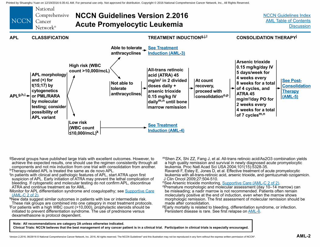
NCCN Guidelines Version 2.2016 Acute Promyelocytic Leukemia
gSeveral groups have published large trials with excellent outcomes. However, to achieve the expected results, one should use the regimen consistently through all components and not mix induction from one trial with consolidation from another.
hTherapy-related APL is treated the same as de novo APL.iIn patients with clinical and pathologic features of APL, start ATRA upon first
suspicion of APL. Early initiation of ATRA may prevent the lethal complication of bleeding. If cytogenetic and molecular testing do not confirm APL, discontinue ATRA and continue treatment as for AML.
jMonitor for APL differentiation syndrome and coagulopathy; see Supportive Care (AML-C 2 of 2).
kNew data suggest similar outcomes in patients with low or intermediate risk. These risk groups are combined into one category in most treatment protocols.
lFor patients with a high WBC count (>10,000), prophylactic steroids should be initiated to prevent differentiation syndrome. The use of prednisone versus dexamethasone is protocol dependent.
mShen ZX, Shi ZZ, Fang J, et al. All-trans retinoic acid/As2O3 combination yields a high quality remission and survival in newly diagnosed acute promyelocytic leukemia. Proc Natl Acad Sci USA 2004;101(15):5328-35. Ravandi F, Estey E, Jones D, et al. Effective treatment of acute promyelocytic leukemia with all-trans-retinoic acid, arsenic trioxide, and gemtuzumab ozogamicin.
J Clin Oncol 2009;27:504-510.nSee Arsenic trioxide monitoring, Supportive Care (AML-C 2 of 2).oPremature morphologic and molecular assessment (day 10–14 marrow) can
be misleading; a nadir marrow is not recommended. Patients often remain molecularly positive at the end of induction, even when the marrow shows morphologic remission. The first assessment of molecular remission should be made after consolidation.
pEarly mortality is related to bleeding, differentiation syndrome, or infection. Persistent disease is rare. See first relapse on AML-6.
APL CLASSIFICATION TREATMENT INDUCTIONg,j,l CONSOLIDATION THERAPYj
APLg,h,i
APL morphology and (+) for t(15;17) by cytogenetics or PML/RARA by molecular testing; consider possibility of APL variant
High risk (WBC count >10,000/mcL)
Low risk (WBC count ≤10,000/mcL)k
Able to tolerate anthracyclines
Not able to tolerate anthracyclines
See Treatment Induction (AML-3)
All-trans retinoic acid (ATRA) 45 mg/m2 in 2 divided doses daily + arsenic trioxide 0.15 mg/kg IV dailym,n until bone marrow remission
See Treatment Induction (AML-4)
At count recovery, proceed with consolidationo,p
Arsenic trioxide 0.15 mg/kg/day IV 5 days/week for 4 weeks every 8 weeks for a total of 4 cycles, and ATRA 45 mg/m2/day PO for 2 weeks every 4 weeks for a total of 7 cyclesm,n
See Post-Consolidation Therapy (AML-5)
Printed by Shuanghu Yuan on 12/19/2016 6:35:41 AM. For personal use only. Not approved for distribution. Copyright © 2016 National Comprehensive Cancer Network, Inc., All Rights Reserved.
NCCN Guidelines IndexAML Table of Contents
Discussion
Note: All recommendations are category 2A unless otherwise indicated.Clinical Trials: NCCN believes that the best management of any cancer patient is in a clinical trial. Participation in clinical trials is especially encouraged.
Version 2.2016, 06/29/16 © National Comprehensive Cancer Network, Inc. 2016, All rights reserved. The NCCN Guidelines® and this illustration may not be reproduced in any form without the express written permission of NCCN®. AML-3
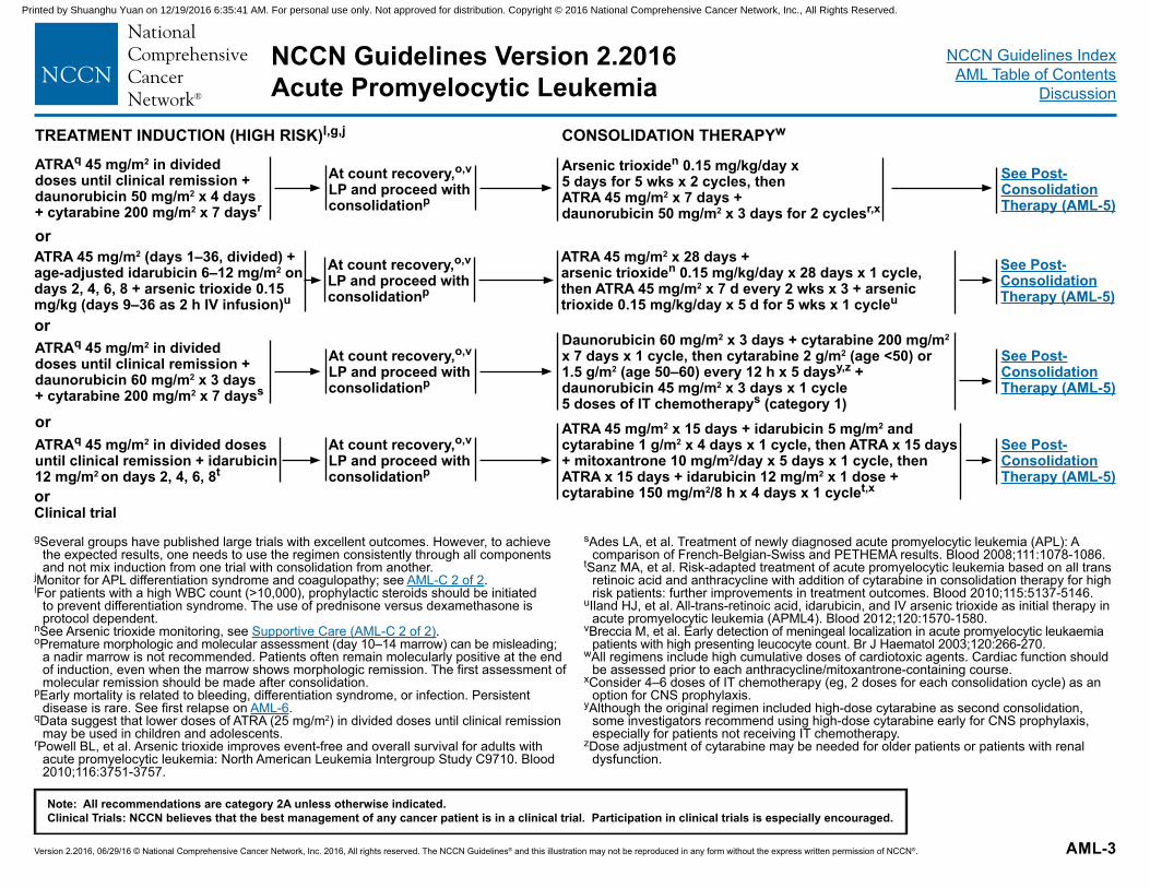
NCCN Guidelines Version 2.2016 Acute Promyelocytic Leukemia
TREATMENT INDUCTION (HIGH RISK)l,g,j CONSOLIDATION THERAPYw
gSeveral groups have published large trials with excellent outcomes. However, to achieve the expected results, one needs to use the regimen consistently through all components and not mix induction from one trial with consolidation from another.
jMonitor for APL differentiation syndrome and coagulopathy; see AML-C 2 of 2. lFor patients with a high WBC count (>10,000), prophylactic steroids should be initiated
to prevent differentiation syndrome. The use of prednisone versus dexamethasone is protocol dependent.
nSee Arsenic trioxide monitoring, see Supportive Care (AML-C 2 of 2).oPremature morphologic and molecular assessment (day 10–14 marrow) can be misleading;
a nadir marrow is not recommended. Patients often remain molecularly positive at the end of induction, even when the marrow shows morphologic remission. The first assessment of molecular remission should be made after consolidation.
pEarly mortality is related to bleeding, differentiation syndrome, or infection. Persistent disease is rare. See first relapse on AML-6.
qData suggest that lower doses of ATRA (25 mg/m2) in divided doses until clinical remission may be used in children and adolescents.
rPowell BL, et al. Arsenic trioxide improves event-free and overall survival for adults with acute promyelocytic leukemia: North American Leukemia Intergroup Study C9710. Blood 2010;116:3751-3757.
sAdes LA, et al. Treatment of newly diagnosed acute promyelocytic leukemia (APL): A comparison of French-Belgian-Swiss and PETHEMA results. Blood 2008;111:1078-1086.
tSanz MA, et al. Risk-adapted treatment of acute promyelocytic leukemia based on all trans retinoic acid and anthracycline with addition of cytarabine in consolidation therapy for high risk patients: further improvements in treatment outcomes. Blood 2010;115:5137-5146.
uIland HJ, et al. All-trans-retinoic acid, idarubicin, and IV arsenic trioxide as initial therapy in acute promyelocytic leukemia (APML4). Blood 2012;120:1570-1580.
vBreccia M, et al. Early detection of meningeal localization in acute promyelocytic leukaemia patients with high presenting leucocyte count. Br J Haematol 2003;120:266-270.
wAll regimens include high cumulative doses of cardiotoxic agents. Cardiac function should be assessed prior to each anthracycline/mitoxantrone-containing course.
xConsider 4–6 doses of IT chemotherapy (eg, 2 doses for each consolidation cycle) as an option for CNS prophylaxis.
yAlthough the original regimen included high-dose cytarabine as second consolidation, some investigators recommend using high-dose cytarabine early for CNS prophylaxis, especially for patients not receiving IT chemotherapy.
zDose adjustment of cytarabine may be needed for older patients or patients with renal dysfunction.
ATRAq 45 mg/m2 in divided doses until clinical remission + daunorubicin 50 mg/m2 x 4 days + cytarabine 200 mg/m2 x 7 daysr
or
ATRAq 45 mg/m2 in divided doses until clinical remission + daunorubicin 60 mg/m2 x 3 days + cytarabine 200 mg/m2 x 7 dayss
orATRAq 45 mg/m2 in divided doses until clinical remission + idarubicin 12 mg/m2 on days 2, 4, 6, 8t
or
ATRA 45 mg/m2 (days 1–36, divided) + age-adjusted idarubicin 6–12 mg/m2 on days 2, 4, 6, 8 + arsenic trioxide 0.15 mg/kg (days 9–36 as 2 h IV infusion)u
At count recovery,o,v LP and proceed with consolidationp
At count recovery,o,v LP and proceed with consolidationp
At count recovery,o,v LP and proceed with consolidationp
At count recovery,o,v LP and proceed with consolidationp
Arsenic trioxiden 0.15 mg/kg/day x 5 days for 5 wks x 2 cycles, then ATRA 45 mg/m2 x 7 days + daunorubicin 50 mg/m2 x 3 days for 2 cyclesr,x
Daunorubicin 60 mg/m2 x 3 days + cytarabine 200 mg/m2 x 7 days x 1 cycle, then cytarabine 2 g/m2 (age <50) or 1.5 g/m2 (age 50–60) every 12 h x 5 daysy,z + daunorubicin 45 mg/m2 x 3 days x 1 cycle 5 doses of IT chemotherapys (category 1)ATRA 45 mg/m2 x 15 days + idarubicin 5 mg/m2 and cytarabine 1 g/m2 x 4 days x 1 cycle, then ATRA x 15 days + mitoxantrone 10 mg/m2/day x 5 days x 1 cycle, then ATRA x 15 days + idarubicin 12 mg/m2 x 1 dose + cytarabine 150 mg/m2/8 h x 4 days x 1 cyclet,x
ATRA 45 mg/m2 x 28 days + arsenic trioxiden 0.15 mg/kg/day x 28 days x 1 cycle, then ATRA 45 mg/m2 x 7 d every 2 wks x 3 + arsenic trioxide 0.15 mg/kg/day x 5 d for 5 wks x 1 cycleu
See Post-Consolidation Therapy (AML-5)
See Post-Consolidation Therapy (AML-5)
See Post-Consolidation Therapy (AML-5)
See Post-Consolidation Therapy (AML-5)
orClinical trial
Printed by Shuanghu Yuan on 12/19/2016 6:35:41 AM. For personal use only. Not approved for distribution. Copyright © 2016 National Comprehensive Cancer Network, Inc., All Rights Reserved.
NCCN Guidelines IndexAML Table of Contents
Discussion
Note: All recommendations are category 2A unless otherwise indicated.Clinical Trials: NCCN believes that the best management of any cancer patient is in a clinical trial. Participation in clinical trials is especially encouraged.
Version 2.2016, 06/29/16 © National Comprehensive Cancer Network, Inc. 2016, All rights reserved. The NCCN Guidelines® and this illustration may not be reproduced in any form without the express written permission of NCCN®. AML-4
NCCN Guidelines Version 2.2016 Acute Promyelocytic Leukemia
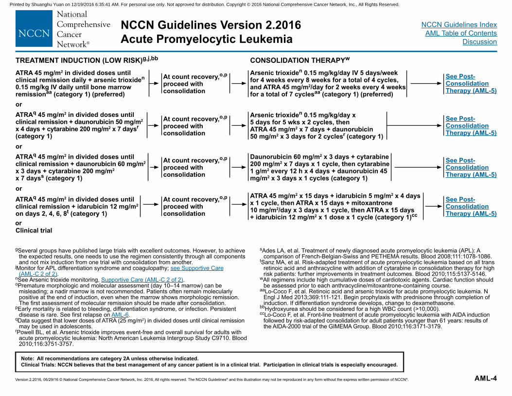
TREATMENT INDUCTION (LOW RISK)g,j,bb CONSOLIDATION THERAPYw
ATRA 45 mg/m2 in divided doses until clinical remission daily + arsenic trioxiden 0.15 mg/kg IV daily until bone marrow remissionaa (category 1) (preferred)orATRAq 45 mg/m2 in divided doses until clinical remission + daunorubicin 50 mg/m2 x 4 days + cytarabine 200 mg/m2 x 7 daysr
(category 1)orATRAq 45 mg/m2 in divided doses until clinical remission + daunorubicin 60 mg/m2 x 3 days + cytarabine 200 mg/m2 x 7 dayss (category 1)orATRAq 45 mg/m2 in divided doses until clinical remission + idarubicin 12 mg/m2 on days 2, 4, 6, 8t (category 1)
At count recovery,o,p proceed with consolidation
At count recovery,o,p proceed with consolidation
At count recovery,o,p proceed with consolidation
At count recovery,o,p
proceed with consolidation
Arsenic trioxiden 0.15 mg/kg/day IV 5 days/week for 4 weeks every 8 weeks for a total of 4 cycles, and ATRA 45 mg/m2/day for 2 weeks every 4 weeks for a total of 7 cyclesaa (category 1) (preferred)
Arsenic trioxiden 0.15 mg/kg/day x 5 days for 5 wks x 2 cycles, then ATRA 45 mg/m2 x 7 days + daunorubicin 50 mg/m2 x 3 days for 2 cyclesr (category 1)
Daunorubicin 60 mg/m2 x 3 days + cytarabine 200 mg/m2 x 7 days x 1 cycle, then cytarabine 1 g/m2 every 12 h x 4 days + daunorubicin 45 mg/m2 x 3 days x 1 cycles (category 1)
ATRA 45 mg/m2 x 15 days + idarubicin 5 mg/m2 x 4 days x 1 cycle, then ATRA x 15 days + mitoxantrone 10 mg/m2/day x 3 days x 1 cycle, then ATRA x 15 days + idarubicin 12 mg/m2 x 1 dose x 1 cycle (category 1)cc
See Post-Consolidation Therapy (AML-5)
See Post-Consolidation Therapy (AML-5)
See Post-Consolidation Therapy (AML-5)
See Post-Consolidation Therapy (AML-5)
Clinical trial
gSeveral groups have published large trials with excellent outcomes. However, to achieve the expected results, one needs to use the regimen consistently through all components and not mix induction from one trial with consolidation from another.
jMonitor for APL differentiation syndrome and coagulopathy; see Supportive Care (AML-C 2 of 2).
nSee Arsenic trioxide monitoring, Supportive Care (AML-C 2 of 2).oPremature morphologic and molecular assessment (day 10–14 marrow) can be
misleading; a nadir marrow is not recommended. Patients often remain molecularly positive at the end of induction, even when the marrow shows morphologic remission. The first assessment of molecular remission should be made after consolidation.
pEarly mortality is related to bleeding, differentiation syndrome, or infection. Persistent disease is rare. See first relapse on AML-6.
qData suggest that lower doses of ATRA (25 mg/m2) in divided doses until clinical remission may be used in adolescents.
rPowell BL, et al. Arsenic trioxide improves event-free and overall survival for adults with acute promyelocytic leukemia: North American Leukemia Intergroup Study C9710. Blood 2010;116:3751-3757.
sAdes LA, et al. Treatment of newly diagnosed acute promyelocytic leukemia (APL): A comparison of French-Belgian-Swiss and PETHEMA results. Blood 2008;111:1078-1086.
tSanz MA, et al. Risk-adapted treatment of acute promyelocytic leukemia based on all trans retinoic acid and anthracycline with addition of cytarabine in consolidation therapy for high risk patients: further improvements in treatment outcomes. Blood 2010;115:5137-5146.
wAll regimens include high cumulative doses of cardiotoxic agents. Cardiac function should be assessed prior to each anthracycline/mitoxantrone-containing course.
aaLo-Coco F, et al. Retinoic acid and arsenic trioxide for acute promyelocytic leukemia. N Engl J Med 2013;369:111-121. Begin prophylaxis with prednisone through completion of induction. If differentiation syndrome develops, change to dexamethasone.
bbHydroxyurea should be considered for a high WBC count (>10,000).ccLo-Coco F, et al. Front-line treatment of acute promyelocytic leukemia with AIDA induction
followed by risk-adapted consolidation for adult patients younger than 61 years: results of the AIDA-2000 trial of the GIMEMA Group. Blood 2010;116:3171-3179.
or
Printed by Shuanghu Yuan on 12/19/2016 6:35:41 AM. For personal use only. Not approved for distribution. Copyright © 2016 National Comprehensive Cancer Network, Inc., All Rights Reserved.
NCCN Guidelines IndexAML Table of Contents
Discussion
Note: All recommendations are category 2A unless otherwise indicated.Clinical Trials: NCCN believes that the best management of any cancer patient is in a clinical trial. Participation in clinical trials is especially encouraged.
Version 2.2016, 06/29/16 © National Comprehensive Cancer Network, Inc. 2016, All rights reserved. The NCCN Guidelines® and this illustration may not be reproduced in any form without the express written permission of NCCN®. AML-5
NCCN Guidelines Version 2.2016 Acute Promyelocytic Leukemia
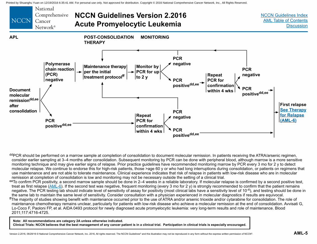
ddPCR should be performed on a marrow sample at completion of consolidation to document molecular remission. In patients receiving the ATRA/arsenic regimen, consider earlier sampling at 3–4 months after consolidation. Subsequent monitoring by PCR can be done with peripheral blood, although marrow is a more sensitive monitoring technique and may give earlier signs of relapse. Prior practice guidelines have recommended monitoring marrow by PCR every 3 mo for 2 y to detect molecular relapse. We continue to endorse this for high-risk patients, those >age 60 y or who had long interruptions during consolidation, or patients on regimens that use maintenance and are not able to tolerate maintenance. Clinical experience indicates that risk of relapse in patients with low-risk disease who are in molecular remission at completion of consolidation is low and monitoring may not be necessary outside the setting of a clinical trial.
eeTo confirm PCR positivity, a second marrow sample should be done in 2–4 weeks in a reliable laboratory. If molecular relapse is confirmed by a second positive test, treat as first relapse (AML-6). If the second test was negative, frequent monitoring (every 3 mo for 2 y) is strongly recommended to confirm that the patient remains negative. The PCR testing lab should indicate level of sensitivity of assay for positivity (most clinical labs have a sensitivity level of 10-4), and testing should be done in the same lab to maintain the same level of sensitivity. Consider consultation with a physician experienced in molecular diagnostics if results are equivocal.
ffThe majority of studies showing benefit with maintenance occurred prior to the use of ATRA and/or arsenic trioxide and/or cytarabine for consolidation. The role of maintenance chemotherapy remains unclear, particularly for patients with low-risk disease who achieve a molecular remission at the end of consolidation. Avvisati G, Lo-Coco F, Paoloni FP, et al. AIDA 0493 protocol for newly diagnosed acute promyelocytic leukemia: very long-term results and role of maintenance. Blood 2011;117:4716-4725.
APL POST-CONSOLIDATION THERAPY
MONITORING
First relapse See Therapy for Relapse (AML-6)
Document molecular remissiondd,ee after consolidation
Polymerase chain reaction (PCR) negative
PCR positivedd,ee
Maintenance therapy per the initial treatment protocolff
Monitor by PCR for up to 2 y
Repeat PCR for confirmation within 4 wks
PCR negative
PCR positivedd,ee
Repeat PCR for confirmation within 4 wks
PCR negative
PCR positivedd,ee
PCR negative
PCR positivedd,ee
Printed by Shuanghu Yuan on 12/19/2016 6:35:41 AM. For personal use only. Not approved for distribution. Copyright © 2016 National Comprehensive Cancer Network, Inc., All Rights Reserved.
NCCN Guidelines IndexAML Table of Contents
Discussion
Note: All recommendations are category 2A unless otherwise indicated.Clinical Trials: NCCN believes that the best management of any cancer patient is in a clinical trial. Participation in clinical trials is especially encouraged.
Version 2.2016, 06/29/16 © National Comprehensive Cancer Network, Inc. 2016, All rights reserved. The NCCN Guidelines® and this illustration may not be reproduced in any form without the express written permission of NCCN®. AML-6
NCCN Guidelines Version 2.2016 Acute Promyelocytic Leukemia
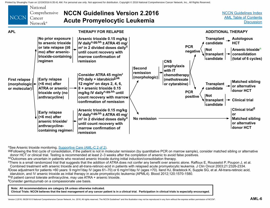
nSee Arsenic trioxide monitoring, Supportive Care (AML-C 2 of 2). ggFollowing the first cycle of consolidation, if the patient is not in molecular remission (by quantitative PCR on marrow sample), consider matched sibling or alternative
donor HCT or clinical trial. Testing is recommended at least 2–3 weeks after the completion of arsenic to avoid false positives.hhOutcomes are uncertain in patients who received arsenic trioxide during initial induction/consolidation therapy. iiThere is a small randomized trial that suggests that the addition of ATRA does not confer any benefit over arsenic alone. Raffoux E, Rousselot P, Poupon J, et al.
Combined treatment with arsenic trioxide and all-trans-retinoic-acid in patients with relapsed acute promyelocytic leukemia. J Clin Oncol 2003;21:2326-2334.jjDose adjustment for patients >60 years: 9 mg/m2/day IV (ages 61–70) or 6 mg/m2/day IV (ages >70). Iland HJ, Bradstock K, Supple SG, et al. All-trans-retinoic acid,
idarubicin, and IV arsenic trioxide as initial therapy in acute promyelocytic leukemia (APML4). Blood 2012;120:1570-1580. kkIf patient cannot tolerate anthracycline, may use ATRA + arsenic trioxide.llConsider gemtuzumab on a compassionate use basis.
APL THERAPY FOR RELAPSE ADDITIONAL THERAPY
First relapse (morphologic or molecular)
Early relapse (<6 mo) after ATRA or arsenic trioxide only (no anthracycline)
Consider ATRA 45 mg/m2 PO daily + idarubicinjj,kk 12 mg/m2 on days 2, 4, 6, 8 + arsenic trioxide 0.15 mg/kg IV dailyn,gg,hh until count recovery with marrow confirmation of remission
No prior exposure to arsenic trioxide or late relapse (≥6 mo) after arsenic-trioxide-containing regimen
Early relapse (<6 mo) after arsenic trioxide/ anthracycline-containing regimen
Arsenic trioxide 0.15 mg/kg IV dailyn,gg,hh ± ATRA 45 mg/m2 in 2 divided doses dailyii until count recovery with marrow confirmation of remission
Arsenic trioxide 0.15 mg/kg IV dailyn,gg,hh ± ATRA 45 mg/m2 in 2 divided doses dailyii until count recovery with marrow confirmation of remission
Second remission (morphologic)
No remission
CNS prophylaxis with IT chemotherapy (methotrexate or cytarabine)
Transplant candidate
Autologous HCTArsenic trioxiden
consolidation (total of 6 cycles)
Not transplant candidate
PCR negative
PCR positive
Transplant candidate
Not transplant candidate
Matched sibling or alternative donor HCT
Clinical trial
Clinical trialllorMatched sibling or alternative donor HCT
Printed by Shuanghu Yuan on 12/19/2016 6:35:41 AM. For personal use only. Not approved for distribution. Copyright © 2016 National Comprehensive Cancer Network, Inc., All Rights Reserved.
NCCN Guidelines Version 2.2016 Acute Myeloid Leukemia
NCCN Guidelines IndexAML Table of Contents
Discussion
Note: All recommendations are category 2A unless otherwise indicated.Clinical Trials: NCCN believes that the best management of any cancer patient is in a clinical trial. Participation in clinical trials is especially encouraged.
Version 2.2016, 06/29/16 © National Comprehensive Cancer Network, Inc. 2016, All rights reserved. The NCCN Guidelines® and this illustration may not be reproduced in any form without the express written permission of NCCN®. AML-7
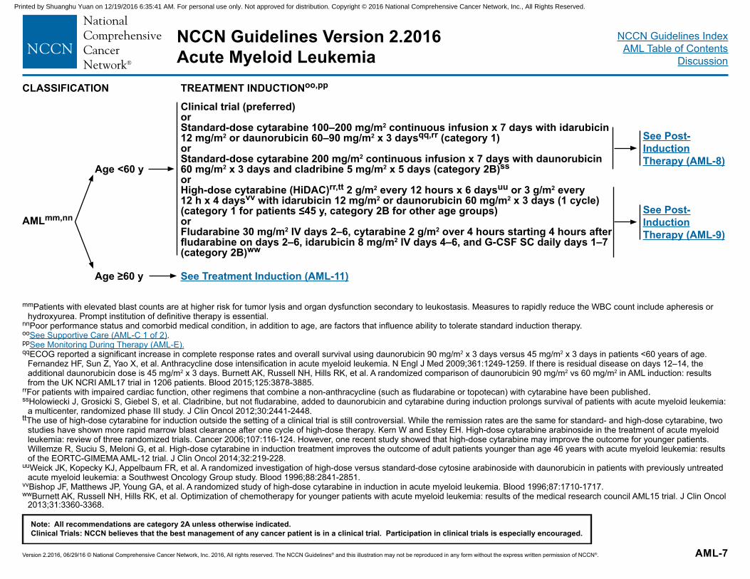
CLASSIFICATION TREATMENT INDUCTIONoo,pp
Clinical trial (preferred) orStandard-dose cytarabine 100–200 mg/m2 continuous infusion x 7 days with idarubicin 12 mg/m2 or daunorubicin 60–90 mg/m2 x 3 daysqq,rr (category 1)orStandard-dose cytarabine 200 mg/m2 continuous infusion x 7 days with daunorubicin 60 mg/m2 x 3 days and cladribine 5 mg/m2 x 5 days (category 2B)ssorHigh-dose cytarabine (HiDAC)rr,tt 2 g/m2 every 12 hours x 6 daysuu or 3 g/m2 every 12 h x 4 daysvv with idarubicin 12 mg/m2 or daunorubicin 60 mg/m2 x 3 days (1 cycle) (category 1 for patients ≤45 y, category 2B for other age groups)or Fludarabine 30 mg/m2 IV days 2–6, cytarabine 2 g/m2 over 4 hours starting 4 hours after fludarabine on days 2–6, idarubicin 8 mg/m2 IV days 4–6, and G-CSF SC daily days 1–7 (category 2B)ww
AMLmm,nn
Age <60 y
Age ≥60 y See Treatment Induction (AML-11)
See Post-Induction Therapy (AML-8)
See Post-Induction Therapy (AML-9)
mmPatients with elevated blast counts are at higher risk for tumor lysis and organ dysfunction secondary to leukostasis. Measures to rapidly reduce the WBC count include apheresis or hydroxyurea. Prompt institution of definitive therapy is essential.
nnPoor performance status and comorbid medical condition, in addition to age, are factors that influence ability to tolerate standard induction therapy. ooSee Supportive Care (AML-C 1 of 2).ppSee Monitoring During Therapy (AML-E).qqECOG reported a significant increase in complete response rates and overall survival using daunorubicin 90 mg/m2 x 3 days versus 45 mg/m2 x 3 days in patients <60 years of age.
Fernandez HF, Sun Z, Yao X, et al. Anthracycline dose intensification in acute myeloid leukemia. N Engl J Med 2009;361:1249-1259. If there is residual disease on days 12–14, the additional daunorubicin dose is 45 mg/m2 x 3 days. Burnett AK, Russell NH, Hills RK, et al. A randomized comparison of daunorubicin 90 mg/m2 vs 60 mg/m2 in AML induction: results from the UK NCRI AML17 trial in 1206 patients. Blood 2015;125:3878-3885.
rrFor patients with impaired cardiac function, other regimens that combine a non-anthracycline (such as fludarabine or topotecan) with cytarabine have been published.ssHolowiecki J, Grosicki S, Giebel S, et al. Cladribine, but not fludarabine, added to daunorubicin and cytarabine during induction prolongs survival of patients with acute myeloid leukemia:
a multicenter, randomized phase III study. J Clin Oncol 2012;30:2441-2448.ttThe use of high-dose cytarabine for induction outside the setting of a clinical trial is still controversial. While the remission rates are the same for standard- and high-dose cytarabine, two
studies have shown more rapid marrow blast clearance after one cycle of high-dose therapy. Kern W and Estey EH. High-dose cytarabine arabinoside in the treatment of acute myeloid leukemia: review of three randomized trials. Cancer 2006;107:116-124. However, one recent study showed that high-dose cytarabine may improve the outcome for younger patients. Willemze R, Suciu S, Meloni G, et al. High-dose cytarabine in induction treatment improves the outcome of adult patients younger than age 46 years with acute myeloid leukemia: results of the EORTC-GIMEMA AML-12 trial. J Clin Oncol 2014;32:219-228.
uuWeick JK, Kopecky KJ, Appelbaum FR, et al. A randomized investigation of high-dose versus standard-dose cytosine arabinoside with daunorubicin in patients with previously untreated acute myeloid leukemia: a Southwest Oncology Group study. Blood 1996;88:2841-2851.
vvBishop JF, Matthews JP, Young GA, et al. A randomized study of high-dose cytarabine in induction in acute myeloid leukemia. Blood 1996;87:1710-1717.wwBurnett AK, Russell NH, Hills RK, et al. Optimization of chemotherapy for younger patients with acute myeloid leukemia: results of the medical research council AML15 trial. J Clin Oncol
2013;31:3360-3368.
Printed by Shuanghu Yuan on 12/19/2016 6:35:41 AM. For personal use only. Not approved for distribution. Copyright © 2016 National Comprehensive Cancer Network, Inc., All Rights Reserved.
NCCN Guidelines Version 2.2016 Acute Myeloid Leukemia
NCCN Guidelines IndexAML Table of Contents
Discussion
Note: All recommendations are category 2A unless otherwise indicated.Clinical Trials: NCCN believes that the best management of any cancer patient is in a clinical trial. Participation in clinical trials is especially encouraged.
Version 2.2016, 06/29/16 © National Comprehensive Cancer Network, Inc. 2016, All rights reserved. The NCCN Guidelines® and this illustration may not be reproduced in any form without the express written permission of NCCN®. AML-8
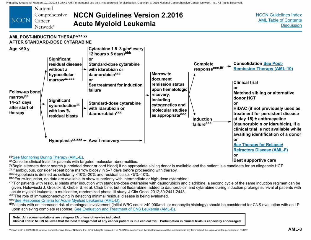
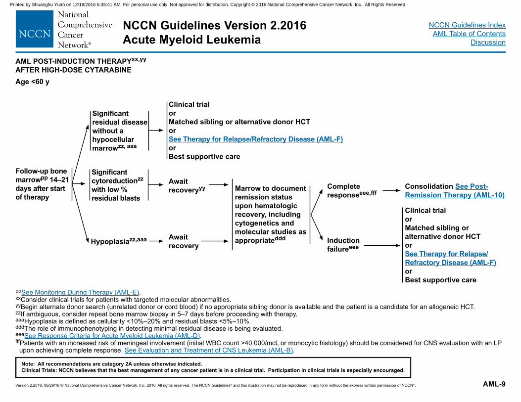
ppSee Monitoring During Therapy (AML-E).xxConsider clinical trials for patients with targeted molecular abnormalities.yyBegin alternate donor search (unrelated donor or cord blood) if no appropriate sibling donor is available and the patient is a candidate for an allogeneic HCT.zzIf ambiguous, consider repeat bone marrow biopsy in 5–7 days before proceeding with therapy. aaaHypoplasia is defined as cellularity <10%–20% and residual blasts <5%–10%.bbbFor re-induction, no data are available to show superiority with intermediate or high-dose cytarabine.cccFor patients with residual blasts after induction with standard-dose cytarabine with daunorubicin and cladribine, a second cycle of the same induction regimen can be
given. Holowiecki J, Grosicki S, Giebel S, et al. Cladribine, but not fludarabine, added to daunorubicin and cytarabine during induction prolongs survival of patients with acute myeloid leukemia: a multicenter, randomized phase III study. J Clin Oncol 2012;30:2441-2448.
dddThe role of immunophenotyping in detecting minimal residual disease is being evaluated.eeeSee Response Criteria for Acute Myeloid Leukemia (AML-D).fffPatients with an increased risk of meningeal involvement (initial WBC count >40,000/mcL or monocytic histology) should be considered for CNS evaluation with an LP
upon achieving complete response. See Evaluation and Treatment of CNS Leukemia (AML-B).
AML POST-INDUCTION THERAPYxx,yy
AFTER STANDARD-DOSE CYTARABINEAge <60 y
Follow-up bone marrowpp 14–21 days after start of therapy
Significant residual disease without a hypocellular marrowzz,aaa
Significant cytoreductionzz with low % residual blasts
Hypoplasiazz,aaa
Cytarabine 1.5–3 g/m2 every 12 hours x 6 days)bbb or Standard-dose cytarabine with idarubicin or daunorubicinccc
orSee treatment for induction failure
Standard-dose cytarabine with idarubicin or daunorubicinccc
Await recovery
Marrow to document remission status upon hematologic recovery, including cytogenetics and molecular studies as appropriateddd
Complete responseeee,fff
Induction failureeee
Consolidation See Post-Remission Therapy (AML-10)
Clinical trialor Matched sibling or alternative donor HCT orHiDAC (if not previously used as treatment for persistent disease at day 15) ± anthracycline (daunorubicin or idarubicin), if a clinical trial is not available while awaiting identification of a donororSee Therapy for Relapse/Refractory Disease (AML-F) orBest supportive care
Printed by Shuanghu Yuan on 12/19/2016 6:35:41 AM. For personal use only. Not approved for distribution. Copyright © 2016 National Comprehensive Cancer Network, Inc., All Rights Reserved.
NCCN Guidelines Version 2.2016 Acute Myeloid Leukemia
NCCN Guidelines IndexAML Table of Contents
Discussion
Note: All recommendations are category 2A unless otherwise indicated.Clinical Trials: NCCN believes that the best management of any cancer patient is in a clinical trial. Participation in clinical trials is especially encouraged.
Version 2.2016, 06/29/16 © National Comprehensive Cancer Network, Inc. 2016, All rights reserved. The NCCN Guidelines® and this illustration may not be reproduced in any form without the express written permission of NCCN®. AML-9
AML POST-INDUCTION THERAPYxx,yy
AFTER HIGH-DOSE CYTARABINEAge <60 y
Clinical trialor Matched sibling or alternative donor HCT orSee Therapy for Relapse/Refractory Disease (AML-F) orBest supportive care
Await recoveryyy
Await recovery
Marrow to document remission status upon hematologic recovery, including cytogenetics and molecular studies as appropriateddd
Complete responseeee,fff
Induction failureeee
Consolidation See Post-Remission Therapy (AML-10)
Clinical trialor Matched sibling or alternative donor HCT orSee Therapy for Relapse/Refractory Disease (AML-F) orBest supportive care
Significant residual disease without a hypocellular marrowzz, aaa
Hypoplasiazz,aaa
Significant cytoreductionzz
with low % residual blasts
ppSee Monitoring During Therapy (AML-E).xxConsider clinical trials for patients with targeted molecular abnormalities.yyBegin alternate donor search (unrelated donor or cord blood) if no appropriate sibling donor is available and the patient is a candidate for an allogeneic HCT.zzIf ambiguous, consider repeat bone marrow biopsy in 5–7 days before proceeding with therapy. aaaHypoplasia is defined as cellularity <10%–20% and residual blasts <5%–10%.dddThe role of immunophenotyping in detecting minimal residual disease is being evaluated.eeeSee Response Criteria for Acute Myeloid Leukemia (AML-D).fffPatients with an increased risk of meningeal involvement (initial WBC count >40,000/mcL or monocytic histology) should be considered for CNS evaluation with an LP
upon achieving complete response. See Evaluation and Treatment of CNS Leukemia (AML-B).
Follow-up bone marrowpp 14–21 days after start of therapy
Printed by Shuanghu Yuan on 12/19/2016 6:35:41 AM. For personal use only. Not approved for distribution. Copyright © 2016 National Comprehensive Cancer Network, Inc., All Rights Reserved.
NCCN Guidelines Version 2.2016 Acute Myeloid Leukemia
NCCN Guidelines IndexAML Table of Contents
Discussion
Note: All recommendations are category 2A unless otherwise indicated.Clinical Trials: NCCN believes that the best management of any cancer patient is in a clinical trial. Participation in clinical trials is especially encouraged.
Version 2.2016, 06/29/16 © National Comprehensive Cancer Network, Inc. 2016, All rights reserved. The NCCN Guidelines® and this illustration may not be reproduced in any form without the express written permission of NCCN®. AML-10
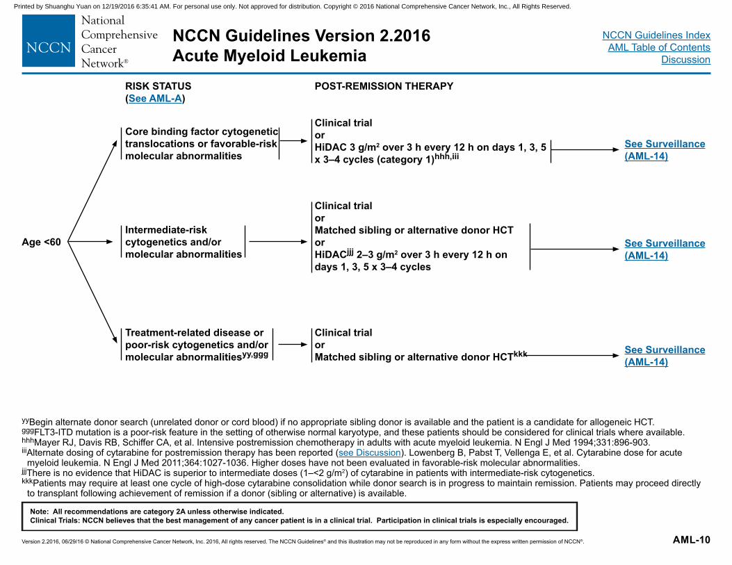
RISK STATUS(See AML-A)
POST-REMISSION THERAPY
Age <60
Core binding factor cytogenetic translocations or favorable-risk molecular abnormalities
Intermediate-risk cytogenetics and/or molecular abnormalities
Treatment-related disease or poor-risk cytogenetics and/or molecular abnormalitiesyy,ggg
Clinical trialorHiDAC 3 g/m2 over 3 h every 12 h on days 1, 3, 5 x 3–4 cycles (category 1)hhh,iii
See Surveillance(AML-14)
Clinical trial or Matched sibling or alternative donor HCTor HiDACjjj 2–3 g/m2 over 3 h every 12 h on days 1, 3, 5 x 3–4 cycles
See Surveillance(AML-14)
Clinical trialorMatched sibling or alternative donor HCTkkk See Surveillance
(AML-14)
yyBegin alternate donor search (unrelated donor or cord blood) if no appropriate sibling donor is available and the patient is a candidate for allogeneic HCT.gggFLT3-ITD mutation is a poor-risk feature in the setting of otherwise normal karyotype, and these patients should be considered for clinical trials where available. hhhMayer RJ, Davis RB, Schiffer CA, et al. Intensive postremission chemotherapy in adults with acute myeloid leukemia. N Engl J Med 1994;331:896-903.iiiAlternate dosing of cytarabine for postremission therapy has been reported (see Discussion). Lowenberg B, Pabst T, Vellenga E, et al. Cytarabine dose for acute
myeloid leukemia. N Engl J Med 2011;364:1027-1036. Higher doses have not been evaluated in favorable-risk molecular abnormalities.jjjThere is no evidence that HiDAC is superior to intermediate doses (1–<2 g/m2) of cytarabine in patients with intermediate-risk cytogenetics.kkkPatients may require at least one cycle of high-dose cytarabine consolidation while donor search is in progress to maintain remission. Patients may proceed directly
to transplant following achievement of remission if a donor (sibling or alternative) is available.
Printed by Shuanghu Yuan on 12/19/2016 6:35:41 AM. For personal use only. Not approved for distribution. Copyright © 2016 National Comprehensive Cancer Network, Inc., All Rights Reserved.
NCCN Guidelines Version 2.2016 Acute Myeloid Leukemia
NCCN Guidelines IndexAML Table of Contents
Discussion
Note: All recommendations are category 2A unless otherwise indicated.Clinical Trials: NCCN believes that the best management of any cancer patient is in a clinical trial. Participation in clinical trials is especially encouraged.
Version 2.2016, 06/29/16 © National Comprehensive Cancer Network, Inc. 2016, All rights reserved. The NCCN Guidelines® and this illustration may not be reproduced in any form without the express written permission of NCCN®. AML-11
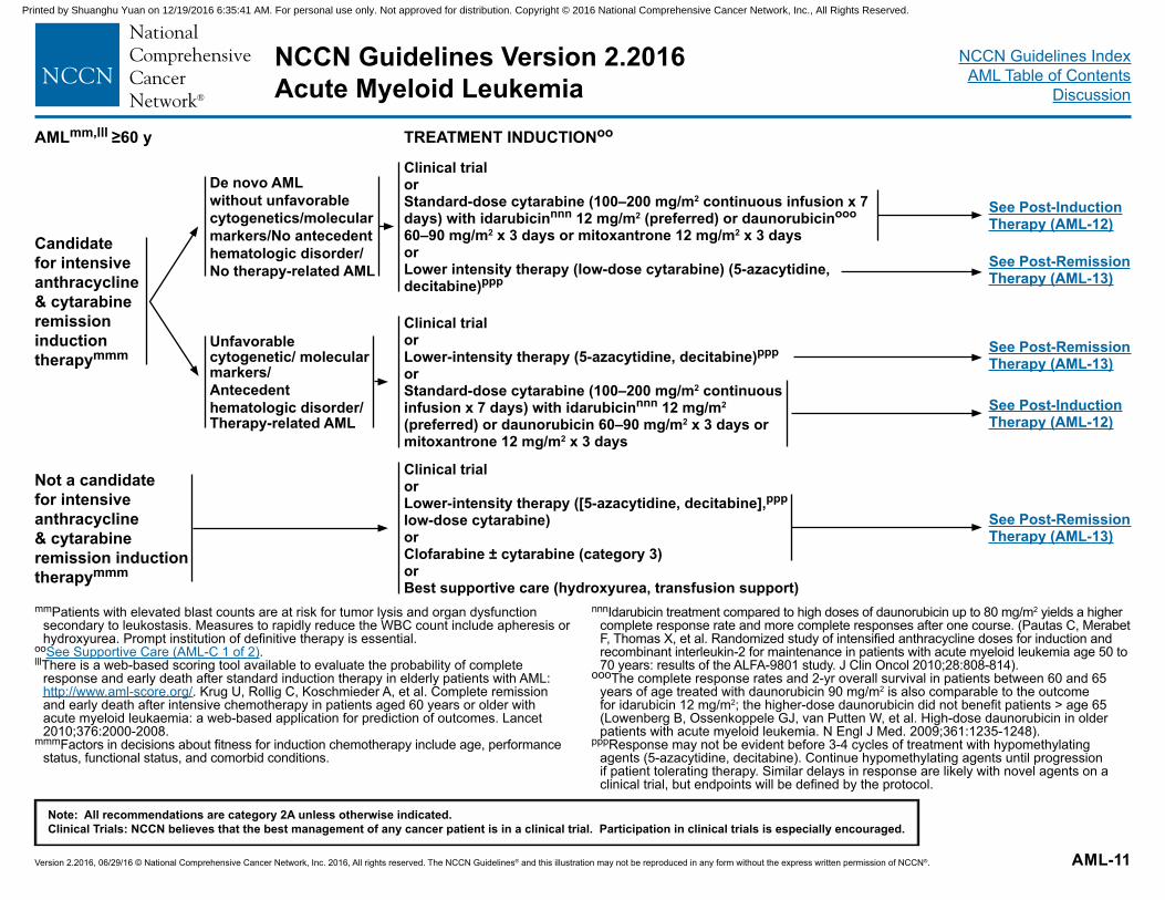
mmPatients with elevated blast counts are at risk for tumor lysis and organ dysfunction secondary to leukostasis. Measures to rapidly reduce the WBC count include apheresis or hydroxyurea. Prompt institution of definitive therapy is essential.
ooSee Supportive Care (AML-C 1 of 2).lllThere is a web-based scoring tool available to evaluate the probability of complete
response and early death after standard induction therapy in elderly patients with AML: http://www.aml-score.org/. Krug U, Rollig C, Koschmieder A, et al. Complete remission and early death after intensive chemotherapy in patients aged 60 years or older with acute myeloid leukaemia: a web-based application for prediction of outcomes. Lancet 2010;376:2000-2008.
mmmFactors in decisions about fitness for induction chemotherapy include age, performance status, functional status, and comorbid conditions.
nnnIdarubicin treatment compared to high doses of daunorubicin up to 80 mg/m2 yields a higher complete response rate and more complete responses after one course. (Pautas C, Merabet F, Thomas X, et al. Randomized study of intensified anthracycline doses for induction and recombinant interleukin-2 for maintenance in patients with acute myeloid leukemia age 50 to 70 years: results of the ALFA-9801 study. J Clin Oncol 2010;28:808-814).
oooThe complete response rates and 2-yr overall survival in patients between 60 and 65 years of age treated with daunorubicin 90 mg/m2 is also comparable to the outcome for idarubicin 12 mg/m2; the higher-dose daunorubicin did not benefit patients > age 65 (Lowenberg B, Ossenkoppele GJ, van Putten W, et al. High-dose daunorubicin in older patients with acute myeloid leukemia. N Engl J Med. 2009;361:1235-1248).
pppResponse may not be evident before 3-4 cycles of treatment with hypomethylating agents (5-azacytidine, decitabine). Continue hypomethylating agents until progression if patient tolerating therapy. Similar delays in response are likely with novel agents on a clinical trial, but endpoints will be defined by the protocol.
AMLmm,lll ≥60 y TREATMENT INDUCTIONoo
Unfavorable cytogenetic/ molecular markers/ Antecedent hematologic disorder/Therapy-related AML
Clinical trial orStandard-dose cytarabine (100–200 mg/m2 continuous infusion x 7 days) with idarubicinnnn 12 mg/m2 (preferred) or daunorubicinooo 60–90 mg/m2 x 3 days or mitoxantrone 12 mg/m2 x 3 days or Lower intensity therapy (low-dose cytarabine) (5-azacytidine, decitabine)ppp
Clinical trial orLower-intensity therapy (5-azacytidine, decitabine)ppp
orStandard-dose cytarabine (100–200 mg/m2 continuous infusion x 7 days) with idarubicinnnn 12 mg/m2 (preferred) or daunorubicin 60–90 mg/m2 x 3 days or mitoxantrone 12 mg/m2 x 3 days
Clinical trial orLower-intensity therapy ([5-azacytidine, decitabine],ppp low-dose cytarabine) or Clofarabine ± cytarabine (category 3)orBest supportive care (hydroxyurea, transfusion support)
See Post-Induction Therapy (AML-12)
See Post-Remission Therapy (AML-13)
See Post-Remission Therapy (AML-13)
See Post-Induction Therapy (AML-12)
See Post-Remission Therapy (AML-13)
De novo AML without unfavorable cytogenetics/molecular markers/No antecedent hematologic disorder/No therapy-related AML
Candidate for intensive anthracycline & cytarabine remission induction therapymmm
Not a candidate for intensive anthracycline & cytarabine remission induction therapymmm
Printed by Shuanghu Yuan on 12/19/2016 6:35:41 AM. For personal use only. Not approved for distribution. Copyright © 2016 National Comprehensive Cancer Network, Inc., All Rights Reserved.

NCCN Guidelines Version 2.2016 Acute Myeloid Leukemia
NCCN Guidelines IndexAML Table of Contents
Discussion
Note: All recommendations are category 2A unless otherwise indicated.Clinical Trials: NCCN believes that the best management of any cancer patient is in a clinical trial. Participation in clinical trials is especially encouraged.
Version 2.2016, 06/29/16 © National Comprehensive Cancer Network, Inc. 2016, All rights reserved. The NCCN Guidelines® and this illustration may not be reproduced in any form without the express written permission of NCCN®. AML-12
ppSee Monitoring During Therapy (AML-E).yyBegin alternate donor search (unrelated donor or cord blood) if no appropriate sibling donor is available and the patient is a candidate for an allogeneic HCT.zzIf ambiguous, consider repeat bone marrow biopsy in 5-7 days before proceeding with therapy. aaaHypoplasia is defined as cellularity <10%–20% and residual blasts <5%–10%.nnnIdarubicin treatment compared to high doses of daunorubicin up to 80 mg/m2 yields higher complete response rate and more complete responses after one course.
(Pautas C, Merabet F, Thomas X, et al. Randomized study of intensified anthracycline doses for induction and recombinant interleukin-2 for maintenance in patients with acute myeloid leukemia age 50 to 70 years: results of the ALFA-9801 study. J Clin Oncol 2010;28:808-814).
oooThe complete response rates and 2-yr overall survival in patients between 60 and 65 years of age treated with daunorubicin 90 mg/m2 is also comparable to the outcome for idarubicin 12 mg/m2; the higher dose daunorubicin did not benefit patients > age 65 (Lowenberg B, Ossenkoppele GJ, van Putten W, et al. High-dose daunorubicin in older patients with acute myeloid leukemia. N Engl J Med 2009;361:1235-1248).
qqqReduced-intensity HCT may be appropriate for patients with a low level of residual disease post-induction (eg, patients with prior MDS who reverted back to MDS with <10% blasts). It is preferred that this approach be given in the context of a clinical trial.
AML POST-INDUCTION THERAPYyy
AFTER STANDARD-DOSE CYTARABINEAge ≥60 y
Clinical trial orAdditional standard-dose cytarabine with anthracycline (idarubicinnnn or daunorubicinooo) or mitoxantroneorIntermediate-dose cytarabine (1-<2 g/m2) containing regimens orReduced-intensity allogeneic HCTqqq
or Await recoveryor Best supportive care
Residual diseasezz,aaa
Hypoplasiazz,aaa Await recovery
Follow-up bone marrowpp 14–21 days after start of therapy
See Post-Remission Therapy (AML-13)
Printed by Shuanghu Yuan on 12/19/2016 6:35:41 AM. For personal use only. Not approved for distribution. Copyright © 2016 National Comprehensive Cancer Network, Inc., All Rights Reserved.
NCCN Guidelines Version 2.2016 Acute Myeloid Leukemia
NCCN Guidelines IndexAML Table of Contents
Discussion
Note: All recommendations are category 2A unless otherwise indicated.Clinical Trials: NCCN believes that the best management of any cancer patient is in a clinical trial. Participation in clinical trials is especially encouraged.
Version 2.2016, 06/29/16 © National Comprehensive Cancer Network, Inc. 2016, All rights reserved. The NCCN Guidelines® and this illustration may not be reproduced in any form without the express written permission of NCCN®. AML-13
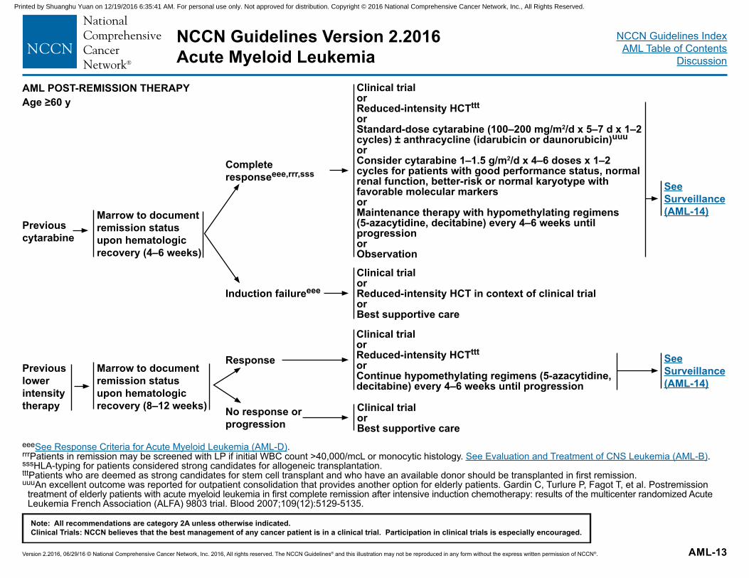
AML POST-REMISSION THERAPYAge ≥60 y
eeeSee Response Criteria for Acute Myeloid Leukemia (AML-D).rrrPatients in remission may be screened with LP if initial WBC count >40,000/mcL or monocytic histology. See Evaluation and Treatment of CNS Leukemia (AML-B).sssHLA-typing for patients considered strong candidates for allogeneic transplantation.tttPatients who are deemed as strong candidates for stem cell transplant and who have an available donor should be transplanted in first remission.uuuAn excellent outcome was reported for outpatient consolidation that provides another option for elderly patients. Gardin C, Turlure P, Fagot T, et al. Postremission
treatment of elderly patients with acute myeloid leukemia in first complete remission after intensive induction chemotherapy: results of the multicenter randomized Acute Leukemia French Association (ALFA) 9803 trial. Blood 2007;109(12):5129-5135.
Clinical trialor Reduced-intensity HCTtttorStandard-dose cytarabine (100–200 mg/m2/d x 5–7 d x 1–2 cycles) ± anthracycline (idarubicin or daunorubicin)uuuorConsider cytarabine 1–1.5 g/m2/d x 4–6 doses x 1–2 cycles for patients with good performance status, normal renal function, better-risk or normal karyotype with favorable molecular markersor Maintenance therapy with hypomethylating regimens (5-azacytidine, decitabine) every 4–6 weeks until progressionor Observation
Complete responseeee,rrr,sss
Induction failureeee
Marrow to document remission status upon hematologic recovery (4–6 weeks)
Clinical trialor Reduced-intensity HCT in context of clinical trialorBest supportive care
See Surveillance (AML-14)
Previous cytarabine
Previous lower intensity therapy
Clinical trialor Reduced-intensity HCTtttorContinue hypomethylating regimens (5-azacytidine, decitabine) every 4–6 weeks until progression
Marrow to document remission status upon hematologic recovery (8–12 weeks) Clinical trial
orBest supportive care
Response
No response or progression
See Surveillance (AML-14)
Printed by Shuanghu Yuan on 12/19/2016 6:35:41 AM. For personal use only. Not approved for distribution. Copyright © 2016 National Comprehensive Cancer Network, Inc., All Rights Reserved.
NCCN Guidelines Version 2.2016 Acute Myeloid Leukemia
NCCN Guidelines IndexAML Table of Contents
Discussion
Note: All recommendations are category 2A unless otherwise indicated.Clinical Trials: NCCN believes that the best management of any cancer patient is in a clinical trial. Participation in clinical trials is especially encouraged.
Version 2.2016, 06/29/16 © National Comprehensive Cancer Network, Inc. 2016, All rights reserved. The NCCN Guidelines® and this illustration may not be reproduced in any form without the express written permission of NCCN®. AML-14
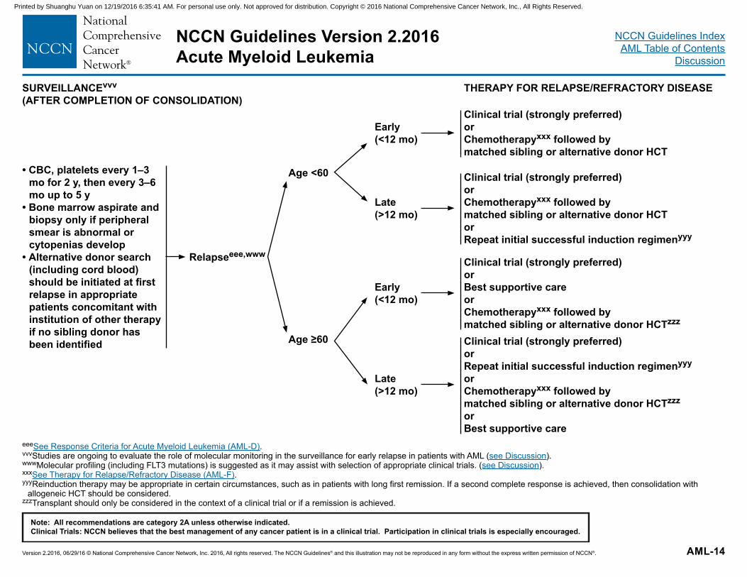
SURVEILLANCEvvv
(AFTER COMPLETION OF CONSOLIDATION)THERAPY FOR RELAPSE/REFRACTORY DISEASE
eeeSee Response Criteria for Acute Myeloid Leukemia (AML-D).vvvStudies are ongoing to evaluate the role of molecular monitoring in the surveillance for early relapse in patients with AML (see Discussion).wwwMolecular profiling (including FLT3 mutations) is suggested as it may assist with selection of appropriate clinical trials. (see Discussion).xxxSee Therapy for Relapse/Refractory Disease (AML-F).yyyReinduction therapy may be appropriate in certain circumstances, such as in patients with long first remission. If a second complete response is achieved, then consolidation with
allogeneic HCT should be considered.zzzTransplant should only be considered in the context of a clinical trial or if a remission is achieved.
• CBC, platelets every 1–3 mo for 2 y, then every 3–6 mo up to 5 y
• Bone marrow aspirate and biopsy only if peripheral smear is abnormal or cytopenias develop
• Alternative donor search (including cord blood) should be initiated at first relapse in appropriate patients concomitant with institution of other therapy if no sibling donor has been identified
Relapseeee,www
Age <60
Age ≥60
Early(<12 mo)
Late (>12 mo)
Early (<12 mo)
Late (>12 mo)
Clinical trial (strongly preferred)or Chemotherapyxxx followed bymatched sibling or alternative donor HCT
Clinical trial (strongly preferred)orChemotherapyxxx followed bymatched sibling or alternative donor HCTorRepeat initial successful induction regimenyyy
Clinical trial (strongly preferred)orBest supportive care orChemotherapyxxx followed bymatched sibling or alternative donor HCTzzz Clinical trial (strongly preferred)orRepeat initial successful induction regimenyyy
orChemotherapyxxx followed bymatched sibling or alternative donor HCTzzz
orBest supportive care
Printed by Shuanghu Yuan on 12/19/2016 6:35:41 AM. For personal use only. Not approved for distribution. Copyright © 2016 National Comprehensive Cancer Network, Inc., All Rights Reserved.
NCCN Guidelines Version 2.2016 Acute Myeloid Leukemia
NCCN Guidelines IndexAML Table of Contents
Discussion
Note: All recommendations are category 2A unless otherwise indicated.Clinical Trials: NCCN believes that the best management of any cancer patient is in a clinical trial. Participation in clinical trials is especially encouraged.
Version 2.2016, 06/29/16 © National Comprehensive Cancer Network, Inc. 2016, All rights reserved. The NCCN Guidelines® and this illustration may not be reproduced in any form without the express written permission of NCCN®. AML-A
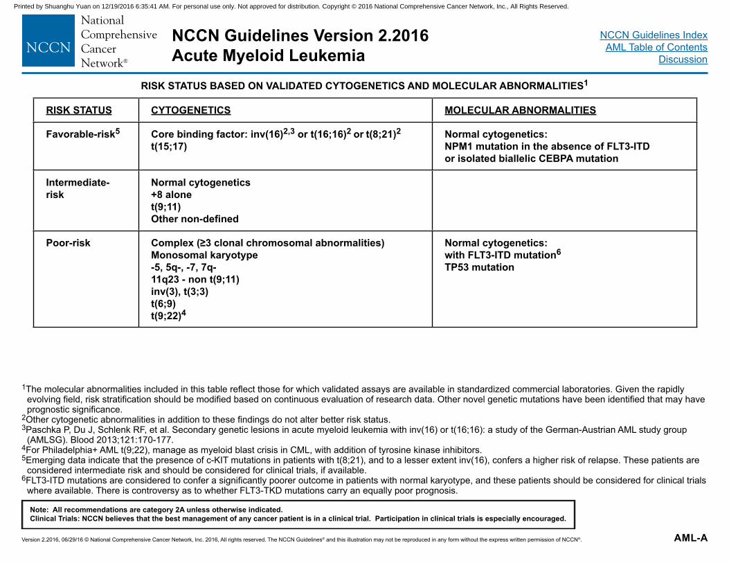
1The molecular abnormalities included in this table reflect those for which validated assays are available in standardized commercial laboratories. Given the rapidly evolving field, risk stratification should be modified based on continuous evaluation of research data. Other novel genetic mutations have been identified that may have prognostic significance.
2Other cytogenetic abnormalities in addition to these findings do not alter better risk status. 3Paschka P, Du J, Schlenk RF, et al. Secondary genetic lesions in acute myeloid leukemia with inv(16) or t(16;16): a study of the German-Austrian AML study group
(AMLSG). Blood 2013;121:170-177.4For Philadelphia+ AML t(9;22), manage as myeloid blast crisis in CML, with addition of tyrosine kinase inhibitors. 5Emerging data indicate that the presence of c-KIT mutations in patients with t(8;21), and to a lesser extent inv(16), confers a higher risk of relapse. These patients are
considered intermediate risk and should be considered for clinical trials, if available. 6FLT3-ITD mutations are considered to confer a significantly poorer outcome in patients with normal karyotype, and these patients should be considered for clinical trials
where available. There is controversy as to whether FLT3-TKD mutations carry an equally poor prognosis.
RISK STATUS BASED ON VALIDATED CYTOGENETICS AND MOLECULAR ABNORMALITIES1
RISK STATUS CYTOGENETICS MOLECULAR ABNORMALITIES
Favorable-risk5 Core binding factor: inv(16)2,3 or t(16;16)2 or t(8;21)2 t(15;17)
Normal cytogenetics: NPM1 mutation in the absence of FLT3-ITD or isolated biallelic CEBPA mutation
Intermediate-risk
Normal cytogenetics +8 alone t(9;11) Other non-defined
Poor-risk Complex (≥3 clonal chromosomal abnormalities) Monosomal karyotype -5, 5q-, -7, 7q- 11q23 - non t(9;11) inv(3), t(3;3) t(6;9) t(9;22)4
Normal cytogenetics: with FLT3-ITD mutation6
TP53 mutation
Printed by Shuanghu Yuan on 12/19/2016 6:35:41 AM. For personal use only. Not approved for distribution. Copyright © 2016 National Comprehensive Cancer Network, Inc., All Rights Reserved.
NCCN Guidelines Version 2.2016 Acute Myeloid Leukemia
NCCN Guidelines IndexAML Table of Contents
Discussion
Note: All recommendations are category 2A unless otherwise indicated.Clinical Trials: NCCN believes that the best management of any cancer patient is in a clinical trial. Participation in clinical trials is especially encouraged.
Version 2.2016, 06/29/16 © National Comprehensive Cancer Network, Inc. 2016, All rights reserved. The NCCN Guidelines® and this illustration may not be reproduced in any form without the express written permission of NCCN®. AML-B
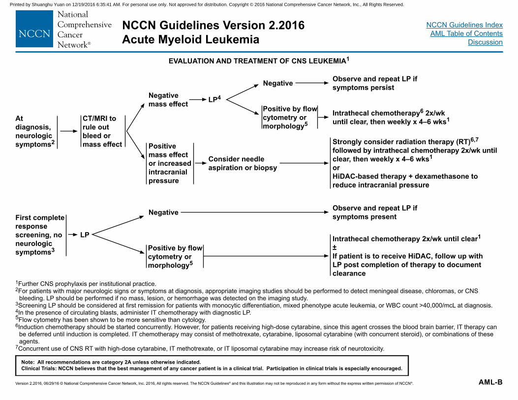
1Further CNS prophylaxis per institutional practice.2For patients with major neurologic signs or symptoms at diagnosis, appropriate imaging studies should be performed to detect meningeal disease, chloromas, or CNS
bleeding. LP should be performed if no mass, lesion, or hemorrhage was detected on the imaging study. 3Screening LP should be considered at first remission for patients with monocytic differentiation, mixed phenotype acute leukemia, or WBC count >40,000/mcL at diagnosis.4In the presence of circulating blasts, administer IT chemotherapy with diagnostic LP.5Flow cytometry has been shown to be more sensitive than cytology. 6Induction chemotherapy should be started concurrently. However, for patients receiving high-dose cytarabine, since this agent crosses the blood brain barrier, IT therapy can
be deferred until induction is completed. IT chemotherapy may consist of methotrexate, cytarabine, liposomal cytarabine (with concurrent steroid), or combinations of these agents.
7Concurrent use of CNS RT with high-dose cytarabine, IT methotrexate, or IT liposomal cytarabine may increase risk of neurotoxicity.
EVALUATION AND TREATMENT OF CNS LEUKEMIA1
At diagnosis, neurologic symptoms2
First complete response screening, no neurologic symptoms3
CT/MRI to rule out bleed or mass effect
LP
Negative mass effect
Positive mass effect or increased intracranial pressure
LP4
Consider needle aspiration or biopsy
Negative Observe and repeat LP if symptoms persist
Intrathecal chemotherapy6 2x/wk until clear, then weekly x 4–6 wks1
Strongly consider radiation therapy (RT)6,7 followed by intrathecal chemotherapy 2x/wk until clear, then weekly x 4–6 wks1
orHiDAC-based therapy + dexamethasone to reduce intracranial pressure
Negative Observe and repeat LP if symptoms present
Intrathecal chemotherapy 2x/wk until clear1
±If patient is to receive HiDAC, follow up with LP post completion of therapy to document clearance
Positive by flow cytometry or morphology5
Positive by flow cytometry or morphology5
Printed by Shuanghu Yuan on 12/19/2016 6:35:41 AM. For personal use only. Not approved for distribution. Copyright © 2016 National Comprehensive Cancer Network, Inc., All Rights Reserved.
NCCN Guidelines Version 2.2016 Acute Myeloid Leukemia
NCCN Guidelines IndexAML Table of Contents
Discussion
Note: All recommendations are category 2A unless otherwise indicated.Clinical Trials: NCCN believes that the best management of any cancer patient is in a clinical trial. Participation in clinical trials is especially encouraged.
Version 2.2016, 06/29/16 © National Comprehensive Cancer Network, Inc. 2016, All rights reserved. The NCCN Guidelines® and this illustration may not be reproduced in any form without the express written permission of NCCN®.
AML-C1 OF 2
See Supportive Care (AML-C 2 of 2)
1Patients who are allo-immunized should receive cross-match compatible and/or HLA-specific blood products.2Smith GA, Damon LE, Rugo HS, et al. High-dose cytarabine dose modification reduces the incidence of neurotoxicity in patients
with renal insufficiency. J Clin Oncol 1997;15(2):833-8393Cornely OA, Maertens J, Winston DJ, et al. Posaconazole vs. fluconazole or itraconazole prophylaxis in patients with neutropenia.
N Engl J Med 2007;356:348-359.
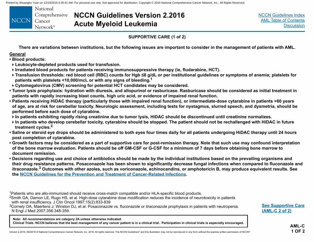
SUPPORTIVE CARE (1 of 2)
There are variations between institutions, but the following issues are important to consider in the management of patients with AML.General• Blood products: �Leukocyte-depleted products used for transfusion.�Irradiated blood products for patients receiving immunosuppressive therapy (ie, fludarabine, HCT).�Transfusion thresholds: red blood cell (RBC) counts for Hgb ≤8 g/dL or per institutional guidelines or symptoms of anemia; platelets for
patients with platelets <10,000/mcL or with any signs of bleeding.1 �Cytomegalovirus (CMV) screening for potential HCT candidates may be considered.
• Tumor lysis prophylaxis: hydration with diuresis, and allopurinol or rasburicase. Rasburicase should be considered as initial treatment in patients with rapidly increasing blast counts, high uric acid, or evidence of impaired renal function.
• Patients receiving HiDAC therapy (particularly those with impaired renal function), or intermediate-dose cytarabine in patients >60 years of age, are at risk for cerebellar toxicity. Neurologic assessment, including tests for nystagmus, slurred speech, and dysmetria, should be performed before each dose of cytarabine. �In patients exhibiting rapidly rising creatinine due to tumor lysis, HiDAC should be discontinued until creatinine normalizes.�In patients who develop cerebellar toxicity, cytarabine should be stopped. The patient should not be rechallenged with HiDAC in future
treatment cycles.2 • Saline or steroid eye drops should be administered to both eyes four times daily for all patients undergoing HiDAC therapy until 24 hours
post completion of cytarabine.• Growth factors may be considered as a part of supportive care for post-remission therapy. Note that such use may confound interpretation
of the bone marrow evaluation. Patients should be off GM-CSF or G-CSF for a minimum of 7 days before obtaining bone marrow to document remission.
• Decisions regarding use and choice of antibiotics should be made by the individual institutions based on the prevailing organisms and their drug resistance patterns. Posaconazole has been shown to significantly decrease fungal infections when compared to fluconazole and itraconazole.3 Outcomes with other azoles, such as voriconazole, echinocandins, or amphotericin B, may produce equivalent results. See the NCCN Guidelines for the Prevention and Treatment of Cancer-Related Infections.
Printed by Shuanghu Yuan on 12/19/2016 6:35:41 AM. For personal use only. Not approved for distribution. Copyright © 2016 National Comprehensive Cancer Network, Inc., All Rights Reserved.
NCCN Guidelines Version 2.2016 Acute Myeloid Leukemia
NCCN Guidelines IndexAML Table of Contents
Discussion
Note: All recommendations are category 2A unless otherwise indicated.Clinical Trials: NCCN believes that the best management of any cancer patient is in a clinical trial. Participation in clinical trials is especially encouraged.
Version 2.2016, 06/29/16 © National Comprehensive Cancer Network, Inc. 2016, All rights reserved. The NCCN Guidelines® and this illustration may not be reproduced in any form without the express written permission of NCCN®.
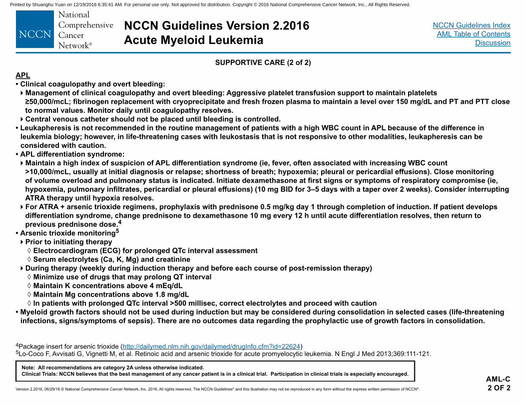
AML-C2 OF 2
4Package insert for arsenic trioxide (http://dailymed.nlm.nih.gov/dailymed/drugInfo.cfm?id=22624)5Lo-Coco F, Avvisati G, Vignetti M, et al. Retinoic acid and arsenic trioxide for acute promyelocytic leukemia. N Engl J Med 2013;369:111-121.
SUPPORTIVE CARE (2 of 2)APL• Clinical coagulopathy and overt bleeding:�Management of clinical coagulopathy and overt bleeding: Aggressive platelet transfusion support to maintain platelets
≥50,000/mcL; fibrinogen replacement with cryoprecipitate and fresh frozen plasma to maintain a level over 150 mg/dL and PT and PTT close to normal values. Monitor daily until coagulopathy resolves.�Central venous catheter should not be placed until bleeding is controlled.
• Leukapheresis is not recommended in the routine management of patients with a high WBC count in APL because of the difference in leukemia biology; however, in life-threatening cases with leukostasis that is not responsive to other modalities, leukapheresis can be considered with caution.
• APL differentiation syndrome:�Maintain a high index of suspicion of APL differentiation syndrome (ie, fever, often associated with increasing WBC count
>10,000/mcL, usually at initial diagnosis or relapse; shortness of breath; hypoxemia; pleural or pericardial effusions). Close monitoring of volume overload and pulmonary status is indicated. Initiate dexamethasone at first signs or symptoms of respiratory compromise (ie, hypoxemia, pulmonary infiltrates, pericardial or pleural effusions) (10 mg BID for 3–5 days with a taper over 2 weeks). Consider interrupting ATRA therapy until hypoxia resolves.�For ATRA + arsenic trioxide regimens, prophylaxis with prednisone 0.5 mg/kg day 1 through completion of induction. If patient develops
differentiation syndrome, change prednisone to dexamethasone 10 mg every 12 h until acute differentiation resolves, then return to previous prednisone dose.4
• Arsenic trioxide monitoring5 �Prior to initiating therapy
◊ Electrocardiogram (ECG) for prolonged QTc interval assessment ◊ Serum electrolytes (Ca, K, Mg) and creatinine
�During therapy (weekly during induction therapy and before each course of post-remission therapy) ◊ Minimize use of drugs that may prolong QT interval ◊ Maintain K concentrations above 4 mEq/dL ◊ Maintain Mg concentrations above 1.8 mg/dL ◊ In patients with prolonged QTc interval >500 millisec, correct electrolytes and proceed with caution
• Myeloid growth factors should not be used during induction but may be considered during consolidation in selected cases (life-threatening infections, signs/symptoms of sepsis). There are no outcomes data regarding the prophylactic use of growth factors in consolidation.
Printed by Shuanghu Yuan on 12/19/2016 6:35:41 AM. For personal use only. Not approved for distribution. Copyright © 2016 National Comprehensive Cancer Network, Inc., All Rights Reserved.
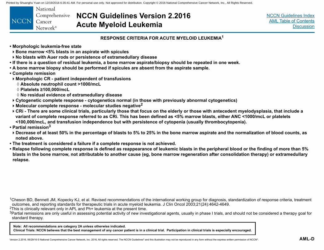
NCCN Guidelines Version 2.2016 Acute Myeloid Leukemia
NCCN Guidelines IndexAML Table of Contents
Discussion
Note: All recommendations are category 2A unless otherwise indicated.Clinical Trials: NCCN believes that the best management of any cancer patient is in a clinical trial. Participation in clinical trials is especially encouraged.
Version 2.2016, 06/29/16 © National Comprehensive Cancer Network, Inc. 2016, All rights reserved. The NCCN Guidelines® and this illustration may not be reproduced in any form without the express written permission of NCCN®. AML-D
1Cheson BD, Bennett JM, Kopecky KJ, et al. Revised recommendations of the international working group for diagnosis, standardization of response criteria, treatment outcomes, and reporting standards for therapeutic trials in acute myeloid leukemia. J Clin Oncol 2003;21(24):4642-4649.
2This is clinically relevant only in APL and Ph+ leukemia at the present time.3Partial remissions are only useful in assessing potential activity of new investigational agents, usually in phase I trials, and should not be considered a therapy goal for
standard therapy.
RESPONSE CRITERIA FOR ACUTE MYELOID LEUKEMIA1
• Morphologic leukemia-free state�Bone marrow <5% blasts in an aspirate with spicules�No blasts with Auer rods or persistence of extramedullary disease
• If there is a question of residual leukemia, a bone marrow aspirate/biopsy should be repeated in one week.• A bone marrow biopsy should be performed if spicules are absent from the aspirate sample.• Complete remission�Morphologic CR - patient independent of transfusions
◊ Absolute neutrophil count >1000/mcL ◊ Platelets ≥100,000/mcL ◊ No residual evidence of extramedullary disease
�Cytogenetic complete response - cytogenetics normal (in those with previously abnormal cytogenetics)�Molecular complete response - molecular studies negative2 �CRi - There are some clinical trials, particularly those that focus on the elderly or those with antecedent myelodysplasia, that include a
variant of complete response referred to as CRi. This has been defined as <5% marrow blasts, either ANC <1000/mcL or platelets <100,000/mcL, and transfusion independence but with persistence of cytopenia (usually thrombocytopenia).
• Partial remission3
�Decrease of at least 50% in the percentage of blasts to 5% to 25% in the bone marrow aspirate and the normalization of blood counts, as noted above.
• The treatment is considered a failure if a complete response is not achieved.• Relapse following complete response is defined as reappearance of leukemic blasts in the peripheral blood or the finding of more than 5%
blasts in the bone marrow, not attributable to another cause (eg, bone marrow regeneration after consolidation therapy) or extramedullary relapse.
Printed by Shuanghu Yuan on 12/19/2016 6:35:41 AM. For personal use only. Not approved for distribution. Copyright © 2016 National Comprehensive Cancer Network, Inc., All Rights Reserved.
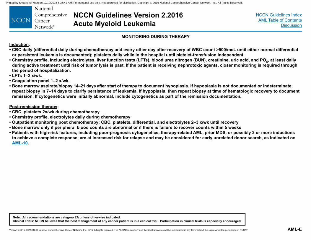
NCCN Guidelines Version 2.2016 Acute Myeloid Leukemia
NCCN Guidelines IndexAML Table of Contents
Discussion
Note: All recommendations are category 2A unless otherwise indicated.Clinical Trials: NCCN believes that the best management of any cancer patient is in a clinical trial. Participation in clinical trials is especially encouraged.
Version 2.2016, 06/29/16 © National Comprehensive Cancer Network, Inc. 2016, All rights reserved. The NCCN Guidelines® and this illustration may not be reproduced in any form without the express written permission of NCCN®. AML-E
MONITORING DURING THERAPY
Induction:• CBC daily (differential daily during chemotherapy and every other day after recovery of WBC count >500/mcL until either normal differential
or persistent leukemia is documented); platelets daily while in the hospital until platelet-transfusion independent.• Chemistry profile, including electrolytes, liver function tests (LFTs), blood urea nitrogen (BUN), creatinine, uric acid, and PO4, at least daily
during active treatment until risk of tumor lysis is past. If the patient is receiving nephrotoxic agents, closer monitoring is required through the period of hospitalization.
• LFTs 1–2 x/wk. • Coagulation panel 1–2 x/wk.• Bone marrow aspirate/biopsy 14–21 days after start of therapy to document hypoplasia. If hypoplasia is not documented or indeterminate,
repeat biopsy in 7–14 days to clarify persistence of leukemia. If hypoplasia, then repeat biopsy at time of hematologic recovery to document remission. If cytogenetics were initially abnormal, include cytogenetics as part of the remission documentation.
Post-remission therapy:• CBC, platelets 2x/wk during chemotherapy• Chemistry profile, electrolytes daily during chemotherapy• Outpatient monitoring post chemotherapy: CBC, platelets, differential, and electrolytes 2–3 x/wk until recovery• Bone marrow only if peripheral blood counts are abnormal or if there is failure to recover counts within 5 weeks• Patients with high-risk features, including poor-prognosis cytogenetics, therapy-related AML, prior MDS, or possibly 2 or more inductions
to achieve a complete response, are at increased risk for relapse and may be considered for early unrelated donor search, as indicated on AML-10.
Printed by Shuanghu Yuan on 12/19/2016 6:35:41 AM. For personal use only. Not approved for distribution. Copyright © 2016 National Comprehensive Cancer Network, Inc., All Rights Reserved.
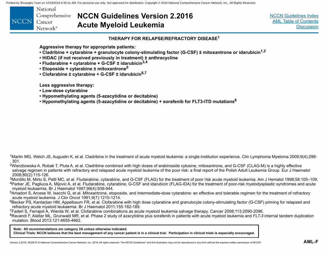
NCCN Guidelines Version 2.2016 Acute Myeloid Leukemia
NCCN Guidelines IndexAML Table of Contents
Discussion
Note: All recommendations are category 2A unless otherwise indicated.Clinical Trials: NCCN believes that the best management of any cancer patient is in a clinical trial. Participation in clinical trials is especially encouraged.
Version 2.2016, 06/29/16 © National Comprehensive Cancer Network, Inc. 2016, All rights reserved. The NCCN Guidelines® and this illustration may not be reproduced in any form without the express written permission of NCCN®. AML-F
1Martin MG, Welch JS, Augustin K, et al. Cladribine in the treatment of acute myeloid leukemia: a single-institution experience. Clin Lymphoma Myeloma 2009;9(4):298-301.
2Wierzbowska A, Robak T, Pluta A, et al. Cladribine combined with high doses of arabinoside cytosine, mitoxantrone, and G-CSF (CLAG-M) is a highly effective salvage regimen in patients with refractory and relapsed acute myeloid leukemia of the poor risk: a final report of the Polish Adult Leukemia Group. Eur J Haematol 2008;80(2):115-126.
3Montillo M, Mirto S, Petti MC, et al. Fludarabine, cytarabine, and G-CSF (FLAG) for the treatment of poor risk acute myeloid leukemia. Am J Hematol 1998;58:105–109. 4Parker JE, Pagliuca A, Mijovic A, et al. Fludarabine, cytarabine, G-CSF and idarubicin (FLAG-IDA) for the treatment of poor-risk myelodysplastic syndromes and acute
myeloid leukaemia. Br J Haematol 1997;99(4):939-944. 5Amadori S, Arcese W, Isacchi G, et al. Mitoxantrone, etoposide, and intermediate-dose cytarabine: an effective and tolerable regimen for the treatment of refractory
acute myeloid leukemia. J Clin Oncol 1991;9(7):1210-1214.6Becker PS, Kantarjian HM, Appelbaum FR, et al. Clofarabine with high dose cytarabine and granulocyte colony-stimulating factor (G-CSF) priming for relapsed and
refractory acute myeloid leukaemia. Br J Haematol 2011;155:182-189.7Faderl S, Ferrajoli A, Wierda W, et al. Clofarabine combinations as acute myeloid leukemia salvage therapy. Cancer 2008;113:2090-2096.8Ravandi F, Alattar ML, Grunwald MR, et al. Phase 2 study of azacytidine plus sorafenib in patients with acute myeloid leukemia and FLT-3 internal tandem duplication
mutation. Blood 2013:121:4655-4662.
THERAPY FOR RELAPSE/REFRACTORY DISEASE1
Aggressive therapy for appropriate patients:• Cladribine + cytarabine + granulocyte colony-stimulating factor (G-CSF) ± mitoxantrone or idarubicin1,2
• HiDAC (if not received previously in treatment) ± anthracycline • Fludarabine + cytarabine + G-CSF ± idarubicin3,4
• Etoposide + cytarabine ± mitoxantrone5
• Clofarabine ± cytarabine + G-CSF ± idarubicin6,7
Less aggressive therapy:• Low-dose cytarabine• Hypomethylating agents (5-azacytidine or decitabine)• Hypomethylating agents (5-azacytidine or decitabine) + sorafenib for FLT3-ITD mutations8
Printed by Shuanghu Yuan on 12/19/2016 6:35:41 AM. For personal use only. Not approved for distribution. Copyright © 2016 National Comprehensive Cancer Network, Inc., All Rights Reserved.
Version 2.2016, 06/29/16 © National Comprehensive Cancer Network, Inc. 2016, All rights reserved. The NCCN Guidelines® and this illustration may not be reproduced in any form without the express written permission of NCCN®. MS-1
NCCN Guidelines IndexAML Table of Contents
Discussion
NCCN Guidelines Version 2.2016 Acute Myeloid Leukemia
Discussion
NCCN Categories of Evidence and Consensus
Category 1: Based upon high-level evidence, there is uniform NCCN consensus that the intervention is appropriate.
Category 2A: Based upon lower-level evidence, there is uniform NCCN consensus that the intervention is appropriate.
Category 2B: Based upon lower-level evidence, there is NCCN consensus that the intervention is appropriate.
Category 3: Based upon any level of evidence, there is major NCCN disagreement that the intervention is appropriate.
All recommendations are category 2A unless otherwise noted.
Table of Contents
Overview ..................................................................................... MS-2
Literature Search Criteria and Guidelines Update Methodology MS-3
Initial Evaluation ........................................................................ MS-3
Workup ..................................................................................... MS-3
Diagnosis ................................................................................. MS-4
Cytogenetics and Risk Stratification ......................................... MS-6
Molecular Markers and Risk Stratification ................................. MS-7
Principles of AML Treatment .................................................. MS-12
Management of Acute Promyelocytic Leukemia .................... MS-13
Induction Therapy for Patients with APL ................................. MS-14
Consolidation Therapy for Patients with APL .......................... MS-18
Post-Consolidation or Maintenance for Patients with APL ....... MS-22
Management of Relapsed APL ............................................... MS-24
Supportive Care for Patients with APL .................................... MS-26
Management of AML ................................................................ MS-27
Management of AML in Patients Younger Than 60 Years ....... MS-28
Management of AML in Patients Older Than 60 Years ........... MS-36
Role of MRD Monitoring .......................................................... MS-45
Postremission Surveillance and Therapy for Relapsed/Refractory AML ........................................................................................ MS-48
Supportive Care for Patients with AML ................................... MS-50
Evaluation and Treatment of CNS Leukemia .......................... MS-51
References ................................................................................ MS-52
Printed by Shuanghu Yuan on 12/19/2016 6:35:41 AM. For personal use only. Not approved for distribution. Copyright © 2016 National Comprehensive Cancer Network, Inc., All Rights Reserved.
Version 2.2016, 06/29/16 © National Comprehensive Cancer Network, Inc. 2016, All rights reserved. The NCCN Guidelines® and this illustration may not be reproduced in any form without the express written permission of NCCN®. MS-2
NCCN Guidelines IndexAML Table of Contents
Discussion
NCCN Guidelines Version 2.2016 Acute Myeloid Leukemia
Overview Acute myeloid leukemia (AML) is a heterogeneous hematologic malignancy characterized by the clonal expansion of myeloid blasts in the peripheral blood, bone marrow, and/or other tissues. It is the most common form of acute leukemia among adults and accounts for the largest number of annual deaths from leukemias in the United States. An estimated 19,950 people will be diagnosed with AML in 2016, and 10,430 patients will die of the disease.1,2 The median age of diagnosis is 67 years, with 54% of patients diagnosed at 65 years or older (and approximately a third diagnosed at ≥75 years of age).3 Thus, as the population ages, the incidence of AML, along with myelodysplasia, seems to be rising. Environmental factors that have long been established to increase the risks of myelodysplastic syndromes (MDS) and AML include prolonged exposure to petrochemicals; solvents such as benzene; pesticides; and ionizing radiation.4 Equally disturbing is the increasing incidence of treatment-related myelodysplasia and acute leukemia in survivors of childhood and young adulthood cancers. The exact incidence of therapy-related MDS/AML (secondary MDS/AML) is unknown and varies depending on the types of treatment modalities used for a given primary tumor. Reports suggest that therapy-related MDS/AML may account for 5% to 20% of patients with MDS/AML.5-7 Therapy-related myeloid leukemia is a well-recognized consequence of cancer treatment in a proportion of patients receiving cytotoxic therapy for solid tumors or hematologic malignancies. The rate of therapy-related MDS/AML is higher among patients with certain primary tumors, including breast cancer, gynecologic cancers, and lymphomas (both non-Hodgkin’s lymphoma and Hodgkin lymphoma), largely owing to the more leukemogenic cytotoxic agents that are commonly used in the treatment of these tumors.7-10 The 2 well-documented categories of cytotoxic agents associated with the development of therapy-related MDS/AML are alkylating agents (eg, cyclophosphamide, melphalan)
and topoisomerase inhibitors/agents (eg, etoposide, doxorubicin, mitoxantrone).5,8,9 Treatment with antimetabolites, such as the purine analog fludarabine, has also been associated with therapy-related MDS/AML in patients with lymphoproliferative disorders, particularly when administered in combination with alkylating agents.11,12 Radiotherapy, especially in the context of myeloablative therapy (eg, total-body irradiation or radioimmunotherapy) given before autologous hematopoietic cell transplantation (HCT), may also increase the risk for therapy-related MDS/AML.13,14 The disease course of therapy-related MDS/AML is generally progressive and may be more resistant to conventional cytotoxic therapies than de novo cases of MDS/AML.9 Importantly, clinical outcomes in patients with therapy-related AML have been shown to be significantly inferior (both in terms of relapse-free survival [RFS] and overall survival [OS]) compared with patients with de novo cases,8,15 except those with the therapy-related acute promyelocytic leukemia (APL) subtype7,16 or the favorable-risk core binding factor (CBF) translocations. The proportion of patients with unfavorable cytogenetics tends to be higher in the population with therapy-related AML. Even among the subgroup with favorable karyotypes, those with therapy-related AML tend to do less well.
The AML Panel for the NCCN Clinical Practice Guidelines in Oncology (NCCN Guidelines®) convenes annually to update recommendations for the diagnosis and treatment of AML in adults. These recommendations are based on a review of recently published clinical trials that have led to significant improvements in treatment or have yielded new information regarding biologic factors that may have prognostic importance. Most improvements in recent years have been in the treatment of patients with APL, which serves as a paradigm for understanding how the biology of the disease can inform treatment.
Printed by Shuanghu Yuan on 12/19/2016 6:35:41 AM. For personal use only. Not approved for distribution. Copyright © 2016 National Comprehensive Cancer Network, Inc., All Rights Reserved.
Version 2.2016, 06/29/16 © National Comprehensive Cancer Network, Inc. 2016, All rights reserved. The NCCN Guidelines® and this illustration may not be reproduced in any form without the express written permission of NCCN®. MS-3
NCCN Guidelines IndexAML Table of Contents
Discussion
NCCN Guidelines Version 2.2016 Acute Myeloid Leukemia
Literature Search Criteria and Guidelines Update Methodology Prior to the update of this version of the NCCN Guidelines for Acute Myeloid Leukemia, an electronic search of the PubMed database was performed to obtain key literature published between July 10, 2014 and August 28, 2015, using the following search terms: acute myeloid leukemia or acute promyelocytic leukemia. The PubMed database was chosen as it remains the most widely used resource for medical literature and indexes only peer-reviewed biomedical literature.17
The search results were narrowed by selecting studies in humans published in English. Results were confined to the following article types: Clinical Trial, Phase II; Clinical Trial, Phase III; Clinical Trial, Phase IV; Guideline; Meta-Analysis; Randomized Controlled Trial; Systematic Reviews; and Validation Studies.
The PubMed search resulted in 70 citations and their potential relevance was examined. The data from key PubMed articles as well as articles from additional sources deemed as relevant to these Guidelines and discussed by the panel have been included in this version of the Discussion section (eg, e-publications ahead of print, meeting abstracts). Recommendations for which high-level evidence is lacking are based on the panel’s review of lower-level evidence and expert opinion.
The complete details of the Development and Update of the NCCN Guidelines are available on the NCCN webpage.
Initial Evaluation The initial evaluation of AML has 2 objectives. The first is to characterize the disease process based on factors such as prior toxic exposure, antecedent myelodysplasia, and karyotypic or molecular
abnormalities, which may provide prognostic information that can impact responsiveness to chemotherapy and risk of relapse. The second objective focuses on patient-specific factors, including assessment of comorbid conditions, which may affect an individual’s ability to tolerate chemotherapy. Both disease-specific and individual patient factors are taken into consideration when deciding treatment.
Workup The evaluation and initial workup for suspected acute leukemias consist of a comprehensive medical history and physical examination. Laboratory evaluations include blood chemistry and a complete blood count including platelets and differentials. Bone marrow analysis with cytogenetics (karyotype, with or without fluorescence in situ hybridization [FISH]) is necessary for risk stratification and to guide therapy of AML. Evaluation of several molecular markers (eg, FLT3, NPM1, CEBPA, KIT) may be important for risk assessment and prognostication, and may also guide treatment decisions. More comprehensive panel arrays are available and institutions may have established sequencing panels that expand beyond the markers identified. Recent studies have reported on the prognostic impact of a number of molecular abnormalities in patients with AML (see Molecular Markers and Risk Stratification). Adequate marrow should be available at the time of diagnosis or relapse for molecular studies as per the institutional practice. Please confer with your local pathologist on how to optimize sample collection. If molecular testing is not available at the patient’s treatment center, evaluation at an outside reference laboratory is recommended.
Extramedullary presentation, including central nervous system (CNS) disease, is uncommon in patients with AML. Patients with significant CNS signs or symptoms at presentation should be evaluated using
Printed by Shuanghu Yuan on 12/19/2016 6:35:41 AM. For personal use only. Not approved for distribution. Copyright © 2016 National Comprehensive Cancer Network, Inc., All Rights Reserved.
Version 2.2016, 06/29/16 © National Comprehensive Cancer Network, Inc. 2016, All rights reserved. The NCCN Guidelines® and this illustration may not be reproduced in any form without the express written permission of NCCN®. MS-4
NCCN Guidelines IndexAML Table of Contents
Discussion
NCCN Guidelines Version 2.2016 Acute Myeloid Leukemia
appropriate imaging techniques, such as radiography, CT, or MRI for the detection of intracranial bleeding, leptomeningeal disease, or mass lesions in either the brain or spinal cord. However, if symptoms persist, and bleeding and mass/lesions are excluded, the patient should have a lumbar puncture (LP) for diagnostic and possible therapeutic purposes once coagulopathy has been corrected and adequate platelet support is available. Routine screening LPs are not warranted at the time of diagnosis in patients with AML. However, for patients at high risk for CNS disease, such as those with monocytic differentiation or high white blood cell (WBC) count (>40,000/mcL)18 at presentation, a diagnostic LP should be considered as part of the documentation of remission status. Screening LPs should be considered in patients with extramedullary disease before first consolidation, particularly in patients not receiving high-dose cytarabine (HiDAC) (ie, older patients). Patients proceeding to transplant should also be considered for screening LPs. For patients who present with solitary extramedullary disease (often referred to as myeloid sarcoma, granulocytic sarcoma, or chloroma) without overt marrow disease, the initial treatment should still be based on systemic induction chemotherapy. Radiation or surgical resection may be incorporated with systemic chemotherapy in emergent situations; however, these modalities, if needed at all, should be optimally deferred until after count recovery to avoid excess toxicity.
Coagulopathy is fairly common at presentation in many leukemias; it is therefore standard clinical practice to screen for coagulopathy by evaluating prothrombin time, partial thromboplastin time, and fibrinogen activity as part of the initial evaluation/workup and before performing any invasive procedure. The need for a cardiac evaluation (eg, echocardiogram or multiple gated acquisition [MUGA] scan) should be determined by individual risk factors. Patients with a history or symptoms of cardiac disease, prior exposure to cardiotoxic drugs or
thoracic radiation, or who are of an older age should have an echocardiogram. In younger patients who are otherwise asymptomatic with no history of cardiac disease, an echocardiogram can be considered. In cases of acutely ill patients, treatment should not be delayed for an echocardiogram. A small study of 76 patients with cancer who were screened for cardiac disease identified only 4 patients with cardiac abnormalities. Of these 4 patients, the presence of cardiac disease did not change the course of treatment.19
Human leukocyte antigen (HLA) typing should be performed in all patients with newly diagnosed AML for whom allogeneic HCT would be considered. HLA typing of family members is recommended for patients younger than 60 years of age who do not have favorable-risk cytogenetics and tissue typing should be broadened to include alternative donor searches. In patients with any non-favorable risk, a donor search should begin while the patient is recovering from induction chemotherapy rather than waiting for remission to be achieved. HLA typing is routinely used in many institutions to select donors for patients who exhibit alloimmunization to HLA-specific antigens. In patients with treatment that fails to respond to HLA-matched products, the use of corticosteroids, intravenous (IV) immunoglobulins, and/or rituximab are strategies to decrease alloimmunization.
Diagnosis Originally, the classification system for AML was defined by the French American British (FAB) system, which relied on cytochemical stains and morphology to separate AML from acute lymphoblastic leukemia (ALL) and to categorize the disease based on degree of myeloid and monocytic differentiation. In 1999, WHO developed a newer classification system, which incorporates information from cytogenetics
Printed by Shuanghu Yuan on 12/19/2016 6:35:41 AM. For personal use only. Not approved for distribution. Copyright © 2016 National Comprehensive Cancer Network, Inc., All Rights Reserved.
Version 2.2016, 06/29/16 © National Comprehensive Cancer Network, Inc. 2016, All rights reserved. The NCCN Guidelines® and this illustration may not be reproduced in any form without the express written permission of NCCN®. MS-5
NCCN Guidelines IndexAML Table of Contents
Discussion
NCCN Guidelines Version 2.2016 Acute Myeloid Leukemia
and evidence of dysplasia, to refine prognostic subgroups that may define treatment strategies.20 During this transition from the FAB system to the WHO classification, the percent blasts threshold for defining high-grade MDS and AML was lowered. The FAB classification had set the threshold between high-grade MDS and AML at 30% blasts, whereas the WHO classification lowered the threshold for diagnosing AML to 20% or more blasts. This change was based on the finding that the biologic behavior (and survival outcomes) of the FAB MDS subgroup of “refractory anemia with excess blasts in transformation (RAEB-T),” defined as patients with 20% to 30% blasts, was similar compared with that of patients with greater than 30% blasts. The WHO classification system further allowed AML to be diagnosed in patients with abnormal hematopoiesis and characteristic clonal structural cytogenetic abnormalities with t(15;17), t(8;21), and inv(16) or t(16;16) regardless of the percentage of marrow blasts.
In 2003, the International Working Group for Diagnosis, Standardization of Response Criteria accepted the cytochemical and immunophenotypic WHO criteria as the standard for diagnosing AML, including the reporting of dysplasia according to morphology.21
However, no evidence shows that dysplasia represents an independent risk factor, because it is frequently linked to poor-risk cytogenetics.
In 2008, WHO revised the diagnostic and response criteria for AML to include additional recurrent genetic abnormalities created by reciprocal translocations/inversions, and a new provisional category for some of the molecular markers that have been found to have a prognostic impact.22 Additionally, the category of AML with recurrent genetic abnormalities was expanded to include the following: t(9;11)(p22;q23), t(6;9)(p23;q34) (provisional entity), inv(3)(q21 q26.2) or inv(3;3)(q21;q26.2) (provisional entity), and t(1;22)(p13;q13) (provisional entity), in addition to the previously recognized
t(8;21)(q22;q22); inv(16)(p13;1q22) or t(16;16)(p13.1;q22); and t(15;17)(q22;q12) [APL subtype]. Other provisional entities include AML with molecular abnormalities such as mutated nucleophosmin (NPM1) or CCAAT/enhancer-binding protein alpha (CEBPA) genes (further information on these genetic lesions is provided later).22 In 2016, WHO expanded the recurrent genetic abnormalities to include two provisional categories, AML with BCR-ABL1 and AML with RUNX1. AML with BCR-ABL1 is a rare de novo AML that may benefit from therapies that entail tyrosine kinase inhibitors. AML with RUNX1 is associated with a poorer prognosis. The accurate classification of AML requires multidisciplinary diagnostic studies (using immunohistochemistry, cytochemistry, or both, in addition to molecular genetics analysis) in accordance with the 2016 WHO classification. The NCCN AML Panel suggests that complementary diagnostic techniques can be used at the discretion of the pathology department of the individual institution. Some cases may still show evidence of both myeloid and lymphoid antigen expression on the leukemic cells and are defined as acute leukemias of ambiguous lineage. This is further subgrouped into acute undifferentiated leukemia, mixed phenotypic acute leukemia (MPAL) with BCR-ABL1, MPAL with KMT2A rearranged, MPAL with B-myeloid features not otherwise specified, and MPAL with T-myeloid features not otherwise specified. The expression of both cytochemical and/or immunophenotypic characteristics of both lineages on the same cells is defined as biphenotypic, whereas expression of lineage-specific characteristics on different populations of leukemia cells is termed bilineal. When presented with rare cases such as acute leukemias of ambiguous lineage as defined by the 2016 WHO classification), consultation with an experienced hematopathologist should be sought.
Printed by Shuanghu Yuan on 12/19/2016 6:35:41 AM. For personal use only. Not approved for distribution. Copyright © 2016 National Comprehensive Cancer Network, Inc., All Rights Reserved.
Version 2.2016, 06/29/16 © National Comprehensive Cancer Network, Inc. 2016, All rights reserved. The NCCN Guidelines® and this illustration may not be reproduced in any form without the express written permission of NCCN®. MS-6
NCCN Guidelines IndexAML Table of Contents
Discussion
NCCN Guidelines Version 2.2016 Acute Myeloid Leukemia
Aberrant expression of differentiation antigens present at diagnosis may allow tracking of residual blasts through flow cytometry in follow-up samples that may appear normal according to conventional morphology. The use of immunophenotyping and molecular markers to monitor minimal residual disease (MRD) in adult AML has not yet been incorporated into postremission monitoring strategies, except in patients with APL. However, ongoing research is moving MRD monitoring to the forefront for all patients with AML (see Role of MRD Monitoring).
Cytogenetics and Risk Stratification Although cytogenetic information is often unknown when treatment is initiated in patients with de novo AML, karyotype represents the single most important prognostic factor for predicting remission rates, relapse risks, and OS outcomes. The cytogenetic risk categories adopted by these guidelines are primarily based on analyses of large datasets from major cooperative group trials (see Risk Status Based on Validated Cytogenetics and Molecular Abnormalities in the algorithm).23-25 In an analysis of data from pediatric and adult patients with AML (N = 1612) enrolled in the United Kingdom Medical Research Council (UK MRC) AML 10 trial, the 5-year survival rates for those with favorable, intermediate, and unfavorable risk cytogenetics were 65%, 41%, and 14%, respectively.24 In a review of data from adult patients treated on a phase III Southwest Oncology Group (SWOG)/Eastern Cooperative Oncology Group (ECOG) intergroup study (N = 609), the 5-year survival rates for those with favorable, intermediate, and adverse risk cytogenetics were 55%, 38%, and 11%, respectively.25 Similarly, in a retrospective review of adult patients with AML treated on Cancer and Leukemia Group B (CALGB) protocols (N = 1213), the 5-year survival rates for patients with favorable-, intermediate-, and poor-risk cytogenetics were 55%, 24%, and 5%, respectively.23 The AML 11 trial
had similar results with 5-year survival rates of the favorable-, intermediate-, and poor-risk cytogenetics of 34%, 13%, and 2%, respectively.26 This last study included an older population of patients, which is believed to attribute to the overall lower percent survival in all groups.
The importance of obtaining adequate samples of marrow or peripheral blood at diagnosis for full karyotyping and FISH cytogenetic analysis for the most common abnormalities cannot be overemphasized. Although FISH studies for common cytogenetic abnormalities may allow for rapid screening to identify either favorable- or unfavorable-risk groups, additional tests are needed to provide a full picture of the genetic factors that contribute to risk (see Molecular Markers and Risk Stratification).
In the past 5 years, the presence of autosomal chromosome monosomies in AML has emerged as an important prognostic factor associated with extremely poor prognosis.27-29 Data from 3 large studies have identified monosomal karyotypes (defined as ≥2 autosomal monosomies, or a single monosomy with an additional structural abnormality) as a subset of unfavorable cytogenetic prognosticators. Although complex karyotype (≥3 clonal cytogenetic abnormalities) and either monosomy 5 or monosomy 7 are categorized as high-risk/unfavorable cytogenetics, the presence of a monosomal karyotype was found to confer further negative prognostic influence within the high-risk group. This high-risk subgroup was first identified in a joint study conducted by the Dutch-Belgian-Swiss cooperative groups (HOVON/SAKK), that evaluated the correlation between cytogenetics and OS outcomes in patients aged 60 years or younger with AML (N = 1975). The 4-year OS rate in patients with monosomal karyotype was 4% compared with 26% in those with complex karyotype (but without monosomal karyotype).27
Printed by Shuanghu Yuan on 12/19/2016 6:35:41 AM. For personal use only. Not approved for distribution. Copyright © 2016 National Comprehensive Cancer Network, Inc., All Rights Reserved.

Version 2.2016, 06/29/16 © National Comprehensive Cancer Network, Inc. 2016, All rights reserved. The NCCN Guidelines® and this illustration may not be reproduced in any form without the express written permission of NCCN®. MS-7
NCCN Guidelines IndexAML Table of Contents
Discussion
NCCN Guidelines Version 2.2016 Acute Myeloid Leukemia
These findings were confirmed in subsequent analyses from other large cooperative group studies. In an analysis of data from patients treated on SWOG protocols (N = 1344; age 16–88 years), 13% of patients were found to have monosomal karyotype; nearly all of these cases (98%) occurred within the unfavorable cytogenetics category.28 The incidence of monosomal karyotype increased with age, from 4% in patients aged 30 years of age or younger to 20% in patients older than 60 years of age. Among patients with unfavorable cytogenetics, the 4-year OS rate in the subgroup of patients with monosomal karyotype was 3% compared with 13% in the subgroup without monosomal karyotype. In patients with monosomy 7, monosomal karyotype did not appear to influence outcomes (4-year OS, 0%–3%); the 4-year OS rates for patients with inv(3)/t(3;3) and t(6;9) and those without monosomal karyotype were 0% and 9%, respectively.28 In a retrospective study that evaluated the prognostic impact of monosomal karyotype in older patients (age >60 years; N = 186) with unfavorable cytogenetics treated in a GOELAMS trial, the 2-year OS rate was significantly decreased among patients with monosomal karyotype compared with patients without this abnormality (7% vs. 22%; P < .0001). Similar outcomes were observed within the subgroup of patients with complex karyotype.29
These studies show that monosomal karyotype, independent of other unfavorable cytogenetic factors, confers very poor prognosis. In the NCCN Guidelines, the presence of monosomal karyotype is included in the unfavorable-risk category of AML based on cytogenetics (see Risk Status Based on Validated Cytogenetics and Molecular Abnormalities in the algorithm).
Molecular Markers and Risk Stratification The intermediate-risk cytogenetic category is the most heterogeneous group in AML, because it encompasses both normal karyotype AML (NK-AML) without gross structural abnormalities and those with structural changes that are considered neither poor-risk nor favorable. Based on retrospective analyses of data from large cooperative group studies, 40% to 50% of patients with de novo AML have normal karyotype, which is associated with intermediate risk as measured in terms of survival outcomes.23,24 However, even in patients with NK-AML, clinical outcome is heterogeneous.
The ability to identify mutations that carry prognostic impact is increasing with the use of molecular profiling. Thus, in addition to basic cytogenetic analysis, new molecular markers can help refine prognostics groups, particularly in patients with a normal karyotype. These markers include NPM1, FMS-like tyrosine kinase 3 (FLT3), CEBPA, isocitrate dehydrogenase 1 and 2 (IDH1/2), DNA (cytosine-5)-methyltransferase 3A (DNMT3A), and KIT gene mutations.30-42 Tests for these molecular markers are becoming more common in commercial reference laboratories and in referral centers. Therefore, it is important for physicians to confer with the local pathologist on how to optimize sample collection from the time of diagnosis for subsequent molecular diagnostic tests, particularly in patients with normal karyotype. Testing for additional mutations may also be recommended.
NPM1 Mutations The NPM1 gene encodes a shuttle protein within the nucleolus of cells. Mutations in this gene occur in 28% to 35% of AML cases.40,43,44 34,44,45 The NPM1 mutation has been shown to be associated with NK-AML with a reported frequency of 48% to 53%.32,38,45 Isolated NPM1 mutation, which localizes to the cytoplasm, confers a higher complete
Printed by Shuanghu Yuan on 12/19/2016 6:35:41 AM. For personal use only. Not approved for distribution. Copyright © 2016 National Comprehensive Cancer Network, Inc., All Rights Reserved.
Version 2.2016, 06/29/16 © National Comprehensive Cancer Network, Inc. 2016, All rights reserved. The NCCN Guidelines® and this illustration may not be reproduced in any form without the express written permission of NCCN®. MS-8
NCCN Guidelines IndexAML Table of Contents
Discussion
NCCN Guidelines Version 2.2016 Acute Myeloid Leukemia
response (CR) rate and improved event-free survival (EFS) and OS compared with patients who are NK-AML and wild-type NPM1, resulting in outcomes similar to patients with favorable cytogenetics (eg, CBF AML).32,33,38,40,41
FLT3 Mutations The FLT3 gene encodes a receptor tyrosine kinase involved in hematopoiesis. Two major classes of activating FLT3 mutations have been identified in patients with AML, which include the internal tandem duplications (ITD) and tyrosine kinase domain (TKD) point mutations.46-
51 FLT3-ITD mutations occur in approximately 30% of cases and are more common than FLT3-TKD mutations, which occur in approximately 10% of patients.30,34,45,50-54 Numerous studies have shown the negative prognostic influence of FLT3-ITD in patients with AML, resulting in shorter remission durations (eg, decreased disease-free survival [DFS] in patients with a CR) and poorer survival outcomes compared with patients who have wild-type FLT3.30,34,47,48,50,52,53,55 Among patients with FLT3-ITD and NK-AML, median OS from the time of diagnosis ranged from 6 to 12 months.30,34,50,53
Interestingly, a study in patients with NK-AML showed that prognosis was worse among patients with FLT3-ITD without wild-type FLT3, compared with those with FLT3-ITD with wild-type FLT3 in the second allele. The median OS among patients with FLT3-ITD in the absence of a wild-type FLT3 was only 7 months compared with 46 months among wild-type FLT3 patients with or without FLT3-ITD.50 The FLT3-TKD mutations predominantly occur independently of FLT3-ITD, and most frequently involve mutations in the D835 residue of a TKD. Although the presence of FLT3-TKD mutations has been shown to be associated with shorter remission durations (eg, decreased DFS) and decreased OS outcomes in some studies,34,47,51,54 other studies have reported no impact of FLT3-TKD on prognosis45,55,56 or even a favorable outcome on
OS with FLT3-TKD mutations.57 In the latter study from the UK MRC, the 5-year OS rates among patients with and without FLT3-TKD mutations were 53% versus 37%, respectively. Patients with a higher level of FLT3-TKD mutations (>25%) had a significantly higher 5-year OS rate compared with those with lower levels of mutations, which showed an OS rate similar to that of patients without FLT3-TKD mutations (71% vs. 37%; adjusted P = .004).57
The discrepant findings from these studies may be a result of important differences such as patient baseline characteristics, presence of concurrent genetic lesions (eg, NPM1, CEBPA mutations), or inclusion of the APL subtypes. Studies have shown that FLT3-TKD mutations can occur in a subgroup of patients with the prognostically favorable NPM1 or CEBPA mutations.45,56 Moreover, FLT3-TKD mutations as the sole genetic aberration or occurring concurrently with t(15;17)/promyelocytic leukemia (PML)-retinoic acid receptor alpha (RARA) (underlying lesion in the APL subtype) or with FLT3-ITD (FLT3 double mutation) has been associated with poorer outcomes.45,56
CEBPA Mutations Another mutation associated with prognosis is the CEBPA gene, a transcription factor that plays a key role in the differentiation of granulocytes.36 Mutations in CEBPA have been reported in 7% to 11% of patients with AML (or 13%–15% of those with NK-AML) and has been associated with a favorable outcome (similar to patients with CBF translocations) with regard to increased remission duration and OS outcome compared with wild-type CEBPA.35,44,45,58-60 One caveat identified was that the OS benefit with CEBPA was observed for patients with double mutations of CEBPA but not for those with a single mutation of the gene. The 8-year OS rates reported in this study for patients with double-mutant-positive, single mutation, and wild-type CEBPA genes were 54%, 31%, and 34%, respectively.59 The revised
Printed by Shuanghu Yuan on 12/19/2016 6:35:41 AM. For personal use only. Not approved for distribution. Copyright © 2016 National Comprehensive Cancer Network, Inc., All Rights Reserved.
Version 2.2016, 06/29/16 © National Comprehensive Cancer Network, Inc. 2016, All rights reserved. The NCCN Guidelines® and this illustration may not be reproduced in any form without the express written permission of NCCN®. MS-9
NCCN Guidelines IndexAML Table of Contents
Discussion
NCCN Guidelines Version 2.2016 Acute Myeloid Leukemia
2016 WHO classification of AML has redefined mutated CEBPA to indicate that biallelic mutation and not the single mutation is associated with improved prognosis.61
IDH1/2 Mutations Mutations in IDH1 have been reported in 6% to 9% of AML cases, with a higher frequency among patients with NK-AML (8%–16%).44,62-67 IDH1 mutations were found to occur concurrently with NK-AML and NPM1 mutations.62-65,67 Additionally, these mutations have been associated with wild-type CEBPA and the absence of FLT3 abnormalities.65 Findings from published reports on the prognostic effects of IDH1 mutations have been inconsistent. Although some studies showed no prognostic effect of IDH1 mutations on OS when considering all IDH mutations (IDH1 and IDH2 combined) or in the overall patient population,62-65 IDH1 mutations correlated with significantly worse outcomes in the subgroup of NK-AML patients with favorable- or intermediate-risk disease.62,65,67 In the subgroup of patients younger than 60 years with favorable-risk AML (NPM1 mutation without FLT3-ITD), IDH1 mutations were associated with a significantly decreased 5-year DFS rate (42% vs. 59%; P = .046) and a trend for decreased OS rate (50% vs. 63%) compared with patients who had wild-type IDH.65 In another study, IDH mutations (IDH1 and IDH2 combined) were associated with significantly inferior 5-year RFS rates (37% vs. 67%; P = .02) and OS rates (41% vs. 65%; P = .03) in the subgroup of patients with favorable-risk AML (NK-AML with NPM1 mutation without FLT3-ITD).67 This prognostic significance was observed when IDH1 and IDH2 mutations were separately analyzed, although patient numbers were small for each subgroup and statistical significance was reached only for the RFS analysis.67 IDH1 mutations were also associated with worse EFS and OS outcomes among the subgroup of patients with intermediate-risk NK-AML (wild-type NPM1
without FLT3-ITD).62 Mutations in IDH2 have been reported in 8% to 12% of patients with AML,44,62,63,67,68 with a higher frequency of 19% among those with NK-AML.65 The presence of IDH2 mutations was mutually exclusive with IDH1 mutation in nearly all cases.62,63,65 Mutations have been identified in R172 and R140 of the IDH2 gene, with the R140 mutation occurring more frequently.65,67,68 Interestingly, the IDH2-R172 mutation seemed to be mutually exclusive with NPM1 mutations and FLT3-ITD.65,67,68
Reports on the prognostic effect of IDH2 mutations have also been inconsistent. Some studies have reported the lack of prognostic value of IDH2 mutations,62,63,67 whereas others have reported favorable outcomes with IDH2 mutations.44,68 In one study, an association was found between IDH2 mutations and poorer prognosis in the subgroup of patients with NK-AML and otherwise favorable risk (NPM1 mutation without FLT3-ITD).67 However, in another study, the IDH2 mutation (restricted to IDH2-R140) was associated with improved survival among the overall study population, and among the subgroup of patients with favorable risk (intermediate-risk AML with NPM1 mutation without FLT3-ITD).44 In this latter subgroup, the presence of IDH1 or IDH2 mutations was associated with a significantly increased 3-year OS rate compared with patients with NPM1 mutation without FLT3-ITD and without IDH1 or IDH2 mutations (89% vs. 31%; P < .0001). These results seem to suggest that in patients with NK-AML without FLT3-ITD, NPM1 mutations confer a survival benefit only in the presence of concurrent IDH mutations.44 The conflicting findings from the above studies require further investigation.
DNMT3A Mutations The DNMT3A mutations have been reported in 18% to 22% of patients with AML,44,69,70 with a frequency of 29% to 34% in those with NK-AML.71-73 R882 is the most commonly mutated residue. This
Printed by Shuanghu Yuan on 12/19/2016 6:35:41 AM. For personal use only. Not approved for distribution. Copyright © 2016 National Comprehensive Cancer Network, Inc., All Rights Reserved.
Version 2.2016, 06/29/16 © National Comprehensive Cancer Network, Inc. 2016, All rights reserved. The NCCN Guidelines® and this illustration may not be reproduced in any form without the express written permission of NCCN®.MS-10
NCCN Guidelines IndexAML Table of Contents
Discussion
NCCN Guidelines Version 2.2016 Acute Myeloid Leukemia
mutation has also been observed in conjunction with NPM1 mutations and FLT3 mutations.70,72,73 Data concerning the prognostic significance of DNMT3A mutations have thus far been conflicting. Some studies in the overall AML population and in patients with intermediate risk reported no significant effect of DNMT3A mutations on survival outcomes,44,72 whereas other studies have shown a negative prognostic effect in the overall population or specific subgroups.69-71,73 Studies have shown significantly decreased OS outcomes among patients with DNMT3A mutations compared with patients who have the wild-type gene (median OS, 12–21 months vs. 40–41 months).69,70 Significantly decreased OS with DNMT3A mutations has also been reported in the subgroup of patients with NK-AML who have wild-type NPM1 with or without FLT3-ITD, or NPM1 mutation in the presence of FLT3-ITD, but not in the favorable subgroup with NPM1 mutation without FLT3-ITD.70 A study reported that in younger patients (age <60 years) with NK-AML, the presence of DNMT3A mutations was associated with significantly decreased OS compared with the wild-type gene (5-year OS rate, 23% vs. 45%; P = .02).73 Another study also showed that in younger patients (age <60 years) with NK-AML, a DNMT3A mutation was associated with significantly decreased DFS (3-year rate, 20% vs. 49%; P = .007) and a trend toward decreased OS.71 In this latter study, non-R882 DNMT3A mutations were significantly associated with poorer outcomes in patients younger than 60 years of age but not R882 mutations; in contrast, DNMT3A-R882 mutations (but not non-R882 mutations) in patients aged 60 years and older were associated with significantly decreased DFS (3-year rate, 3% vs. 21%; P = .006) and OS (3-year rate, 4% vs. 24%; P = .01).71 The authors concluded that the prognostic relevance of DNMT3A mutations may depend on age and mutation type. Currently, the interactions of IDH1 or IDH2 and DNMT3 mutations with other molecular changes require further investigation to determine the prognostic value in patients with NK-AML. Although commercial
testing is available for FLT3 and CEBPA, most of the other genetic mutations are not available for testing outside of the research setting. Other candidate genes that are associated with an adverse impact on outcome are TET2 and RUNX1.74,75
KIT Mutations KIT mutations have been reported in approximately 20% of patients with CBF AML.37,76 Studies have shown that KIT mutations are associated with decreased remission duration (eg, EFS and RFS) and decreased OS in patients with t(8;21).31,37,39,76 However, the association of KIT mutations on CBF AML with inv(16) is less clear than the data for t(8;21), with several studies showing no association.31,76,77 In a recent analysis from the German-Austrian AML Study Group, the frequency and prognostic impact of secondary genetic lesions were evaluated in patients with CBF AML who were treated in prospective trials (n = 176).78 Secondary chromosomal abnormalities were found in 39% of patients, with the most common abnormalities being trisomy 22 (18%), trisomy 8 (16%), and 7q deletion (5%). Secondary genetic lesions were found in 84% of patients, including mutations in RAS (53%; NRAS in 45%; KRAS in 13%), KIT (37%), and FLT3 (17%; FLT3-TKD in 14%; FLT3-ITD in 5%; both mutations present in 2%). In addition, 25% of patients had more than one of these mutations. Mutations in KIT and RAS were less likely to occur concurrently, whereas mutations in KIT and FLT3 occurred concurrently in 6% of patients.78 Of these secondary genetic lesions, KIT mutation and trisomy 22 were significant independent factors predictive of RFS in multivariable analysis; FLT3 mutations, trisomy 22, and trisomy 8 were significant independent predictors for OS.78 These studies demonstrate the importance of secondary genetic mutations in the prognostic classification of patients with otherwise favorable-risk CBF AML(see
Printed by Shuanghu Yuan on 12/19/2016 6:35:41 AM. For personal use only. Not approved for distribution. Copyright © 2016 National Comprehensive Cancer Network, Inc., All Rights Reserved.
Version 2.2016, 06/29/16 © National Comprehensive Cancer Network, Inc. 2016, All rights reserved. The NCCN Guidelines® and this illustration may not be reproduced in any form without the express written permission of NCCN®.MS-11
NCCN Guidelines IndexAML Table of Contents
Discussion
NCCN Guidelines Version 2.2016 Acute Myeloid Leukemia
Risk Status Based on Validated Cytogenetics and Molecular Abnormalities in the algorithm).
Classification and Prognostic Relevance of Gene Mutations Both NCCN and the European LeukemiaNet (ELN) classify patients with NK-AML and mutated NPM1 or CEBPA (without FLT3-ITD) as having favorable risk.79,80 Specifically, within the NCCN Guidelines, patients with NK-AML with mutated NPM1 (without FLT3-ITD) or with isolated biallelic CEBPA mutation are categorized as having favorable risk (see Risk Status Based on Validated Cytogenetics and Molecular Abnormalities in the algorithm). In the ELN guidelines, patients with NK-AML with both mutated NPM1 and FLT3, and those with wild-type NPM1 and mutated FLT3 or wild-type NPM1 and FLT3, are categorized as having intermediate-risk AML (“Intermediate I” group).79,80 ELN classifies patients with t(9;11)(p22;q23), MLLT3-MLL, and other cytogenetic abnormalities that fall into neither the favorable nor adverse category into the “Intermediate II” group. An analysis that evaluated the prognostic value of the ELN risk classification (based on data from the German AML96 study) showed that for patients aged 60 years and younger, median RFS was shorter for the Intermediate I than for the Intermediate II group (7.9 vs. 39.1 months, respectively). In patients older than 60 years, no major difference was observed (9.6 vs. 11.6 months, respectively).80 In this analysis, median OS between the Intermediate I and Intermediate II groups was not as widely separated among patients aged 60 years and younger (13.6 vs. 18.7 months, respectively); in patients older than 60 years, median OS was similar between the 2 intermediate groups (9.5 vs. 9.2 months, respectively).80 However, based on the substantial difference in RFS data between the Intermediate I and Intermediate II groups defined by ELN, NCCN has continued to place NK-AML with FLT3-ITD mutations in the unfavorable risk group rather than the intermediate risk group (see Risk Status
Based on Validated Cytogenetics and Molecular Abnormalities in the algorithm).
A recent study proposes a predictive model in patients with NK-AML based on the combined molecular and clinical prognostic markers.81 In addition to WBC count, age, and ECOG performance status, various combinations of FLT3-ITD and NPM1 mutations reached statistical significance as independent prognostic factors. Additionally, CEBPA biallelic versus monoallelic or wild-type expression had prognostic value.81 This is the first model to incorporate molecular abnormalities and signals the growing acceptance that molecular abnormalities can further refine predictive models for AML to determine risk-adapted therapy.
As seen from the earlier discussions, patients with NK-AML may present with multiple molecular abnormalities. NPM1 mutations can occur concurrently with FLT3-ITD, and patients who have both genetic lesions have an outcome more similar to those with isolated FLT3-ITD mutations.32,38 Thus, NPM1 mutation confers favorable prognosis only in the absence of FLT3-ITD.45 Similarly, the benefit in OS outcomes seen with CEBPA mutations seems to be lost in the presence of concurrent FLT3-ITD.59 As previously mentioned, studies suggest that FLT3-TKD in the presence of FLT3-ITD is associated with poorer prognosis. In contrast, FLT3-TKD may be associated with an additional favorable prognosis in the presence of NPM1 or CEBPA mutations.56 A systematic review and meta-analysis in patients younger than 60 years of age with NK-AML further established the prognostic role of these markers.42 OS and RFS predicted unfavorable prognosis for FLT3-ITD (HR, 1.86 and 1.75, respectively) and favorable prognosis for NPM1 (HR, 0.56 and 0.37, respectively) and CEBPA (HR, 0.56 and 0.42, respectively).
Printed by Shuanghu Yuan on 12/19/2016 6:35:41 AM. For personal use only. Not approved for distribution. Copyright © 2016 National Comprehensive Cancer Network, Inc., All Rights Reserved.
Version 2.2016, 06/29/16 © National Comprehensive Cancer Network, Inc. 2016, All rights reserved. The NCCN Guidelines® and this illustration may not be reproduced in any form without the express written permission of NCCN®.MS-12
NCCN Guidelines IndexAML Table of Contents
Discussion
NCCN Guidelines Version 2.2016 Acute Myeloid Leukemia
The clinical significance of FLT3 mutations in patients with APL remains controversial. FLT3-ITD is associated with a higher incidence of several hematologic features associated with APL (eg, higher WBC count, decreased fibrogen levels, higher Sanz risk score)82,83. However, there remains a paucity of data to support a correlation of FLT3-ITD on OS and rate of relapse.82,84,85 Although mutation status alone may not reflect patient outcome, there was a trend for decreased OS and EFS with a higher FLT3-ITD mutational load suggesting that further studies are necessary to elucidate the clinical significance of this mutation.85 Conversely, FLT3-TKD has not been associated with the hematologic features of APL and studies do not show a correlation of FLT3-TKD on outcome.82,83,85-87
Despite emerging data on the prognostic relevance of mutations in the IDH and DNMT3A genes (see earlier discussions), the role of these molecular abnormalities on the risk stratification of patients with AML has yet to be defined. Therefore, these molecular markers have not been incorporated into the current risk categorization schema. Although none of the genetic abnormalities discussed earlier affects the initial course of AML treatment, each provides prognostic information that may influence subsequent treatment decisions. Research into basic leukemia biology using banked samples from clinical trials may provide keys to altered cellular pathways, which may lead to new treatment options. Risk stratification incorporating molecular data along with cytogenetics is summarized in the guidelines (see Risk Status Based on Validated Cytogenetics and Molecular Abnormalities in the algorithm). The NCCN AML Panel recognizes that molecular genetics is a rapidly evolving field in AML; therefore, risk stratification should be modified based on continuous evaluation of evolving research data. Again, it is important for physicians to confer with the local pathologist on how to optimize sample collection from the time of diagnosis for
future molecular diagnostics in patients who have NK-AML or in other situations where molecular analysis may refine the prognostic category.
Principles of AML Treatment Treatment of acute leukemia has been divided into induction chemotherapy and postremission (eg, consolidation) therapy. Although obtaining a remission is the first step in controlling the disease, it is also important for patients to emerge from the induction phase in a condition to tolerate subsequent, more intensive treatments during consolidation to achieve durable disease control. Patients who do not receive postremission therapy may experience relapse, usually within 6 to 9 months. Postremission therapy is recommended for patients younger than 60 years of age. However, there are trials that by design do not include postremission treatment for patients and the results have been promising; these trials are generally in older patients with AML. The induction strategy is influenced by individual patient characteristics such as age, presence of comorbid conditions affecting performance status, and preexisting myelodysplasia. This is particularly true of elderly patients with AML. Patients whose performance status would make them poor candidates for the standard antineoplastic regimens may still be able to participate in clinical trials using epigenetic agents designed to target this underserved patient population. If a clinical trial is not an option, then low-intensity therapy or supportive care may be the appropriate choice. In younger patients, strategies for consolidation are based on the potential risk of relapse, with higher-risk patients receiving more aggressive therapy. Cytogenetic and molecular abnormalities are the most significant prognostic indicators; however, failure to achieve remission after 1 cycle of induction therapy or high tumor burden, defined as a WBC ≥40,000/mcL,18 are included as poor-risk factors for long-term remission. Therefore, response is assessed based on bone marrow morphology and cytogenetic and
Printed by Shuanghu Yuan on 12/19/2016 6:35:41 AM. For personal use only. Not approved for distribution. Copyright © 2016 National Comprehensive Cancer Network, Inc., All Rights Reserved.
Version 2.2016, 06/29/16 © National Comprehensive Cancer Network, Inc. 2016, All rights reserved. The NCCN Guidelines® and this illustration may not be reproduced in any form without the express written permission of NCCN®.MS-13
NCCN Guidelines IndexAML Table of Contents
Discussion
NCCN Guidelines Version 2.2016 Acute Myeloid Leukemia
molecular responses taken at several points during the course of treatment (see Response Criteria for Acute Myeloid Leukemia and Monitoring During Therapy in the algorithm for definitions of complete and partial response and disease relapse).
Finally, all patients require attentive supportive care related to the underlying leukemia (ie, tumor lysis syndrome) and the adverse effects of chemotherapy (see Supportive Care in the algorithm).
Management of Acute Promyelocytic Leukemia APL is a particularly aggressive subtype of AML, comprising approximately 10% of AML cases. APL has a distinct morphology and clinical presentation that may be associated with a high early death rate due to potentially fatal coagulopathy.88-90 In an analysis of data (from 1992–2007) from the National Cancer Institute SEER registry, the age-adjusted annual incidence rate of APL was 0.23 per 100,000 persons.91 The median age of APL diagnosis was 44 years, which is younger than that of patients with AML (median age 67 years).91,92 APL is cytogenetically distinguished by the t(15;17) chromosomal translocation. The translocation of the PML gene on chromosome 15 to the RARA gene on chromosome 17 [ie, t(15;17)(q24.1;q21.1)] produces a PML-RARA fusion gene that can be quantitatively monitored using polymerase chain reaction (PCR) to document disease burden and to ultimately confirm molecular remission. As further emphasis of the cytogenetic attribute of APL, the most recent WHO classification of myeloid neoplasms and acute leukemia changed the definition of APL from the cytogenetic criteria of t(15;17) to the molecular definition of “APL with PML-RARA” to be inclusive of complex or cryptic rearrangements that lead to a functional transcription factor.61
APL may be de novo or therapy-related. Some of the following attributes of therapy-related APL (t-APL) were highlighted in a
systematic review: 1) the average age of diagnosis is 47 years with a higher incidence in women; 2) the risk significantly declines 2 years after completion of treatment for the primary antecedent disease; 3) breast cancer, hematologic malignancy, multiple sclerosis, and genitourinary malignancy are the most common antecedent diseases; 4) topoisomerase II inhibitors and radiation have the highest risk associated with developing t-APL; 5) the clinicopathology of t-APL is not different from de novo APL; 6) the single mutation t(15;17) is most common; and 7) the remission rate of t-APL is 80%, which is comparable to de novo APL.93 Therefore, t-APL and de novo APL are treated similarly.
The incorporation of all-trans retinoic acid (ATRA) and the use of risk stratification (based on WBC counts) in the management of APL has largely improved outcomes for patients with this subtype. The unique ability of ATRA to produce differentiation in APL blasts can reverse the coagulopathy, which is the major cause of death during induction. To minimize early induction mortality due to coagulopathy, patients with a presumptive diagnosis of APL based on morphology, immunophenotype, and/or coagulopathy with a positive disseminated intravascular coagulation screen should promptly start ATRA. It is not necessary to wait for molecular testing or bone marrow with cytogenetics to confirm the diagnosis. If the initial clinical diagnosis of APL is not confirmed by FISH or PCR, ATRA will be discontinued and standard AML induction will be continued.
Studies have demonstrated the necessity of early recognition and prompt initiation ATRA based on a presumed diagnosis of APL to reduce the rate of early mortality. This is evidenced by early death rates below 10% reported for patients enrolled in clinical trials94-98 compared to the general population where early mortality rates are still in excess of 15%.91,99-101 Data from the SEER registry measured 2-year survival
Printed by Shuanghu Yuan on 12/19/2016 6:35:41 AM. For personal use only. Not approved for distribution. Copyright © 2016 National Comprehensive Cancer Network, Inc., All Rights Reserved.
Version 2.2016, 06/29/16 © National Comprehensive Cancer Network, Inc. 2016, All rights reserved. The NCCN Guidelines® and this illustration may not be reproduced in any form without the express written permission of NCCN®.MS-14
NCCN Guidelines IndexAML Table of Contents
Discussion
NCCN Guidelines Version 2.2016 Acute Myeloid Leukemia
and 30-day mortality from 1977 to 2007 and found a 61% improvement in 3-year survival per decade (P = .001) but a consistent rate of 30-day mortality averaging 20%.99 Education of heath care providers to identify the first suspicion of APL may extend the improved outcomes seen in clinical trials to the general population if treatment is not delayed.
There is a high frequency of FLT3 mutations in APL. In a systematic review including 11 studies, FLT3-ITD frequency in APL occurred in about 12% to 38% of cases and FLT3-TKD occurred in 2% to 20% of cases.102 Data are inconsistent about whether FLT3-ITD in APL results in a negative prognosis. Several studies support this association and further correlate FLT3-ITD with higher WBC counts, lower platelet counts, and the expression of the bcr3 PML-RARA fusion transcript.102-
105 However, data from other studies have not shown a correlation.52,106 It has been proposed that the discrepancy between studies may be at least partially resolved by incorporation of a FLT3-ITD/wildtype ratio to measure the effect on prognosis.85,107 Data showed that a ratio of greater than 0.66 resulted in a shorter 5-year RFS.107 Similarly, shorter EFS and OS were observed in patients with equal to or greater than a 0.5 ratio compared to patients with less than 0.5 (EFS, P = .029; OS, P = .084).85 While data may correlate with prognosis, there currently remains no change in treatment course depending on expression of FLT3-ITD.
Induction Therapy for Patients with APL The evolution of treatment strategies for APL, built on clinical observation and well-constructed clinical trials, represents one of the most rewarding sagas of modern hematology. An early study by the group in Shanghai reported a CR rate of 85% in response to the single-agent ATRA. 108 The first North American Intergroup study confirmed a 70% CR rate with single-agent ATRA, which was
equivalent to rates obtained with conventional doses of cytarabine and daunorubicin.109,110 Induction regimens with ATRA combined with anthracyclines (with or without cytarabine) are associated with CR rates exceeding 90%, as demonstrated in several large cooperative group trials.111-114 Using ATRA-based induction regimens followed by consolidation with regimens containing either ATRA with anthracyclines, or cytarabine with anthracyclines, more than 80% of patients with APL can be cured of their disease.111,113-115 ATRA with arsenic trioxide (ATO) has resulted in promising outcomes for patients with APL.116 Risk stratification is a major consideration in the treatment of APL (see APL Classification in the algorithm).114 Although clinical trials may group patients into those with low-, intermediate-, or high-risk disease, the NCCN Panel categorizes patients with APL as having low-risk disease (WBC count ≤ 10,000/cmL) or high-risk disease (WBC > 10,000mcL). Patients with low-risk disease are typically treated with less intensive consolidation regimens compared with regimens used for high-risk patients.
The French APL 93 trial compared sequential therapy of ATRA followed by chemotherapy (cytarabine and daunorubicin) with concurrent ATRA plus chemotherapy. CR rates were 92% in both arms, but the relapse rate at 2 years was 6% in combined ATRA plus chemotherapy group versus 16% for the sequential group.95,117 Induction regimens were pared down to ATRA and idarubicin (the AIDA schedule) in both the Italian GIMEMA 93 trial and the Spanish PETHEMA LPA 94 trial, which produced CR rates of 89% to 95%, raising the question of whether there was a need for cytarabine in APL induction.94,98 In these trials, 51% to 61% of evaluable patients achieved PCR-negative status for PML-RARA following induction therapy; 93% to 98% were PCR-negative after consolidation. The estimated 2-year EFS rate was
Printed by Shuanghu Yuan on 12/19/2016 6:35:41 AM. For personal use only. Not approved for distribution. Copyright © 2016 National Comprehensive Cancer Network, Inc., All Rights Reserved.
Version 2.2016, 06/29/16 © National Comprehensive Cancer Network, Inc. 2016, All rights reserved. The NCCN Guidelines® and this illustration may not be reproduced in any form without the express written permission of NCCN®.MS-15
NCCN Guidelines IndexAML Table of Contents
Discussion
NCCN Guidelines Version 2.2016 Acute Myeloid Leukemia
79% in both trials. 94,98 In the PETHEMA trial, the 2-year OS rate was 82%.98
Following observational data that correlated elevated WBC counts and high-risk disease (based on both the higher number of deaths during induction and the increased rates of relapse), the PETHEMA LPA 94 trials, Sanz et al118,119 devised a risk stratification study based solely on WBC and platelet counts at presentation. In this study, the induction regimen remained the same (AIDA), but ATRA was added to consolidation cycles 1 to 3 for all but low-risk patients (ie, WBC ≤10,000/mcL and platelets >40,000/mcL). The CR rate in this trial was 90% with almost all the failure attributed to hemorrhage, infection, or differentiation syndrome. Factors predictive of death during induction were a WBC count greater than 10,000/mcL, age older than 60 years, creatinine of 1.4 or greater, and male sex.118,119 In 2006, Ades et al120 reported the outcome of the French APL 2000 trial (N = 340) in which patients younger than 60 years of age with WBC counts less than 10,000/mcL were randomized to receive ATRA (45 mg/m2) and daunorubicin (60 mg/m2/d for 3 days) as induction therapy with or without cytarabine (200 mg/m2/d for 7 days). Those randomized to cytarabine for induction also received cytarabine during consolidation.120 Patients with WBC counts greater than 10,000/mcL or age older than 60 years received cytarabine. While the CR rates were similar between the randomized groups (99% with cytarabine and 94% without cytarabine), those receiving cytarabine had a lower 2-year cumulative incidence of relapse (5% with cytarabine and 16% without cytarabine) that translated into an improved EFS rate (93% with cytarabine and 77% with no cytarabine) at 2 years. The 2-year OS rate was 98% with cytarabine and 90% without cytarabine. Among patients with a WBC count greater than 10,000/mcL, the CR rate was 97%; the 2-year EFS rate was 89% for those younger than 60 years of age and
79% for those older than 60 years of age.120 A report of a joint analysis of the outcomes in the PETHEMA 99 and the French APL 2000 trials in patients younger than 65 years of age showed that in patients with a WBC count less than 10,000/mcL, CR rates were similar, but the relapse rates at 3 years were lower in the PETHEMA trial, which used AIDA and no cytarabine during induction (with ATRA during consolidation), than in the APL 2000 cytarabine-containing regimen (4% vs. 14%; P = .03).112 However, for patients with WBC count greater than 10,000/mcL, the cytarabine-containing protocol resulted in higher CR (95% vs. 84%; P = .018) and 3-year OS rates (91.5% vs. 81%; P = .026).112 The second North American Intergroup trial also used ATRA (45 mg/m2), daunorubicin (50 mg/m2/d for 4 days), and cytarabine (200 mg/m2/d for 7 days) with a similar initial CR rate of 90%.113 Consolidation in this trial differed in that 2 cycles of ATO were given following induction and prior to the final 2 cycles of anthracycline.
ATO has been found to be a potent promoter of apoptosis in APL cells.121,122 In 2004, Shen et al123 first published outcomes using single-agent ATRA, single-agent ATO, or the combination of both drugs.123 While CR rates exceeded 90% in all three treatment arms, the decline in quantity of PML/RARA fusion transcripts (as measured by quantitative PCR) was significantly higher with the combination. Time to hematologic response was more rapid and RFS (after a median follow-up of 18 months) was improved with the combination regimen compared with the monotherapy regimens.123 Subsequently, Estey et al124 used a similar combination of ATRA and ATO to treat patients with low-risk APL.124 High-risk patients in the same study were treated with ATRA and ATO combined with gemtuzumab ozogamicin (GO; 9 mg/m2 on day 1 of induction therapy). In the final report from this study (N = 82), the CR rate in all patients was 92% (95% for low-risk and 81% for high-risk patients) and the estimated 3-year OS rate was 85%.125
Printed by Shuanghu Yuan on 12/19/2016 6:35:41 AM. For personal use only. Not approved for distribution. Copyright © 2016 National Comprehensive Cancer Network, Inc., All Rights Reserved.
Version 2.2016, 06/29/16 © National Comprehensive Cancer Network, Inc. 2016, All rights reserved. The NCCN Guidelines® and this illustration may not be reproduced in any form without the express written permission of NCCN®.MS-16
NCCN Guidelines IndexAML Table of Contents
Discussion
NCCN Guidelines Version 2.2016 Acute Myeloid Leukemia
The authors suggested that ATRA combined with ATO, with or without GO, may be an alternative to conventional chemotherapy in patients with untreated APL. As of October 2010, GO is no longer commercially available in the United States after it was voluntarily discontinued by the manufacturer in agreement with the FDA. GO is only available on compassionate use, but it may be an effective alternative for patients with APL who have exhausted all other treatment options. However, clinicians should be aware of possible adverse events associated with GO including sinusoidal obstruction syndrome (previously termed hepatic veno-occlusive disease).126,127
A phase II study (APML4) from Australia/New Zealand evaluated an induction regimen with ATO added to a backbone of AIDA in patients with previously untreated APL (N = 124; median age, 44 years).128 Patients received 1 cycle of induction therapy with ATRA (45 mg/m2 days 1–36 in divided doses), age-adjusted idarubicin (6–12 mg/m2 days 2, 4, 6, and 8), and ATO (0.15 mg/kg days 9–36 as a 2-hour IV infusion). All patients received prednisone (1 mg/kg/d for at least 10 days) regardless of initial WBC count as prophylaxis for differentiation syndrome.128 The most common grade 3 or 4 non-hematologic adverse events during induction included infections (76%; including febrile neutropenia), hepatic toxicity (44%), gastrointestinal toxicity (28%), metabolic abnormalities (16%), and prolonged QTc interval (14%); grade 3 or 4 differentiation syndrome occurred in 14% of patients. Patients with a CR to induction received consolidation with 2 cycles of ATRA and ATO. Maintenance therapy was administered for 2 years and consisted of eight 3-month cycles of treatment with ATRA, oral methotrexate, and 6-mercaptopurine.128 Grade 3 or 4 adverse events occurred primarily during induction (as above); the most common grade 3 or 4 events during consolidation (cycle 1) included infections (19%) and hepatic toxicity (12%), and no deaths occurred during consolidation
cycles. The hematologic CR rate after induction was 95%; early death (during induction) occurred in 3% of patients. The 2-year DFS and failure-free survival rates were 97.5% and 88%, respectively. The 2-year OS rate was 93%.128 This trial enrolled 24 patients that were defined as high risk based on the Sanz criteria. Overall survival was not affected by the Sanz risk group (P[trend] = .17), although a correlation was made with the failure-free survival rate (P[trend] = .03). This association may be attributed to the method of analysis that included patients who withdrew from the study due to refusal of treatment or excessive toxicity, as well as patients who had relapse, death, or failure to achieve a molecular CR.
In a phase III randomized trial of the Italian-German Cooperative Group, induction with ATRA combined with ATO was compared with the AIDA regimen in patients with newly diagnosed, low-, or intermediate-risk APL (N = 162; APL0406 study).116 Patients in Arm A received ATRA (45 mg/m2) plus ATO (0.15 mg/kg) daily until CR, then ATO 5 days per week for 4 weeks every 8 weeks for a total of 4 courses, and ATRA daily for 2 weeks every 4 weeks for a total of 7 courses. Patients in Arm B received standard AIDA induction followed by consolidation with 3 cycles of anthracycline-based consolidation combined with ATRA and then maintenance comprising low-dose chemotherapy and ATRA.115 In addition, all patients received prednisone (0.5 mg/kg/d from day 1 until the end of induction) as prophylaxis for differentiation syndrome. The primary endpoint of this study was the 2-year EFS rate. Among evaluable patients (n = 156), CR rates were not different between Arm A and Arm B (100% vs. 95%). After a median follow-up period of 34.4 months, the 2-year EFS rate was significantly higher in Arm A compared with Arm B (97% vs. 86%; P < .001 for non-inferiority; P = .02 for superiority). The 2-year OS probability was also significantly higher in Arm A compared with Arm B
Printed by Shuanghu Yuan on 12/19/2016 6:35:41 AM. For personal use only. Not approved for distribution. Copyright © 2016 National Comprehensive Cancer Network, Inc., All Rights Reserved.
Version 2.2016, 06/29/16 © National Comprehensive Cancer Network, Inc. 2016, All rights reserved. The NCCN Guidelines® and this illustration may not be reproduced in any form without the express written permission of NCCN®.MS-17
NCCN Guidelines IndexAML Table of Contents
Discussion
NCCN Guidelines Version 2.2016 Acute Myeloid Leukemia
(99% vs. 91%; P = .02). Four patients in Arm B died during induction therapy (2 deaths were caused by differentiation syndrome). One patient in Arm A and 3 patients in Arm B died during consolidation. Grade 3 or 4 neutropenia and thrombocytopenia lasting more than 15 days were significantly more frequent in Arm B compared with Arm A throughout induction and consolidation cycles. Grade 3 or 4 hepatic toxicities also occurred more frequently in Arm A compared with Arm B (63% vs. 6%; P < .001).116 Health-related quality of life outcomes were not significantly different between treatment groups except for fatigue severity. There was improvement in fatigue following induction in the ATRA plus ATO group (P = .022), though the benefit was negligible by third consolidation (P = .660).129 This randomized study showed non-inferiority of an ATRA plus ATO regimen compared with AIDA, which may allow for elimination of chemotherapy agents in the initial treatment of patients with non-high-risk APL.
All 4 induction regimens discussed above offer excellent outcomes. These regimens are ATRA plus ATO (with the addition of idarubicin for high-risk patients only); ATRA plus daunorubicin (50 mg/m2 daily for 4 days) plus cytarabine; ATRA plus daunorubicin (60 mg/m2 daily for 3 days) plus cytarabine; or AIDA. Choice of regimen will be influenced by risk group, age, and cardiovascular risks. The NCCN AML Panel recommends that patients with APL be treated according to one of the regimens established from the clinical trials; importantly, one should use a regimen consistently through all components of the protocol and not mix induction regimens from one trial with consolidation regimens from another trial. With the advances in treatment regimens, the panel emphasizes the importance of receiving treatment from an established treatment center for the monitoring and treatment of adverse events, regardless of risk stratification. The recommendations within the guidelines are broken down by: 1) risk classification using WBC count
(cut off of 10,000/mcL) at diagnosis; and 2) the patient’s ability to tolerate anthracyclines.
For low-risk patients (WBC counts ≤10,000/mcL), the panel recommends initial induction with ATRA plus ATO116 (category 1, preferred); ATRA plus daunorubicin and cytarabine110,112,113 (category 1112); AIDA114 (category 1); or enrollment in a clinical trial.
For high-risk patients (WBC counts >10,000/mcL), the NCCN AML panel historically recommended a regimen that included cytarabine along with ATRA plus daunorubicin (PETHEMA LPA 99 trial) over AIDA (APL2000 trial) because of higher CR and 3-year OS rates.112,114 To improve patient outcome, the PETHEMA LPA 99 trial and the GIMEMA AIDA-0493 study were modified to incorporate the combination of ATRA with cytarabine either during induction (LPA 2005)114 or during consolidation (AIDA-2000).115 The improved outcomes in both these studies suggest a supra-additive effect with ATRA plus cytarabine, independent of the anthracycline. The APML4 trial has shown the benefit of induction that includes ATRA and ATO. Unlike the other regimens, the APML4 trial does not use cytarabine during induction. In light of these new studies, the panel recommends initial induction with ATRA plus daunorubicin and cytarabine110,112,113; AIDA and ATO116; AIDA alone114; or enrollment in a clinical trial.
The sudden onset of differentiation syndrome and the severity of the complications have resulted in the frequent use of preemptive dexamethasone, because there are no markers to predict its development. The panel recommends the prophylactic administration of corticosteroids in patients with a WBC count greater than 10,000/mcL (or in patients receiving induction with both ATRA and ATO, regardless of WBC count) to prevent differentiation syndrome. Dexamethasone is generally preferred except if the patient is being treated with the ATRA
Printed by Shuanghu Yuan on 12/19/2016 6:35:41 AM. For personal use only. Not approved for distribution. Copyright © 2016 National Comprehensive Cancer Network, Inc., All Rights Reserved.
Version 2.2016, 06/29/16 © National Comprehensive Cancer Network, Inc. 2016, All rights reserved. The NCCN Guidelines® and this illustration may not be reproduced in any form without the express written permission of NCCN®.MS-18
NCCN Guidelines IndexAML Table of Contents
Discussion
NCCN Guidelines Version 2.2016 Acute Myeloid Leukemia
plus ATO regimens defined by Lo-Coco et al116 or Iland et al128,130, in which case prednisone is the steroid of choice to remain consistent with the trial designs. For patients who develop differentiation syndrome on these regimens despite prednisone prophylaxis, prednisone should be stopped and replaced with dexamethasone as above (see Supportive Care in the algorithm). While the panel recommends the use of prophylactic corticosteroids, it is acknowledged that corticosteroids may not be necessary in all patients. Some institutions may advocate a low threshold for initiating corticosteroids instead of defaulting to prophylaxis. Until more studies are done to address this issue, consistency to the selected protocol should be sought. For patients with high-risk APL who cannot tolerate anthracyclines, the guidelines list induction and consolidation regimens using ATRA plus ATO as an alternative (see Treatment Induction and Consolidation Therapy in the algorithm).
Consolidation Therapy for Patients with APL Because the differentiating action of ATRA occurs over a longer time period than the cytoreduction of conventional chemotherapy, early marrow evaluations for hematologic response at days 7 to 14 post induction are misleading and may lead to overtreatment. Marrow evaluation is not recommended until recovery of blood counts, usually 4 to 6 weeks after induction. Cytogenetic analysis is usually normal by this point, but molecular remission often requires at least 2 cycles of consolidation. Thus, the first assessment of molecular remission should only be made after completion of the first cycle of consolidation therapy. At count recovery following induction therapy, patients should proceed with consolidation; for patients with high-risk disease, LP should be considered at count recovery following induction therapy, before proceeding with consolidation.131 Many consolidation regimens involve high cumulative doses of cardiotoxic agents. It is therefore important to
assess the cardiac function of patients prior to initiating each anthracycline- or mitoxantrone-containing consolidation cycle. Consolidation regimens employing ATO will require monitoring of the QTc interval and optimizing electrolytes (see Supportive Care in the algorithm and Supportive Care for Patients with APL in the discussion). According to the package insert, for QTc greater than 500 msec, corrective measures should be initiated and reassessment with serial ECGs should be performed prior to ATO treatment.132
The goal of consolidation therapy for APL is a durable molecular remission. Data from the two sequential PETHEMA trials,98,118,119 which produced the current risk model, were used to construct subsequent trials that intensify therapy for the high-risk groups. In the second PETHEMA trial (LPA 99), 15 days of ATRA (45 mg/m2) were added to each of three cycles of anthracycline-based consolidation therapy. Overall, relapse rates were reduced from 20% to 9% with the incorporation of ATRA in the consolidation phase.118 For the low-risk group, there was no difference in relapse rate (3%–6%) or in 3-year DFS rate (93%–97%) between the ATRA group compared with a similar consolidation without ATRA in the LPA 94 trial.118 Among patients with intermediate risk, the relapse rate was reduced from 14% to 2.5% with the incorporation of ATRA; the 3-year DFS rate was 97% with ATRA consolidation versus 82% in historical controls.118 Although the addition of ATRA to the high-risk group improved relapse and DFS rates, there were significant rates of relapse (26%) and 3-year DFS (77%). In the PETHEMA LPA 2005 study, both ATRA and cytarabine were included in the anthracycline-containing consolidation regimen for the high-risk patients.114 In this high-risk group, the 3-year relapse rate was reduced to 11% (compared with 26% from the LPA 99 study), and the 3-year DFS and OS rates were 82% and 79%, respectively. The LPA 2005 trial also began to approach the question of how to reduce
Printed by Shuanghu Yuan on 12/19/2016 6:35:41 AM. For personal use only. Not approved for distribution. Copyright © 2016 National Comprehensive Cancer Network, Inc., All Rights Reserved.
Version 2.2016, 06/29/16 © National Comprehensive Cancer Network, Inc. 2016, All rights reserved. The NCCN Guidelines® and this illustration may not be reproduced in any form without the express written permission of NCCN®.MS-19
NCCN Guidelines IndexAML Table of Contents
Discussion
NCCN Guidelines Version 2.2016 Acute Myeloid Leukemia
toxicity during consolidation therapy in low- and intermediate-risk patients by dose reduction of mitoxantrone (from 10 mg/m2/d for 5 days to 10 mg/m2/d for 3 days in cycle 2) and a small reduction of idarubicin dose for low- and intermediate-risk groups (from 7 mg/m2/d for 4 days to 5 mg/m2/d for 4 days in cycle 1 and from 2 doses of 12 mg/m2/d to 1 dose of 12 mg/m2/d in cycle 3). Based on results in the low- and intermediate-risk groups, lowering the dose of mitoxantrone resulted in reduction of toxicity and hospital stay while maintaining the anti-leukemic activity (compared with results in low- and intermediate-risk groups from the LPA 99 study). With the consolidation regimens evaluated in the LPA 2005 study, outcomes were similar between low-risk and intermediate-risk groups with regard to the 3-year cumulative incidence of relapse (6% vs. 6%), the 3-year DFS (93% vs. 94%), and the 3-year OS rate (96% vs. 93%).114
The AIDA-2000 trial of the Italian GIMEMA group has confirmed that inclusion of ATRA in consolidation significantly improved outcome, most notably for high-risk patients; the high-risk group received a consolidation regimen containing ATRA and cytarabine along with anthracyclines.115 In this study, the 6-year cumulative incidence of relapse was 9% for patients in the high-risk group; the 6-year DFS and OS rates in this group were 84.5% and 83%, respectively. In the AIDA-2000 study, the low- and intermediate-risk groups were collapsed into a single category, and received the same consolidation regimen with ATRA, mitoxantrone, and idarubicin (ATRA 45 mg/m2 for 15 days + idarubicin 5 mg/m2 for 4 days in cycle 1; ATRA for 15 days and mitoxantrone 10 mg/m2/d for 5 days in cycle 2; and ATRA for 15 days and idarubicin 12 mg/m2 for 1 dose in cycle 3). For patients in the low- and intermediate-risk group, the 6-year cumulative incidence of relapse was 11%; the 6-year DFS and OS rates in this group were 86% and 89%, respectively.115
In the European APL 2000 trial, which randomized daunorubicin with or without cytarabine for the consolidation phase (no ATRA during consolidation) for the low- and intermediate-risk (ie, “standard risk”) groups, the 2-year EFS rate was higher with the addition of cytarabine.120 Long-term follow-up from this study showed that in patients with standard risk, the addition of cytarabine substantially reduced cumulative incidence of relapse (7-year relapse rate 13% vs. 29%; P = .0065) and increased 7-year EFS rates (83% vs. 65%; P = .0029) compared with the regimen without cytarabine.133 A poorer response was seen in patients who did not receive cytarabine despite maintenance treatment of continuous 6-mercaptopurine plus methotrexate and intermittent ATRA. Furthermore, all high-risk patients received cytarabine during induction and consolidation resulting in a 7-year relapse rate, EFS rate, and OS rate of 7.1%, 82.2%, and 87.6%, respectively, an outcome that was slightly improved over standard-risk patients treated without cytarabine. Although the results of the European APL 2000 trial are limited by the use of a single anthracycline in all study arms, the data support the use of cytarabine in standard-risk APL with the anthracycline daunorubicin.
The North American Intergroup trial also focused on decreasing toxicity during consolidation by incorporating ATO into the consolidation schema directly after achieving remission.113 In this trial, patients who were randomized to receive 2 courses of 25 days of ATO (5 days a week for 5 weeks) immediately after entering CR followed by the standard post-remission regimen with 2 more courses of ATRA plus daunorubicin, had a significantly higher 3-year EFS rate (80% vs. 63%; P < .0001) and improved OS outcomes (3-year OS rate 86% vs. 81%; P = .06) compared with those who received only the 2 courses of ATRA plus chemotherapy. The 3-year DFS rate was also significantly improved with the addition of ATO (90% vs. 70%; P < .0001). The
Printed by Shuanghu Yuan on 12/19/2016 6:35:41 AM. For personal use only. Not approved for distribution. Copyright © 2016 National Comprehensive Cancer Network, Inc., All Rights Reserved.
Version 2.2016, 06/29/16 © National Comprehensive Cancer Network, Inc. 2016, All rights reserved. The NCCN Guidelines® and this illustration may not be reproduced in any form without the express written permission of NCCN®.MS-20
NCCN Guidelines IndexAML Table of Contents
Discussion
NCCN Guidelines Version 2.2016 Acute Myeloid Leukemia
favorable outcomes with the incorporation of ATO were observed in patients with low-/intermediate-risk and high-risk disease.113 Notably, in the high-risk group, DFS outcomes with the addition of ATO were similar to the DFS rate observed for the low-/intermediate-risk group, suggesting that ATO may help to overcome the negative prognostic influence of high-risk disease. The overall outcomes do not appear to be superior to the less complex consolidation schedules used in either of the two most recent European trials for patients in the low- and intermediate-risk groups, but did appear to offer improved survival for patients with high-risk disease. However, the consolidation phase in the North American Intergroup protocol is longer and may be difficult for some patients to complete.
The ongoing French APL 2006 randomized trial is evaluating the role of ATO in consolidation therapy for previously untreated APL, both for standard-risk patients (WBC count <10,000/mcL; ATO vs. cytarabine vs. ATRA, all in combination with idarubicin during consolidation) and high-risk patients (WBC >10,000/mcL; cytarabine vs. ATO + cytarabine, both in combination with idarubicin during consolidation).134,135 Based on results from the interim analysis (median follow-up, 22–24 months), all regimens resulted in CR rates exceeding 95% with low rates of relapse. However, the use of ATO in the consolidation phase was associated with longer durations of myelosuppression, which necessitated a protocol amendment to further reduce the chemotherapy dose in patients receiving ATO.134 In the second interim analysis, the only change was a decrease of idarubicin during second consolidation. Data from this analysis show a 99.4% CR across all groups encompassing a total of 347 patients.135 While the two-year EFS and OS rates were above 95% for all three groups, there was a reduction of myelosuppression in the group treated with AIDA compared to idarubicin plus cytarabine and idarubicin plus ATO, which had similar
durations.135 The potential benefits of the use of ATO or ATRA in consolidation may rest in a lower risk for long-term cardiovascular complications and a lower risk for secondary myelodysplasia.
In the phase II APML4 study from Australia/New Zealand, 2 cycles of ATO and ATRA were used as consolidation in patients who achieved a CR after a 3-drug induction with ATRA, idarubicin, and ATO.128 Among the patients who proceeded to consolidation (n = 112), all achieved molecular remission, and the 2-year DFS rate was 97.5%. The 2-year OS rate in all evaluable patients in this study (N = 124) was 93%.128 As discussed earlier, in the phase III randomized trial of ATRA combined with ATO versus the AIDA regimen (APL0406 study) in patients with newly diagnosed, low-, or intermediate-risk APL (N = 162), patients in the ATRA plus ATO arm received consolidation with ATO 5 days per week for 4 weeks every 8 weeks for a total of 4 courses, and ATRA daily for 2 weeks every 4 weeks for a total of 7 courses (Arm A).116 Patients in the AIDA arm (Arm B) received 3 cycles of anthracycline-based consolidation combined with ATRA and then maintenance with low-dose chemotherapy and ATRA.115 After a median follow-up period of 31 months, the 2-year EFS rate was significantly longer in Arm A compared with Arm B (97% vs. 86%; P < .001 for noninferiority; P = .02 for superiority of ATRA-ATO). In addition, the 2-year OS was also longer in Arm A (99% vs. 91%; P = .02), with no differences in 2-year DFS (97% vs. 90%; P = .11) or cumulative incidence of relapse (1% vs. 6%; P = .24) between treatment arms.116
Recent data from the randomized phase III AML17 trial compared ATRA plus ATO to AIDA in a cohort of 235 patients. ATRA was given to both groups in daily divided oral doses (45 mg/m2) until remission or until day 60, after which patients were treated 2 weeks on then 2 weeks off.136 The AIDA group received four cycles of consolidation consisting of 12 mg/m2 IV idarubicin on days 2, 4, 6, and 8 in the first course; 5
Printed by Shuanghu Yuan on 12/19/2016 6:35:41 AM. For personal use only. Not approved for distribution. Copyright © 2016 National Comprehensive Cancer Network, Inc., All Rights Reserved.
Version 2.2016, 06/29/16 © National Comprehensive Cancer Network, Inc. 2016, All rights reserved. The NCCN Guidelines® and this illustration may not be reproduced in any form without the express written permission of NCCN®.MS-21
NCCN Guidelines IndexAML Table of Contents
Discussion
NCCN Guidelines Version 2.2016 Acute Myeloid Leukemia
mg/m2 IV idarubicin on days 1 through 4 in course 2; 10 mg/m2 mitoxantrone on days 1 through 4 in course 3; and 12 mg/m2 idarubicin on day 1 of the final course.136 The ATRA plus ATO treatment entailed 0.3 mg/kg IV ATO on days 1 through 5 in the first week and 0.25 mg/kg twice weekly in weeks 2 through 8 in course 1 and then twice weekly in weeks 2 through 4 during courses 2 through 5. High-risk patients could receive an initial dose of GO (6 mg/m2 IV). Comparison between the ATRA plus ATO group and the AIDA group showed a higher 4-year EFS (91% vs. 70%; P = .002) and lower 4-year cumulative incidence of morphological relapse (1% vs. 18%; P = .0007) for ATRA plus ATO compared to AIDA, though no statistically significant difference in 4-year survival was seen (93% vs. 89%; P = .25). Quality of life was equivalent in the treatment groups for both high- and low-risk patients as measured by the primary outcome of global functioning (effect size, 2.17; 95% CI, -2.79–7.12; P = .39).136 However, the data from the trial measured more supportive care treatments and higher liver toxicity with AIDA. Treatment schedule differed from previous trials by moving to a higher dose of ATO given at a lower frequency of twice weekly. Though data are limited to this singular trial, the NCCN AML Panel recognizes that this alternative dosing schedule may be more manageable for patients who have difficulty getting to the clinic.
In the French APL 93 trial, a 4% incidence of CNS relapse was reported in patients with WBC counts greater than 10,000/mcL. In the APL 2000 trial, that high-risk population received five doses of IT chemotherapy using a combination of methotrexate, cytarabine, and steroids, upon count recovery following induction therapy. These patients also received a higher dose of cytarabine (2 g/m2) during consolidation (in cycle 2) as compared with 1 g/m2 in the APL 93 trial. There were no cases of CNS relapse in the APL 2000 trial, compared with 5 cases in the APL 93 trial. While the original treatment protocol on
APL 2000 used HiDAC in the second cycle of consolidation, some investigators suggest the use of HiDAC earlier, particularly in those patients who are not receiving IT therapy for CNS prophylaxis.
For patients with high-risk disease, the NCCN AML Panel suggests that a regimen should be used consistently through all components and physicians should not mix induction therapy from one trial with consolidation therapy from another. Recommended consolidation therapies include cytarabine with daunorubicin as used in the French APL 2000 trial120; cytarabine with AIDA as used in the PETHEMA LPA 2005114 and GIMEMA AIDA-2000 trials;115 2 cycles of ATO followed by 2 additional cycles of standard chemotherapy as used in the North American Intergroup trial 113; and ATRA plus ATO as used in the APML4 trial128. When using a cytarabine-containing regimen, dose adjustments of cytarabine may be needed for older patients or for patients with renal dysfunction.112,113 In patients who could not tolerate anthracyclines and who received ATRA and ATO for induction therapy, the reported trials continued with repeated cycles of these two agents following induction without anthracycline.124,125 For patients with high-risk disease who cannot receive anthracycline-containing therapy, the NCCN Guidelines Panel recommends ATO (0.15 mg/kg IV daily for 5 d/wk for 2 weeks every 8 weeks for 4 cycles) with ATRA (45 mg/m2 daily PO for 2 weeks every 4 weeks for a total of 7 cycles) for consolidation.
In general, it is recommended that 4 to 6 doses of IT chemotherapy be given during consolidation for high-risk patients with APL. IT chemotherapy may include agents such as methotrexate and liposomal cytarabine combined with corticosteroids; the choice of single drug versus combinations may vary based on clinical situation and institutional practice. Multiple cycles of higher dose cytarabine or 2 doses of IT methotrexate or cytarabine (with or without hydrocortisone)
Printed by Shuanghu Yuan on 12/19/2016 6:35:41 AM. For personal use only. Not approved for distribution. Copyright © 2016 National Comprehensive Cancer Network, Inc., All Rights Reserved.
Version 2.2016, 06/29/16 © National Comprehensive Cancer Network, Inc. 2016, All rights reserved. The NCCN Guidelines® and this illustration may not be reproduced in any form without the express written permission of NCCN®.MS-22
NCCN Guidelines IndexAML Table of Contents
Discussion
NCCN Guidelines Version 2.2016 Acute Myeloid Leukemia
in conjunction with non-HIDAC consolidation for a total of 6 doses may be used. Since the half-life of liposomal cytarabine is longer, 2 doses per cycle is not recommended.
For low risk patients, the NCCN Guidelines Panel has positioned the ATRA plus ATO regimen first, based on results from the APL0406 phase III randomized trial in comparison with the AIDA regimen.116 The GIMEMA AIDA-2000 regimen may be positioned slightly higher than either the French APL 2000 or the North American Intergroup regimens because of the ease of administration and potentially decreased toxicity. However, all four of these regimens will yield excellent results. Again, it is important to note that clinicians should use a regimen consistently through all components of the treatment protocol and not mix induction regimens from one trial with consolidation regimens from another trial.
Post-Consolidation or Maintenance for Patients with APL Following consolidation therapy, patients are assessed for molecular remission using RT-PCR techniques on bone marrow samples. For patients who are PCR negative, a 1- to 2-year course of ATRA maintenance therapy, which may be combined with 6-mercaptopurine and methotrexate, may be a reasonable approach. The recommendations for maintenance ATRA arose from several early trials that showed superior RFS for patients receiving ATRA alone or in combination as maintenance therapy. The French APL 93 trial randomized eligible patients (n = 289) to four different maintenance regimens: no maintenance, continuous chemotherapy with 6-mercaptopurine and methotrexate, intermittent ATRA, and the combination of ATRA with 6-mercaptopurine and methotrexate.95
Results showed decreased 2-year relapse rates with continuous chemotherapy (11.5% vs. 27% with no chemotherapy) and with ATRA
(13.5% vs. 25% with no ATRA). The estimated 2-year relapse rate for patients who received maintenance with ATRA in combination with chemotherapy was 7.4%, suggesting an additive benefit with the combination. The 2-year EFS rate was also improved with continuous chemotherapy (92% vs. 77% without chemotherapy) and with ATRA (87% vs. 82% without ATRA); the 2-year EFS rate among patients who received ATRA in combination with chemotherapy was 93%.95 Results from the long-term follow-up of the APL 93 study showed a beneficial effect of maintenance treatment with intermittent ATRA and continuous chemotherapy, with an additive effect of the 2 modalities. The 10-year cumulative relapse rates with no maintenance, ATRA alone, continuous chemotherapy, and ATRA combined with chemotherapy were 43%, 33%, 23%, and 13%, respectively (P < .001).111 Patients considered to be at high risk (WBC count >5000/mcL) appeared to derive the most benefit from maintenance therapy. The 10-year cumulative relapse rate among high-risk patients with no maintenance, ATRA alone, continuous chemotherapy, and ATRA combined with chemotherapy was 68%, 53%, 33%, and 21%, respectively (P < .001). No statistically significant difference in the 10-year relapse rates was observed among patients with lower-risk disease, although the relapse rate dropped from 29% without maintenance to 11.5% with ATRA combined with chemotherapy. Overall, the 10-year OS rates with no maintenance, ATRA alone, continuous chemotherapy, and ATRA combined with chemotherapy were 74%, 88%, 93%, and 94%, respectively (P < .001).111
The first North American Intergroup trial showed superior DFS outcomes for patients receiving maintenance ATRA compared with no maintenance.110 In this trial, patients were randomized to induction therapy with daunorubicin plus cytarabine or with ATRA alone, and subsequently underwent a second randomization to maintenance
Printed by Shuanghu Yuan on 12/19/2016 6:35:41 AM. For personal use only. Not approved for distribution. Copyright © 2016 National Comprehensive Cancer Network, Inc., All Rights Reserved.
Version 2.2016, 06/29/16 © National Comprehensive Cancer Network, Inc. 2016, All rights reserved. The NCCN Guidelines® and this illustration may not be reproduced in any form without the express written permission of NCCN®.MS-23
NCCN Guidelines IndexAML Table of Contents
Discussion
NCCN Guidelines Version 2.2016 Acute Myeloid Leukemia
therapy with ATRA or no maintenance (observation only). Consolidation therapy comprised the initial induction therapy regimen for course 1, and then daunorubicin and HiDAC for course 2. The 5-year DFS rates for the four randomization groups, chemotherapy induction plus observation, chemotherapy induction plus ATRA maintenance, ATRA induction plus observation, and ATRA induction plus ATRA maintenance, were 16%, 47%, 55%, and 74%, respectively.110 Thus, the incorporation of ATRA during induction and maintenance appeared to improve long-term remission durations. It should be noted that in the above North American Intergroup trial, molecular remission status was not assessed prior to randomization to maintenance treatment.
The Japanese APL 97 randomized study evaluated the role of maintenance with intensified chemotherapy compared with observation in patients with APL who were in molecular remission following consolidation (n = 175).137 The estimated 6-year DFS was not significantly different between the chemotherapy maintenance and observation arms (63% vs. 80%). In fact, the estimated 6-year OS was significantly lower with maintenance (86% vs. 99%; P = .014), which the investigators attributed to possible effects of chemotherapy maintenance on the development of secondary malignancies and responses to subsequent (second-line) therapies.137
Data from the AIDA 0493 trial suggested that there was no long-term benefit to maintenance therapy (ie, combination chemotherapy with 6-mercaptopurine and methotrexate, ATRA alone, or ATRA in combination with chemotherapy) in patients who were in molecular remission (PCR negative) at the end of consolidation therapy.138 In this trial, ATRA was not given during consolidation. The above studies have not demonstrated long-term benefit with the use of maintenance therapy in patients who achieve molecular remission following consolidation therapy. As treatment strategies have evolved to
incorporate ATRA or ATO into consolidation regimens, the role of maintenance therapy is less clear, particularly for patients with low-risk disease who achieve a molecular remission at the end of consolidation. Further data from randomized trials are needed to address the question of maintenance. A phase III cooperative group trial (SWOG 0521) is designed to examine the need for maintenance therapy (using the combination of ATRA, 6-mercaptopurine, and methotrexate) in patients with low-risk APL. In this trial, patients receive induction therapy with ATRA, daunorubicin, and cytarabine, followed by consolidation therapy with ATO, ATRA, and daunorubicin. Patients are then randomized to receive maintenance therapy or no further treatment (observation only). No benefit for maintenance was observed.139 The benefit of maintenance therapy likely depends upon the regimens used during induction and consolidation therapies. Therefore, it is important to use maintenance therapy in conjunction with the treatment protocols in which they have been shown to confer benefit.
RT-PCR should be performed on a marrow sample at completion of consolidation to document molecular remission. It is at the discretion of the treating physician to determine the appropriate frequency of monitoring for individual patients. Subsequent monitoring of patients by PCR can be performed on peripheral blood samples, although monitoring of marrow samples is a more sensitive technique and may detect earlier signs of relapse. Periodic monitoring is recommended for up to 2 years during maintenance therapy to detect molecular relapse in patients with high-risk disease, patients older than 60 years or who had long interruptions during consolidation, or patients on regimens that use maintenance and are not able to tolerate maintenance. Clinical experience indicates that the risk of relapse in patients with low-risk disease who are in molecular remission at completion of consolidation is low, and monitoring may not be necessary outside the setting of a
Printed by Shuanghu Yuan on 12/19/2016 6:35:41 AM. For personal use only. Not approved for distribution. Copyright © 2016 National Comprehensive Cancer Network, Inc., All Rights Reserved.
Version 2.2016, 06/29/16 © National Comprehensive Cancer Network, Inc. 2016, All rights reserved. The NCCN Guidelines® and this illustration may not be reproduced in any form without the express written permission of NCCN®.MS-24
NCCN Guidelines IndexAML Table of Contents
Discussion
NCCN Guidelines Version 2.2016 Acute Myeloid Leukemia
clinical trial. At the current level of test sensitivity/specificity, a change from PCR negative to positive status should be confirmed in a bone marrow sample by a reliable laboratory within 2 to 4 weeks. If molecular relapse is confirmed by a second positive test, the patient should be treated for relapsed disease (see Therapy for Relapse in the algorithm). If the second test was negative, maintenance therapy and frequent monitoring (eg, every 2–3 months) for up to an additional 2 years may be considered to ensure that the patient remains PCR negative. Testing should be done in the same laboratory to maintain a consistent level of sensitivity. For patients who develop cytopenias and who have a negative RT-PCR, a bone marrow aspirate is recommended to assess for new cytogenetic abnormalities, as secondary MDS and AML can occur following APL therapy.
Management of Relapsed APL ATO has been the recommended therapy for patients who do not achieve molecular remission at completion of consolidation or who subsequently demonstrate molecular relapse. As a single agent, ATO produced CR rates of 80% to 90% in patients with hematologic relapse and achieved molecular remissions in 70% to 80% of those patients.122,140-142 In a retrospective analysis of patients with APL who relapsed after first-line therapy with ATRA combined with chemotherapy (n = 23), reinduction therapy with ATO-containing regimens (ATO monotherapy, n = 20; ATO combined with ATRA and anthracycline, n = 2; ATO combined with mitoxantrone, n = 1) resulted in hematologic CR in 95% and molecular remission in 83% of patients.143 ATRA and ATO appear to be synergistic and one could consider using the combination in patients who have not received ATRA during consolidation.121-123 However, in a small randomized study of patients with relapsed APL (N = 20), all patients previously treated with ATRA-containing chemotherapy showed no improvement in
response by adding ATRA to ATO compared with ATO alone.144 The role of retreatment with ATO for patients who relapse following therapy with ATO-containing regimens during initial induction and/or consolidation therapy remains unknown. A retrospective analysis in a small number of patients reported a second CR rate of 93% (both for hematologic CR and molecular remission) among patients who were retreated with ATO combined with ATRA (with or without anthracyclines) after a relapse following first-line therapy with single-agent ATO (n = 14).143 For patients with APL who relapse after an initial CR to first-line therapy with ATRA-containing regimens (no prior ATO) or who experience a late relapse (≥6 months) to ATO-containing regimens, ATO with or without ATRA is recommended as first-line therapy after relapse. For patients who experience an early relapse (<6 months) after an initial CR to ATO-containing first-line regimens (but with no anthracyclines or only limited cycles of anthracyclines), it would be reasonable to consider therapy with AIDA, with or without ATO. In the rare instance of a patient who presents with an early relapse after ATO- and anthracycline-containing regimens, it is recommended that the patient receive ATO with or without ATRA until count recovery with marrow confirms remission. Following completion of the first cycle of consolidation, if the patient does not enter molecular remission, a matched sibling or alternative donor HCT or clinical trial is recommended.
A small phase II trial in patients with relapsed APL evaluated ATO during induction and consolidation followed by a peripheral blood hematopoietic cell harvest after HiDAC chemotherapy and autologous HCT.145 The study enrolled 35 patients (16 with hematologic relapse and 9 with molecular relapse) between the ages of 18 and 65 years. The EFS after 1 year was 77% (90% CI, 63%–86%). At a median follow-up of 4.9 years (range, 0.3–6.3 years), the 5-year EFS was 65%
Printed by Shuanghu Yuan on 12/19/2016 6:35:41 AM. For personal use only. Not approved for distribution. Copyright © 2016 National Comprehensive Cancer Network, Inc., All Rights Reserved.
Version 2.2016, 06/29/16 © National Comprehensive Cancer Network, Inc. 2016, All rights reserved. The NCCN Guidelines® and this illustration may not be reproduced in any form without the express written permission of NCCN®.MS-25
NCCN Guidelines IndexAML Table of Contents
Discussion
NCCN Guidelines Version 2.2016 Acute Myeloid Leukemia
and the 5-year OS was 77% with an estimated 59% probability of failure-free survival.145 The data suggest that this sequential treatment regimen may provide improved outcomes with greater duration.
A retrospective analysis conducted by the European APL Group showed that in patients who received HCT following a second hematologic remission (primarily with ATRA-containing regimens), outcomes were more favorable with autologous HCT (n = 50) compared with allogeneic HCT (n = 23). The 7-year RFS (79% vs. 92%) and EFS (61% vs. 52%) rates did not reach statistical significance between patients who received autologous HCT versus allogeneic HCT; however, 7-year OS rates were significantly improved with autologous compared with allogeneic HCT (60% vs. 52%; P = .04).146 Among patients who received a PCR-negative autograft, the 7-year RFS and OS rates were 87% and 75%, respectively. Although the relapse rates were low with allogeneic HCT, the reduced OS with this procedure was accounted for by the higher treatment-related mortality observed in the allogeneic HCT group compared with the autologous HCT group (39% vs. 6%).146
A second study also suggested that autologous transplant could have a survival advantage over allogeneic transplant in this population.147 Chakrabarty et al147 looked at 294 patients who received either allogeneic transplant (n = 232) or autologous transplant (n = 62) between 1995 and 2006. The 5-year DFS in the autologous transplant recipients was 63% (range, 49%–75%) versus 50% (range, 44%–57%) in patients receiving allogeneic transplant. Although the DFS was not statistically significant (P = .1), the difference in OS did reach statistical significance (P = .002). In the patients receiving autologous transplant, OS was 75% (range, 63%–85%) versus 50% (range, 48%–61%). The authors attribute this benefit to the increased treatment-related mortality
seen with patients receiving allogeneic transplant (30%) compared to autologous transplant (2%).
It should be noted that only limited evidence from retrospective studies exist with regard to the role of autologous and allogeneic HCT following relapse of APL in the era of ATO therapy. The optimal consolidation strategy following therapy with ATO-containing regimens in patients with relapsed disease remains to be defined.148 In a small retrospective study in patients with relapsed APL treated with ATO-containing induction and consolidation therapy, outcome of further consolidation with autologous HCT was compared with maintenance (without autologous HCT) consisting of ATO with or without ATRA.143 In this analysis, all patients had achieved second molecular remission following induction and consolidation therapy with the ATO-containing regimens; subsequently, 14 patients underwent autologous HCT and 19 patients opted for an ATO-containing maintenance regimen. Consolidation with autologous HCT was associated with a significantly higher 5-year EFS rate (83% vs. 34.5%; P = .001) and OS rate (100% vs. 38.5%; P = .001) compared with ATO-containing maintenance therapy.143 The authors concluded that consolidation with autologous HCT was superior to ATO-containing maintenance alone in patients who achieved molecular remission after relapse. Outcome data from the European LeukemiaNet registry reported a 3-year OS after transplant in second CR of 80% compared with 59% in patients without transplant (P = .03).149
In the context of a clinical trial or on compassionate use, GO is a potential treatment option for relapsed APL. The voluntary withdrawal of the drug was based on data from a randomized trial in AML (not APL) comparing induction regimens of cytarabine and daunorubicin with or without GO in which there was no improvement in outcomes and a small but significant increase in early mortality in the GO arm. The
Printed by Shuanghu Yuan on 12/19/2016 6:35:41 AM. For personal use only. Not approved for distribution. Copyright © 2016 National Comprehensive Cancer Network, Inc., All Rights Reserved.
Version 2.2016, 06/29/16 © National Comprehensive Cancer Network, Inc. 2016, All rights reserved. The NCCN Guidelines® and this illustration may not be reproduced in any form without the express written permission of NCCN®.MS-26
NCCN Guidelines IndexAML Table of Contents
Discussion
NCCN Guidelines Version 2.2016 Acute Myeloid Leukemia
sample size was too small for subset analysis to identify sub populations in which GO might be beneficial. Since its withdrawal from the market in 2010, studies have demonstrated a significant benefit in specific patient populations.150 One complication to evaluating the benefit of GO is that APL occurs in a small population of patients, and therefore studies do not have the numbers to enroll for a suitable trial. The benefit of GO must be weighed against the possibility for adverse events. Clinicians should be advised of the possible complication of sinusoidal obstructive syndromes when administering GO.126,127
A small percentage of relapsed APL has a CNS component.151,152 Therefore, for patients who are in second morphologic remission, the NCCN Guidelines Panel strongly recommends the use of intrathecal (IT) therapy for CNS prophylaxis. Patients who achieve a molecular remission after second-line therapy should be considered for autologous HCT if they do not have contraindications to high-dose therapy. Allogeneic transplant should be reserved for patients who have persistent disease despite therapy for relapsed disease. For patients in second CR who have contraindications to HCT, continued therapy with ATO for six cycles is recommended in the absence of a suitable clinical trial.
Supportive Care for Patients with APL Specific supportive care issues should be considered when treating patients with APL. Therapy for APL is often associated with a constellation of symptoms and physiologic abnormalities, including fluid retention, dyspnea, episodic hypotension, pulmonary infiltrates, and pulmonary or pericardial effusions now referred to as “differentiation syndrome.” Approximately 15% to 25% of previously untreated patients receiving ATRA-containing therapy develop this syndrome.153,154 Patients may begin to develop evidence of differentiation syndrome
early in the treatment with either ATRA or ATO as single agents or in combination. These patients develop fever, often accompanied by rapidly rising WBC counts (>10,000/mcL). Patients should be closely monitored for hypoxia and the development of pulmonary infiltrates or pleural effusion. Differentiation syndrome along with hemorrhage, are the leading causes of death during induction therapy. Early recognition and prompt initiation of corticosteroids are key components in the management of this complication. In some studies, low mortality and morbidity rates were reported when corticosteroids were administered prophylactically in patients presenting with high WBC counts.118,155
Kelaidi et al156 assessed the outcomes of patients with high WBC (>10,000/mcL) enrolled in the APL 93 and APL 2000 trials.156 A fundamental difference between these two trials was the use of dexamethasone (10 mg every 12 hours beginning on day 1) for patients on APL 2000. The early death rate from differentiation syndrome dropped from 8 in 139 patients (6%) in the APL 93 trial to 2 in 133 patients (1.5%) in the APL 2000 trial.
There should be a high index of suspicion for differentiation syndrome in APL patients who may be triggered by symptoms including fever, an increasing WBC count greater than 10,000/mcL, shortness of breath, hypoxemia, and pleural or pericardial effusion. Close monitoring of volume overload and pulmonary status is warranted in these patients and initiation of dexamethasone should occur at the first signs or symptoms of respiratory compromise (ie, hypoxia, pulmonary infiltrates, pericardial or pleural effusions). The NCCN AML Panel recommends treating with dexamethasone 10 mg twice a day for 3 to 5 days, then tapering the dose over 2 weeks (see Supportive Care in the algorithm). ATRA may need to be withheld during the initial acute symptomatic period but may be resumed when symptoms resolve. Other factors that have been reported to increase the risk of differentiation syndrome
Printed by Shuanghu Yuan on 12/19/2016 6:35:41 AM. For personal use only. Not approved for distribution. Copyright © 2016 National Comprehensive Cancer Network, Inc., All Rights Reserved.
Version 2.2016, 06/29/16 © National Comprehensive Cancer Network, Inc. 2016, All rights reserved. The NCCN Guidelines® and this illustration may not be reproduced in any form without the express written permission of NCCN®.MS-27
NCCN Guidelines IndexAML Table of Contents
Discussion
NCCN Guidelines Version 2.2016 Acute Myeloid Leukemia
include a high body mass index and age older than 40 years. For induction regimens that include both ATRA and ATO, prophylaxis with prednisone (0.5 mg/kg) should be given from day 1 through completion of induction therapy (see Supportive Care in the algorithm). It is recommended that the prophylaxis regimen follow the specific treatment protocol used. In the Australia/New Zealand study that evaluated induction with ATO added to a backbone of AIDA (phase II APML4 trial), all patients received prednisone (1 mg/kg/d for at least 10 days) as prophylaxis for differentiation syndrome regardless of initial WBC count (see Treatment Induction (High Risk) in the algorithm).128 In the Italian-German Cooperative Group study that evaluated ATRA combined with ATO versus the AIDA regimen (phase III APL0406 trial), patients received prophylaxis with prednisone (0.5 mg/kg/d) from day 1 until the end of induction (see Treatment Induction (Low Risk) in the algorithm).116 If a patient develops differentiation syndrome, it is recommended that treatment be changed from prednisone to dexamethasone 10 mg every 12 hours until acute differentiation resolves. The patient may then be returned to the previous prednisone dose.116
Leukapheresis is not routinely recommended in the management of patients with high WBC counts in APL because of the difference in leukemia biology. However, in cases of potentially life-threatening leukostasis not responsive to other modalities, leukapheresis can be considered with caution.
Because coagulopathy is common in patients with APL, it is important to screen for this problem with evaluation of prothrombin time, partial thromboplastin time, and fibrinogen concentration during the initial workup and before any invasive procedure. Clinical coagulopathy is managed by aggressive transfusion support to maintain platelet counts of 50,000/mcL or greater, by fibrinogen replacement with
cryoprecipitate and frozen plasma to maintain a level of 150 mg/dL, and by maintenance of prothrombin time and partial thromboplastin time close to normal. Patients with clinical coagulopathy need to be monitored daily until resolution.
ATO therapy may prolong the QT interval, making patients susceptible to ventricular arrhythmias. Therefore, prior to initiation of therapy, an ECG is recommended to assess the QT interval. Routine monitoring (eg, weekly) during therapy is suggested for older patients. Serum electrolytes should also be monitored prior to and during therapy to maintain electrolytes (Ca 9.0, K 4.0, Mg 1.8) in the upper normal range. Other drugs that prolong the QT interval should be avoided during ATO therapy to minimize the risk of cardiac arrhythmias. Patients with an absolute QT interval greater than 500 milliseconds should be reassessed on a weekly basis during induction therapy, and prior to each course of post-remission therapy.
Growth factors are not recommended during induction for patients with APL as they can complicate assessment of response and increase the risk of differentiation syndrome. There is no evidence for whether growth factors have a positive or negative impact on long-term outcome if used during consolidation. Therefore, growth factors may be considered during consolidation in selected cases, including in the event of life-threatening infections, or when signs/symptoms of sepsis are present, in an attempt to shorten the duration of neutropenia.
Management of AML Most initial treatment decisions for AML are based on age, history of prior myelodysplasia or cytotoxic therapy, and performance status. Although karyotype and molecular markers are powerful predictors of DFS outcomes, induction chemotherapy will be initiated before this information is available in most instances. The intent of traditional
Printed by Shuanghu Yuan on 12/19/2016 6:35:41 AM. For personal use only. Not approved for distribution. Copyright © 2016 National Comprehensive Cancer Network, Inc., All Rights Reserved.
Version 2.2016, 06/29/16 © National Comprehensive Cancer Network, Inc. 2016, All rights reserved. The NCCN Guidelines® and this illustration may not be reproduced in any form without the express written permission of NCCN®.MS-28
NCCN Guidelines IndexAML Table of Contents
Discussion
NCCN Guidelines Version 2.2016 Acute Myeloid Leukemia
induction chemotherapy is to produce a major reduction in the leukemic burden and to restore normal hematopoiesis.
Recommendations for induction chemotherapy in patients with AML consider age 60 years as a therapeutic divergence point. This is based on the higher prevalence of unfavorable cytogenetics and antecedent myelodysplasia, along with a higher incidence of multidrug resistance in patients older than 60 years, and an increased frequency of comorbid medical conditions that affect the patient’s ability to tolerate intensive treatment.157 Because complete remission rates rarely exceed 70% in younger patients and 50% in older patients, substantial opportunity exists for innovative clinical trials involving both patient populations. The guidelines consider recommendations for patients older or younger than 60 years of age separately.
Management of AML in Patients Younger Than 60 Years Induction Therapy Standard induction regimens used for patients younger than age 60 years are based on a backbone of cytarabine plus an anthracycline, and have changed little in the past 25 years. Historically, in most large cooperative group trials, daunorubicin has been the most commonly used anthracycline at doses of 45 to 60 mg/m2 daily for 3 days. Idarubicin, which has a longer intracellular retention time, used at doses of 12 mg/m2 daily for 3 days, has had comparable remission rates with fewer patients requiring additional therapy at day 15 to achieve remission. CR rates for patients who are 50 years or younger have consistently been in the range of 60% to 70% in most large cooperative group trials of infusional cytarabine and anthracycline.
A large randomized phase III ECOG study reported a significant increase in CR rate (71% vs. 57%; P < .001) and median OS (24 vs. 16 months; P = .003) using daunorubicin 90 mg/m2 daily for 3 days
(n = 327) versus 45 mg/m2 daily for 3 days (n = 330) in patients with previously untreated AML younger than 60 years.158 Based on subgroup analyses, however, the survival benefit with high-dose daunorubicin was shown to be restricted to patients with favorable- and intermediate-risk cytogenetic profiles (median OS, 34 vs. 21 months; P = .004) and those younger than 50 years (median OS, 34 vs. 19 months; P = .004). The survival outcome for patients with unfavorable cytogenetics was poor, with a median OS of only 10 months in both treatment arms.158 In an update of the E1900 trial, high dose daunorubicin maintained a higher response than standard dose daunorubicin in patients younger than 50 years of age (HR, 0.66; P = .002).159 This benefit was seen regardless of risk cytogenetics. In addition, patients with FLT3-ITD, DNMT3A, and NPM1 mutant AML had improved OS. Patients between 50 and 60 years of age with FLT3-ITD or NPM1 also benefitted from high dose daunorubicin.159 High dose daunorubicin was previously evaluated in a European trial that compared idarubicin 12 mg/m2 daily for 3 or 4 days versus daunorubicin 80 mg/m2 daily for 3 days in patients between ages 50 and 70 years; CR rates were 83% and 70%, respectively (P = .024).160 No difference was seen in relapse rate, EFS, or OS outcomes between the treatment arms.
A recent systematic review and meta-analysis of 29 randomized controlled trials compared idarubicin to daunorubicin.161 Idarubicin had a lower remission failure rate compared to daunorubicin (RR, 0.81; 95% CI, 0.66–0.99; P = .04), but no difference in early death or overall mortality was observed. Furthermore, this benefit was only seen when the dose ratio between daunorubicin and idarubicin was less than 5. When higher doses of daunorubicin (defined as 90 mg/m2 daily for 3 days or 50 mg/m2 daily for 5 days) were compared to lower doses of daunorubicin, there was also a reduction in remission failure and overall
Printed by Shuanghu Yuan on 12/19/2016 6:35:41 AM. For personal use only. Not approved for distribution. Copyright © 2016 National Comprehensive Cancer Network, Inc., All Rights Reserved.
Version 2.2016, 06/29/16 © National Comprehensive Cancer Network, Inc. 2016, All rights reserved. The NCCN Guidelines® and this illustration may not be reproduced in any form without the express written permission of NCCN®.MS-29
NCCN Guidelines IndexAML Table of Contents
Discussion
NCCN Guidelines Version 2.2016 Acute Myeloid Leukemia
mortality (RR, 0.83; 95% CI, 0.75–0.93; P < .001). Both high-dose daunorubicin and idarubicin resulted in 5-year survival rates between 40% and 50%.161
It has been suggested that a higher dose of 60 mg/m2 daunorubicin may be equally as effective as 90 mg/m2 and have a lower toxicity. A study from Burnett et al162 compared these two doses in 1206 patients who were predominately younger than 60 years of age. There was no difference in CR (73% vs. 75%; OR, 1.07; 95% CI, 0.83–1.39; P = .60). The 60-day mortality was higher in the patients receiving 90 mg/m2 (10% vs. 5%; HR, 1.98; 95% CI, 1.30–3.02; P = .001), though the 2-year OS was similar (59% vs. 60%; HR, 1.16; 95% CI, 0.95–1.43; P = .15).161 A phase III randomized trial from the Polish Adult Leukemia Group evaluated the efficacy and safety of adding a purine analog to an induction regimen comprising daunorubicin and cytarabine in patients 60 years or younger with previously untreated AML (n = 652).163 In this study, patients were randomized to the following treatment arms: daunorubicin and cytarabine (daunorubicin 60 mg/m2 daily for 3 days and cytarabine 200 mg/m2 continuous infusion for 7 days; DA arm); DA with addition of cladribine (5 mg/m2 daily for 5 days; DAC arm); and DA with addition of fludarabine (25 mg/m2 daily for 5 days; DAF arm). Patients with a partial response after induction could receive a second cycle of the assigned induction regimen. Post-remission treatment was the same in the 3 arms. Patients with a CR after induction received consolidation with a course of intermediate-dose cytarabine (1.5 g/m2 on days 1–3) and mitoxantrone (10 mg/m2 on days 3–5), followed by a course of HiDAC (2 g/m2 every 12 hours on days 1, 3, and 5).163 A similar proportion of patients in the 3 arms proceeded to allogeneic HCT. The DAC regimen resulted in a significantly higher CR rate after induction (67.5% vs. 56%; P = .01) and improved OS outcomes (median, 24 months vs. 14 months; 3-year OS, 45% vs. 33%; P = .02)
compared with the DA arm. Based on subgroup analysis, significant improvements in OS with DAC compared with DA were observed for patients 50 years and older, those with initial WBC count 50 × 109/L or greater, and patients with high-risk karyotype.163 No significant improvements in efficacy were observed in the overall DAF arm with regard to CR rate (59%) or OS (median, 16 months; 3-year OS rate, 35%); however, in subgroup analysis, significant improvements with DAF compared with DA were observed among patients with high-risk karyotype. The incidence of hematologic toxicities and other adverse events were similar among treatment arms.163 This randomized trial showed that the addition of cladribine to a standard induction regimen improved the CR rate and OS for patients 60 years or younger with AML.
HiDAC therapy continues to be a complex treatment option. The most recent study from the EORTC-GIMEMA AML-12 trial suggests that HiDAC (3 g/m2 every 12 hours on days 1, 2, 5, and 7) improves outcome in patients who are younger than 46 years of age.164 This study randomized 1900 patients between the ages of 15 and 60 years into two treatment groups, HiDAC and standard-dose cytarabine (SDAC; 100 mg/m2/d by continuous infusion for 10 days). Both groups were also given daunorubicin (50 mg/m2/d on days 1, 3, and 5) and etoposide (50 mg/m2/d on days 1–5). Data from a median 6-year follow-up indicate an OS near statistical significance (HiDAC, 42.5% vs. SDAC, 38.7%; P = .06), and when separated by age with a cutoff of 46 years, the benefit was relegated to the younger patient cohort (HiDAC, 51.9% vs. SDAC, 43.3%; P = .009) compared to patients 46 years of age or older (HiDAC, 32.9% vs. SDAC, 33.9%; P = .91). Other populations that benefited from HiDAC were high-risk patients including patients with very poor-risk cytogenetic abnormalities and/or FLT3-ITD mutation or with secondary AML. There was no significant increase in
Printed by Shuanghu Yuan on 12/19/2016 6:35:41 AM. For personal use only. Not approved for distribution. Copyright © 2016 National Comprehensive Cancer Network, Inc., All Rights Reserved.
Version 2.2016, 06/29/16 © National Comprehensive Cancer Network, Inc. 2016, All rights reserved. The NCCN Guidelines® and this illustration may not be reproduced in any form without the express written permission of NCCN®.MS-30
NCCN Guidelines IndexAML Table of Contents
Discussion
NCCN Guidelines Version 2.2016 Acute Myeloid Leukemia
grade 3 or 4 toxicities except for an increase in conjunctivitis (grade 2–3) with HiDAC (12.4%) versus SDAC (0.5%). Incidence of adverse events was equivalent (SDAC, 67.6% vs. HiDAC, 66.2%). Patients in CR received a single consolidation cycle of daunorubicin and cytarabine (500 mg/m2 every 12 hours for 6 days) and subsequent HCT.164
HiDAC therapy during induction was explored previously in 2 large cooperative group trials. In an Australian Leukemia Study Group trial,165,166 patients younger than 60 years were randomized (N = 301) to receive either HiDAC (3 g/m2 every 12 hours on days 1, 3, 5, and 7 for a total of 24 g/m2) or standard cytarabine therapy (100 mg/m2 daily for 7 days via continuous infusion); patients in both arms received daunorubicin (50 mg/m2 on days 1–3) and etoposide (75 mg/m2 daily for 7 days). The CR rates were equivalent in both arms (71% and 74%, respectively), with significantly higher 5-year RFS rates with HiDAC (48% vs. 25%; P = .007).166 Patients in both treatment arms received only 2 cycles of standard-dose cytarabine, daunorubicin, and etoposide for consolidation therapy. Median remission duration was 45 months for the high-dose arm, compared with 12 months for the standard treatment arm.165 However, treatment-related morbidity and mortality were higher in the HiDAC arm; the 5-year OS rates were 33% in the high-dose arm compared with 25% in the standard-dose arm.166
In a large SWOG study,167 patients younger than 65 years (N = 665) were randomized to receive HiDAC (2 g/m2 every 12 hours for 6 days for a total of 24 g/m2; patients aged <50 years were initially randomized to receive 3 g/m2 at the above schedule before the high-dose arm was redefined to 2 g/m2 because of toxicity concerns) or standard-dose cytarabine (200 mg/m2 daily for 7 days); patients in both treatment arms also received daunorubicin (45 mg/m2 daily for 3 days). Patients treated in the HiDAC arm received a second high-dose cycle for consolidation,
whereas patients in the standard-dose arm were randomized to receive consolidation therapy with either 2 cycles of standard-dose cytarabine or 1 cycle of HiDAC plus daunorubicin. The CR rates were similar, with 55% for the high-dose arm compared with 58% for the standard-dose arm for patients younger than 50 years, and 45% for HiDAC versus 53% for standard-dose therapy for patients 50 to 65 years of age. DFS rate (for patients with a CR) and OS rate (for all patients) at 4 years was not significantly different among treatment arms. Induction therapy with HiDAC was associated with significantly higher rates of treatment-related mortality (14% vs. 5% for patients age <50 years; 20% vs. 12% for patients aged 50–64 years; P = .003) and grade 3 or higher neurologic toxicity (8% vs. 2% for patients <50 years; 5% vs. 0.5% for patients aged 50–64 years; P < .0001).167 For patients younger than 50 years, consolidation with HiDAC was associated with similar rates of treatment-related mortality (2% vs. 0%) and grade 3 or higher neurologic toxicity (2% vs. 0%) compared with the standard dose. For the original cohort of patients younger than 50 years who received 3 g/m2 HiDAC for induction, the rates of treatment-related deaths (10% vs. 5%) and grade 3 or greater neurologic toxicity (16% vs. 2%) were higher than for those who received the standard dose. Similarly, for patients younger than 50 years who received 3 g/m2 HiDAC for consolidation, the rates of treatment-related deaths (4% vs. 0%) and grade 3 or greater neurologic toxicity (16% vs. 0%) were higher than for those who received the standard dose.167
Younger patients (age <50 years) who received HiDAC induction and consolidation in the SWOG trial had the highest OS and DFS rates at 4 years (52% and 34%, respectively) compared with those who received standard-dose induction and consolidation (34% and 24%, respectively) or standard induction with high-dose consolidation (23% and 14%, respectively).167 However, the percentage of patients achieving a CR
Printed by Shuanghu Yuan on 12/19/2016 6:35:41 AM. For personal use only. Not approved for distribution. Copyright © 2016 National Comprehensive Cancer Network, Inc., All Rights Reserved.
Version 2.2016, 06/29/16 © National Comprehensive Cancer Network, Inc. 2016, All rights reserved. The NCCN Guidelines® and this illustration may not be reproduced in any form without the express written permission of NCCN®.MS-31
NCCN Guidelines IndexAML Table of Contents
Discussion
NCCN Guidelines Version 2.2016 Acute Myeloid Leukemia
who did not proceed to consolidation was twice as high in the HiDAC induction arm.167 The risks for neurotoxicity and renal insufficiency are increased with HiDAC; therefore, both renal and neurologic function should be closely monitored in patients receiving this treatment. In a CALGB trial,168 the subgroup of patients aged 60 years or younger (n = 156) who received standard-dose cytarabine-daunorubicin induction therapy and 4 courses of HiDAC consolidation (3 g/m2 every 12 hours on days 1, 3, and 5, per course) experienced a 4-year DFS rate of 44%. Among all patients who received consolidation with HiDAC, the rates of treatment-related deaths and serious neurotoxicity were 5% and 12%, respectively.168
Because the OS outcomes for the high-dose arm in the SWOG trial consisting of HiDAC induction and 2 cycles of HiDAC consolidation (4-year OS rate of 52% for patients aged <50 years) were comparable to those of the CALGB trial with standard-dose infusional cytarabine induction and 4 cycles of HiDAC consolidation (4-year OS rate of 52% for patients aged ≤60 years), the use of HiDAC in the induction phase outside of a clinical trial remains controversial. A meta-analysis including 22 trials and 5945 patients with de novo AML younger than 60 years of age demonstrated improved RFS and reduced risk of relapse, particularly in the favorable-risk cytogenetics, for patients receiving HiDAC versus standard chemotherapy.169 However, toxicity was acknowledged as a limiting factor and emphasis was placed on the importance of future studies to define the populations that would most benefit from HiDAC and to optimize dosing recommendations. The decision to use high- versus standard-dose cytarabine for induction might be influenced by consolidation strategies; fewer high-dose consolidation cycles may be needed for patients induced with HiDAC or for those who will undergo early autologous HCT. Although the remission rates are similar for high- and standard-dose cytarabine, 2
studies have shown more rapid marrow blast clearance after 1 cycle of high-dose therapy and a DFS advantage for patients aged 50 years or younger who received the high-dose therapy.170 No data are available using more than 60 mg/m2 of daunorubicin or 12 mg/m2 of idarubicin with HiDAC. With either high- or standard-dose cytarabine-based induction for younger patients, between 20% and 45% of these patients will not enter remission. In a report of 122 patients treated with HiDAC and daunorubicin, the remission rates were strongly influenced by cytogenetics, with CR rates of 87%, 79%, and 62% for favorable-, intermediate-, and poor-risk groups, respectively.171
The NCCN AML Panel recommends enrollment in a clinical trial for treatment induction of younger patients (<60 y) with AML (preferred). For patients not enrolled in a clinical trial, infusional standard-dose cytarabine (100–200 mg/m2 continuous infusion) for 7 days combined with either idarubicin (12 mg/m2 for 3 days) or daunorubicin (60–90 mg/m2 for 3 days) is a category 1 recommendation.158 Standard-dose cytarabine (200 mg/m2 continuous infusion for 7 days) combined with daunorubicin (60 mg/m2 for 3 days) and cladribine (5 mg/m2 for 5 days) is a category 2B recommendation.163 HiDAC plus an anthracycline as induction therapy is a category 1 recommendation for patients 45 years of age or younger, though it remains a category 2B recommendation for other age groups.164,165,167,170 The study from Willemze et al164 that demonstrated improved OS for patients between the ages of 15 and 45 years treated on this regimen was integral in the change of the recommendation to category 1 for this age group. Fludarabine (30 mg/m2 IV for days 2–6) plus cytarabine (2 g/m2) over 4 hours starting 4 hours after fludarabine in combination with idarubicin (8 mg/m2 IV days 4–6) and G-CSF (SC daily on days 1–7) is a category 2B recommendation.172
Printed by Shuanghu Yuan on 12/19/2016 6:35:41 AM. For personal use only. Not approved for distribution. Copyright © 2016 National Comprehensive Cancer Network, Inc., All Rights Reserved.
Version 2.2016, 06/29/16 © National Comprehensive Cancer Network, Inc. 2016, All rights reserved. The NCCN Guidelines® and this illustration may not be reproduced in any form without the express written permission of NCCN®.MS-32
NCCN Guidelines IndexAML Table of Contents
Discussion
NCCN Guidelines Version 2.2016 Acute Myeloid Leukemia
For patients with impaired cardiac function, other regimens that combine a nonanthracycline agent (such as fludarabine173 or topotecan174) with cytarabine have been published.
Patients with antecedent hematologic disease or treatment-related secondary leukemia are considered poor-risk, unless they have favorable cytogenetics such as t(8;21), inv(16), t(16;16), or t(15;17). In addition, patients with unfavorable karyotypes, such as 11q23 abnormalities, monosomy -5 or -7, or complex cytogenetic abnormalities, are also considered to have poor risk. Although all patients with AML are best managed within the context of an appropriate clinical trial, it is particularly important that this poor-risk group of patients should be entered into a clinical trial (incorporating either chemotherapy or low-intensity therapy), if available, because only 40% to 50% of these patients experience a CR with standard induction therapy. In addition, HLA testing should be performed promptly in those who may be candidates for either fully ablative or reduced-intensity conditioning (RIC) allogeneic HCT from a matched sibling or an alternative donor, which constitutes the best option for long-term disease control.175
Postinduction Therapy To judge the efficacy of the induction therapy, a bone marrow aspirate and biopsy should be performed 14 to 21 days after start of therapy. In patients who have received standard-dose cytarabine induction and have significant residual disease without hypoplasia, additional therapy with standard-dose cytarabine and anthracycline should be considered. For patients who have residual blasts after induction with standard-dose cytarabine combined with daunorubicin and cladribine, a second cycle of the same induction regimen may be administered.163 Escalation to HiDAC (1.5–3 g/m2 every 12 hours for 6 days) may be considered for re-induction; no data are available to determine
superiority of standard-dose cytarabine or HiDAC. Treatments for induction failure (see text below) may also be considered. For patients with significant cytoreduction and a low percentage of residual blasts, standard-dose cytarabine with idarubicin or daunorubicin is recommended. If the marrow is hypoplastic (defined as cellularity <10%–20% and residual blasts <5%–10%), additional treatment selection may be deferred until marrow recovery, when the remission status can be assessed.
Patients initially treated with HiDAC and who have significant residual disease without a hypocellular marrow 14 to 21 days after start of therapy are considered to have experienced induction failure. These patients should be considered for a clinical trial, allogeneic HCT with matched sibling or alternative donor, regimens for relapsed/refractory disease (see Postremission Surveillance and Therapy for Relapsed/Refractory AML), or best supportive care. Additional HiDAC at this time is unlikely to induce remission in these cases. If an HLA-matched sibling or matched alternative donor has been identified, an allogeneic HCT may be effective in 25% to 30% of patients with induction failure. If no donor is immediately available, patients should be considered for a clinical trial. If the patient’s clinical condition has deteriorated to a point at which active therapy would be detrimental, best supportive care may be the most appropriate option. If the patient has a significant cytoreduction with a small quantity of residual blasts or hypoplasia, additional therapy may be delayed for an additional 10 to 14 days and the marrow status may be reassessed.
For patients with disease that has a CR to post-induction therapy, consolidation therapy can be initiated. For clear-cut induction failure, a clinical trial or matched sibling or alternate donor HCT is recommended. For patients who were treated with standard-dose cytarabine during induction, HiDAC (if not previously used as treatment for persistent
Printed by Shuanghu Yuan on 12/19/2016 6:35:41 AM. For personal use only. Not approved for distribution. Copyright © 2016 National Comprehensive Cancer Network, Inc., All Rights Reserved.
Version 2.2016, 06/29/16 © National Comprehensive Cancer Network, Inc. 2016, All rights reserved. The NCCN Guidelines® and this illustration may not be reproduced in any form without the express written permission of NCCN®.MS-33
NCCN Guidelines IndexAML Table of Contents
Discussion
NCCN Guidelines Version 2.2016 Acute Myeloid Leukemia
disease at day 15) with or without an anthracycline is a strategy if a clinical trial is not available and a donor has yet to be identified. For patients who received HiDAC during induction, additional HiDAC at this time is unlikely to induce remission. Initiation of regimens for relapsed or refractory disease can be considered for patients with disease that fails post-induction therapy (see Postremission Surveillance and Therapy for Relapsed/Refractory AML). For patients whose clinical condition has deteriorated such that active treatment is no longer appropriate, best supportive care should be continued.
Occasionally, patients with both myeloid and lymphoid markers at diagnosis may experience response to ALL therapy if an AML induction regimen failed.4 Treatment decisions for patients with significant reduction without hypoplasia or those with hypoplasia are deferred until the blood counts recover and a repeat marrow is performed to document remission status. Response is then categorized as a CR or induction failure.
Postremission or Consolidation Therapy Although successful induction therapy clears the visible signs of leukemia in the marrow and restores normal hematopoiesis in patients with de novo AML, additional postremission therapy (ie, consolidation) may be needed to reduce the residual abnormal cells to a level that can be contained by immune surveillance. For patients younger than 60 years of age, postremission therapy is based on risk status defined by cytogenetics and molecular abnormalities (see Evaluation for Acute Leukemia in the algorithm and Initial Evaluation in the Discussion).
In the EORTC/GIMEMA trial, a 43% 4-year DFS rate was reported in the donor group of patients with poor-risk cytogenetics (n = 64; 73% underwent HCT); this was significantly higher than the 4-year DFS rate (18%; P = .008) among the no-donor group (n = 94; 46% underwent
HCT), although only approximately half of the patients were able to proceed with the planned HCT in the no-donor group.176 The 4-year DFS rate among patients with intermediate-risk AML was 45% for the donor group (n = 61; 75% underwent HCT) and 48.5% for the no-donor group (n = 104; 62.5% underwent HCT).176 The incidence of relapse was 35% and 47%, respectively, and the incidence of death in CR was 20% and 5%, respectively. The 4-year OS rate among intermediate-risk patients was 53% for the donor group and 54% for the no-donor group.176
The SWOG/ECOG trial reported a 5-year survival rate (from time of CR) of 44% with allogeneic HCT (n = 18; 61% underwent HCT) and 13% with autologous HCT (n = 20; 50% underwent HCT) among the subgroup of patients with unfavorable cytogenetics. Moreover, the 5-year survival rate was similar between those allocated to autologous HCT and those intended for chemotherapy consolidation alone (13% and 15%, respectively).25 The 5-year survival rates (from time of CR) for patients with intermediate-risk cytogenetics were 52% for the allogeneic HCT group (n = 47; 66% underwent HCT) and 36% for the autologous HCT group (n = 37; 59% underwent HCT).25
In the UK MRC AML 10 trial, significant benefit with allogeneic HCT was observed for the subgroup of patients with intermediate-risk cytogenetics (but not for those with favorable or high-risk cytogenetics). In this subgroup, the DFS (50% vs. 39%; P = .004) and OS rates (55% vs. 44%; P = .02) were significantly higher among the donor groups than the no-donor groups.177
Since 1994, multiple (3–4) cycles of HiDAC therapy have been the standard consolidation regimen for patients younger than 60 years with either good- or intermediate-risk cytogenetics. This consolidation therapy is based on a CALGB trial comparing 100 mg/m2, 400 mg/m2,
Printed by Shuanghu Yuan on 12/19/2016 6:35:41 AM. For personal use only. Not approved for distribution. Copyright © 2016 National Comprehensive Cancer Network, Inc., All Rights Reserved.
Version 2.2016, 06/29/16 © National Comprehensive Cancer Network, Inc. 2016, All rights reserved. The NCCN Guidelines® and this illustration may not be reproduced in any form without the express written permission of NCCN®.MS-34
NCCN Guidelines IndexAML Table of Contents
Discussion
NCCN Guidelines Version 2.2016 Acute Myeloid Leukemia
and 3 g/m2 doses of cytarabine.168 The 4-year DFS rate for patients receiving consolidation with 3 g/m2 of HiDAC was 44%, with a 5% treatment-related mortality rate and a 12% incidence of severe neurologic toxicity. Although the initial report did not break down remission duration by cytogenetic groups, subsequent analysis showed a 5-year RFS (continuous CR measured from time of randomization) rate of 50% for CBF AML, 32% for patients with NK-AML, and 15% for patients in other cytogenetic categories (overall P < .001). Among the patients who received HiDAC consolidation, the 5-year RFS rate was 78% for CBF AML, 40% for NK-AML, and 21% for other cytogenetic categories.171 Notably, in patients with CBF AML who received postremission therapy with HiDAC, the presence of KIT mutations resulted in poorer outcomes.37 In an analysis of patients with CBF AML treated on CALGB trials (n = 110), KIT mutations among patients with inv(16) were associated with a higher cumulative incidence of relapse at 5 years (56% vs. 29%; P = .05) and a decreased 5-year OS rate (48% vs. 68%) compared with wild-type KIT; in multivariate analysis, the presence of KIT mutations remained a significant predictor of decreased OS in the subgroup with inv(16). In patients with t(8;21), KIT mutations were associated with a higher incidence of relapse at 5 years (70% vs. 36%: P = .017), but no difference was observed in 5-year OS (42% vs. 48%).37 The CALGB trial also included maintenance chemotherapy following the consolidation phase; however, not all patients in remission received maintenance (55% of patients in CR) following HiDAC consolidation.168 Subsequent clinical trials have eliminated maintenance during postremission therapy.
The recent shortages of several chemotherapy agents have raised the question of how best to use cytarabine. The HOVON/SAKK study compared a double-induction concept using intermediate- or HiDAC as part of an induction/consolidation regimen in a phase III randomized
study in patients (age 18–60 years) with newly diagnosed AML (N = 860).178 Patients were randomized to treatment with an “intermediate-dose” cytarabine regimen (12 g/m2 cytarabine; cycle 1: cytarabine, 200 mg/m2 daily for 7 days + idarubicin, 12 mg/m2 daily for 3 days; cycle 2: cytarabine, 1 g/m2 every 12 hours for 6 days + amsacrine, 120 mg/m2 daily for 3 days) or a “high-dose” cytarabine regimen (26 g/m2 cytarabine; cycle 1: cytarabine, 1 g/m2 every 12 hours for 5 days + idarubicin, 12 mg/m2 daily for 3 days; cycle 2: cytarabine, 2 g/m2 every 12 hours for 4 days + amsacrine, 120 mg/m2 daily for 3 days). Patients who experienced a CR after both treatment cycles were eligible to receive consolidation with a third cycle of chemotherapy or autologous or allogeneic HCT.178 A similar proportion of patients in each treatment arm received consolidation, specifically 26% to 27% of third chemotherapy cycle patients, 10% to 11% of autologous HCT patients, and 27% to 29% of allogeneic HCT patients. No significant differences were observed between the intermediate- and high-dose arms in rates of CR (80% vs. 82%), 5-year EFS (34% vs. 35%), or 5-year OS (40% vs. 42%).178 These results are comparable to those from the CALGB study with HiDAC.168 More than 50% of patients in each arm had already experienced a CR when they received cycle 2. The 5-year cumulative rate of relapse risk was also similar between treatment arms (39% vs. 27%, respectively).178 Outcomes were poor for patients with monosomal karyotype at baseline (n = 83), although the high-dose regimen was associated with significantly improved rates of 5-year EFS (13% vs. 0%; P = .02) and OS (16% vs. 0%; P = .02) compared with patients in this subgroup receiving the intermediate-dose. The incidence of grade 3 or 4 toxicities after cycle 1 was higher in the high-dose arm than in the intermediate-dose arm (61% vs. 51%; P = .005), but the incidence of 30-day mortality was the same in both arms (10%).178 This study suggests that 2 cycles of intermediate-dose cytarabine (1 g/m2 every 12 hours for 6 days; total dose 12 g/m2 per
Printed by Shuanghu Yuan on 12/19/2016 6:35:41 AM. For personal use only. Not approved for distribution. Copyright © 2016 National Comprehensive Cancer Network, Inc., All Rights Reserved.
Version 2.2016, 06/29/16 © National Comprehensive Cancer Network, Inc. 2016, All rights reserved. The NCCN Guidelines® and this illustration may not be reproduced in any form without the express written permission of NCCN®.MS-35
NCCN Guidelines IndexAML Table of Contents
Discussion
NCCN Guidelines Version 2.2016 Acute Myeloid Leukemia
cycle) for each consolidation cycle may be a feasible alternative to the current NCCN recommendations of 3 cycles of HiDAC (3 g/m2 for 6 doses; total dose of 18 g/m2 per cycle). However, what importance amsacrine may have served in the outcomes of the HOVON/SAKK study is currently unknown.
During the past 3 to 5 years, “normal” cytogenetics have been shown to encompass several molecular abnormalities with divergent risk behaviors.32 The presence of an isolated NPM1 or CEBPA mutation improves prognosis only slightly less than for patients with CBF translocations, placing these patients in the favorable-risk molecular abnormalities category.32 In contrast, patients with an isolated FLT3-ITD mutation and NK-AML have an outlook similar to those with poor-risk cytogenetics.39 In a report that evaluated the ELN risk classification in a large cohort of patients, for those in the “Intermediate I” risk group (which includes all patients with NK-AML with FLT3 abnormalities and those lacking both FLT3 and NPM1 mutations), RFS was more favorable with allogeneic HCT (94 vs. 7.9 months without allogeneic HCT).80 179-181 Studies using sorafenib have implicated FLT3 inhibitors as actionable targets in AML.182 Long-term follow-up data from a phase II study of sorafenib in combination with idarubicin and cytarabine in younger patients showed an improved CR rate, particularly in FLT3-mutated patients; however, this improvement was not statistically significant (95% vs. 83%; P = .23).183 183,184Sorafenib with azacitidine has been shown to be well-tolerated and result in improved survival.185 186 Recent abstract data for midostaurin demonstrated improved survival of young patients with AML when this FLT3 inhibitor was added to standard chemotherapy as part of frontline treatment.187-189 This ultimately led to its breakthrough designation by the FDA. Two other FLT3 inhibitors, quizartinib and gilteritinib, are in clinical trials for
patients with AML.190,191 FLT3-ITD mutations are considered to confer a significantly poorer outcome in patients with NK-AML.
The panel has provided the following options for consolidation therapy for patients with favorable-risk cytogenetics (those with CBF leukemia, without KIT mutations): 1) participation in a clinical trial; or 2) 3 to 4 cycles of HiDAC (category 1). There are not sufficient data to evaluate the use of allogeneic HCT in first remission for patients with AML and favorable-risk cytogenetics outside of a clinical trial.192 Data suggest that the response to treatment is similar regardless of whether the favorable-risk cytogenetics are de novo and treatment-related.192 However, outcomes in favorable-risk patients who have KIT mutations are more similar to those of patients with intermediate-risk karyotype, and these patients should be considered for either clinical trials targeted toward the molecular abnormality or consolidation strategies similar to those used in the intermediate-risk group. A well-designed plan for relapsed/refractory therapy with either a matched sibling or alternative donor HCT should be an important part of the treatment decision for these patients.
The panel members agreed that transplant-based options (either matched sibling or alternate donor allogeneic HCT) or 3 to 4 cycles of HiDAC afforded a lower risk of relapse and a somewhat higher DFS when given as consolidation for patients with intermediate-risk cytogenetics. While 2 to 3 g/m2 HiDAC is preferred, a range of 1 to less than 2 g/m2 can be used to accommodate patients who are less fit. The role of autologous HCT in the intermediate-risk group outside of clinical trials is diminishing due to improvements in allogeneic transplants, which are expanding the pool of potential donors outside the family setting. While autologous HCT is still incorporated into the clinical trial design in Europe, the consensus of the NCCN AML Panel was that autologous HCT should not be a recommended consolidation therapy
Printed by Shuanghu Yuan on 12/19/2016 6:35:41 AM. For personal use only. Not approved for distribution. Copyright © 2016 National Comprehensive Cancer Network, Inc., All Rights Reserved.
Version 2.2016, 06/29/16 © National Comprehensive Cancer Network, Inc. 2016, All rights reserved. The NCCN Guidelines® and this illustration may not be reproduced in any form without the express written permission of NCCN®.MS-36
NCCN Guidelines IndexAML Table of Contents
Discussion
NCCN Guidelines Version 2.2016 Acute Myeloid Leukemia
outside that context. Clinical trial participation is also encouraged. Other options for this group include clinical trials or multiple courses (3–4) of HiDAC consolidation.193 Alternative regimens incorporating intermediate doses of cytarabine (1–<2 g/m2) may be reasonable in patients with intermediate-risk disease. Comparable 5-year DFS rates were reported in patients younger than 60 years with NK-AML after either 4 cycles of intermediate-dose or HiDAC (41%) or autologous HCT (45%).193 At this time, there is no evidence that HiDAC (2–3 g/m2) is superior to intermediate-dose (1–<2 g/m2) cytarabine in patients with intermediate-risk AML.
The panel strongly recommends clinical trials as standard therapy for patients with poor prognostic features, which include FLT3 abnormalities in the setting of otherwise NK-AML, high WBC (>50,000/mcL) at diagnosis, or 2 cycles of induction therapy needed to achieve CR. The data are not sufficient to recommend sorafenib as upfront therapy during induction outside the context of a clinical trial, but it may be considered in the relapsed setting. Allogeneic HCT with matched sibling or matched alternative donor (including umbilical cord blood products) as consolidation therapy for patients with poor-risk cytogenetics or molecular abnormalities is a treatment option. HiDAC-based consolidation may be required to maintain remission while searching for a potential matched donor.
Management of AML in Patients Older Than 60 Years Induction Therapy The creation of separate guidelines for patients older than 60 years recognizes the poor outcomes in this group treated with standard cytarabine and an anthracycline. In patients older than 60 years, the proportion of those with favorable CBF translocations decreases, as does the number with isolated NPM1 mutations, whereas the number of patients with unfavorable karyotypes and mutations increases.
However, it should be noted that NPM1 mutations in older patients remains a positive prognostic factor as seen in the UK NCRI AML16 trial where higher remission rates were seen in this age group irrespective of the treatment approach.194 Similar to younger patients, only the combined wild-type FLT3 and NPM1 mutant group had improved survival. This same study also demonstrated that the FLT3 mutation did not affect remission rates, though there was an association with inferior survival. Secondary AML, either related to prior myelodysplasia or prior chemotherapy, also increases along with a higher rate of multidrug resistance protein expression. Although studies in the Swedish Acute Leukemia Registry documented improvement in outcomes for patients younger than 60 years over the past 3 decades, no similar improvement was observed for the older population.157,174 Treatment-related mortality frequently exceeds any expected transient response in this group, particularly in patients older than 75 years or in those who have significant comorbid conditions or ECOG performance status greater than 2.
For older patients (age >60 years) with AML, the panel recommends using patient performance status, in addition to adverse features (eg, de novo AML without unfavorable cytogenetics or molecular markers; therapy-related AML; antecedent hematologic disorder) and comorbid conditions, to select treatment options rather than rely on a patient’s chronologic age alone. A treatment decision-making algorithm for previously untreated, medically fit, elderly patients (age ≥60 years) with AML was developed by the German AML cooperative group. Based on data from a large study in elderly patients (N = 1406), patient and disease factors significantly associated with CR and/or early death were identified and risk scores were developed based on multivariate regression analysis.195 The predictive model was subsequently validated in an independent cohort of elderly patients (N = 801) treated
Printed by Shuanghu Yuan on 12/19/2016 6:35:41 AM. For personal use only. Not approved for distribution. Copyright © 2016 National Comprehensive Cancer Network, Inc., All Rights Reserved.
Version 2.2016, 06/29/16 © National Comprehensive Cancer Network, Inc. 2016, All rights reserved. The NCCN Guidelines® and this illustration may not be reproduced in any form without the express written permission of NCCN®.MS-37
NCCN Guidelines IndexAML Table of Contents
Discussion
NCCN Guidelines Version 2.2016 Acute Myeloid Leukemia
with 2 courses of induction therapy with cytarabine and daunorubicin. The algorithm, with or without knowledge of cytogenetic or molecular risk factors, predicts the probability of achieving a CR and the risk for an early death for elderly patients with untreated AML who are medically fit and therefore considered eligible for standard treatments.195 The factors included in the algorithm are the following: body temperature (≤38°C and >38 °C), hemoglobin levels (≤10.3 and >10.3 g/dL), platelet counts (≤28K, >28K–≤53K, >53K–≤10K, and >10K counts/mcL), fibrinogen levels (≤150 and >150 mg/dL), age at diagnosis (60–64, >64–67, >67–72, and >72 years), and type of leukemia (de novo and secondary). The algorithm can be accessed online at http://www.aml-score.org/.
Another comprehensive predictive model for early death following induction in patients with newly diagnosed AML suggests that age may be a reflection of other covariants, and the evaluation of these factors may provide a more accurate predictive model. The model includes performance score, age, platelet count, serum albumin, presence or absence of secondary AML, WBC count, peripheral blood blast percentage, and serum creatinine. These factors, when taken together, result in a predictive accuracy based on the area under the curve (AUC) of 0.82 (a perfect correlation is an AUC of 1.0).196 This model is complex, and currently there is not a tool available to implement this model. A shortened form of the model was based on covariants that include age, PS, and platelet count. The simplified model provides an AUC of 0.71, which is less accurate than the complex model but may be more accurate than decision-making strategies based solely on age.196
Older adults with intact functional status (ie, ECOG score 0–2), minimal comorbidity, and de novo AML without unfavorable cytogenetics or molecular markers, without antecedent hematologic disorder, and
without therapy-related AML may benefit from standard therapies regardless of chronologic age. A reasonable treatment regimen for these patients includes standard-dose cytarabine (100–200 mg/m2 by continuous infusion per day for 7 days) along with 3 days of anthracycline. Although patients older than 75 years with significant comorbidities generally do not benefit from conventional chemotherapy treatment, the rare patient with favorable-risk or NK-AML and no significant comorbidities might be the exception to this dogma. For patients with NK-AML, the remission rates are 40% to 50% with cytarabine combined with idarubicin, daunorubicin, or mitoxantrone. The randomized study from the Acute Leukemia French Association (ALFA)-9801 study (N = 468) showed that idarubicin induction (the standard 12 mg/m2 daily for 3 days or intensified with 12 mg/m2 daily for 4 days) compared with high-dose daunorubicin (up to 80 mg/m2) yielded a significantly higher CR rate in patients aged 50 to 70 years (80% vs. 70%, respectively; P = .03). 160 The median OS for all patients was 17 months. The estimated 2-year EFS and OS rates were 23.5% and 38%, respectively, and the estimated 4-year EFS and OS rates were 18% and 26.5%, respectively; no differences were observed between treatment arms with regard to EFS, OS, and cumulative relapse rates.160
The ALFA-9803 study (N = 416) evaluated (during first randomization) induction with idarubicin (9 mg/m2 daily for 4 days) compared with daunorubicin (45 mg/m2 daily for 4 days) in patients aged 65 years or older.197 In this trial, the CR rate after induction was 57% and induction death occurred in 10% of patients. The median OS for all patients was 12 months; the estimated 2-year OS rate was 27%. No significant differences in these outcomes were seen between anthracycline treatment arms.197 Long-term outcomes based on a combined analysis of data from the two ALFA trials above (9801 and 9803 studies;
Printed by Shuanghu Yuan on 12/19/2016 6:35:41 AM. For personal use only. Not approved for distribution. Copyright © 2016 National Comprehensive Cancer Network, Inc., All Rights Reserved.
Version 2.2016, 06/29/16 © National Comprehensive Cancer Network, Inc. 2016, All rights reserved. The NCCN Guidelines® and this illustration may not be reproduced in any form without the express written permission of NCCN®.MS-38
NCCN Guidelines IndexAML Table of Contents
Discussion
NCCN Guidelines Version 2.2016 Acute Myeloid Leukemia
N = 727) showed superior results with standard idarubicin induction (36 mg/m2 total dose) compared with daunorubicin induction (240 mg/m2 total dose for patients <65 years; 180 mg/m2 total dose for patients ≥65 years) in older patients with AML (age ≥50 years).198 At a median actuarial follow-up of 7.5 years, the median OS for all patients included in the analysis was 14.2 months. The estimated 5-year OS rate was 15.3%, and the overall cure rate was 13.3%. Induction with standard idarubicin was associated with a significantly higher cure rate compared with daunorubicin (16.6% vs. 9.8%; P = .018). In the group of patients younger than age 65 years, standard idarubicin was still associated with a significantly higher cure rate than daunorubicin despite the high dose (240 mg/m2 total dose) of daunorubicin (27.4% vs. 15.9%; P = .049).198
In the HOVON trial, which randomized patients aged 60 years and older to induction therapy with standard-dose cytarabine combined with either standard-dose daunorubicin (45 mg/m2 daily for 3 days; n = 411) or dose-escalated daunorubicin (90 mg/m2 daily for 3 days; n = 402), the CR rate was 54% and 64%, respectively (P = .002).199 No significant differences were observed in EFS, DFS, or OS outcomes between treatment arms. Among the subgroup of patients aged 60 to 65 years (n = 299), an advantage with dose-escalated compared with standard-dose daunorubicin was observed with regard to rates of CR (73% vs. 51%), 2-year EFS (29% vs. 14%), and 2-year OS (38% vs. 23%). These outcomes with dose-escalated daunorubicin seemed similar to those with idarubicin (12 mg/m2 daily for 3 days) from the ALFA-9801 study, in which the 4-year EFS and OS rates were 21% and 32%, respectively.160 In the HOVON trial, the benefit in OS outcomes for the dose-escalated daunorubicin group was observed only in patients aged 65 years and younger or in those with CBF translocations.199
There are conflicting data about the use of GO for older patients with AML. Three phase III randomized trials evaluated the efficacy and safety of adding the anti-CD33 antibody-drug conjugate GO to induction therapy with daunorubicin and cytarabine in older patients with previously untreated AML.200,201 In the phase III ALFA-0701 trial, patients aged 50 to 70 years with de novo AML (N = 280) were randomized to receive induction with daunorubicin (60 mg/m2 daily for 3 days) and cytarabine (200 mg/m2 continuous infusion for 7 days), with or without (control arm) fractionated GO 3 mg/m2 given on days 1, 4, and 7.201 Patients with persistent marrow blasts at day 15 received additional daunorubicin and cytarabine. Patients with a CR/CR with incomplete recovery of peripheral blood counts (CRi) after induction received two consolidation courses with daunorubicin and cytarabine, with or without GO (3 mg/m2 on day 1). The CR/CRi after induction was similar between the GO and control arms (81% vs. 75%). The GO arm was associated with significantly higher estimated 2-year EFS (41% vs. 17%; P = .0003), RFS (50% vs. 23%; P = .0003), and OS (53% vs. 42%; P = .0368) rates compared with control.201 The GO arm was associated with a higher incidence of hematologic toxicity (16% vs. 3%; P < .0001); this was not associated with an increase in the risk of death from toxicity.201 In another multicenter, phase III, randomized trial from the UK and Denmark (AML-16 trial), patients older than 50 years with previously untreated AML or high-risk MDS (N = 1115) were randomized to receive daunorubicin-based induction (daunorubicin combined with cytarabine or clofarabine) with or without (control) GO (3 mg/m2 on day 1 of course 1 of induction).200 The median age was 67 years (range, 51–84 years) and 98% of patients were age 60 years or older; 31% were age 70 years or older. The CR/CRi rate after induction was similar between the GO and control arms (70% vs. 68%). The GO arm was associated with significantly lower 3-year cumulative incidence of relapse (68% vs. 76%; P = .007) and higher 3-year RFS (21% vs.
Printed by Shuanghu Yuan on 12/19/2016 6:35:41 AM. For personal use only. Not approved for distribution. Copyright © 2016 National Comprehensive Cancer Network, Inc., All Rights Reserved.
Version 2.2016, 06/29/16 © National Comprehensive Cancer Network, Inc. 2016, All rights reserved. The NCCN Guidelines® and this illustration may not be reproduced in any form without the express written permission of NCCN®.MS-39
NCCN Guidelines IndexAML Table of Contents
Discussion
NCCN Guidelines Version 2.2016 Acute Myeloid Leukemia
16%; P = .04) and OS (25% vs. 20%; P = .05) rates compared with the control arm. The early mortality rates were not different between treatment arms (30-day mortality rate, 9% vs. 8%); in addition, no major increase in adverse events were observed with GO.200 These two trials suggest that the addition of GO to standard induction regimens reduced the risk of relapse and improved OS outcomes in older patients with previously untreated AML.
The third phase III trial combining GO with chemotherapy showed a different result than the other two. In this study, patients between the ages of 61 and 75 years were given chemotherapy consisting of mitoxantrone, cytarabine, and etoposide (N = 472).202 Half of the patients were given 6 mg/m2 GO prior to chemotherapy on days 1 and 15. In remission, treatment included two courses of consolidation with or without 3 mg/m2 GO on day 0. The OS between the two groups was similar (GO, 45% vs. no GO, 49%), but the induction and 60-day mortality rates were higher in the patients given GO (17% vs. 12% and 22% vs. 18%, respectively). Only a small subgroup of patients younger than 70 years of age with secondary AML showed any benefit to treatment. Combined with the increased toxicity, the results of this study suggest that GO does not provide an advantage over standard chemotherapy for older patients with AML.202
Conflicting studies have led to the publication of several recent systematic reviews and meta-analyses. A larger systematic review, inclusive of any randomized RCTs that investigated the benefit of anti-CD33 antibody therapy, regardless of whether treatment was in de novo or secondary disease, concluded that the data from 11 trials showed increased induction deaths (P = .02) and reduced residual disease (P = .0009).203 Despite improved RFS (HR, 0.90; 95% CI, 0.84–0.98; P = .01), no OS benefit was measured (HR, 0.96; 95% CI, 0.90–1.02; P = .2). Two other meta-analyses showed improved RFS, though
induction death was elevated.204,205 Conversely, a fourth meta-analysis evaluating 5 trials with 3325 patients aged 15 years and older, showed a reduced risk of relapse (P = .0001) and improved 5-year OS (OR, 0.90; 95% CI, 0.82–0.98; P = .01) with the addition of GO to conventional induction therapy.206 It was noted that the greatest survival benefit was seen in patients with favorable cytogenetics. Some benefit was seen in patients with intermediate cytogenetics, but no benefit was reported with the addition of GO in patients with adverse cytogenetics. These studies underscore the need for further investigation into the possible benefits of GO for the treatment of AML. As previously mentioned, GO is currently not available in the United States after the FDA withdrew its prior approval of the drug for treatment of older patients in the relapsed AML setting due to concerns for early, non-relapse mortality rate in clinical trials in younger patients, further complicating its use.
Another option for patients who are medically fit is the purine nucleoside analogue clofarabine (currently FDA-approved only for the treatment of relapsed or refractory pediatric ALL). In a large phase II study from the MD Anderson Cancer Center, older patients (n = 112; age >60 years; median age 71 years), who frequently had additional risk factors present, received clofarabine (30 mg/m2 IV for 5 days).207 CR/CRi was achieved in 46% of patients, with a 30-day mortality rate of 10%. Patients who experienced a remission continued to receive therapy every 4 to 6 weeks to maintain remission for up to 6 additional treatment cycles. For the entire patient cohort, the median DFS and OS were 37 and 41 weeks, respectively; patients experiencing a CR had a median OS of 72 weeks.207 In a pooled analysis of data from two European phase II studies that also evaluated first-line clofarabine (30 mg/m2 IV for 5 days, up to 4–6 courses) in older patients considered unsuitable for intensive chemotherapy (age ≥60 years; median age, 71
Printed by Shuanghu Yuan on 12/19/2016 6:35:41 AM. For personal use only. Not approved for distribution. Copyright © 2016 National Comprehensive Cancer Network, Inc., All Rights Reserved.
Version 2.2016, 06/29/16 © National Comprehensive Cancer Network, Inc. 2016, All rights reserved. The NCCN Guidelines® and this illustration may not be reproduced in any form without the express written permission of NCCN®.MS-40
NCCN Guidelines IndexAML Table of Contents
Discussion
NCCN Guidelines Version 2.2016 Acute Myeloid Leukemia
years), monotherapy with clofarabine resulted in a CR in 32% of patients.150 An additional 16% achieved CRi. Unfavorable risk cytogenetics were present in 30% of patients, and 36% had a WHO performance status score of 2 or worse. The 30-day mortality rate was 18% in this analysis. The median OS for all patients was 19 weeks; the median OS among the patients achieving a CR was 47 weeks.150 A recent randomized trial from the United Kingdom National Cancer Research Institute (UK NCRI) compared the efficacy and safety of first-line therapy with clofarabine (20 mg/m2 IV for 5 days, up to 4 courses) versus low-dose cytarabine (20 mg twice daily subcutaneously for 10 days, every 6 weeks up to 4 courses) in previously untreated older patients with AML and high-risk MDS (n = 406; median age, 74 years).208 Treatment with clofarabine resulted in a significantly higher overall response rate (ORR) (38% vs. 19%; P < .0001) and CR rate (22% vs. 12%; P = .005) compared with low-dose cytarabine. However, no differences were observed in the 2-year OS rate (13% vs. 12%, respectively). The 30-day mortality rate (induction death) was not significantly different (18% vs. 13%, respectively). Treatment with clofarabine was associated with significantly higher incidences of grade 3 or 4 gastrointestinal toxicities and hepatic toxicity, as well as a higher mean number of days in the hospital and days on antibiotics, compared with low-dose cytarabine.208
Several studies have evaluated the combination of clofarabine with low-dose cytarabine in older patients with AML. In an earlier study from the MD Anderson Cancer Center, older patients with previously untreated AML (age ≥60 years, median age, 71 years) were randomized to receive induction with clofarabine alone (n = 16; 30 mg/m2 IV for 5 days) or clofarabine combined with low-dose cytarabine (n = 54; 20 mg/m2 subcutaneously for 14 days).209 All patients were admitted to a laminar air flow room during induction (generally lasting
30 days), and anti-infective prophylaxis included antiviral and antifungal therapies. Patients received consolidation with 3 days of clofarabine, with or without 7 days of cytarabine. The combination regimen resulted in a significantly higher CR rate compared with clofarabine alone (63% vs. 31%; P = .025), with a lower induction mortality rate (19% vs. 31%; P = NS). Although the combination regimen resulted in an improved EFS (median, 7.1 months vs. 1.7 months; P = .04), median OS was not significantly different (11.4 months vs. 5.8 months) compared with clofarabine alone.209 A phase II Spanish study evaluated the combination of clofarabine (20 mg/m2 IV for 5 days) and cytarabine (20 mg/m2 subcutaneously for 14 days) in older patients with previously untreated AML (age ≥60 years).210 Patients with less than a CR with the first course could receive another induction course; consolidation comprised 5 days of clofarabine (15 mg/m2) and 7 days of low-dose cytarabine (20 mg/m2) up to 10 courses. The study was designed to enroll 75 patients. However, after enrolling 11 patients (median age, 74 years), the study was discontinued due to high toxicity and unacceptable mortality rates. The mortality rate at 4 weeks was 46% (5 patients) and at 8 weeks was 73% (8 patients).210 The poorer outcomes reported in this trial compared with the earlier MD Anderson trial may, in part, be explained by the older age and frequent comorbidity of patients in the former study, as well as potential differences in the extent of monitoring (eg, outpatient vs. inpatient) and supportive care practices (eg, anti-infective prophylaxis and infection monitoring) between the studies. Although the combination of clofarabine and low-dose cytarabine appears promising in older patients who may not be suitable for standard induction therapies, rigorous monitoring and supportive care measures are needed to minimize toxicities.
The role of clofarabine monotherapy compared with standard induction regimens in the treatment of older patients with AML remains
Printed by Shuanghu Yuan on 12/19/2016 6:35:41 AM. For personal use only. Not approved for distribution. Copyright © 2016 National Comprehensive Cancer Network, Inc., All Rights Reserved.
Version 2.2016, 06/29/16 © National Comprehensive Cancer Network, Inc. 2016, All rights reserved. The NCCN Guidelines® and this illustration may not be reproduced in any form without the express written permission of NCCN®.MS-41
NCCN Guidelines IndexAML Table of Contents
Discussion
NCCN Guidelines Version 2.2016 Acute Myeloid Leukemia
undefined. The ECOG-ACRIN Cancer Research Group phase III trial was designed to compare induction therapy with single-agent clofarabine versus cytarabine/daunorubicin in patients older than 60 years. Consolidation therapy in this trial would be either continuation of clofarabine or intermediate-dose cytarabine. Data presented at the 2015 ASH Annual Meeting analyzed the 686 patients randomized in the trial with 659 patients with complete treatment information.211 At the time of median follow-up (7.6 months), 374 patients had died (174 in the cytarabine/daunorubicin arm and 200 in the clofarabine arm). Although the CR and induction mortality were similar between the two groups, a significantly inferior OS was measured in the clofarabine monotherapy treatment arm (hazard ration [HR], 1.41; 95% CI, 1.12–1.78).
An international, randomized, phase III study by Fenaux et al212 compared the hypomethylating agent 5-azacitidine with conventional care (best supportive care, low-dose cytarabine, or intensive chemotherapy) in patients with MDS (N = 358). Although this study was designed for evaluation of treatment in patients with high-risk MDS (based on FAB criteria), 113 study patients (32%) fulfilled criteria for AML using the 2008 WHO classification, with marrow-blast percentages between 20% and 30%.212,213 In the subgroup of these patients with AML, a significant survival benefit was found with 5-azacitidine compared with conventional care regimens, with a median OS of 24.5 months versus 16 months (HR, 0.47; 95% CI, 0.28–0.79; P = .005).213 The 2-year OS rates were 50% and 16%, respectively (P = .001).
Another hypomethylating agent, decitabine, has also been evaluated as remission induction therapy for older patients with AML.214 In a phase II study in previously untreated patients aged 60 years and older (N = 55; median age, 74 years), the overall CR rate with this agent (20 mg/m2 for 5 days every 28 days) was 24% (including 6 out of 25 patients [24%]
with poor-risk cytogenetics), and the median EFS and OS were 6 months and 8 months, respectively.214 An earlier phase I study evaluated different dose schedules of decitabine in patients with relapsed/refractory leukemias (n = 50; AML diagnosis, n = 37).215 In this study decitabine was given at 5, 10, 15, or 20 mg/m2 for 5 days per week for 2 to 4 consecutive weeks (ie, 10, 15, or 20 days). The decitabine dose of 15 mg/m2 for 10 days (n = 17) was associated with the highest response rates, with an ORR of 65% and CR rate of 35%. Among the patients with relapsed/refractory AML (n = 37), the ORR was 22% with a CR in 14% across all dose levels.215
In an open-label randomized phase III study, decitabine (20 mg/m2 for 5 days every 28 days) was compared with physician’s choice (either low-dose cytarabine or supportive care) in older patients (age ≥65 years) with newly diagnosed AML.216 Based on the protocol-specified final analysis of the primary endpoint (OS), decitabine was associated with a statistically nonsignificant trend for increased median OS compared with physician’s choice (7.7 months vs. 5 months; HR, 0.85; 95% CI, 0.69–1.04; P = .108). A subsequent post hoc analysis of OS with additional follow-up time showed the same median OS with a statistically significant advantage associated with decitabine (HR, 0.82; 95% CI, 0.68–0.99; P = .037). The CR (including CRi) rate was significantly higher with decitabine (18% vs. 8%; P = .001).216 The most common treatment-related adverse events with decitabine versus cytarabine included thrombocytopenia (27% vs. 26%), neutropenia (24% vs. 15%), febrile neutropenia (21% vs. 15%), and anemia (21% vs. 20%). The 30-day mortality rates were similar between the decitabine and cytarabine groups (9% vs. 8%).216 Both azacitidine and decitabine are approved by the FDA for the treatment of patients with MDS.
Printed by Shuanghu Yuan on 12/19/2016 6:35:41 AM. For personal use only. Not approved for distribution. Copyright © 2016 National Comprehensive Cancer Network, Inc., All Rights Reserved.
Version 2.2016, 06/29/16 © National Comprehensive Cancer Network, Inc. 2016, All rights reserved. The NCCN Guidelines® and this illustration may not be reproduced in any form without the express written permission of NCCN®.MS-42
NCCN Guidelines IndexAML Table of Contents
Discussion
NCCN Guidelines Version 2.2016 Acute Myeloid Leukemia
The UK NCRI AML 14 trial randomized 217 older patients (primarily age >60 years; de novo AML, n = 129; secondary AML, n = 58; high-risk MDS, n = 30) unfit for chemotherapy to receive either low-dose cytarabine subcutaneously (20 mg twice daily for 10 consecutive days, every 4–6 weeks) or hydroxyurea (given to maintain target WBC counts <10,000/mcL).217 Patients were also randomized to receive ATRA or no ATRA. Low-dose cytarabine resulted in a CR rate of 18% (vs. 1% with hydroxyurea) and a survival benefit compared with hydroxyurea in patients with favorable or NK-AML. No advantage was observed with the addition of ATRA. The median DFS in patients who achieved a CR with low-dose cytarabine was 8 months.217 Even with this “low-intensity” treatment approach, induction death occurred in 26% of patients, and overall prognosis remained poor for older patients who cannot tolerate intensive chemotherapy regimens. A phase II study evaluated a regimen with low-dose cytarabine (20 mg twice daily for 10 days) combined with clofarabine (20 mg/m2 daily for 5 days) in patients aged 60 years or older with previously untreated AML (n = 60; median age, 70 years; range, 60–81 years).218 Patients with a response received consolidation (up to 17 courses) with clofarabine plus low-dose cytarabine alternated with decitabine. Among evaluable patients (n = 59), the CR rate was 58% and median RFS was 14 months. The median OS for all patients was 12.7 months. The induction mortality rate was 7% at 8 weeks.218 Although this regimen appeared to be active in older patients with AML, the authors noted that the benefits of prolonged consolidation remain unknown.
Novel regimens that incorporate non-chemotherapy agents are currently under investigation in the management of older patients with AML. Lenalidomide—a thalidomide analog—is an immunomodulating agent that has demonstrated activity against myeloid malignancies including MDS. In a phase I/II study that evaluated sequential therapy
with 5-azacitidine followed by lenalidomide in older patients with previously untreated AML (n = 18), the regimen resulted in a CR in 44% of patients (including CRi).219 The median duration of response was approximately 6 months. The maximum tolerated dose of the regimen was not reached in this study. The most common adverse events included fatigue, injection site reactions, gastrointestinal events, and febrile neutropenia.219 A recent trial evaluated this regimen with sequential 5-azacitidine and lenalidomide in older patients (age ≥60 years) with previously untreated AML not eligible for standard induction chemotherapy (N = 45; n = 42 evaluated).220 Seven patients (17%) had a prior diagnosis of MDS, and five of these patients had received prior treatment with hypomethylating agents for MDS (5-azacitidine, n = 5; decitabine, n = 1). The ORR was 41%, including a CR in 19% and CRi in 9% of patients.220 The median duration of response was 28 weeks and the median OS for patients with cancer that responded to treatment was 69 weeks. Early death (death within 4 weeks from start of treatment) occurred in 17% of patients. The median OS for all patients was 20 weeks.220 The most common treatment-related adverse events included grade 1 or 2 gastrointestinal toxicities, injection site reactions, fatigue, and rash/pruritus; grade 3 adverse events were uncommon, and no grade 4 or 5 treatment-related toxicities were reported. Additional studies with larger numbers of patients are needed to further evaluate the efficacy and safety profile of this combination approach.
Another novel regimen is volasertib, a cell cycle inhibitor that received breakthrough therapy designation in September 2013 for the treatment of AML in patients who are aged 65 years or older. Data from the randomized phase II trial comparing low-dose cytarabine alone or in combination with volasertib in patients who are unable to receive standard induction therapy, showed a higher response rate with the combination (31.0% vs. 13.3%; OR, 2.91; P = .052) with improved EFS
Printed by Shuanghu Yuan on 12/19/2016 6:35:41 AM. For personal use only. Not approved for distribution. Copyright © 2016 National Comprehensive Cancer Network, Inc., All Rights Reserved.
Version 2.2016, 06/29/16 © National Comprehensive Cancer Network, Inc. 2016, All rights reserved. The NCCN Guidelines® and this illustration may not be reproduced in any form without the express written permission of NCCN®.MS-43
NCCN Guidelines IndexAML Table of Contents
Discussion
NCCN Guidelines Version 2.2016 Acute Myeloid Leukemia
(5.6 months vs. 2.3 months; HR. 0.57, 95% CI, 0.35–0.92; P = .021) and OS (8.0 months vs. 5.2 months; HR, 0.63; 95% CI, 0.40–1.00; P = .047).221 Though a higher frequency of adverse events occurred, no increase in death rate was seen. A phase III study has been initiated based on the results of this trial.222
Older adults with newly diagnosed AML who are candidates for intensive anthracycline and cytarabine remission induction therapy may be managed with one of the following options: clinical trial, standard infusional cytarabine and anthracycline (idarubicin preferred), or lower-intensity therapy (eg, low-dose cytarabine, azacitidine, decitabine). Standard induction with infusional cytarabine combined with anthracycline may be an appropriate option for patients who are candidates for subsequent HCT, whereas lower-intensity therapy may be more appropriate for patients with unfavorable cytogenetic/molecular markers, antecedent hematologic disorder, or therapy-related AML.
For patients who are not candidates for intensive anthracycline and cytarabine remission induction therapy, treatment options include a clinical trial, lower-intensity therapy with epigenetic agents such as the hypomethylating drugs 5-azacitadine and decitabine (alone or in combination with histone deacetylase inhibitors), or low-dose cytarabine. Clofarabine with or without cytarabine is a category 3 recommendation. Best supportive care with hydroxyurea and transfusion support should also be considered.
Postinduction Therapy Similar to younger patients, older patients who receive standard cytarabine/anthracycline induction receive a bone marrow evaluation 14 to 21 days after start of therapy and are categorized according to the presence of blasts or hypoplasia. Patients with hypoplasia should await recovery of counts before continuing to post-remission therapy.
Patients with residual disease without hypoplasia may receive additional standard-dose cytarabine with an anthracycline or mitoxantrone. Intermediate-dose cytarabine-containing regimens, RIC allogeneic HCT, or best supportive care are also treatment options. Additionally, it is acceptable to await recovery in these patients as many will enter remission without further treatment. Regardless of treatment, all patients receiving post-induction therapy after standard-dose cytarabine should have a repeat bone marrow evaluation to document remission status. Because many older patients have some evidence of antecedent myelodysplasia, full normalization of peripheral blood counts often does not occur even if therapy clears the marrow blasts. Thus, many phase I/II trials for AML in the older patient include categories such as CRi for patients who have fewer than 5% marrow blasts but mild residual cytopenias.
Many of the newer treatment strategies are designed to work more gradually using agents that may allow expression of tumor suppressor genes (eg, a methyltransferase inhibitor such as decitabine or 5-azacitidine) or increase apoptosis (eg, histone deacetylase inhibitors). Thus, success in these trials may be assessed using indirect measures, such as hematologic improvement or decreased transfusion requirements and survival, without actually achieving CR. Frequently, in these trials, marrow examination is not performed until completion of 1 to 2 cycles of therapy.
Postremission Therapy Patients who achieve a CR (including CRi) with standard induction chemotherapy may receive further consolidation with these same agents. The French ALFA 98 trial randomized patients aged 65 years and older who achieved remission (n = 164; randomized for postremission therapy) to consolidation with either 1 additional course of standard-dose cytarabine (200 mg/m2 daily for 7 days) plus the
Printed by Shuanghu Yuan on 12/19/2016 6:35:41 AM. For personal use only. Not approved for distribution. Copyright © 2016 National Comprehensive Cancer Network, Inc., All Rights Reserved.
Version 2.2016, 06/29/16 © National Comprehensive Cancer Network, Inc. 2016, All rights reserved. The NCCN Guidelines® and this illustration may not be reproduced in any form without the express written permission of NCCN®.MS-44
NCCN Guidelines IndexAML Table of Contents
Discussion
NCCN Guidelines Version 2.2016 Acute Myeloid Leukemia
anthracycline to which they had been randomized for induction (idarubicin, 9 mg/m2 daily for 4 days or daunorubicin, 45 mg/m2 daily for 4 days) or 6 monthly courses of anthracycline (1 day only) at the above doses and 60 mg/m2 of cytarabine every 12 hours as a subcutaneous infusion at home for 5 days each month.197 Based on intent-to-treat analysis, patients randomized to the ambulatory arm had a significantly higher 2-year DFS rate (28% vs. 17%; P = .04) and OS rate (from time of CR; 56% vs. 37%; P = .04) compared with the single course of intense chemotherapy consolidation. In addition, the 2-year death rate in CR was significantly lower in the ambulatory arm (0% vs. 5%; P = .04) and no difference was observed in the cumulative relapse rate between arms.197 Although the CALGB trial did not show an overall benefit for higher doses of cytarabine consolidation in older patients, a subset of patients with a good performance status, normal renal function, and a normal or low-risk karyotype might be considered for a single cycle of cytarabine (1.0–1.5 g/m2 daily for 4–6 doses) without an anthracycline.
The role of myeloablative allogeneic HCT is limited in older patients because of significant comorbidities; however, ongoing interest has been shown in RIC allogeneic HCT as consolidation therapy.223,224 Case series and analysis of registry data have reported encouraging results, with 40% to 60% 2-year OS rates and 20% nonrelapse mortality for patients who underwent transplant in remission.223,224 In a retrospective analysis comparing outcomes with RIC allogeneic HCT and autologous HCT in patients aged 50 years and older based on large registry data, RIC allogeneic HCT was associated with lower risk for relapse and superior DFS and OS relative to autologous HCT.223 The authors also noted that a survival benefit was not observed in the subgroup of patients undergoing RIC allogeneic HCT in first CR because of an increased incidence of nonrelapse mortality.
Estey et al225 prospectively evaluated a protocol in which patients aged 50 years and older with unfavorable cytogenetics would be evaluated for a RIC allogeneic HCT.225 Of the 259 initial patients, 99 experienced a CR and were therefore eligible for HCT evaluation. Of these patients, only 14 ultimately underwent transplantation because of illness, lack of donor, refusal, or unspecified reasons. The authors compared the results of RIC allogeneic HCT with those from matched subjects receiving conventional-dose chemotherapy. This analysis suggested that RIC allogeneic HCT was associated with improved RFS, and the authors concluded that this approach remains of interest.225 In an analysis of outcomes between 2 different strategies for matched sibling allogeneic HCT, outcomes in younger patients (age ≤50 years; n = 35) receiving conventional myeloablative allogeneic HCT were compared with those in older patients (age >50 years; n = 39) receiving RIC allogeneic HCT.226 This study showed similar rates of 4-year nonrelapse mortality (19% and 20%, respectively), and no difference was seen in relapse and OS rates.226
A retrospective study based on data in older patients (range, 50–70 years) with AML compared outcomes in patients who underwent allogeneic HCT (either myeloablative conditioning or RIC; n = 152) and those who did not receive HCT in first CR (chemotherapy only; n = 884).227 Allogeneic HCT in first CR was associated with a significantly lower 3-year cumulative relapse rate (22% vs. 62%; P < .001) and a higher 3-year RFS rate (56% vs. 29%; P < .001) compared with the non-HCT group. Although HCT was associated with a significantly higher rate of nonrelapse mortality (21% vs. 3%; P < .001), the 3-year OS rate showed a survival benefit with HCT (62% vs. 51%; P = .012).227 Among the patients who underwent allogeneic HCT, myeloablative conditioning was used in 37% of patients, whereas
Printed by Shuanghu Yuan on 12/19/2016 6:35:41 AM. For personal use only. Not approved for distribution. Copyright © 2016 National Comprehensive Cancer Network, Inc., All Rights Reserved.
Version 2.2016, 06/29/16 © National Comprehensive Cancer Network, Inc. 2016, All rights reserved. The NCCN Guidelines® and this illustration may not be reproduced in any form without the express written permission of NCCN®.MS-45
NCCN Guidelines IndexAML Table of Contents
Discussion
NCCN Guidelines Version 2.2016 Acute Myeloid Leukemia
RIC was used in 61%. Survival outcomes between these groups were similar, with 3-year OS rates of 63% and 61%, respectively.227
Another study evaluating treatment in older patients (range, 60–70 years) compared outcomes between RIC allogeneic HCT reported to the Center for International Blood and Marrow Transplant Research (n = 94) and standard chemotherapy induction and postremission therapy from the CALGB studies (n = 96).228 Allogeneic HCT in first CR was associated with significantly lower 3-year relapse (32% vs. 81%; P < .001) and higher 3-year leukemia-free survival rates (32% vs. 15%; P < .001) compared with the chemotherapy-only group. As would be expected, allogeneic HCT was associated with a significantly higher rate of nonrelapse mortality (36% vs. 4%; P < .001) at 3 years; the 3-year OS rate was not significantly different between the groups (37% vs. 25%; P = .08), although there was a trend favoring allogeneic HCT.228
Collectively, these studies suggest that RIC allogeneic HCT is a feasible treatment option for patients aged 60 years and older, particularly those in first CR with minimal comorbidities and who have an available donor. For this strategy to be better used, potential transplant options should be considered during induction therapy, and alternative donor options/searches should be explored earlier in the disease management. The guidelines note that RIC allogeneic HCT is considered an additional option for patients aged 60 years and older for the following situations: 1) as postremission therapy in those experiencing a CR to induction therapy; or 2) as treatment of induction failure (in the context of a clinical trial) only in patients with low-volume disease.
For patients who had previously received cytarabine, a marrow to document remission status upon hematologic recovery should be
performed after 4 to 6 weeks. If a CR is observed, a clinical trial, standard dose cytarabine with or without an anthracycline, intermediate-dose cytarabine (for patients who are more fit), maintenance therapy with hypomethylating regimens (ie, 5-azacitidine, decitabine), or observation may be appropriate. Observation is recommended as some patients have been able to maintain a CR without further treatment. For patients with induction failure, a clinical trial, RIC allogeneic HCT in the context of a clinical trial, or best supportive care are recommended treatment options.
For patients who previously received lower-intensity therapy, a marrow to document remission status upon hematologic recovery should be performed after 8 to 12 weeks. If a response is observed, a clinical trial, reduced-intensity HCT, or continuation with hypomethylating regimens (every 4–6 weeks until progression) may be appropriate. If no response or progression is seen, a clinical trial or best supportive care are recommended treatment options.
Role of MRD Monitoring Currently, NCCN does not provide recommendations on the use of MRD monitoring until further studies can provide consistent and reliable results; however, due to the rapidly evolving field and the undeniable need for monitoring, current trends in this field are discussed below.
While morphologic assessment is the first step in a cure for AML, there remains a level of MRD that currently lacks any standardized method of monitoring. Two promising techniques are real-time quantitative PCR (RQ-PCR) and flow cytometry. RQ-PCR amplifies leukemia-associated genetic abnormalities, while flow cytometric profiling detects leukemia-associated immunophenotypes (LAIPs).229-231 Both methods have a higher sensitivity than conventional morphology. RQ-PCR has a detection range of 1 in 1000 to 1 in 100,000, while flow cytometry has
Printed by Shuanghu Yuan on 12/19/2016 6:35:41 AM. For personal use only. Not approved for distribution. Copyright © 2016 National Comprehensive Cancer Network, Inc., All Rights Reserved.
Version 2.2016, 06/29/16 © National Comprehensive Cancer Network, Inc. 2016, All rights reserved. The NCCN Guidelines® and this illustration may not be reproduced in any form without the express written permission of NCCN®.MS-46
NCCN Guidelines IndexAML Table of Contents
Discussion
NCCN Guidelines Version 2.2016 Acute Myeloid Leukemia
sensitivity between 10-4 to 10-5. The challenge to incorporating these techniques into routine practice is a lack of standardization and established cutoff values, though ongoing research is focused on addressing these limitations. Most of what is known about MRD monitoring has been done in the APL population;232,233 however, these techniques are now expanding to include other AML subtypes. The data from these methods have been correlated with AML treatment outcome and the preliminary results are promising. Refinement of these methods to take into account variables including the intrinsic nature of the transcript as well as factors of the patient population, including age, disease severity, and treatment, will make MRD monitoring in patients with AML a more reliable tool.
RQ-PCR There are three classifications of RQ-PCR targets: leukemic fusion genes, mutations, and gene overexpression. The most investigated leukemic fusion genes are RUNX1-RUNX1T1, CBFB-MYH11, and MLL (KMT2A) fusion transcripts. Gene fusions are found in 20% and 35% of adult and childhood non-APL AML cases, respectively.234,235 Mutations in AML include NPM1, DNMT3A, and FLT3-ITD mutations. NPM1 mutations are seen in approximately one-third of adult AML cases, while less than 10% of childhood cases have this mutation.236,237 Similarly, the DMNT3A mutation is found at a higher percentage in adult (15%–20%) compared to childhood (2%) AML.69,238,239 The FLT3-ITD mutation is found in 25% of adult and 15% of childhood AML.48,240 Two less well-studied mutations that may serve as MRD markers include CEBPA and MLL-partial tandem duplications.241 Finally, the main target of gene overexpression in AML is the Wilms’ tumor (WT1) gene. Taken together, these putative targets for MRD monitoring encompass the majority of AML cases.
A study of 29 patients with either RUNX1-RUNX1T1 or CBFB-MYH11 AML during postinduction and postconsolidation chemotherapy did not observe a correlation with survival.242 However, the authors did correlate a greater than or equal to 1 log rise in RQ-PCR transcript relative to the remission bone marrow sample as indicative of inferior leukemia-free survival and imminent morphologic relapse.242 Another study evaluated bone marrow from 53 patients during consolidation therapy and was the first to establish clinically relevant MRD cut-off values for the CBFB-MYH11 transcript to stratify patients with increased risk of relapse.243 PCR negativity in at least one bone marrow sample during consolidation therapy was predictive of a 2-year RFS of 79% as compared to the 54% seen in PCR-positive patients. Similarly, Yin et al3 found that a less than 3-log reduction in RUNX1-RUNX1T1 transcript in bone marrow or a greater than 10 CBFB-MYH11 copy number in peripheral blood after 1 course of induction chemotherapy was highly predictive of relapse.3 A study in 15 patients with childhood AML showed that increased RUNX1-RUNX1T1 transcript levels were predictive of relapse.244 MLL fusion transcripts for MRD monitoring have also been analyzed in 19 patients with t(9;11)(q22;q23) AML. Eleven of these patients showed negative PCR for the MLL fusion transcripts, which associated with a better outcome. While most studies have shown a correlation between transcript level and outcome, a study of childhood AML showed RQ-PCR of RUNX1-RUNX1T1 to be a poor marker for relapse and the method to be inferior to flow cytometry.245 The different outcomes of the studies highlight the need for standardization of these methods. It also may be an indication of variability between adult and pediatric populations, a factor that must be taken into account when establishing methods and cutoffs.
The use of RQ-PCR in mutations is hampered by the inability to distinguish the number of cells containing transcripts as each cell may
Printed by Shuanghu Yuan on 12/19/2016 6:35:41 AM. For personal use only. Not approved for distribution. Copyright © 2016 National Comprehensive Cancer Network, Inc., All Rights Reserved.
Version 2.2016, 06/29/16 © National Comprehensive Cancer Network, Inc. 2016, All rights reserved. The NCCN Guidelines® and this illustration may not be reproduced in any form without the express written permission of NCCN®.MS-47
NCCN Guidelines IndexAML Table of Contents
Discussion
NCCN Guidelines Version 2.2016 Acute Myeloid Leukemia
have variable levels. Furthermore, these transcripts may still be detected in cells that have differentiated in response to treatment and are no longer clonogenic, thereby giving a false positive.246,247 Another caveat is the instability of mutations that may result in false negatives. This is particularly true for FLT3-ITD248-250 and NPM1 mutations.251-253 Despite these complications, several studies have correlated NPM1 mutations and outcome.84,252,254-258 In a small study of 25 patients, the use of a higher sensitivity RQ-PCR was shown to circumvent transcript instability, ultimately showing that FLT3-ITD MRD monitoring was predictive of relapse.259 In comparison to FLT3-ITD, data suggest that NPM1 mutations may be more stable.255 Schittger et al256 developed and tested primers for 17 different mutations of NPM1.256 Serial analyses of 252 NPM1-mutated AML samples at 4 time points showed a strong correlation between the level of NPM1mut and outcome. Kronke et al253 further modified this method to show that NPM1mut levels after double induction and consolidation therapy reflected OS and cumulative incidence of relapse.253 In 245 patients, PCR negativity had a 6.5% 4-year cumulative incidence of relapse versus 53% for patients with a positive PCR.253 This correlation was also seen when taken after completion of therapy. CEBPA and MLL-partial tandem duplications are additional targets for MRD monitoring by RQ-PCR.241,260 While data suggest both transcripts may be suitable MRD markers, the small sample sizes limit current use of these markers until data can be extrapolated to a larger population.
Gene overexpression studies have focused on WT1. Retrospective data show that a lower level of WT1 after induction therapy is associated with long-term remission.261 A meta-analysis of 11 trials, encompassing 1297 patients, showed the poor prognostic significance of WT1 level.262 WT1 was overexpressed in 86% of marrow and 91% of blood samples from 504 patients with AML when compared to 204
healthy donors.263 However, when using the cutoff values of greater than 100-fold detection, only 46% of blood and 13% of marrow samples in the cohort were positive.263 This reflects the outliers of the healthy population that have higher WT1 transcripts. Furthermore, only 19% of childhood AML samples met this criterion in a study.264 While WT1 is a strong candidate for MRD monitoring, early studies show that there is variability in the detection of this transcript that must first be addressed.
Flow Cytometry Flow cytometry for the monitoring of AML measures the presence of tumor-specific antigens and abnormalities not found on normal bone marrow cells. Several known markers identify abnormal cells or cell maturation, and when used as a panel these markers can define cell populations.265 Studies in both adult and childhood AML cases show a correlation between flow cytometry and relapse. Loken et al266 showed that 7 of 27 patients who had not achieved morphologic remission had negative MRD by flow cytometry. All 7 patients were long-term survivors when compared with the remaining 20 patients. Conversely, in a separate study of 188 patients in morphologic remission, less than 5% had high levels of MRD by flow cytometry.266 A larger study of 1382 follow-up bone marrow samples from 202 children with AML demonstrated MRD to be a predictor of relapse. In this study 28 of the 38 samples (74%) with greater than 15% myeloblasts had measurements of 0.1% or greater by flow cytometry. In patients with 5% to 15% myeloblasts, 43 of the 129 patients (33%) were detected by the same threshold and only 100 of the 1215 samples (8%) with less than 5% myeloblasts fell into this category. The ability of MRD monitoring to predict an unfavorable EFS was statistically significant (P < .0001).245
The most difficult issue facing flow cytometry as an effective method for MRD monitoring is standardization and training. Flow cytometry relies
Printed by Shuanghu Yuan on 12/19/2016 6:35:41 AM. For personal use only. Not approved for distribution. Copyright © 2016 National Comprehensive Cancer Network, Inc., All Rights Reserved.
Version 2.2016, 06/29/16 © National Comprehensive Cancer Network, Inc. 2016, All rights reserved. The NCCN Guidelines® and this illustration may not be reproduced in any form without the express written permission of NCCN®.MS-48
NCCN Guidelines IndexAML Table of Contents
Discussion
NCCN Guidelines Version 2.2016 Acute Myeloid Leukemia
heavily on the expertise of the technician who must take into account variability in instruments, fluorochromes, analysis software, and individual antigens. Variations in the treatment schedule, dosing, type of treatment, and time of draw are also potential variables. Despite the issues with flow cytometry, research is focused on improving the method by defining threshold cutoff values267-270 as well as generating standards to equalize data among different instruments and software programs. A recent study by Feller et al271 further defined LAIPs and evaluated whether data from an established MRD monitoring laboratory could be replicated in four centers with no significant prior experience. Increased success rates of defining LAIPs were seen in all four centers after extensive group discussion. The inexperienced laboratories had a success rate of 82% to 93% for defining at least one LAIP in a sample from 35 evaluable samples. The missed LAIPs would have resulted in 7% to 18% of the patients being unevaluable by MRD in these centers. The number of samples incorrectly evaluated increases if they included samples in which at least two LAIPs were identified by the primary lab, but the other labs only detected one LAIP. This accounted for an additional 9% to 20% of cases that would have resulted in false negatives. LAIPs with high specificity and sensitivity (MRD levels of .01%) were very well-defined in the multicenter analysis. With regard to the missed LAIPs, the authors proposed the design of redundant panels to account for immunophenotypic shift. Inconsistencies in LAIPs with MRD of 0.1% or lower may be resolved with the use of a greater number of fluorochromes.272 Another important conclusion from this publication was the ability of these methods to be applied to different instruments; both the Beckman Coulter and the Becton Dickinson instruments were tested and obtained similar results. MRD monitoring is a more feasible option if performed in core facilities until greater research is done on the method to eliminate variability. Enrollment in clinical trials that provide MRD monitoring is encouraged. A currently
enrolling trial is entitled, Monitoring Minimal Residual Disease Following Treatment of Patients with Acute Myeloid Leukemia (AML) or High Grade Myelodysplastic Syndrome (MDS) (NCT01311258).273
Postremission Surveillance and Therapy for Relapsed/Refractory AML Monitoring for complete blood counts, including platelets, every 1 to 3 months for the first 2 years after patients have completed consolidation therapy, then every 3 to 6 months thereafter up to 5 years is recommended. Bone marrow evaluation should be performed only if the hemogram becomes abnormal, rather than as routine surveillance at fixed intervals, unless the bone marrow evaluation is being performed as part of a clinical research protocol.
A matched alternative donor search (including umbilical cord blood) should be initiated for high-risk patients who would be candidates for HCT in first CR, or considered at first relapse in appropriate patients concomitant with initiation of reinduction therapy.
Treatment strategies for relapse are categorized according to patient age (see Surveillance in the algorithm). For patients younger than 60 years who have experienced a relapse, enrollment in clinical trials is considered an appropriate strategy and is a strongly preferred option by the panel. If the relapse is detected when the tumor burden is low and the patient has a previously identified sibling or alternative donor, chemotherapy followed by allogeneic HCT can be considered. Transplant should be considered only if the patient has entered remission or in the context of a clinical trial. If the relapse occurs “late” (>12 months), retreatment with the previously successful induction regimen is an option.
Printed by Shuanghu Yuan on 12/19/2016 6:35:41 AM. For personal use only. Not approved for distribution. Copyright © 2016 National Comprehensive Cancer Network, Inc., All Rights Reserved.
Version 2.2016, 06/29/16 © National Comprehensive Cancer Network, Inc. 2016, All rights reserved. The NCCN Guidelines® and this illustration may not be reproduced in any form without the express written permission of NCCN®.MS-49
NCCN Guidelines IndexAML Table of Contents
Discussion
NCCN Guidelines Version 2.2016 Acute Myeloid Leukemia
Similarly, patients 60 years or older who are physically fit and wish to pursue treatment after relapse may be offered the following options: 1) therapy on clinical trial (strongly preferred); 2) chemotherapy followed by RIC allogeneic HCT (again, transplant should be considered only if the patient has entered remission or in the context of a clinical trial); or 3) retreatment with the initial successful induction for patients with a long initial remission duration (ie, relapse >12 months). Best supportive care is always an option for patients who cannot tolerate or do not wish to pursue further intensive treatment.
The guidelines provide a list of several commonly used regimens for relapsed/refractory disease that are grouped as either aggressive or less aggressive therapy (see Therapy for Relapsed/Refractory Disease in the algorithm). The regimens represent purine analog (eg, fludarabine, cladribine, clofarabine)–containing regimens, which have shown remission rates of 30% to 45% in several clinical trials, and those that have been used as the comparator arms in U.S. cooperative group trials in the past decade. The representative regimens for aggressive therapy include: 1) cladribine, cytarabine, and granulocyte colony-stimulating factor (G-CSF), with or without mitoxantrone or idarubicin274,275; 2) HiDAC, if not received previously in treatment, with or without anthracycline; 3) fludarabine, cytarabine, and G-CSF (FLAG regimen) with or without idarubicin276,277; 4) etoposide and cytarabine, with or without mitoxantrone278; 5) clofarabine (25 mg/m2 daily for 5 days), cytarabine (2 g/m2 daily for 5 days), and G-CSF279; 6) clofarabine (22.5 mg/m2 daily for 5 days), idarubicin (6 mg/m2 daily for 3 days), cytarabine (0.75 g/m2 daily for 5 days), and G-CSF280; 7) clofarabine (22.5 mg/m2 daily for 5 days) and idarubicin (10 mg/m2 daily for 3 days) 280; or 8) clofarabine alone. Less intensive therapy may consist of low-dose cytarabine or hypomethylating agents. Sorafenib may be added to hypomethylating agents for patients with FLT3-ITD mutations.
A regimen with clofarabine (40 mg/m2) combined with cytarabine (2 g/m2) was evaluated in a randomized, placebo-controlled, phase III trial (CLASSIC I trial) in relapsed/refractory AML, resulting in an ORR of 47% (CR rate, 35%) and a median OS of 6.6 months.281 A recent retrospective study compared clofarabine versus fludarabine in combination with HiDAC with or without G-CSF.282 Patients treated with a clofarabine-based regimen (n = 50) compared to a fludarabine-based regimen (n = 101) had a higher CR rate (OR, 9.57; P <.0001) and a longer survival (mortality HR, 0.43; P = .0002).282 In addition, HiDAC, if not previously used as treatment for persistent disease at day 15, with or without anthracycline, may also be considered in the setting of relapsed/refractory disease. Notably, these treatment options are aggressive regimens intended for appropriate patients who can tolerate such therapies; for other patients, less aggressive treatment options may include low-dose cytarabine217,283 or hypomethylating agents.213-
216,284,285
A recent study suggests that azacitidine followed by donor lymphocyte infusions may be a treatment option for therapy in patients who have AML that relapses after allogeneic HCT.286 These data are based on a prospective phase II trial of 28 patients with AML. In this study, 22 patients received donor lymphocyte infusions and an ORR of 30% was achieved. This included 7 CR and 2 partial responses. At publication, there were 5 patients still in CR with a median of 777 days (range, 461–888 days). Neutropenia and thrombocytopenia grade III/IV were the most common adverse events (65% and 63%, respectively). Acute and chronic graft-versus-host disease was seen in 37% and 17% of patients, respectively. Correlations suggest a better response in patients with myelodysplasia-related changes (P = .011) and lower blast count (P = .039) or patients with high-risk cytogenetics (P =.035).
Printed by Shuanghu Yuan on 12/19/2016 6:35:41 AM. For personal use only. Not approved for distribution. Copyright © 2016 National Comprehensive Cancer Network, Inc., All Rights Reserved.
Version 2.2016, 06/29/16 © National Comprehensive Cancer Network, Inc. 2016, All rights reserved. The NCCN Guidelines® and this illustration may not be reproduced in any form without the express written permission of NCCN®.MS-50
NCCN Guidelines IndexAML Table of Contents
Discussion
NCCN Guidelines Version 2.2016 Acute Myeloid Leukemia
However, interpretation of results is limited by the small size of the study.286
Supportive Care for Patients with AML Although variations exist between institutional standards and practices, several supportive care issues are important to consider in the management of patients with AML. In general, supportive care measures may include the use of blood products or transfusion support, tumor lysis prophylaxis, neurologic assessments, anti-infective prophylaxis, and growth factors. These supportive care measures are tailored to address the specific needs and infection susceptibility of each individual patient.
When transfusion support is required, leukocyte-depleted blood products should be used for transfusion. Radiation of all blood products is advised in all patients receiving immunosuppressive therapy, particularly for patients receiving fludarabine-based regimens and those undergoing HCT. Cytomegalovirus (CMV) screening for potential HCT candidates is left to institutional policies regarding provision of CMV-negative blood products to patients who are CMV-negative at the time of diagnosis.
Standard tumor lysis prophylaxis includes hydration with diuresis, and allopurinol administration or rasburicase treatment. Rasburicase is a genetically engineered recombinant form of urate oxidase enzyme. Rasburicase should be considered as initial treatment in patients with rapidly increasing blast counts, high uric acid, or evidence of impaired renal function. Urine alkalinization was previously recommended as a means to increase uric acid solubility and reduce the potential for uric acid precipitation in the tubules. However, this method is not generally favored as there are no data to support this practice and similar effects could be seen with saline hydration alone.287 Alkalinization can
complicate care by increasing calcium phosphate deposits in vital organs (eg, kidney, heart) as a result of hyperphosphatemia. Furthermore, in contrast to allopurinol, rasburicase has the added benefit of rapid breakdown of serum uric acid, eliminating the need for urine alkalinization.
Patients who receive HiDAC should be closely monitored for changes in renal function, because renal dysfunction is highly correlated with increased risk of cerebellar toxicity. Patients should be monitored and assessed for nystagmus, dysmetria, slurred speech, and ataxia before each dose of HiDAC; patients exhibiting any neurologic signs should discontinue HiDAC, and all subsequent cytarabine therapy must be administered as standard dose. Patients who develop cerebellar toxicity should not be rechallenged with HiDAC in future treatment cycles.288 HiDAC should also be discontinued in patients with rapidly rising creatinine caused by tumor lysis.
Decisions regarding the use and choice of antibiotics to prevent and treat infections should be made by the individual institutions based on the prevailing organisms and their drug resistance patterns.289 Greater detail regarding the prevention and treatment of cancer-related infections can be found in the NCCN supportive care guidelines (see NCCN Clinical Practice Guidelines for Prevention and Treatment of Cancer-Related Infections).
Growth factors are not recommended during induction for patients with APL as they can complicate assessment of response and increase the risk of differentiation syndrome. However, in patients with AML (non-APL), growth factors may be considered during induction for patients who are septic and who have a life-threating infection in an attempt to shorten the duration of neutropenia. There is no evidence for
Printed by Shuanghu Yuan on 12/19/2016 6:35:41 AM. For personal use only. Not approved for distribution. Copyright © 2016 National Comprehensive Cancer Network, Inc., All Rights Reserved.
Version 2.2016, 06/29/16 © National Comprehensive Cancer Network, Inc. 2016, All rights reserved. The NCCN Guidelines® and this illustration may not be reproduced in any form without the express written permission of NCCN®.MS-51
NCCN Guidelines IndexAML Table of Contents
Discussion
NCCN Guidelines Version 2.2016 Acute Myeloid Leukemia
whether growth factors have a positive or negative impact on long-term outcome if used during consolidation.
Growth factors may be considered as part of supportive care for postremission therapy. Growth factors are not routinely recommended in postremission therapy, except in life-threatening infections or when signs and symptoms of sepsis are present and the leukemia is believed to be in remission. Use of growth factors may be a confounding factor in the interpretation of pathology results from bone marrow evaluations. Therefore, G-CSFs or granulocyte-macrophage colony-stimulating factors should be discontinued for a minimum of 7 days before bone marrow samples are assessed when documenting remission status.
Evaluation and Treatment of CNS Leukemia Leptomeningeal involvement is much less frequent (<3%) in patients with AML than in those with ALL; therefore, the panel does not recommend LP as part of the routine diagnostic workup. However, if neurologic symptoms (eg, headache, confusion, altered sensory input) are present at diagnosis, an initial CT/MRI should be performed to rule out the possibility of intracranial hemorrhage or presence of a mass or lesion. If no mass effect is seen, cerebrospinal fluid cytology should be sampled by LP. If the LP is negative for leukemic cells, the patient can be followed with a repeat LP if symptoms persist. If the LP is positive, IT chemotherapy is recommended, given concurrently with systemic induction therapy. IT therapy may include agents such as HiDAC, IT methotrexate, and liposomal cytarabine with corticosteroids. The selection of agents (eg, single agent, combination, triple IT therapy) and dose schedules for IT therapy largely depend upon the specific clinical situation (eg, extent of CNS leukemia, symptoms, systemic therapies given concurrently) and institutional practices. Initially, IT therapy is generally given twice weekly until the cytology shows no blasts, and
then weekly for 4 to 6 weeks. IT therapy with the liposomal formulation of cytarabine, which has a longer half-life, offers the benefit of less frequent once weekly administration. Importantly, IT therapy should only be administered by clinicians with experience and expertise in the delivery of IT agents. HiDAC, when used as part of induction therapy, may substitute for IT chemotherapy because it crosses the blood-brain barrier. The cerebrospinal fluid must then be reassessed after completion of induction therapy, and further IT therapy should be given as appropriate.
If the initial CT/MRI identifies a mass effect or increased intracranial pressure due to a parenchymal lesion in the brain, a needle aspiration or biopsy should be considered. If the results are positive, then radiation therapy should be strongly considered, followed by IT therapy, as described earlier. IT therapy or HiDAC should not be administered concurrently with cranial radiation because of the increased risks of neurotoxicity. Another option for these patients includes HiDAC-containing therapy with dexamethasone to help reduce intracranial pressure.
The panel does not recommend routine screening for occult CNS disease in most patients with AML in remission. The exceptions are patients with monocytic differentiation, biphenotypic leukemia, or WBC count greater than 40,000/mcL at diagnosis. For patients with positive cytology, the panel recommends either IT chemotherapy, as outlined earlier, or documenting clearance of CNS disease after the first cycle of HiDAC chemotherapy. In addition to the recommended evaluation and treatment of CNS leukemia, further CNS surveillance should be followed based on institutional practice.
Printed by Shuanghu Yuan on 12/19/2016 6:35:41 AM. For personal use only. Not approved for distribution. Copyright © 2016 National Comprehensive Cancer Network, Inc., All Rights Reserved.
Version 2.2016, 06/29/16 © National Comprehensive Cancer Network, Inc. 2016, All rights reserved. The NCCN Guidelines® and this illustration may not be reproduced in any form without the express written permission of NCCN®.MS-52
NCCN Guidelines IndexAML Table of Contents
Discussion
NCCN Guidelines Version 2.2016 Acute Myeloid Leukemia
References 1. Siegel R, Ma J, Zou Z, Jemal A. Cancer statistics, 2014. CA: A Cancer Journal for Clinicians 2014;64:9-29. Available at: http://dx.doi.org/10.3322/caac.21208.
2. Siegel RL, Miller KD, Jemal A. Cancer statistics, 2016. CA Cancer J Clin 2016;66:7-30. Available at: http://www.ncbi.nlm.nih.gov/pubmed/26742998.
3. Yin JA, O'Brien MA, Hills RK, et al. Minimal residual disease monitoring by quantitative RT-PCR in core binding factor AML allows risk stratification and predicts relapse: results of the United Kingdom MRC AML-15 trial. Blood 2012;120:2826-2835. Available at: http://www.ncbi.nlm.nih.gov/pubmed/22875911.
4. Smith M, Barnett M, Bassan R, et al. Adult acute myeloid leukaemia. Crit Rev Oncol Hematol 2004;50:197-222. Available at: http://www.ncbi.nlm.nih.gov/pubmed/15182826.
5. Leone G, Pagano L, Ben-Yehuda D, Voso MT. Therapy-related leukemia and myelodysplasia: susceptibility and incidence. Haematologica 2007;92:1389-1398. Available at: http://www.ncbi.nlm.nih.gov/pubmed/17768113.
6. Pagana L, Pulsoni A, Tosti ME, et al. Clinical and biological features of acute myeloid leukaemia occurring as second malignancy: GIMEMA archive of adult acute leukaemia. Br J Haematol 2001;112:109-117. Available at: http://www.ncbi.nlm.nih.gov/pubmed/11225603.
7. Pulsoni A, Pagano L, Lo Coco F, et al. Clinicobiological features and outcome of acute promyelocytic leukemia occurring as a second tumor: the GIMEMA experience. Blood 2002;100:1972-1976. Available at: http://www.ncbi.nlm.nih.gov/pubmed/12200354.
8. Kayser S, Dohner K, Krauter J, et al. The impact of therapy-related acute myeloid leukemia (AML) on outcome in 2853 adult patients with newly diagnosed AML. Blood 2011;117:2137-2145. Available at: http://www.ncbi.nlm.nih.gov/pubmed/21127174.
9. Larson RA. Etiology and management of therapy-related myeloid leukemia. Hematology Am Soc Hematol Educ Program 2007:453-459. Available at: http://www.ncbi.nlm.nih.gov/pubmed/18024664.
10. Mauritzson N, Albin M, Rylander L, et al. Pooled analysis of clinical and cytogenetic features in treatment-related and de novo adult acute myeloid leukemia and myelodysplastic syndromes based on a consecutive series of 761 patients analyzed 1976-1993 and on 5098 unselected cases reported in the literature 1974-2001. Leukemia 2002;16:2366-2378. Available at: http://www.ncbi.nlm.nih.gov/pubmed/12454741.
11. Carney DA, Westerman DA, Tam CS, et al. Therapy-related myelodysplastic syndrome and acute myeloid leukemia following fludarabine combination chemotherapy. Leukemia 2010;24:2056-2062. Available at: http://www.ncbi.nlm.nih.gov/pubmed/20962860.
12. Czader M, Orazi A. Therapy-related myeloid neoplasms. Am J Clin Pathol 2009;132:410-425. Available at: http://www.ncbi.nlm.nih.gov/pubmed/19687318.
13. Hosing C, Munsell M, Yazji S, et al. Risk of therapy-related myelodysplastic syndrome/acute leukemia following high-dose therapy and autologous bone marrow transplantation for non-Hodgkin's lymphoma. Ann Oncol 2002;13:450-459. Available at: http://www.ncbi.nlm.nih.gov/pubmed/11996478.
14. Lenz G, Dreyling M, Schiegnitz E, et al. Moderate increase of secondary hematologic malignancies after myeloablative radiochemotherapy and autologous stem-cell transplantation in patients with indolent lymphoma: results of a prospective randomized trial of the German Low Grade Lymphoma Study Group. J Clin Oncol 2004;22:4926-4933. Available at: http://www.ncbi.nlm.nih.gov/pubmed/15611507.
15. Borthakur G, Lin E, Jain N, et al. Survival is poorer in patients with secondary core-binding factor acute myelogenous leukemia compared
Printed by Shuanghu Yuan on 12/19/2016 6:35:41 AM. For personal use only. Not approved for distribution. Copyright © 2016 National Comprehensive Cancer Network, Inc., All Rights Reserved.
Version 2.2016, 06/29/16 © National Comprehensive Cancer Network, Inc. 2016, All rights reserved. The NCCN Guidelines® and this illustration may not be reproduced in any form without the express written permission of NCCN®.MS-53
NCCN Guidelines IndexAML Table of Contents
Discussion
NCCN Guidelines Version 2.2016 Acute Myeloid Leukemia
with de novo core-binding factor leukemia. Cancer 2009;115:3217-3221. Available at: http://www.ncbi.nlm.nih.gov/pubmed/19441109.
16. Beaumont M, Sanz M, Carli PM, et al. Therapy-related acute promyelocytic leukemia. J Clin Oncol 2003;21:2123-2137. Available at: http://www.ncbi.nlm.nih.gov/pubmed/12775738.
17. U.S. National Library of Medicine-Key MEDLINE® Indicators. Available at: http://www.nlm.nih.gov/bsd/bsd_key.html. Accessed June 16, 2016.
18. Cassileth PA, Sylvester LS, Bennett JM, Begg CB. High peripheral blast count in adult acute myelogenous leukemia is a primary risk factor for CNS leukemia. J Clin Oncol 1988;6:495-498. Available at: http://www.ncbi.nlm.nih.gov/pubmed/3162514.
19. Bryant A, Sheppard D, Sabloff M, et al. A single-institution analysis of the utility of pre-induction ejection fraction measurement in patients newly diagnosed with acute myeloid leukemia. Leuk Lymphoma 2015;56:135-140. Available at: http://www.ncbi.nlm.nih.gov/pubmed/24913512.
20. Harris NL, Jaffe ES, Diebold J, et al. World Health Organization classification of neoplastic diseases of the hematopoietic and lymphoid tissues: report of the Clinical Advisory Committee meeting-Airlie House, Virginia, November 1997. J Clin Oncol 1999;17:3835-3849. Available at: http://www.ncbi.nlm.nih.gov/pubmed/10577857.
21. Cheson BD, Bennett JM, Kopecky KJ, et al. Revised recommendations of the International Working Group for Diagnosis, Standardization of Response Criteria, Treatment Outcomes, and Reporting Standards for Therapeutic Trials in acute myeloid leukemia. J Clin Oncol 2003;21:4642-4649. Available at: http://www.ncbi.nlm.nih.gov/pubmed/14673054.
22. Swerdlow SH, Campo E, Harris NL, et al., eds. WHO classification of tumours of haematopoietic and lymphoid tissues (ed 4th). Lyon: IARC; 2008.
23. Byrd JC, Mrozek K, Dodge RK, et al. Pretreatment cytogenetic abnormalities are predictive of induction success, cumulative incidence of relapse, and overall survival in adult patients with de novo acute myeloid leukemia: results from Cancer and Leukemia Group B (CALGB 8461). Blood 2002;100:4325-4336. Available at: http://www.ncbi.nlm.nih.gov/pubmed/12393746.
24. Grimwade D, Walker H, Oliver F, et al. The importance of diagnostic cytogenetics on outcome in AML: analysis of 1,612 patients entered into the MRC AML 10 trial. The Medical Research Council Adult and Children's Leukaemia Working Parties. Blood 1998;92:2322-2333. Available at: http://www.ncbi.nlm.nih.gov/pubmed/9746770.
25. Slovak ML, Kopecky KJ, Cassileth PA, et al. Karyotypic analysis predicts outcome of preremission and postremission therapy in adult acute myeloid leukemia: a Southwest Oncology Group/Eastern Cooperative Oncology Group Study. Blood 2000;96:4075-4083. Available at: http://www.ncbi.nlm.nih.gov/pubmed/11110676.
26. Grimwade D, Walker H, Harrison G, et al. The predictive value of hierarchical cytogenetic classification in older adults with acute myeloid leukemia (AML): analysis of 1065 patients entered into the United Kingdom Medical Research Council AML11 trial. Blood 2001;98:1312-1320. Available at: http://www.ncbi.nlm.nih.gov/pubmed/11520776.
27. Breems DA, Van Putten WL, De Greef GE, et al. Monosomal karyotype in acute myeloid leukemia: a better indicator of poor prognosis than a complex karyotype. J Clin Oncol 2008;26:4791-4797. Available at: http://www.ncbi.nlm.nih.gov/pubmed/18695255.
28. Medeiros BC, Othus M, Fang M, et al. Prognostic impact of monosomal karyotype in young adult and elderly acute myeloid leukemia: the Southwest Oncology Group (SWOG) experience. Blood 2010;116:2224-2228. Available at: http://www.ncbi.nlm.nih.gov/pubmed/20562328.
29. Perrot A, Luquet I, Pigneux A, et al. Dismal prognostic value of monosomal karyotype in elderly patients with acute myeloid leukemia: a
Printed by Shuanghu Yuan on 12/19/2016 6:35:41 AM. For personal use only. Not approved for distribution. Copyright © 2016 National Comprehensive Cancer Network, Inc., All Rights Reserved.
Version 2.2016, 06/29/16 © National Comprehensive Cancer Network, Inc. 2016, All rights reserved. The NCCN Guidelines® and this illustration may not be reproduced in any form without the express written permission of NCCN®.MS-54
NCCN Guidelines IndexAML Table of Contents
Discussion
NCCN Guidelines Version 2.2016 Acute Myeloid Leukemia
GOELAMS study of 186 patients with unfavorable cytogenetic abnormalities. Blood 2011;118:679-685. Available at: http://www.ncbi.nlm.nih.gov/pubmed/21622650.
30. Bienz M, Ludwig M, Leibundgut EO, et al. Risk assessment in patients with acute myeloid leukemia and a normal karyotype. Clin Cancer Res 2005;11:1416-1424. Available at: http://www.ncbi.nlm.nih.gov/pubmed/15746041.
31. Cairoli R, Beghini A, Grillo G, et al. Prognostic impact of c-KIT mutations in core binding factor leukemias: an Italian retrospective study. Blood 2006;107:3463-3468. Available at: http://www.ncbi.nlm.nih.gov/pubmed/16384925.
32. Dohner K, Schlenk RF, Habdank M, et al. Mutant nucleophosmin (NPM1) predicts favorable prognosis in younger adults with acute myeloid leukemia and normal cytogenetics: interaction with other gene mutations. Blood 2005;106:3740-3746. Available at: http://www.ncbi.nlm.nih.gov/pubmed/16051734.
33. Falini B, Nicoletti I, Martelli MF, Mecucci C. Acute myeloid leukemia carrying cytoplasmic/mutated nucleophosmin (NPMc+ AML): biologic and clinical features. Blood 2007;109:874-885. Available at: http://www.ncbi.nlm.nih.gov/pubmed/17008539.
34. Frohling S, Schlenk RF, Breitruck J, et al. Prognostic significance of activating FLT3 mutations in younger adults (16 to 60 years) with acute myeloid leukemia and normal cytogenetics: a study of the AML Study Group Ulm. Blood 2002;100:4372-4380. Available at: http://www.ncbi.nlm.nih.gov/pubmed/12393388.
35. Frohling S, Schlenk RF, Stolze I, et al. CEBPA mutations in younger adults with acute myeloid leukemia and normal cytogenetics: prognostic relevance and analysis of cooperating mutations. J Clin Oncol 2004;22:624-633. Available at: http://www.ncbi.nlm.nih.gov/pubmed/14726504.
36. Pabst T, Mueller BU, Zhang P, et al. Dominant-negative mutations of CEBPA, encoding CCAAT/enhancer binding protein-alpha (C/EBPalpha), in acute myeloid leukemia. Nat Genet 2001;27:263-270. Available at: http://www.ncbi.nlm.nih.gov/pubmed/11242107.
37. Paschka P, Marcucci G, Ruppert AS, et al. Adverse prognostic significance of KIT mutations in adult acute myeloid leukemia with inv(16) and t(8;21): A Cancer and Leukemia Group B Study. J Clin Oncol 2006;24:3904-3911. Available at: http://www.ncbi.nlm.nih.gov/pubmed/16921041.
38. Schnittger S, Schoch C, Kern W, et al. Nucleophosmin gene mutations are predictors of favorable prognosis in acute myelogenous leukemia with a normal karyotype. Blood 2005;106:3733-3739. Available at: http://www.ncbi.nlm.nih.gov/pubmed/16076867.
39. Schnittger S, Kohl TM, Haferlach T, et al. KIT-D816 mutations in AML1-ETO-positive AML are associated with impaired event-free and overall survival. Blood 2006;107:1791-1799. Available at: http://www.ncbi.nlm.nih.gov/pubmed/16254134.
40. Thiede C, Koch S, Creutzig E, et al. Prevalence and prognostic impact of NPM1 mutations in 1485 adult patients with acute myeloid leukemia (AML). Blood 2006;107:4011-4020. Available at: http://www.ncbi.nlm.nih.gov/pubmed/16455956.
41. Verhaak RG, Goudswaard CS, van Putten W, et al. Mutations in nucleophosmin (NPM1) in acute myeloid leukemia (AML): association with other gene abnormalities and previously established gene expression signatures and their favorable prognostic significance. Blood 2005;106:3747-3754. Available at: http://www.ncbi.nlm.nih.gov/pubmed/16109776.
42. Port M, Bottcher M, Thol F, et al. Prognostic significance of FLT3 internal tandem duplication, nucleophosmin 1, and CEBPA gene mutations for acute myeloid leukemia patients with normal karyotype and younger than 60 years: a systematic review and meta-analysis.
Printed by Shuanghu Yuan on 12/19/2016 6:35:41 AM. For personal use only. Not approved for distribution. Copyright © 2016 National Comprehensive Cancer Network, Inc., All Rights Reserved.
Version 2.2016, 06/29/16 © National Comprehensive Cancer Network, Inc. 2016, All rights reserved. The NCCN Guidelines® and this illustration may not be reproduced in any form without the express written permission of NCCN®.MS-55
NCCN Guidelines IndexAML Table of Contents
Discussion
NCCN Guidelines Version 2.2016 Acute Myeloid Leukemia
Ann Hematol 2014;93:1279-1286. Available at: http://www.ncbi.nlm.nih.gov/pubmed/24801015.
43. Falini B, Mecucci C, Tiacci E, et al. Cytoplasmic nucleophosmin in acute myelogenous leukemia with a normal karyotype. N Engl J Med 2005;352:254-266. Available at: http://www.ncbi.nlm.nih.gov/pubmed/15659725.
44. Patel JP, Gonen M, Figueroa ME, et al. Prognostic relevance of integrated genetic profiling in acute myeloid leukemia. N Engl J Med 2012;366:1079-1089. Available at: http://www.ncbi.nlm.nih.gov/pubmed/22417203.
45. Schlenk RF, Dohner K, Krauter J, et al. Mutations and treatment outcome in cytogenetically normal acute myeloid leukemia. N Engl J Med 2008;358:1909-1918. Available at: http://www.ncbi.nlm.nih.gov/pubmed/18450602.
46. Abu-Duhier FM, Goodeve AC, Wilson GA, et al. Identification of novel FLT-3 Asp835 mutations in adult acute myeloid leukaemia. Br J Haematol 2001;113:983-988. Available at: http://www.ncbi.nlm.nih.gov/pubmed/11442493.
47. Kiyoi H, Naoe T, Nakano Y, et al. Prognostic implication of FLT3 and N-RAS gene mutations in acute myeloid leukemia. Blood 1999;93:3074-3080. Available at: http://www.ncbi.nlm.nih.gov/pubmed/10216104.
48. Kottaridis PD, Gale RE, Frew ME, et al. The presence of a FLT3 internal tandem duplication in patients with acute myeloid leukemia (AML) adds important prognostic information to cytogenetic risk group and response to the first cycle of chemotherapy: analysis of 854 patients from the United Kingdom Medical Research Council AML 10 and 12 trials. Blood 2001;98:1752-1759. Available at: http://www.ncbi.nlm.nih.gov/pubmed/11535508.
49. Nakao M, Yokota S, Iwai T, et al. Internal tandem duplication of the flt3 gene found in acute myeloid leukemia. Leukemia 1996;10:1911-1918. Available at: http://www.ncbi.nlm.nih.gov/pubmed/8946930.
50. Whitman SP, Archer KJ, Feng L, et al. Absence of the wild-type allele predicts poor prognosis in adult de novo acute myeloid leukemia with normal cytogenetics and the internal tandem duplication of FLT3: a cancer and leukemia group B study. Cancer Res 2001;61:7233-7239. Available at: http://www.ncbi.nlm.nih.gov/pubmed/11585760.
51. Yamamoto Y, Kiyoi H, Nakano Y, et al. Activating mutation of D835 within the activation loop of FLT3 in human hematologic malignancies. Blood 2001;97:2434-2439. Available at: http://www.ncbi.nlm.nih.gov/pubmed/11290608.
52. Kainz B, Heintel D, Marculescu R, et al. Variable prognostic value of FLT3 internal tandem duplications in patients with de novo AML and a normal karyotype, t(15;17), t(8;21) or inv(16). Hematol J 2002;3:283-289. Available at: http://www.ncbi.nlm.nih.gov/pubmed/12522450.
53. Whitman SP, Maharry K, Radmacher MD, et al. FLT3 internal tandem duplication associates with adverse outcome and gene- and microRNA-expression signatures in patients 60 years of age or older with primary cytogenetically normal acute myeloid leukemia: a Cancer and Leukemia Group B study. Blood 2010;116:3622-3626. Available at: http://www.ncbi.nlm.nih.gov/pubmed/20656931.
54. Whitman SP, Ruppert AS, Radmacher MD, et al. FLT3 D835/I836 mutations are associated with poor disease-free survival and a distinct gene-expression signature among younger adults with de novo cytogenetically normal acute myeloid leukemia lacking FLT3 internal tandem duplications. Blood 2008;111:1552-1559. Available at: http://www.ncbi.nlm.nih.gov/pubmed/17940205.
55. Santos FP, Jones D, Qiao W, et al. Prognostic value of FLT3 mutations among different cytogenetic subgroups in acute myeloid leukemia. Cancer 2011;117:2145-2155. Available at: http://www.ncbi.nlm.nih.gov/pubmed/21523727.
Printed by Shuanghu Yuan on 12/19/2016 6:35:41 AM. For personal use only. Not approved for distribution. Copyright © 2016 National Comprehensive Cancer Network, Inc., All Rights Reserved.
Version 2.2016, 06/29/16 © National Comprehensive Cancer Network, Inc. 2016, All rights reserved. The NCCN Guidelines® and this illustration may not be reproduced in any form without the express written permission of NCCN®.MS-56
NCCN Guidelines IndexAML Table of Contents
Discussion
NCCN Guidelines Version 2.2016 Acute Myeloid Leukemia
56. Bacher U, Haferlach C, Kern W, et al. Prognostic relevance of FLT3-TKD mutations in AML: the combination matters--an analysis of 3082 patients. Blood 2008;111:2527-2537. Available at: http://www.ncbi.nlm.nih.gov/pubmed/17965322.
57. Mead AJ, Linch DC, Hills RK, et al. FLT3 tyrosine kinase domain mutations are biologically distinct from and have a significantly more favorable prognosis than FLT3 internal tandem duplications in patients with acute myeloid leukemia. Blood 2007;110:1262-1270. Available at: http://www.ncbi.nlm.nih.gov/pubmed/17456725.
58. Barjesteh van Waalwijk van Doorn-Khosrovani S, Erpelinck C, Meijer J, et al. Biallelic mutations in the CEBPA gene and low CEBPA expression levels as prognostic markers in intermediate-risk AML. Hematol J 2003;4:31-40. Available at: http://www.ncbi.nlm.nih.gov/pubmed/12692518.
59. Green CL, Koo KK, Hills RK, et al. Prognostic significance of CEBPA mutations in a large cohort of younger adult patients with acute myeloid leukemia: impact of double CEBPA mutations and the interaction with FLT3 and NPM1 mutations. J Clin Oncol 2010;28:2739-2747. Available at: http://www.ncbi.nlm.nih.gov/pubmed/20439648.
60. Preudhomme C, Sagot C, Boissel N, et al. Favorable prognostic significance of CEBPA mutations in patients with de novo acute myeloid leukemia: a study from the Acute Leukemia French Association (ALFA). Blood 2002;100:2717-2723. Available at: http://www.ncbi.nlm.nih.gov/pubmed/12351377.
61. Arber DA, Orazi A, Hasserjian R, et al. The 2016 revision to the World Health Organization classification of myeloid neoplasms and acute leukemia. Blood 2016;127:2391-2405. Available at: http://www.ncbi.nlm.nih.gov/pubmed/27069254.
62. Abbas S, Lugthart S, Kavelaars FG, et al. Acquired mutations in the genes encoding IDH1 and IDH2 both are recurrent aberrations in acute myeloid leukemia: prevalence and prognostic value. Blood
2010;116:2122-2126. Available at: http://www.ncbi.nlm.nih.gov/pubmed/20538800.
63. Chotirat S, Thongnoppakhun W, Promsuwicha O, et al. Molecular alterations of isocitrate dehydrogenase 1 and 2 (IDH1 and IDH2) metabolic genes and additional genetic mutations in newly diagnosed acute myeloid leukemia patients. J Hematol Oncol 2012;5:5. Available at: http://www.ncbi.nlm.nih.gov/pubmed/22397365.
64. Chou WC, Hou HA, Chen CY, et al. Distinct clinical and biologic characteristics in adult acute myeloid leukemia bearing the isocitrate dehydrogenase 1 mutation. Blood 2010;115:2749-2754. Available at: http://www.ncbi.nlm.nih.gov/pubmed/20097881.
65. Marcucci G, Maharry K, Wu YZ, et al. IDH1 and IDH2 gene mutations identify novel molecular subsets within de novo cytogenetically normal acute myeloid leukemia: a Cancer and Leukemia Group B study. J Clin Oncol 2010;28:2348-2355. Available at: http://www.ncbi.nlm.nih.gov/pubmed/20368543.
66. Mardis ER, Ding L, Dooling DJ, et al. Recurring mutations found by sequencing an acute myeloid leukemia genome. N Engl J Med 2009;361:1058-1066. Available at: http://www.ncbi.nlm.nih.gov/pubmed/19657110.
67. Paschka P, Schlenk RF, Gaidzik VI, et al. IDH1 and IDH2 mutations are frequent genetic alterations in acute myeloid leukemia and confer adverse prognosis in cytogenetically normal acute myeloid leukemia with NPM1 mutation without FLT3 internal tandem duplication. J Clin Oncol 2010;28:3636-3643. Available at: http://www.ncbi.nlm.nih.gov/pubmed/20567020.
68. Chou WC, Lei WC, Ko BS, et al. The prognostic impact and stability of Isocitrate dehydrogenase 2 mutation in adult patients with acute myeloid leukemia. Leukemia 2011;25:246-253. Available at: http://www.ncbi.nlm.nih.gov/pubmed/21079611.
Printed by Shuanghu Yuan on 12/19/2016 6:35:41 AM. For personal use only. Not approved for distribution. Copyright © 2016 National Comprehensive Cancer Network, Inc., All Rights Reserved.
Version 2.2016, 06/29/16 © National Comprehensive Cancer Network, Inc. 2016, All rights reserved. The NCCN Guidelines® and this illustration may not be reproduced in any form without the express written permission of NCCN®.MS-57
NCCN Guidelines IndexAML Table of Contents
Discussion
NCCN Guidelines Version 2.2016 Acute Myeloid Leukemia
69. Ley TJ, Ding L, Walter MJ, et al. DNMT3A mutations in acute myeloid leukemia. N Engl J Med 2010;363:2424-2433. Available at: http://www.ncbi.nlm.nih.gov/pubmed/21067377.
70. Thol F, Damm F, Ludeking A, et al. Incidence and prognostic influence of DNMT3A mutations in acute myeloid leukemia. J Clin Oncol 2011;29:2889-2896. Available at: http://www.ncbi.nlm.nih.gov/pubmed/21670448.
71. Marcucci G, Metzeler KH, Schwind S, et al. Age-related prognostic impact of different types of DNMT3A mutations in adults with primary cytogenetically normal acute myeloid leukemia. J Clin Oncol 2012;30:742-750. Available at: http://www.ncbi.nlm.nih.gov/pubmed/22291079.
72. Markova J, Michkova P, Burckova K, et al. Prognostic impact of DNMT3A mutations in patients with intermediate cytogenetic risk profile acute myeloid leukemia. Eur J Haematol 2012;88:128-135. Available at: http://www.ncbi.nlm.nih.gov/pubmed/21967546.
73. Renneville A, Boissel N, Nibourel O, et al. Prognostic significance of DNA methyltransferase 3A mutations in cytogenetically normal acute myeloid leukemia: a study by the Acute Leukemia French Association. Leukemia 2012;26:1247-1254. Available at: http://www.ncbi.nlm.nih.gov/pubmed/22289988.
74. Mendler JH, Maharry K, Radmacher MD, et al. RUNX1 mutations are associated with poor outcome in younger and older patients with cytogenetically normal acute myeloid leukemia and with distinct gene and microRNA expression signatures. J Clin Oncol 2012;30:3109-3118. Available at: http://www.ncbi.nlm.nih.gov/pubmed/22753902.
75. Metzeler KH, Maharry K, Radmacher MD, et al. TET2 mutations improve the new European LeukemiaNet risk classification of acute myeloid leukemia: a Cancer and Leukemia Group B study. J Clin Oncol 2011;29:1373-1381. Available at: http://www.ncbi.nlm.nih.gov/pubmed/21343549.
76. Boissel N, Leroy H, Brethon B, et al. Incidence and prognostic impact of c-Kit, FLT3, and Ras gene mutations in core binding factor acute myeloid leukemia (CBF-AML). Leukemia 2006;20:965-970. Available at: http://www.ncbi.nlm.nih.gov/pubmed/16598313.
77. Park SH, Chi HS, Min SK, et al. Prognostic impact of c-KIT mutations in core binding factor acute myeloid leukemia. Leuk Res 2011;35:1376-1383. Available at: http://www.ncbi.nlm.nih.gov/pubmed/21715005.
78. Paschka P, Du J, Schlenk RF, et al. Secondary genetic lesions in acute myeloid leukemia with inv(16) or t(16;16): a study of the German-Austrian AML Study Group (AMLSG). Blood 2013;121:170-177. Available at: http://www.ncbi.nlm.nih.gov/pubmed/23115274.
79. Dohner H, Estey EH, Amadori S, et al. Diagnosis and management of acute myeloid leukemia in adults: recommendations from an international expert panel, on behalf of the European LeukemiaNet. Blood 2010;115:453-474. Available at: http://www.ncbi.nlm.nih.gov/pubmed/19880497.
80. Rollig C, Bornhauser M, Thiede C, et al. Long-term prognosis of acute myeloid leukemia according to the new genetic risk classification of the European LeukemiaNet recommendations: evaluation of the proposed reporting system. J Clin Oncol 2011;29:2758-2765. Available at: http://www.ncbi.nlm.nih.gov/pubmed/21632498.
81. Pastore F, Dufour A, Benthaus T, et al. Combined molecular and clinical prognostic index for relapse and survival in cytogenetically normal acute myeloid leukemia. J Clin Oncol 2014;32:1586-1594. Available at: http://www.ncbi.nlm.nih.gov/pubmed/24711548.
82. Callens C, Chevret S, Cayuela JM, et al. Prognostic implication of FLT3 and Ras gene mutations in patients with acute promyelocytic leukemia (APL): a retrospective study from the European APL Group. Leukemia 2005;19:1153-1160. Available at: http://www.ncbi.nlm.nih.gov/pubmed/15889156.
Printed by Shuanghu Yuan on 12/19/2016 6:35:41 AM. For personal use only. Not approved for distribution. Copyright © 2016 National Comprehensive Cancer Network, Inc., All Rights Reserved.
Version 2.2016, 06/29/16 © National Comprehensive Cancer Network, Inc. 2016, All rights reserved. The NCCN Guidelines® and this illustration may not be reproduced in any form without the express written permission of NCCN®.MS-58
NCCN Guidelines IndexAML Table of Contents
Discussion
NCCN Guidelines Version 2.2016 Acute Myeloid Leukemia
83. Barragan E, Montesinos P, Camos M, et al. Prognostic value of FLT3 mutations in patients with acute promyelocytic leukemia treated with all-trans retinoic acid and anthracycline monochemotherapy. Haematologica 2011;96:1470-1477. Available at: http://www.ncbi.nlm.nih.gov/pubmed/21685470.
84. Barragan E, Pajuelo JC, Ballester S, et al. Minimal residual disease detection in acute myeloid leukemia by mutant nucleophosmin (NPM1): comparison with WT1 gene expression. Clin Chim Acta 2008;395:120-123. Available at: http://www.ncbi.nlm.nih.gov/pubmed/18590714.
85. Schnittger S, Bacher U, Haferlach C, et al. Clinical impact of FLT3 mutation load in acute promyelocytic leukemia with t(15;17)/PML-RARA. Haematologica 2011;96:1799-1807. Available at: http://www.ncbi.nlm.nih.gov/pubmed/21859732.
86. Au WY, Fung A, Chim CS, et al. FLT-3 aberrations in acute promyelocytic leukaemia: clinicopathological associations and prognostic impact. Br J Haematol 2004;125:463-469. Available at: http://www.ncbi.nlm.nih.gov/pubmed/15142116.
87. Shih LY, Kuo MC, Liang DC, et al. Internal tandem duplication and Asp835 mutations of the FMS-like tyrosine kinase 3 (FLT3) gene in acute promyelocytic leukemia. Cancer 2003;98:1206-1216. Available at: http://www.ncbi.nlm.nih.gov/pubmed/12973844.
88. Arber DA, Vardiman JW, Brunning RD, et al. Acute myeloid leukemia with recurrent genetic abnormalities. In: Swerdlow SH, Campo E, Harris NL, et al., eds. WHO Classification of Tumours of Haematopoietic and Lymphoid Tissues (ed 4th). Lyon: IARC; 2008:110-123.
89. Powell BL. Arsenic trioxide in acute promyelocytic leukemia: potion not poison. Expert Rev Anticancer Ther 2011;11:1317-1319. Available at: http://www.ncbi.nlm.nih.gov/pubmed/21929304.
90. Tallman MS, Altman JK. Curative strategies in acute promyelocytic leukemia. Hematology Am Soc Hematol Educ Program 2008:391-399. Available at: http://www.ncbi.nlm.nih.gov/pubmed/19074116.
91. Park JH, Qiao B, Panageas KS, et al. Early death rate in acute promyelocytic leukemia remains high despite all-trans retinoic acid. Blood 2011;118:1248-1254. Available at: http://www.ncbi.nlm.nih.gov/pubmed/21653939.
92. Howlader N NA, Krapcho M, Garshell J, Neyman N, Altekruse SF, Kosary CL, Yu M, Ruhl J, Tatalovich Z, Cho H, Mariotto A, Lewis DR, Chen HS, Feuer EJ, Cronin KA (eds). SEER Cancer Statistics Review, 1975-2010, National Cancer Institute. (http://seer.cancer.gov/csr/1975_2010/). Bethesda, MD; 2013. Available at: http://seer.cancer.gov/csr/1975_2010/browse_csr.php?sectionSEL=1&pageSEL=sect_01_table.12.html.
93. Rashidi A, Fisher SI. Therapy-related acute promyelocytic leukemia: a systematic review. Med Oncol 2013;30:625. Available at: http://www.ncbi.nlm.nih.gov/pubmed/23771799.
94. Mandelli F, Diverio D, Avvisati G, et al. Molecular remission in PML/RAR alpha-positive acute promyelocytic leukemia by combined all-trans retinoic acid and idarubicin (AIDA) therapy. Gruppo Italiano-Malattie Ematologiche Maligne dell'Adulto and Associazione Italiana di Ematologia ed Oncologia Pediatrica Cooperative Groups. Blood 1997;90:1014-1021. Available at: http://www.ncbi.nlm.nih.gov/pubmed/9242531.
95. Fenaux P, Chastang C, Chevret S, et al. A randomized comparison of all transretinoic acid (ATRA) followed by chemotherapy and ATRA plus chemotherapy and the role of maintenance therapy in newly diagnosed acute promyelocytic leukemia. The European APL Group. Blood 1999;94:1192-1200. Available at: http://www.ncbi.nlm.nih.gov/pubmed/10438706.
Printed by Shuanghu Yuan on 12/19/2016 6:35:41 AM. For personal use only. Not approved for distribution. Copyright © 2016 National Comprehensive Cancer Network, Inc., All Rights Reserved.
Version 2.2016, 06/29/16 © National Comprehensive Cancer Network, Inc. 2016, All rights reserved. The NCCN Guidelines® and this illustration may not be reproduced in any form without the express written permission of NCCN®.MS-59
NCCN Guidelines IndexAML Table of Contents
Discussion
NCCN Guidelines Version 2.2016 Acute Myeloid Leukemia
96. Yanada M, Matsushita T, Asou N, et al. Severe hemorrhagic complications during remission induction therapy for acute promyelocytic leukemia: incidence, risk factors, and influence on outcome. Eur J Haematol 2007;78:213-219. Available at: http://www.ncbi.nlm.nih.gov/pubmed/17241371.
97. Di Bona E, Avvisati G, Castaman G, et al. Early haemorrhagic morbidity and mortality during remission induction with or without all-trans retinoic acid in acute promyelocytic leukaemia. Br J Haematol 2000;108:689-695. Available at: http://www.ncbi.nlm.nih.gov/pubmed/10792270.
98. Sanz MA, Martin G, Rayon C, et al. A modified AIDA protocol with anthracycline-based consolidation results in high antileukemic efficacy and reduced toxicity in newly diagnosed PML/RARalpha-positive acute promyelocytic leukemia. PETHEMA group. Blood 1999;94:3015-3021. Available at: http://www.ncbi.nlm.nih.gov/pubmed/10556184.
99. Lehmann S, Ravn A, Carlsson L, et al. Continuing high early death rate in acute promyelocytic leukemia: a population-based report from the Swedish Adult Acute Leukemia Registry. Leukemia 2011;25:1128-1134. Available at: http://www.ncbi.nlm.nih.gov/pubmed/21502956.
100. Juliusson G, Antunovic P, Derolf A, et al. Age and acute myeloid leukemia: real world data on decision to treat and outcomes from the Swedish Acute Leukemia Registry. Blood 2009;113:4179-4187. Available at: http://www.ncbi.nlm.nih.gov/pubmed/19008455.
101. Jeddi R, Kacem K, Ben Neji H, et al. Predictive factors of all-trans-retinoic acid related complications during induction therapy for acute promyelocytic leukemia. Hematology 2008;13:142-146. Available at: http://www.ncbi.nlm.nih.gov/pubmed/18702871.
102. Beitinjaneh A, Jang S, Roukoz H, Majhail NS. Prognostic significance of FLT3 internal tandem duplication and tyrosine kinase domain mutations in acute promyelocytic leukemia: a systematic review. Leuk Res 2010;34:831-836. Available at: http://www.ncbi.nlm.nih.gov/pubmed/20096459.
103. Gale RE, Hills R, Pizzey AR, et al. Relationship between FLT3 mutation status, biologic characteristics, and response to targeted therapy in acute promyelocytic leukemia. Blood 2005;106:3768-3776. Available at: http://www.ncbi.nlm.nih.gov/pubmed/16105978.
104. Noguera NI, Breccia M, Divona M, et al. Alterations of the FLT3 gene in acute promyelocytic leukemia: association with diagnostic characteristics and analysis of clinical outcome in patients treated with the Italian AIDA protocol. Leukemia 2002;16:2185-2189. Available at: http://www.ncbi.nlm.nih.gov/pubmed/12399960.
105. Kuchenbauer F, Schoch C, Kern W, et al. Impact of FLT3 mutations and promyelocytic leukaemia-breakpoint on clinical characteristics and prognosis in acute promyelocytic leukaemia. Br J Haematol 2005;130:196-202. Available at: http://www.ncbi.nlm.nih.gov/pubmed/16029447.
106. Kiyoi H, Naoe T, Yokota S, et al. Internal tandem duplication of FLT3 associated with leukocytosis in acute promyelocytic leukemia. Leukemia Study Group of the Ministry of Health and Welfare (Kohseisho). Leukemia 1997;11:1447-1452. Available at: http://www.ncbi.nlm.nih.gov/pubmed/9305596.
107. Chillon MC, Santamaria C, Garcia-Sanz R, et al. Long FLT3 internal tandem duplications and reduced PML-RARalpha expression at diagnosis characterize a high-risk subgroup of acute promyelocytic leukemia patients. Haematologica 2010;95:745-751. Available at: http://www.ncbi.nlm.nih.gov/pubmed/20133893.
108. Huang ME, Ye YC, Chen SR, et al. Use of all-trans retinoic acid in the treatment of acute promyelocytic leukemia. Blood 1988;72:567-572. Available at: http://www.ncbi.nlm.nih.gov/pubmed/3165295.
109. Tallman MS, Andersen JW, Schiffer CA, et al. All-trans-retinoic acid in acute promyelocytic leukemia. N Engl J Med 1997;337:1021-1028. Available at: http://www.ncbi.nlm.nih.gov/pubmed/9321529.
Printed by Shuanghu Yuan on 12/19/2016 6:35:41 AM. For personal use only. Not approved for distribution. Copyright © 2016 National Comprehensive Cancer Network, Inc., All Rights Reserved.
Version 2.2016, 06/29/16 © National Comprehensive Cancer Network, Inc. 2016, All rights reserved. The NCCN Guidelines® and this illustration may not be reproduced in any form without the express written permission of NCCN®.MS-60
NCCN Guidelines IndexAML Table of Contents
Discussion
NCCN Guidelines Version 2.2016 Acute Myeloid Leukemia
110. Tallman MS, Andersen JW, Schiffer CA, et al. All-trans retinoic acid in acute promyelocytic leukemia: long-term outcome and prognostic factor analysis from the North American Intergroup protocol. Blood 2002;100:4298-4302. Available at: http://www.ncbi.nlm.nih.gov/pubmed/12393590.
111. Ades L, Guerci A, Raffoux E, et al. Very long-term outcome of acute promyelocytic leukemia after treatment with all-trans retinoic acid and chemotherapy: the European APL Group experience. Blood 2010;115:1690-1696. Available at: http://www.ncbi.nlm.nih.gov/pubmed/20018913.
112. Ades L, Sanz MA, Chevret S, et al. Treatment of newly diagnosed acute promyelocytic leukemia (APL): a comparison of French-Belgian-Swiss and PETHEMA results. Blood 2008;111:1078-1084. Available at: http://www.ncbi.nlm.nih.gov/pubmed/17975017.
113. Powell BL, Moser B, Stock W, et al. Arsenic trioxide improves event-free and overall survival for adults with acute promyelocytic leukemia: North American Leukemia Intergroup Study C9710. Blood 2010;116:3751-3757. Available at: http://www.ncbi.nlm.nih.gov/pubmed/20705755.
114. Sanz MA, Montesinos P, Rayon C, et al. Risk-adapted treatment of acute promyelocytic leukemia based on all-trans retinoic acid and anthracycline with addition of cytarabine in consolidation therapy for high-risk patients: further improvements in treatment outcome. Blood 2010;115:5137-5146. Available at: http://www.ncbi.nlm.nih.gov/pubmed/20393132.
115. Lo-Coco F, Avvisati G, Vignetti M, et al. Front-line treatment of acute promyelocytic leukemia with AIDA induction followed by risk-adapted consolidation for adults patients younger than 61 years: results of the AIDA-2000 trial of the GIMEMA Group. Blood 2010;116:3171-3179. Available at: http://www.ncbi.nlm.nih.gov/pubmed/20644121.
116. Lo-Coco F, Avvisati G, Vignetti M, et al. Retinoic acid and arsenic trioxide for acute promyelocytic leukemia. N Engl J Med 2013;369:111-121. Available at: http://www.ncbi.nlm.nih.gov/pubmed/23841729.
117. Fenaux P, Le Deley MC, Castaigne S, et al. Effect of all transretinoic acid in newly diagnosed acute promyelocytic leukemia. Results of a multicenter randomized trial. European APL 91 Group. Blood 1993;82:3241-3249. Available at: http://www.ncbi.nlm.nih.gov/pubmed/8241496.
118. Sanz MA, Martin G, Gonzalez M, et al. Risk-adapted treatment of acute promyelocytic leukemia with all-trans-retinoic acid and anthracycline monochemotherapy: a multicenter study by the PETHEMA group. Blood 2004;103:1237-1243. Available at: http://www.ncbi.nlm.nih.gov/pubmed/14576047.
119. Sanz MA, Lo Coco F, Martin G, et al. Definition of relapse risk and role of nonanthracycline drugs for consolidation in patients with acute promyelocytic leukemia: a joint study of the PETHEMA and GIMEMA cooperative groups. Blood 2000;96:1247-1253. Available at: http://www.ncbi.nlm.nih.gov/pubmed/10942364.
120. Ades L, Chevret S, Raffoux E, et al. Is cytarabine useful in the treatment of acute promyelocytic leukemia? Results of a randomized trial from the European Acute Promyelocytic Leukemia Group. J Clin Oncol 2006;24:5703-5710. Available at: http://www.ncbi.nlm.nih.gov/pubmed/17116939.
121. Shen ZX, Chen GQ, Ni JH, et al. Use of arsenic trioxide (As2O3) in the treatment of acute promyelocytic leukemia (APL): II. Clinical efficacy and pharmacokinetics in relapsed patients. Blood 1997;89:3354-3360. Available at: http://www.ncbi.nlm.nih.gov/pubmed/9129042.
122. Soignet SL, Maslak P, Wang ZG, et al. Complete remission after treatment of acute promyelocytic leukemia with arsenic trioxide. N Engl J Med 1998;339:1341-1348. Available at: http://www.ncbi.nlm.nih.gov/pubmed/9801394.
Printed by Shuanghu Yuan on 12/19/2016 6:35:41 AM. For personal use only. Not approved for distribution. Copyright © 2016 National Comprehensive Cancer Network, Inc., All Rights Reserved.
Version 2.2016, 06/29/16 © National Comprehensive Cancer Network, Inc. 2016, All rights reserved. The NCCN Guidelines® and this illustration may not be reproduced in any form without the express written permission of NCCN®.MS-61
NCCN Guidelines IndexAML Table of Contents
Discussion
NCCN Guidelines Version 2.2016 Acute Myeloid Leukemia
123. Shen ZX, Shi ZZ, Fang J, et al. All-trans retinoic acid/As2O3 combination yields a high quality remission and survival in newly diagnosed acute promyelocytic leukemia. Proc Natl Acad Sci U S A 2004;101:5328-5335. Available at: http://www.ncbi.nlm.nih.gov/pubmed/15044693.
124. Estey E, Garcia-Manero G, Ferrajoli A, et al. Use of all-trans retinoic acid plus arsenic trioxide as an alternative to chemotherapy in untreated acute promyelocytic leukemia. Blood 2006;107:3469-3473. Available at: http://www.ncbi.nlm.nih.gov/pubmed/16373661.
125. Ravandi F, Estey E, Jones D, et al. Effective treatment of acute promyelocytic leukemia with all-trans-retinoic acid, arsenic trioxide, and gemtuzumab ozogamicin. J Clin Oncol 2009;27:504-510. Available at: http://www.ncbi.nlm.nih.gov/pubmed/19075265.
126. McKoy JM, Angelotta C, Bennett CL, et al. Gemtuzumab ozogamicin-associated sinusoidal obstructive syndrome (SOS): an overview from the research on adverse drug events and reports (RADAR) project. Leuk Res 2007;31:599-604. Available at: http://www.ncbi.nlm.nih.gov/pubmed/16959316.
127. Rajvanshi P, Shulman HM, Sievers EL, McDonald GB. Hepatic sinusoidal obstruction after gemtuzumab ozogamicin (Mylotarg) therapy. Blood 2002;99:2310-2314. Available at: http://www.ncbi.nlm.nih.gov/pubmed/11895761.
128. Iland HJ, Bradstock K, Supple SG, et al. All-trans-retinoic acid, idarubicin, and IV arsenic trioxide as initial therapy in acute promyelocytic leukemia (APML4). Blood 2012;120:1570-1580. Available at: http://www.ncbi.nlm.nih.gov/pubmed/22715121.
129. Efficace F, Mandelli F, Avvisati G, et al. Randomized phase III trial of retinoic acid and arsenic trioxide versus retinoic acid and chemotherapy in patients with acute promyelocytic leukemia: health-related quality-of-life outcomes. J Clin Oncol 2014;32:3406-3412. Available at: http://www.ncbi.nlm.nih.gov/pubmed/25245446.
130. Iland H, Bradstock K, Seymour J, et al. Results of the APML3 trial incorporating all-trans-retinoic acid and idarubicin in both induction and consolidation as initial therapy for patients with acute promyelocytic leukemia. Haematologica 2012;97:227-234. Available at: http://www.ncbi.nlm.nih.gov/pubmed/21993673.
131. Breccia M, Carmosino I, Diverio D, et al. Early detection of meningeal localization in acute promyelocytic leukaemia patients with high presenting leucocyte count. Br J Haematol 2003;120:266-270. Available at: http://www.ncbi.nlm.nih.gov/pubmed/12542484.
132. Food and Drug Administration. Prescribing Information. Trisenox® (arsenic trioxide) For injection USP. 2000. Available at: http://www.accessdata.fda.gov/drugsatfda_docs/label/2000/21248lbl.pdf. Accessed June 16, 2016.
133. Ades L, Chevret S, Raffoux E, et al. Long-term follow-up of European APL 2000 trial, evaluating the role of cytarabine combined with ATRA and Daunorubicin in the treatment of nonelderly APL patients. Am J Hematol 2013;88:556-559. Available at: http://www.ncbi.nlm.nih.gov/pubmed/23564205.
134. Ades L, Raffoux E, Chevret S, et al. Arsenic Trioxide (ATO) In the Consolidation Treatment of Newly Diagnosed APL - First Interim Analysis of a Randomized Trial (APL 2006) by the French Belgian Swiss APL Group [abstract]. Blood 2010;116:Abstract 505. Available at: http://abstracts.hematologylibrary.org/cgi/content/abstract/ashmtg;116/21/505.
135. Ades L, Chevret S, Raffoux E, et al. Arsenic trioxide (ATO) or ATRA for consolidation treatment of standard risk non elderly newly diagnosed APL– second interim analysis of a randomized trial (APL 2006) by the French Belgian Swiss APL Group. Blood 2013;122:495. Available at: http://bloodjournal.hematologylibrary.org/content/122/21/495.abstract.
136. Burnett AK, Russell NH, Hills RK, et al. Arsenic trioxide and all-trans retinoic acid treatment for acute promyelocytic leukaemia in all
Printed by Shuanghu Yuan on 12/19/2016 6:35:41 AM. For personal use only. Not approved for distribution. Copyright © 2016 National Comprehensive Cancer Network, Inc., All Rights Reserved.
Version 2.2016, 06/29/16 © National Comprehensive Cancer Network, Inc. 2016, All rights reserved. The NCCN Guidelines® and this illustration may not be reproduced in any form without the express written permission of NCCN®.MS-62
NCCN Guidelines IndexAML Table of Contents
Discussion
NCCN Guidelines Version 2.2016 Acute Myeloid Leukemia
risk groups (AML17): results of a randomised, controlled, phase 3 trial. Lancet Oncol 2015;16:1295-1305. Available at: http://www.ncbi.nlm.nih.gov/pubmed/26384238.
137. Asou N, Kishimoto Y, Kiyoi H, et al. A randomized study with or without intensified maintenance chemotherapy in patients with acute promyelocytic leukemia who have become negative for PML-RARalpha transcript after consolidation therapy: the Japan Adult Leukemia Study Group (JALSG) APL97 study. Blood 2007;110:59-66. Available at: http://www.ncbi.nlm.nih.gov/pubmed/17374742.
138. Avvisati G, Lo-Coco F, Paoloni FP, et al. AIDA 0493 protocol for newly diagnosed acute promyelocytic leukemia: very long-term results and role of maintenance. Blood 2011;117:4716-4725. Available at: http://www.ncbi.nlm.nih.gov/pubmed/21385856.
139. Coutre SE, Othus M, Powell B, et al. Arsenic trioxide during consolidation for patients with previously untreated low/intermediate risk acute promyelocytic leukaemia may eliminate the need for maintenance therapy. Br J Haematol 2014;165:497-503. Available at: http://www.ncbi.nlm.nih.gov/pubmed/24528179.
140. Lazo G, Kantarjian H, Estey E, et al. Use of arsenic trioxide (As2O3) in the treatment of patients with acute promyelocytic leukemia: the M. D. Anderson experience. Cancer 2003;97:2218-2224. Available at: http://www.ncbi.nlm.nih.gov/pubmed/12712474.
141. Leoni F, Gianfaldoni G, Annunziata M, et al. Arsenic trioxide therapy for relapsed acute promyelocytic leukemia: a bridge to transplantation. Haematologica 2002;87:485-489. Available at: http://www.ncbi.nlm.nih.gov/pubmed/12010661.
142. Soignet SL, Frankel SR, Douer D, et al. United States multicenter study of arsenic trioxide in relapsed acute promyelocytic leukemia. J Clin Oncol 2001;19:3852-3860. Available at: http://www.ncbi.nlm.nih.gov/pubmed/11559723.
143. Thirugnanam R, George B, Chendamarai E, et al. Comparison of clinical outcomes of patients with relapsed acute promyelocytic leukemia induced with arsenic trioxide and consolidated with either an autologous stem cell transplant or an arsenic trioxide-based regimen. Biol Blood Marrow Transplant 2009;15:1479-1484. Available at: http://www.ncbi.nlm.nih.gov/pubmed/19822309.
144. Raffoux E, Rousselot P, Poupon J, et al. Combined treatment with arsenic trioxide and all-trans-retinoic acid in patients with relapsed acute promyelocytic leukemia. J Clin Oncol 2003;21:2326-2334. Available at: http://www.ncbi.nlm.nih.gov/pubmed/12805334.
145. Yanada M, Tsuzuki M, Fujita H, et al. Phase 2 study of arsenic trioxide followed by autologous hematopoietic cell transplantation for relapsed acute promyelocytic leukemia. Blood 2013;121:3095-3102. Available at: http://www.ncbi.nlm.nih.gov/pubmed/23412094.
146. de Botton S, Fawaz A, Chevret S, et al. Autologous and allogeneic stem-cell transplantation as salvage treatment of acute promyelocytic leukemia initially treated with all-trans-retinoic acid: a retrospective analysis of the European acute promyelocytic leukemia group. J Clin Oncol 2005;23:120-126. Available at: http://www.ncbi.nlm.nih.gov/pubmed/15534358.
147. Holter Chakrabarty JL, Rubinger M, Le-Rademacher J, et al. Autologous is superior to allogeneic hematopoietic cell transplantation for acute promyelocytic leukemia in second complete remission. Biol Blood Marrow Transplant 2014;20:1021-1025. Available at: http://www.ncbi.nlm.nih.gov/pubmed/24691221.
148. Douer D, Hu W, Giralt S, et al. Arsenic trioxide (trisenox) therapy for acute promyelocytic leukemia in the setting of hematopoietic stem cell transplantation. Oncologist 2003;8:132-140. Available at: http://www.ncbi.nlm.nih.gov/pubmed/12697938.
149. Lengfelder E, Lo-Coco F, Ades L, et al. Arsenic trioxide-based therapy of relapsed acute promyelocytic leukemia: registry results from
Printed by Shuanghu Yuan on 12/19/2016 6:35:41 AM. For personal use only. Not approved for distribution. Copyright © 2016 National Comprehensive Cancer Network, Inc., All Rights Reserved.
Version 2.2016, 06/29/16 © National Comprehensive Cancer Network, Inc. 2016, All rights reserved. The NCCN Guidelines® and this illustration may not be reproduced in any form without the express written permission of NCCN®.MS-63
NCCN Guidelines IndexAML Table of Contents
Discussion
NCCN Guidelines Version 2.2016 Acute Myeloid Leukemia
the European LeukemiaNet. Leukemia 2015;29:1084-1091. Available at: http://www.ncbi.nlm.nih.gov/pubmed/25627637.
150. Burnett AK, Russell NH, Kell J, et al. European development of clofarabine as treatment for older patients with acute myeloid leukemia considered unsuitable for intensive chemotherapy. J Clin Oncol 2010;28:2389-2395. Available at: http://www.ncbi.nlm.nih.gov/pubmed/20385984.
151. de Botton S, Sanz MA, Chevret S, et al. Extramedullary relapse in acute promyelocytic leukemia treated with all-trans retinoic acid and chemotherapy. Leukemia 2006;20:35-41. Available at: http://www.ncbi.nlm.nih.gov/pubmed/16307026.
152. Specchia G, Lo Coco F, Vignetti M, et al. Extramedullary involvement at relapse in acute promyelocytic leukemia patients treated or not with all-trans retinoic acid: a report by the Gruppo Italiano Malattie Ematologiche dell'Adulto. J Clin Oncol 2001;19:4023-4028. Available at: http://www.ncbi.nlm.nih.gov/pubmed/11600603.
153. De Botton S, Dombret H, Sanz M, et al. Incidence, clinical features, and outcome of all trans-retinoic acid syndrome in 413 cases of newly diagnosed acute promyelocytic leukemia. The European APL Group. Blood 1998;92:2712-2718. Available at: http://www.ncbi.nlm.nih.gov/pubmed/9763554.
154. Tallman MS, Andersen JW, Schiffer CA, et al. Clinical description of 44 patients with acute promyelocytic leukemia who developed the retinoic acid syndrome. Blood 2000;95:90-95. Available at: http://www.ncbi.nlm.nih.gov/pubmed/10607690.
155. Wiley JS, Firkin FC. Reduction of pulmonary toxicity by prednisolone prophylaxis during all-trans retinoic acid treatment of acute promyelocytic leukemia. Australian Leukaemia Study Group. Leukemia 1995;9:774-778. Available at: http://www.ncbi.nlm.nih.gov/pubmed/7769839.
156. Kelaidi C, Chevret S, De Botton S, et al. Improved outcome of acute promyelocytic leukemia with high WBC counts over the last 15 years: the European APL Group experience. J Clin Oncol 2009;27:2668-2676. Available at: http://www.ncbi.nlm.nih.gov/pubmed/19414681.
157. Appelbaum FR, Gundacker H, Head DR, et al. Age and acute myeloid leukemia. Blood 2006;107:3481-3485. Available at: http://www.ncbi.nlm.nih.gov/pubmed/16455952.
158. Fernandez HF, Sun Z, Yao X, et al. Anthracycline dose intensification in acute myeloid leukemia. N Engl J Med 2009;361:1249-1259. Available at: http://www.ncbi.nlm.nih.gov/pubmed/19776406.
159. Luskin MR, Lee JW, Fernandez HF, et al. Benefit of high-dose daunorubicin in AML induction extends across cytogenetic and molecular groups. Blood 2016;127:1551-1558. Available at: http://www.ncbi.nlm.nih.gov/pubmed/26755712.
160. Pautas C, Merabet F, Thomas X, et al. Randomized study of intensified anthracycline doses for induction and recombinant interleukin-2 for maintenance in patients with acute myeloid leukemia age 50 to 70 years: results of the ALFA-9801 study. J Clin Oncol 2010;28:808-814. Available at: http://www.ncbi.nlm.nih.gov/pubmed/20048183.
161. Teuffel O, Leibundgut K, Lehrnbecher T, et al. Anthracyclines during induction therapy in acute myeloid leukaemia: a systematic review and meta-analysis. Br J Haematol 2013;161:192-203. Available at: http://www.ncbi.nlm.nih.gov/pubmed/23398482.
162. Burnett AK, Russell NH, Hills RK, et al. A randomized comparison of daunorubicin 90 mg/m2 vs 60 mg/m2 in AML induction: results from the UK NCRI AML17 trial in 1206 patients. Blood 2015;125:3878-3885. Available at: http://www.ncbi.nlm.nih.gov/pubmed/25833957.
163. Holowiecki J, Grosicki S, Giebel S, et al. Cladribine, but not fludarabine, added to daunorubicin and cytarabine during induction
Printed by Shuanghu Yuan on 12/19/2016 6:35:41 AM. For personal use only. Not approved for distribution. Copyright © 2016 National Comprehensive Cancer Network, Inc., All Rights Reserved.
Version 2.2016, 06/29/16 © National Comprehensive Cancer Network, Inc. 2016, All rights reserved. The NCCN Guidelines® and this illustration may not be reproduced in any form without the express written permission of NCCN®.MS-64
NCCN Guidelines IndexAML Table of Contents
Discussion
NCCN Guidelines Version 2.2016 Acute Myeloid Leukemia
prolongs survival of patients with acute myeloid leukemia: a multicenter, randomized phase III study. J Clin Oncol 2012;30:2441-2448. Available at: http://www.ncbi.nlm.nih.gov/pubmed/22508825.
164. Willemze R, Suciu S, Meloni G, et al. High-dose cytarabine in induction treatment improves the outcome of adult patients younger than age 46 years with acute myeloid leukemia: results of the EORTC-GIMEMA AML-12 trial. J Clin Oncol 2014;32:219-228. Available at: http://www.ncbi.nlm.nih.gov/pubmed/24297940.
165. Bishop JF, Matthews JP, Young GA, et al. A randomized study of high-dose cytarabine in induction in acute myeloid leukemia. Blood 1996;87:1710-1717. Available at: http://www.ncbi.nlm.nih.gov/pubmed/8634416.
166. Bishop JF, Matthews JP, Young GA, et al. Intensified induction chemotherapy with high dose cytarabine and etoposide for acute myeloid leukemia: a review and updated results of the Australian Leukemia Study Group. Leuk Lymphoma 1998;28:315-327. Available at: http://www.ncbi.nlm.nih.gov/pubmed/9517503.
167. Weick JK, Kopecky KJ, Appelbaum FR, et al. A randomized investigation of high-dose versus standard-dose cytosine arabinoside with daunorubicin in patients with previously untreated acute myeloid leukemia: a Southwest Oncology Group study. Blood 1996;88:2841-2851. Available at: http://www.ncbi.nlm.nih.gov/pubmed/8874180.
168. Mayer RJ, Davis RB, Schiffer CA, et al. Intensive postremission chemotherapy in adults with acute myeloid leukemia. Cancer and Leukemia Group B. N Engl J Med 1994;331:896-903. Available at: http://www.ncbi.nlm.nih.gov/pubmed/8078551.
169. Li W, Gong X, Sun M, et al. High-dose cytarabine in acute myeloid leukemia treatment: a systematic review and meta-analysis. PLoS One 2014;9:e110153. Available at: http://www.ncbi.nlm.nih.gov/pubmed/25299623.
170. Kern W, Estey EH. High-dose cytosine arabinoside in the treatment of acute myeloid leukemia: Review of three randomized trials. Cancer 2006;107:116-124. Available at: http://www.ncbi.nlm.nih.gov/pubmed/16721819.
171. Bloomfield CD, Lawrence D, Byrd JC, et al. Frequency of prolonged remission duration after high-dose cytarabine intensification in acute myeloid leukemia varies by cytogenetic subtype. Cancer Res 1998;58:4173-4179. Available at: http://www.ncbi.nlm.nih.gov/pubmed/9751631.
172. Burnett AK, Russell NH, Hills RK, et al. Optimization of chemotherapy for younger patients with acute myeloid leukemia: results of the medical research council AML15 trial. J Clin Oncol 2013;31:3360-3368. Available at: http://www.ncbi.nlm.nih.gov/pubmed/23940227.
173. Borthakur G, Kantarjian H, Wang X, et al. Treatment of core-binding-factor in acute myelogenous leukemia with fludarabine, cytarabine, and granulocyte colony-stimulating factor results in improved event-free survival. Cancer 2008;113:3181-3185. Available at: http://www.ncbi.nlm.nih.gov/pubmed/18932257.
174. Kantarjian H, O'Brien S, Cortes J, et al. Results of intensive chemotherapy in 998 patients age 65 years or older with acute myeloid leukemia or high-risk myelodysplastic syndrome: predictive prognostic models for outcome. Cancer 2006;106:1090-1098. Available at: http://www.ncbi.nlm.nih.gov/pubmed/16435386.
175. Al-Ali HK, Brand R, van Biezen A, et al. A retrospective comparison of autologous and unrelated donor hematopoietic cell transplantation in myelodysplastic syndrome and secondary acute myeloid leukemia: a report on behalf of the Chronic Leukemia Working Party of the European Group for Blood and Marrow Transplantation (EBMT). Leukemia 2007;21:1945-1951. Available at: http://www.ncbi.nlm.nih.gov/pubmed/17611571.
Printed by Shuanghu Yuan on 12/19/2016 6:35:41 AM. For personal use only. Not approved for distribution. Copyright © 2016 National Comprehensive Cancer Network, Inc., All Rights Reserved.
Version 2.2016, 06/29/16 © National Comprehensive Cancer Network, Inc. 2016, All rights reserved. The NCCN Guidelines® and this illustration may not be reproduced in any form without the express written permission of NCCN®.MS-65
NCCN Guidelines IndexAML Table of Contents
Discussion
NCCN Guidelines Version 2.2016 Acute Myeloid Leukemia
176. Suciu S, Mandelli F, de Witte T, et al. Allogeneic compared with autologous stem cell transplantation in the treatment of patients younger than 46 years with acute myeloid leukemia (AML) in first complete remission (CR1): an intention-to-treat analysis of the EORTC/GIMEMAAML-10 trial. Blood 2003;102:1232-1240. Available at: http://www.ncbi.nlm.nih.gov/pubmed/12714526.
177. Burnett AK, Wheatley K, Goldstone AH, et al. The value of allogeneic bone marrow transplant in patients with acute myeloid leukaemia at differing risk of relapse: results of the UK MRC AML 10 trial. Br J Haematol 2002;118:385-400. Available at: http://www.ncbi.nlm.nih.gov/pubmed/12139722.
178. Lowenberg B, Pabst T, Vellenga E, et al. Cytarabine dose for acute myeloid leukemia. N Engl J Med 2011;364:1027-1036. Available at: http://www.ncbi.nlm.nih.gov/pubmed/21410371.
179. O'Farrell AM, Foran JM, Fiedler W, et al. An innovative phase I clinical study demonstrates inhibition of FLT3 phosphorylation by SU11248 in acute myeloid leukemia patients. Clin Cancer Res 2003;9:5465-5476. Available at: http://www.ncbi.nlm.nih.gov/pubmed/14654525.
180. Zhang W, Konopleva M, Shi YX, et al. Mutant FLT3: a direct target of sorafenib in acute myelogenous leukemia. J Natl Cancer Inst 2008;100:184-198. Available at: http://www.ncbi.nlm.nih.gov/pubmed/18230792.
181. Shah NP, Talpaz M, Deininger MW, et al. Ponatinib in patients with refractory acute myeloid leukaemia: findings from a phase 1 study. Br J Haematol 2013;162:548-552. Available at: http://www.ncbi.nlm.nih.gov/pubmed/23691988.
182. Rollig C, Serve H, Huttmann A, et al. Addition of sorafenib versus placebo to standard therapy in patients aged 60 years or younger with newly diagnosed acute myeloid leukaemia (SORAML): a multicentre, phase 2, randomised controlled trial. Lancet Oncol 2015;16:1691-1699. Available at: http://www.ncbi.nlm.nih.gov/pubmed/26549589.
183. Ravandi F, Arana Yi C, Cortes JE, et al. Final report of phase II study of sorafenib, cytarabine and idarubicin for initial therapy in younger patients with acute myeloid leukemia. Leukemia 2014;28:1543-1545. Available at: http://www.ncbi.nlm.nih.gov/pubmed/24487412.
184. Ravandi F, Cortes JE, Jones D, et al. Phase I/II study of combination therapy with sorafenib, idarubicin, and cytarabine in younger patients with acute myeloid leukemia. J Clin Oncol 2010;28:1856-1862. Available at: http://www.ncbi.nlm.nih.gov/pubmed/20212254.
185. Ravandi F, Alattar ML, Grunwald MR, et al. Phase 2 study of azacytidine plus sorafenib in patients with acute myeloid leukemia and FLT-3 internal tandem duplication mutation. Blood 2013;121:4655-4662. Available at: http://www.ncbi.nlm.nih.gov/pubmed/23613521.
186. Serve H, Krug U, Wagner R, et al. Sorafenib in combination with intensive chemotherapy in elderly patients with acute myeloid leukemia: results from a randomized, placebo-controlled trial. J Clin Oncol 2013;31:3110-3118. Available at: http://www.ncbi.nlm.nih.gov/pubmed/23897964.
187. Stone RM, Fischer T, Paquette R, et al. Phase IB study of the FLT3 kinase inhibitor midostaurin with chemotherapy in younger newly diagnosed adult patients with acute myeloid leukemia. Leukemia 2012;26:2061-2068. Available at: http://www.ncbi.nlm.nih.gov/pubmed/22627678.
188. Fischer T, Stone RM, Deangelo DJ, et al. Phase IIB trial of oral Midostaurin (PKC412), the FMS-like tyrosine kinase 3 receptor (FLT3) and multi-targeted kinase inhibitor, in patients with acute myeloid leukemia and high-risk myelodysplastic syndrome with either wild-type or mutated FLT3. J Clin Oncol 2010;28:4339-4345. Available at: http://www.ncbi.nlm.nih.gov/pubmed/20733134.
189. Stone RM, Mandrekar S, Sanford BL, et al. The multi-kinase inhibitor midostaurin (M) prolongs survival compared with placebo (P) in
Printed by Shuanghu Yuan on 12/19/2016 6:35:41 AM. For personal use only. Not approved for distribution. Copyright © 2016 National Comprehensive Cancer Network, Inc., All Rights Reserved.
Version 2.2016, 06/29/16 © National Comprehensive Cancer Network, Inc. 2016, All rights reserved. The NCCN Guidelines® and this illustration may not be reproduced in any form without the express written permission of NCCN®.MS-66
NCCN Guidelines IndexAML Table of Contents
Discussion
NCCN Guidelines Version 2.2016 Acute Myeloid Leukemia
combination with daunorubicin (D)/cytarabine (C) induction (ind), high-dose C consolidation (consol), and as maintenance (maint) therapy in newly diagnosed acute my…. Blood 2015;126:6-6. Available at: http://www.bloodjournal.org/content/126/23/6.
190. Altman JK, Perl AE, Cortes JE, et al. Antileukemic activity and tolerability of ASP2215 80 mg and greater in FLT3 mutation-positive subjects with relapsed or refractory acute myeloid leukemia: results from a phase 1/2, open-label, dose-escalation/dose-response study. Blood 2015;126:321-321. Available at: http://www.bloodjournal.org/content/126/23/321.
191. Hills RK, Gammon G, Trone D, Burnett AK. Quizartinib significantly improves overall survival in FLT3-ITD positive AML patients relapsed after stem cell transplantation or after failure of salvage chemotherapy: a comparison with historical AML database (UK NCRI data). Blood 2015;126:2557-2557. Available at: http://www.bloodjournal.org/content/126/23/2557.
192. Aldoss I, Pullarkat V. Therapy-related acute myeloid leukemia with favorable cytogenetics: still favorable? Leuk Res 2012;36:1547-1551. Available at: http://www.ncbi.nlm.nih.gov/pubmed/23031555.
193. Farag SS, Ruppert AS, Mrozek K, et al. Outcome of induction and postremission therapy in younger adults with acute myeloid leukemia with normal karyotype: a cancer and leukemia group B study. J Clin Oncol 2005;23:482-493. Available at: http://www.ncbi.nlm.nih.gov/pubmed/15534356.
194. Lazenby M, Gilkes AF, Marrin C, et al. The prognostic relevance of flt3 and npm1 mutations on older patients treated intensively or non-intensively: a study of 1312 patients in the UK NCRI AML16 trial. Leukemia 2014;28:1953-1959. Available at: http://www.ncbi.nlm.nih.gov/pubmed/24573385.
195. Krug U, Rollig C, Koschmieder A, et al. Complete remission and early death after intensive chemotherapy in patients aged 60 years or older with acute myeloid leukaemia: a web-based application for
prediction of outcomes. Lancet 2010;376:2000-2008. Available at: http://www.ncbi.nlm.nih.gov/pubmed/21131036.
196. Walter RB, Othus M, Borthakur G, et al. Prediction of early death after induction therapy for newly diagnosed acute myeloid leukemia with pretreatment risk scores: a novel paradigm for treatment assignment. J Clin Oncol 2011. Available at: http://www.ncbi.nlm.nih.gov/pubmed/21969499.
197. Gardin C, Turlure P, Fagot T, et al. Postremission treatment of elderly patients with acute myeloid leukemia in first complete remission after intensive induction chemotherapy: results of the multicenter randomized Acute Leukemia French Association (ALFA) 9803 trial. Blood 2007;109:5129-5135. Available at: http://www.ncbi.nlm.nih.gov/pubmed/17341661.
198. Gardin C, Chevret S, Pautas C, et al. Superior long-term outcome with idarubicin compared with high-dose daunorubicin in patients with acute myeloid leukemia age 50 years and older. J Clin Oncol 2013;31:321-327. Available at: http://www.ncbi.nlm.nih.gov/pubmed/23248249.
199. Lowenberg B, Ossenkoppele GJ, van Putten W, et al. High-dose daunorubicin in older patients with acute myeloid leukemia. N Engl J Med 2009;361:1235-1248. Available at: http://www.ncbi.nlm.nih.gov/pubmed/19776405.
200. Burnett AK, Russell NH, Hills RK, et al. Addition of gemtuzumab ozogamicin to induction chemotherapy improves survival in older patients with acute myeloid leukemia. J Clin Oncol 2012;30:3924-3931. Available at: http://www.ncbi.nlm.nih.gov/pubmed/22851554.
201. Castaigne S, Pautas C, Terre C, et al. Effect of gemtuzumab ozogamicin on survival of adult patients with de-novo acute myeloid leukaemia (ALFA-0701): a randomised, open-label, phase 3 study. Lancet 2012;379:1508-1516. Available at: http://www.ncbi.nlm.nih.gov/pubmed/22482940.
Printed by Shuanghu Yuan on 12/19/2016 6:35:41 AM. For personal use only. Not approved for distribution. Copyright © 2016 National Comprehensive Cancer Network, Inc., All Rights Reserved.
Version 2.2016, 06/29/16 © National Comprehensive Cancer Network, Inc. 2016, All rights reserved. The NCCN Guidelines® and this illustration may not be reproduced in any form without the express written permission of NCCN®.MS-67
NCCN Guidelines IndexAML Table of Contents
Discussion
NCCN Guidelines Version 2.2016 Acute Myeloid Leukemia
202. Amadori S, Suciu S, Stasi R, et al. Sequential combination of gemtuzumab ozogamicin and standard chemotherapy in older patients with newly diagnosed acute myeloid leukemia: results of a randomized phase III trial by the EORTC and GIMEMA consortium (AML-17). J Clin Oncol 2013;31:4424-4430. Available at: http://www.ncbi.nlm.nih.gov/pubmed/24127442.
203. Loke J, Khan JN, Wilson JS, et al. Mylotarg has potent anti-leukaemic effect: a systematic review and meta-analysis of anti-CD33 antibody treatment in acute myeloid leukaemia. Ann Hematol 2015;94:361-373. Available at: http://www.ncbi.nlm.nih.gov/pubmed/25284166.
204. Kharfan-Dabaja MA, Hamadani M, Reljic T, et al. Gemtuzumab ozogamicin for treatment of newly diagnosed acute myeloid leukaemia: a systematic review and meta-analysis. Br J Haematol 2013;163:315-325. Available at: http://www.ncbi.nlm.nih.gov/pubmed/24033280.
205. Li X, Xu SN, Qin DB, et al. Effect of adding gemtuzumab ozogamicin to induction chemotherapy for newly diagnosed acute myeloid leukemia: a meta-analysis of prospective randomized phase III trials. Ann Oncol 2014;25:455-461. Available at: http://www.ncbi.nlm.nih.gov/pubmed/24478322.
206. Hills RK, Castaigne S, Appelbaum FR, et al. Addition of gemtuzumab ozogamicin to induction chemotherapy in adult patients with acute myeloid leukaemia: a meta-analysis of individual patient data from randomised controlled trials. Lancet Oncol 2014;15:986-996. Available at: http://www.ncbi.nlm.nih.gov/pubmed/25008258.
207. Kantarjian HM, Erba HP, Claxton D, et al. Phase II study of clofarabine monotherapy in previously untreated older adults with acute myeloid leukemia and unfavorable prognostic factors. J Clin Oncol 2010;28:549-555. Available at: http://www.ncbi.nlm.nih.gov/pubmed/20026805.
208. Burnett AK, Russell NH, Hunter AE, et al. Clofarabine doubles the response rate in older patients with acute myeloid leukemia but does
not improve survival. Blood 2013;122:1384-1394. Available at: http://www.ncbi.nlm.nih.gov/pubmed/23838349.
209. Faderl S, Ravandi F, Huang X, et al. A randomized study of clofarabine versus clofarabine plus low-dose cytarabine as front-line therapy for patients aged 60 years and older with acute myeloid leukemia and high-risk myelodysplastic syndrome. Blood 2008;112:1638-1645. Available at: http://www.ncbi.nlm.nih.gov/pubmed/18565853.
210. Martinez-Cuadron D, Montesinos P, Oriol A, et al. Phase II trial to assess the safety and efficacy of clofarabine in combination with low-dose cytarabine in elderly patients with acute myeloid leukemia. Ann Hematol 2014;93:43-46. Available at: http://www.ncbi.nlm.nih.gov/pubmed/24081577.
211. Foran JM, Sun Z, Claxton DF, et al. North American Leukemia, Intergroup phase III randomized trial of single agent clofarabine as induction and post-remission therapy, and decitabine as maintenance therapy in newly-diagnosed acute myeloid leukemia in older adults (age ≥60 years): a trial o…. Blood 2015;126:217-217. Available at: http://www.bloodjournal.org/content/126/23/217.
212. Fenaux P, Mufti GJ, Hellstrom-Lindberg E, et al. Efficacy of azacitidine compared with that of conventional care regimens in the treatment of higher-risk myelodysplastic syndromes: a randomised, open-label, phase III study. Lancet Oncol 2009;10:223-232. Available at: http://www.ncbi.nlm.nih.gov/pubmed/19230772.
213. Fenaux P, Mufti GJ, Hellstrom-Lindberg E, et al. Azacitidine prolongs overall survival compared with conventional care regimens in elderly patients with low bone marrow blast count acute myeloid leukemia. J Clin Oncol 2010;28:562-569. Available at: http://www.ncbi.nlm.nih.gov/pubmed/20026804.
214. Cashen AF, Schiller GJ, O'Donnell MR, DiPersio JF. Multicenter, phase II study of decitabine for the first-line treatment of older patients
Printed by Shuanghu Yuan on 12/19/2016 6:35:41 AM. For personal use only. Not approved for distribution. Copyright © 2016 National Comprehensive Cancer Network, Inc., All Rights Reserved.
Version 2.2016, 06/29/16 © National Comprehensive Cancer Network, Inc. 2016, All rights reserved. The NCCN Guidelines® and this illustration may not be reproduced in any form without the express written permission of NCCN®.MS-68
NCCN Guidelines IndexAML Table of Contents
Discussion
NCCN Guidelines Version 2.2016 Acute Myeloid Leukemia
with acute myeloid leukemia. J Clin Oncol 2010;28:556-561. Available at: http://www.ncbi.nlm.nih.gov/pubmed/20026803.
215. Issa JP, Garcia-Manero G, Giles FJ, et al. Phase 1 study of low-dose prolonged exposure schedules of the hypomethylating agent 5-aza-2'-deoxycytidine (decitabine) in hematopoietic malignancies. Blood 2004;103:1635-1640. Available at: http://www.ncbi.nlm.nih.gov/pubmed/14604977.
216. Kantarjian HM, Thomas XG, Dmoszynska A, et al. Multicenter, randomized, open-label, phase III trial of decitabine versus patient choice, with physician advice, of either supportive care or low-dose cytarabine for the treatment of older patients with newly diagnosed acute myeloid leukemia. J Clin Oncol 2012;30:2670-2677. Available at: http://www.ncbi.nlm.nih.gov/pubmed/22689805.
217. Burnett AK, Milligan D, Prentice AG, et al. A comparison of low-dose cytarabine and hydroxyurea with or without all-trans retinoic acid for acute myeloid leukemia and high-risk myelodysplastic syndrome in patients not considered fit for intensive treatment. Cancer 2007;109:1114-1124. Available at: http://www.ncbi.nlm.nih.gov/pubmed/17315155.
218. Faderl S, Ravandi F, Huang X, et al. Clofarabine plus low-dose cytarabine followed by clofarabine plus low-dose cytarabine alternating with decitabine in acute myeloid leukemia frontline therapy for older patients. Cancer 2012;118:4471-4477. Available at: http://www.ncbi.nlm.nih.gov/pubmed/22282348.
219. Pollyea DA, Kohrt HE, Gallegos L, et al. Safety, efficacy and biological predictors of response to sequential azacitidine and lenalidomide for elderly patients with acute myeloid leukemia. Leukemia 2012;26:893-901. Available at: http://www.ncbi.nlm.nih.gov/pubmed/22033493.
220. Pollyea DA, Zehnder J, Coutre S, et al. Sequential azacitidine plus lenalidomide combination for elderly patients with untreated acute
myeloid leukemia. Haematologica 2013;98:591-596. Available at: http://www.ncbi.nlm.nih.gov/pubmed/23242596.
221. Döhner H, Lübbert M, Fiedler W, et al. Randomized, phase 2 trial of low-dose cytarabine with or without volasertib in AML patients not suitable for induction therapy. Blood 2014;124:1426-1433. Available at: http://www.bloodjournal.org/content/bloodjournal/124/9/1426.full.pdf.
222. Dennis M, Russell N, Hills RK, et al. Vosaroxin and vosaroxin plus low-dose Ara-C (LDAC) vs low-dose Ara-C alone in older patients with acute myeloid leukemia. Blood 2015;125:2923-2932. Available at: http://www.bloodjournal.org/content/bloodjournal/125/19/2923.full.pdf.
223. Herr AL, Labopin M, Blaise D, et al. HLA-identical sibling allogeneic peripheral blood stem cell transplantation with reduced intensity conditioning compared to autologous peripheral blood stem cell transplantation for elderly patients with de novo acute myeloid leukemia. Leukemia 2007;21:129-135. Available at: http://www.ncbi.nlm.nih.gov/pubmed/17128198.
224. Storb R. Can reduced-intensity allogeneic transplantation cure older adults with AML? Best Pract Res Clin Haematol 2007;20:85-90. Available at: http://www.ncbi.nlm.nih.gov/pubmed/17336258.
225. Estey E, de Lima M, Tibes R, et al. Prospective feasibility analysis of reduced-intensity conditioning (RIC) regimens for hematopoietic stem cell transplantation (HSCT) in elderly patients with acute myeloid leukemia (AML) and high-risk myelodysplastic syndrome (MDS). Blood 2007;109:1395-1400. Available at: http://www.ncbi.nlm.nih.gov/pubmed/17038533.
226. Martino R, Valcarcel D, Brunet S, et al. Comparable non-relapse mortality and survival after HLA-identical sibling blood stem cell transplantation with reduced or conventional-intensity preparative regimens for high-risk myelodysplasia or acute myeloid leukemia in first remission. Bone Marrow Transplant 2008;41:33-38. Available at: http://www.ncbi.nlm.nih.gov/pubmed/17982504.
Printed by Shuanghu Yuan on 12/19/2016 6:35:41 AM. For personal use only. Not approved for distribution. Copyright © 2016 National Comprehensive Cancer Network, Inc., All Rights Reserved.
Version 2.2016, 06/29/16 © National Comprehensive Cancer Network, Inc. 2016, All rights reserved. The NCCN Guidelines® and this illustration may not be reproduced in any form without the express written permission of NCCN®.MS-69
NCCN Guidelines IndexAML Table of Contents
Discussion
NCCN Guidelines Version 2.2016 Acute Myeloid Leukemia
227. Kurosawa S, Yamaguchi T, Uchida N, et al. Comparison of allogeneic hematopoietic cell transplantation and chemotherapy in elderly patients with non-M3 acute myelogenous leukemia in first complete remission. Biol Blood Marrow Transplant 2011;17:401-411. Available at: http://www.ncbi.nlm.nih.gov/pubmed/20667478.
228. Farag SS, Maharry K, Zhang MJ, et al. Comparison of reduced-intensity hematopoietic cell transplantation with chemotherapy in patients age 60-70 years with acute myelogenous leukemia in first remission. Biol Blood Marrow Transplant 2011;17:1796-1803. Available at: http://www.ncbi.nlm.nih.gov/pubmed/21699879.
229. Shook D, Coustan-Smith E, Ribeiro RC, et al. Minimal residual disease quantitation in acute myeloid leukemia. Clin Lymphoma Myeloma 2009;9 Suppl 3:S281-285. Available at: http://www.ncbi.nlm.nih.gov/pubmed/19778853.
230. Grimwade D, Vyas P, Freeman S. Assessment of minimal residual disease in acute myeloid leukemia. Curr Opin Oncol 2010;22:656-663. Available at: http://www.ncbi.nlm.nih.gov/pubmed/20805746.
231. Buccisano F, Maurillo L, Del Principe MI, et al. Prognostic and therapeutic implications of minimal residual disease detection in acute myeloid leukemia. Blood 2012;119:332-341. Available at: http://www.ncbi.nlm.nih.gov/pubmed/22039260.
232. Sanz MA, Lo-Coco F. Modern approaches to treating acute promyelocytic leukemia. J Clin Oncol 2011;29:495-503. Available at: http://www.ncbi.nlm.nih.gov/pubmed/21220600.
233. Grimwade D, Tallman MS. Should minimal residual disease monitoring be the standard of care for all patients with acute promyelocytic leukemia? Leuk Res 2011;35:3-7. Available at: http://www.ncbi.nlm.nih.gov/pubmed/20674017.
234. Grimwade D, Hills RK, Moorman AV, et al. Refinement of cytogenetic classification in acute myeloid leukemia: determination of prognostic significance of rare recurring chromosomal abnormalities
among 5876 younger adult patients treated in the United Kingdom Medical Research Council trials. Blood 2010;116:354-365. Available at: http://www.hubmed.org/display.cgi?uids=20385793.
235. Harrison CJ, Hills RK, Moorman AV, et al. Cytogenetics of childhood acute myeloid leukemia: United Kingdom Medical Research Council Treatment trials AML 10 and 12. J Clin Oncol 2010;28:2674-2681. Available at: http://www.ncbi.nlm.nih.gov/pubmed/20439644.
236. Falini B, Martelli MP, Bolli N, et al. Acute myeloid leukemia with mutated nucleophosmin (NPM1): is it a distinct entity? Blood 2011;117:1109-1120. Available at: http://www.ncbi.nlm.nih.gov/pubmed/21030560.
237. Hollink IH, Zwaan CM, Zimmermann M, et al. Favorable prognostic impact of NPM1 gene mutations in childhood acute myeloid leukemia, with emphasis on cytogenetically normal AML. Leukemia 2009;23:262-270. Available at: http://www.ncbi.nlm.nih.gov/pubmed/19020547.
238. Hollink IH, Feng Q, Danen-van Oorschot AA, et al. Low frequency of DNMT3A mutations in pediatric AML, and the identification of the OCI-AML3 cell line as an in vitro model. Leukemia 2012;26:371-373. Available at: http://www.ncbi.nlm.nih.gov/pubmed/21836609.
239. Hou HA, Kuo YY, Liu CY, et al. DNMT3A mutations in acute myeloid leukemia: stability during disease evolution and clinical implications. Blood 2012;119:559-568. Available at: http://www.ncbi.nlm.nih.gov/pubmed/22077061.
240. Meshinchi S, Woods WG, Stirewalt DL, et al. Prevalence and prognostic significance of Flt3 internal tandem duplication in pediatric acute myeloid leukemia. Blood 2001;97:89-94. Available at: http://www.ncbi.nlm.nih.gov/pubmed/11133746.
241. Weisser M, Kern W, Schoch C, et al. Risk assessment by monitoring expression levels of partial tandem duplications in the MLL gene in acute myeloid leukemia during therapy. Haematologica
Printed by Shuanghu Yuan on 12/19/2016 6:35:41 AM. For personal use only. Not approved for distribution. Copyright © 2016 National Comprehensive Cancer Network, Inc., All Rights Reserved.
Version 2.2016, 06/29/16 © National Comprehensive Cancer Network, Inc. 2016, All rights reserved. The NCCN Guidelines® and this illustration may not be reproduced in any form without the express written permission of NCCN®.MS-70
NCCN Guidelines IndexAML Table of Contents
Discussion
NCCN Guidelines Version 2.2016 Acute Myeloid Leukemia
2005;90:881-889. Available at: http://www.ncbi.nlm.nih.gov/pubmed/15996925.
242. Lane S, Saal R, Mollee P, et al. A >or=1 log rise in RQ-PCR transcript levels defines molecular relapse in core binding factor acute myeloid leukemia and predicts subsequent morphologic relapse. Leuk Lymphoma 2008;49:517-523. Available at: http://www.ncbi.nlm.nih.gov/pubmed/18297529.
243. Corbacioglu A, Scholl C, Schlenk RF, et al. Prognostic impact of minimal residual disease in CBFB-MYH11-positive acute myeloid leukemia. J Clin Oncol 2010;28:3724-3729. Available at: http://www.ncbi.nlm.nih.gov/pubmed/20625124.
244. Viehmann S, Teigler-Schlegel A, Bruch J, et al. Monitoring of minimal residual disease (MRD) by real-time quantitative reverse transcription PCR (RQ-RT-PCR) in childhood acute myeloid leukemia with AML1/ETO rearrangement. Leukemia 2003;17:1130-1136. Available at: http://www.ncbi.nlm.nih.gov/pubmed/12764380.
245. Inaba H, Coustan-Smith E, Cao X, et al. Comparative analysis of different approaches to measure treatment response in acute myeloid leukemia. J Clin Oncol 2012;30:3625-3632. Available at: http://www.ncbi.nlm.nih.gov/pubmed/22965955.
246. Varella-Garcia M, Hogan CJ, Odom LF, et al. Minimal residual disease (MRD) in remission t(8;21) AML and in vivo differentiation detected by FISH and CD34+ cell sorting. Leukemia 2001;15:1408-1414. Available at: http://www.ncbi.nlm.nih.gov/pubmed/11516101.
247. Sexauer A, Perl A, Yang X, et al. Terminal myeloid differentiation in vivo is induced by FLT3 inhibition in FLT3/ITD AML. Blood 2012;120:4205-4214. Available at: http://www.ncbi.nlm.nih.gov/pubmed/23012328.
248. Cloos J, Goemans BF, Hess CJ, et al. Stability and prognostic influence of FLT3 mutations in paired initial and relapsed AML samples.
Leukemia 2006;20:1217-1220. Available at: http://www.ncbi.nlm.nih.gov/pubmed/16642044.
249. Chou WC, Hou HA, Liu CY, et al. Sensitive measurement of quantity dynamics of FLT3 internal tandem duplication at early time points provides prognostic information. Ann Oncol 2011;22:696-704. Available at: http://www.ncbi.nlm.nih.gov/pubmed/20693296.
250. Schiller J, Praulich I, Krings Rocha C, Kreuzer KA. Patient-specific analysis of FLT3 internal tandem duplications for the prognostication and monitoring of acute myeloid leukemia. Eur J Haematol 2012;89:53-62. Available at: http://www.ncbi.nlm.nih.gov/pubmed/22458420.
251. Suzuki T, Kiyoi H, Ozeki K, et al. Clinical characteristics and prognostic implications of NPM1 mutations in acute myeloid leukemia. Blood 2005;106:2854-2861. Available at: http://www.ncbi.nlm.nih.gov/pubmed/15994285.
252. Papadaki C, Dufour A, Seibl M, et al. Monitoring minimal residual disease in acute myeloid leukaemia with NPM1 mutations by quantitative PCR: clonal evolution is a limiting factor. Br J Haematol 2009;144:517-523. Available at: http://www.ncbi.nlm.nih.gov/pubmed/19055671.
253. Kronke J, Schlenk RF, Jensen KO, et al. Monitoring of minimal residual disease in NPM1-mutated acute myeloid leukemia: a study from the German-Austrian acute myeloid leukemia study group. J Clin Oncol 2011;29:2709-2716. Available at: http://www.ncbi.nlm.nih.gov/pubmed/21555683.
254. Kristensen T, Moller MB, Friis L, et al. NPM1 mutation is a stable marker for minimal residual disease monitoring in acute myeloid leukaemia patients with increased sensitivity compared to WT1 expression. Eur J Haematol 2011;87:400-408. Available at: http://www.ncbi.nlm.nih.gov/pubmed/21707751.
255. Gorello P, Cazzaniga G, Alberti F, et al. Quantitative assessment of minimal residual disease in acute myeloid leukemia carrying
Printed by Shuanghu Yuan on 12/19/2016 6:35:41 AM. For personal use only. Not approved for distribution. Copyright © 2016 National Comprehensive Cancer Network, Inc., All Rights Reserved.
Version 2.2016, 06/29/16 © National Comprehensive Cancer Network, Inc. 2016, All rights reserved. The NCCN Guidelines® and this illustration may not be reproduced in any form without the express written permission of NCCN®.MS-71
NCCN Guidelines IndexAML Table of Contents
Discussion
NCCN Guidelines Version 2.2016 Acute Myeloid Leukemia
nucleophosmin (NPM1) gene mutations. Leukemia 2006;20:1103-1108. Available at: http://www.ncbi.nlm.nih.gov/pubmed/16541144.
256. Schnittger S, Kern W, Tschulik C, et al. Minimal residual disease levels assessed by NPM1 mutation-specific RQ-PCR provide important prognostic information in AML. Blood 2009;114:2220-2231. Available at: http://www.ncbi.nlm.nih.gov/pubmed/19587375.
257. Stahl T, Badbaran A, Kroger N, et al. Minimal residual disease diagnostics in patients with acute myeloid leukemia in the post-transplant period: comparison of peripheral blood and bone marrow analysis. Leuk Lymphoma 2010;51:1837-1843. Available at: http://www.ncbi.nlm.nih.gov/pubmed/20849383.
258. Miglino M, Colombo N, Grasso R, et al. Nucleophosmin gene-based monitoring in de novo cytogenetically normal acute myeloid leukemia with nucleophosmin gene mutations: comparison with cytofluorimetric analysis and study of Wilms tumor gene 1 expression. Leuk Lymphoma 2012;53:2214-2217. Available at: http://www.ncbi.nlm.nih.gov/pubmed/22475129.
259. Schnittger S, Schoch C, Dugas M, et al. Analysis of FLT3 length mutations in 1003 patients with acute myeloid leukemia: correlation to cytogenetics, FAB subtype, and prognosis in the AMLCG study and usefulness as a marker for the detection of minimal residual disease. Blood 2002;100:59-66. Available at: http://www.ncbi.nlm.nih.gov/pubmed/12070009.
260. Smith LL, Pearce D, Smith ML, et al. Development of a quantitative real-time polymerase chain reaction method for monitoring CEBPA mutations in normal karyotype acute myeloid leukaemia. Br J Haematol 2006;133:103-105. Available at: http://www.ncbi.nlm.nih.gov/pubmed/16512836.
261. Cilloni D, Messa F, Arruga F, et al. Early prediction of treatment outcome in acute myeloid leukemia by measurement of WT1 transcript levels in peripheral blood samples collected after chemotherapy.
Haematologica 2008;93:921-924. Available at: http://www.ncbi.nlm.nih.gov/pubmed/18443273.
262. Yi-Ning Y, Xiao-rui W, Chu-xian Z, et al. Prognostic significance of diagnosed WT1 level in acute myeloid leukemia: a meta-analysis. Ann Hematol 2015;94:929-938. Available at: http://www.ncbi.nlm.nih.gov/pubmed/25572170.
263. Cilloni D, Renneville A, Hermitte F, et al. Real-time quantitative polymerase chain reaction detection of minimal residual disease by standardized WT1 assay to enhance risk stratification in acute myeloid leukemia: a European LeukemiaNet study. J Clin Oncol 2009;27:5195-5201. Available at: http://www.ncbi.nlm.nih.gov/pubmed/19752335.
264. Willasch AM, Gruhn B, Coliva T, et al. Standardization of WT1 mRNA quantitation for minimal residual disease monitoring in childhood AML and implications of WT1 gene mutations: a European multicenter study. Leukemia 2009;23:1472-1479. Available at: http://www.ncbi.nlm.nih.gov/pubmed/19322206.
265. Venditti A, Buccisano F, Del Poeta G, et al. Level of minimal residual disease after consolidation therapy predicts outcome in acute myeloid leukemia. Blood 2000;96:3948-3952. Available at: http://www.ncbi.nlm.nih.gov/pubmed/11090082.
266. Loken MR, Alonzo TA, Pardo L, et al. Residual disease detected by multidimensional flow cytometry signifies high relapse risk in patients with de novo acute myeloid leukemia: a report from Children's Oncology Group. Blood 2012;120:1581-1588. Available at: http://www.ncbi.nlm.nih.gov/pubmed/22649108.
267. Buccisano F, Maurillo L, Gattei V, et al. The kinetics of reduction of minimal residual disease impacts on duration of response and survival of patients with acute myeloid leukemia. Leukemia 2006;20:1783-1789. Available at: http://www.ncbi.nlm.nih.gov/pubmed/16838027.
268. Maurillo L, Buccisano F, Del Principe MI, et al. Toward optimization of postremission therapy for residual disease-positive
Printed by Shuanghu Yuan on 12/19/2016 6:35:41 AM. For personal use only. Not approved for distribution. Copyright © 2016 National Comprehensive Cancer Network, Inc., All Rights Reserved.
Version 2.2016, 06/29/16 © National Comprehensive Cancer Network, Inc. 2016, All rights reserved. The NCCN Guidelines® and this illustration may not be reproduced in any form without the express written permission of NCCN®.MS-72
NCCN Guidelines IndexAML Table of Contents
Discussion
NCCN Guidelines Version 2.2016 Acute Myeloid Leukemia
patients with acute myeloid leukemia. J Clin Oncol 2008;26:4944-4951. Available at: http://www.ncbi.nlm.nih.gov/pubmed/18606980.
269. Maurillo L, Buccisano F, Spagnoli A, et al. Monitoring of minimal residual disease in adult acute myeloid leukemia using peripheral blood as an alternative source to bone marrow. Haematologica 2007;92:605-611. Available at: http://www.ncbi.nlm.nih.gov/pubmed/17488683.
270. Al-Mawali A, Gillis D, Lewis I. The use of receiver operating characteristic analysis for detection of minimal residual disease using five-color multiparameter flow cytometry in acute myeloid leukemia identifies patients with high risk of relapse. Cytometry B Clin Cytom 2009;76:91-101. Available at: http://www.ncbi.nlm.nih.gov/pubmed/18727068.
271. Feller N, van der Velden VH, Brooimans RA, et al. Defining consensus leukemia-associated immunophenotypes for detection of minimal residual disease in acute myeloid leukemia in a multicenter setting. Blood Cancer J 2013;3:e129. Available at: http://www.ncbi.nlm.nih.gov/pubmed/23912609.
272. Voskova D, Schnittger S, Schoch C, et al. Use of five-color staining improves the sensitivity of multiparameter flow cytomeric assessment of minimal residual disease in patients with acute myeloid leukemia. Leuk Lymphoma 2007;48:80-88. Available at: http://www.ncbi.nlm.nih.gov/pubmed/17325851.
273. Monitoring Minimal Residual Disease Following Treatment of Patients with Acute Myeloid Leukemia (AML) or High Grade Myelodysplastic Syndrome (MDS) (NCT01311258). Available at: http://clinicaltrials.gov/ct2/show/NCT01311258?term=mrd+monitoring+aml&rank=3.
274. Martin MG, Augustin KM, Uy GL, et al. Salvage therapy for acute myeloid leukemia with fludarabine, cytarabine, and idarubicin with or without gemtuzumab ozogamicin and with concurrent or sequential G-CSF. Am J Hematol 2009;84:733-737. Available at: http://www.ncbi.nlm.nih.gov/pubmed/19806665.
275. Wierzbowska A, Robak T, Pluta A, et al. Cladribine combined with high doses of arabinoside cytosine, mitoxantrone, and G-CSF (CLAG-M) is a highly effective salvage regimen in patients with refractory and relapsed acute myeloid leukemia of the poor risk: a final report of the Polish Adult Leukemia Group. Eur J Haematol 2008;80:115-126. Available at: http://www.ncbi.nlm.nih.gov/pubmed/18076637.
276. Montillo M, Mirto S, Petti MC, et al. Fludarabine, cytarabine, and G-CSF (FLAG) for the treatment of poor risk acute myeloid leukemia. Am J Hematol 1998;58:105-109. Available at: http://www.ncbi.nlm.nih.gov/pubmed/9625576.
277. Parker JE, Pagliuca A, Mijovic A, et al. Fludarabine, cytarabine, G-CSF and idarubicin (FLAG-IDA) for the treatment of poor-risk myelodysplastic syndromes and acute myeloid leukaemia. Br J Haematol 1997;99:939-944. Available at: http://www.ncbi.nlm.nih.gov/pubmed/9432047.
278. Amadori S, Arcese W, Isacchi G, et al. Mitoxantrone, etoposide, and intermediate-dose cytarabine: an effective and tolerable regimen for the treatment of refractory acute myeloid leukemia. J Clin Oncol 1991;9:1210-1214. Available at: http://www.ncbi.nlm.nih.gov/pubmed/2045861.
279. Becker PS, Kantarjian HM, Appelbaum FR, et al. Clofarabine with high dose cytarabine and granulocyte colony-stimulating factor (G-CSF) priming for relapsed and refractory acute myeloid leukaemia. Br J Haematol 2011;155:182-189. Available at: http://www.ncbi.nlm.nih.gov/pubmed/21848522.
280. Faderl S, Ferrajoli A, Wierda W, et al. Clofarabine combinations as acute myeloid leukemia salvage therapy. Cancer 2008;113:2090-2096. Available at: http://www.ncbi.nlm.nih.gov/pubmed/18756533.
281. Faderl S, Wetzler M, Rizzieri D, et al. Clofarabine plus cytarabine compared with cytarabine alone in older patients with relapsed or refractory acute myelogenous leukemia: results from the CLASSIC I
Printed by Shuanghu Yuan on 12/19/2016 6:35:41 AM. For personal use only. Not approved for distribution. Copyright © 2016 National Comprehensive Cancer Network, Inc., All Rights Reserved.
Version 2.2016, 06/29/16 © National Comprehensive Cancer Network, Inc. 2016, All rights reserved. The NCCN Guidelines® and this illustration may not be reproduced in any form without the express written permission of NCCN®.MS-73
NCCN Guidelines IndexAML Table of Contents
Discussion
NCCN Guidelines Version 2.2016 Acute Myeloid Leukemia
Trial. J Clin Oncol 2012;30:2492-2499. Available at: http://www.ncbi.nlm.nih.gov/pubmed/22585697.
282. Becker PS, Kantarjian HM, Appelbaum FR, et al. Retrospective comparison of clofarabine versus fludarabine in combination with high-dose cytarabine with or without granulocyte colony-stimulating factor as salvage therapies for acute myeloid leukemia. Haematologica 2013;98:114-118. Available at: http://www.ncbi.nlm.nih.gov/pubmed/22801963.
283. Jensen MK, Johansen P, Stentoft J. Salvage therapy with low-dose cytosine arabinoside in refractory or relapsed acute non-lymphocytic leukaemia: a report on 25 patients. Eur J Haematol 1994;52:236-239. Available at: http://www.ncbi.nlm.nih.gov/pubmed/8005235.
284. Al-Ali HK, Jaekel N, Junghanss C, et al. Azacitidine in patients with acute myeloid leukemia medically unfit for or resistant to chemotherapy: a multicenter phase I/II study. Leuk Lymphoma 2012;53:110-117. Available at: http://www.ncbi.nlm.nih.gov/pubmed/21767242.
285. Blum W, Klisovic RB, Hackanson B, et al. Phase I study of decitabine alone or in combination with valproic acid in acute myeloid leukemia. J Clin Oncol 2007;25:3884-3891. Available at: http://www.ncbi.nlm.nih.gov/pubmed/17679729.
286. Schroeder T, Czibere A, Platzbecker U, et al. Azacitidine and donor lymphocyte infusions as first salvage therapy for relapse of AML or MDS after allogeneic stem cell transplantation. Leukemia 2013;27:1229-1235. Available at: http://www.ncbi.nlm.nih.gov/pubmed/23314834.
287. Conger JD, Falk SA. Intrarenal dynamics in the pathogenesis and prevention of acute urate nephropathy. J Clin Invest 1977;59:786-793. Available at: http://www.ncbi.nlm.nih.gov/pubmed/16037.
288. Smith GA, Damon LE, Rugo HS, et al. High-dose cytarabine dose modification reduces the incidence of neurotoxicity in patients with renal insufficiency. J Clin Oncol 1997;15:833-839. Available at: http://www.ncbi.nlm.nih.gov/pubmed/9053511.
289. Cornely OA, Maertens J, Winston DJ, et al. Posaconazole vs. fluconazole or itraconazole prophylaxis in patients with neutropenia. N Engl J Med 2007;356:348-359. Available at: http://www.ncbi.nlm.nih.gov/pubmed/17251531.
Printed by Shuanghu Yuan on 12/19/2016 6:35:41 AM. For personal use only. Not approved for distribution. Copyright © 2016 National Comprehensive Cancer Network, Inc., All Rights Reserved.