Embed Size (px)
Citation preview

UNIVERSITÉ DE STRASBOURG
ÉCOLE DOCTORALE _ Sciences de la vie (ED414) _____________
[ Institute de virologie, Inserm U748 ]
THÈSE présentée par :
[ MUHAMMAD NAUMAN ZAHID ]
soutenue le : 27 Avril 2012
pour obtenir le grade de : Docteur de l’université de Strasbourg
Discipline/ Spécialité : Aspects Moléculaire et Cellulaire de la Biologie
Impact of SR-BI and CD81 on Hepatitis C virus entry and evasion
THÈSE dirigée par :
[Baumert Thomas] Directeur de Recherche, Inserm U748, Université de Strasbourg RAPPORTEURS :
[Boulanger Nathalie] MC, MD, PHD, Université de Strasbourg
AUTRES MEMBRES DU JURY : [Hirsch Ivan] Directeur de Recherche, Inserm U1068, Marseille [Durantel David] Chargé de Recherche, Inserm U1052, Lyon

Résumé
Le virus de l’hépatite C (VHC) est l’une des causes majeures de cirrhose du foie et de carcinome hépatocellulaire. Il n’existe à ce jour pas de vaccin et les options thérapeutiques actuelles sont limitées par la résistance, la toxicité et le coût élevé du traitement. L’entrée du VHC dans les hépatocytes est un processus complexe impliquant les glycoprotéines de l’enveloppe virale E1 et E2, de même que de nombreux autres facteurs de l’hôte comme le récepteur scavenger de type BI (SR-BI), le CD81, la claudine 1, l’occludine et les récepteurs à activité tyrosine kinase tels que le récepteur du facteur de croissance épidermique (EGFR) et l’ephrine A2 (EphA2). Au courant de la première partie de ma thèse, nous nous sommes intéressés à caractériser plus en détail le rôle de SR-BI dans l’infection par le VHC. Le SR-BI humain est impliqué dans la capture sélective des esters de cholestérol HDL et le transport bidirectionnel du cholestérol libre à la membrane. Il a été démontré que SR-BI joue un rôle dans l’infection par le VHC lors de la liaison du virus à la cellule hôte et lors d’étapes suivant la liaison. Bien que les mécanismes impliquant SR-BI dans la liaison du virus à l’hépatocyte aient été partiellement caractérisés, le rôle de SR-BI dans les étapes suivant la liaison du VHC reste encore largement méconnu. Afin de mieux caractériser le rôle de l’interaction VHC/SR-BI dans l’infection par le VHC, notre laboratoire à généré une nouvelle classe d’anticorps monoclonaux anti-SR-BI inhibant l’infection virale. Nous avons pu démontrer que SR-BI humain jouait un rôle dans le processus d’entrée du virus à la fois lors de l’étape de liaison du virus à la cellule hôte mais aussi au cours d’étapes suivant cette liaison. Nos données indiquent que la fonction de SR-BI impliquée dans les processus suivant l’attachement du virus aux hépatocytes peut être dissociée de sa fonction de liaison du virus aux hépatocytes. Par ailleurs, nous avons démontré que cette fonction de SR-BI est également importante pour l’initiation et la dissémination du VHC. Ainsi il serait intéressant de cibler cette fonction de SR-BI dans le cadre d’une stratégie antivirale pour lutter contre l’infection par le VHC. Dans la seconde partie de ma thèse, nous avions pour but de caractériser les mécanismes moléculaires intervenant dans la réinfection du greffon lors de la transplantation hépatique (TH). En effet, la réinfection systématique du greffon hépatique est la limitation majeure de la TH. Il a été montré précédemment au sein de notre laboratoire que l’entrée virale et l’échappement aux anticorps neutralisants jouent un rôle déterminant dans la sélection des variants du VHC lors des phases précoces de TH. Cependant, les mécanismes moléculaires par lesquels le virus échappe à la réponse immunitaire de l’hôte ne sont toujours pas élucidés. Nous avons ainsi identifiés 3 mutations adaptatives dans la glycoprotéine d’enveloppe E2 responsables de l’entrée virale augmentée du variant hautement infectieux. Ces mutations influent sur la dépendance au récepteur CD81 du VHC résultant en une entrée virale accrue. Cette étude nous a permis d’identifier un nouveau mécanisme moléculaire de l’échappement viral dans lequel on observe une association entre l’utilisation des facteurs d’entrée par le VHC et l’échappement viral. L’identification de ces mécanismes va nous permettre une meilleure compréhension de la pathogénèse de l’infection par le VHC, et est un premier pas pour le développement d’une stratégie préventive antivirale ou vaccinale. De plus les anticorps anti-SR-BI développés au sein de notre laboratoire, compte tenu des mécanismes d'action novateur et du profil de toxicité potentiellement différent, représentent une nouvelle classe d'anticorps anti-SR-BI qui pourrait être utilisée comme antiviraux dans la prévention de l'infection par le VHC lors de la transplantation hépatique et / ou dans le traitement de l’infection chronique par le VHC.

- 2 -
Abstract
Hepatitis C virus (HCV) is a major cause of liver cirrhosis and hepatocellular carcinoma. Preventive modalities are absent and the current antiviral treatment is limited by resistance, toxicity and high costs. HCV entry into hepatocytes is a complex and multistep process involving the viral envelope glycoproteins E1 and E2, as well as several host factors such as SR-BI, CD81, CLDN1, OCLN, RTKs and NPC1L1. In the first part of my PhD, we aimed to further characterize the role of scavenger receptor class B type I (SR-BI) in HCV infection. Human SR-BI is a glycoprotein involved in the selective uptake of HDL cholesterol ester as well as the bidirectional free cholesterol transport at the cell membrane. SR-BI has been demonstrated to act during binding and post-binding steps of HCV entry. While the SR-BI determinants involved in HCV binding have been partially characterized, the post-binding function of SR-BI remains remained largely unknown. To further explore the role of HCV-SR-BI interaction during HCV infection, we generated a novel class of anti-SR-BI monoclonal antibodies inhibiting HCV infection. We demonstrated that human SR-BI plays a dual role in the HCV entry process during both binding and post-binding steps. Our data indicate that the HCV post-binding function of human SR-BI can be dissociated from its binding function. Moreover, we demonstrated that the post-binding function of SR-BI is most relevant for initiation of HCV infection and viral dissemination. Targeting the post-binding function of SR-BI thus represents an interesting antiviral strategy against HCV infection. In the second part of my PhD, we aimed to characterize the molecular mechanisms underlying HCV re-infection of the graft after liver transplantation (LT). A major limitation of LT is the universal re-infection of the liver graft with accelerated recurrence of liver disease. It had been previously shown in our laboratory that viral entry and escape from host neutralizing responses are important determinants allowing the virus to rapidly infect the liver during the early phase of transplantation. However, the molecular mechanisms by which the virus evades host immunity to persistently re-infect the liver graft are unknown. We identified three adaptive mutations in envelope glycoprotein E2 mediating enhanced entry and evasion of a highly infectious escape variant. These mutations markedly modulated CD81 receptor dependency resulting in enhanced viral entry. We identified a novel and clinically important mechanism of viral evasion, where co-evolution simultaneously occurs between cellular entry factor usage and escape from neutralization. The identification of these mechanisms advances our understanding of the pathogenesis of HCV infection and paves the way for the development of novel antiviral strategies and vaccines. Moreover, given the novel mechanism of action and the potential differential toxicity profile, our anti-SR-BI antibodies represent a novel class of antibodies that may be used as antivirals for prevention of HCV infection, such as during liver transplantation, and/or treatment of HCV infection.

- 2 -
Acknowledgements These three and half years in Strasbourg have been extremely rich for me. Now that this thesis arrives at its end, I have many people to thank for having made my stay and my work here so lively. It is a pleasure to convey my indebtedness to them in my humble acknowledgment. First and foremost I would like to thank God the most beneficent and merciful. I could never have done this without the faith I have in you, the Almighty. I would like to express my deep and sincere gratitude to my supervisor, Professor Dr. Thomas Baumert, for making it possible for me to work in such a prestigious scientific environment. Thomas has been very supportive, encouraging and kind. I owe a great deal of appreciation for his valuable advice, constructive criticism and extensive discussions around my work which will not only nourish my intellectual maturity but also be helpful in my future perspectives. I feel immense pleasure to gratefully acknowledge and to express deep sense of gratitude to my supervising guide and mentor, Dr. Mirjam Zeisel, for her enthusiasm, creative suggestions, motivation and exemplary guidance throughout the course of my doctoral research and also during thesis writing. I am also truly indebted to Mirjam for her understanding, patience and personal attention which have provided good and smooth basis for my PhD tenure. Apart from her scientific knowledge and devotion to research, I found her an adorable, upright, pure and kind-hearted human being. I solemnly submit my honest and humble thanks to her for bringing my dreams into reality. I wish to express my cordial appreciation to my examiners, Dr. Ivan Hirsch, Dr. David Durantel and Dr. Nathalie Boulanger, for the acceptance to be my referees. My heartiest thanks go in particular to Dr. Isabel Fofana. I learnt first experiment of my PhD from her and she has been very kind to me during all these years. Her charming and delightful personality has always pleasant affect on environment of the lab. It is a pleasure to pay attribute to Marine Turek, my sweet office fellow, for her support during my projects and for all scientific as well as social discussions. I offer my profound gratitude to Dr. Joachim Lupberger for his scientific and moral support. It has always been a pleasure talking to him. I sincerely acknowledge my colleagues especially Laetitia, Fei, Daniel, Patric, Catherine, Roxane, Sarah and Laura for the time we spent together in the lab and also at “beer time” where I found my favorite “orangina”. I will always feel the warmth and geniality of those moments. I also benefited by outstanding support of Christine, Samira, Jochen and offer them my zealous thanks. I would like to express my gratitude to Dan and Rajeev. It has always been nice to discuss with Rajeev on scientific issues as well as on cricket. I would like to extend my thanks to Sigis, Dominique and Patricia for their assistance and humor during my stay and also to Catherine C and Anne Z for facilitating all administrative issues. Words are lacking to express my gratitude for Higher Education Commission of Pakistan, for providing me with scholarship during my study in France. The opportunity of studying in France has really broadened my horizon and widened my perspectives in life.

- 3 -
I cherish the friendship I have and would like to thank each one of them. I would like to thank Sultan Ali, Azeem Sultan, Sarfraz Shafiq, Rizwan Aslam, Asghar Shabbir and Ghulam Hussain for their support, guidance and affection. I have spent some most beautiful moments of my life with them. I would also like to extend huge, warm thanks to my friends in Pakistan, Zulfiqar, Zubair, Abbas, Ahsan and Imran, for their love and sincerity. I am eternally grateful to my beloved parents, Ch. M. Riaz and Naseem Akhtar, for their unconditional love, fidelity, endurance and encouragement. They have been selfless in giving me the best of everything. I would also like to express my gratitude and deep love for my brother, M.Imran, who has always been a source of inspiration for me. Behind my every success, my brother is always there. I wish to thank my sister-in-law, Saima, for her affection and prayers. My heartfelt thanks to my better half, Ayesha, for her support, understanding and love and last but not least my mother, though no longer with us, remains the compass of my life. There are so many others whom I may have inadvertently left out and I sincerely thank all of them for their help.

- 4 -
TABLE OF CONTENT
Index of figures and tables ............................................................................................... 5
Abbreviations .................................................................................................................... 6
1. Introduction ................................................................................................................... 8
1.1. Epidemiology, mode of transmission and clinical signs .......................................... 8
1.2. Molecular biology of hepatitis C virus .................................................................. 11
1.3. Model systems to study HCV-host cell interactions .............................................. 20
1.3.1. In vitro model systems ..................................................................................... 21
1.3.2. In vivo model systems ...................................................................................... 25
1.4. HCV host factors required for viral attachment and entry ..................................... 26
1.5. HCV life cycle ....................................................................................................... 35
1.6. Adaptive immune response to HCV and escape from antibody mediated
neutralization ................................................................................................................. 39
1.7. Treatment of chronic HCV infection ..................................................................... 44
1.8. HCV infection after liver transplantation ............................................................... 46
2. Aims of the study ......................................................................................................... 48
3. Results .......................................................................................................................... 49
3.1. The post-binding activity of SR-BI mediates initiation of hepatitis C virus
infection and viral dissemination .................................................................................. 49
Publication n°1 (Zahid et al; Hepatology, in revision)
3.2. Mutations that alter hepatitis C virus cell entry factor usage mediate escape
from neutralizing antibodies ......................................................................................... 51
Publication n°2 (Fofana, Fafi-Kremer, Carolla et al; Gastroenterology, accepted with minor revision)
4. Discussion ..................................................................................................................... 54
5. Conclusions and perspectives ..................................................................................... 60
6. References .................................................................................................................... 61
Annex Publication n°3 (Lupberger, Zeisel et al; 2011, Nature Medicine) Publication n°4 (Dhillon et al; 2010, Journal of Virology)

- 5 -
INDEX OF FIGURES AND TABLES
Figure index:
Figure no. Title
Page
1 Geographical distribution of HCV infection 9
2 Natural history of HCV infection 10
3 Schematic representation of HCV 11
4 Genomic organization of HCV 13
5 Production of HCV pseudoparticles 23
6 Production of HCVcc 25
7 Structure of CD81 29
8 Structure of SR-BI 31
9 Model of HCV entry 39
10 Evolution of HCV variants before and 7 d after LT 52
Table index:
Table no. Title Page
1 HCV proteins and their functions in the viral life cycle 19
2 Genetic variability of HCV 20

- 6 -
ABBREVIATIONS
ALT Alanine aminotransferaseApoE Apolipoprotein EARFP Alternate reading frame protein AST Aspartate aminotransferaseBOC Boceprevir C Core CARs Coxsackie virus B adenovirus receptors CE Cholesteryl esterCETP Cholesterol ester transfer protein CHO Chinese hamster ovary cellsCLDN1 Claudin-1CRE cis-acting replication element DAA Direct-acting antiviral agents ECL1 Extracellular loop 1EGF Epidermal growth factor EGFR Epidermal growth factor receptor EHM Extra-hepatic manifestationselF 3 Eukaryotic translation initiation factor 3 EMCV Encephalomyocarditis virusEphA2 Ephrin A2 ER Endoplasmic reticulum EVR Early virological responseF FrameshiftFDA Food and Drug Administration GAGs GlycosaminoglycansGFP Green fluorescent proteinHCC Hepatocellular carcinoma HCV Hepatitic C virus HCVcc Cell culture-derived HCVHCV-LPs HCV-like particlesHCVpp HCV pseudoparticles HDL High density lipoproteinsHis Histidine HIV Human immunodeficiency virus HS Heparan sulfate HVR Hypervariable region IDU Injecting drug useIFN Interferon IRES Internal ribosome entry siteJFH1 Japanese fulminant hepatitis LDL Low-density-lipoproteinsLEL Large extracellular loopLT Liver transplantation mAb Monoclonal antibody MHC Major histocompatibility complex MHC Major histocompatibility complex miRNA microRNAMLV Murine leukemia virusnAb Neutralizing antibody

- 7 -
NPC1L1 Niemann-Pick C1-Like 1NS Non-structural OCLN Occludin ORF Open reading framePHH Primary human hepatocytes PIs Protease inhibitors PKIs Protein kinase inhibitors RBV Ribavirin RC Replication complex RdRP RNA-dependent RNA polymerase RFP Red fluorescent proteinRTKs Receptor tyrosine kinasesRVR Rapid virological responseSAA Serum amyloid ASCID Severe combined immunodeficiency disorder SEL Small extracellular loopSL Stem-loop SR-BI Scavenger receptor class B type I SVR Sustained virological response TEM Tetraspanin-enriched microdomains TJ Tight junctionsTMD Transmembrane domainTPV Telaprevir uPA Urokinase plasminogen activator UTR Untranslated regionsVLDL Very-low-density lipoproteinVSV Vesicular stomatitis virus

- 8 -
1. Introduction
Hepatitis C is an infectious disease caused by the hepatitis C virus (HCV). HCV has a major impact
on public health with over 170 million infected individuals. HCV infects only humans and
chimpanzees. HCV mainly affects injecting drug users. Early diagnosis is difficult because acute
infection is usually asymptomatic and in 70% of cases, it leads to chronic infection. Development of
liver cirrhosis is about 20-30% in chronically infected patients and up to 2.5% of chronic cases will
develop hepatocellular carcinoma. The rate of progression of liver disease varies in different
individuals, but usually takes 15-20 years with a risk of 5% liver cancer per year. HCV is a leading
indication for liver transplantation in Europe and the United States. Re-infection of the graft occurs in
all patients. A vaccine protecting against HCV infection is not available. Although novel direct acting
antivirals were recently approved for HCV therapy in Europe and the United States, the current
antiviral therapies and treatment options, e.g. pegylated interferon-alpha and ribavirin in association
or not with protease inhibitors, are still characterized by limited efficiency, high costs and substantial
side effects. Thus, the development of new antiviral strategies remains an important issue. The lack of
data on mechanisms involved in HCV infection has long been a hurdle to develop effective strategies
to treat this disease. A better understanding of the molecular mechanisms involved in HCV entry into
cells and re-infection of graft after liver transplantation will help to combat HCV infection.
1.1. Epidemiology, mode of transmission and clinical signs HCV infection is responsible for major global health hazard. There are around 170 million people
worldwide who are chronically infected by HCV (George et al. 2001). HCV has been considered to
cause 25% of hepatocellular carcinoma (HCC) and 27% of cirrhosis all over the world (Alter, 2007).
Death rate due to HCV infection is very high and approximately 350 000 people die every year after
being infected with HCV. It is thought that HCV is 10 times more infectious than human
immunodeficiency virus (HIV) (Hatzakis et al., 2011). HCV has a heterogenous geographical
distribution (Figure 1). The lowest prevalence has been recorded in United Kingdom and Scandinavia
(0.01%-0.1%) and Egypt is the country showing the highest prevalence (15%-20%) (Alter, 2007). In
Europe, chronically infected patients are around 9 million in comparison with 1.5 million infected by
HIV (Hatzakis et al., 2011). Prevalence of HCV in Pakistan is 4.7% whereas in India, Nepal,
Myanmar, Iran, China, Taiwan and Afghanistan is 0.66%, 1%, 2,5%, 0.87%, 1%, 4.4% and 1,1%
respectively (Attaullah et al., 2011; Sievert et al., 2011). In France, it is considered that 550,000 to
600,000 people are carriers of this virus, representing 1 to 1.2% of the population. HCC induced by

- 9 -
HCV is 60%- 70% in Europe, 50%-60% in North America and 20 % in Asia and Africa (Hatzakis et
al., 2011).
Figure 1: Geographical distribution of HCV infection (WHO 2007)
HCV is transmitted mainly through parenteral route. Blood transfusion was one of the major
threats for HCV infection before the launching of improved blood screening measures in 1990 and
1992 (Lauer and Walker, 2001). Injecting drug use (IDU) is the most significant HCV transmission
risk in most developed countries like United Kingdom where 90% of infectious cases are due to
injecting drug abuse (Martin et al., 2012). The rate of occurrence of HCV infection among at-risk
IDUs is quite high; normally it is 25-40 per 100 individual years (Grebely et al., 2011). Iatrogenic
exposures are also major causes of HCV transmission. It involves unsafe therapeutic injections and
usage of poorly sterilized surgical and dental equipments. Hemodialysis and organ transplants are also
important factors for HCV transmission (Qureshi, 2007). The other modes of transmission include
intranasal drug use, body-piercing, tattooing, circumcision and acupuncture (Alter, 2007). Vertical
transmission occurs but not frequently and it is mostly associated with coinfection with HIV in the
mother (Lauer and Walker, 2001). Sexual transmission is very rare as compared to HIV but certain
sexual activities may involve exposure to blood and may enhance the risk of transmission (O'Reilly et
al., 2011).
HCV infection exists in two forms i.e. acute hepatitis and chronic hepatitis (Figure 2). Acute
phase of HCV infection is not diagnosed frequently. Only 15% to 30% of infected patients show
clinical signs which usually appear 2 to 26 weeks after the infection (Lauer and Walker, 2001). In
acute hepatitis, majority of individuals are able to clear the infection without showing any symptoms.
Symptomatic acute infection involves nonspecific symptoms like malaise, lethargy, jaundice and
Prevalence of HCV infection
> 10 %2,5 % - 10 %1 % - 2,5 %Non déterminé
Prévalence de l’infection
No data

- 10 -
nausea. There is also an increase in the level of liver-associated serum enzymes like alanine
aminotransferase (ALT) and aspartate aminotransferase (AST) after initial HCV infection (Grebely et
al., 2011). Fulminant hepatitis is rare as it is observed within less than 1% patients. Viral clearance
after acute HCV infection is evident in around 25% of patients. After the initial infection, HCV
persists in approximately 70% of individuals despite the presence of cellular and humoral immunity.
Chronic HCV infection is defined as the presence of HCV RNA 6 months after the estimated time of
infection. Hepatitis will be developed in majority of chronic infections and to some extent of fibrosis
which may be linked to some nonspecific signs as fatigue. It has been observed that spontaneous
elimination of chronic HCV infection appears in 0.5%-0.74% per person-year annually (Craxi et al.,
2008). The chronic infection will gradually show severe complications and almost 15 to 20 percent
individuals develop liver cirrhosis which may lead to HCC, hepatic decompesation or ultimately
death. The frequency of HCC is 1-4% per year after the development of liver cirrhosis. HCC can
appear without cirrhosis but not often (Lauer and Walker, 2001) (Figure 2). It has become one of the
major indications for liver transplantation.
Acute infection Chronic infection Cirrhosis HCC
Figure 2: Natural history of HCV infection. Approximately 25% of infected individuals recover spontaneously after acute infection but around 75% become chronically infected and it can be complicated by cirrhosis and hepatocellular carcinoma (HCC) in 20-30 years after infection.
Along with acute and chronic infections, HCV is also responsible for extra-hepatic
manifestations (EHMs). It has been reported that at least one EHM is found in approximately 60% of
patients infected with HCV like autoimmune disorders and lymphoma (Bockle et al., 2012). Among
EHMs, mixed cryoglobulinemia, a prototype of B-cell lymphoproliferative disorders, has been mostly
scrutinized in HCV patients (Craxi et al., 2008). Some other extra-hepatic manifestations include
malignant lymphoproliferative disorders, cutaneous diseases like porphyria cutanea tarda and oral
lichen planus (Zignego et al., 2007). Neurological disorders (Lidove et al., 2001) and diabetes mellitus
type 2 is also found in chronically infected HCV individuals (Antonelli et al., 2005).
20-30% recovery
70-80% 20-30%
< 5%
1-4%
- 11 -
1.2. Molecular biology of hepatitis C virus
HCV is an enveloped positive-strand RNA virus which belongs to the genus hepacivirus of the
Flaviviridae family. The Flaviviridea includes two other genra: flavivirus (dengue fever virus, yellow
fever virus, tick-borne encephalitis virus and Japanese encephalitis virus) and pestivirus (bovine viral
diarrhea, swine fever virus and Border disease virus) (Lindenbach et al., 2007). HCV was identified
through expression cloning of immunoreactive cDNA derived from the infectious non-A, non-B
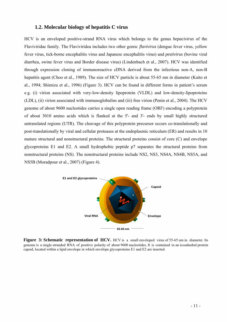
hepatitis agent (Choo et al., 1989). The size of HCV particle is about 55-65 nm in diameter (Kaito et
al., 1994; Shimizu et al., 1996) (Figure 3). HCV can be found in different forms in patient’s serum
e.g. (i) virion associated with very-low-density lipoprotein (VLDL) and low-density-lipoproteins
(LDL), (ii) virion associated with immunoglobulins and (iii) free virion (Penin et al., 2004). The HCV
genome of about 9600 nucleotides carries a single open reading frame (ORF) encoding a polyprotein
of about 3010 amino acids which is flanked at the 5'- and 3'- ends by small highly structured
untranslated regions (UTR). The cleavage of this polyprotein precursor occurs co-translationally and
post-translationally by viral and cellular proteases at the endoplasmic reticulum (ER) and results in 10
mature structural and nonstructural proteins. The structural proteins consist of core (C) and envelope
glycoproteins E1 and E2. A small hydrophobic peptide p7 separates the structural proteins from
nonstructural proteins (NS). The nonstructural proteins include NS2, NS3, NS4A, NS4B, NS5A, and
NS5B (Moradpour et al., 2007) (Figure 4).
55-65 nm
Glycoprotéines d’enveloppe E1 et E2
Enveloppe
Capside
ARN viralViral RNA
E1 and E2 glycoproteins
Envelope
Capsid
Figure 3: Schematic representation of HCV. HCV is a small enveloped virus of 55-65 nm in diameter. Its genome is a single-stranded RNA of positive polarity of about 9600 nucleotides. It is contained in an icosahedral protein capsid, located within a lipid envelope in which envelope glycoproteins E1 and E2 are inserted.

- 12 -
Untranslated regions 5' and 3'
The 5' UTR is a highly conserved, 341 nucleotides long element. It contains four well structured
domains containing numerous stem-loops and a pseudoknot (Lindenbach and Rice, 2001). The
pseudoknot is present in domain III and the domain IV contains the ORF translation initiation codon.
The 5' UTR carries an internal ribosome entry site (IRES) which is crucial for cap-independent
translation of the viral RNA (Bartenschlager et al., 2004a). Domain I has no role in IRES activity but
domain II, III and IV along with first 24 to 40 nucleotides of core-encoding region constitute the
IRES. Electron microscopy revealed that domain II, III and IV constitute distinct regions within the
molecules and a flexible hinge exists between domain II and III (Beales et al., 2001).
The 5'UTR region contains both the determinants for translation and the elements for RNA replication
(Astier-Gin et al., 2005). The upstream sequence of the IRES is essential for viral RNA replication
(Friebe et al., 2001) and the stem-loop of domain II of the IRES is essential for replication (Appel and
Bartenschlager, 2006). The formation of a binary complex between the IRES and the 40S ribosomal
subunit is required for initiation of HCV translation. The IRES-40S complex then binds to eukaryotic
translation initiation factor 3 (elF 3) and ternary complex i.e. elF2.GTP.Met-tRNAi, to constitute a 48S
intermediate complex at the AUG initiation codon. Finally, after GTP hydrolysis and recruitment of
the 60S ribosomal subunit, the 48S intermediate complex is converted into translationally active 80S
complex.
It has been shown that an abundant liver-specific microRNA (miRNA), miR-122, is able to
increase HCV RNA replication after binding to the 5' UTR (Jopling et al., 2005). In vivo experiments
in chimpanzees showed that the suppression of miR-122 by an antagomir results in a decrease in viral
load (Lanford et al., 2010).
The 3' UTR contains around 225 nucleotides and is essential for viral replication (Friebe and
Bartenschlager, 2002; Kolykhalov et al., 2000). It is composed of a short (about 40 nucleotides)
variable region, a polyuridine/polypyrimidine (poly U/UC) tract of an average length of 80
nucleotides and a highly conserved 98 nucleotides long sequence which is designated as X-tail and
contains three stable stem-loop structures SL1, SL2 and SL3 (Appel et al., 2006; Kolykhalov et al.,
1996; Tanaka et al., 1996). It has been suggested that the complete X-tail as well as at least 25
nucleotides of poly U/UC are compulsory for RNA replication in cell culture and for the infectivity of
the viral genome in vivo (Yi and Lemon, 2003; You et al., 2004). An essential cis-acting replication
element (CRE) was identified in the 3'-terminal coding region of NS5B. This CRE (designated
5BSL3.2) was found to interact with a stem-loop (SL2) in the X-tail, suggesting that a pseudoknot is
formed at the 3'-end of the HCV genome which is indispensible for RNA replication (Friebe et al.,
2005; You et al., 2004).

- 13 -
Structural proteins
Core protein
The first structural protein encoded by HCV is called core, C or capsid protein, which constitutes the
viral nucleocapsid. The nascent polypeptide is targeted by an internal signal sequence located between
the core and E1 sequence, to the host ER membrane for translocation of the E1 ectodomain into the
ER lumen. The signal peptidase cuts the signal sequence and yields an immature form of core protein
(191 amino acids), further C-terminal processing by signal peptide peptidase results in mature 21-kDa
core protein (173-179 amino acids) (McLauchlan et al., 2002).
The core protein is composed of three distinct domains. Domain D1 is an N-terminal
hydrophilic domain which contains 120 amino acids. It contains high portion of basic amino acids and
mainly participates in RNA binding and nuclear localization (Suzuki et al., 2005). Domain D2 is a C-
terminal hydrophobic domain of about 50 amino acids. This domain is involved in binding of core
protein with ER membranes, outer mitochondrial membranes and lipid droplets (Schwer et al., 2004;
Suzuki et al., 2005). The last domain is of about 20 amino acids that work as a signal peptide for the
5' UTR 3' UTR
Figure 4: Genomic organization of HCV. The HCV genome contains a positive RNA of 9.6 kb. The 5‘UTR region containing the IRES, is followed by an open reading frame encoding the structural proteins and nonstructural proteins (NS), and the 3‘UTR region required for replication. The polyprotein of about 3011 amino acids is cleaved co-and post-translationally by cellular and viral proteases to yield the structural proteins and NS proteins. Solid diamonds denote cleavage sites of HCV polyprotein precursor by the endoplasmic reticulum signal peptidase and open diamond shows further C-terminal processing of the core protein by signal peptide peptidase (Moradpour et al., 2007).
- 14 -
downstream envelope protein E1 (Grakoui et al., 1993). The association of core protein with lipid
droplets may affect lipid metabolism and may play a role in stetosis (Asselah et al., 2006). In addition
to nucleocapsid formation, the core protein has been involved in many cellular pathways including
gene transcription, apoptosis, cell signaling and cellular transformation (Kato, 2001; Lai and Ware,
2000).
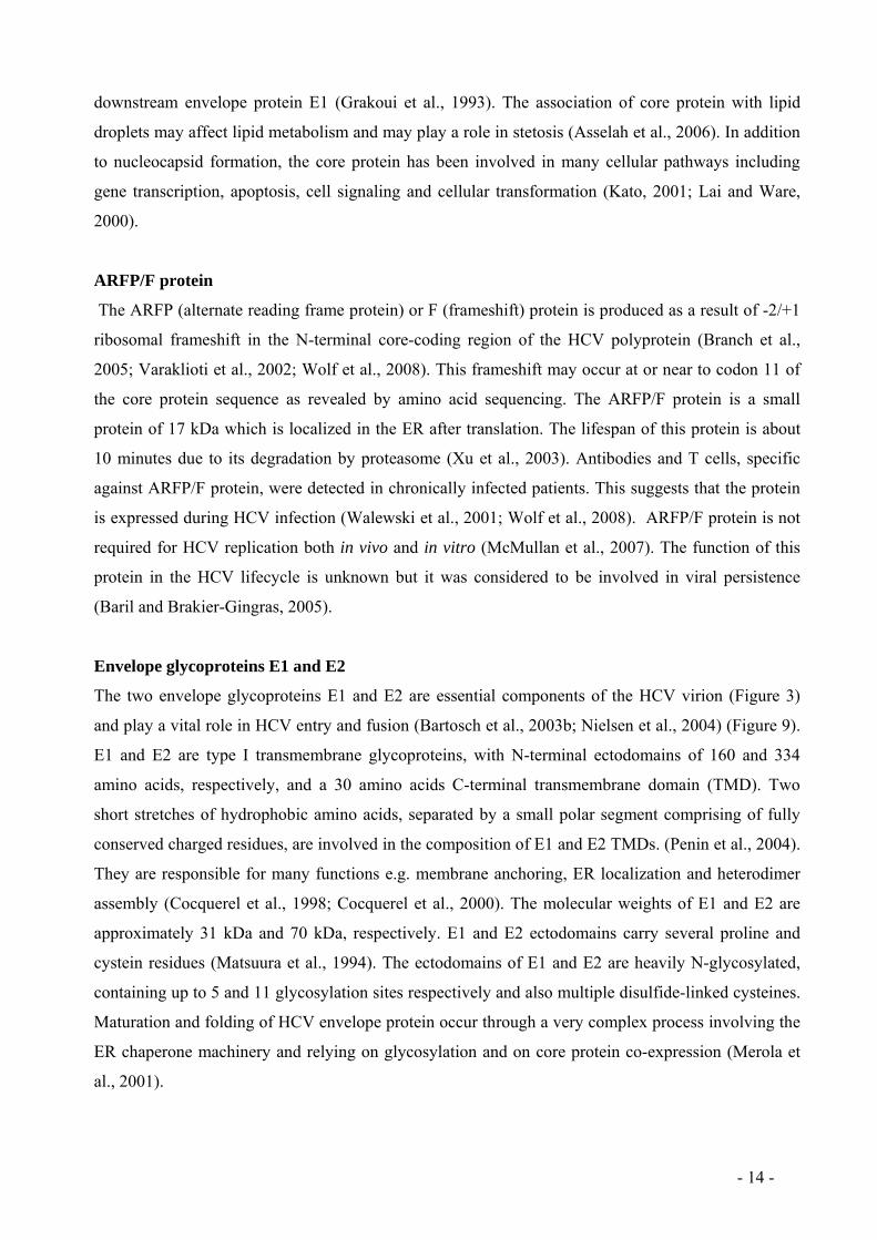
ARFP/F protein
The ARFP (alternate reading frame protein) or F (frameshift) protein is produced as a result of -2/+1
ribosomal frameshift in the N-terminal core-coding region of the HCV polyprotein (Branch et al.,
2005; Varaklioti et al., 2002; Wolf et al., 2008). This frameshift may occur at or near to codon 11 of
the core protein sequence as revealed by amino acid sequencing. The ARFP/F protein is a small
protein of 17 kDa which is localized in the ER after translation. The lifespan of this protein is about
10 minutes due to its degradation by proteasome (Xu et al., 2003). Antibodies and T cells, specific
against ARFP/F protein, were detected in chronically infected patients. This suggests that the protein
is expressed during HCV infection (Walewski et al., 2001; Wolf et al., 2008). ARFP/F protein is not
required for HCV replication both in vivo and in vitro (McMullan et al., 2007). The function of this
protein in the HCV lifecycle is unknown but it was considered to be involved in viral persistence
(Baril and Brakier-Gingras, 2005).
Envelope glycoproteins E1 and E2
The two envelope glycoproteins E1 and E2 are essential components of the HCV virion (Figure 3)
and play a vital role in HCV entry and fusion (Bartosch et al., 2003b; Nielsen et al., 2004) (Figure 9).
E1 and E2 are type I transmembrane glycoproteins, with N-terminal ectodomains of 160 and 334
amino acids, respectively, and a 30 amino acids C-terminal transmembrane domain (TMD). Two
short stretches of hydrophobic amino acids, separated by a small polar segment comprising of fully
conserved charged residues, are involved in the composition of E1 and E2 TMDs. (Penin et al., 2004).
They are responsible for many functions e.g. membrane anchoring, ER localization and heterodimer
assembly (Cocquerel et al., 1998; Cocquerel et al., 2000). The molecular weights of E1 and E2 are
approximately 31 kDa and 70 kDa, respectively. E1 and E2 ectodomains carry several proline and
cystein residues (Matsuura et al., 1994). The ectodomains of E1 and E2 are heavily N-glycosylated,
containing up to 5 and 11 glycosylation sites respectively and also multiple disulfide-linked cysteines.
Maturation and folding of HCV envelope protein occur through a very complex process involving the
ER chaperone machinery and relying on glycosylation and on core protein co-expression (Merola et
al., 2001).

- 15 -
Several hypervariable regions in envelope glycoprotein E2 have been identified where amino
acid sequences differ about 80%, not only among HCV genotypes but even among subtypes of a same
genotype (Kato, 2001; Weiner et al., 1991). This variability may result in viral escape from the host
immune system and persistence of the virus (von Hahn et al., 2007) (see HCV heterogeneity on page
19). Hypervariable region 1 (HVR1) is composed of 27 amino acids and serves as HCV neutralizing
epitope (Farci et al., 1996; Zibert et al., 1997). In vivo studies in chimpanzees have demonstrated that
after the deletion of HVR1, HCV was still infectious but highly attenuated suggesting a role of this
region in host cell entry (Bankwitz et al., 2010; Callens et al., 2005; Forns et al., 2000). The
physicochemical properties of HVR1 residues at each position and its conformation are highly
conserved among the various genotypes (Penin et al., 2001). The positively charged residues of HVR1
can interact with negatively charged molecules at the cell surface. This association can take part in
host cell recognition and attachment as well as in cell or tissue compartmentalization (Barth et al.,
2003; Bartosch et al., 2003c). HVR2 is another hypervariable region consisting of 7 amino acids
(Kato, 2001). The functional role of HVR2 is not well defined. This region seems to be involved in E2
binding to cellular factors such as CD81 (Roccasecca et al., 2003). A third region called HVR3 has
been identified between HVR1 and HVR2 (Troesch et al., 2006), which also appears to be involved in
binding to host factors (Callens et al., 2005).
E1 and E2 have a crucial role in the early steps of viral infection. Interaction of E2 with one or
several components of the receptor complex results in viral attachment (Barth et al., 2003; Barth,
2006; Flint and McKeating, 2000; Scarselli et al., 2002). It has been shown that both E1 and E2
interact with heparan sulfate (HS) (Barth et al., 2006; Barth et al., 2003), while only E2 interacts
CD81 (Pileri et al., 1998), scavenger receptor class B type I (SR-BI) (Scarselli et al., 2002) and
probably occludin (OCLN) (Liu et al., 2009). The two glycoproteins E1 and E2 have one for the other
chaperone activity (Lavillette et al., 2007) and both appear to be involved in the process of membrane
fusion required for internalization of the virus into the host cell (Flint and McKeating, 2000; Lavillette
et al., 2007). The precise role of envelope glycoproteins E1 and E2 in the fusion step is not yet well
defined. Given their importance in virus-host interactions, the envelope glycoproteins are major
targets of neutralizing antibodies (El Abd et al., 2011; Kachko et al., 2011; Owsianka et al., 2005) (see
chapter 1.6 adaptive immune response to HCV and escape from antibody mediated neutralization on
page 39).
p7 protein
Partial cleavage of E2 results in a small polypeptide, p7, which contains 63 amino acids and has been
described to be an integral membrane protein (Carrere-Kremer et al., 2002; Steinmann et al., 2007). It
comprises two transmembrane domains organized in α-helices, linked together by a cytoplasmic loop.

- 16 -
The orientation of its both N-terminus and C-terminus is towards the ER lumen. p7 is not needed for
RNA replication in vitro but data have shown that it is essential for in vivo HCV infection in
chimpanzees (Sakai et al., 2003). It has been suggested that p7 forms oligomers and could act as a
calcium ion channel which indicate its belonging to the viroporin family of proteins (Gonzalez and
Carrasco, 2003; Luik et al., 2009). p7 plays a critical role in assembly and release of HCV particles
(Bankwitz et al., 2010; Brohm et al., 2009; Jones et al., 2007; Steinmann et al., 2007). Using a trans-
complementation system, Brohm and colleagues described that p7-defective full length genomes are
rescued by HCV replicons expressing p7 in trans (Brohm et al., 2009). They also showed that p7
function cannot be replaced by viroporins from other viruses (Brohm et al., 2009). The importance of
p7 for virus production has made it another target for antiviral strategy. Several p7 inhibitors e.g.
amantadine (Bankwitz et al., 2010; Cook and Opella, 2010; Griffin et al., 2008) and amilorides
(Griffin et al., 2008; Steinmann and Pietschmann, 2010) have shown antiviral activity in cell culture.
Non-structural proteins Non-structural (NS) proteins play a crucial role in replication, translation and assembly of HCV.
NS2 protein
NS2, a non-glycosylated transmembrane protein of 21-23 kDa, is not crucial for the replication
complex but takes part in production of infectious particles (Jirasko et al., 2008; Jones et al., 2007). It
has been described that NS2 is composed of three transmembrane segments (TMS) (Yamaga and Ou,
2002). The C-terminal half of NS2 and the N-terminal one-third of NS3 participate in the catalytic
activity of the NS2-3 protease (Grakoui et al., 1993). It has been demonstrated by site directed
mutagenesis that amino acid His 143, Glu 143 and Cys 184 are crucial for NS2 catalytic activity
(Moradpour et al., 2007). It has been suggested that the protease domain of NS2, but not its enzymatic
activity, is required for infectious virus production (Jirasko et al., 2008; Jones et al., 2007). Full length
NS2 protein has been shown to be essential for HCV assembly (Jirasko et al., 2008). Mutations in
NS2 that hinder HCV assembly can be rescued by trans-complementation (Jirasko et al., 2008). It has
been reported that NS2 interacts with envelope glycoproteins, p7 and NS3 and seems to recruit viral
proteins to lipid droplets, so NS2 acts as a key organizer of the assembly of infectious HCV particles
(Jirasko et al., 2010). Moreover, genetic data suggested that functional interactions exist among NS2,
E1-E2 and NS3-NS4A during virus assembly (Phan et al., 2009; Stapleford and Lindenbach, 2011).
Recently, it has been reported that interaction of p7 and NS2 induces core-ER colocalization which is
required for initiation of viral assembly (Boson et al., 2011). The life span of NS2 is short and its
protease activity is lost after self-cleavage from NS3 (Franck et al., 2005).

- 17 -
NS3-NS4A poteins
HCV NS3 is a multifunctional protein which contains an N-terminal serine protease domain and a C-
terminal RNA helicase/NTPase domain. Enzyme activity of both is essential for viral replication
(Bartenschlager et al., 2004b; Lindenbach and Rice, 2001). NS4A polypeptide serves as a cofactor for
the NS3 serine protease. NS3-NS4A protease plays a critical role in HCV life cycle and catalyzes the
cleavage of HCV polyprotein at NS3/NS4A, NS4A/NS4B, NS4B/NS5A and NS5A/NS5B junctions
(Kim et al., 1996; Penin et al., 2004). The NS3 helicase-NTPase domain performs several functions
such as RNA-stimulated NTPase activity, RNA binding and unwinding of RNA regions with
secondary structures. Recently, it has been suggested that NS3 protein takes part in the early steps of
morphogenesis of viral particles i.e. it is involved in the recruitment of NS5A to lipid droplets and in
the assembly of viral particles (Ma et al., 2008). NS3-NS4A protease is considered to be an important
target of antiviral therapy and indeed in 2011, two protease inhibitors (telaprevir and boceprevir) have
obtained approval from U.S. Food and Drug Administration (FDA) for treatment of HCV infection
(genotype 1).
NS4B protein
NS4B is an integral membrane protein with a molecular weight of 27 kDa. It is associated with ER or
ER-derived membranes (Hugle et al., 2001; Lundin et al., 2003). NS4B contains 4 TMDs which
separate cytoplasmic N- and C-terminals (Elazar et al., 2004). It also plays a crucial role in
membrane-bound replication complex. (Gosert et al., 2003; Gretton et al., 2005). The induction of
membranous web, the specific membrane alteration that serves as a scaffold for HCV replication, is
an important function of NS4B (Egger et al., 2002). However, the detailed characteristics of this
protein have still to be elucidated.
NS5A protein
NS5A is a 56-58 kDa phosphoprotein which plays a key role in RNA replication. The N-terminal of
NS5A carries an amphipathic α-helix which is involved in protein-protein interaction essential for the
formation of a functional HCV replication complex (Brass et al., 2002; Penin et al., 2004). NS5A
contains 3 domains (I, II and III). Domain I is an N-terminal Zn2+ binding domain, domain II is
central and may be helix-rich and domain III is an unfolded C-terminal domain (Tellinghuisen et al.,
2004; Tellinghuisen et al., 2005). Domain III has been recently described as a key factor in the
assembly of viral particles and the phosphorylation of this domain may regulate assembly (Appel et
al., 2008; Hughes et al., 2009; Tellinghuisen et al., 2008). The amino acid sequence of domain III is
- 18 -
poorly conserved among different HCV genotypes (Hanoulle et al., 2009). Noteworthy, it has been
reported that NS5A of genotype 1a (H77S) shares only 58% amino acid identity with genotype 2a
protein overall and only 46% identity within domain III (Kim et al., 2011). Furthermore, it has been
suggested that existence of major differences in the sequences and/or structures of the genotypes 1a
and 2a NS5A proteins hinders them from functioning interchangeably in support of viral RNA
replication (Kim et al., 2011). Cyclophilin A has been shown to bind with domain II of NS5A protein
(Foster et al., 2011). It has been reported that the isomerase activity of Cyclophilin A plays a vital role
in HCV replication (Chatterji et al., 2009; Foster et al., 2011) and importantly, specific residues
within NS5A are the target for this isomerase activity (Hanoulle et al., 2009) The interaction of NS5A
and apolipoprotein (ApoE) is required for the assembly and export of infectious virions (Benga et al.,
2010). NS5A takes part in different functions depending on its interaction with cellular proteins
(Tellinghuisen and Rice, 2002). It can play a role in interferon resistance by binding to and inhibiting
PKR, an antiviral effector of interferon-α (Gale et al., 1998). NS5A is also involved in regulation of
cell growth and cellular signaling pathways (Tan and Katze, 2001). NS5A is also a target for direct
acting antivirals such as the BMS-790052 compound (Bourliere et al., 2011). Cyclosporin, a
cyclophilin inhibitor, has been reported to inhibit HCV replication in vitro and in patients as well.
Alisporivir (Debio 025) is a synthetic form of cyclosporine is in a phase-I study (Flisiak et al., 2008).
NS5B protein
NS5B, a RNA-dependent RNA polymerase (RdRP), is the key enzyme of HCV RNA replication.
NS5B belongs to a class of membrane proteins termed tail-anchored proteins (Ivashkina et al., 2002;
Schmidt-Mende et al., 2001). Its C-terminal post-translationally inserts into the ER membrane
(Moradpour and Blum, 2004). Like other polymerases, NS5B has a classical right hand structure with
distinct finger, palm and thumb domains (Bressanelli et al., 1999; Lesburg et al., 1999). The catalytic
domain of NS5B is membrane-associated via a C-terminal transmembrane domain that is critical for
HCV RNA replication (Appel et al., 2006). The interaction of viral proteins NS3 and NS5A modulate
the activity of NS5B (Bartenschlager et al., 2004a). Recently, it has been suggested that NS5B may
also be involved in virus assembly (Gouklani et al., 2012). The RdRP is an important target for the
development of anti-HCV drugs, polymerase inhibitors (Di Marco et al., 2005; Pawlotsky, 2006;
Qureshi, 2007).
- 19 -
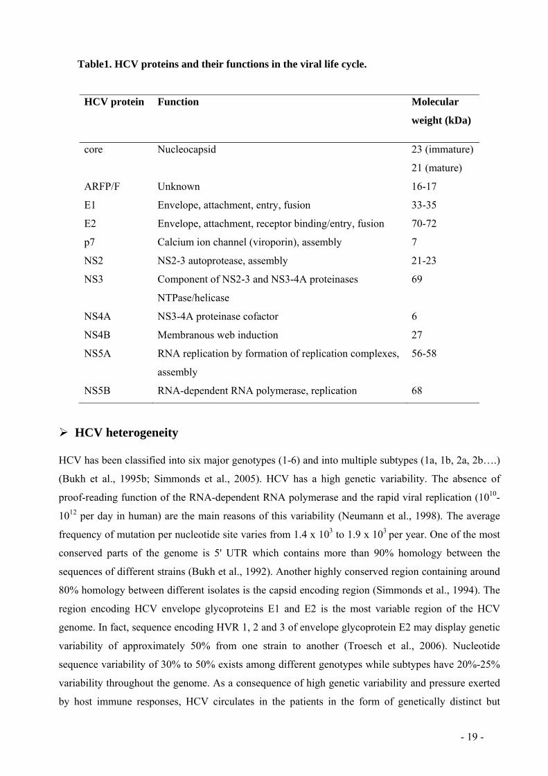
Table1. HCV proteins and their functions in the viral life cycle.
HCV protein Function Molecular
weight (kDa)
core Nucleocapsid 23 (immature)
21 (mature)
ARFP/F Unknown 16-17
E1 Envelope, attachment, entry, fusion 33-35
E2 Envelope, attachment, receptor binding/entry, fusion 70-72
p7 Calcium ion channel (viroporin), assembly 7
NS2 NS2-3 autoprotease, assembly 21-23
NS3 Component of NS2-3 and NS3-4A proteinases
NTPase/helicase
69
NS4A NS3-4A proteinase cofactor 6
NS4B Membranous web induction 27
NS5A RNA replication by formation of replication complexes,
assembly
56-58
NS5B RNA-dependent RNA polymerase, replication 68
HCV heterogeneity
HCV has been classified into six major genotypes (1-6) and into multiple subtypes (1a, 1b, 2a, 2b….)
(Bukh et al., 1995b; Simmonds et al., 2005). HCV has a high genetic variability. The absence of
proof-reading function of the RNA-dependent RNA polymerase and the rapid viral replication (1010-
1012 per day in human) are the main reasons of this variability (Neumann et al., 1998). The average
frequency of mutation per nucleotide site varies from 1.4 x 10³ to 1.9 x 10³ per year. One of the most
conserved parts of the genome is 5' UTR which contains more than 90% homology between the
sequences of different strains (Bukh et al., 1992). Another highly conserved region containing around
80% homology between different isolates is the capsid encoding region (Simmonds et al., 1994). The
region encoding HCV envelope glycoproteins E1 and E2 is the most variable region of the HCV
genome. In fact, sequence encoding HVR 1, 2 and 3 of envelope glycoprotein E2 may display genetic
variability of approximately 50% from one strain to another (Troesch et al., 2006). Nucleotide
sequence variability of 30% to 50% exists among different genotypes while subtypes have 20%-25%
variability throughout the genome. As a consequence of high genetic variability and pressure exerted
by host immune responses, HCV circulates in the patients in the form of genetically distinct but
- 20 -
closely related viral variants termed quasispecies. Viral variants within a quasispecies differ by 1%-
5% in their nucleotide sequences. The presence of distinct viral variants results in rapid and
continuous selection of variants best suited to the environment. These selected variants play a key role
in viral pathogenesis, persistence and resistance to antiviral therapy.
Genotype 1a is common in North Europe and the United States, 1b is the most frequent
genotype and has a worldwide distribution. Genotypes 2a and 2b, representing 10% to 30% of HCV
types, are mainly common in north Italy and Japan but are also worldwide distributed. Genotype 3 is
most common in the Indian subcontinent whereas genotype 4 is most common in the Middle East and
Africa. Genotypes 5 and 6 are relatively rare and can be found in South Africa and Southeast Asia,
respectively. Interestingly, the genotypes have little impact on clinical expression and are not
evidently related to a different clinical outcome (Hoofnagle, 2002). However, response to pegylated
interferon-alfa/ribavirin therapy differs between HCV genotypes. Response rates of patients infected
with genotypes 2 and 3 range from 76% - 80% in contrast to genotype 1 and 4 with rates from 42% -
46% (Feld and Hoofnagle, 2005).
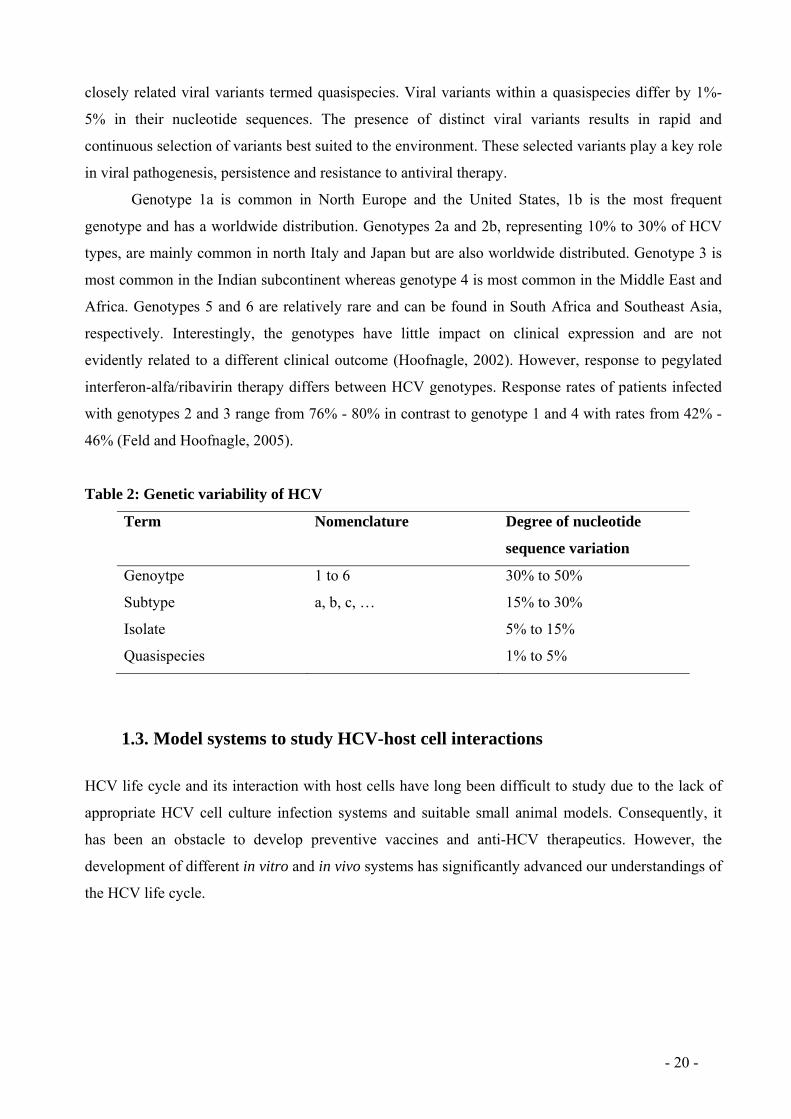
Table 2: Genetic variability of HCV
Term Nomenclature Degree of nucleotide
sequence variation
Genoytpe 1 to 6 30% to 50%
Subtype a, b, c, … 15% to 30%
Isolate 5% to 15%
Quasispecies 1% to 5%
1.3. Model systems to study HCV-host cell interactions
HCV life cycle and its interaction with host cells have long been difficult to study due to the lack of
appropriate HCV cell culture infection systems and suitable small animal models. Consequently, it
has been an obstacle to develop preventive vaccines and anti-HCV therapeutics. However, the
development of different in vitro and in vivo systems has significantly advanced our understandings of
the HCV life cycle.

- 21 -
1.3.1. In vitro systems
The in vitro model systems include plasma derived HCV, recombinant HCV envelope glycoproteins,
HCV-like particles (HCV-LPs), HCV pseudoparticles (HCVpp), HCV replicons and cell culture-
derived HCV (HCVcc).
Plasma-derived HCV
Inoculation of primary hepatocytes with serum-derived HCV was one of the first approaches to study
HCV infection in vitro (Rumin et al., 1999; Shimizu et al., 1992). Primary hepatocytes of humans,
chimpanzees, or tree shrews can be successfully infected with serum-derived HCV (Barth et al.,
2005a; Castet et al., 2002). Serum-derived HCV has been used to identify the role of LDL-receptor
(LDL-R) in HCV infection (Agnello et al., 1999). The drawback of this system was the low level of
replication of HCV which required RT-PCR for the detection of viral RNA in infected cells.
Secondly, there was absence or very low production of infectious virus particles (von Hahn and Rice,
2008). Moreover, due to heterogeneity of the virus in the serum and its association with lipoproteins,
it was difficult to obtain a homogenous and well-characterized inoculum.
Recombinant E1 and E2 glycoproteins
A truncated, soluble form of recombinant E2 glycoprotein was used to study virus-host cell
interaction leading to the identification of putative HCV receptor candidates involved in HCV entry.
These include tetraspanin CD81 (Pileri et al., 1998) and SR-BI (Scarselli et al., 2002). It also helped
to study the interaction of E1 and E2 with heparan sulfate (HS) proteoglycan (Barth et al., 2003;
Barth, 2006; Haberstroh et al., 2008). Recombinant E1 and E2 glycoproteins have been used to detect
virus neutralizing antibodies (Rosa et al., 1996). Recently, it has been demonstrated that the
immunization of mice and chimpanzees with recombinant E1E2 proteins induces neutralizing
antibodies (Kachko et al., 2011). As in this system E1 and E2 form a heterodimer on the viral
envelope and the isolated recombinant E2 may act differently (Burlone and Budkowska, 2009), it
cannot be used to study the entire attachment and entry process.
HCV-like particles (HCV-LPs)
Virus-like particles are defined as particles generated by self-assembly of the HCV structural proteins
core, E1, E2 and p7 in a baculovirus-insect cell expression system (Baumert et al., 1998). They do not
replicate because of the lack of the viral genome. HCV-LPs are characterized by morphological,
biophysical and antigenic properties similar to those of putative virions isolated from HCV-infected
patients. The E1 and E2 heterodimeric complex similar to native virions and the ability of HCV-LPs
to attach and enter hepatic cell lines, primary human hepatocytes (PHH) and dendritic cells, make this

- 22 -
an attractive model to study virus-host interactions (Barth et al., 2005b; Triyatni et al., 2002; Wellnitz
et al., 2002). In addition, HCV-LP have been shown also to have antigenic properties similar to those
of virions isolated from HCV infected patients (Baumert et al., 1998), so it has been proposed as
potential vaccine (Baumert et al., 1999; Steinmann et al., 2004). Interestingly, HCV-LP induced
HCV-specific cellular immune responses protected chimpanzees from persistent HCV infection
following HCV challenge (Elmowalid et al., 2007). A limitation of this model is the fact that these
particles do not contain a reporter gene, therefore the mechanism of attachment and cell entry require
the use of microscopy techniques or flow cytometry.
HCV pseudoparticles (HCVpp)
HCV pseudotyped particles (HCVpp) were the first robust in vitro model to study the early steps of
virus binding and cell entry that can be used in high-throughput assays. Infectious HCVpp consist of
unmodified HCV envelope glycoproteins E1 and E2 assembled onto retroviral or lentiviral core
particles (Bartosch et al., 2003b; Hsu et al., 2003). HCVpp are produced by transfecting human
embryonic kidney cells (HEK 293T) with three expression vectors. The first vector encodes the capsid
protein of retrovirus i.e. murine leukemia virus (MLV) or lentivirus (HIV), the second vector
expresses the unmodified E1 and E2 envelope glycoproteins and the third one carries a retrovirus
genome containing only the long terminal repeats and packaging signal and encoding a reporter gene
such as green fluorescent protein (GFP) or luciferase (Bartosch et al., 2003b; Hsu et al., 2003) (Figure
5). The presence of marker gene encoding for GFP or luciferase reporter gene allows reliable and fast
determination of infectivity mediated through the envelope glycoproteins (Bartosch et al., 2003b).
HCVpp are considered as reference tools to study the properties of HCV envelope glycoproteins.
HCVpp are infectious for hepatoma cells lines, like Huh-7 cells, as well as for primary human
hepatocytes (Bartosch et al., 2003b; Hsu et al., 2003) showing HCV tropism. This model has been
used to identify two co-receptors of HCV: claudin 1 (CLDN1) (Evans et al., 2007) and occludin
(OCLN) (Ploss et al., 2009). HCVpp infectivity has been demonstrated to be neutralized by anti-E1
and anti-E2 antibodies as well as by sera from human and chimpanzees infected with HCV, but not
sera from healthy controls (Bartosch et al., 2003b; Hsu et al., 2003; Lavillette et al., 2005; Law et al.,
2008; Meunier et al., 2005; Pestka et al., 2007; Vanwolleghem et al., 2008; von Hahn et al., 2007).
HCVpp indeed mimic the entry of HCV into cell and have antigenic properties similar to those of
native HCV but unlike the natural virus, HCVpp are not associated with lipoproteins, as they are
produced in 293T kidney cells that do not synthesize lipoproteins.

- 23 -
Plasmid 1
Plasmid 2
Plasmid 3
Transfection of 293T cells
HCVpp
Infection of human hepatoma cells and primary human hepatocytes (PHH)
Quantification of luciferase activity
Luciferase
Plasmid 1
Plasmid 2
Plasmid 3
Transfection of 293T cells
HCVpp
Infection of human hepatoma cells and primary human hepatocytes (PHH)
Quantification of luciferase activity
Luciferase
Figure 5: Production of HCV pseudoparticles (HCVpp). The HCVpp are infectious chimeric viruses obtained by incorporation of the E1 and E2 glycoproteins, in their native form on the surface of retroviral particles. HCVpp are generated by transfecting human embryonic kidney cells with expression vectors encoding the entire E1E2 polyprotein, capsid protein of a retrovirus/lentivirus and a defective retroviral genome carrying a marker gene that will allow to assess the infectivity of the HCVpp. HCvpp infect hepatoma cells, especially Huh7 and PHH. (LTR- long terminal repeat, PBS- primer binding site, PPT- polyurine tract, Ψ- packaging sequence).
HCV replicons
The subgenomic HCV replicons have made it possible to study viral replication (Lohmann et al.,
1999). These bicistronic RNAs replicate autonomously and contain (i) 5' IRES of HCV, which
provides the translation of an antibiotic gene (neomycin), (ii) IRES of encephalomyocarditis virus
(EMCV) ensuring translation of non-structural proteins and (iii) all framed by 5' and 3' UTR of HCV.
Only neomycin-resistant clones replicate HCV RNA (Lohmann et al., 1999). It has been demonstrated
that the cell culture replicons contain adaptive mutations in the virus, mainly in NS3, NS4B and
NS5A (Bartenschlager et al., 2004a), which markedly increase the rate of replication of transfected
cells (Lohmann et al., 2001). Many of these mutations alter the phosphorylation of NS5A, and the
hyperphophorylated form is deleterious for efficient replication of HCV (Evans et al., 2004).
Noteworthy, genomic replicons replicate efficiently under antibiotic pressure but do not allow the
production of virus particles (Pietschmann et al., 2002).

- 24 -
Cell culture-derived HCV (HCVcc)
The ability to recapitulate the entire viral life cycle in vitro was achieved in 2005. The transfection of
RNA of a viral isolate of a Japanese patient with fulminant hepatitis C (JFH-1) into highly permissive
Huh-7-derived cell clones led to efficient HCVcc production in vitro (Lindenbach et al., 2005; Wakita
et al., 2005; Zhong et al., 2005). The cell culture supernatant containing the virions successfully
infects naïve Huh-7 and Huh-7-derived hepatoma cells (Lindenbach et al., 2005; Wakita et al., 2005)
(Figure 6). HCVcc infection and replication can be easily monitored using different assays. These
include assays to determine focus forming units (FFU), 50% tissue culture infectivity dose (TCID50)
(Lindenbach et al., 2005), immunostaining of viral proteins and highly reproducible time-dependent
increase of viral RNA in infected cells (Lindenbach et al., 2005; Wakita et al., 2005) or alternatively
by the expression of a firefly luciferase reporter gene (Koutsoudakis et al., 2006), or green fluorescent
protein (GFP) (Suratanee et al., 2010) or red fluorescent protein (RFP) (Jones et al., 2010) as reporter
genes. Recently, a new construction strategy was developed to produce a dual reporter HCV virus
containing a humanized Renilla luciferase gene and an enhanced GPF gene (Wu et al., 2010). HCVcc
are able to infect chimpanzees and uPA-SCID mice transplanted with human hepatocytes (Lindenbach
et al., 2006; Wakita et al., 2005; Zhong et al., 2005). HCVcc production of different genotypes is also
possible through the use of intra-genotypic (Lindenbach et al., 2005; Pietschmann et al., 2006) or
inter-genotypic (Pietschmann et al., 2006) chimeric viruses. Recombinant HCVcc with core-NS2
(Scheel et al., 2011a), NS3/4A (Gottwein et al., 2011) and NS5A (Scheel et al., 2011b) for all major
genotypes have been developed to study resistance to antiviral therapy. The HCVcc system allows
major advances in HCV research as it helps to study the complete life cycle of HCV. Moreover, this
model has confirmed the results obtained with previous model systems such as the role of envelope
glycoproteins in virus entry (Wakita et al., 2005), the role of host cell factors involved in attachment
and entry of the virus (Koutsoudakis et al., 2006; Lindenbach et al., 2005; Wakita et al., 2005; Zeisel
et al., 2007a; Zhong et al., 2005) and activity of neutralizing antibodies (Haberstroh et al., 2008; Law
et al., 2008). However, the handling of HCVcc requires a BSL3 laboratory which is less user friendly.

- 25 -
HCV RNA JFH1 or chimeric
Electroporation of Huh7, Huh7.5, Huh7.5.1 cells
JFH-1
JFH-1J6
5' UTR
5' UTR3' UTR
3' UTR
L3 Production of HCVcc
Infection of Human hepatoma cells and PHH
HCV RNA JFH1 or chimeric
Electroporation of Huh7, Huh7.5, Huh7.5.1 cells
JFH-1
JFH-1J6
5' UTR
5' UTR3' UTR
3' UTR
JFH-1
JFH-1J6
5' UTR
5' UTR3' UTR
3' UTR
L3 Production of HCVcc
Infection of Human hepatoma cells and PHH
Figure 6: Production of HCVcc. Cell culture-derived HCV (HCVcc) are produced by electroporation of Huh7-derived cells with JFH1 RNA or a chimeric RNA. Viruses are secreted in the supernatants few days after the transfection. The infectivity and replication potential of HCVcc can be assessed on Huh7-derived cell lines and analysing by expression of viral or reporter proteins or by quantification of intracellular viral RNA.
1.3.2. In vivo systems
The study of HCV infection and pathogenesis was long performed in chimpanzees (Pan troglodytes).
The infection follows a progression similar to that observed in humans. HCV RNA can be detected in
the blood several days after infection, followed by an acute hepatitis which is characterized by
increase of ALT. Liver cirrhosis or fibrosis in chimpanzees is rare. There are several drawbacks of
this model: the chronic infection is less severe as compared to human, chimpanzees are expensive and
difficult to handle as they require special housing (Barth et al., 2008a); moreover, since 1988 the
chimpanzee has been listed as an endangered species. These limitations of the chimpanzee model
have stimulated progress toward developing alternative animal models for HCV research.
Mice or rats are the key candidates to generate such a model but the strict tropism of HCV requires
the hepatocytes of man or chimpanzee to be transplanted in these rodents. The survival and expansion
of xenogenic donor hepatocytes in the recipient animal need an environment that is permissive for the
engraftment and the expansion of liver cells. An immune deficient animal suffering from a severe
liver disease can provide the desired environment. Therefore, the current state-of-the-art small animal
model was developed by using transgenic mice carrying the transgene "urokinase plasminogen
activator" (alb-uPA) and crossing of these mice with SCID (severe combined immunodeficiency
disorder) mice for the complete reconstruction of the liver of mice with xenograft of human
hepatocytes (Mercer et al., 2001). This model has allowed the study of hepatitis B and C virus
infection. In 2001, Mercer and collaborators have shown for the first time that uPA-SCID mice
transplanted with human hepatocytes could be infected with HCV in vivo (Mercer et al., 2001). The

- 26 -
measurement of albumin is used to evaluate the integrity and functionality of transplanted human
hepatocytes. Once stabilized, the uPA-SCID mice can be infected either by the serum of patients or
chimpanzees infected with HCV, or by HCVcc (Law et al., 2008; Lindenbach et al., 2006; Mercer et
al., 2001; Meuleman et al., 2005; Vanwolleghem et al., 2008). HCV loads measured in the serum of
these mice are comparable to those observed in humans. In addition, plasma derived from these mice
is able to infect other mice allowing massed infection. The HCV infection in this model can be
maintained for at least 4 months. During this period, the function and architecture of the liver are not
altered (Barth et al., 2008a). This mouse model allowed to confirm the role of anti-receptor antibodies
and neutralizing antibodies in controlling viral infection (Law et al., 2008; Meuleman et al., 2008;
Meuleman et al., 2012; Vanwolleghem et al., 2008). This mouse model has the advantage of being
cheaper than chimpanzees, more easily maintainable and breeding faster than the chimpanzee.
However, this animal model is very difficult to implement. It requires considerable expertise to isolate
and transplant human hepatocytes and maintain colonies of mice because of their immunosuppression.
The mortality rate of infants is estimated at about 35% (Mercer et al., 2001). In addition, the study of
virus-host interactions is limited by the mouse genetic background, as the absence of functional
immune system precludes the study of HCV interaction with the host immune system. To further
improve the in vivo study of HCV, Bissig and colleagues have developed a new mouse model. They
described a regulatable system for repopulating the liver of immunodeficient mice [specifically mice
lacking fumaryl acetoacetate hydrolase (Fah), recombination activating gene 2 (Rag2) and the γ-chain
of the receptor for IL-2 (Il-2r γ)] with human hepatocytes (Bissig et al., 2010). Selection pressure for
transplanted human hepatocytes in these animals can be regulated by oral administration of 2-(2-nitro-
4-trifluoro-methylbenzoyl)-1.3-cyclohexanedione (NTBC), absence of which results in the death of
mouse hepatocytes due to accumulation of toxic tyrosine catabolites caused by the lack of Fah,
whereas presence of human homolog keeps human hepatocytes healthy (Bissig et al., 2010). The
advantage of this Fah-/-RAg-/-Il2rg-/- mouse is that animals with low human chimerism can be put back
on the drug NTBC and therefore do not result in liver failure and eventually death (Bissig et al.,
2010).
1.4. HCV host factors required for viral attachment and entry
HCV entry into host cells is a complex and multistep process. Many efforts have been made to
develop different model systems to study HCV-host interactions in order to identify several host cell
surface molecules such as the tetraspanin CD81 (Pileri et al., 1998), the low-density lipoprotein
(LDL) receptor (Agnello et al., 1999), highly sulfated heparan sulfate (HS) (Barth et al., 2003;
Koutsoudakis et al., 2006), the scavenger receptor class B type I (SR-BI) (Bartosch et al., 2005;
Scarselli et al., 2002; Zeisel et al., 2007b), the C-type lectins (DC-SIGN/L-SIGN) (Lozach et al.,

- 27 -
2004; Pohlmann et al., 2003), the tight junction proteins claudin-1 (CLDN-1) (Evans et al., 2007) and
occludin (OCLN) (Ploss et al., 2009) receptor tyrosine kinases (RTKs) such as epidermal growth
factor receptor (EGFR) and ephrin A2 (EphA2) (Lupberger et al., 2011) as well as the recently
described Niemann-Pick C1-Like 1 receptor (NPC1L1) (Sainz et al., 2012).
Glycosaminoglycans
Glycosaminoglycans (GAGs) are thought to be the first attachment sites of HCV (Barth et al., 2003;
Germi et al., 2002). There are several different types of glycosaminoglycans e.g. chondroitin sulfate,
dermatan sulfate, keratan sulfate, heparan sulfate, heparin and hyaluronan (Helle and Dubuisson,
2008). Among them, heparan sulfate (HS) is involved in attachment of many viruses like human
herpes virus 8 or dengue virus. The glycosaminoglycan HS is composed of a family of linear
polysaccharides located at the surface of mammalian cells and in the extracellular matrix. The
repeating disaccharide units [GLcA-GlcNAc]n define the structure of HS. GlcA is the glucuronic acid
and GlcNAc is N-acetylglucosamine (Esko and Lindahl, 2001). It has been shown that HCV envelope
glycoproteins E1 and E2 interact with HS (Barth et al., 2006). Moreover, the use of heparin, which is
an analogue of HS, and heparinase, an enzyme which degrades HS, hamper the attachment of HCV to
cells (Haberstroh et al., 2008; Koutsoudakis et al., 2006). Similarly, glycosidase treatment of the cells
decreases the infectivity of HCV (Barth et al., 2006; Basu et al., 2007; Morikawa et al., 2007). These
findings demonstrate the important role of HS in HCV binding to cells. HS may play a role in HCV
infection by concentrating the virus on the surface of target cells and allow subsequent interaction
with other host factors responsible for viral entry (Morikawa et al., 2007).
The low-density lipoprotein receptor (LDL-R)
HCV is able to associate with high-density lipoproteins (HDL), low-density lipoproteins (LDL) and
very low-density lipoproteins (VLDL). The low-density lipoprotein receptor (LDL-R) has been
suggested for HCV entry (Agnello et al., 1999; Burlone and Budkowska, 2009; Wunschmann et al.,
2000). LDL-R is an endocytotic receptor with a molecular weight of 160 kDa. LDL-R plays an
important role in cholesterol homeostasis. The apolipoprotein B (apoB)-containing LDL and
apolipoprotein E (apoE)-containing VLDL are the major ligands of LDL-R (Hishiki et al., 2010;
Owen et al., 2009). Serum-derived HCV has been suggested to be internalized by binding of virus-
LDL particles to LDL-R (Agnello et al., 1999). Moreover, antibodies directed against LDL-R as well
as anti-apoB and anti-apoE antibodies inhibited HCV endocytosis (Agnello et al., 1999; Chang et al.,
2007; Jiang and Luo, 2009; Long et al., 2011; Wunschmann et al., 2000). It was also observed that
LDL-R plays a role in an early step of serum-derived HCV infection of primary human hepatocytes
(Molina et al., 2007). HCVpp are not associated with lipoproteins, so the interaction of LDL-R and

- 28 -
HCVpp could not be studied to understand the role of LDL-R in viral entry (Bartosch et al., 2003c).
Most recently, using HCVcc, it has been shown that LDL-R could participate in non-productive entry
of HCV particles and the physiological function of LDL-R plays a critical role in optimal replication
of HCV genome (Albecka et al., 2011). In conclusion, similar to SR-BI, LDL-R may be involved in
viral cell entry through interaction with lipoproteins that are associated with HCV at an early stage of
infection.
Lectins: DC-SIGN and L-SIGN
HCV enters the liver through blood. Liver macrophages (Kupffer cells) and endothelial cells may
capture the infectious virus particles and transfer them to adjacent hepatocytes which are not directly
in contact with circulating blood. This process could be mediated by C-type lectins such as dendritic
cell-specific intercellular adhesion molecule-3-grabbing non-integrin (DC-SIGN) and lymph node-
specific intercellular adhesion molecule-3 (ICAM-3)-grabbing integrin (L-SIGN or CD209L). DC-
SIGN and L-SIGN could be involved in viral pathogenesis and tissue tropism (Gardner et al., 2003;
Lozach et al., 2004; Lozach et al., 2003; Pohlmann et al., 2003). DC-SIGN is expressed in Kupffer
cells, dendritic cells and lymphocytes while L-SIGN is expressed in liver sinusoidal endothelial cells
(van Kooyk and Geijtenbeek, 2003). Both lectins participate in binding, internalization and
elimination of many pathogens (Cambi et al., 2005). Studies have shown that binding of E2 to L-
SIGN could induce transmission of HCVpp to adjacent hepatocytes (Cormier et al., 2004a). However,
these molecules do not facilitate entry of HCVpp and HCVcc on their own behalf (Lai et al., 2006).
Because neither molecule is expressed on hepatocytes, they are unlikely to function as direct entry
receptors (von Hahn and Rice, 2008) but DC-SIGN and L-SIGN may function as capture receptors
which have the ability to transmit the virus to permissive cells and may be involved in the initiation of
HCV infection and tissue tropism (Cormier et al., 2004a; Lozach et al., 2004).
The tetraspanin CD81
The first host factor revealed to be required for HCV entry was the tetraspanin CD81 which interacts
with soluble E2 (sE2) (Pileri et al., 1998). CD81 is 25 kDa tetraspanin which is ubiquitously
expressed. It comprises four transmembrane domains, one small extracellular loop (SEL), one large
extracellular loop (LEL) and N- and C-terminal intracellular domains (Figure 7). It is involved in
pleiotropic activities such as cell adhesion, motility, metastasis, cell activation, and signal
transduction (Levy et al., 1998). The CD81-LEL has been demonstrated to play its role in HCV
binding through interaction with sE2 (Pileri et al., 1998). Several amino acids were considered to be
crucial for the interaction between E2 and CD81-LEL (Bertaux and Dragic, 2006; Boo et al., 2012;
Drummer et al., 2002; Higginbottom et al., 2000; Pileri et al., 1998). Recently, it has been suggested

- 29 -
that deletion of HVR2 of E2 results in decrease of 50% in the CD81-binding ability of HCVpp
(McCaffrey et al., 2011). The binding of sE2 is species-specific as it does not bind to mouse or rat
CD81 (Flint et al., 2006). Furthermore, using HCVpp and HCVcc infection, it has been reported that
the glycoprotein E2 residues at position 415, 420, 527. 529. 530 and 535 play a critical role in HCV
E2-CD81 interaction (Dhillon et al., 2010; Owsianka et al., 2006). The role of CD81 in HCV infection
was elucidated by using different human hepatoma cell lines which do not express CD81 and are non-
permissive for HCV such as HepG2 and HH29. These cell lines became susceptible to both HCVcc
and HCVpp infection upon ectotopic expression of CD81 after transduction (Bartosch et al., 2003b;
Cormier et al., 2004b; Lavillette et al., 2005; Zhang et al., 2004a). Anti-CD81 antibodies as well as a
soluble form of the CD81 extracellular loop have been shown to inhibit HCVpp and HCVcc entry into
Huh-7 hepatoma cells and human hepatocytes (Bartosch et al., 2003b{Wakita, 2005 #1791;
Lindenbach et al., 2005; McKeating et al., 2004; Zhang et al., 2004a). Moreover, using uPA-SCID
mouse model, it has been shown that CD81 is an essential factor for HCV infection in vivo
(Meuleman et al., 2008). The down regulation of CD81 expression by siRNA also resulted in the
inhibition of serum-derived HCV (sHCV) (Molina et al., 2008; Zhang et al., 2004a). It is worth
mentioning that CD81 is one of the two HCV entry factors responsible for the species-specificity of
HCV as expression of human CD81 and human OCLN may confer HCV permissivity to mouse cell
lines (Ploss et al., 2009).
Extracellular
Cytoplasmic
COOHNH2
SEL
LEL
Extracellular
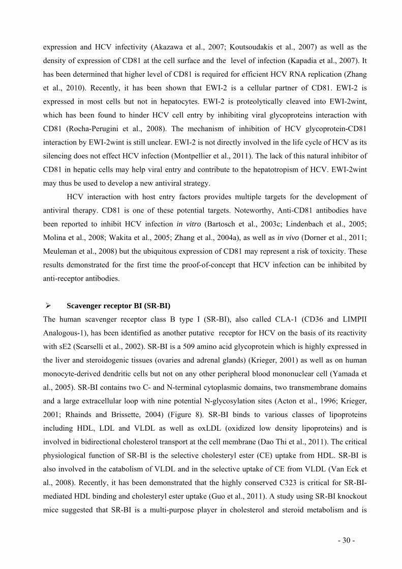
Cytoplasmic
COOHNH2
SEL
LEL
Figure 7: Structure of CD81. Amino acids are shown as circles; specific residues of human CD81 are indicated by single letter designation, bolded circles mark the position of residues that are conserved in the core (CD9, CD37, CD53, CD63, CD81, CD82, CO-029, CD151, A15, SAS, sm23 and late bloomer) tetraspanins (Levy et al., 1998).
Antibodies directed against CD81 and human recombinant CD81-LEL inhibit HCV infection
after virus attachment suggesting that CD81 serves as a postbinding entry factor (Cormier et al.,
2004b; Flint et al., 2006; Koutsoudakis et al., 2006). A correlation exists between the level of CD81
- 30 -
expression and HCV infectivity (Akazawa et al., 2007; Koutsoudakis et al., 2007) as well as the
density of expression of CD81 at the cell surface and the level of infection (Kapadia et al., 2007). It
has been determined that higher level of CD81 is required for efficient HCV RNA replication (Zhang
et al., 2010). Recently, it has been shown that EWI-2 is a cellular partner of CD81. EWI-2 is
expressed in most cells but not in hepatocytes. EWI-2 is proteolytically cleaved into EWI-2wint,
which has been found to hinder HCV cell entry by inhibiting viral glycoproteins interaction with
CD81 (Rocha-Perugini et al., 2008). The mechanism of inhibition of HCV glycoprotein-CD81
interaction by EWI-2wint is still unclear. EWI-2 is not directly involved in the life cycle of HCV as its
silencing does not effect HCV infection (Montpellier et al., 2011). The lack of this natural inhibitor of
CD81 in hepatic cells may help viral entry and contribute to the hepatotropism of HCV. EWI-2wint
may thus be used to develop a new antiviral strategy.
HCV interaction with host entry factors provides multiple targets for the development of
antiviral therapy. CD81 is one of these potential targets. Noteworthy, Anti-CD81 antibodies have
been reported to inhibit HCV infection in vitro (Bartosch et al., 2003c; Lindenbach et al., 2005;
Molina et al., 2008; Wakita et al., 2005; Zhang et al., 2004a), as well as in vivo (Dorner et al., 2011;
Meuleman et al., 2008) but the ubiquitous expression of CD81 may represent a risk of toxicity. These
results demonstrated for the first time the proof-of-concept that HCV infection can be inhibited by
anti-receptor antibodies.
Scavenger receptor BI (SR-BI)
The human scavenger receptor class B type I (SR-BI), also called CLA-1 (CD36 and LIMPII
Analogous-1), has been identified as another putative receptor for HCV on the basis of its reactivity
with sE2 (Scarselli et al., 2002). SR-BI is a 509 amino acid glycoprotein which is highly expressed in
the liver and steroidogenic tissues (ovaries and adrenal glands) (Krieger, 2001) as well as on human
monocyte-derived dendritic cells but not on any other peripheral blood mononuclear cell (Yamada et
al., 2005). SR-BI contains two C- and N-terminal cytoplasmic domains, two transmembrane domains
and a large extracellular loop with nine potential N-glycosylation sites (Acton et al., 1996; Krieger,
2001; Rhainds and Brissette, 2004) (Figure 8). SR-BI binds to various classes of lipoproteins
including HDL, LDL and VLDL as well as oxLDL (oxidized low density lipoproteins) and is
involved in bidirectional cholesterol transport at the cell membrane (Dao Thi et al., 2011). The critical
physiological function of SR-BI is the selective cholesteryl ester (CE) uptake from HDL. SR-BI is
also involved in the catabolism of VLDL and in the selective uptake of CE from VLDL (Van Eck et
al., 2008). Recently, it has been demonstrated that the highly conserved C323 is critical for SR-BI-
mediated HDL binding and cholesteryl ester uptake (Guo et al., 2011). A study using SR-BI knockout
mice suggested that SR-BI is a multi-purpose player in cholesterol and steroid metabolism and is
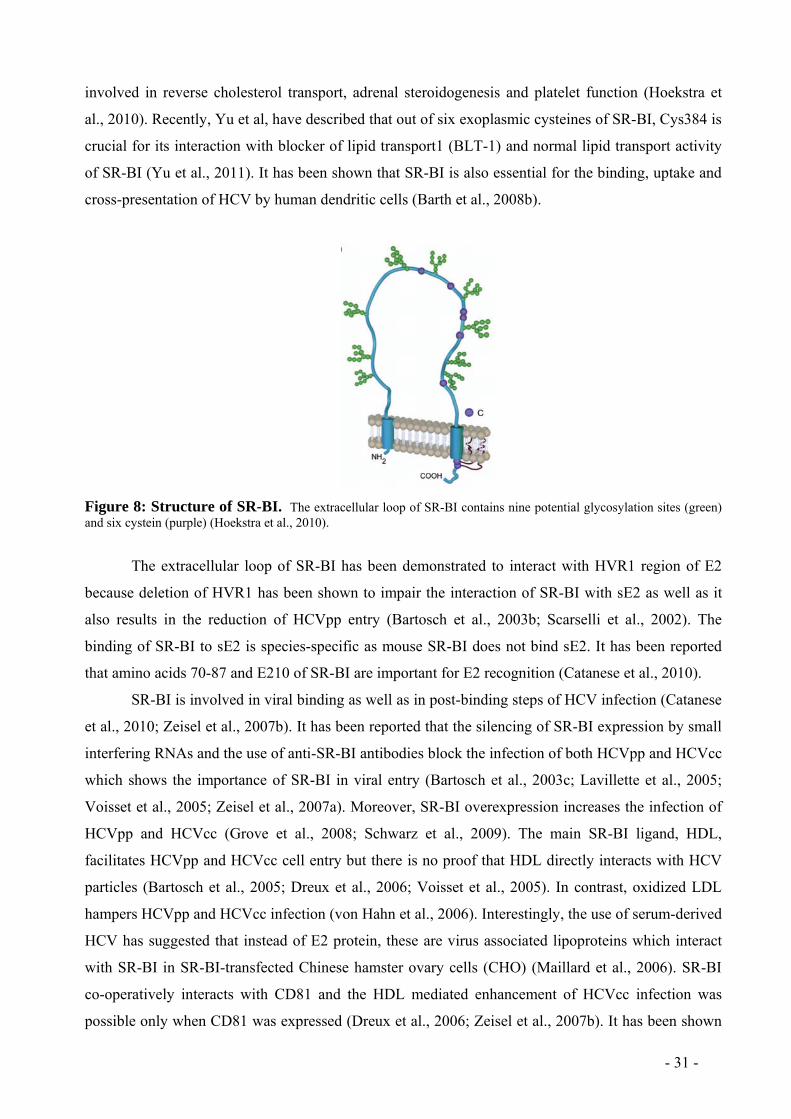
- 31 -
involved in reverse cholesterol transport, adrenal steroidogenesis and platelet function (Hoekstra et
al., 2010). Recently, Yu et al, have described that out of six exoplasmic cysteines of SR-BI, Cys384 is
crucial for its interaction with blocker of lipid transport1 (BLT-1) and normal lipid transport activity
of SR-BI (Yu et al., 2011). It has been shown that SR-BI is also essential for the binding, uptake and
cross-presentation of HCV by human dendritic cells (Barth et al., 2008b).
Figure 8: Structure of SR-BI. The extracellular loop of SR-BI contains nine potential glycosylation sites (green) and six cystein (purple) (Hoekstra et al., 2010).
The extracellular loop of SR-BI has been demonstrated to interact with HVR1 region of E2
because deletion of HVR1 has been shown to impair the interaction of SR-BI with sE2 as well as it
also results in the reduction of HCVpp entry (Bartosch et al., 2003b; Scarselli et al., 2002). The
binding of SR-BI to sE2 is species-specific as mouse SR-BI does not bind sE2. It has been reported
that amino acids 70-87 and E210 of SR-BI are important for E2 recognition (Catanese et al., 2010).
SR-BI is involved in viral binding as well as in post-binding steps of HCV infection (Catanese
et al., 2010; Zeisel et al., 2007b). It has been reported that the silencing of SR-BI expression by small
interfering RNAs and the use of anti-SR-BI antibodies block the infection of both HCVpp and HCVcc
which shows the importance of SR-BI in viral entry (Bartosch et al., 2003c; Lavillette et al., 2005;
Voisset et al., 2005; Zeisel et al., 2007a). Moreover, SR-BI overexpression increases the infection of
HCVpp and HCVcc (Grove et al., 2008; Schwarz et al., 2009). The main SR-BI ligand, HDL,
facilitates HCVpp and HCVcc cell entry but there is no proof that HDL directly interacts with HCV
particles (Bartosch et al., 2005; Dreux et al., 2006; Voisset et al., 2005). In contrast, oxidized LDL
hampers HCVpp and HCVcc infection (von Hahn et al., 2006). Interestingly, the use of serum-derived
HCV has suggested that instead of E2 protein, these are virus associated lipoproteins which interact
with SR-BI in SR-BI-transfected Chinese hamster ovary cells (CHO) (Maillard et al., 2006). SR-BI
co-operatively interacts with CD81 and the HDL mediated enhancement of HCVcc infection was
possible only when CD81 was expressed (Dreux et al., 2006; Zeisel et al., 2007b). It has been shown

- 32 -
in a mapping study that HCV and HDL binding to SR-BI and lipid transfer function of SR-BI are
required for SR-BI to behave as HCV entry factor (Dreux et al., 2009). Anti-SR-BI antibodies and
genetically humanized mouse have been used to describe the critical role of SR-BI in HCV infection
in vivo (Dorner et al., 2011; Meuleman et al., 2012). PDZK1, a four PDZ domain-containing adaptor
protein that is predominantly expressed in liver, kidney and small intestines, interacts with SR-BI and
indirectly enhances HCV entry (Eyre et al., 2010).
SR-BI represents another interesting target for anti-HCV therapy. SR-BI binds and internalizes
serum amyloid A (SAA), which inhibits HCV entry by interacting with the virus (Lavie et al., 2006).
Anti-SR-BI antibodies have been demonstrated to inhibit HCV infection in vitro (Bartosch et al.,
2003c; Catanese et al., 2010; Catanese et al., 2007; Zeisel et al., 2007b), as well as in vivo (Dorner et
al., 2011; Meuleman et al., 2012). ITX 5061, a small molecule SR-BI antagonist that inhibits HCV
infection, has entered phase I clinical trials in HCV-infected patients (Syder et al., 2011).
Claudin-1 (CLDN1)
The tight junction protein claudin-1 (CLDN1) has been identified as another entry factor for HCV by
expression cloning (Evans et al., 2007). CLDN1, a 24 kDa protein, is expressed in all epithelial tissues
but predominantly in the liver, forming networks at tight junctions (TJ) (Furuse et al., 1998). TJs are
multiprotein complexes that contain four kinds of transmembrane proteins including claudins,
occludin, junction-associated molecules and the coxsackie virus B adenovirus receptors (CARs)
(Burlone and Budkowska, 2009). TJs are responsible for the control of paracellular transport and
maintenance of cell polarity. CLDN1 is comprised of two extracellular loops, three intra cellular
domains and four transmembrane segments (Furuse and Tsukita, 2006). It has been shown that
CLDN1 is localized at TJ of hepatocytes but also on the sinusoidal basolateral surfaces of these cells
(Reynolds et al., 2008). Noteworthy, non-junctional CLDN1 has been suggested to be involved in
HCV entry (Cukierman et al., 2009; Evans et al., 2007; Harris et al., 2010; Krieger et al., 2010) while
other studies demonstrated that the distribution of CLDN1 in tight junctions is affiliated with
permissiveness to HCV infection (Liu et al., 2009; Yang et al., 2008), so subcellular localization of
CLDN1 appears to be critical for viral entry and cellular tropism of HCV (Burlone and Budkowska,
2009).
Studies suggest that the first extracellular loop (ECL1) and residues in the highly conserved
claudin motif W(30)-GLW(51)-C(54)-C(64) are crucial for HCV entry (Cukierman et al., 2009;
Evans et al., 2007; Zhang et al., 2007). Expression of CLDN1 in non-permissive cell lines such as
293T and SW13, make them permissive for HCV infection while the silencing of CLDN1 hinders
infection of HCV in susceptible cells like Huh7.5 (Evans et al., 2007). However, there is no evidence
of direct interaction between CLDN1 and HCV (Krieger et al., 2010). In contrast to SR-BI,

- 33 -
overexpression of CLDN1 does not increase the infectivity of HCV (Schwarz et al., 2009). The study
of HCV entry kinetics using anti-Flag and anti-CLDN1 antibodies demonstrated that CLDN1 is
involved in post-binding stages of HCV infection (Evans et al., 2007; Krieger et al., 2010). CLDN1-
HCV interaction had initially been considered to take place after the virus-SR-BI/CD81 complex is
laterally migrated to the tight junctions (Coyne et al., 2007), while more recent data have shown that
SR-BI, CD81 and CLDN1 act at closely related time points in the viral entry process (Krieger et al.,
2010). Moreover, it has been demonstrated that CD81 and CLDN1 co-localize at the apical and
basolateral regions of hepatocytes (Mee et al., 2009; Reynolds et al., 2008). The formation of
CLDN1-CD81 complexes is critical for HCV infection (Harris et al., 2010; Harris et al., 2008;
Krieger et al., 2010). Indeed, mutations at residues 32 and 48 in ECL1 of CLDN1 disrupt the
association with CD81 which results in obstruction of the viral receptor activity (Harris et al., 2010).
Some other members of the claudin family i.e. CLDN6 and CLDN9 are also able to mediate HCV
entry (Meertens et al., 2008; Zhang et al., 2007). Both CLDN6 and CLDN9 contain a highly
conserved ECL1 (Zhang et al., 2007).
CLDN1 is a promising antiviral target as it is critical for HCV entry. Recently, anti-CLDN1
antibodies have been shown to inhibit HCV infection in vitro (Fofana et al., 2010; Krieger et al.,
2010). Polyclonal anti-CLDN1 antibodies reduce HCV E2 interaction with cell surface and disrupt
CLDN1-CD81 interaction (Krieger et al., 2010). Noteworthy, monoclonal anti-CLDN1 antibodies
markedly block entry of highly infectious escape variants of HCV that are resistant to host
neutralizing antibodies (Fofana et al., 2010). These data indicate that antibodies against CLDN1
represent interesting new antivirals to inhibit HCV infection.
Occludin (OCLN)
Occludin (OCLN) has been identified as another host cell factor essential for HCV entry which plays
its role probably at a late post-binding stage (Benedicto et al., 2009; Liu et al., 2009; Ploss et al.,
2009). OCLN is a 65 kDa protein expressed in TJ. OCLN is comprised of four transmembrane
regions, two extracellular loops and N- and C- terminal cytoplasmic regions. It is important to note
that OCLN is one of the two HCV entry factors responsible for the species-specificity of HCV as
expression of human OCLN and human CD81 may confer HCV permissivity in vitro and in vivo
(Dorner et al., 2011; Ploss et al., 2009). Amino acids responsible for the species-specificity are found
in the second extracellular loop of OCLN (Ploss et al., 2009). Glucocorticoid treatment results in
enhancement of expression of OCLN as well as HCV entry in Huh7.5 cells and PHH (Ciesek et al.,
2010).
In contrast, the silencing of OCLN by siRNA suggested that reduction of OCLN expression
inhibits HCVpp and HCVcc cell entry (Liu et al., 2009). Confocal microscopy studies demonstrated

- 34 -
that in HCV infected cells OCLN accumulates in the endoplasmic reticulum and co-localizes with
HCV glycoprotein E2 (Benedicto et al., 2008; Liu et al., 2009). Interestingly, HCV infection may
result in the reduction of TJ proteins as it has been shown that following HCV infection, the
expressions of OCLN and CLDN were downregulated to prevent superinfection (Liu et al., 2009).
OCLN may also be targeted to prevent HCV infection. To date, there is unavailability of any
anti-OCLN antibody inhibiting HCV infection. Further characterization of the role of OCLN in HCV
entry may lead to the development of novel compounds interfering with HCV infection.
Receptor tyrosine kinases (RTKs)
Recently, using a functional siRNA screen, our laboratory identified receptor tyrosine kinases (RTKs)
such as epidermal growth factor receptor (EGFR) and ephrin A2 (EphA2) as novel HCV entry factor
(Lupberger et al., 2011). EGFR exists on the cell surface and is activated by binding of its endogenous
ligands including epidermal growth factor (EGF) and transforming growth factor α (TGF-α). EGFR is
involved in cell proliferation, survival, differentiation during development, tissue homeostasis and
tumorigenesis (Schneider and Wolf, 2009). EphA2 is a member of the largest class of RTKs and
mediates cell positioning, cell morphology and motility (Lackmann and Boyd, 2008). The functional
significance of the RTKs for HCV entry was studied using protein kinase inhibitors (PKIs) such as
Erlotinib (EGFR inhibitor) and Dasatinib (EphA2 inhibitor). Inhibition of RTKs by Erlotinib and
Dasatinib suggests their role in HCV entry process. Usage of PKIs and silencing of RTK expression
by siRNA did not effect E2 binding to target cells, which shows that RTKs are not involved in HCV
binding and RTK-mediated HCV entry does not require direct E2-RTK binding. Moreover, it has
been suggested that RTKs act at post-binding steps of viral entry (Lupberger et al., 2011).
Interestingly, it has been demonstrated that EGFR and EphA2 take part in regulating the formation of
CD81-CLDN1 complexes that are crucial for HCV entry and both Erlotinib and Dasatinib hinder
HCV entry by interfering with the CD81-CLDN1 co-receptor association (Lupberger et al., 2011).
Inhibition of HCV entry at late steps in the kinetic infection assay and HCV cell fusion assay
demonstrated a functional role for EGFR in pH-dependent fusion of viral and host cell membranes
(Lupberger et al., 2011). Furthermore, EGF significantly accelerates the rate of HCV entry (Lupberger
et al., 2011). Moreover, using uPA-SCID mouse model, functional role of EGFR as a co-factor for
HCV entry and dissemination has been demonstrated in vivo (Lupberger et al., 2011).
Thus, EGFR may be another promising target to control HCV infection. Importantly, clinically
licensed PKIs have shown marked antiviral activity in vitro and in vivo (Lupberger et al., 2011).
Furthermore, EGFR-specific antibody has been identified to inhibit HCV infection (Lupberger et al.,
2011) which shows that RTKs may offer a perspsective for novel antiviral strategies against HCV
infection.

- 35 -
Niemann-Pick C1-Like 1 (NPC1L1)
HCV is associated with cellular lipoproteins (LDL and VLDL) and dependence of HCV infectivity on
cholesterol (Gastaminza et al., 2007) suggests the involvement of cholesterol-uptake receptors in
HCV cell entry. As NPC1L1 receptor is involved in cellular cholesterol absorption and whole-body
cholesterol homeostasis (Altmann et al., 2004), NPC1L1 has recently been identified as a putative
HCV host factor. NPC1L1, a 1332 amino acid protein, is a cell surface cholesterol sensing receptor
(Yu, 2008). NPC1L1 contains 13 transmembrane domains, a conserved amino-terminal ‘NPC’
domain and extensive N-linked glycosylation sites (Yu, 2008). NPC1L1 is expressed on the apical
surface of enterocytes and on the canalicular membrane of hepatocytes (Jia et al., 2011; Sainz et al.,
2012; Temel et al., 2007; Yu, 2008). It has been demonstrated that NPC1L1 takes part in cholesterol
absorption into enterocytes from the apical surface and on the other side; it recovers cholesterol from
canalicular bile and transfers it back into hepatocytes (Temel et al., 2007; Yamanashi et al., 2007; Yu
et al., 2006). It has been demonstrated that silencing and antibody mediated blocking of NPC1L1
decreased HCV infection (Sainz et al., 2012). NPC1L1 has three large extracellular loops (LEL) but
only first large extracellular loop (LEL1) mediates HCV infection (Sainz et al., 2012). Furthermore,
pharmacological inhibition of NPC1L1 by ezetimibe has been shown to reduce HCV infection by
direct inhibition of HCV entry at or before virus-host fusion which also suggests that NPC1L1 acts at
post-binding steps of viral entry (Sainz et al., 2012). It has also been shown that a correlation exists
between amount of virion-linked cholesterol and NPC1L1 reliance for entry of HCV (Sainz et al.,
2012). The presence of NPC1L1 in only human and primate hepatocytes makes this receptor a
potential HCV tropism determinant (Davis et al., 2004). Moreover, blocking of NPC1L1 by ezetimibe
can delay HCV infection in uPA-SCID mice suggesting a role of NPC1L1 for HCV infection in vivo.
Thus, this new receptor may represent another therapeutic target for controlling HCV infection.
1.5. HCV life cycle HCV seems to interact initially with the basolateral surface of hepatocytes in vivo. HS
glycosaminoglycans may serve as the first attachment site for HCV (Barth et al., 2003), (Barth, 2006;
Koutsoudakis et al., 2006) and then the virus requires several entry factors to gain access into its host
cell: SR-BI (Barth et al., 2008b; Bartosch et al., 2003b; Scarselli et al., 2002; Voisset et al., 2005;
Zeisel et al., 2007b), CD81 (Koutsoudakis et al., 2006; Pileri et al., 1998), CDLN1 (Evans et al.,
2007; Krieger et al., 2010), OCLN (Liu et al., 2009; Ploss et al., 2009; Yang et al., 2008) and
NPC1L1 (Sainz et al., 2012). This suggests that HCV entry may be mediated through well organized
HCV-entry factor complexes at the plasma membrane (Krieger et al., 2010; Zeisel et al., 2007b). The
- 36 -
formation of such complexes between entry factors was demonstrated by FRET (fluorescence
resonance energy transfer). Indeed, CLDN1-CD81 complexes have been shown to participate in HCV
infection (Harris et al., 2010; Harris et al., 2008). Interestingly, only the members of CLDN family
involved in HCV entry i.e. CLDN1, CLDN6 and CLDN9, are able to form complexes with CD81
(Harris et al., 2010). To date, the formation of other potential complexes is poorly understood. Early
studies have shown that the majority of CLDN1 proteins at the plasma membrane interact with OCLN
but there is no evidence of any relationship between the formation of CLDN1-OCLN association and
HCV infection (Harris et al., 2010).
Following the interaction of HCV with different host factors, it was shown that HCV entry
into hepatoma cells and into primary human hepatocytes occurs through clathrin-mediated
endocytosis (Blanchard et al., 2006; Codran et al., 2006). The sequence of events leading to virus
internalization, fusion and replication are still not well known. It was shown that contact between cells
modulates the expression levels of SR-BI and CLDN1 and promotes internalization of the virus
(Schwarz et al., 2009). Recently, it has been shown that during internalization, HCV is associated
with CD81 and CLDN1 (Coller et al., 2009). It has also been suggested that OCLN may interact with
E2 to promote HCV entry (Liu et al., 2009). In addition, it has been suggested that PKA also takes
part in this process as inhibition of PKA results in reorganization of CDLN1 from plasma membrane
to intracellular vesicular location and disrupts CLDN1-CD81 complexes (Farquhar et al., 2008).
Recently, EGFR has been reported to play a role in HCV entry by promoting particle internalization
(Lupberger et al., 2011). Moreover, it has been reported that RTks such as EGFR and EphA2 play a
role in regulating the formation of CD81-CLDN complex that is crucial for HCV entry (Lupberger et
al., 2011). Furthermore, it has been demonstrated that HCV promotes CD81 and CLDN1 endocytosis
suggesting a direct role of these receptors in virus internalization (Farquhar et al., 2012).
After clathrin-mediated endocytosis, viruses along with their receptors are directed towards
early and late endosomes (Marsh and Helenius, 2006). HCVpp has been described to be transported to
early endosomes (Meertens et al., 2006). It is still unknown whether either all or part of the membrane
expressing host factors is internalized along with HCV. As a result of acidification of pH in early
endosomes, membranes of HCV fuse with membranes of endosomes to release the genomic viral
RNA into the cytosol. This is shown by a recent study, demonstrating co-localization between HCV
and Rab5a, a marker of early endosomes (Coller et al., 2009), also by the fact that the entry of HCVpp
(Bartosch et al., 2003b; Lavillette et al., 2006), and the infection of HCVcc is pH-dependent
(Blanchard et al., 2006; Tscherne et al., 2006). Furthermore, it has been shown that EGFR may be
involved in pH-dependent fusion of virus and host membranes (Lupberger et al., 2011). Recently,
using anti-CD81 mAbs that inhibit HCV infection after virus internalization, it has been suggested

- 37 -
that CD81 plays a role in trafficking the virus to the endosomes for subsequent fusion events
(Farquhar et al., 2012).
There are two broad classes of fusion proteins: (i) fusion proteins of class I (such as the
hemagglutinin of influenza virus or HIV gp41) and (ii) fusion proteins of class II (as in Flaviridae)
which have some internal fusion peptide that is synthesized in the form of a complex, which acts as
chaperone on the other (Sollner, 2004). In case of HCV, it is thought that the envelope glycoproteins
have a way of folding similar to that of fusion proteins of class II, since it belongs to the family of
Flaviridae. The precise role of envelope glycoproteins E1 and E2 in the fusion step is not yet well
defined. The two glycoproteins E1 and E2 have one for the other chaperone activity (Lavillette et al.,
2007) and appear to be involved both in the fusion process (Lavillette et al., 2007). These fusion
assays are pH-dependent and interestingly, these assays described the role of CD81 and CLDN1 in the
fusion process (Evans et al., 2007; Kobayashi et al., 2006). The role of both viral and host factors in
HCV fusion has been demonstrated through cell-to-cell fusion assay where HCV envelope
glycoproteins are expressed on one cell type whereas the host entry factors on other cell type
(Kobayashi et al., 2006). Yet, it is not clear whether host factors are directly involved in the fusion
process of HCV or if they only play a role in early steps required for this process.
Following the release of the genomic RNA in the cytoplasm, replication of RNA strands of
positive polarity can be started with the synthesis of a complementary RNA strand of negative
polarity, which serves as a template for the production of RNA strands of positive polarity. The non-
structural proteins of HCV i.e. NS3, NS4A, NS4B, NS5A and NS5B, form the replication machinery.
NS4B induces the formation of a specific membranous web that serves as a scaffold for the HCV
replication complex (RC) (Egger et al., 2002). NS3 is the only soluble protein among all the non-
structural proteins, and its serine protease and RNA helicase activities play a crucial role in HCV
RNA replication. NS4A serves as a cofactor for the NS3 serine protease and its N-terminal assists in
the anchoring of NS3-4A complex to the membrane (Wolk et al., 2000). Several host cell proteins are
involved in HCV replication. Interestingly, using siRNA screen of the human kinome, 13 different
kinases, including phosphatidylinositol-4 kinase III alpha (PI4KIIIa), have been identified to be
required for HCV replication (Reiss et al., 2010). Recently, a host cell protein, annexin A2 has been
reported to be involved in the formation of HCV RC (Saxena et al., 2012). Noteworthy, it has been
demonstrated that miR-122 also plays important role in HCV replication and infectious virus
production (Jopling et al., 2005).
HCVcc were used to investigate the late stages of the replicative cycle, such as particle
assembly and release of virions. The assembly of viral particles at the interface of endoplasmic
reticulum (ER) and storage organelles of fat called "lipid droplets" is triggered by the association of
protein C to lipids (Miyanari et al., 2007). Co-localization of RC with HCV envelope proteins E1 and

- 38 -
E2 facilitates the production of infectious virus. It is in the ER that the envelope proteins E1 and E2
will undergo various post-translational modifications including N-glycosylation (Goffard et al., 2005).
Associated glycans play a major role in the folding, but also in the entry functions of the virus and in
modulating the immune response (Lavie et al., 2006). Indeed, glycosylation plays a key role in HCV
life cycle, since deletion or mutation of some glycosylation sites induces a reduction of HCVpp and
HCVcc entry (Falkowska et al., 2007; Helle et al., 2007). In addition, glycans associated with E2
mask functional domains important for the accessibility of neutralizing antibodies (Falkowska et al.,
2007; Helle et al., 2007). The newly synthesized virions are then released into the ER lumen and may
leach out of the cell through the secretion of VLDL (Gastaminza et al., 2007; Huang et al., 2007).
Notably, apolipoprotein E (apoE) is required for HCV assembly (Benga et al., 2010; Chang et al.,
2007) and is also part of infectious HCV particles (Chang et al., 2007). It has been shown that
antibodies against apoE can inhibit HCV entry (Agnello et al., 1999; Chang et al., 2007). The viral
particles produced from the infectious cells at this time can infect naive cells through a receptor-
dependent pathway.
There are two mechanisms for viral dissemination within a host: release of cell-free virions
and direct cell-to-cell transfer between infected and uninfected cells. Direct cell-to-cell transmission
seems to be more efficient and quick as it precludes early steps of the virus life cycle (Johnson and
Huber, 2002). Moreover, this route of transmission may allow viruses to escape from immune
responses such as neutralizing antibodies. It has recently been shown in vitro that HCV can be
transmitted directly from cell to cell (Timpe et al., 2008) and thus escapes the action of neutralizing
antibodies present in the extracellular medium. SR-BI, CLDN1, OCLN and CD81 appear to be
involved in this mode of transmission (Brimacombe et al., 2011; Russell et al., 2008; Schwarz et al.,
2009; Timpe et al., 2008). In addition, CD81-independent routes of cell-to-cell transmission have also
been demonstrated (Timpe et al., 2008; Witteveldt et al., 2009). Recently, it has been described that
SR-BI plays a critical role in cell-to-cell transmission and SR-BI antagonists markedly reduced HCV
cell-to-cell transmission (Brimacombe et al., 2011). It has also been shown that viral envelope
glycoproteins are crucial for HCV cell-to-cell transmission (Witteveldt et al., 2009). Interestingly,
although cell-to-cell transmission has been reported to be resistant to neutralizing antibodies, some
monoclonal antibodies directed against the HCV glycoproteins, particularly, monoclonal anti-HVR
antibodies reduced HCV cell-to-cell transmission (Brimacombe et al., 2011). This mode of
transmission has a direct impact on the development of antiviral drugs, as this transmission process
allows viral spread by escaping most of the neutralizing antibodies present in the extracellular
medium.

- 39 -
Figure 9: Model of HCV entry. Virus binding to the cell surface may involve heparan sulfate (HS) and LDL receptor (LDL-R) and viral entry may be mediated by scavenger receptor class B type I (SR-BI), the tetraspanin CD81 (CD81), claudin-1 (CLDN1) and occludin (OCLN). Recently discovered host factors like receptor tyrosine kinases (TRKs) and NPC1L1 also contribute in the entry process. Internalization depends on clathrin-mediated endocytosis. The fusion between membranes of the virus and endosomes results in release of genomic RNA into the cytoplasm where translation and replication occur. HCV particles are then assembled and secreted outside the cell. Cell-to-cell transmission is an alternative route of HCV infection. (adapted from Zeisel et al. J Hepatol 2011 and Turek et al. Med./Sci. 2011)
1.6. Adaptive immune response to HCV and escape from antibody mediated
neutralization
The immune system protects organisms from infection with layered defenses of increasing specificity.
The immune system is divided into two parts: the innate immune system and the adaptive immune
system. The innate immune system provides an immediate, but non-specific response (Litman et al.,
2005). The major functions of the innate immune system include acting as physical and chemical
barrier to pathogens, recruiting immune cells to the site of infection through specialized chemical
mediators called cytokines, activation of complement cascade and activation of adaptive immune
system through a process called antigen presentation. The adaptive immune system is composed of
highly specialized cells and processes to eliminate or prevent pathogens to infect human body. The
adaptive immune response is antigen-specific. The adaptive immune system is composed of T
lymphocytes and B lymphocytes. T cells are involved in cell-mediated immune response and B cells
are involved in humoral immune response. T cells recognize an antigen when it is processed and
presented with major histocompatibility complex (MHC) molecules. There are two major types of T
cells: the CD8+ killer T cells and the CD4+ helper T cells. The killer T cells recognize an antigen
which is coupled with class I MHC molecules while the helper T cells recognize an antigen which is
CLDN-1CD81
SR-BI
RTKs
RTK ligands
Cell-cell transmission
LDL-R
HS
CD81
HCV
Fusion
OCLN
ZO-1Actine
JAM
CLDN-1
Uncoating
Translation
CLDN-1
OCLN
SR-BI
Endocytosis
Replication

- 40 -
coupled with class II MHC molecules (Holtmeier and Kabelitz, 2005). B cells are involved in the
production of antibodies. B cells recognize pathogens without any need for antigen processing. In
addition, B cells differentiate into plasma cells after encountering with their specific antigen. Plasma
cells are short lived cells which secrete antibodies. About 10% of plasma cells will survive to become
long-lived antigen specific memory B cells.
Cellular and humoral immune responses are generated by the body during HCV infection.
However, in the majority of individuals, they are insufficient to achieve viral clearance. It has been
indicated that after 5 to 9 weeks of HCV infection, HCV-specific T lymphocytes emerge and play
their part to control the virus and liver injury (Zeisel et al., 2008). Regulatory function of CD4+ T cells
supports CD8+ T cells to eliminate infected cells and help B cells for the production of antibodies. It
has been shown that dynamic multi-epitope-specific CD4+ and CD8+ T cell responses are required for
spontaneous clearance of HCV infection (Diepolder et al., 1995; Missale et al., 1996; Rehermann,
2009; Thimme et al., 2001). Proliferative HCV-specific CD4+ T Cell responses are usually
undetectable in acute persisting and chronic HCV infection (Chang et al., 2001; Schulze zur Wiesch et
al., 2005; Shoukry et al., 2004). It has been demonstrated that HCV-specific CD4+ T cell response is
directly dependent on HCV viremia and early initiation of antiviral treatment may protect HCV-
specific CD4+ T cells from complete deletion. In addition, failure to build up a broad CD4+ T cell
response is not the cause of progression of acute HCV infection into chronic infection because the
breadth of CD4+ T cell response during early acute infection is similar in patients who spontaneously
clear viral infection and also who develop chronic infection(Schulze Zur Wiesch et al., 2012). The
emergence of viral variants containing escape mutations in CD8+ T cell epitopes is related to the
development of chronic infection (Cox et al., 2005; Erickson et al., 2001; Timm et al., 2004). Von
Hahn and colleagues screened PBMCs from patient H for reactivity to a series of overlapping peptides
representing the H77 E1E2 sequence and recognized three peptides located at residues E1 226-240,
E1 296-310 and E2 436-450, representing new T cell epitopes. Both E1 peptides were recognized by
CD4+ T cell while E2 peptide was recognized by both CD4+ T and CD8+ T cells (von Hahn et al.,
2007). Interestingly, the mutations in the sequences of these peptides abrogated CD4+ T cell
recognition of one of the E1 peptide and CD4+ T and CD8+ T cells recognition of E2 peptide
suggesting that HCV sequence change mediates viral escape from T cell responses (von Hahn et al.,
2007). In another study, Osburn and colleagues investigated whether re-infection altered cellular
immune response to HCV. They have demonstrated that exposure to a genetically distinct virus
following a period of aviremia resulted in acquisition of a significant greater number of new T cell
responses than in persistent viremia, however one of the re-infected subject with new T cell responses
developed a persistent infection suggesting that the development of new T cell responses does not
provide absolute protection against persistence (Osburn et al., 2010). Studies in chimpanzees have

- 41 -
demonstrated that animals re-challenged with homologous or heterologous strains of HCV are not
consistently protected against re-infection following acute resolving infection (Bukh et al., 2008).
Barth and colleagues have demonstrated the importance of T cells in viral clearance and protection
following HCV re-exposure of two chimpanzees that has previously recovered from HCV-JFH1
infection (Barth et al., 2011). They reported that one of the chimpanzees became infected with
heterologous re-challenge with HCV H77 virus while the other chimpanzee was protected from a
similar challenge. Interestingly, peripheral HCV-specific T cell responses were present in both
chimpanzees but uninfected chimpanzee exhibited a more robust CD8+ T cell response. In addition,
the protective immunity in uninfected chimpanzee was associated with a rapid and durable increase of
specific T, natural killer (NK) and natural killer T (NKT) cell markers and increased level of IFN-γ
mRNA in the liver. In contrast, chimpanzee that became re-infected showed a weak increase of T, NK
and NKT cell marker with marginally induced IFN-γ mRNA in the liver (Barth et al., 2011). After the
clearance of virus from infected individual, cellular immune responses stay for a long period as
compare to neutralizing antibody responses which appear to be feeble or vanish after viral clearance
(Pestka et al., 2007; Takaki et al., 2000).
Detection of antibodies against HCV is possible after 4 to 14 weeks of infection (Farci et al.,
1991; Netski et al., 2005). There may be a delayed appearance of antibodies in persistent infection
(Rahman et al., 2004). Moreover, it has been demonstrated that HCV-specific antibodies appear after
cellular immune response and aminotransferase elevation (Heller and Rehermann, 2005). High-titer
neutralizing antibody (nAbs) levels were detected in plasma from chronically infected chimpanzees
and humans (Bartosch et al., 2003a; Bartosch et al., 2003b; Hsu et al., 2003; Lavillette et al., 2006). A
large range of epitopes of both structural and nonstructural proteins are targeted by these antibodies.
A hypervariable region in the E2 envelope glycoprotein has been proposed to be a target for nAbs
(Bartosch et al., 2003a; Farci et al., 1996). Antibodies mediated neutralization may occur through
many different mechanisms (Hangartner et al., 2006; Parren and Burton, 2001; Reading and
Dimmock, 2007). nAbs can block viral spread by directly inhibiting attachment of the virus to the
host cell or interfere with entry of virus into cells or they may target post-binding steps of viral
infection. Neutralizing antibodies can be divided into two classes: isolate-specific antibodies which
neutralize only autologous virus (a well defined strain that exists in patient of interest) or cross-
neutralizing antibodies which neutralize heterologous viral strain (these strains are taken from
individuals different from the patient of interest) (Zeisel et al., 2008).
The prognosis of infection depends upon the strength of the immune system of patient during
the acute phase of HCV infection. A study conducted by Lavillette and colleagues demonstrated that a
correlation may exist between the viral load and the presence of nAbs during the early phase of
infection. Appearance of strong neutralizing responses resulted in low viremia and control of HCV

- 42 -
replication while high viremia and inability to control HCV infection was a consequence of absence
of neutralizing antibodies (Lavillette et al., 2005). In another study from our laboratory, it has been
suggested that a rapid induction of high titer and cross-neutralizing antibodies in the acute phase of
HCV infection may contributed to clear the virus. Contrarily, in majority of patients, absence or
decreased ability to neutralize the virus resulted in chronic HCV infection (Pestka et al., 2007). Von
Hahn and colleagues have shown that nAbs against HCVpp-bearing glycoproteins representing the
infecting strain are detected at seroconversion and these nAbs target HVR (von Hahn et al., 2007).
They further demonstrated that cross-reactive nAb responses were first detected at 111 weeks after
infection and their titer and breadth increased to recognize distant HCV genotypes but yet the nAb
response was less able to neutralize the viruses that were dominating in the serum at the time of
sampling which suggests that nAb response lags behind the rapidly evolving glycoprotein sequences
present within the quasispecies (von Hahn et al., 2007). In another study, Osburn and colleagues
showed that nAbs with high titers against heterologous virus were detected in re-infected individuals
independent of the sequence of the stimulating virus during initial infection and re-infection,
indicating that the presence of cross-reactive nAbs is able to neutralize heterologous HCVpp.
Moreover, detection of cross-reactive nAbs during re-infection of short duration suggests that
clearance of an initial infection alters the subsequent humoral response to repeated HCV infection,
therefore resulting in rapid generation of broadly nAbs (Osburn et al., 2010). It has been demonstrated
that viral clearance in acute HCV infection does not correlate with the development of nAbs in
chimpanzees (Bukh et al., 2008). Recently, it has been reported that nAbs were not able to prevent re-
infection with H77 strain in chimpanzees (Barth et al., 2011). Taken together these studies suggest
that induction of neutralizing antibodies in initial phase of infection contributes to clear or control the
viral infection.
The immune system of most of the patients is unable to clear the virus during the first 6
months which results in persistence of HCV infection. Both kind of nAbs i.e. isolate-specific and
cross-neutralizing antibodies, produced during chronic infection fail to eliminate the virus. It has been
suggested that viral escape from antibody-mediated neutralization involves different mechanisms. The
existence of quasispecies has been shown to contribute in viral evasion from neutralizing antibodies
(Farci, 2011; Forns et al., 1999). HCV circulates in an infected individual as a quasispecies, which is a
dynamic population of closely related but divergent genomes subjected to a continuous process of
genetic variation, competition, and selection (Bukh et al., 1995a; Farci, 2011; Martell et al., 1992). It
has been demonstrated that the humoral immune response may mediate quasispecies selection by
exerting selection pressure against the predominant strain which results in the production of new
variants (Forns et al., 1999). It has been suggested that the acute phase of infection is characterized by
a high level of viral mutations due to a high level of immune pressure during this stage, whereas

- 43 -
during chronic phase, less immune pressure is exerted which results in decreased level of viral
mutations (Fernandez et al., 2004; Ray et al., 2000). Moreover, mutations in HVR also result in
escape from neutralization suggesting its role in maintenance of persistent HCV infection (Farci et al.,
2000; Kato et al., 1993). It has been reported that HVR1 mutates more quickly in vivo than the rest of
viral genome (Kurosaki et al., 1993). Interestingly, HVR1 has been suggested to function as a
immunological decoy during infection as it masks highly conserved structure within the viral
envelope (Bankwitz et al., 2010; Mondelli et al., 2001; Ray et al., 1999). In addition to HVR1, escape
from nAbs has been shown in other regions of the HCV E2 envelope glycoprotein (Dhillon et al.,
2010; Gal-Tanamy et al., 2008; Keck et al., 2011).
Induction of interfering antibodies has been shown to be a novel escape mechanism (Zhang et
al., 2007; Zhang et al., 2009). Zhang and colleagues reported that the existence of non-neutralizing
antibodies in HCV-specific immunoglobulins impedes the function of neutralizing antibodies. They
mapped two epitopes within HCV envelope glycoprotein E2 and showed that epitope I (amino acids
412-419) took part in the viral neutralization process while epitope II (amino acids 434-446) did not
participate in this process. The binding of epitope II by an antibody results in the shielding of epitope
I, so epitope I is not recognized by specific nAbs. These findings demonstrated that the existence of
such an interfering antibody can disrupt the function of an nAb produced against epitope I and it will
lead to the chronic infection even in the presence of high titer of neutralizing antibodies (Zhang et al.,
2007; Zhang et al., 2009).
Furthermore, it has been demonstrated that glycans associated with viral envelope proteins
protect HCV from nAbs by shielding important epitopes (Balzarini, 2005; Goffard et al., 2005;
Goffard and Dubuisson, 2003; Zhang et al., 2004b). HCV envelope glycoproteins E1 and E2 have
been demonstrated to contain 5 and 11 N-linked glycans respectively. These glycans take part in the
folding of glycoproteins, viral entry or in evasion of neutralizing responses. Helle and colleagues
indicated that N-linked glycans of E1 do not help in shielding of neutralizing epitopes but three
glycans on E2 (E2N1, E2N6 and E2N11) enhance the ability of HCVpp to evade neutralization (Helle
et al., 2007). Interestingly, mutation of different glycosylation sites can contribute to boost the
immunogenicity of viral proteins (Fournillier et al., 2001; Liu et al., 2007).
The association of lipoproteins with HCV has been considered to be another mechanism that
could help in viral evasion (Andre et al., 2002; Bartosch et al., 2005; Dreux and Cosset, 2007; Molina
et al., 2007). HCV associated with LDL and VLDL and has been shown to be internalized through
LDL-R (Agnello et al., 1999; Monazahian et al., 1999). Moreover, only low-density fractions of
infectious serum have been demonstrated to transmit infection to chimpanzees (Bradley et al., 1991)
and to cultured cells in vitro (Agnello et al., 1999; Andre et al., 2002). It has been reported that
immature intracellular HCVcc virions which contain less lipoprotein content than released virions, are
- 44 -
better neutralized by anti-E2 antibodies and show less sensitivity to anti apo-E antibodies than
released virions (Di Lorenzo et al., 2011; Merz et al., 2011). In line with this, a cell culture-adaptive
mutation in E2 (I414T) that decreased the lipoprotein content of HCVcc virions also enhanced the
sensitivity to neutralization (Tao et al., 2009). Furthermore, HDL has also been shown to attenuate
antibody-mediated neutralization of HCV (Bartosch et al., 2005; Lavillette et al., 2005; Voisset et al.,
2006). It has been reported that HDL facilitates HCV entry which limits the virus exposure to nAbs
(Dreux et al., 2006). The interaction between SR-BI and HVR1 region of E2 seems to be involved in
this process (Dreux et al., 2006). Moreover, apoC-I, an essential component of HDL appears to be
involved in infection enhancement (Dreux et al., 2007). Antibodies against apoC-I have been shown
to neutralize HCVcc and virus derived from infected chimpanzees suggesting that apoC-I is a
component of HCV (Dreux et al., 2007; Merz et al., 2011; Meunier et al., 2008).
Cell-to-cell transmission also allows HCV to escape from nAbs (see chapter 1.5 HCV life
cycle). It has been shown that HCV is efficiently transmitted in the presence of anti-HCV
glycoprotein antibodies suggesting a direct cell-to-cell transfer (Timpe et al., 2008; Witteveldt et al.,
2009). These data suggest a role of cell-to-cell transmission in evasion from neutralizing antibodies
and viral persistence.
All above studies describe the different mechanisms executed by HCV to evade the immune
response of host. These findings suggest a need for the development of a prophylactic vaccine
competent enough to stimulate robust and long-lasting humoral and cellular immune response against
different HCV genotypes.
1.7. Treatment of chronic HCV infection
To date, there is unavailability of preventive or therapeutic vaccine against HCV. Current standard of
care is based on combination of pegylated (PEG) interferon (IFN) alfa-2a or -2b with ribavirin. IFNα
is a key component of host innate response to viral infection while ribavirin (RBV) is a guanosine
analog with broad antiviral activity. A weekly injection of IFN is administered and ribavirin is given
twice a day through oral route. The combination of PEG-IFN with ribavirin yields sustained
virological response (SVR) which is defined as the absence of detectable HCV RNA in the serum 6
months after the end of treatment, of about 54-56% of treated patients(Fried et al., 2002; Manns et al.,
2001). The duration of treatment is variable from 24 weeks for patients infected with HCV genotype
2/3 to 48 weeks for the patients infected with genotype 1/4. Measurement of virological response at 4
and 12 weeks of combination therapy is a simple and reliable tool that allows a treatment regimen to
be administered to the patient. The treatment is discontinued for patients for whom a decrease of
≥2log10 in HCV RNA after 12 weeks is not achieved because the probability of cure is very low.
Contrarily, the patients showing rapid decrease in viral load means undetectable HCV RNA by 4

- 45 -
weeks, termed as rapid virological response (RVR), have significantly high chances of cure. Patients
with a >2log10 decrease in HCV RNA at week 12 are said to have an early virological response
(EVR). For better clinical utility, EVR can be further divided into two sub-classes, (i) patients who
have complete EVR (cEVR) i.e. no RVR and HCV RNA negative after 12 weeks and (ii) patients
who have partial EVR i.e. no RVR and HCV RNA positive but have a >2log10 decrease in HCV
RNA at week 12 (Ferenci, 2012). Patients infected with HCV genotype 1 who obtain RVR are
potential candidates for 24-week treatment regimens, while those having cEVR are recommended for
48-week regimens. Conversely, the patients who have pEVR are slow responders and they are
potential candidates for intense treatment of 72-week regimens (Ferenci, 2012). Current HCV therapy
is expensive and is constrained by resistance and adverse effects (Qureshi, 2007; Zeisel et al., 2011).
The side effects of HCV therapy include influenza like symptoms (rigors, pyrexia, fatigue, and
myalgia), depression, neutropenia, anemia, thrombocytopenia and neuropsychiatric symptoms (Russo
and Fried, 2003).
Several new strategies have recently obtained FDA approval or are under development for the
treatment of HCV. Direct-acting antiviral agents (DAAs) and host cofactor inhibitors show promising
results against HCV. DAAs inhibit many HCV proteins, including NS3/4A serine protease, NS5B
RNA polymerase, NS5A and NS4B. Since 2011 two NS3/4A protease inhibitors (telaprevir and
boceprevir) have obtained FDA approval in Europe and the United States in combination with IFN-α
and ribavirin for the treatment of chronic hepatitis C related to HCV genotype 1, in both treatment-
naïve and treatment-experienced patients. These two patient populations have obtained SVR rates in
the range of 66-75% and 59-66%, respectively, with treatment duration of 24 to 48 weeks (Sarrazin et
al., 2012). The peptidomimetic NS3/4A protease inhibitors (PIs) hinder enzymatic activity by
mimicking the cleavage end product of the proteolytic reaction. Protease inhibitors are of two
type:,linear (covalent) ketoamide derivatives and macrocyclic (non-covalent) PIs (Fusco and Chung,
2011). FDA approved Telaprevir, boceprevir and BI201335, linear PIs which are in clinical
development. Macrocyclic PIs comprise TMC435350, danoprevir, MK7009, GS-9256 and many
others are at initial phases of development. In a phase III trial that included treatment-naïve patients
infected with HCV genotype 1, telaprevir (750mg tid) was administered for 12 or 8 weeks in
combination with PEG IFN-α (180 µg/week) and ribavirin (1000 or 1200 mg/day). PEG IFN-α and
ribavirin were administered until week 24 in patients who achieved an extended RVR (eRVR),
defined as an undetectable HCV RNA (< 10 IU/ml) at week 4 of therapy that was still undetectable at
week 12. Patients without eRVR received PEG IFN-α and ribavirin until week 48 (Jacobson et al.,
2011). SVR rates were 75% and 69% in 12-week and 8-week of telaprevir treatment, respectively.
Another study has revealed that telaprevir in combination with IFN and ribavirin markedly improved

- 46 -
rates of SVR in patients with genotype 1 but has limitation of high rates of discontinuation due to
adverse effects like (Jacobson et al., 2011; McHutchison et al., 2009).
Triple regimens i.e. PIs in association with PEG IFN and ribavirin, are now the new standard
of care in genotype 1 patients. However, due to increased rate of side effects, there are more treatment
withdrawals. In addition, they have been restricted to only genotype 1 patients. The other major
problem associated with first-generation PIs is their low genetic barrier to resistance. Therefore,
second-wave PIs seem to have many advantages over first-generation. They have higher genetic
barrier to resistance, improved activity against multiple genotypes and better tolerability (Bourliere et
al., 2011). Among the second-generation PIs, MK-5172 and ACH-264 have shown promising activity
against broad range of HCV genotypes (Bourliere et al., 2011).
Other drugs under study include NS5B RNA-dependent RNA polymerase (RdRp) inhibitors
which are competing with nucleotide triphosphate at the polymerase active site for incorporation into
nascent HCV RNA termed as nucleoside inhibitors (NIs) or stimulate conformational changes in
RdRp by inhibiting chain elongation known as non-nucleoside inhibitors (NNIs). Recently, BMS-
790052 has been reported to be the first documented inhibitor of NS5A (Bourliere et al., 2011).
Cyclosporin, a cyclophilin inhibitor was found to inhibit HCV replication in vitro and in patients as
well. Alisporivir (Debio 025), a synthetic form of cyclosporine, has shown potent antiviral activity
against a broad range of HCV genotypes is in a phase-I study (Flisiak et al., 2008).
1.8. HCV infection after liver transplantation
In the majority of patients, HCV infection leads to chronic liver disease, cirrhosis and hepatocellular
carcinoma which are major indications for liver transplantation. However, universal re-infection of the
graft leads to critical complications and threats to both allograft and survival of patient. HCV re-
infects the graft in the reperfusion phase during liver transplantation (LT) and after one to three
months of transplantation, acute hepatitis can be observed (Roche and Samuel, 2008). It has been
observed that 8% to 30% of the recipients will develop cirrhosis within 5 years after transplantation
(Garcia-Retortillo et al., 2002; Sugawara et al., 2010). After the development of cirrhosis, around 40%
of patients are in danger of decompensation within one year and more than 50% have risk of death
after initiation of decompensation. It has been illustrated that 10% of patients who receive a new graft
after transplantation due to HCV cirrhosis will be in need for re-transplantation because of loss of first
graft (Roche and Samuel, 2008). To date, no therapy is available to prevent the re-infection of the
transplanted graft. After LT, combined treatment of PEG-IFN and ribavirin is used which allows to
achieved 30% SVR with histological improvements. To date, PIs are contraindicated
in liver transplant patients due to severe drug toxicity (Mukherjee, 2012).

- 47 -
The factors reported for severe recurrent HCV include donor age, treatment of acute rejection,
high viral titer in the pre-operative or early post-operative phases and long gap between the antiviral
therapy and transplantation (Sugawara et al., 2010). It has been reported that the titer of virus starts to
increase after 15 hours of anhepatic phase, around 19% of hepatocytes are infected after one month
and the virus titer reaches its peak at fourth post-operative month (Charlton, 2005; Powers et al.,
2006). The level of HCV RNA after one year of transplantation is 10-20 folds higher than its pre-
transplant level. More than 70% of the patients develop acute lobular hepatitis after 6 months of liver
transplantation (Charlton, 2005). Garcia-Retrotillo and colleagues studied the kinetics of HCV during
and after transplantation of liver. Their study was based on 20 consecutive patients undergoing liver
transplantation. They showed that during the anhepatic phase which ranges from 45 to 207 minutes,
HCV-RNA level decreased in 18 patients. The mean half-life of HCV was 2.2 hours. After 8 to 24
hours of reperfusion, the HCV viral load was at its lower level and the elimination half life of HCV
virions was 3.44 hours. After this decline phase of viral load, HCV-RNA level increased quickly in 10
patients and the average HCV doubling time was 13.8 hours. There was a progressive increase in the
concentration of HCV-RNA after first week and it attained the plateau in a month after transplantation
(Garcia-Retortillo et al., 2002). In the same way, Powers and colleagues also studied kinetics of HCV
re-infection after LT in a cohort of 6 patients. They found the half life of HCV virion around 40
minutes during the anhepatic period which is much quicker than the estimation of Garcia- Retrotillo et
al. i.e. 2.2 hours. The HCV-RNA load in the serum continued to reduced up to 23 hours after
reperfusion and the elimination half life was similar as revealed by Garcia-Retrotillo et al (Powers et
al., 2006).
Noteworthy, it has been reported that the population of HCV quasispeciesis is more
homogenous among patients after LT (Garcia et al., 2003; Schvoerer et al., 2007). Hughes et al.
conducted a study of 8 patients where biopsies of graft were taken 1.5 to 2.5 hours after reperfusion.
They described that HCV quasispecies genetic complexity and diversity were decreased (Hughes et
al., 2004). HCV quasispecies displayed a marked decrease in HCV amino acid diversity when
estimated after one week and one month of LT as compare to its level before transplantation (Feliu et
al., 2004). It has been shown that after LT, the diversity of HVR1 region of E2 glycoprotein declined
in 70% patients while its genetic complexity has decreased in 61% patients. It has recently been
demonstrated by our laboratory that the selection of HCV population during liver transplantation is
characterized by efficient entry and poor neutralization by the pre-transplant serum circulating
antibodies as compared to HCV population which is undetectable after transplantation (Fafi-Kremer
et al., 2010). Recently, it has been suggested that HCV recurrence after LT is associated with
increased levels of CLDN1 and OCLN in hepatocytes membranes without altering their localization
or expression pattern within the tight junctions (Mensa et al., 2011).
- 48 -
2. Aims of the study
HCV infection is a major health problem of the world affecting approximately 200 million people
worldwide. After the initial infection, HCV persists in approximately 70% of individuals despite the
presence of cellular and humoral immunity. Chronic hepatitis leads to liver cirrhosis and HCC which
is a major indication for liver transplantation. The current antiviral treatment is based on combination
of PEG-IFN and ribavirin and is still limited by resistance, toxicity and high costs. Thus, novel
antiviral treatment and a vaccine are urgently needed. HCV entry is needed for initiation, spread and
persistence of infection and hence represents an interesting target for novel antiviral strategy.
Several host entry factors have been described during the past years including SR-BI, CD81,
CLDN, OCLN, RTKs and NPC1L1. Yet detailed mechanism of entry is not completely understood.
Human SR-BI is a glycoprotein highly expressed in tissues with a high cholesterol need for
steroidogenesis and the liver. The physiological role of SR-BI is to mediate the selective uptake of
HDL cholesterol ester as well as the bidirectional free cholesterol transport at the cell membrane. SR-
BI has been demonstrated to act during binding and post-binding steps of HCV entry suggesting that
this receptor can play a dual role in HCV infection (Zeisel et al., 2007b). While the SR-BI
determinants involved in HCV binding had been partially characterized, the post-binding function of
SR-BI remained largely unknown. In the first part of my PhD, we aimed to further characterize the
role of SR-BI in HCV infection by using a novel class of anti-SR-BI mAbs specifically targeting the
post-binding function of SR-BI.
HCV-related cirrhosis and hepatocellular carcinoma are leading indications for liver
transplantation (LT). A major limitation is the universal HCV re-infection of the graft followed by an
accelerated course of virus-induced liver disease. Re-infection occurs within few hours of graft
reperfusion despite the presence of anti-HCV antibodies. Currently, there is no strategy to prevent re-
infection of liver grafts. Only a small portion of viral variants present before transplantation is
selected following LT. Our laboratory has previously shown that viral entry and escape from host
neutralizing antibodies are important factors which permit the virus to quickly infect the liver during
the early phase of LT (Fafi-Kremer et al., 2010). However, the molecular mechanisms involved in the
virus evasion from the immunity of host to re-infect the graft are unknown. In the second part of my
PhD, we aimed to characterize the molecular mechanisms underlying HCV re-infection of the graft
after liver transplantation. Using HCVpp and HCVcc expressing patient-derived viral envelopes, we
uncovered viral and host factors mediating evasion from immune responses.
- 49 -
3. Results 3.1. The post-binding activity of SR-BI mediates initiation of hepatitis C virus
infection and viral dissemination
A manuscript presenting the results from this study is currently in revision in Hepatology. Publication n°1:
M. N. Zahid, M. Turek, F. Xiao, V. L. D. Thi, M. Guérin, M. Dreux, F-L. Cosset, I. Fofana, P.
Bachellier, J. Thompson, F. Grunert, T. F. Baumert*, M. B. Zeisel*
* TFB and MBZ equally contributed to this work
The post-binding activity of scavenger receptor BI mediates initiation of hepatitis C virus infection
and viral dissemination.
Hepatology, in revision
In the first part of my PhD, we aimed to further explore the role of HCV-SR-BI interaction
during HCV infection. SR-BI has been identified as a putative host cell factor for HCV on the basis of
its reactivity with sE2 (Scarselli et al., 2002). SR-BI is a 509 amino acid glycoprotein which is highly
expressed in the liver and steroidogenic tissues. Earlier, it was identified as the major physiological
receptor for HDL (Krieger, 2001) (Rigotti et al., 2003). It has been demonstrated that SR-BI is
involved in viral binding as well as in post-binding steps of HCV infection (Catanese et al., 2010;
Zeisel et al., 2007b). The SR-BI determinants involved in HCV binding have been partially
characterized but the post-binding function of SR-BI is still not well known.
To find out the mechanistic role SR-BI in viral initiation and dissemination, we generated a
novel class of anti-SR-BI monoclonal antibodies (mAbs) inhibiting HCV infection. To characterize
the steps of the viral entry process targeted by these anti-SR-BI antibodies, I first assessed the ability
of these antibodies to interfere with HCV E2 binding to human SR-BI. None of the anti-SR-BI mAbs
inhibited binding of recombinant soluble E2 (sE2) to Huh7.5.1 cells (Figure 2A of the manuscript)
suggesting that these antibodies do not directly interfere with E2-SR-BI but most probably inhibit a
post-binding step during HCV entry. I next investigated the HCVcc entry kinetics into Huh7.5.1 cells
in the presence of anti-SR-BI mAbs which demonstrated that the anti-SR-BI mAbs target HCV
infection during post-binding steps of viral entry. These data indicate that a post-binding function of
SR-BI is essential for initiation of HCV infection (Figure 2C of the manuscript). These antibodies are
the first molecules exclusively targeting the post-binding function of SR-BI and thus enabled us to
more thoroughly assess the relevance of this function for HCV infection. Additional experiments
performed in our laboratory demonstrated that these anti-SR-BI mAbs also interfered with HCV cell-

- 50 -
cell transmission which plays a major role in viral dissemination. Taken together, these data suggest
that SR-BI-E2 binding is not required for cell-free infection and cell-cell transmission but a
postbinding function of SR-BI is essential for these processes.
SR-BI has been demonstrated to be important for both HDL binding and cholesteryl-ester
uptake but the SR-BI determinants involved in these processes are largely unknown. I demonstrated
that these antibodies do not block HDL binding to SR-BI but partially inhibit lipid transfer function of
SR-BI (Figure 5B, C of the manuscript). These data suggest that SR-BI determinants involved in
HCV post-binding events do not mediate HDL binding but may play a role in lipid transfer, in line
with the reported link between the lipid transfer function of SR-BI and HCV infection (Bartosch et al.,
2005; Dreux et al., 2009).
Taken together, in this study we confirmed the hypothesis that human SR-BI plays a
multifunctional role in the HCV entry process during both binding and postbinding steps. Our data
indicate that the HCV post-binding function of human SR-BI can be dissociated from its binding
function. Moreover, we demonstrated that the post-binding function of SR-BI is most relevant for
initiation of HCV infection and viral dissemination. Targeting the post-binding function of SR-BI thus
represents an interesting antiviral strategy against HCV infection.

1
The post-binding activity of scavenger receptor BI mediates initiation
of hepatitis C virus infection and viral dissemination
Muhammad N. Zahid1,2, Marine Turek1,2, Fei Xiao1,2, Viet Loan Dao Thi3, Maryse Guérin4, Isabel
Fofana1,2, Philippe Bachellier5, John Thompson6, Marlène Dreux3,
François-Loïc Cosset3, Fritz Grunert6, Thomas F. Baumert1,2,7*, Mirjam B. Zeisel1,2*
* these authors contributed equally
1Inserm, U748, Université de Strasbourg, Strasbourg, France ; 2Université de Strasbourg,
Strasbourg, France; 3Inserm, U758, Ecole Normale Supérieure de Lyon, IFR128 BioSciences Lyon-
Gerland, Lyon, France ; 4Inserm, U939, Hôpital de la Pitié, Paris, France; 5Pôle des Pathologies
Digestives Hépatiques et Transplantation, Hôpitaux Universitaires de Strasbourg, France ; 6Aldevron
GmbH, Freiburg, Germany, 7Pôle Hépato-digestif, Nouvel Hôpital Civil, Strasbourg, France
Word count
Abstract: 255 words; main manuscript: 4950 words; figures: 6; tables: 2
Corresponding authors
Mirjam B. Zeisel, PhD, PharmD, Inserm U748, Université de Strasbourg, 3 Rue Koeberlé, F-67000
Strasbourg, France; Phone: (++33) 3 68 85 37 03, Fax: (++33) 3 68 85 37 24, e-mail:
[email protected] and Thomas F. Baumert, M. D., Inserm U748, Université de Strasbourg, 3
Rue Koeberlé and Pôle Hépato-digestif, Nouvel Hôpital Civil, F-67000 Strasbourg, France; Phone:
(++33) 3 68 85 37 03, Fax: (++33) 3 68 85 37 24, e-mail: [email protected]
Keywords
Hepatitis C virus, scavenger receptor BI, virus entry, viral dissemination, antiviral
Abbreviations

2
CE: cholesteryl ester; CLDN1: claudin-1; HCV: hepatitis C virus; HCVcc: cell culture-derived HCV;
HCVpp: HCV pseudotype particles; HDL: high-density lipoprotein; SR-BI: scavenger receptor class B
type I
Financial support
This work was supported by the European Union (ERC-2008-AdG-233130-HEPCENT, INTERREG-
IV-Rhin Supérieur-FEDER-Hepato-Regio-Net 2009), ANR-05-CEXC-008, ANRS (2008/354,
2009/183, 2011/132), Région Alsace, Inserm, University of Strasbourg, and Aldevron Freiburg. M. N.
Z. was supported by HEC fellowship.
Author contribution
M. B. Z. and T. F. B. designed and supervised research. M. N. Z., M. T., F. X., V. L. D. T., M. G., I.
F., P. B., J. T., F.-L. C, F. G. and M. B. Z. performed experiments. M. N. Z., M. T., F. X., V. L. D. T.,
M. G., I. F., J. T., F.-L. C., F. G., T. F. B. and M. B. Z. analyzed data. M.B.Z. and T. F. B. wrote the
paper. The authors declare no conflict of interest. * these authors contributed equally

3
Abstract
Scavenger receptor class B type I (SR-BI) is a high-density lipoprotein (HDL) receptor highly
expressed in the liver and modulating HDL metabolism. Hepatitis C virus (HCV) is able to directly
interact with SR-BI and requires this receptor to efficiently enter into hepatocytes to establish
productive infection. A complex interplay between lipoproteins, SR-BI and HCV envelope
glycoproteins has been reported to take place during this process. SR-BI has been demonstrated to
act during binding and post-binding steps of HCV entry. While the SR-BI determinants involved in
HCV binding have been partially characterized, the post-binding function of SR-BI remains largely
unknown. To uncover the mechanistic role of SR-BI in viral initiation and dissemination we generated
a novel class of anti-SR-BI monoclonal antibodies that interfere with post-binding steps during the
HCV entry process without interfering with envelope glycoprotein E2 binding to the target cell
surface. Using the novel class of antibodies and cell lines expressing murine and human SR-BI we
demonstrate that the post-binding function of SR-BI is of key impact for both initiation of HCV
infection and viral dissemination. Interestingly, this post-binding function of SR-BI seems not to be
related to HDL interaction but appears to be directly linked to its lipid transfer function. Conclusion:
Taken together, our results uncover a crucial role of the SR-BI post-binding function for initiation and
maintenance of viral HCV infection which does not require receptor-E2/HDL interactions. The
dissection of the molecular mechanisms of SR-BI-mediated HCV entry opens a novel perspective for
entry inhibitors interfering specifically with the proviral function of SR-BI.
4
Hepatitis C virus (HCV) is a major cause of liver cirrhosis and hepatocellular carcinoma. Preventive
modalities are absent and the current antiviral treatment is limited by resistance, toxicity and high
costs.1 Viral entry is required for initiation, spread, and maintenance of infection, and thus is a
promising target for antiviral therapy. HCV binding and entry into hepatocytes is a complex process
involving the viral envelope glycoproteins E1 and E2, as well as several host factors, among which
highly sulfated heparan sulfate glycosaminoglycans, CD81, the low-density lipoprotein (LDL)
receptor, scavenger receptor class B type I (SR-BI), claudin-1 (CLDN1), occludin (OCLN), as well as
receptor tyrosine kinases2 (reviewed in3).
Human SR-BI is a glycoprotein highly expressed in tissues with a high cholesterol need for
steroidogenesis and the liver.4 SR-BI is a multifunctional molecule well known to modulate high-
densitiy lipoprotein (HDL) metabolism. Indeed, SR-BI binds a variety of lipoproteins and mediates the
selective uptake of HDL cholesterol ester (CE) as well as the bidirectional free cholesterol transport
at the cell membrane. Genetic SR-BI variants have been associated with HDL levels in humans and
a recent study uncovered a functional mutation in SR-BI impairing SR-BI function and affecting
cholesterol homeostasis.5 SR-BI also interacts with different pathogens, including HCV6-8,
plasmodium sporozoites9 and various bacteria10, and mediates their entry/uptake into host cells.
Noteworthy, is the importance of SR-BI for HCV infection in vivo and its potential as an antiviral
target has recently been reported.11
SR-BI directly binds HCV E26, 8 but virus-associated lipoproteins also contribute to host cell
binding and uptake.12 Moreover, physiological SR-BI ligands modulate HCV infection.13-16 This
suggests the existence of a complex interplay between lipoproteins, SR-BI and HCV envelope
glycoproteins for HCV entry. Furthermore, SR-BI has also been demonstrated to mediate post-
binding events during HCV entry.17-19 The HCV-SR-BI interaction during post-binding steps occurs at
similar time-points to HCV interaction with CD81 and CLDN1, suggesting that HCV entry may be
mediated through the formation of co-receptor complex(es).17, 20, 21 These data suggest that SR-BI
plays a multifunctional role during HCV entry at both binding and post-binding steps.17, 22 This is
corroborated by the fact that murine SR-BI does not bind HCV E222, 23 although promoting HCV
entry.22, 24

5
To elucidate the mechanistic function of SR-BI in the HCV entry process and to explore its
impact as an antiviral target, we generated a novel class of monoclonal antibodies (mAbs) directed
against human SR-BI that inhibit HCV entry during post-binding steps without preventing HCV E2
binding to target cells.

6
Material and Methods
Cells. HEK293T, Chinese hamster ovary (CHO), Buffalo Rat Liver (BRL3A), Huh7 and Huh7.5.1
cells were cultured as described.25-27 Primary human hepatocytes (PHH) were isolated as
described.20 CHO and BRL3A cells expressing human, mouse or human/mouse chimeric SR-BI
(CLA-1, BD Bioscience) were produced as described.17, 27 Briefly, cDNAs encoding three
human/murine SR-BI chimeras were generated through PCR by swapping three SR-BI domains
between amino acid positions 38-215, 216-398 and 399-432, respectively (primer sequences are
available upon request). While the HHH and MMM SR-BI constructs refer to the wild-type human (H)
and murine (M) SR-BI molecules, respectively, the human/mouse SR-BI chimeras were denominated
according to the origin of either SR-BI domain, e.g., HMM bears the domain 1 from human SR-BI and
the domains 2 and 3 from murine SR-BI. All mutants were sequenced to ensure that the clones
possessed the expected sequences (Dao Thi et al. submitted manuscript).
Antibodies. Polyclonal and monoclonal antibodies directed against the extracellular loop of SR-BI
(QQ-4A3-A1, QQ-2A10-A5, QQ-4G9-A6, PS-6A7-C4, PS-7B11-E3, NK-8H5-E3, NK-6B10-E6, NK-
6G8-B5) were raised by genetic immunization of Wistar rats and Balb/c mice as previously
described17 according to proprietary technology (Aldevron Freiburg GmbH, Freiburg, Germany).
Antibodies were selected by flow cytometry for their ability to bind to human SR-BI expressed on
CHO cells as described.17 Anti-CD81 (JS-81), anti-SR-BI (CLA-1) and phycoerythrin (PE)-conjugated
anti-mouse antibodies were from Beckman Coulter. Anti-His and FITC-conjugated anti-His antibodies
were from Qiagen and rabbit anti-actin (AA20-30) antibodies from Sigma-Aldrich. Anti-E1 mAb
(IGH526; Innogenetics), anti-E2 mAb (IGH461; Innogenetics) and patient-derived heterologous anti-
HCV IgG have been described.18, 28
Cell culture-derived HCV (HCVcc) and HCV pseudoparticle (HCVpp) production and infection.
Production of HCVcc, HCVpp, MLVpp and VSVpp, infection and kinetic experiments have been
described.17, 20, 26, 28-30 For combination experiments, each antibody was tested individually or in
combination with the second antibody. Huh7.5.1 cells were pre-incubated with anti-SR-BI or control

7
antibody for 1 h and then incubated for 4 h at 37°C with HCVcc or HCVpp (pre-incubated for 1 h with
or without anti-envelope antibodies). The Combination Index (CI) was calculated as described.31 A CI
less than, equal to, and more than 1 indicates synergy, additivity, and antagonism, respectively31.
Cell viability was assessed using a MTT test as described.2
Cellular binding of envelope glycoprotein E2. Recombinant His-tagged soluble E2 (sE2) was
produced as described.27 Huh7.5.1 cells were preincubated with control or polyclonal anti-SR-BI
serum (1:50), monoclonal anti-SR-BI or control antibodies (20 µg/mL) for 1 h at room temperature
(RT) and then incubated with sE2 for 1 h at RT. Binding of sE2 was revealed using flow cytometry as
described.20, 27
Epitope mapping. BRL3A or CHO cells were transduced with retroviral vectors expressing human,
mouse or human-mouse chimeric SR-BI (Dao Thi et al. submitted manuscript). Transduced cells
were selected using antibiotics and proper SR-BI expression was studied using flow cytometry and
commercial anti-SR-BI antibodies. Anti-SR-BI mAb binding was assessed using flow cytometry.27
HCV cell-to-cell transmission. Cell-to-cell transmission of HCV was assessed as described.2, 32
Briefly, producer Huh7.5.1 cells were electroporated with HCV Jc1 RNA and cultured with naive
target Huh7.5-GFP cells in the presence or absence of anti-SR-BI or control monoclonal antibodies.
An HCV E2–neutralizing antibody (AP33, 25 μg/mL) was added to block cell-free transmission.32
After 24 h of co-culture, cells were fixed with paraformaldehyde, stained with an NS5A-specific
antibody (0.1 μg/mL) (Virostat) and analyzed by flow cytometry.2, 32
Immunoblotting. Huh7.5.1 cells were lysed with Glo lysis buffer (Promega) and 50 μg of protein of
each sample were separated by 12% SDS-PAGE, transferred to HyBond-P nitrocellulose
membranes (GE Healthcare) and then incubated with monoclonal anti-SR-BI (5 µg/mL) and AP-
labelled secondary antibodies.17

8
HDL binding. HDL was labeled using Amersham Cy5 Mono-Reactive Dye Pack (GE Healthcare).
Unbound Cy5 was removed by applying labeled HDL on illustra MicroSpin G-25 Columns (GE
Healthcare). Blocking of Cy5-HDL binding with indicated reagents was performed for 1 h at RT prior
to Cy5-HDL binding for 1 h at 4°C on 106 target cells.
Lipid transfer assays. Selective HDL-CE uptake and lipid efflux assays were performed as
previously described.27, 33 Briefly, HDL-CE uptake was assessed in the presence or absence of
monoclonal anti-SR-BI antibodies (20 µg/mL) and 3H-CE-labelled HDL (60 µg protein) for 5 h at 37◦
C. Selective uptake was calculated from the known specific radioactivity of radiolabelled HDL-CE and
is denoted in µg HDL-CE/ µg cell protein. For lipid efflux assay, Huh7 cells were labeled with 3H-
cholesterol (1 µCi/mL) and incubated at 37◦ C for 48 h as previously described.27, 34 Cells were
incubated with monoclonal anti-SR-BI (20 µg/mL) for 1 h prior to incubation with unlabeled HDL at
different concentrations for 4 h. Fractional cholesterol efflux was calculated as the amount of label
obtained in the medium divided by the total in each well (radioactivity in the medium + radioactivity in
the cells) regained after lipid extraction from cells.

9
Results
Production of SR-BI-specific monoclonal antibodies interfering with the post-binding steps of
viral entry. To further explore the role of HCV-SR-BI interaction during HCV infection, we generated
five rat and three mouse monoclonal antibodies (mAbs) directed against the human SR-BI
ectodomain (Table 1). These antibodies bound to endogenous SR-BI on human hepatoma Huh7.5.1
cells and primary human hepatocytes (PHH) but did not bind to mouse SR-BI expressed on rat BRL
cells (Figure 1A, B, Table 1). Among these antibodies, three rat (QQ-4A3-A1, QQ-2A10-A5 and QQ-
4G9-A6) and one mouse mAb (NK-8H5-E3) markedly inhibited HCVcc infection in a dose-dependent
manner with 50% inhibitory concentrations (IC50) between 0.2 to 8 µg/mL (Figure 1C, D,Table 1). The
apparent Kd (Kdapp) corresponding to the half-saturating concentrations for binding to Huh7.5.1 cells
ranged from 0.5 to 7.4 nM demonstrating that these antibodies recognize SR-BI with high affinity
(Table 1). To characterize the steps of the viral entry process targeted by these anti-SR-BI mAbs, we
first assessed their ability to interfere with HCV E2 binding to human SR-BI. In contrast to a
polyclonal anti-SR-BI rat serum and an anti-CD81 mAb, none of the anti-SR-BI mAbs inhibited
binding of recombinant soluble E2 (sE2) to SR-BI on the cell surface of Huh7.5.1 cells (Figure 2A).
Moreover, while rat anti-SR-BI mAbs increased sE2 binding to human SR-BI expressed on BRL
cells, mouse anti-SR-BI mAbs did not modulate sE2 binding in this assay (Figure 2B). These data
suggest that interference with HCV E2 binding to target cells does not account for the antiviral action
of anti-SR-BI mAbs. Next, to characterize potential post-binding steps targeted by these anti-SR-BI
mAbs, we assessed HCVcc entry kinetics into Huh7.5.1 cells in the presence of anti-SR-BI mAbs
inhibiting HCV infection (QQ-4A3-A1, QQ-2A10-A5 and QQ-4G9-A6 and NK-8H5-E3). These anti-
SR-BI mAbs inhibited HCVcc infection when added immediately after viral binding as well as 20 to 30
min after initiation of viral entry (Figure 2C) demonstrating that QQ-4A3-A1, QQ-2A10-A5 and QQ-
4G9-A6 and NK-8H5-E3 indeed target post-binding steps of the HCV entry process. Taken together,
these data indicate that a post-binding function of SR-BI is essential for initiation of HCV infection. In
contrast to previous anti-SR-BI mAbs inhibiting HCV binding22 and small molecules interfering with
both viral binding and post-binding19, 27, these antibodies are the first molecules exclusively targeting

10
the post-binding function of SR-BI and thus represent a unique tool to more thoroughly assess the
relevance of this function for HCV infection.
A post-binding function of SR-BI is essential for cell-to-cell transmission and viral spread.
HCV disseminates via direct cell-to-cell transmission.32, 35 To assess the role of SR-BI post-binding
function in viral dissemination, we first investigated the ability of the anti-SR-BI mAbs to interfere with
neutralizing antibody-resistant viral spread by studying direct HCV cell-to-cell transmission in the
presence of anti-SR-BI mAbs QQ-2A10-A5 and QQ-4G9-A6. Both anti-SR-BI mAbs efficiently
blocked HCV cell-to-cell transmission (Figure 3A-C) indicating that these antibodies may prevent viral
spread in vitro. As these anti-SR-BI mAbs do not block sE2-SR-BI binding (Figure 2A, B) but inhibit
HCV entry during post-binding steps (Figure 2C), these data suggest that a post-binding function of
SR-BI plays an important role during HCV cell-to-cell transmission. To ascertain the importance of
the SR-BI post-binding function in this process, we performed additional HCV cell-to-cell
transmission assays using Huh7.5 cells or Huh7.5 cells overexpressing either mouse SR-BI, unable
to bind HCV E2, or human SR-BI, able to bind HCV E2, as target cells. Cell-to-cell transmission was
enhanced in Huh7.5 cells overexpressing either human or mouse SR-BI as compared to parental
cells (Figure 3D). These data indicate that HCV E2-SR-BI binding is not mandatory for viral
dissemination and confirm the crucial role of SR-BI post-binding function in this process. Finally, to
assess whether anti-SR-BI mAbs may prevent viral dissemination when added post-infection, we
performed a long-term analysis of HCVcc infection in the presence or absence of control or anti-SR-
BI mAbs QQ-4G9-A6 and NK-8H5-E3. When added 48 h after infection, these anti-SR-BI mAbs
efficiently inhibited HCV spread over 2 weeks in a dose-dependent manner without affecting cell
viability (Figure 3E, F and data not shown). Taken together, these data indicate that a post-binding
function of SR-BI is required for HCV cell-to-cell transmission and spread.
Protein determinants relevant for HCV post-binding steps lie within the N-terminal
ectodomain of human SR-BI. To map the protein determinants important for SR-BI post-binding
function during HCV entry, we first performed cross-competition studies in order to determine
whether these antibodies recognize overlapping or distinct epitopes. Labeled anti-SR-BI mAb NK-
11
8H5-E3 was incubated with Huh7.5.1 cells in the presence of increasing concentrations of unlabeled
anti-SR-BI mAbs. In contrast to unlabeled mouse NK-8H5-E3, none of the three unlabeled rat mAbs
(QQ-4A3-A1, QQ-2A10-A5 and QQ-4G9-A6) reduced binding of NK-8H5-E3 to Huh7.5.1 cells,
comparable to control isotype mAb (Figure 4A). In contrast, cross-competition experiments with
labeled versions of QQ-4A3-A1, QQ-2A10-A5 and QQ-4G9-A6 demonstrated that each of these
mAbs reduced binding of unlabeled rat mAbs but not mouse mAb (Figure 4B and data not shown).
The mutual cross competition between the three rat mAbs suggests that they recognize overlapping
or closely related epitopes on SR-BI while the mouse mAb recognizes a distinct epitope. To further
define the epitopes targeted by these antibodies, we investigated their ability to bind to human-
mouse SR-BI chimeras, where part of the mouse SR-BI ectodomain was replaced by the
corresponding human sequence (Table 2). Side-by-side flow cytometry binding studies using anti-
SR-BI mAbs inhibiting HCV infection (QQ-4A3-A1, QQ-2A10-A5, QQ-4G9-A6 and NK-8H5-E3) and
anti-SR-BI mAbs that have no effect on HCV infection (NK-6B10-E6, NK-6G8-B5) suggest that the
epitopes responsible for inhibition of HCV infection lie within the N-terminal ectodomain of human
SR-BI (Table 2). Finally, to determine whether these epitopes are linear or conformationally
dependent we assessed the ability of the anti-SR-BI mAbs to bind to human SR-BI using SDS-PAGE
and Western blot. Staining of SR-BI by anti-SR-BI mAb PS-6A7-C4, NK-6B10-E6 and NK-6G8-B5
suggest that these antibodies may recognize linear epitopes (data not shown). In contrast, none of
the antibodies inhibiting HCV infection interacted with linear SR-BI (data not shown). Taken together,
these data indicate that anti-SR-BI mAbs inhibiting HCVcc infection recognize conformational
epitopes within the N-terminal ectodomain of SR-BI. Moreover, these data suggest that the N-
terminal ectodomain of SR-BI contains protein determinants relevant for the SR-BI post-binding
function in HCV entry.
SR-BI determinants relevant for HCV post-binding steps may be linked to the lipid transfer
function of the entry factor. The SR-BI ectodomain has been demonstrated to be important for
both HDL binding and CE uptake but the determinants involved in these processes have not been
precisely defined yet. To assess whether anti-SR-BI mAbs inhibiting HCV post-binding steps affect
HDL binding to SR-BI, we studied Cy5-labeled HDL binding to human SR-BI in the presence or
12
absence of anti-SR-BI mAbs. In contrast to polyclonal anti-SR-BI serum which inhibited Cy5-labeled
HDL binding, none of the anti-SR-BI mAbs markedly interfered with HDL-SR-BI binding at
concentrations inhibiting HCV infection by up to 90% (Figure 5A). Furthermore, we investigated the
effect of these mAbs on CE uptake and cholesterol efflux. While PS-6A7-C4, PS-7B11-E3, NK-6B10-
E6 and NK-6G8-B5 had no effect on lipid transfer, QQ-4A3-A1, QQ-2A10-A5, QQ-4G9-A6 and NK-
8H5-E3 partially reduced both CE uptake and cholesterol efflux at concentrations inhibiting HCV
infection by up to 90% (Figure 5B, C). These data indicate that the anti-SR-BI mAbs inhibiting HCVcc
infection also partially inhibit SR-BI mediated lipid transfer (Table 1). Taken together, these data
suggest that SR-BI determinants involved in HCV post-binding events do not mediate HDL binding
but may contribute to lipid transfer, in line with the reported link between the SR-BI lipid transfer
function and HCV infection.13, 27
Synergy between antibodies targeting SR-BI post-binding function and neutralizing
antibodies on inhibition of HCV infection. Finally, to assess the clinical relevance of blocking SR-
BI post-binding function to inhibit HCV infection, we determined the effect of anti-SR-BI mAbs on
entry into Huh7.5.1 cells of HCVpp bearing the envelope glycoproteins of genotypes 1 to 6 and of
highly infectious HCV strains selected during liver transplantation and reinfecting the liver graft
(P02VJ).30 All anti-SR-BI mAbs inhibiting HCVcc genotype 2a infection (QQ-4A3-A1, QQ-2A10-A5,
QQ-4G9-A6 and NK-8H5-E3) also inhibited entry of HCVpp of all major genotypes into Huh7.5.1
cells (data not shown). Moreover, entry of patient-derived HCVpp P02VJ into both Huh7.5.1 cells and
PHH was also efficiently inhibited by these anti-SR-BI mAbs while VSVpp entry was not affected
(Figure 6 and data not shown). Given that combining compounds targeting both viral and host cell
factors represents a promising future approach to prevent and treat HCV infection, we next
determined whether the combination of anti-SR-BI mAb NK-8H5-E3 and anti-HCV envelope
glycoprotein antibodies results in an additive or synergistic effect on inhibiting HCV infection.
Combination of anti-SR-BI and anti-HCV envelope antibodies resulted in a synergistic effect on
inhibition of HCVpp P02VJ entry and HCVcc infection as assessed by calculation of the combination
index.31 It is noteworthy, that these combinations reduced the IC50 of anti-SR-BI mAb by up to 100-
fold (Figure 6A-D). The marked synergy may be explained by the fact the E2- and SR-BI-specific

13
antibodies target highly complementary steps during HCV entry. Taken together, these data indicate
that interfering with SR-BI post-binding function may hold promise for the design of novel antiviral
strategies targeting HCV entry factors.

14
Discussion
In this study, we generated novel anti-SR-BI mAbs specifically inhibiting HCV entry during post-
binding steps that enabled us for the first time, using endogenous SR-BI, to explore and validate the
hypothesis that SR-BI has a multifunctional role during HCV entry and to elucidate the functional role
of SR-BI post-binding activity for HCV infection. Our data demonstrate that the HCV post-binding
function of human SR-BI can indeed be dissociated from its E2-binding function. Moreover, we
demonstrate that the post-binding activity of SR-BI is of key relevance for cell-free HCV infection as
well as cell-to-cell transmission.
SR-BI mediates uptake of HDL-CE in a two-step process including HDL binding and
subsequent transfer of CE into the cell without internalization of the HDL molecule. At the same time,
SR-BI also participates in HCV binding and entry into target cells. SR-BI is able to directly bind HCV
E2 and virus-associated lipoproteins but additional function(s) of SR-BI have been reported to be at
play during HCV infection.13, 17, 27 The results from this study highlight the importance of a SR-BI post-
binding function for HCV entry and further extend the relevance of this function for HCV cell-to-cell
transmission.
The molecular mechanisms underlying HCV cell-to-cell transmission are only partially
understood. A recent study showed that SR-BI contributes to this process35 and that HCV E2-SR-BI
interaction and/or SR-BI-mediated lipid transfer likely takes place during HCV dissemination as
antibodies and small molecule inhibitors targeting both SR-BI binding and lipid transfer reduce HCV
cell-to-cell transmission.11, 19 However, which SR-BI functions are relevant for this process remained
to be determined. Taking advantage of our novel mAbs uniquely inhibiting SR-BI post-binding activity
required for HCV entry, we demonstrated that an E2 binding-independent post-binding function is
involved in neutralizing antibody-resistant cell-to-cell transmission. E2-independent SR-BI function in
HCV dissemination is in line with the observation that cell-to-cell transmission is insensitive to E2-
specific antiviral mAbs.35 Given that mouse SR-BI does not bind sE2 but mediates HCV entry and
promotes HCV cell-to-cell transmission, the post-binding function of SR-BI seems to be essential for
HCV infection and dissemination while the binding function may be dispensable.

15
Previous studies using small molecule inhibitors indicated a role for SR-BI lipid transfer
function in HCV infection and HDL-mediated HCV entry enhancement.13, 14, 27 As inhibition of cell-free
HCV entry and cell-to-cell transmission by our novel anti-SR-BI mAbs was associated with
interference with lipid transfer, our data suggest that the SR-BI lipid transfer function may be relevant
for both initiation of HCV infection and viral dissemination. It is noteworthy that our anti-SR-BI mAbs
are the first anti-SR-BI mAbs that do not inhibit HDL binding to SR-BI. These data suggest that HCV
entry and dissemination can be inhibited without blocking HDL-SR-BI binding. The further
characterization of the SR-BI post-binding function will allow to determine whether the SR-BI-
mediated post-binding steps of HCV entry and dissemination are directly linked to its lipid transfer
function.
Using SR-BI chimeras, we demonstrate that the determinants relevant for HCV post-binding
steps lie within the human SR-BI N-terminal ectodomain. Amino acids 70 to 87 and residue E210 of
SR-BI are required for HCV E2 binding while distinct protein regions are involved in HDL binding.22, 36
Although the SR-BI determinants involved in HDL binding and CE uptake have not been precisely
defined yet, a recent study reported that amino acid C323 is critical for these processes36. Given that
our anti-SR-BI mAbs do not interfere with E2 and HDL binding, amino acids 70-87 and residues
E210 and C323 are most likely not part of the targeted epitope(s). Interestingly, the amino acid
associated with cholesterol homeostasis5 probably also lies outside these epitope(s). The further
characterization of the(se) epitope(s) may allow to more thoroughly determine the regions of SR-BI
relevant for its post-binding function during initiation of HCV infection and spread.
Finally, our data suggest that the SR-BI post-binding function is a highly relevant target for
antivirals. Therapeutic options for a large proportion of HCV-infected patients are still limited by drug
resistance and adverse effects.1 Furthermore, a strategy for prevention of HCV liver graft infection is
absent. Antivirals targeting essential host factors required for the HCV life cycle are attractive since
they may increase the genetic barrier for antiviral resistance.2, 3 Indeed, our data demonstrate a
marked synergistic effect on the inhibition of HCV entry when combining antibodies directed against
the viral envelope and SR-BI. These results suggest that combination of molecules directed against
the virus and host entry factors are a promising strategy for prevention of HCV infection such as liver

16
graft infection. The potent effect on cell-to-cell transmission and viral spread also opens a
perspective of SR-BI-based entry inhibitors for treatment of chronic infection.
Small molecules and mAbs targeting SR-BI and interfering with HCV infection have previously
been described.13, 19, 37 A human anti-SR-BI mAb has been reported to inhibit HDL binding, to
interfere with cholesterol efflux and to decrease HCVcc entry during attachment steps without having
a relevant impact on SR-BI mediated post-binding steps.22, 37 Indeed, a codon-optimized version of
this mAb has been demonstrated to prevent HCV spread in vivo11 underscoring the potential of SR-
BI as an antiviral target. The mAbs generated in our study are highly novel in their function as they
do not interfere with sE2-SR-BI binding but inhibit HCV entry during post-binding steps of cell-free
infection and cell-to-cell transmission. Furthermore, in contrast to previously described anti-SR-BI
mAbs37, these mAbs do not hinder HDL binding to SR-BI and only partially inhibit lipid transfer at
concentrations significantly inhibiting HCV infection. Given their novel mechanism of action and their
potential differential toxicity profile, QQ-4A3-A1, QQ-2A10-A5, QQ-4G9-A6 and NK-8H5-E3 define a
novel class of anti-SR-BI mAbs for prevention and treatment of HCV infection.
17
Acknowledgements
We thank R. Bartenschlager (University of Heidelberg) for providing Luc-Jc1 expression vectors, T.
Wakita (National Institute of Infectious Diseases, Japan) for the JFH1 construct, C. M. Rice (The
Rockefeller University) and F. V. Chisari (The Scripps Research Institute) for Huh7.5 and Huh7.5.1
cells, respectively. Moreover, we would like to thank A. H. Patel (MRC University of Glasgow Centre
for Virus Research) for the Huh7.5-GFP cells and the AP33 antibody. We also acknowledge Eva
Schnober (University of Freiburg) for contributing to sE2 binding assays and excellent technical
assistance of Sarah Durand (Inserm U748) and Sandra Glauben (Aldevron Freiburg).
18
References
1. Pawlotsky JM. Treatment failure and resistance with direct-acting antiviral drugs against
hepatitis C virus. Hepatology 2011;53:1742-1751.
2. Lupberger J, Zeisel MB, Xiao F, Thumann C, Fofana I, Zona L, Davis C, et al. EGFR and
EphA2 are host factors for hepatitis C virus entry and possible targets for antiviral therapy. Nature
Medicine 2011;17:589-595.
3. Zeisel MB, Fofana I, Fafi-Kremer S, Baumert TF. Hepatitis C virus entry into hepatocytes:
Molecular mechanisms and targets for antiviral therapies. J Hepatol 2011;54:566-576.
4. Krieger M. Scavenger receptor class B type I is a multiligand HDL receptor that influences
diverse physiologic systems. J Clin Invest 2001;108:793-797.
5. Vergeer M, Korporaal SJ, Franssen R, Meurs I, Out R, Hovingh GK, Hoekstra M, et al.
Genetic variant of the scavenger receptor BI in humans. N Engl J Med 2011;364:136-145.
6. Scarselli E, Ansuini H, Cerino R, Roccasecca RM, Acali S, Filocamo G, Traboni C, et al. The
human scavenger receptor class B type I is a novel candidate receptor for the hepatitis C virus.
EMBO J 2002;21:5017-5025.
7. Bartosch B, Vitelli A, Granier C, Goujon C, Dubuisson J, Pascale S, Scarselli E, et al. Cell
entry of hepatitis C virus requires a set of co-receptors that include the CD81 tetraspanin and the SR-
B1 scavenger receptor. J Biol Chem 2003;278:41624-41630.
8. Evans MJ, von Hahn T, Tscherne DM, Syder AJ, Panis M, Wolk B, Hatziioannou T, et al.
Claudin-1 is a hepatitis C virus co-receptor required for a late step in entry. Nature 2007;446:801-
805.
9. Rodrigues CD, Hannus M, Prudencio M, Martin C, Goncalves LA, Portugal S, Epiphanio S, et
al. Host scavenger receptor SR-BI plays a dual role in the establishment of malaria parasite liver
infection. Cell Host Microbe 2008;4:271-282.
10. Vishnyakova TG, Kurlander R, Bocharov AV, Baranova IN, Chen Z, Abu-Asab MS, Tsokos M,
et al. CLA-1 and its splicing variant CLA-2 mediate bacterial adhesion and cytosolic bacterial
invasion in mammalian cells. Proc Natl Acad Sci U S A 2006;103:16888-16893.

19
11. Meuleman P, Catanese M, Verhoye L, Desombere I, Farhoudi A, Jones C, Sheahan T, et al.
A human monoclonal antibody targeting SR-BI precludes hepatitis C virus infection and viral spread
in vitro and in vivo. Hepatology 2011;Sep 27 [Epub ahead of print].
12. Maillard P, Huby T, Andreo U, Moreau M, Chapman J, Budkowska A. The interaction of
natural hepatitis C virus with human scavenger receptor SR-BI/Cla1 is mediated by ApoB-containing
lipoproteins. Faseb J 2006;20:735-737.
13. Bartosch B, Verney G, Dreux M, Donot P, Morice Y, Penin F, Pawlotsky JM, et al. An
interplay between hypervariable region 1 of the hepatitis C virus E2 glycoprotein, the scavenger
receptor BI, and high-density lipoprotein promotes both enhancement of infection and protection
against neutralizing antibodies. J Virol 2005;79:8217-8229.
14. Voisset C, Callens N, Blanchard E, Op De Beeck A, Dubuisson J, Vu-Dac N. High density
lipoproteins facilitate hepatitis C virus entry through the scavenger receptor class B type I. J Biol
Chem 2005;280:7793-7799.
15. von Hahn T, Lindenbach BD, Boullier A, Quehenberger O, Paulson M, Rice CM, McKeating
JA. Oxidized low-density lipoprotein inhibits hepatitis C virus cell entry in human hepatoma cells.
Hepatology 2006;43:932-942.
16. Molina S, Castet V, Fournier-Wirth C, Pichard-Garcia L, Avner R, Harats D, Roitelman J, et al.
The low-density lipoprotein receptor plays a role in the infection of primary human hepatocytes by
hepatitis C virus. J Hepatol 2007;46:411-419.
17. Zeisel MB, Koutsoudakis G, Schnober EK, Haberstroh A, Blum HE, Cosset F-L, Wakita T, et
al. Scavenger receptor BI is a key host factor for Hepatitis C virus infection required for an entry step
closely linked to CD81. Hepatology 2007; 46:1722-1731.
18. Haberstroh A, Schnober EK, Zeisel MB, Carolla P, Barth H, Blum HE, Cosset FL, et al.
Neutralizing host responses in hepatitis C virus infection target viral entry at postbinding steps and
membrane fusion. Gastroenterology 2008;135:1719-1728 e1711.
19. Syder AJ, Lee H, Zeisel MB, Grove J, Soulier E, Macdonald J, Chow S, et al. Small molecule
scavenger receptor BI antagonists are potent HCV entry inhibitors. J Hepatol 2011;54:48-55.

20
20. Krieger SE, Zeisel MB, Davis C, Thumann C, Harris HJ, Schnober EK, Mee C, et al. Inhibition
of hepatitis C virus infection by anti-claudin-1 antibodies is mediated by neutralization of E2-CD81-
claudin-1 associations. Hepatology 2010;51:1144-1157.
21. Harris HJ, Farquhar MJ, Mee CJ, Davis C, Reynolds GM, Jennings A, Hu K, et al. CD81 and
claudin 1 coreceptor association: role in hepatitis C virus entry. J Virol 2008;82:5007-5020.
22. Catanese MT, Ansuini H, Graziani R, Huby T, Moreau M, Ball JK, Paonessa G, et al. Role of
scavenger receptor class B type I in hepatitis C virus entry: kinetics and molecular determinants. J
Virol 2010;84:34-43.
23. Barth H, Cerino R, Arcuri M, Hoffmann M, Schurmann P, Adah MI, Gissler B, et al. Scavenger
receptor class B type I and hepatitis C virus infection of primary tupaia hepatocytes. J Virol
2005;79:5774-5785.
24. Ploss A, Evans MJ, Gaysinskaya VA, Panis M, You H, de Jong YP, Rice CM. Human
occludin is a hepatitis C virus entry factor required for infection of mouse cells. Nature 2009;457:882-
886.
25. Zhong J, Gastaminza P, Cheng G, Kapadia S, Kato T, Burton DR, Wieland SF, et al. Robust
hepatitis C virus infection in vitro. Proc Natl Acad Sci U S A 2005;102:9294-9299.
26. Pestka JM, Zeisel MB, Blaser E, Schurmann P, Bartosch B, Cosset FL, Patel AH, et al. Rapid
induction of virus-neutralizing antibodies and viral clearance in a single-source outbreak of hepatitis
C. Proc Natl Acad Sci U S A 2007;104:6025-6030.
27. Dreux M, Dao Thi VL, Fresquet J, Guerin M, Julia Z, Verney G, Durantel D, et al. Receptor
complementation and mutagenesis reveal SR-BI as an essential HCV entry factor and functionally
imply its intra- and extra-cellular domains. PLoS Pathog 2009;5:e1000310.
28. Fofana I, Krieger SE, Grunert F, Glauben S, Xiao F, Fafi-Kremer S, Soulier E, et al.
Monoclonal anti-claudin 1 antibodies prevent hepatitis C virus infection of primary human
hepatocytes. Gastroenterology 2010;39:953-964.
29. Bartosch B, Dubuisson J, Cosset FL. Infectious hepatitis C virus pseudo-particles containing
functional E1-E2 envelope protein complexes. J Exp Med 2003;197:633-642.

21
30. Fafi-Kremer S, Fofana I, Soulier E, Carolla P, Meuleman P, Leroux-Roels G, Patel AH, et al.
Viral entry and escape from antibody-mediated neutralization influence hepatitis C virus reinfection in
liver transplantation. J Exp Med 2010;207:2019-2031.
31. Zhao L, Wientjes MG, Au JL. Evaluation of combination chemotherapy: integration of
nonlinear regression, curve shift, isobologram, and combination index analyses. Clin Cancer Res
2004;10:7994-8004.
32. Witteveldt J, Evans MJ, Bitzegeio J, Koutsoudakis G, Owsianka AM, Angus AG, Keck ZY, et
al. CD81 is dispensable for hepatitis C virus cell-to-cell transmission in hepatoma cells. J Gen Virol
2009;90:48-58.
33. Le Goff W, Settle M, Greene DJ, Morton RE, Smith JD. Reevaluation of the role of the
multidrug-resistant P-glycoprotein in cellular cholesterol homeostasis. J Lipid Res 2006;47:51-58.
34. de la Llera Moya M, Atger V, Paul JL, Fournier N, Moatti N, Giral P, Friday KE, et al. A cell
culture system for screening human serum for ability to promote cellular cholesterol efflux. Relations
between serum components and efflux, esterification, and transfer. Arterioscler Thromb
1994;14:1056-1065.
35. Brimacombe CL, Grove J, Meredith LW, Hu K, Syder AJ, Flores MV, Timpe JM, et al.
Neutralizing antibody-resistant hepatitis C virus cell-to-cell transmission. J Virol 2011;85:596-605.
36. Guo L, Chen M, Song Z, Daugherty A, Li XA. C323 of SR-BI is required for SR-BI-mediated
HDL binding and cholesteryl ester uptake. J Lipid Res 2011;52:2272-2278.
37. Catanese MT, Graziani R, von Hahn T, Moreau M, Huby T, Paonessa G, Santini C, et al.
High-avidity monoclonal antibodies against the human scavenger class B type I receptor efficiently
block hepatitis C virus infection in the presence of high-density lipoprotein. J Virol 2007;81:8063-
8071.

22
Figure legends
Figure 1. Binding of monoclonal anti-SR-BI antibodies to human hepatocytes and inhibition of
HCV infection. (A) Huh7.5.1 cells and (B) primary human hepatocytes (PHH) were incubated with
anti-SR-BI mAbs and antibody binding was assessed using flow cytometry. (C) Inhibition of HCVcc
infection by monoclonal anti-SR-BI antibodies. Huh7.5.1 cells were preincubated for 1 h at 37°C with
anti-SR-BI or control monoclonal antibodies (100 µg/mL) before infection with HCVcc (Luc-Jc1) for 4
h at 37°C. HCV infection was assessed by luciferase activity in lysates of infected Huh7.5.1 cells 72
h post-infection. Results are expressed as means ± SD % HCVcc infectivity in the absence of
antibody of three independent experiments. (D) Dose-dependent inhibition of HCVcc infection by
monoclonal anti-SR-BI antibodies. Huh7.5.1 cells were preincubated for 1 h at 37°C with anti-SR-BI
or control monoclonal antibodies at the indicated concentrations before infection with HCVcc (Luc-
Jc1) for 4 h at 37°C. HCV infection was assessed by luciferase activity in lysates of infected Huh7.5.1
cells 72 h post-infection. Results are expressed as mean ± SD % HCVcc infectivity in the absence of
antibody of three independent experiments performed in duplicate.
Figure 2. Monoclonal anti-SR-BI antibodies do not interfere with sE2 binding to SR-BI but
inhibit HCV entry at post-binding steps. Huh7.5.1 cells (A) or BRL cells engineered to express
human SR-BI (B) were preincubated with anti-CD81 (5 µg/mL), anti-SR-BI (20 µg/mL) or control
antibodies for 1 h at room temperature before incubation with sE2 for 1 h at room temperature. sE2
binding was detected using FITC-labelled mouse anti-His antibody or mouse anti-His antibody
followed by PE-labelled anti-mouse antibody and flow cytometry. Results are expressed as mean ±
SD % sE2 binding in the absence of antibody of two independent experiments performed in
duplicate. (C) HCV entry kinetics. To discriminate between virus binding and post-binding events,
HCVcc binding to Huh7.5.1 cells was performed in the presence or absence of anti-CD81 (5 µg/mL),
anti-SR-BI (20 µg/mL) or control monoclonal antibodies (20 µg/mL) at 4 °C, before cells were washed
and incubated at 37 °C with antibodies added at different time-points during infection. Time-course of
HCVcc infection of Huh7.5.1 cells following addition of the indicated antibodies at different time-
points during infection is shown. HCV infection was assessed by luciferase activity in lysates of
23
infected Huh7.5.1 cells 48 h post-infection. Results are expressed as mean % HCVcc infectivity in
the absence of antibody of three independent experiments performed in triplicate.
Figure 3. The SR-BI post-binding function is relevant for HCV cell-to-cell transmission and
viral spread. (A-B) Quantification of HCV–infected target cells (Ti) after co–cultivation with HCV
producer cells (Pi) during incubation with control (A) or anti-SR-BI (B) monoclonal antibodies in the
presence of anti-HCV E2 antibody by flow cytometry. (C) HCV cell-to-cell transmission in the
presence of anti-SR-BI or control monoclonal antibodies is shown. Data are expressed as % infected
target cells in the presence of anti-HCV E2 antibody and represent means ± SD of three independent
experiments. (D) Quantification of HCV cell-to-cell transmission in parental target cells compared to
target cells overexpressing mouse (m) or human (h) SR-BI. Data are expressed as means ± SD from
three different experiments. (E) Long-term analysis of HCVcc infection in the presence or absence of
control (10 μg/mL) or anti-SR-BI mAb (QQ-4G9-A6) at the indicated concentrations. Antibodies were
added 48 h after HCVcc infection and control medium or medium containing antibodies were
replenished every 4 days. Luciferase activity was determined in cell lysates every 2 days. Data are
expressed as Log10 RLU and represent means ± SD of three different experiments performed in
duplicate. (F) Cell viability after long-term exposure to anti-SR-BI mAbs. Cell viability was assessed
using MTT assay after incubation of Huh7.5.1 cells for 14 days in the presence or absence of control
or anti-SR-BI (QQ-4G9-A6) mAbs at 1 μg/mL, 10 μg/mL or 100 μg/mL. Control medium and medium
containing antibodies were replenished every 4 days. Data are expressed as % cell viability relative
to cells incubated in the absence of mAb and represent means ± SD from one experiment.
Figure 4. Competition of monoclonal anti-SR-BI antibodies for cellular binding. Huh7.5.1 cells were
incubated with 0.1 µg/mL of biotinylated anti-SR-BI mAb (A) NK-8H5-E3 or (B) QQ-4A3-A1 together
with increasing concentrations of unlabeled anti-SR-BI mAb (QQ-4A3-A1, QQ-2A10-A5, QQ-4G9-A6,
NK-8H5-E3) as competitors. Following washing of cells with PBS, binding of labelled mAbs was
determined by flow cytometry and is shown as relative fluorescence.
Figure 5. Anti-SR-BI mAbs do not interfere with HDL binding but partially inhibit lipid transfer.
(A) HDL binding to BRL3-hSR-BI cells. BRL3-hSR-BI cells were incubated in the presence or
24
absence of anti-SR-BI mAbs (20 µg/mL) or polyclonal serum (1:50) or respective controls, prior to
Cy5-HDL binding for 1 h at 4°C. Bound Cy5-HDL was quantified using flow cytometry. Results
represent mean ± SD of two different experiments performed in duplicate. (B) Lipid uptake by Huh7
cells. Huh7 cells were incubated with a mixture of anti-SR-BI mAbs (20 µg/mL) and 3H-CE-labeled
HDL for 5 h before incubation with unlabelled HDL for 30 min. Selective uptake was calculated from
the known specific radioactivity of radiolabelled HDL-CE and is denoted in µg HDL-CE/µg cell
protein. Results represent mean ± SD of two different experiments performed in duplicate. (C)
Cholesterol efflux from Huh7 cells. Huh7 cells were first incubated with 3H-cholesterol for 48 h and
then with BSA (0.5%) for 24 h. Subsequently, cells were first incubated with anti-SR-BI mAbs (20
µg/mL) for 1 h and then with unlabeled HDL for 4 h. Fractional cholesterol efflux was calculated as
the amount of the label obtained in the medium divided by the total label in each well regained after
lipid extraction from cells. Results represent mean ± SD of two different experiments performed in
duplicate.
Figure 6. Synergy between anti-SR-BI and neutralizing antibodies in inhibiting HCV infection.
Patient derived HCVpp P02VJ (A-C) or HCVcc (D) were pre-incubated with (A) anti-E1 or (B) anti-E2
mAbs or (C-D) purified heterologous anti-HCV IgG (1 or 10 µg/mL) obtained from an unrelated
chronically infected subject or isotype control IgGs for 1 h at 37°C and added to Huh7.5.1 cells pre-
incubated with increasing concentrations of control or anti-SR-BI mAbs (NK-8H5-E3). HCVpp and
HCVcc infection was analyzed by luciferase reporter gene expression. Results are expressed as
mean % HCVpp entry or HCVcc infection from a representative experiment.

25
Table 1. Monoclonal antibodies directed against human SR-BI. Isotype, binding affinity to
Huh7.5.1 cells (Kdapp) as well as inhibition of HCVcc infection (IC50) and lipid transfer of anti-SR-BI
mAbs are shown. IC50 was determined after incubation of Huh7.5.1 cells with serial dilutions of anti-
SR-BI mAbs for 1 h at room temperature before infection with HCVcc. The results represent means
of three independent experiments. Lipid uptake and efflux was assessed in Huh7 cells as described
in Material and Methods in the presence of anti-SR-BI mAbs (20 µg/mL). The results are
representative of two independent experiments.
mAb Isotype Kdapp Huh7.5.1 (nM)
IC50 HCVcc (µg/mL)
Inhibition of lipid transfer
QQ-4A3-A1 rat IgG2b 1.0 0.7 yes
QQ-2A10-A5 rat IgG2b 0.5 0.2 yes
QQ-4G9-A6 rat IgG2b 0.5 1.0 yes
PS-6A7-C4 rat IgG2b low no inhibition no
PS-7B11-E3 rat IgG2b low no inhibition no
NK-8H5-E3 mouse IgG2b 7.4 8.0 yes
NK-6B10-E6 mouse IgG1 low no inhibition no
NK-6G8-B5 mouse IgG1 low no inhibition no
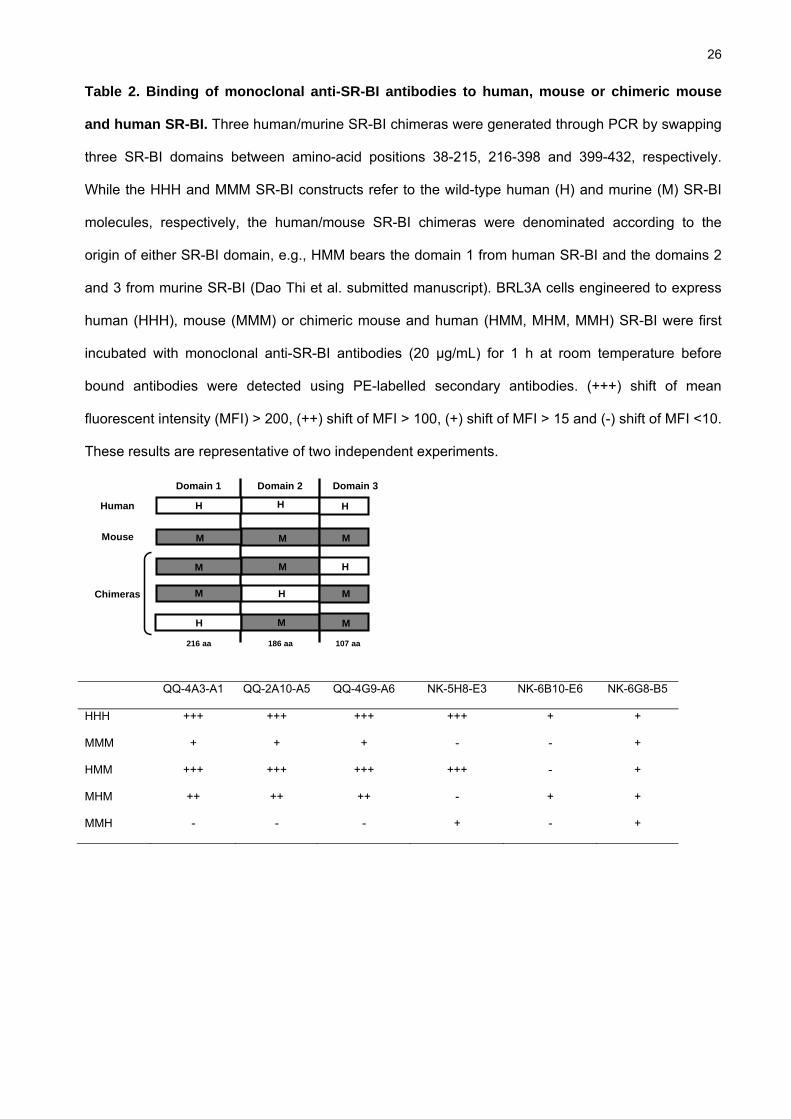
26
Table 2. Binding of monoclonal anti-SR-BI antibodies to human, mouse or chimeric mouse
and human SR-BI. Three human/murine SR-BI chimeras were generated through PCR by swapping
three SR-BI domains between amino-acid positions 38-215, 216-398 and 399-432, respectively.
While the HHH and MMM SR-BI constructs refer to the wild-type human (H) and murine (M) SR-BI
molecules, respectively, the human/mouse SR-BI chimeras were denominated according to the
origin of either SR-BI domain, e.g., HMM bears the domain 1 from human SR-BI and the domains 2
and 3 from murine SR-BI (Dao Thi et al. submitted manuscript). BRL3A cells engineered to express
human (HHH), mouse (MMM) or chimeric mouse and human (HMM, MHM, MMH) SR-BI were first
incubated with monoclonal anti-SR-BI antibodies (20 µg/mL) for 1 h at room temperature before
bound antibodies were detected using PE-labelled secondary antibodies. (+++) shift of mean
fluorescent intensity (MFI) > 200, (++) shift of MFI > 100, (+) shift of MFI > 15 and (-) shift of MFI <10.
These results are representative of two independent experiments.
Human
Chimeras
Mouse
216 aa 186 aa 107 aa
Domain 1 Domain 2 Domain 3
M
M
M
M M
M
M
MM
H
H
H
H HH
QQ-4A3-A1 QQ-2A10-A5 QQ-4G9-A6 NK-5H8-E3 NK-6B10-E6 NK-6G8-B5
HHH +++ +++ +++ +++ + +
MMM + + + - - +
HMM +++ +++ +++ +++ - +
MHM ++ ++ ++ - + +
MMH - - - + - +

C.
A.
Figure 1
QQ-4A3-
A1
%H
CV
ccin
fect
ion
0
2040
6080
100
120
140160
180 100 µg/mL
10 µg/mL
1 µg/ml
0.1 µg/mL0.01 µg/mL
QQ-2A10
-A5
QQ-4G9-
A6
NK-8H5-
E3
0
500
1000
1500
2000
2500
CTRL IgG
QQ-4A3-A1
QQ-2A10-A5
QQ-4G9-A6
PS-6A7-C4
PS-7B11-E3
NK-8H5-E3
NK-6B10-E6
NK-6G8-B5
Rel
ativ
e flu
ores
cenc
e
0
500
1000
1500
2000
2500
3000
3500
CTRL IgG
QQ-4A3-A1
QQ-2A10-A5
QQ-4G9-A6
PS-6A7-C4
PS-7B11-E3
NK-8H5-E3
NK-6B10-E6
NK-6G8-B5
Rel
ativ
e flu
ores
cenc
e
B.
D.
020
4060
80100
120140
160180
CTRL
CTRL IgG
PS-6A7-C
4
PS-7B11
-E3
QQ-4A3-A
1
QQ-2A10
-A5
QQ-4G9-
A6
NK-8H5-E
3
NK6B10
-E6
NK-6G8-
B5
%H
CV
ccin
fect
ion
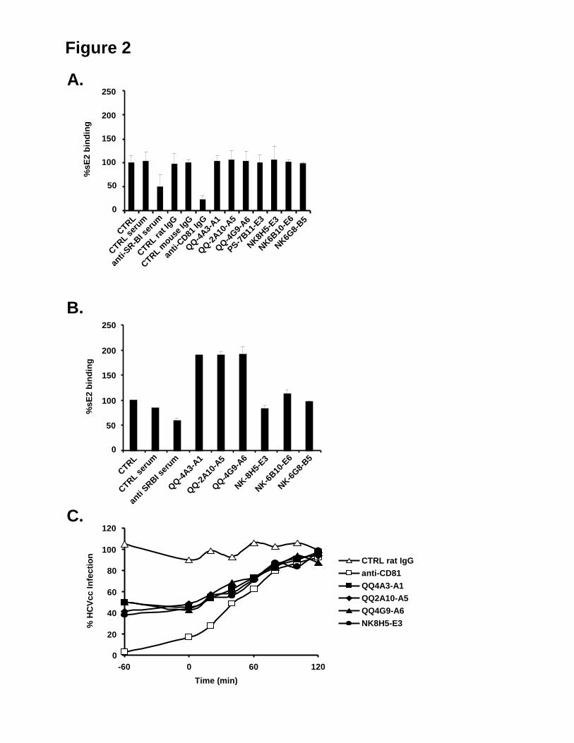
Figure 2
A.
B.
C.
0
50
100
150
200
250
CTRL
CTRL ser
um
anti S
RBI ser
um
QQ-4A3-
A1
QQ-2A10
-A5
QQ-4G9-
A6
NK-8H5-E
3
NK-6B10
-E6
NK-6G8-
B5
%sE
2 bi
ndin
g
0
20
40
60
80
100
120
-60 0 60 120
Time (min)
% H
CV
ccIn
fect
ion CTRL rat IgG
anti-CD81
QQ4A3-A1QQ2A10-A5QQ4G9-A6NK8H5-E3
CTRL
CTRL ser
um
anti-
SR-BI s
erum
CTRL ra t I
gG
CTRL mou
se Ig
G
PS-7B11
-E3
%sE
2 bi
ndin
g
QQ-4A3-
A1
QQ-2A10
-A5
QQ-4G9-
A6
NK8H5-
E3
NK6B10
-E6
NK6G8-
B50
50
100
150
200
250
anti-
CD81 Ig
G

Figure 3
A.
C.
3
4
5
6
7
8
0 2 4 6 8 10 12 14 16Postinfection (days)
Vira
l loa
d(lo
g10
RLU
)
CTRL IgGCTRL1 µg/mL10 µg/mL100 µg/mL
E. F.
0
10
20
30
40
50
60
CTRL mSRBI hSRBI
%In
fect
edta
rget
cells
B.
D.
QQ-4G9-A60
5
10
15
20
25
30
CTRL
%In
fect
edta
rget
cells
QQ-2A10-A5
TiPi
T
20.50%
CTRL5.74%TiPi
T
QQ-4G9-A6
CTRL CTRL IgG QQ-4G9-A6
0
20
4060
80
100
120
160
140
% C
ell v
iabi
lity

Figure 4
A. B.
0
20
40
60
80
100
120
1 10 100 1000 10000 100000
Inhibiting antibody (ng/mL)
% B
indi
ng
CTRL rat IgGCTRL mouse IgGQQ-4A3-A1
QQ-2A10-A5QQ-4G99-A6NK-8H5-E3
0
20
40
60
80
100
120
1 10 100 1000 10000 100000
Inhibiting antibody (ng/mL)
% B
indi
ng
CTRL rat IgGCTRL mouse IgGQQ-4A3-A1
QQ-2A10-A5QQ-4G99-A6NK-8H5-E3

Figure 5
A.
B.
0
20
40
60
80
100
120
140
160
180
CTRL ra
t IgG
QQ-4A3-
A1
QQ-2A10
-A5
QQ-4G9-
A6
CTRL mou
se Ig
G
NK-8H5-
E3
NK-6B10
-E6
NK-6G8-
B5
anti-S
RBI ser
um
CTRL se
rum
% C
y5 H
DL
bind
ing
0
2
4
6
8
10
12
CTRL ra
t IgG
CTRL mou
se Ig
G
QQ-4A3-
A1
QQ-2A10
-A5
QQ-4G9-
A6
PS-6A7-C
4
PS-7B11
-E3
NK-8H5-
E3
NK-6B10
-E6
NK-6G8-
B5
HD
L-C
E u
ptak
e/ µ
g pr
otei
nce
lls(%
)
C.
0
1
2
3
4
5
6
0 10 20 30 40 50
Fra
ctio
nalc
hole
ster
olef
flux
(%)
HDL
CTRL rat IgG
CTRL mouse IgG
QQ-4A3-A1
QQ-2A10-A5
QQ-4G9-A6
PS-6A7-C4
PS-7B11-E3
NK-8H5-E3
NK-6B10-E6
NK-6G8-B5

Figure 6
A. B.
C.
0
20
40
60
80
100
120
0,001 0,01 0,1 1 10 100
Ab concentration ( µg/mL)
% H
CV
ppen
try
CTRL anti-SRBIanti-SRBI + anti-E1 (1 µg/mL)
anti-SRBI + anti-E1 (10 µg/mL)
anti-E1
0
20
40
60
80
100
120
0,001 0,01 0,1 1 10 100
Ab concentration ( µg/mL)
% H
CV
ppen
try
CTRL anti-SRBIanti-SRBI + anti-E2 (1 µg/mL) anti-SRBI + anti-E2 (10 µg/mL) anti-E2
Ab concentration ( µg/mL)
% H
CV
ppen
try
CTRL anti-SRBIanti-SRBI + anti-HCV IgG (1 µg/mL)
anti-SRBI + anti-HCV IgG (10 µg/mL)
anti-HCV IgG
0
20
40
60
80
100
120
0,001 0,01 0,1 1 10 100
Ab concentration ( µg/mL)
% H
CV
ccin
fect
ion
CTRL
anti-SRBI
anti-SRBI + anti-HCV IgG (1 µg/mL)
anti-SRBI + anti-HCV IgG (10 µg/mL)
anti-HCV IgG
D.
0
20
40
60
80
100
120
0,01 0,1 1 10 100
- 51 -
3.2. Mutations that alter hepatitis C virus cell entry factor usage mediate escape
from neutralizing antibodies
A manuscript presenting the results from this study is accepted with minor revision in
Gastroenterology. Publication n°2:
Isabel Fofana*, Samira Fafi-Kremer*, Patric Carolla*, Catherine Fauvelle, Muhammad Nauman
Zahid, Marine Turek, Laura Heydmann , Karine Cury, Juliette Hayer, Christophe Combet, François-
Loïc Cosset, Thomas Pietschmann, Marie-Sophie Hiet, Ralf Bartenschlager, François Habersetzer,
Michel Doffoёl, Zhen-Yong Keck, Steven K.H. Foung, Mirjam B. Zeisel, Françoise Stoll-Keller,
Thomas F. Baumert
* IF, SF-K and PC equally contributed to this work
Mutations that alter hepatitis C virus cell entry factor usage mediate escape from neutralizing
antibodies
Gastroenterology, accepted with minor revision
In the second part of my PhD, we aimed to characterize the molecular mechanisms underlying
HCV re-infection of the graft after liver transplantation (LT). A major limitation of LT is the universal
re-infection of the liver graft with accelerated recurrence of liver disease. It had been previously
shown in the lab that viral entry and escape from host neutralizing responses are important
determinants allowing the virus to rapidly infect the liver during the early phase of transplantation.
However, the molecular mechanisms by which the virus evades host immunity to persistently re-
infect the liver graft are unknown.
To uncover enhanced viral entry and evasion mechanisms, we analyzed genetically closely
related prototype variants derived from a well-characterized patient undergoing LT (Fafi-Kremer et
al., 2010) (Figure 10). One variant, termed P01 VL, had re-infected the liver graft and was
characterized by high-infectivity and escape from neutralizing antibodies present in autologous pre-
transplant serum. The other variants, termed P01 VA and VC, were not selected during graft infection
and were characterized by lower infectivity and high-sensitivity to neutralization by autologous pre-
transplant serum (Fafi-Kremer et al., 2010).

- 52 -
VL
19
1
VL
VAVB 4
7
83
VD to
VJ
(1)
VK
****VC
Before LT After LT
VL
19
1
VL
VAVB 4
7
83
VD to
VJ
(1)
VK
****VC
VL
19
1
VL
VAVB 4
7
83
VD to
VJ
(1)
VK
****VC
VLVL
19
1
VL
VAVB 4
7
83
VD to
VJ
(1)
VK
****VC
Before LT After LT
Figure 10: Evolution of HCV variants before and 7 d after LT. Distribution of full-length E1E2 variants is shown in the figure. Circle graphs represent the percentage of each clone detected. Viral isolates are indicated by an individual color and capital letters. Variants re-infecting the liver graft are depicted in blue, and non-selected variants not detected after transplantation are depicted in white, grey or black (Fafi-Kremer et al., 2010).
To investigate the molecular mechanism of enhanced entry of the selected variant VL,
individual mutations of envelope glycoprotein region F447, S458G and R478C of non selected mutant
VA and VC were introduced into infectious HCV pseudoparticles (HCVpp) expressing envelope
glycoproteins of escape variant VL. The results obtained from these mutations demonstrate that
residues F447L, S458G and R478C are largely responsible for the high infectivity of the escape
variant VL.
In the framework of this project, I assessed that the envelope glycoprotein expression was
similar for parental and chimeric strains which indicates that the differences in viral entry are not due
to impaired production in HCVpp 293T producer cells or impaired assembly (Figure S3A, C of the
manuscript). Then I investigated internalization kinetics of the parental and chimeric variants in the
presence of anti-CD81 antibody. Since entry kinetics of parental and chimeric variants were similar, it
is unlikely that the mutant induced modulation of CD81-dependency alters the velocity of viral entry
(Figure 3D of the manuscript). In another experiment, we showed that overexpression of SR-BI and
CD81 enhanced viral entry of the VL and the VC variants, but in contrast to CD81 overexpression, no
specific increase was observed for the chimeric strains containing substitutions at positions 458 and
478 in SR-BI overexpression. I further investigated whether the mutations affect HCV-CD81 binding.
We showed that E1E2 complexes of the escape variant VL bound less efficiently to shCD81-Huh7.5
cells than glycoproteins of the variants VC and VA. Exchange of the mapped residues conferred
similar phenotypes suggesting that the residues at positions 447, 458 and 478 alter E1E2 interactions
with cell surface CD81 (Figure 3B of the manuscript).
Next, I investigated the neutralization kinetics of parental and chimeric variants using a well-
defined human antibody directed against HCV envelope glycoprotein E2 (HMAb CBH-23). The
HMAb CBH-23 inhibited viral entry of VC and VLVC458+478 at post-binding steps during time points
- 53 -
closely related to HCV-CD81 interaction (Figure 5C of the manuscript) and partially inhibited VA
and VLVA447 (Figure 5D of the manuscript). The VL variant escaped antibody mediated
neutralization at the same steps. These data indicate that the positions 447, 458 and 478 mediate viral
evasion from neutralizing antibodies at post-binding steps and time points closely related to HCV-
CD81 interaction.
Taken together, in this study we identified a novel and clinically important mechanism of viral
evasion, where co-evolution simultaneously occurs between cellular entry factor usage and escape
from neutralization. The identification of these mechanisms advances our understanding of the
pathogenesis of HCV infection and paves the way for the development of novel antiviral strategies
and vaccines.
Mutations that Alter Use of Hepatitis C Virus Cell Entry Factors Mediate Escape from Neutralizing Antibodies
Molecular mechanisms of viral evasion in HCV infection
Isabel Fofana1,2,*, Samira Fafi-Kremer1,2,3,*, Patric Carolla1,2,*, Catherine Fauvelle1,2,
Muhammad Nauman Zahid1,2, Marine Turek1,2, Laura Heydmann1,2, Karine Cury1,2,, Juliette
Hayer4, Christophe Combet4, François-Loïc Cosset5, Thomas Pietschmann6, Marie-Sophie
Hiet7, Ralf Bartenschlager7, François Habersetzer1,2,8, Michel Doffoёl1,2,8, Zhen-Yong Keck9,
Steven K.H. Foung9, Mirjam B. Zeisel1,2, Françoise Stoll-Keller1,2,3, Thomas F. Baumert1,2,8,§
1Inserm, U748, 67000 Strasbourg, France, 2Université de Strasbourg, 67000 Strasbourg,
France, 3Laboratoire de Virologie, Hôpitaux Universitaires de Strasbourg, 67000 Strasbourg,
France, 4Bases Moléculaires et Structurales des Systèmes Infectieux, UMR 5086, CNRS,
Université de Lyon, Institut de Biologie et Chimie des Proteines, 69367 Lyon, France, 5Université de Lyon, Université Claude Bernard Lyon1; IFR 128 ; Inserm, U758; Ecole
Normale Supérieure de Lyon, 69364 Lyon, France, 6Division of Experimental Virology,
TWINCORE, Centre for Experimental and Clinical Infection Research, a joint venture
between the Medical School Hannover and the Helmholtz Centre for Infection Research,
30625 Hannover, Germany, 7The Department of Infectious Diseases, Molecular Virology,
Heidelberg University, 69120 Heidelberg, Germany, 8Pôle Hepato-digestif, Hôpitaux
Universitaires de Strasbourg, 67000 Strasbourg, France, 9Department of Pathology,
Stanford University School of Medicine, Stanford, 94304 California, USA
Grant support: This work was supported by Inserm, the European Union (ERC-2008-AdG-
233130-HEPCENT and Interreg IV FEDER-Hepato-Regio-Net 2009), the ANR chair of
excellence program (ANR-05-CEXC-008), ANRS (2007/306, 2008/354, 2009/183,
2011/132), the Région d’Alsace (2007/09), the Else Kröner-Fresenius Stiftung (EKFS
P17//07//A83/06), the Ligue Contre le Cancer (CA 06/12/08), INCA (2009-143) and
Canceropôle du Grand-Est (30/03/09), the Finovi Foundation, the IBiSA GIS, the SFERE
program and PHS grants HL079381 and AI081903.

Acknowledgements: The authors thank F. Chisari (The Scripps Research Institute, La
Jolla) for the gift of Huh7.5.1 cells, J. A. McKeating (University of Birmingham, Birmingham,
UK), C. Rice (Rockefeller University, New York City), C. Schuster (Inserm U748, Strasbourg,
France), M. Heim (University of Basel, Basel Switzerland), F. Wong-Staal (Itherx, San
Diego), J. Dubuisson (Inserm U1019, Lille, France) and F. Rey (Institut Pasteur, Paris) for
helpful discussions. We acknowledge excellent technical assistance of Michèle Bastien-Valle
(Inserm U748, Strasbourg, France).
Abbreviations: CLDN - claudin; CTRL - control; HCV - hepatitis C virus; HCVpp - HCV
pseudoparticles; HMAb - human monoclonal antibody; HVR - hypervariable region; IgG -
immune globulin G; mAb - monoclonal antibody; RLU - relative light units; SR-BI - scavenger
receptor class B type I; LT - liver transplantation; OCLN – occludin ; V- viral variant.
§Correspondence: Thomas F. Baumert, M. D., Inserm Unit 748, Hôpitaux Universitaires de
Strasbourg, 3 rue Koeberlé, F-67000 Strasbourg, France; phone: (33) 3 68 85 37 02; fax:
(33) 3 68 85 37 50, e-mail: [email protected]
Disclosures: This author discloses the following: T.P. is member of the advisory board of
Biotest AG and reports receiving consultancies. The remaining authors disclose no conflicts.
Author contribution: I.F., S. F.-K and P.C. contributed equally to this work. S. F.-K, M. B.
Z., F. S.-K and T. F. B. designed and supervised research. I. F., S. F.-K, P. C, C. F., M. N.
Z., M. T., L. H., K. C., J. H., C. C., M.-S. H., F.-L. C, F. H., Z.-Y. K., S. K. H. F, M. B. Z., F.
S.-K and T. F. B. performed research. I. F., S. F.-K, P. C, C. F., M. N. Z., M. T., M. B. Z., F.
S.-K and T. F. B analyzed data. F.-L. C, T. P., R. B., F. H., M. D. F. S.-K provided reagents.
I. F., S. F.-K, P. C, M. B. Z. and T. F. B. wrote the paper.
Word count: title – 93 characters, abstract – 261 words, text – 5,993 words

ABSTRACT Background & Aims: The development of vaccines and other strategies to prevent hepatitis
C virus (HCV) infection is limited by rapid viral evasion. HCV entry is the first step of
infection; this process involves several viral and host factors and is targeted by host
neutralizing responses. Although the roles of host factors in HCV entry have been well
characterized, their involvement in evasion of immune responses is poorly understood. We
used acute infection of liver graft as a model to investigate the molecular mechanisms of
viral evasion.
Methods: We studied factors that contribute to evasion of host immune responses using
patient-derived antibodies, HCV pseudoparticles, and cell culture-derived HCV that express
viral envelopes from patients who have undergone liver transplantation. These viruses were
used to infect hepatoma cell lines that express different levels of HCV entry factors.
Results: Using reverse genetic analyses, we identified altered use of host-cell entry factors
as a mechanism by which HCV evades host immune responses. Mutations that alter use of
the CD81 receptor also allowed the virus to escape neutralizing antibodies. Kinetic studies
demonstrated that these mutations affect virus–antibody interactions during post-binding
steps of the HCV entry process. Functional studies with a large panel of patient-derived
antibodies showed that this mechanism mediates viral escape, leading to persistent infection
in general.
Conclusion: We identified a mechanism by which HCV evades host immune responses, in
which use of cell entry factors evolves with escape from neutralizing antibodies. These
findings advance our understanding of the pathogenesis of HCV infection and might be used
to develop antiviral strategies and vaccines.
Keywords: virology; liver disease; tissue culture model; immunity
INTRODUCTION
Hepatitis C virus (HCV) infection is a major cause of liver disease.1 A vaccine is not available
and antiviral treatment is limited by resistance and adverse effects.2 HCV-induced liver
disease is a leading indication for liver transplantation (LT).3 A major limitation of LT is the
universal re-infection of the liver graft with accelerated recurrence of liver disease. A strategy
to prevent re-infection is lacking.3 Thus, there is an urgent unmet medical need for the
development of efficient and safe antivirals and vaccines.
HCV entry is required for initiation, maintenance and dissemination of infection. Viral
entry is a key target for adaptive host responses and antiviral strategies.4, 5 Functional
studies in clinical cohorts highlight that viral entry and escape from antibody-mediated
neutralization play an important role in viral persistence and liver disease.6-12 HCV entry is a
highly orchestrated process mediated by viral envelope glycoproteins E1 and E2 and several
host factors including heparan sulfate, CD81, scavenger receptor BI (SR-BI), claudin-1
(CLDN1), occludin (OCLN) (reviewed in 5) and kinases.13 While the role of E1E2 in antibody-
mediated neutralization has intensively been studied,4, 5, 14 the role of host factors for viral
evasion in vivo is only poorly understood.
Acute graft infection is an established in vivo model to study viral evasion since viral
infection and host neutralizing responses can be precisely monitored.8 Viral entry and
escape from host neutralizing responses are important determinants allowing the virus to
rapidly infect the liver during transplantation.8 However, the molecular mechanisms by which
the virus evades host immunity to persistently re-infect the liver graft are unknown.
To uncover viral and host factors mediating enhanced viral entry and escape, we
functionally analyzed genetically closely related prototype variants derived from a well-
characterized patient undergoing LT.8 One variant P01VL re-infecting the liver graft was
characterized by high infectivity and escape from neutralizing antibodies present in
autologous pre-transplant serum.8 The other closely related variants, P01VA and VC, were
not selected during LT and characterized by lower infectivity and high sensitivity to
neutralization by autologous pre-transplant serum.8 Previous studies had indicated that an
E2 region comprising amino acids 425-483 most likely contained mutations responsible for
the phenotype of enhanced entry and viral evasion of variants re-infecting the liver graft.8

MATERIALS AND METHODS Patients. Evolution and functional analysis of viral variants of patient P01 have been
described.8 Anti-HCV-positive serum samples from patients undergoing transplantation and
chronic HCV infection were obtained with approval from the Strasbourg University Hospital
IRB (ClinicalTrial.gov Identifiers NCT00638144 and NCT00213707).
Plasmids. Plasmids for HCVpp production of variants VL, VA and VC have been described.8
E1E2-encoding sequences were used as templates for individual and combinations of
mutations using the QuikChange II XL site-directed mutagenesis kit (Stratagene). Mutations
were confirmed by DNA sequence analysis (GATC Biotech) for the desired mutation and for
exclusion of unexpected residue changes in the full-length E1E2 encoding sequences.
Mutated constructs were designated X#Y, where # is the residue location in H77c,15 X is the
mutated and Y the original amino acid.
Antibodies. Monoclonal anti-E1 (11B7) and anti-E2 (AP33, IGH461, 16A6), human anti-
HCV IgG,10, 16 HMAbs CBH-2, CBH-5, CBH-23 and HC-1 have been described.9, 17 Anti-
CD81 (JS-81) was from BD Biosciences, AP33 from Genentech, 11B7, IGH461 and 16A6
from Innogenetics.
Cell lines. HEK 293T and Huh7.5.1 cells were cultured as described.10, 13, 16 Huh7.5.1 cells
overexpressing HCV entry factors were created by stable lentiviral gene transfer of CLDN1,
OCLN, SR-BI or CD81.18 Huh7.5 stably transduced with retroviral vectors encoding for CD81
and CD13-specific shRNAs have been described.19 Receptor expression was assessed by
flow cytometry .13
HCVpp and HCVcc production, infection and neutralization. Lentiviral HCVpp bearing
patient-derived envelope glycoproteins were produced as described.8, 10, 20 The amount of
HCVpp was normalized following quantification of HIV p24 antigen expression (Innotest HIV
Antigen mAb Kit, Innogenetics) and HCVpp entry was performed as described. 8, 10, 11, 16
Chimeric HCVcc expressing patient-derived structural proteins were constructed and
produced as described in Supplementary Materials and Methods. HCVcc infectivity was
determined by determining the TCID5021 or intracellular HCV RNA levels as described.13, 21, 22
HCVpp and HCVcc neutralization were performed as described.8, 10, 11, 16
Kinetic assays. HCVpp kinetic assays were performed in Huh7.5.1 cells using anti-CD81
(JS-81) and anti-E2 (CBH-23) mAbs as described.16, 23

Statistical analysis. Statistical analysis (Repeated Measures ANOVA) was performed using
the SPSS 16.0 software for Windows (SPSS Inc., Chicago, IL).

RESULTS HCV E2 residues at positions 447, 458 and 478 confer enhanced viral entry of a high-infectivity variant re-infecting the liver graft. To investigate the molecular mechanism of
enhanced entry of the variant VL re-infecting the liver graft, we first introduced individual
mutations of region E2425-4838 of the low-entry and neutralization-sensitive mutant VC into
HCV pseudoparticles (HCVpp) expressing envelope glycoproteins of the highly infectious
escape variant VL (Fig. 1A). Previous studies had indicated that this region most likely
contains the mutations responsible for the high-infectivity phenotype of VL.8 Following
normalization of HCVpp levels by p24 antigen expression, viral entry was quantified relative
to the escape variant VL. The entry level of the nonselected variant VC was 5% compared to
the escape variant VL (Fig. 1B). By introducing the mutations S458G and R478C into VC,
chimeric HCVpp showed similar viral entry level as the paternal variant VL whereas
introduction of individual or combination of other mutations only had a partial effect (Fig. 1B,
Fig. S1). To explore the impact of other positions on viral entry we introduced mutations
from another nonselected variant termed VA into VL (Fig. 1A) and identified position F447
as an additional residue relevant for enhanced entry of the escape variant VL (Fig. 1C).
These results demonstrate that residues F447L, S458G and R478C are largely responsible
for the high-infectivity of the escape variant VL.
Enhanced viral entry by mutations F447L, S458G and R478C of the escape variant is the result of altered use of CD81. To address whether the mutations affect viral entry by
different usage of cell entry factors SR-BI, CD81, CLDN1 and OCLN, we studied viral entry
of HCVpp derived from parental and chimeric variants in Huh7.5.1 cells stably
overexpressing individually the four main entry factors (Fig. 2A). Overexpression of either
SR-BI, CD81, CLDN1 or OCLN, did not affect the stability or proportion of other cell surface
HCV receptors (Fig. 2B and data not shown).
Overexpression of CD81 significantly enhanced viral entry of VL (3.2fold) and VC
(2fold) compared to parental cells (P < .001) (Fig. 2C). The fold change in HCVpp entry was
significantly higher for VL than for VC (P < .001). Exchanging the two residues at position
458 and 478 similarly increased viral entry. This suggests that combination of the two
individual mutations modulates viral entry by altering CD81-dependency. Overexpression of
SR-BI also increased viral entry of VL and VC, but no specific increase was observed for the
chimeric strains containing substitutions at positions 458 and 478 (Fig. 2C). These data
confirm an important role for SR-BI as an entry factor for patient-derived variants, but also
demonstrate that positions 458 and 478 do not significantly alter SR-BI-dependency. Thus,

increased entry efficiency of VL in SR-BI-overexpressing cells is most likely due to other
mutations, e. g. in HVR1. Viral entry enhancement was less pronounced in cells
overexpressing CLDN1 or OCLN than CD81 and SR-BI (Fig. 2C) and no specific modulation
of viral entry was associated with the two variants or chimeric strains.
The CD81 usage of viral variants VL, VC and VA was further investigated using
Huh7.5 cells with silenced CD81 expression (Fig. 3A).19 The escape variant VL showed the
highest decrease (5.4fold) of viral entry in shCD81-Huh7.5 cells compared to the decrease
of variants VC (4.3fold, P < .001) and VA (2.9fold, P < .001) (Fig. 3B-C). Exchange of the
mapped residues into chimeric expression plasmids conferred the phenotype of decreased
entry of VL (Fig. 3B-C) confirming that identified residues modulate viral entry by different
CD81 usage. Moreover, using a relevant model system for HCV-CD81 interactions occurring
in vivo consisting of cell surface-expressed CD81, we demonstrate that E1E2 complexes of
the escape variant VL bound less efficiently to shCD81-Huh7.5 cells than glycoproteins of
variants VC and VA (Fig. S2A). Exchange of the mapped residues conferred similar
phenotypes as the parental glycoproteins (Fig. S2B) suggesting that the residues at
positions 447, 458 and 478 alter E1E2 interactions with cell surface CD81.
Taken together, these data demonstrate that (i) the escape variant is characterized by
markedly altered CD81 usage and (ii) altered CD81 usage of the variant is mediated by
residues at positions 447, 458 and 478.
Since the levels of E1E2 incorporation into HCVpp and lentiviral p24 antigen
expression were similar for all strains (Fig. S3A-D), it is unlikely that the differences in viral
entry are the result of impaired HCVpp assembly or release.
Next, to assess whether enhanced entry is due to more rapid internalization of viral
particles we investigated internalization kinetics of the parental and chimeric variants in the
presence of anti-CD81 antibody.16, 21, 23, 24 Since entry kinetics of parental and chimeric
variants were similar (Fig. 3D), it is unlikely that the mutant-induced modulation of CD81-
dependency alters the velocity of viral entry.
Positions 447, 458 and 478 mediate escape from autologous transplant serum during graft re-infection. To assess whether the residues in region E2425-483 influencing viral entry
(Fig. 1) were also responsible for escape from antibody-mediated neutralization, we studied
the impact of each single and combined substitutions of the nonselected variant VC on
neutralization by autologous pre-transplant serum. Autologous pre-transplant serum only
poorly neutralized the selected variant VL as well as the variants substituted at position 434,
444, and 445 while individual substitution at positions 458 and 478 significantly (P < .001, P

≤ .05) increased the sensitivity of VLVC458 and VLVC478 to autologous neutralizing antibodies
(1:400 and 1:200) (Fig. 4A). Noteworthy, only the variant VLVC458+478 showed a similar
neutralization titer as the nonselected variant VC (1:6,400, P < .001). To confirm that these
mutations were indeed responsible for the phenotype of the parental variant VL, we
investigated neutralization of VCVL458+478 by autologous serum. The variant VCVL458+478
escaped autologous neutralization similarly to the escape variant VL (Fig. 4A). A similar
phenotype was observed when mutation 447 of VA was introduced into the VL cDNA (Fig.
4B). In contrast, the introduction of other residues into VL only had a minor effect on
neutralization (Fig. 4B). Taken together, these findings suggest that the residues at positions
447, 458 and 478 are simultaneously responsible for both enhanced viral entry and evasion
from antibody-mediated neutralization.
Positions 447, 458 and 478 define a conformational epitope involved in evasion from host neutralizing responses. To further elucidate the mechanism of viral evasion of the
escape variant VL from patient-derived neutralizing antibodies, we investigated whether the
identified mutations F447L, S458G and R478C confer resistance or sensitivity to a panel of
mAbs directed against conformational9, 17 and linear E2 epitopes.16 The conformational
HMAbs (CBH-2, CBH-5, CBH-23, HC-1) have been shown to exhibit a broad cross-
neutralizing activity by interfering with E2-CD81 interaction9, 17 and their epitopes are only
partially defined (Table S1). AP33 is directed against a conserved epitope comprising aa
412-423.25 While the escape variant VL was poorly neutralized by several HMAbs directed
against conformational epitopes, VC and VA were efficiently neutralized by all HMAbs (Fig.
5A-B). Moreover, by substituting the residues at positions 458 and 478 or 447, the well
neutralized nonselected variants VC (VCVL458+478) and VA (VAVL447) became neutralization-
resistant as the escape variant VL. Introducing the residues of VC or VA into VL
(VLVC458+478 and VLVA447) restored neutralization by HMAbs, suggesting that these residues
are part of the HMAbs epitopes. In contrast, anti-E2 antibodies (AP33, 16A6, IGH461)
targeting linear epitopes similarly neutralized parental and chimeric variants (Fig. 5A-B and
Table S1).
Antibody-mediated neutralization occurs at binding and post-binding steps during viral
entry.16 To map the entry step involved in viral evasion from neutralizing antibodies by VL,
we investigated the neutralization kinetics of parental and chimeric variants.16, 21, 23 The anti-
E2 HMAb CBH-23 inhibited viral entry of VC and VLVC458+478 at post-binding steps during
time points closely related to HCV-CD81 interaction (Fig. 5C). Partial inhibition at post-

binding steps by CBH-23 was also observed for VA and VLVA447 (Fig. 5D). The VL variant
escaped antibody-mediated neutralization at the same steps.
Interestingly, purified HCVpp expressing envelope glycoproteins of the escape variant
bound similarly to neutralizing anti-E2 antibody CBH-23 as the envelope glycoproteins of
non selected variants or variants containing mutations of the identified escape residue (Fig.
S4). Thus, it is likely that viral evasion is not due to decreased antibody-binding to circulating
virions but rather occurs during post-binding steps of viral entry where E2-host entry factor
interactions result in conformational changes of the envelope and failure of antibodies to
inhibit entry. Taken together, these data indicate that positions 447, 458 and 478 mediate
viral evasion from neutralizing antibodies at post-binding steps and time points closely
related to HCV-CD81 interaction.
Positions 447, 458 and 478 mediate escape from antiviral antibodies in non-related patients with chronic HCV infection. To investigate whether these mutations not only
result in escape from antibodies from the same patient but also confer resistance to antiviral
antibodies of non-related HCV infected patients, we studied the neutralization of the parental
variants by a large panel of sera randomly selected from chronically infected patients (n =
102). While VL was not neutralized by 53 out of 102 patient sera (mean neutralizing titer,
1:144) VC was significantly neutralized by 90 out of 102 patient sera (mean neutralizing titer,
1:1,088, P < .001) (Fig. 6 and Table S2). Similar results were obtained for VA (neutralization
by 80 out of 102 patient sera, mean neutralizing titer of 1:322, P = .01). Functional analysis
of HCVpp expressing chimeric envelope glycoproteins demonstrated that neutralization of
VC and VA was predominantly mediated by the identified mutations in residues 447, 458
and 478 (Fig. 6).
Confirmation of differential cell entry factor usage and viral evasion using chimeric HCVcc. Finally, we confirmed the functional impact of the three residues on virus-host
interactions using the HCVcc system. To address this issue we constructed chimeric JFH-1
based HCVcc expressing the VL wild-type envelope or VL containing VC and VA-specific
functional residues. Viruses containing patient-derived envelopes showed similar levels of
replication and envelope production (data not shown). Phenotypic analyses of infection and
neutralization of chimeric HCVcc confirmed the relevance of the identified residues for
enhanced entry, differential CD81 usage and viral evasion (Fig. 7A-D). While the escape
variant VL was poorly neutralized, the identified mutations at positions 447, 458 and 478
restored its sensitivity to conformational HMAb CBH-23 (Fig. 7C) as well as to heterologous

sera from chronically infected patients (Fig. 7D). These data confirm the functional relevance
of the obtained results in the HCVcc system expressing authentic patient-derived envelopes.

DISCUSSION
Using acute infection of the liver graft as an in vivo model, we identified a novel, clinically
and therapeutically important mechanism of viral evasion, where co-evolution simultaneously
occurs between cellular entry factor usage and escape from neutralization.
Several host selection forces operate concomitantly during HCV infection. These
include pro-viral host factors resulting in selection of most infectious viruses best adapted to
host factors and anti-viral host immune responses leading to escape from immune
responses. Antibody-mediated selective pressure is thought to be an important driver of viral
evolution.8, 11 The immune response may fail to resolve HCV infection because neutralizing
antibody-mediated response lags behind the rapidly and continuously evolving HCV
glycoprotein sequences.11 However, continuous generation of escape mutations during
chronic HCV infection may also compromise virus infectivity: indeed, it has been reported
that structural changes in E2 leading to complete escape from neutralizing antibodies
simultaneously compromised viral fitness by reducing CD81-binding.9 Moreover, escape
from T cell responses has been associated with impaired viral replication.26, 27 We show for
the first time that clinically occurring mutations simultaneously lead to enhanced viral
infectivity by optimizing host factor usage and escape from host immune responses. Since
this mechanism was uncovered in patient strains isolated during acute liver graft infection it
is likely that the novel and unique mechanism of co-evolution between host factor usage and
viral evasion ensures optimal initiation, dissemination and maintenance of viral infection in
the early phase of liver graft infection. In addition, since the VL strain escapes autologous
antibodies from the transplant patient (Fig. 4) and resists to monoclonal and polyclonal
antibodies of heterologous patients (Figs. 5, 6, 7 and Tables S1, S2), and given the high
prevalence of the identified mutations in a large genomic database of viral isolates (Fig. S5
and Supplementary Results), the co-evolution of receptor usage and escape from
neutralizing antibodies may also play an important role for viral evasion in chronic HCV
infection in general.
Our mechanistic studies demonstrate that the identified viral evasion factors are part
of a conformational neutralizing epitope modulating E2-CD81 interactions at post-binding
entry steps.28, 29 Noteworthy, the same mutations were also responsible for immune escape
of VL. Neutralization studies using HMAbs directed against discontinuous envelope
glycoprotein regions termed domain B and C30, 31 demonstrate that the three positions are
part of an epitope which plays a key role for neutralization and viral evasion. Since the
mutations are outside the known contact residues within the epitopes of the HMAbs CBH-2,
CBH-5, CBH-23 and HC-19, 17 (Table S1) and complementary to previously identified regions
associated with escape from neutralizing monoclonal antibodies,25 positions 447, 458 and
478 either modulate the interaction of the majority of antibodies directed against domain B
and C epitopes or are part of a novel E2 epitope mediating evasion from host neutralizing
antibodies.
Based on previous functional observations and structural predictions, Krey and
colleagues proposed a model for a potential tertiary organisation of E2. In this model, E2
comprises three subdomains with the CD81 binding regions located within domain I (W420,
A440LFY, Y527, W529, G530 and D535) and potential CD81 binding sites overlapping with
domain III (Y613RLWHY).28, 29, 32, 33 In this model, positions 447, 458 and 478 are located
outside but in close proximity of the previously suggested CD81 binding domains. Moreover,
position 447 is located immediately downstream a conserved motif between HVR1 and
HVR2 which has been shown to play an important role in CD81 recognition as well as pre- or
post-CD81 dependent stages of viral entry.32 Position 478 is located within HVR2 which
modulates, by a complex interplay with HVR1, binding of E2 glycoprotein to CD81.34
Since mutations F447L, S458G and R478C (i) modulate CD81-dependency of HCV
entry (Fig. 2 and 3), (ii) alter the interaction with cell surface CD81 (Fig. S2), (iii) mediate
viral evasion from antibodies at post-binding steps closely related to HCV-CD81 interactions
(Fig. 5) and (iv) are located within E2 loops of the predicted E2 secondary structure and
tertiary organization29 positions 447, 458 and 478 may be part of two loops belonging to a
larger cluster of closely related surface-exposed E2 loops. These loops are most likely
involved in E2-CD81 binding either directly or indirectly as a key point for structural
rearrangement during viral entry. 34, 35
The polar S and R residues present in the escape variant can form non-bonded
interactions with other residues by hydrogen bonds and salt bridge, respectively. These
interactions could increase the stability of the interacting E2-CD81 interface allowing efficient
entry of the VL escape variant through E2-CD81-CLDN1 co-receptor complexes which are
key determinants for viral entry.13, 23, 36 Furthermore, the E2 cluster of loops containing the
mutations bears linear epitopes but also defines at least one conformational epitope that is a
target of neutralizing antibodies. According to residue physical-chemical properties, the VL
variant S458 and R478 residues enhance the hydrophilicity of the loops they belong to and
may promote the surface exposure of the loops. This change could further modulate E2-
CD81 interactions and impair the binding of neutralizing antibodies by blocking access to
their target epitopes. The F to L substitution present in the VA strain most likely does not
profoundly alter the tertiary or quaternary structure of E2. This is suggested by the fact that
this position is located in a loop as predicted by the proposed E2 model.29 Thus, it is

conceivable that this mutation which increases E2 hydrophobicity may reduce accessibility of
the loop and its interactions with CD81 or CD81-CLDN1 co-receptor complexes.
Alternatively, allosteric mechanisms may play a role in the observed virus-antibody-host
interactions.
Taken together, our data identified key determinants of immune evasion in vivo.
Mutations conferring neutralization escape altered CD81 receptor usage and enhanced cell
entry. Moreover, our data suggest that mutations in HVR1 which may modulate entry and
neutralization by altering SR-BI-dependency (Fig. 1, 2, 4 and data not shown) may
contribute to the high-entry and escape phenotype of the escape variant. Furthermore,
interfering non-neutralizing antibodies may constitute another mechanism of escape (data
not shown).
Although proof-of-concept studies in animal models have demonstrated a potential
role for HMAbs in prevention of HCV infection,37, 38 the partial or complete escape of the VL
variant from autologous and heterologous serum-derived antibodies as well as many broadly
cross-neutralizing HMAbs (Fig. 5; Table S1) demonstrates the ability of the virus to evade
cross-neutralizing anti-envelope mAbs. By identifying viral and host factors mediating
immune evasion in the HCV-infected patient, our results may open new perspectives for the
development of broadly cross-neutralizing anti-envelope or anti-receptor antibodies
overcoming viral escape.

REFERENCES
1. Alter H. Viral hepatitis. Hepatology 2006;43:S230-4.
2. Hofmann WP, Zeuzem S. A new standard of care for the treatment of chronic HCV
infection. Nat Rev Gastroenterol Hepatol 2011;8:257-64.
3. Watt K, Veldt B, Charlton M. A practical guide to the management of HCV infection
following liver transplantation. Am J Transplant 2009;9:1707-13.
4. Zeisel MB, Cosset FL, Baumert TF. Host neutralizing responses and pathogenesis of
hepatitis C virus infection. Hepatology 2008;48:299-307.
5. Zeisel MB, Fofana I, Fafi-Kremer S, Baumert TF. HCV entry into hepatocytes:
mechanism and potential therapeutic implications. J Hepatol 2011;54:566-576.
6. Lavillette D, Morice Y, Germanidis G, Donot P, Soulier A, Pagkalos E, Sakellariou G,
Intrator L, Bartosch B, Pawlotsky JM, Cosset FL. Human serum facilitates hepatitis C
virus infection, and neutralizing responses inversely correlate with viral replication
kinetics at the acute phase of hepatitis C virus infection. J Virol 2005;79:6023-34.
7. Dowd KA, Netski DM, Wang XH, Cox AL, Ray SC. Selection Pressure From
Neutralizing Antibodies Drives Sequence Evolution During Acute Infection With
Hepatitis C Virus. Gastroenterology 2009;136:2377-86.
8. Fafi-Kremer S, Fofana I, Soulier E, Carolla P, Meuleman P, Leroux-Roels G, Patel
AH, Cosset F-L, Pessaux P, Doffoël M, Wolf P, Stoll-Keller F, Baumert TF. Enhanced
viral entry and escape from antibody-mediated neutralization are key determinants for
hepatitis C virus re-infection in liver transplantation. J Exp Med 2010;207:2019-2031.
9. Keck ZY, Li SH, Xia J, von Hahn T, Balfe P, McKeating JA, Witteveldt J, Patel AH,
Alter H, Rice CM, Foung SK. Mutations in hepatitis C virus E2 located outside the
CD81 binding sites lead to escape from broadly neutralizing antibodies but
compromise virus infectivity. J Virol 2009;83:6149-60.
10. Pestka JM, Zeisel MB, Blaser E, Schurmann P, Bartosch B, Cosset FL, Patel AH,
Meisel H, Baumert J, Viazov S, Rispeter K, Blum HE, Roggendorf M, Baumert TF.
Rapid induction of virus-neutralizing antibodies and viral clearance in a single-source
outbreak of hepatitis C. Proc Natl Acad Sci U S A 2007;104:6025-30.
11. von Hahn T, Yoon JC, Alter H, Rice CM, Rehermann B, Balfe P, McKeating JA.
Hepatitis C virus continuously escapes from neutralizing antibody and T-cell
responses during chronic infection in vivo. Gastroenterology 2007;132:667-78.
12. Osburn WO, Fisher BE, Dowd KA, Urban G, Liu L, Ray SC, Thomas DL, Cox AL.
Spontaneous control of primary hepatitis C virus infection and immunity against
persistent reinfection. Gastroenterology 2010;138:315-24.

13. Lupberger J, Zeisel MB, Xiao F, Thumann C, Fofana I, Zona L, Davis C, Mee CJ,
Turek M, Gorke S, Royer C, B. F, Zahid MN, Lavillette D, J. F, Cosset F-L,
Rothenberg SM, Pietschmann T, Patel AH, Pessaux P, Doffoël M, Raffelsberger W,
Poch O, McKeating JA, Brino L, Baumert TF. EGFR and EphA2 are hepatitis C virus
host entry factors and targets for antiviral therapy. Nat Med 2011;17:589-95.
14. Stamataki Z, Grove J, Balfe P, McKeating JA. Hepatitis C virus entry and
neutralization. Clin Liver Dis 2008;12:693-712.
15. Kuiken C, Combet C, Bukh J, Shin IT, Deleage G, Mizokami M, Richardson R, Sablon
E, Yusim K, Pawlotsky JM, Simmonds P. A comprehensive system for consistent
numbering of HCV sequences, proteins and epitopes. Hepatology 2006;44:1355-61.
16. Haberstroh A, Schnober EK, Zeisel MB, Carolla P, Barth H, Blum HE, Cosset FL,
Koutsoudakis G, Bartenschlager R, Union A, Depla E, Owsianka A, Patel AH,
Schuster C, Stoll-Keller F, Doffoel M, Dreux M, Baumert TF. Neutralizing host
responses in hepatitis C virus infection target viral entry at postbinding steps and
membrane fusion. Gastroenterology 2008;135:1719-1728.
17. Hadlock KG, Lanford RE, Perkins S, Rowe J, Yang Q, Levy S, Pileri P, Abrignani S,
Foung SK. Human monoclonal antibodies that inhibit binding of hepatitis C virus E2
protein to CD81 and recognize conserved conformational epitopes. J Virol
2000;74:10407-16.
18. Haid S, Windisch MP, Bartenschlager R, Pietschmann T. Mouse-specific residues of
claudin-1 limit hepatitis C virus genotype 2a infection in a human hepatocyte cell line.
J Virol 2009;84:964-75.
19. Koutsoudakis G, Herrmann E, Kallis S, Bartenschlager R, Pietschmann T. The level
of CD81 cell surface expression is a key determinant for productive entry of Hepatitis
C Virus into host cells. J Virol 2007;81:588-598.
20. Bartosch B, Dubuisson J, Cosset FL. Infectious hepatitis C virus pseudo-particles
containing functional E1-E2 envelope protein complexes. J. Exp. Med. 2003;197:633-
42.
21. Zeisel MB, Koutsoudakis G, Schnober EK, Haberstroh A, Blum HE, Cosset F-L,
Wakita T, Jaeck D, Doffoel M, Royer C, Soulier E, Schvoerer E, Schuster C, Stoll-
Keller F, Bartenschlager R, Pietschmann T, Barth H, Baumert TF. Scavenger receptor
BI is a key host factor for Hepatitis C virus infection required for an entry step closely
linked to CD81. Hepatology 2007; 46:1722-1731.
22. Fofana I, Krieger SE, Grunert F, Glauben S, Xiao F, Fafi-Kremer S, Soulier E, Mee
CJ, McKeating JA, Dragic T, Schuster C, Thompson J, Baumert TF. Monoclonal anti-

claudin 1 antibodies for prevention of hepatitis C virus infection. Gastroenterology
2010;139:953-964, 964.e1-4.
23. Krieger SE, Zeisel MB, Davis C, Thumann C, Harris HJ, Schnober EK, Mee C, Soulier
E, Royer C, Lambotin M, Grunert F, Dao Thi VL, Dreux M, Cosset FL, McKeating JA,
Schuster C, Baumert TF. Inhibition of hepatitis C virus infection by anti-claudin-1
antibodies is mediated by neutralization of E2-CD81-claudin-1 associations.
Hepatology 2010;51:1144-57.
24. Koutsoudakis G, Kaul A, Steinmann E, Kallis S, Lohmann V, Pietschmann T,
Bartenschlager R. Characterization of the early steps of hepatitis C virus infection by
using luciferase reporter viruses. J Virol 2006;80:5308-20.
25. Owsianka A, Tarr AW, Juttla VS, Lavillette D, Bartosch B, Cosset FL, Ball JK, Patel
AH. Monoclonal antibody AP33 defines a broadly neutralizing epitope on the hepatitis
C virus E2 envelope glycoprotein. J Virol 2005;79:11095-104.
26. Dazert E, Neumann-Haefelin C, Bressanelli S, Fitzmaurice K, Kort J, Timm J,
McKiernan S, Kelleher D, Gruener N, Tavis JE, Rosen HR, Shaw J, Bowness P, Blum
HE, Klenerman P, Bartenschlager R, Thimme R. Loss of viral fitness and cross-
recognition by CD8+ T cells limit HCV escape from a protective HLA-B27-restricted
human immune response. J Clin Invest 2009;119:376-86.
27. Uebelhoer L, Han JH, Callendret B, Mateu G, Shoukry NH, Hanson HL, Rice CM,
Walker CM, Grakoui A. Stable cytotoxic T cell escape mutation in hepatitis C virus is
linked to maintenance of viral fitness. PLoS Pathog 2008;4:e1000143.
28. Owsianka AM, Timms JM, Tarr AW, Brown RJ, Hickling TP, Szwejk A, Bienkowska-
Szewczyk K, Thomson BJ, Patel AH, Ball JK. Identification of conserved residues in
the E2 envelope glycoprotein of the hepatitis C virus that are critical for CD81 binding.
J Virol 2006;80:8695-704.
29. Krey T, d'Alayer J, Kikuti CM, Saulnier A, Damier-Piolle L, Petitpas I, Johansson DX,
Tawar RG, Baron B, Robert B, England P, Persson MA, Martin A, Rey FA. The
disulfide bonds in glycoprotein E2 of hepatitis C virus reveal the tertiary organization
of the molecule. PLoS Pathog 2010;6:e1000762.
30. Keck ZY, Op De Beeck A, Hadlock KG, Xia J, Li TK, Dubuisson J, Foung SK.
Hepatitis C virus E2 has three immunogenic domains containing conformational
epitopes with distinct properties and biological functions. J Virol 2004;78:9224-32.
31. Helle F, Goffard A, Morel V, Duverlie G, McKeating J, Keck ZY, Foung S, Penin F,
Dubuisson J, Voisset C. The neutralizing activity of anti-hepatitis C virus antibodies is
modulated by specific glycans on the E2 envelope protein. J Virol 2007;81:8101-11.

32. Drummer HE, Boo I, Maerz AL, Poumbourios P. A Conserved Gly436-Trp-Leu-Ala-
Gly-Leu-Phe-Tyr Motif in Hepatitis C Virus Glycoprotein E2 Is a Determinant of CD81
Binding and Viral Entry. J Virol 2006;80:7844-7853.
33. Boo I, Tewierek K, Douam F, Lavillette D, Poumbourios P, Drummer HE. Distinct
roles in folding, CD81 receptor binding and viral entry for conserved histidines of HCV
glycoprotein E1 and E2. Biochem J 2012.
34. Roccasecca R, Ansuini H, Vitelli A, Meola A, Scarselli E, Acali S, Pezzanera M,
Ercole BB, McKeating J, Yagnik A, Lahm A, Tramontano A, Cortese R, Nicosia A.
Binding of the hepatitis C virus E2 glycoprotein to CD81 is strain specific and is
modulated by a complex interplay between hypervariable regions 1 and 2. J Virol
2003;77:1856-67.
35. McCaffrey K, Boo I, Poumbourios P, Drummer HE. Expression and characterization
of a minimal hepatitis C virus glycoprotein E2 core domain that retains CD81 binding.
J Virol 2007;81:9584-90.
36. Harris HJ, Davis C, Mullins JG, Hu K, Goodall M, Farquhar MJ, Mee CJ, McCaffrey K,
Young S, Drummer H, Balfe P, McKeating JA. Claudin association with CD81 defines
hepatitis C virus entry. J Biol Chem 2010;285:21092-102.
37. Law M, Maruyama T, Lewis J, Giang E, Tarr AW, Stamataki Z, Gastaminza P, Chisari
FV, Jones IM, Fox RI, Ball JK, McKeating JA, Kneteman NM, Burton DR. Broadly
neutralizing antibodies protect against hepatitis C virus quasispecies challenge. Nat
Med 2008;14:25-7.
38. Vanwolleghem T, Bukh J, Meuleman P, Desombere I, Meunier JC, Alter H, Purcell
RH, Leroux-Roels G. Polyclonal immunoglobulins from a chronic hepatitis C virus
patient protect human liver-chimeric mice from infection with a homologous hepatitis
C virus strain. Hepatology 2008;47:1846-55.
39. Rychlowska M, Owsianka AM, Foung SK, Dubuisson J, Bienkowska-Szewczyk K,
Patel AH. Comprehensive linker-scanning mutagenesis of the hepatitis C virus E1
and E2 envelope glycoproteins reveals new structure-function relationships. J Gen
Virol 2011;92:2249-61.

FIGURE LEGENDS Figure 1. Positions 447, 458 and 478 confer enhanced viral entry of a high-infectivity variant re-infecting the liver graft. (A) Genomic organisation and mutations of envelope
glycoproteins of escape variant VL and nonselected variants VC and VA. HVR1 and HVR2
are depicted in green; E2 domains in red (DI), yellow (DII) and blue (DIII); and CD81 binding
domains in dark blue. 29, 33, 39 Positions 447, 458 and 478 are highlighted in black vertical
lines. Differences between VL, VC and VA in region E1E2384-483 are displayed. (B-C) Viral
entry in Huh7.5.1 cells of the escape variant VL, the nonselected variants VC and VA as well
as chimeric variants containing defined mutations of VC and VA in VL or vice-versa (see Fig.
S1). HCVpp infection was analyzed by luciferase reporter gene expression. Results are
expressed as percentage of viral entry compared to VL. Means±SD from at least four
independent experiments performed in triplicate are shown. Significant differences in HCVpp
entry between variants are indicated (*, P ≤ .05; **, P < .001). Abbreviations: aa - amino acid;
BD - binding domain; n.s. - not significant
Figure 2. Altered usage of CD81 is responsible for enhanced viral entry of the escape variant. (A) Entry factor expression in clones of SR-BI-, CD81-, CLDN1- or OCLN-
transduced Huh7.5.1 cells. The relative overexpression of each entry factors was
determined by flow cytometry and is indicated as fold expression compared to parental
Huh7.5.1 cells. (B) Entry factor expression in pools of CD81-overexpressing Huh7.5.1 cells
(grey bars). The relative entry factor expression was determined as described in (A). (C)
Receptor-dependency of patient-derived HCVpp entry. Parental and transduced Huh7.5.1
cells were incubated with parental or chimeric HCVpp and viral entry was determined as
described in Fig. 1. Viral entry is expressed as fold change of viral entry compared to
parental cells. Means±SD from three independent experiments performed in triplicate are
shown. Significant differences in HCVpp entry between variants are indicated (**, P < .001).
Figure 3. Different CD81 usage of viral variants in Huh7.5 cells with silenced CD81 expression. (A) Entry factor expression in Huh7.5 cells with silenced CD81 (grey bars) or
CD13 (black bars) expression. CD81 expression was determined by flow cytometry and is
indicated as fold expression compared to control shCD13-Huh7.5 cells. (B-C) Entry of
patient-derived HCVpp VL, VC (B) and VA (C). Huh7.5 cells with silenced CD81 or CD13
expression were incubated with parental or chimeric HCVpp and viral entry was determined
as described in Fig. 1. Viral entry is expressed as fold change of viral entry compared to
shCD13-Huh7.5 control cells. Means±SD from three independent experiments performed in

triplicate are shown. Significant differences in HCVpp entry between wildtype and chimeric
variants are indicated (**, P < .001). (D) Entry kinetics of patient-derived variants. Kinetics of
HCVpp entry was performed using anti-CD81 or isotype control antibody (5 µg/ml). HCV
entry was determined as described in Fig.1. A representative experiment out of four is
shown.
Figure 4. Positions 447, 458 and 478 mediate viral escape from neutralization by autologous transplant serum. Neutralization of the escape variant VL, variants VC and VA
and the chimeric strains. HCVpp were incubated with autologous anti-HCV positive or
control serum in serial dilutions for 1 h at 37°C before incubation with Huh7.5.1 cells.
Neutralization titers obtained by endpoint dilution are indicated. Dotted line indicates the
threshold for a positive neutralization titer (1/40). Means±SD from at least four experiments
performed in triplicate are shown. (A) Neutralization of variants VL, VL containing individual
or combined mutations of VC and VC with double substitutions of VL by autologous anti-
HCV positive pre-transplant serum. (B) Neutralization of variants VL, VL containing individual
mutations of VA and VA with single substitution of VL by autologous anti-HCV positive pre-
transplant serum. Significant differences in neutralization between variants are indicated (*,
P ≤ .05; **, P < .001).
Figure 5. Mechanisms of viral evasion from neutralizing antibodies. (A-B) Escape from
neutralization by HMAbs directed against conformational and linear epitopes. HCVpp
produced from isolates shown in Fig. 1 were incubated with HMAbs (Table S1) or control Ab
(10 µg/ml) for 1 h at 37°C prior to incubation with Huh7.5.1 cells. Results are expressed as
percentage of viral entry relative to HCVpp incubated with control mAb. Means±SD from at
least four experiments performed in triplicate are shown. Significant differences in HCVpp
entry between variants are indicated (**, P < .001). (C-D) Escape from neutralization of anti-
E2 antibody CBH-23 in kinetic assays. Kinetics were performed as described in Fig. 3
(HMAb 10 µg/ml; JS-81: 5 µg/ml). A representative experiment out of four is shown.
Figure 6. HCV VL strain is poorly neutralized by antibodies present in sera from a large panel of non-related patients with chronic HCV infection. Parental HCVpp (VL, VC
and VA) and chimeric HCVpp (VLVC458+478 and VLVA447) strains, adjusted for p24 antigen
expression, were preincubated for 1 h with serial dilutions of anti-HCV positive sera from
randomly selected patients with chronic hepatitis C prior to incubation with Huh7.5.1 target
cells. Patient number, gender, HCV genotype and viral load are indicated in Table S2.

Neutralization was determined as in Fig. 4. Mean neutralization titers are marked by lines.
Means from at least three independent experiments performed in triplicate are shown.
Significant differences in neutralization are indicated.
Figure 7. Entry viral and escape from neutralization of chimeric HCVcc expressing patient-derived viral envelopes. (A) Infectivity of HCVcc expressing envelopes of variant
VL and functional residues of VA and VC is indicated by TCID50. Means±SD from one
representative experiment are shown. (B) Relative infectivity of chimeric HCVcc expressing
patient-derived viral envelopes in Huh7.5 cells with silenced CD81 or CD13 expression.
Means±SD from three independent experiments performed in triplicate are shown. (C)
Escape from neutralization by HMAb CBH-23. Neutralization was performed as described in
Fig. 5. Results are expressed as percentage of viral infectivity relative to HCVcc incubated
with control mAb. Means±SD from at least three experiments performed in triplicate are
shown. (D) Inhibition of HCVcc infection by anti-HCV positive sera described in Table S2B.
Neutralization was performed as described in Fig. 6. Means from one representative
experiment performed in triplicate are shown. Significant differences in HCVcc infection
between wildtype and chimeric variants are indicated (*, P ≤ .05; **, P < .001)
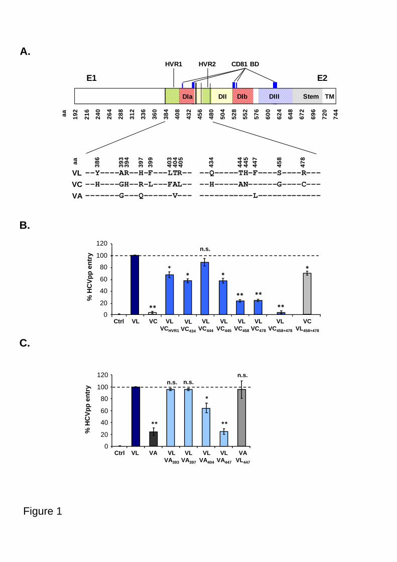
A.
DIa DII TM
E1 E2HVR1 HVR2 CD81 BD
DIIIDIb Stem
--Y----AR--H-F---LTR-- --Q-----TH-F----S----R---VL
386
393
394
397
399
403
404
405
434
445
447
458
478
444aa
192
216
240
264
288
312
336
360
384
408
432
456
480
504
528
552
576
600
624
648
672
696
720
744aa
B.120
--H----GH--R-L---FAL--
-------G---Q------V---
--H-----AN------G----C---
-----------L-------------VAVC
% H
CVp
p en
try
0
20
40
60
80
100
**
*
** **
**
**
n.s.
*
C.
n.s.120n.s. n.s.
Ctrl0
VLVC434
VL VC VLVC444
VLVC445
VLVC458
VLVC478
VLVC458+478
VCVL458+478
VLVCHVR1
% H
CVp
p en
try
****
Ctrl0
20
40
60
80
100
VL VA VL VL VL VL VA
n.s.
*
n.s.
Figure 1
Ctrl VL VA VLVA393
VLVA397
VLVA404
VLVA447
VAVL447

A. B.
C.
Fo
ld o
f o
vere
xpre
ssio
n
0
5
10
15
20
25
30
35
40
45C
trl
1
OC
LN
39.8
CL
DN
1
6.6
CD
81
19.7
SR
-BI
1.6
OCLNCLDN1CD81SR-BI
Fo
ld o
f o
vere
xpre
ssio
n
n.s. n.s. n.s.
0123456789
1011
Fo
ld in
crea
se o
f H
CV
pp
en
try
2
2.5
33.5
**
**
****n.s.
**
**
Figure 2
Fo
ld in
crea
se o
f H
CV
pp
en
try
0
0.5
11.5
2
Ctrl
VL
VC
458+
478
VC
VL
458+
478
VL VC
SR-BI
VL
VC
458+
478
VC
VL
458+
478
VL
VC
CD81
VL
VC
458+
478
VC
VL
458+
478
VL
VC
CLDN1
VL
VC
458+
478
VC
VL
458+
478
VL
VC
OCLN
VL
VC
458+
478
VC
VL
458+
478
VL
VC
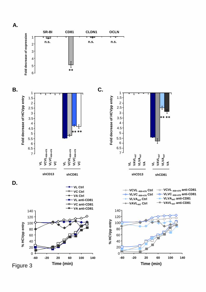
A.
B. C.
Fo
ld d
ecre
ase
of
HC
Vp
p e
ntr
y
6
5
4
3
2
1
**
1.5
2.5
3.5
4.5
5.5
**
5
4
3
2
1
OCLNCLDN1CD81SR-BI
Fo
ld d
ecre
ase
of
exp
ress
ion
**6
n.s. n.s. n.s.
Fo
ld d
ecre
ase
of
HC
Vp
p e
ntr
y
6
5
4
3
2
1
** **
1.5
2.5
3.5
4.5
5.5
Figure 3
D.
shCD81
VL
VC
VL
458+
478
VC
VL
VC
458+
478
VL
VC
VL
458+
478
VC
shCD13
VL
VC
458+
478
7
6.5
VL
VA
VL
447
VA
shCD13
VL
VA
447
shCD81
VL
VA
VL
447
VA
VL
VA
4477
6.5
0
20
40
60
80
100
120
140
-60 -20 20 60 100 140
% H
CV
pp
en
try
VL CtrlVC Ctrl
VL anti-CD81VC anti-CD81
VA Ctrl
VA anti-CD81
Time (min) Time (min)
0
20
40
60
80
100
120
140
-60 -20 20 60 100 140
% H
CV
pp
en
try
VCVL 458+478 CtrlVLVC 458+478 Ctrl
VCVL 458+478 anti-CD81VLVC 458+478 anti-CD81
VLVA447 Ctrl VLVA447 anti-CD81
VAVL447 Ctrl VAVL447 anti-CD81

A.
B.
++
Neu
tral
izat
ion
tite
r(1
/dilu
tio
n)
10
102
103
104
Autologous serumCtrl
VL VLVC434
VL VLVC444
VLVC445
VLVC458
VLVC478
VLVC458+478
**
**
*
n.s. n.s. n.s.
VC VCVL458+478
**
n.s.
B.
Figure 4
++
Neu
tral
izat
ion
tit
er
(1/d
iluti
on
)
10
102
103
104
Autologous serumCtrl
VL VL VLVA393
VLVA397
VLVA404
VLVA447
VAVL447
*
**
n.s. n.s.
VA
n.s.
**

A.
B.
% H
CV
pp
en
try
CBH-23
VL
VC
458+
478
VC
VL
458+
478
VL
VC
HC-1
VL
VC
458+
478
VC
VL
458+
478
VL
VC
AP33
VL
VC
458+
478
VC
VL
458+
478
VL
VC
0
20
40
60
80
100
120
VL
VC
458+
478
VC
VL
458+
478
VL
VC
VL
VC
458+
478
VC
VL
458+
478
VL
VC
CBH-5
VL
VC
458+
478
VC
VL
458+
478
VL
VC
**** **
**
****
**
**
VL
VA
447
VA
VL
447
VL
VA
% H
CV
pp
en
try
0
20
40
60
80
100
120
VL
VA
447
VA
VL
447
VL
VA
Ctrl CBH-2 CBH-5 CBH-23 HC-1 AP33
VL
VA
447
VA
VL
447
VL
VA
VL
VA
447
VA
VL
447
VL
VA
VL
VA
447
VA
VL
447
VL
VA
VL
VA
447
VA
VL
447
VL
VA
****
**
**
**
**
** *
Ctrl CBH-2
Figure 5
C.
D.
Ctrl CBH-2 CBH-5 CBH-23 HC-1 AP33
% H
CV
pp
en
try
VCVL458+478 CtrlVLVC458+478 CtrlVCVL458+478 CBH-23VLVC458+478 CBH-23VCVL458+478 anti-CD81
Time (min)
-60 -20 20 60 100 1400
20
40
60
80
100
120
140
% H
CV
pp
en
try
Time (min)
0
20
40
60
80
100
120
-60 -20 20 60 100 140
140
VAVL ctrlVLVA447 ctrlVAVL447 CBH-23VLVA 447CBH-23VAVL447 anti CD81
% H
CV
pp
en
try
140
Time (min)
0
20
40
60
80
100
120
-60 -20 20 60 100 140
VL ctrlVA ctrlVL CBH-23VA CBH-23VL anti CD81
VL CtrlVC CtrlVL CBH-23VC CBH-23VL anti-CD81
Time (min)
-60 -20 20 60 100 140
% H
CV
pp
en
try
0
20
40
60
80
100
120
140

Neu
tral
izat
ion
tit
er (
1/d
iluti
on
)
103
104
P < .01
P < .0001
P < .001
P = .01
Neu
tral
izat
ion
tit
er (
1/d
iluti
on
)
VLVC458+478VL VC VA10
102
VLVA447
Figure 6

A.
C. D.
Fo
ld d
ecre
ase
of H
CV
cc in
fect
ion
54.54
3.53
2.52
1.51
VL
VL
VC
458
+478
VL
VA
447
shCD13
VL
VL
VC
458
+478
VL
VA
447
shCD81
**
*
Lo
g10
TC
ID50
/ ml
1
2
3
4
5
VL
VL
VA
447
VL
VC
458
+478
Ctr
l
B.
HC
Vcc
infe
ctio
n (%
)
VL
VC
458
+478
VL
VA
447
Ctrl
VL
0
20
40
60
80
100
120
VL
VC
458
+478
VL
VA
447
CBH-23
VL
VL
VC
458
+478
VL
VA
447
AP33
VL
****
Figure 7
Neu
tral
izat
ion
tite
r (1
/dilu
tio
n)
VLVC458+478VL10
102
103
104
VLVA447
p = .001
p < .001

SUPPLEMENTARY DATA
SUPPLEMENTARY MATERIALS AND METHODS Analysis of HCVpp envelope glycoprotein expression. Expression of HCV glycoproteins
was characterized in HEK 293T producer cells and HCVpp purified through a 20% sucrose
cushion ultracentrifugation as described.1 Immunoblots of HCV glycoproteins were
performed using anti-E1 11B7 and anti-E2 AP33 mAbs as described.2
Cellular binding of envelope glycoproteins. Envelope glycoprotein-expressing HEK 293T
cells were lysed in PBS by four freezing and thawing cycles. Cell debris and nuclei were
removed by low-speed centrifugation and supernatants containing native intracellular E1E2
complexes were used for binding studies. shCD81- or shCD13-Huh7.5 cells (2 x 10⁵ cells
per well) were seeded in 96 well plates. Following incubation with lysates containing patient-
derived E1E2 proteins, Huh7.5.1 target cells were first incubated with mAb AP33 (10 µg/ml)
and then with phycoerythrin-conjugated anti-mouse Ab (5 µg/ml, BD). Bound E2 was
analyzed by flow cytometry as described.3
Construction of plasmids for production of chimeric HCVcc expressing patient-derived envelopes. Genotype 1 JFH-based HCVcc chimeras expressing the structural
proteins of patient-derived viruses were produced as previously described for Con1/C3-
JFH1-V2440L.4, 5 Briefly, the cDNA region encoding for the HCV core to first
transmembrane domain of NS2 (C3 junction site) from variant VL was inserted into pFK-
Con1/C3-JFH1-V2440L using fusion polymerase chain reaction (PCR) with Pfu DNA
polymerase (Stratagene) and standard cloning procedures using appropriate restriction sites
including BsmI and AvrII. The obtained construct was designated VL/JFH1. VL/JFH1
encoding sequence was used as template to insert individual and combined mutations using
the QuikChange II XL site-directed as described previously. 1
GNA Capture ELISA. Binding of HMAb CBH-23 to viral envelopes was analyzed using an
ELISA with HCVpp as a capture antigen as described.6 HCVpp expressing the E1E2
glycoproteins of HCV variants or control (Ctrl) pseudoparticles with absent HCV envelope
glycoprotein expression were partially purified and enriched through ultracentrifugation as
described.1 Purified particles were quantified as described before.1 Partially purified HCVpp
or control pseudoparticles were captured onto GNA-coated microtiter plates as described.6
Soluble E2 (sE2, derived from strain HCV-H77 and expressed in 293T cells as described

previously3) was used as a positive control for antibody binding. Neutralizing human anti-E2
antibody CBH-23 (25 µg/ml diluted in PBS) was then added to captured HCVpp or sE2 (1 h
at RT). Following washing and removal of nonbound antibody, mAb binding to HCV
envelopes was detected using horseradish peroxidase anti-human IgG (GEhealthcare) at a
concentration of 1/3000 for 1 h at RT, followed by incubation with 1-stepTM Turbo TMB-
ELISA (Thermo Scientific) for color development. Absorbance was measured at 450 nm
using a microplate reader (Molecular Devices) and the Softmax program.
Bioinformatics. Multiple sequence alignment of complete E2 proteins was performed using
the European HCV databases (http://euhcvdb.ibcp.fr).7 Two amino-acid repertoires were
computed with all E2 sequences of provisional/confirmed genotype 1b using the
ComputeRepertoire tool as part of the euHCVdb Extract tool.

SUPPLEMENTARY RESULTS Prevalence of the identified mutations in a large genomic database of viral isolates.
Bioinformatic sequence analysis of a large panel of 2,074 HCV strains within the European
HCV database further supports the potential relevance of the identified positions for
pathogenesis of HCV infection in general.7 Residues F, S and R are much more frequently
observed at positions 447, 458 and 478 than L, G and C. F and S are the most predominant
residues at positions 447 and 458 in the large majority of 1b strains, respectively (F447 all:
98.4%, 1b: 96.2%; S458 all: 94% for 1b: 90.3%; Fig. S5). The position 478 is variable but R
(all: 2.4% for 1b: 10.8%) is more frequent than C (all: 0.2%, 1b: 0.9%) (Fig. S5). The high
prevalence of identified residues supports their functional relevance for virus survival and
selection as more structurally and functionally relevant residues will be more frequently
observed. These data suggest that the epitope containing the identified residues at positions
447, 458 and 478 is not only responsible for viral evasion from autologous antiviral
antibodies during LT but may also contribute to viral evasion in chronic HCV infection in
general.
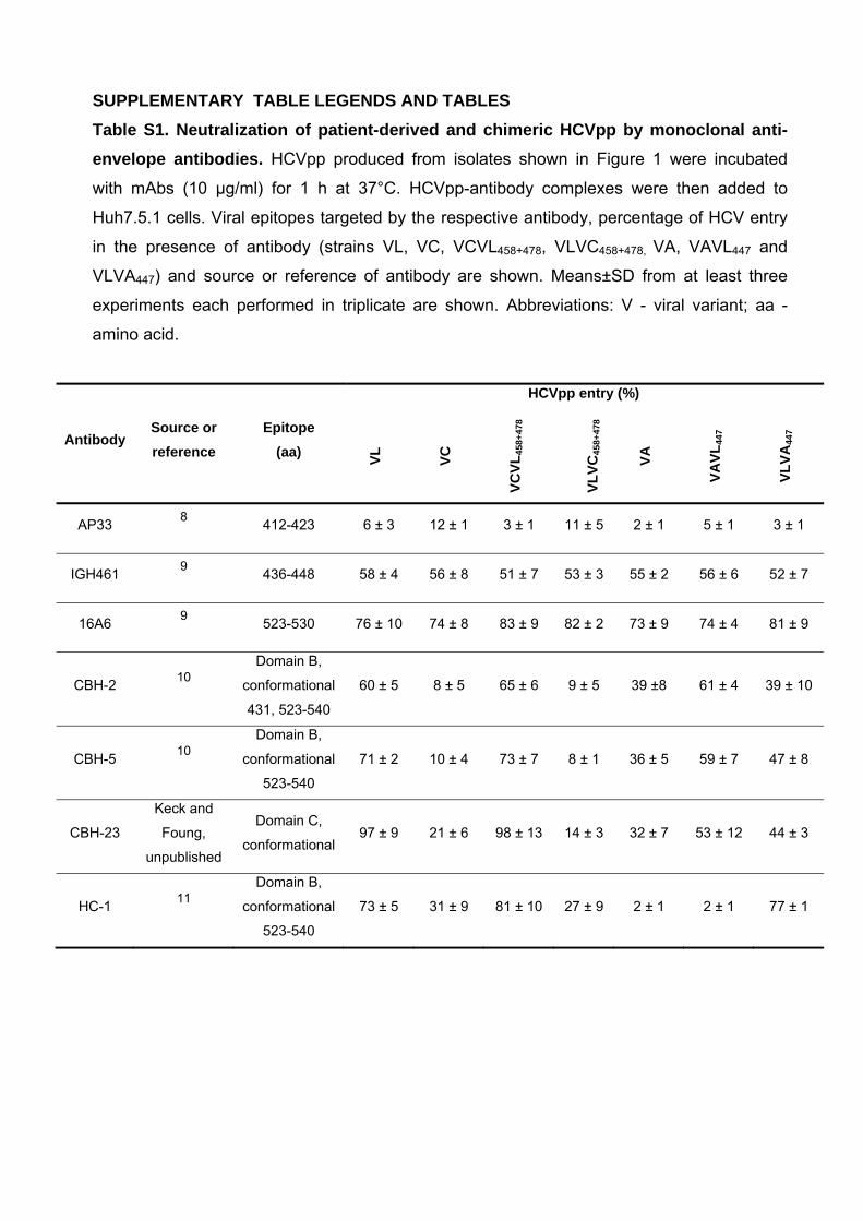
SUPPLEMENTARY TABLE LEGENDS AND TABLES Table S1. Neutralization of patient-derived and chimeric HCVpp by monoclonal anti-envelope antibodies. HCVpp produced from isolates shown in Figure 1 were incubated
with mAbs (10 µg/ml) for 1 h at 37°C. HCVpp-antibody complexes were then added to
Huh7.5.1 cells. Viral epitopes targeted by the respective antibody, percentage of HCV entry
in the presence of antibody (strains VL, VC, VCVL458+478, VLVC458+478, VA, VAVL447 and
VLVA447) and source or reference of antibody are shown. Means±SD from at least three
experiments each performed in triplicate are shown. Abbreviations: V - viral variant; aa -
amino acid.
Antibody Source or reference
Epitope
(aa)
HCVpp entry (%) VL
VC
VCVL
458+
478
VLVC
458+
478
VA
VAVL
447
VLVA
447
AP33 8 412-423 6 ± 3 12 ± 1 3 ± 1 11 ± 5 2 ± 1 5 ± 1 3 ± 1
IGH461 9 436-448 58 ± 4 56 ± 8 51 ± 7 53 ± 3 55 ± 2 56 ± 6 52 ± 7
16A6 9 523-530 76 ± 10 74 ± 8 83 ± 9 82 ± 2 73 ± 9 74 ± 4 81 ± 9
CBH-2 10 Domain B,
conformational
431, 523-540
60 ± 5 8 ± 5 65 ± 6 9 ± 5 39 ±8 61 ± 4 39 ± 10
CBH-5 10 Domain B,
conformational
523-540
71 ± 2 10 ± 4 73 ± 7 8 ± 1 36 ± 5 59 ± 7 47 ± 8
CBH-23
Keck and
Foung,
unpublished
Domain C,
conformational 97 ± 9 21 ± 6 98 ± 13 14 ± 3 32 ± 7 53 ± 12 44 ± 3
HC-1 11 Domain B,
conformational
523-540
73 ± 5 31 ± 9 81 ± 10 27 ± 9 2 ± 1 2 ± 1 77 ± 1

Table S2. Characteristics of patients and viruses used for neutralization studies. (A) HCVpp were incubated with anti-HCV positive sera from 102 patients with chronic HCV
infection (ClinicalTrial.gov Identifier NCT00638144). Patient number, age, gender, viral
genotype and load in serum are indicated. HCVpp- antibody complexes were added to
Huh7.5.1 cells and infection was analyzed as described in Fig. 4. Calculation of
neutralization and determination of background and thresholds for neutralization were
performed as described in Fig. 6. Neutralization titers obtained by endpoint dilution are
indicated for each variant. (B) Results were confirmed using chimeric HCVcc expressing the
HCV envelope glycoproteins depicted in Fig. 7 and using 12 representative sera from
patients. Neutralization assays were performed using a similar protocol as described in (A).
Means from at least three independent experiments each performed in triplicate are shown.
Abbreviations: V - viral variant ; M - male ; F - female.
A. Patient number
Age Gender Genotype Viral Load
(IU/mL) HCVpp neutralization titer (1/dilution)
VL VC VA
1 65 M 1b 2.29 x 10⁵ 100 100 100
2 27 F 1b 9.7 x 10⁴ 100 3200 200
3 31 F 1b 1.53 x 10⁵ 400 3200 400
4 47 M 3a 1.02 x 10⁶ 20 20 100
5 58 M 1b 1.15 x 10⁶ 100 3200 200
6 72 M 1b 1.50 x 10⁶ 20 200 100
7 51 M 4 4.38 x 10⁶ 20 20 20
8 69 F 1b 9.7 x 10⁵ 20 400 100
9 36 F 1 1.29 x 10⁵ 800 1600 100
10 46 M 1a 1.05 x 10⁶ 100 800 100
11 55 M 1a 1.54 x 10⁶ 400 3200 200
12 56 M 4c/4d 2.41 x 10⁴ 20 800 200
13 56 F 4a 1.09 x 10⁶ 100 400 400
14 59 F 1b 3.54 x 10⁵ 200 800 200
15 62 M 1a 3.37 x 10⁶ 20 20 20
16 50 M 4a 1.48 x 10⁶ 20 200 20
17 46 M 4a 4 x 10⁵ 20 200 100
18 70 F 1b 1.3 x 10⁶ 100 800 20
19 77 F 1b 6.2 x 10⁴ 20 100 100
20 61 F 1b 2.58 x 10⁴ 200 800 200
21 46 F 1b 2.11 x 10⁵ 100 400 800
22 36 M 1a 2.04 x 10⁶ 20 200 400
23 52 F 4a 9.12 x 10⁵ 20 3200 400

24 54 M 1a 9.77 x 10⁵ 100 800 200
25 54 M 1b 1.12 x 10⁶ 20 100 200
26 54 F 1a 3.38 x 10⁶ 20 400 20
27 47 M 3a 6.16 x 10⁵ 100 3200 3200
28 43 M 1a 5.75 x 10⁶ 20 800 200
29 51 M 4a 1.44 x 10⁶ 100 400 400
30 54 M 2c 4.67 x 10⁵ 100 100 3200
31 51 M 1a 6.16 x 10⁶ 100 400 100
32 39 M 4a 1.12 x 10⁶ 20 200 800
33 62 F 4f 2.88 x 10⁶ 20 800 20
34 46 M 4k 3.54 x 10⁵ 20 20 100
35 42 M 1a 9.54 x 10⁵ 400 800 400
36 54 M 2c 4.67 x 10⁵ 200 3200 100
37 34 M 3a 3.23 x 10⁶ 20 20 100
38 47 M 3a 7.94 x 10⁴ 20 400 20
39 30 F 1b 1.00 x 10⁶ 20 200 400
40 47 F 1b 2.29 x 10⁶ 100 400 200
41 52 M 1a 1.73 x 10⁶ 200 3200 400
42 34 M 1b 1.45 x 10⁶ 3200 3200 200
43 46 M 1a 4.34 x 10⁶ 200 800 400
44 66 F 1b 3.89 x 10⁵ 200 1600 200
45 29 F 1a 1.08 x 10⁵ 400 400 200
46 45 M 3a 2.78 x 10⁵ 20 200 200
47 65 F 4f 1.46 x 10⁶ 20 3200 20
48 55 M 1a 8.81 x 10⁶ 20 800 100
49 53 M 1a 1.15 x 10⁶ 100 100 100
50 40 M 3a 2.46 x 10⁶ 100 3200 200
51 48 F 1a 1.00 x 10⁵ 20 800 20
52 37 M 1a 5.08 x 10⁶ 20 400 200
53 47 M 3a 6.8 x 10⁶ 100 1600 400
54 37 M 1a 1.84 x 10⁶ 800 800 200
55 65 F 1b 2.18 x 10⁶ 100 100 800
56 45 F 1a 3.93 x 10⁶ 1600 1600 400
57 49 M 4a 2.06 x 10⁶ 800 3200 200
58 30 M 1b 7.21 x 10⁵ 100 800 200
59 31 M 3a 6.66 x 10⁶ 100 200 200
60 37 M 1a 6.70 x 10⁶ 20 100 100
61 49 M 1a 3.16 x 10⁵ 20 800 20
62 43 M 1 6.83 x 10⁵ 20 20 20
63 69 M 1b 4.7 x 10⁵ 20 20 200
64 48 M 1a 3.28 x 10⁶ 20 3200 100
65 46 M 3a 8.55 x 10⁵ 20 800 100

66 51 M 1b 1.07 x 10⁶ 20 200 1600
67 43 M 1b 4.27 x 10⁵ 20 100 800
68 36 M 3a 1.14 x 10⁶ 20 800 20
69 53 F 1b 3.06 x 10⁵ 20 400 20
70 24 F 3a 1.29 x 10⁶ 20 20 20
71 63 M 1b 3.01 x 10⁶ 100 200 100
72 44 M 1 1.10 x 10⁵ 20 3200 200
73 28 M 3a 1.85 x 10⁶ 20 3200 20
74 54 M 1b 1.29 x 10⁵ 20 3200 20
75 17 F 1b 2.41 x 10⁵ 20 20 200
76 40 M 3a 1.26 x 10⁶ 20 20 100
77 35 M 1b 8.89 x 10⁵ 20 20 800
78 36 F 6a 1.4 x 10⁷ 20 100 400
79 70 F 1b 1.13 x 10⁵ 100 100 400
80 62 M 1a 2.68 x 10⁶ 100 200 20
81 70 M 1b 2.85 x 10⁵ 20 200 3200
82 63 M 1b 1.95 x 10⁵ 200 400 400
83 33 M 1a 1.76 x 10⁶ 100 200 800
84 35 M 1a 2.78 x 10⁶ 20 20 200
85 60 F 1 6.39 x 10⁵ 20 200 100
86 57 M 3a 1.22 x 10⁶ 200 3200 400
87 60 M 1 3.6 x 10⁶ 100 3200 20
88 49 M 4 2.24 x 10⁶ 20 1600 20
89 37 M 4 9.35 x 10⁵ 100 800 100
90 55 M 1a 3.77 x 10⁶ 20 3200 100
91 47 M 1a 2.36 x 10⁶ 20 1600 20
92 72 M 3a 3.83 x 10⁵ 20 400 20
93 79 M 1b 2.81 x 10⁵ 100 1600 100
94 58 F 1b 6.58 x 10⁵ 100 3200 200
95 50 M 3a 6.07 x 10⁵ 20 3200 100
96 67 F 1b 4.13 x 10⁵ 100 800 20
97 49 M 3a 5.22 x 10⁵ 200 400 200
98 53 F 1b 2.31 x 10⁶ 20 400 1600
99 37 M 1a 1.87 x 10⁵ 100 3200 200
100 54 F 4a 9.23 x 10⁵ 20 200 100
101 39 M 1a 1.76 x 10⁵ 100 800 200
102 51 F 2b 1.10 x 10⁶ 100 3200 800

B. Patient number
HCVcc neutralization titer (1/dilution)
VL VLVC458+478 VLVA447
11 400 1600 800
28 20 1600 800
33 20 400 400
35 400 1600 1600
36 200 1600 3200
45 800 1600 800
65 20 1600 1600
66 20 3200 800
68 20 1600 1600
94 100 3200 800
98 100 800 3200
99 100 3200 1600
REFERENCES 1. Fafi-Kremer S, Fofana I, Soulier E, Carolla P, Meuleman P, Leroux-Roels G, Patel
AH, Cosset F-L, Pessaux P, Doffoël M, Wolf P, Stoll-Keller F, Baumert TF. Enhanced
viral entry and escape from antibody-mediated neutralization are key determinants for
hepatitis C virus re-infection in liver transplantation. J Exp Med 2010;207:2019-2031.
2. Pestka JM, Zeisel MB, Blaser E, Schurmann P, Bartosch B, Cosset FL, Patel AH,
Meisel H, Baumert J, Viazov S, Rispeter K, Blum HE, Roggendorf M, Baumert TF.
Rapid induction of virus-neutralizing antibodies and viral clearance in a single-source
outbreak of hepatitis C. Proc Natl Acad Sci U S A 2007;104:6025-30.
3. Krieger SE, Zeisel MB, Davis C, Thumann C, Harris HJ, Schnober EK, Mee C, Soulier
E, Royer C, Lambotin M, Grunert F, Dao Thi VL, Dreux M, Cosset FL, McKeating JA,
Schuster C, Baumert TF. Inhibition of hepatitis C virus infection by anti-claudin-1
antibodies is mediated by neutralization of E2-CD81-claudin-1 associations.
Hepatology 2010;51:1144-57.
4. Pietschmann T, Kaul A, Koutsoudakis G, Shavinskaya A, Kallis S, Steinmann E, Abid
K, Negro F, Dreux M, Cosset FL, Bartenschlager R. Construction and characterization
of infectious intragenotypic and intergenotypic hepatitis C virus chimeras. Proc Natl
Acad Sci U S A 2006;103:7408-13.
5. Kaul A, Woerz I, Meuleman P, Leroux-Roels G, Bartenschlager R. Cell culture
adaptation of hepatitis C virus and in vivo viability of an adapted variant. J Virol
2007;81:13168-79.
6. Wang Y, Keck ZY, Saha A, Xia J, Conrad F, Lou J, Eckart M, Marks JD, Foung SK.
Affinity maturation to improve human monoclonal antibody neutralization potency and
breadth against hepatitis C virus. J Biol Chem 2011;286:44218-33.
7. Combet C, Garnier N, Charavay C, Grando D, Crisan D, Lopez J, Dehne-Garcia A,
Geourjon C, Bettler E, Hulo C, Le Mercier P, Bartenschlager R, Diepolder H,
Moradpour D, Pawlotsky JM, Rice CM, Trepo C, Penin F, Deleage G. euHCVdb: the
European hepatitis C virus database. Nucleic Acids Res 2007;35:D363-6.
8. Owsianka A, Tarr AW, Juttla VS, Lavillette D, Bartosch B, Cosset FL, Ball JK, Patel
AH. Monoclonal antibody AP33 defines a broadly neutralizing epitope on the hepatitis
C virus E2 envelope glycoprotein. J Virol 2005;79:11095-104.
9. Haberstroh A, Schnober EK, Zeisel MB, Carolla P, Barth H, Blum HE, Cosset FL,
Koutsoudakis G, Bartenschlager R, Union A, Depla E, Owsianka A, Patel AH,
Schuster C, Stoll-Keller F, Doffoel M, Dreux M, Baumert TF. Neutralizing host
responses in hepatitis C virus infection target viral entry at postbinding steps and
membrane fusion. Gastroenterology 2008;135:1719-1728.
10. Hadlock KG, Lanford RE, Perkins S, Rowe J, Yang Q, Levy S, Pileri P, Abrignani S,
Foung SK. Human monoclonal antibodies that inhibit binding of hepatitis C virus E2
protein to CD81 and recognize conserved conformational epitopes. J Virol
2000;74:10407-16.
11. Keck ZY, Li SH, Xia J, von Hahn T, Balfe P, McKeating JA, Witteveldt J, Patel AH,
Alter H, Rice CM, Foung SK. Mutations in hepatitis C virus E2 located outside the
CD81 binding sites lead to escape from broadly neutralizing antibodies but
compromise virus infectivity. J Virol 2009;83:6149-60.
12. Dimitrova M, Affolter C, Meyer F, Nguyen I, Richard DG, Schuster C, Bartenschlager
R, Voegel JC, Ogier J, Baumert TF. Sustained delivery of siRNAs targeting viral
infection by cell-degradable multilayered polyelectrolyte films. Proc Natl Acad Sci U S
A 2008;105:16320-5.
13. Lupberger J, Zeisel MB, Xiao F, Thumann C, Fofana I, Zona L, Davis C, Mee CJ,
Turek M, Gorke S, Royer C, B. F, Zahid MN, Lavillette D, J. F, Cosset F-L,
Rothenberg SM, Pietschmann T, Patel AH, Pessaux P, Doffoël M, Raffelsberger W,
Poch O, McKeating JA, Brino L, Baumert TF. EGFR and EphA2 are hepatitis C virus
host entry factors and targets for antiviral therapy. Nat Med 2011;17:589-95.
14. Bartosch B, Vitelli A, Granier C, Goujon C, Dubuisson J, Pascale S, Scarselli E,
Cortese R, Nicosia A, Cosset FL. Cell entry of hepatitis C virus requires a set of co-
receptors that include the CD81 tetraspanin and the SR-B1 scavenger receptor. J Biol
Chem 2003;278:41624-41630.

SUPPLEMENTARY FIGURE LEGENDS
Figure S1. Actual viral infectivity of HCVpp derived from variants VL, VC and VA shown as relative light units of luciferase reporter gene expression. (A-B) Comparative
analysis of viral entry of HCVpp shown in Fig. 1. Results are expressed in relative light units
(RLU) plotted in a logarithmic scale. The threshold for a detectable infection in this system is
indicated by dashed lines. The detection limit for positive luciferase reporter protein
expression was 3 × 10³ RLU/assay, corresponding to the mean±3 SD of background levels,
i.e., luciferase activity of naive noninfected cells or cells infected with pseudotypes without
HCV envelopes.1, 12, 13 Background levels of the assay were determined in each experiment.
Means±SD from at least four independent experiments performed in triplicate are shown.
Significant differences in HCVpp entry VC, VA and VL wildtype and mutant variants are
indicated by stars (*, P ≤ .05; **, P < .001). Abbreviations: Ctrl - control; HVR - hypervariable
region; n.s. - not significant; V - viral variant. Figure S2. Positions 447, 458 and 478 modulate binding of envelope glycoproteins to CD81 expressed at the cell surface. Binding of native E1E2 complexes expressed from
patient-derived cDNAs to Huh7.5 cells with silenced CD81 expression (described in Fig. 3)
was detected by flow cytometry. Results are expressed as percentage of E1E2 binding
compared to shCD13-Huh7.5 control cells. Means±SD from three independent experiments
performed in triplicate are shown. Significant differences in binding between variants are
indicated by stars (**, P < .001).
Figure S3. Differences in viral entry are not due to impaired HCVpp production. (A)
Analysis of envelope glycoprotein expression. Protein expression was analyzed by
immunoblotting as described in Materials and Methods. Molecular markers (kDa) are
indicated on the right. (B) Transfection efficiency during HCVpp production. Transfection
effciency was analyzed for each variant and quantified by determining luciferase expression
in HEK 293T producer cells expressed as normalized percentage compared to control
transfected cells. (C) Envelope glycoprotein expression in HCVpp. HCVpp were purified as
described previously1, 2 and subjected to immunoblot as described in panel (A). (D) Lentiviral
p24 antigen expression was analyzed by ELISA and is indicated as optical density (O.D.)
values at 450 nm. Abbreviations: Da - Dalton; MW - molecular weight; n.s. - not significant.

Figure S4. Binding of neutralizing anti-E2 HMAb CBH-23 to patient derived-envelope glycoproteins expressed on HCVpp as capture antigens in an ELISA. HCVpp
expressing envelope glycoproteins of variants VL, VA, VC, VLVA447 and VLVC458+478 were
used as capture antigens on GNA-coated ELISA plates. Control (Ctrl) pseudoparticles with
absent HCV envelope glycoprotein expression and recombinant soluble E2 (sE2 derived
from strain H77)14 served as negative and positive controls, respectively. Anti-E2 CBH-23
reactivity was detected as described in supplementary Materials and Methods and is
indicated as optical density (O.D.) values at 450 nm. Means±SD from one representative
experiment are shown.
Figure S5. Distribution of residues at positions 447, 458 and 478 of HCV E2 sequences in the European HCV databases. Distribution of residues at positions 447, 458 and 478 for
HCV complete E2 sequences from all subtypes (black) and from subtype 1b only (white)
within the European Hepatitis C Virus databases 7, (http://euhcvdb.ibcp.fr). F and S are the
predominant residue at positions 447 and 458 (F447: 98.4%, 1b: 96.2%; S458 all: 94%, 1b:
90.3%). The position 478 is variable (it belongs to HVR2) but R (all: 2.4% for, 1b: 10.8%) is
more frequent than C (all: 0.2%, 1b: 0.9%).

A.
B.
Ctrl VLVC434
VL VC VLVC444
VLVC445
VLVC458
VLVC478
VLVC458+478
**
*
** **
**
*
VCVL458+478
*
n.s.
3
4
5
6
7
HC
Vp
p e
ntr
y (L
og
10 R
LU
)
VLVCHVR1
*
HC
Vp
p e
ntr
y (L
og
10 R
LU
)
**
n.s.
**
4
5
6
7n.s.n.s.
*
Figure S1
HC
Vp
p e
ntr
y
Ctrl VLVA393
VL VA VLVA397
VLVA404
VLVA447
VAVL447
3

A. B.
120
110
**
**
% E
1E2
bin
din
g
0
10
20
30
40
50
60
70
80
90
100
% E
1E2
bin
din
g**
**
0
10
20
30
40
50
60
70
80
90
100
110
120
Figure S2
VL
shCD81
VC
VA
shCD13
VL
VC
VA
0
shCD81
VC
VL
458+
478
VL
VC
458+
478
VA
VL
447
VL
VA
447
0
shCD13
VC
VL
458+
478
VL
VC
458+
478
VA
VL
447
VL
VA
447
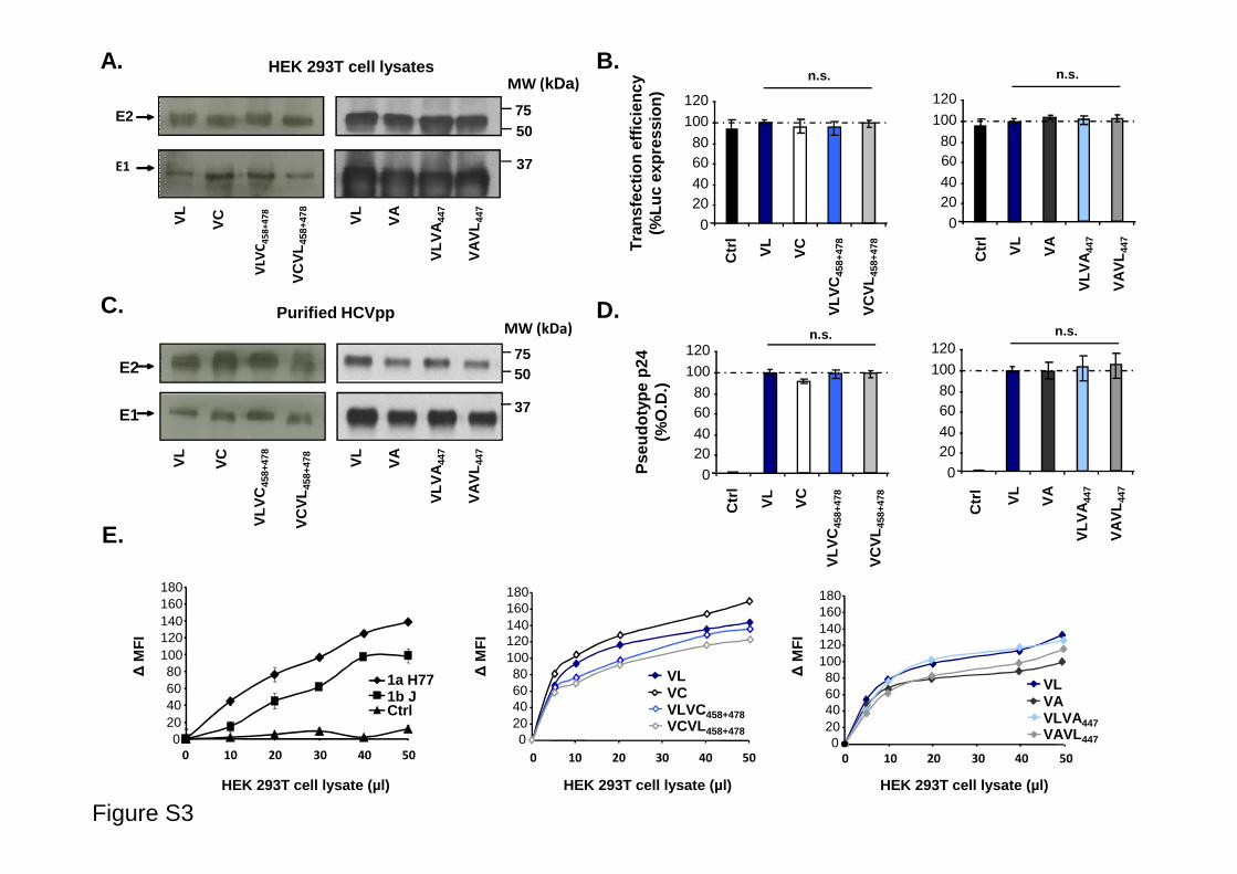
Tra
nsfe
ctio
n ef
ficie
ncy
(%
Luc
expr
essi
on)
B.
Pse
udot
ype
p24
(%
O.D
.)
HEK 293T cell lysates
VLV
C4
58
+4
78
VC
VL
458+
478
VL
VC
7550
MW (kDa)
37
7550
MW (kDa)
37
VLV
A44
7
VA
VL
447
VL
VA
E2
E1
E2
E1
Purified HCVpp
A.
C. D. VLV
C45
8+47
8
VC
VL
458+
478
0
20406080
100120
VL
VC
Ctr
l
n.s .
VLV
A44
7
VA
VL
447
n.s .
0
2040
6080
100
120
VL
VA
Ctr
l
n.s .
40
6080
100
120n.s .
40
6080
100
120
020406080
100120140160180
0 10 20 30 40 50
1a H771b JCtrl
∆M
FI
HEK 293T cell lysate (µl)
Pse
udot
ype
p24
(%
O.D
.)
Figure S3
E.
VL
VC
VLV
C45
8+47
8
VC
VL
458+
478
VL
VA
VLV
A44
7
VA
VL
447
0
2040
VL
VA
Ctr
l
VLV
A44
7
VA
VL
447
VLV
C45
8+47
8
VC
VL
458+
478
0
2040
VL
VC
Ctr
l
∆M
FI
HEK 293T cell lysate (µl)
VLVCVLVC458+478VCVL458+478
020406080
100120140160180
0 10 20 30 40 50
∆M
FI
VLVAVLVA447VAVL 447
0 10 20 30 40 50
020406080
100120140160180
HEK 293T cell lysate (µl)

Figure S4
O.D
. 450
nm
(an
ti-E
2 C
BH
-23)
0
0.1
0.2
0.3
0.4
0.5
0.6
0.7
HCVpp
Ctr
l
VL
VA
VC
H77
VL
VA
447
VL
VC
458+
478
sE2
strain
antigen pp

Figure S5
0
20
40
60
80
100
S G Other R C Other
% o
bse
rved
res
idu
e
Position 458 Position 478
F L Other
Position 447

- 54 -
4. Discussion
HCV entry into hepatocytes is a multistep process involving a variety of receptors such as. HS
proteoglycans, LDL-R, CD81, SR-BI, CLDN1, OCLN and recently described RTKs and NPC1L1
(Albecka et al., 2011; Evans et al., 2007; Lupberger et al., 2011; Pileri et al., 1998; Ploss et al., 2009;
Sainz et al., 2012; Scarselli et al., 2002). However, the role of these receptors in virus binding, entry
into target cells and release of viral particles, is not yet completely understood. Moreover, little is
known about the sequence of events leading to virus internalization.
Different tools including truncated forms of receptors (CD81 large extracellular loop),
molecular mimics (heparin) and neutralizing antibodies have been used to describe the kinetics of
HCV entry. It has been reported that heparin, a homolog of HS, inhibits HCV infection only when
present before or during virus binding suggesting that HS proteoglycans are involved in the initial
attachment of HCV (Barth et al., 2003; Koutsoudakis et al., 2006). Studies have shown that anti-
CD81 antibodies can inhibit HCV infection at postbinding steps which suggest that CD81 acts as
HCV entry coreceptor after the docking of the virus to attachment factors (Cormier et al., 2004b;
Koutsoudakis et al., 2007). The tight junction molecules CLDN1 and OCLN have been described to
be involved in HCV infection (Evans et al., 2007; Ploss et al., 2009) but for the moment, there is no
evidence for their direct binding to the virus (Evans et al., 2007; Krieger et al., 2010). Initial kinetic
studies using anti-Flag antibody and Flag-tagged CLDN1 have suggested that CLDN1 acts late in the
HCV entry process (Evans et al., 2007). Subsequently, studies from our laboratory have shown that
CLDN1, SR-BI and CD81 act at closely related time points in the viral entry process (Krieger et al.,
2010; Zeisel et al., 2007b). Downregulation of both CLDN1 and OCLN resulted in a decrease in HCV
glycoprotein dependent fusion which suggests their possible role in the fusion process (Benedicto et
al., 2009; Evans et al., 2007). Recently, it has been demonstrated that LDL-R may be involved in non-
productive entry of HCV particles and as a lipid providing receptor; it can modulate HCV RNA
replication (Albecka et al., 2011).
SR-BI is a multifunctional molecule that modulate high-density lipoprotein (HDL)
metabolism. SR-BI was initially identified as host factor for HCV binding step (Scarselli et al., 2002).
Later, it has been described that SR-BI also mediates post-binding events during HCV entry
(Haberstroh et al., 2008; Syder et al., 2011; Zeisel et al., 2007b). Interestingly, it has been reported
that HDL which is a physiological ligand of SR-BI, enhances HCVpp entry and HCVcc infection
(Bartosch et al., 2005; Dreux et al., 2006; Voisset et al., 2005). These findings indicate that SR-BI
may play a multifunctional role in HCV infection. Therefore, in the first part of my work, we aimed to
further characterize the role of SR-BI in the HCV entry process.

- 55 -
SR-BI binds a wide variety of lipoproteins and plays an important role in cholesterol
homeostasis. One of its physiological functions is the bilateral cholesterol transfer at the cell
membrane. For cholesteryl ester (CE) uptake, HDL first binds to SR-BI and then CE is transferred
into the cell without internalization of HDL molecule. It has been proposed that the extracellular
domain of SR-BI is crucial for these steps. Recently, it has been reported that amino acid C323 is
highly conserved site in SR-BI and it takes part in HDL binding and SR-BI mediated CE uptake (Guo
et al., 2011). During my PhD, we have characterized novel anti-SR-BI mAbs that do not interfere with
HDL binding which is an indication that our mAbs do not target C323 amino acid and also that these
antibodies do not modulate this physiological function of SR-BI. In addition to SR-BI, there are some
other molecules in human hepatocytes that participate in cholesterol transport. These include LDL-R,
NPC1L1 and CETP (cholesterol ester transfer protein) (Agnello et al., 1999; Barter et al., 2003; Sainz
et al., 2012). CETP mediates the exchange of cholesterol and triglycerides between HDL and LDL
and it is present in human but absent in mice (Barter et al., 2003). Moreover, in contrast to humans, it
has been demonstrated that in mice, SR-BI is the only molecule in hepatic cells which takes part in
selective uptake of CE from HDL (Hoekstra et al., 2010). Noteworthy, SR-BI-knockout mice faces
severe complications like atherosclerosis, adrenal corticoid insufficiency under stress and altered
cholesterol distribution in platelets whereas there is no evidence that impaired SR-BI function affects
human physiology. This may be due to the presence of alternative routes for cholesterol metabolism
in human.
SR-BI is expressed at the sinusoidal surface of hepatocytes (Reynolds et al., 2008), which
appears as physiological route of viral access. The interaction between SR-BI and sE2 was found to
be specific as closely related human scavenger receptor CD36 and mouse SR-BI were not able to bind
to sE2 (Bartosch et al., 2003c; Scarselli et al., 2002). Further, it has been demonstrated that HVR1 of
E2 envelope glycoprotein plays a crucial role in the functional interaction between SR-BI and E2
(Bartosch et al., 2005). The deletion of this region results in reduction of E2 binding to SR-BI and SR-
BI mediated cell entry (Bartosch et al., 2003c; Scarselli et al., 2002). Moreover, HDL enhances HCV
infectivity and there exists a complex interplay between SR-BI, HDL and HVR1 of HCV envelope
glycoprotein E2 (Bartosch et al., 2005; Dreux and Cosset, 2007; Voisset et al., 2005). SR-BI binding
to sE2 has been described to be hampered by polyclonal sera, monoclonal antibodies and small
molecules targeting SR-BI which resulted in decrease in HCV infection (Barth et al., 2005a; Catanese
et al., 2007; Syder et al., 2011). Catanese and colleagues described a panel of monoclonal antibodies
directed against human SR-BI. Among these monoclonal antibodies, C167 inhibited the interaction of
sE2 with SR-BI and HCVcc infection during attachment steps but did not affect the post-binding
function of SR-BI during HCV entry (Catanese et al., 2010; Catanese et al., 2007). This antibody also
blocked HDL binding to SR-BI and hampered the SR-BI mediated cholesterol efflux. A codon-

- 56 -
optimized version of this monoclonal antibody ( mAb 16-71) has been reported to inhibit HCV
infection in vitro and in vivo by interfering with direct cell-to-cell transmission (Meuleman et al.,
2012). Noteworthy, our anti-SR-BI mAbs are novel in their functions as these antibodies unlike
previously described antibodies do not block binding of sE2 to SR-BI. In fact, they inhibit entry of
HCV only during post-binding steps of cell-free infection and cell-to-cell transmission. This indicates
that E2 and SR-BI binding is not the sole function of SR-BI during HCV entry and that the post-
binding function of SR-BI may play a crucial role for HCV infection.
Recently, it has been demonstrated that SR-BI is involved in HCV cell-to-cell transmission
which plays a major role in viral dissemination (Brimacombe et al., 2011; Meuleman et al., 2012;
Syder et al., 2011). Our novel mAbs specifically inhibiting post-binding function of SR-BI enabled us
to understand the importance of the SR-BI post-binding function in cell-free HCV entry and in
neutralizing antibody-resistant cell-to-cell transmission. Taking into consideration the findings that
our mAbs do not hamper sE2 and HDL binding to SR-BI and inhibit HCV entry only during post-
binding steps, it comes into view that our mAbs target different epitope(s) than those targeted by other
anti-SR-BI mAbs. Thus, our novel mAbs will open the way to further characterize the post-binding
function of SR-BI in HCV entry. Moreover, these mAbs also inhibit entry of patient-derived HCVpp
of escape variants selected during liver transplantation. It is worth mentioning that the combination of
anti-SR-BI and anti-HCV envelope antibodies resulted in a synergistic effect on inhibition of escape
variants entry and HCVcc infection. Importantly, these combinations allow to reduce the IC50 of anti-
SR-BI mAb by up to 100-fold. These data suggest an attractive antiviral approach against HCV
infection by targeting the post-binding function of SR-BI.
Two previous studies using HCVcc, demonstrated a cooperation of CD81 and SR-BI in HCV
infection (Kapadia et al., 2007; Zeisel et al., 2007b). It has been reported that all cells showing
permissivity to HCVpp co-express CD81 and SR-BI (Heo et al., 2006). HCV envelope glycoproteins
play a critical role in virus attachment and entry into host cells and it was reported that soluble forms
of CD81 and SR-BI could have indirect link with each other by direct association with HCV envelope
glycoprotein E2 (Heo et al., 2006). CD81 belongs to the tetraspanin family consisting of four
transmembrane domains, short intracellular N and C terminals, a small extracellular loop (SEL) and a
large extracellular loop (LEL) (Levy et al., 1998). It has been shown that the LEL plays a critical role
in CD81-sE2 binding (Flint et al., 1999; Flint et al., 2006; Pileri et al., 1998). Molina and colleague
showed that CD81 is compulsory for HCV infection of cultured primary human hepatocytes (Molina
et al., 2008). Moreover, CD81 is also considered to be indispensible HCV receptor for in vivo HCV
infection (Meuleman et al., 2008). CD81 may not only contribute to initial virus binding to cellular
surface but also regulates the endogenous cellular responses that assist the virus at different stages of
its life cycle e.g. CD81-mediated signals result in Rho GTPase-dependent actin rearrangement may

- 57 -
help the lateral movement of the CD81-E2 complex (Brazzoli et al., 2008; Farquhar et al., 2012).
Further, it was demonstrated that the Raf/MEK/ERK signalling pathway is also activated by CD81
engagement (Brazzoli et al., 2008). Tetraspanin family members have the ability to interact with each
other and with other transmembrane proteins to form tetraspanin-enriched microdomains (TEM).
While association of CD81 with TEM is important for Plasmodium infection, it has been suggested
that CD81 linked with TEM is not essential for early events of HCV infection (Rocha-Perugini et al.,
2009). Recently, another function of CD81 in the HCV life cycle has been demonstrated; it also plays
a critical role in HCV replication (Zhang et al., 2010). The sequence in which cell receptors interact
with HCV and the mechanism by which HCV glycoproteins are activated to mediate membrane
fusion are still unknown. It has been described that CD81 primes HCV for low pH-dependent fusion
during early steps of HCV entry (Sharma et al., 2011). However, CD81 is not the sole determinant of
HCV infection as transgenic mice expressing human CD81 failed to support HCV infection (Dorner
et al., 2011; Flint et al., 1999; Higginbottom et al., 2000; Ploss et al., 2009). Furthermore, many cell
lines expressing both SR-BI and CD81 remained unable to support HCV infection which suggests that
additional host factors such as OCLN, CLDN1, RTKs and NPC1L1 are required for HCV entry.
HCV, after using host factors for entry and fusion, replicates and infects neighbouring cells.
The persistence of infection causes chronic hepatitis which leads to cirrhosis and HCC. Due to
absence of potent therapy, LT is the only remedy for the patients affected by cirrhosis and HCC
related to HCV. Re-infection of the liver graft with HCV is a major problem in patients infected with
HCV. Our lab has previously demonstrated that HCV variants re-infecting the liver graft were
characterized by efficient entry and poor neutralization by antibodies present in pre-transplant serum
compared to variants not detected after transplantation (Fafi-Kremer et al., 2010). However, the
molecular mechanisms by which the virus evades host immunity to persistently re-infect the liver
graft are unknown. As described above, HCV uses different host factors for entry into host cells, so in
second part of my PhD, we aimed to asses the role of host factors in efficient HCV entry and evasion
from neutralizing antibodies.
During LT, the new liver is re-infected by HCV in the early hours of reperfusion and only a
fraction of HCV variants circulating prior to transplant is selected and persists after transplantation. In
chronic infection, the composition of quasispecies is gradually changing and it has been observed that
highly neutralized variants and variants resistant to humoral immune response co-exist. Our lab has
previously demonstrated that the change of the host environment during LT results in an abrupt
change in the composition of HCV quasispecies (Fafi-Kremer et al., 2010). Phylogenetic analysis
revealed a reduction in genetic diversity after LT with the selection of a relatively homogeneous
fraction of variants. It appears that the implantation of a new liver and the start of immunosuppressive
therapy results in a bottleneck effect by selecting the variants which are able to efficiently penetrate
- 58 -
the new liver cells. Viral entry is a key aspect of rapid initiation of HCV infection and neutralizing
antibodies are the first line host defense in transplanted liver. Study of patient-derived viral particles
has revealed that viral variants selected during LT were characterized by enhanced viral entry and
escape from neutralizing antibodies as compared to those variants which were undetectable after
transplantation (Fafi-Kremer et al., 2010). This shows that entry and escape from antibody-mediated
neutralization are key determinants for the selection of viral variants in the early steps of LT. HCV
infection in chimeric uPA-SCID mouse model has demonstrated that the variants which were selected
after LT were the most prevalent variant in both plasma and liver of infected uPA-SCID mouse (Fafi-
Kremer et al., 2010).
To elucidate the mechanism of enhanced viral entry and escape from neutralizing antibodies,
we studied genetically close variants derived from a well characterized patient undergoing LT. The
selected variant VL showed high-infectivity and escape from neutralizing antibodies while non-
selected variants (VA and VC) had lower infectivity and they were highly sensitive to neutralization
by autologous serum. To investigate the molecular mechanisms of enhanced entry of VL, mutations
of the envelope glycoprotein region F447, S458G and R478C of the non-selected variants VA and VC
were introduced into infectious HCV pseudoparticles (HCVpp) expressing envelope glycoproteins of
the escape variant VL. The results show that these mutations play a critical role for high infectivity
and escape from neutralizing antibodies of the selected variant VL.
We further studied whether the mutations F447, S458G and R478C have an effect on viral
entry due to different usage of host cell factors including CD81, SR-BI, CLDN1 and OCLN. Using
HCVpp derived from parental and chimeric strains, we showed that overexperssion of CD81 on
Huh7.5.1 cells markedly increased viral entry of VL as compared to VC. The pattern of enhancement
of viral entry was similar when residues at position 458 and 478 were exchanged between VL and
VC. These results indicate that the double mutation in the viral strain modifies the ability of the virus
to enter into target cells by changing the CD81-usage. Moreover, SR-BI overexpression also showed a
tendency to enhance the viral entry of parental strains but there was no effect of altered expression of
SR-BI on chimeric strains. These data suggest the importance of SR-BI as an entry factor for patient-
derived variants, but also demonstrate that positions 458 and 478 do not significantly alter SR-BI-
dependency. Therefore, other mutations might be involved in the increased entry efficiency of the VL
variant in SR-BI-overexpressing cells e. g. in HVR1. In addition, cells overexpressing CLDN1 and
OCLN illustrated a mild increase in viral entry and modulations of chimeric strains were non-specific
suggesting a significant role of CD81 in enhancement of viral entry. These data are in line with
another study demonstrating absence of increase of HCV entry after CLDN1 overexpression
(Schwarz et al., 2009).
- 59 -
The silencing of CD81 expression further uncovered the importance of CD81 usage. The entry
of the selected variant and related chimeric strains was highly affected in CD81 silenced cells as
compared to non-selected variants and chimeric strains. It has been demonstrated that there are three
subdomains of envelope glycoprotein E2, domain I contains CD81 binding regions while potential
CD81 binding sites overlaps with domain III (Boo et al., 2012; Drummer et al., 2006; Krey et al.,
2010; Owsianka et al., 2006). The residues 447, 458 and 478 are located close to previously described
CD81 binding domains. These findings suggest that acquired mutations at position 447, 458 and 478
play a critical role in E2-CD81 interaction and the interaction between the virus and CD81 is an
important factor in the selection and escape from neutralizing antibodies of viral variants during liver
transplantation. Taken together, our data indicate that mutations F447, S458G and R478C modulate
CD81 dependency; they increase affinity to cell surface CD81 and result in viral escape at post-
binding steps closely related to HCV-CD81 interaction. We can assume the possibility of some other
mechanisms that may contribute to the high entry of HCV particles and their escape from neutralizing
antibodies. These may include some uncovered entry factors or physico-chemical properties of HCV
like association of HCV with lipoproteins. Further understanding of HCV life cycle will help to
develop a better antiviral strategy.
Viral attachment and entry are important targets of host cellular defenses and neutralizing
antibodies against HCV (Haberstroh et al., 2008). Our work highlights that virus-host factor
interactions play a key role in evasion from neutralizing antibodies. This suggests that targeting the
virus and/or host factors represents a promising approach to develop novel antiviral strategies against
HCV infection. Targeting the virus to control HCV infection would be advantageous because there
will be less side effects. However, it will be a challenge to develop neutralizing antibodies capable of
targeting epitopes conserved across genotypes as the virus rapidly adapts to its environment and
constantly escapes the host’s immune responses. Some nAbs have been shown to inhibit HCV cell-to-
cell transmission (Brimacombe et al., 2011) and cross-neutralizing ability of these antibodies would
make them an interesting antiviral strategy. Indeed, the rapid emergence of mutants resistant to
autologous neutralizing antibodies has been reported (Gal-Tanamy et al., 2008; von Hahn et al.,
2007). Thus targeting host entry factors which are essential for viral spread and are less subject to
mutation will represent another exciting way in the development of novel antiviral strategies against
HCV infection. Finally, the synergistic effect between antivirals targeting host factors and nAbs
represents another interesting combination to prevent HCV re-infection and/or chronic HCV
infection.
- 60 -
5. Conclusions and perspectives
In this study, we have described the impact of host cell receptors, particularly SR-BI and CD81 on
initiation of HCV infection, viral dissemination as well as on the mechanisms involved in the re-
infection of liver graft. In the first part of my work, we characterized the role of SR-BI during HCV
infection. Our new anti-SR-BI monoclonal antibodies allowed us to distinguish between the binding
and post-binding function of SR-BI. In addition, we demonstrated that the SR-BI post-binding
function plays a critical role in both cell-free infection of HCV and cell-to-cell transmission of HCV.
In contrast to previously described anti-SR-BI monoclonal antibodies, our data showed that these
novel antibodies have less effects on physiological functions of SR-BI i.e. (i) HDL binding and (ii)
bidirectional cholesterol transfer and no effect on E2 binding which suggest that these antibodies
target distinct epitopes and represent a novel class of anti-SR-BI antibodies.
In the second part of my thesis, we characterized the molecular mechanisms involved in the
selection of variants after liver transplantation. We have identified three residues in the region 425-
483 of E2 glycoprotein responsible for the selection of the escape variant re-infecting the liver graft.
These three residues i.e. F447, S458G and R478C play a crucial role in the interaction of HCV and
host cell factor CD81. These results provide a deeper understanding of the molecular mechanisms of
viral escape during acute infection of the liver transplant and highlight the importance of the
interaction between virus and host factors for this process.
The dependence of viral variants on host entry factors during liver transplantation opens new
therapeutic prospects for the large number of individuals infected with HCV. Therapeutic options for
HCV-infected individuals are still limited by drug resistance and adverse effects. Furthermore, to date
there is no therapy available to prevent re-infection of HCV liver graft. Host entry factors are
interesting targets for antiviral therapy against HCV infection as they may enhance the genetic barrier
for antiviral resistance. In the near future, the optimal treatment for HCV is likely to be based on
combination of several molecules targeting the virus and host cell factors and interfering with
different stages of the viral cycle. We have shown the potential of combination of antiviral targeting
the viral envelope and a host factor as we observed synergistic inhibition in HCV infection when a
combination of anti-E2 and SR-BI antibodies were used. Taken together, our data suggest that
targeting the virus and host entry factors represents a promising strategy to prevent re-infection of the
graft during liver transplantation as well as viral dissemination during chronic HCV infection.

- 61 -
6. References
A Acton, S., Rigotti, A., Landschulz, K.T., Xu, S., Hobbs, H.H., and Krieger, M. (1996). Identification of scavenger receptor SR-BI as a high density lipoprotein receptor. Science 271, 518-520.
Agnello, V., Abel, G., Elfahal, M., Knight, G.B., and Zhang, Q.X. (1999). Hepatitis C virus and other flaviviridae viruses enter cells via low density lipoprotein receptor. Proc Natl Acad Sci U S A 96, 12766-12771.
Akazawa, D., Date, T., Morikawa, K., Murayama, A., Miyamoto, M., Kaga, M., Barth, H., Baumert, T.F., Dubuisson, J., and Wakita, T. (2007). Cd81 Expression Is Important For Heterogeneous Hcv Permissiveness Of Huh7 Cell Clones. Journal of Virology 81, 5036-5045.
Albecka, A., Belouzard, S., de Beeck, A.O., Descamps, V., Goueslain, L., Bertrand-Michel, J., Terce, F., Duverlie, G., Rouille, Y., and Dubuisson, J. (2011). Role of LDL receptor in the hepatitis c virus life cycle. Hepatology .
Alter, M.J. (2007). Epidemiology of hepatitis C virus infection. World J Gastroenterol 13, 2436-2441. Altmann, S.W., Davis, H.R., Jr., Zhu, L.J., Yao, X., Hoos, L.M., Tetzloff, G., Iyer, S.P., Maguire, M., Golovko, A., Zeng, M., et al. (2004). Niemann-Pick C1 Like 1 protein is critical for intestinal cholesterol absorption. Science 303, 1201-1204.
Andre, P., Komurian-Pradel, F., Deforges, S., Perret, M., Berland, J.L., Sodoyer, M., Pol, S., Brechot, C., Paranhos-Baccala, G., and Lotteau, V. (2002). Characterization of low- and very-low-density hepatitis C virus RNA-containing particles. Journal of Virology 76, 6919-6928.
Antonelli, A., Ferri, C., Fallahi, P., Pampana, A., Ferrari, S.M., Goglia, F., and Ferrannini, E. (2005). Hepatitis C virus infection: evidence for an association with type 2 diabetes. Diabetes Care 28, 2548-2550.
Appel, N., and Bartenschlager, R. (2006). A novel function for a micro RNA: negative regulators can do positive for the hepatitis C virus. Hepatology 43, 612-615.
Appel, N., Schaller, T., Penin, F., and Bartenschlager, R. (2006). From structure to function: new insights into hepatitis C virus RNA replication. The Journal of Biological Chemistry 281, 9833-9836.
Appel, N., Zayas, M., Miller, S., Krijnse-Locker, J., Schaller, T., Friebe, P., Kallis, S., Engel, U., and Bartenschlager, R. (2008). Essential role of domain III of nonstructural protein 5A for hepatitis C virus infectious particle assembly. PLoS Pathogens 4, e1000035.
Asselah, T., Rubbia-Brandt, L., Marcellin, P., and Negro, F. (2006). Steatosis in chronic hepatitis C: why does it really matter? Gut 55, 123-130.
Astier-Gin, T., Bellecave, P., Litvak, S., and Ventura, M. (2005). Template requirements and binding of hepatitis C virus NS5B polymerase during in vitro RNA synthesis from the 3'-end of virus minus-strand RNA. Febs J 272, 3872-3886.
Attaullah, S., Khan, S., and Ali, I. (2011). Hepatitis C virus genotypes in Pakistan: a systemic review. Virol J 8, 433.
B Balzarini, J. (2005). Targeting the glycans of gp120: a novel approach aimed at the Achilles heel of HIV. The Lancet Infectious Diseases 5, 726-731.
Bankwitz, D., Steinmann, E., Bitzegeio, J., Ciesek, S., Friesland, M., Herrmann, E., Zeisel, M.B., Baumert, T.F., Keck, Z.Y., Foung, S.K., et al. (2010). Hepatitis C virus hypervariable region 1

- 62 -
modulates receptor interactions, conceals the CD81 binding site, and protects conserved neutralizing epitopes. Journal of Virology 84, 5751-5763.
Baril, M., and Brakier-Gingras, L. (2005). Translation of the F protein of hepatitis C virus is initiated at a non-AUG codon in a +1 reading frame relative to the polyprotein. Nucleic Acids Res 33, 1474-1486.
Bartenschlager, R., Frese, M., and Pietschmann, T. (2004a). Novel insights into hepatitis C virus replication and persistence. Adv Virus Res 63, 71-180.
Bartenschlager, R., Frese, M., and Pietschmann, T. (2004b). Novel insights into hepatitis C virus replication and persistence. Adv Virus Res 63, 71-180.
Barter, P.J., Brewer, H.B., Jr., Chapman, M.J., Hennekens, C.H., Rader, D.J., and Tall, A.R. (2003). Cholesteryl ester transfer protein: a novel target for raising HDL and inhibiting atherosclerosis. Arteriosclerosis, thrombosis, and vascular biology 23, 160-167.
Barth, H., Cerino, R., Arcuri, M., Hoffmann, M., Schurmann, P., Adah, M.I., Gissler, B., Zhao, X., Ghisetti, V., Lavezzo, B., et al. (2005a). Scavenger receptor class B type I and hepatitis C virus infection of primary tupaia hepatocytes. Journal of Virology 79, 5774-5785.
Barth, H., Liang, T.J., and Baumert, T.F. (2006). Hepatitis C virus entry: molecular biology and clinical implications. Hepatology 44, 527-535.
Barth, H., Robinet, E., Liang, T.J., and Baumert, T.F. (2008a). Mouse models for the study of HCV infection and virus-host interactions. Journal of Hepatology 49, 134-142.
Barth, H., Rybczynska, J., Patient, R., Choi, Y., Sapp, R.K., Baumert, T.F., Krawczynski, K., and Liang, T.J. (2011). Both innate and adaptive immunity mediate protective immunity against hepatitis C virus infection in chimpanzees. Hepatology 54, 1135-1148.
Barth, H., Schäfer, C., Adah, M.I., Zhang, F., Linhardt, R.J., Toyoda, H., Kinoshita-Toyoda, A., Toida, T., Van Kuppevelt, T.H., Depla, E., et al. (2003). Cellular binding of hepatitis C virus envelope glycoprotein E2 requires cell surface heparan sulfate. J Biol Chem 278, 41003-41012.
Barth, H., Schnober E.K., Zhang F., Linhardt R.J., Depla E., Boson B., Cosset F.-L., Patel A.H., Blum H.E., Baumert T.F. (2006). Viral and cellular determinants of hepatitis C virus envelope-heparan sulfate interaction. Journal of Virology 80, 10579-10590.
Barth, H., Schnober, E.K., Neumann-Haefelin, C., Thumann, C., Zeisel, M.B., Diepolder, H.M., Hu, Z., Liang, T.J., Blum, H.E., Thimme, R., et al. (2008b). Scavenger Receptor Class B is Required for Hepatitis C Virus Uptake and Cross-presentation by Human Dendritic Cells. Journal of virology 82, 3466-3479.
Barth, H., Ulsenheimer, A., Pape, G.R., Diepolder, H.M., Hoffmann, M., Neumann-Haefelin, C., Thimme, R., Henneke, P., Klein, R., Paranhos-Baccala, G., et al. (2005b). Uptake and presentation of hepatitis C virus-like particles by human dendritic cells. Blood 105, 3605-3614. Epub 2005 Jan 3618.
Bartosch, B., Bukh, J., Meunier, J.C., Granier, C., Engle, R.E., Blackwelder, W.C., Emerson, S.U., Cosset, F.L., and Purcell, R.H. (2003a). In vitro assay for neutralizing antibody to hepatitis C virus: evidence for broadly conserved neutralization epitopes. Proceedings of the National Academy of Sciences of the United States of America 100, 14199-14204. .
Bartosch, B., Dubuisson, J., and Cosset, F.L. (2003b). Infectious hepatitis C virus pseudo-particles containing functional E1-E2 envelope protein complexes. J Exp Med 197, 633-642.
Bartosch, B., Verney, G., Dreux, M., Donot, P., Morice, Y., Penin, F., Pawlotsky, J.M., Lavillette, D., and Cosset, F.L. (2005). An interplay between hypervariable region 1 of the hepatitis C virus E2 glycoprotein, the scavenger receptor BI, and high-density lipoprotein promotes both enhancement of infection and protection against neutralizing antibodies. Journal of Virology 79, 8217-8229.
Bartosch, B., Vitelli, A., Granier, C., Goujon, C., Dubuisson, J., Pascale, S., Scarselli, E., Cortese, R., Nicosia, A., and Cosset, F.L. (2003c). Cell entry of hepatitis C virus requires a set of co-receptors that

- 63 -
include the CD81 tetraspanin and the SR-B1 scavenger receptor. The Journal of Biological Chemistry 278, 41624-41630.
Basu, A., Kanda, T., Beyene, A., Saito, K., Meyer, K., and Ray, R. (2007). Sulfated homologues of heparin inhibit hepatitis C virus entry into mammalian cells. Journal of Virology 81, 3933-3941.
Baumert, T.F., Ito, S., Wong, D.T., and Liang, T.J. (1998). Hepatitis C virus structural proteins assemble into viruslike particles in insect cells. Journal of Virology 72, 3827-3836.
Baumert, T.F., Vergalla, J., Satoi, J., Thomson, M., Lechmann, M., Herion, D., Greenberg, H.B., Ito, S., and Liang, T.J. (1999). Hepatitis C virus-like particles synthesized in insect cells as a potential vaccine candidate. Gastroenterology 117, 1397-1407.
Beales, L.P., Rowlands, D.J., and Holzenburg, A. (2001). The internal ribosome entry site (IRES) of hepatitis C virus visualized by electron microscopy. RNA 7, 661-670.
Benedicto, I., Molina-Jimenez, F., Barreiro, O., Maldonado-Rodriguez, A., Prieto, J., Moreno-Otero, R., Aldabe, R., Lopez-Cabrera, M., and Majano, P.L. (2008). Hepatitis C virus envelope components alter localization of hepatocyte tight junction-associated proteins and promote occludin retention in the endoplasmic reticulum. Hepatology 48, 1044-1053.
Benedicto, I., Molina-Jimenez, F., Bartosch, B., Cosset, F.L., Lavillette, D., Prieto, J., Moreno-Otero, R., Valenzuela-Fernandez, A., Aldabe, R., Lopez-Cabrera, M., et al. (2009). The tight junction-associated protein occludin is required for a postbinding step in hepatitis C virus entry and infection. Journal of Virology 83, 8012-8020.
Benga, W.J., Krieger, S.E., Dimitrova, M., Zeisel, M.B., Parnot, M., Lupberger, J., Hildt, E., Luo, G., McLauchlan, J., Baumert, T.F., et al. (2010). Apolipoprotein E interacts with hepatitis C virus nonstructural protein 5A and determines assembly of infectious particles. Hepatology 51, 43-53.
Bertaux, C., and Dragic, T. (2006). Different domains of CD81 mediate distinct stages of hepatitis C virus pseudoparticle entry. Journal of Virology 80, 4940-4948. Bissig, K.D., Wieland, S.F., Tran, P., Isogawa, M., Le, T.T., Chisari, F.V., and Verma, I.M. (2010). Human liver chimeric mice provide a model for hepatitis B and C virus infection and treatment. The Journal of Clinical Investigation 120, 924-930.
Blanchard, E., Belouzard, S., Goueslain, L., Wakita, T., Dubuisson, J., Wychowski, C., and Rouille, Y. (2006). Hepatitis C virus entry depends on clathrin-mediated endocytosis. Journal of Virology 80, 6964-6972.
Bockle, B.C., Baltaci, M., Ratzinger, G., Graziadei, I., Vogel, W., and Sepp, N.T. (2012). Hepatitis C and autoimmunity: a therapeutic challenge. J Intern Med 271, 104-106.
Boo, I., Tewierek, K., Douam, F., Lavillette, D., Poumbourios, P., and Drummer, H.E. (2012). Distinct roles in folding, CD81 receptor binding and viral entry for conserved histidines of HCV glycoprotein E1 and E2. The BiochemicalJournal 443, 85-94.
Boson, B., Granio, O., Bartenschlager, R., and Cosset, F.L. (2011). A concerted action of hepatitis C virus p7 and nonstructural protein 2 regulates core localization at the endoplasmic reticulum and virus assembly. PLoS Pathogens 7, e1002144.
Bourliere, M., Khaloun, A., Wartelle-Bladou, C., Oules, V., Portal, I., Benali, S., Adhoute, X., and Castellani, P. (2011). Chronic hepatitis C: Treatments of the future. Clinics and research in hepatology and gastroenterology 35 Suppl 2, S84-95.
Bradley, D., McCaustland, K., Krawczynski, K., Spelbring, J., Humphrey, C., and Cook, E.H. (1991). Hepatitis C virus: buoyant density of the factor VIII-derived isolate in sucrose. J Med Virol 34, 206-208.
Branch, A.D., Stump, D.D., Gutierrez, J.A., Eng, F., and Walewski, J.L. (2005). The hepatitis C virus alternate reading frame (ARF) and its family of novel products: the alternate reading frame protein/F-protein, the double-frameshift protein, and others. Seminars in liver disease 25, 105-117.
- 64 -
Brass, V., Bieck, E., Montserret, R., Wolk, B., Hellings, J.A., Blum, H.E., Penin, F., and Moradpour, D. (2002). An amino-terminal amphipathic alpha-helix mediates membrane association of the hepatitis C virus nonstructural protein 5A. The Journal of Biological Chemistry 277, 8130-8139.
Brazzoli, M., Bianchi, A., Filippini, S., Weiner, A., Zhu, Q., Pizza, M., and Crotta, S. (2008). CD81 is a central regulator of cellular events required for hepatitis C virus infection of human hepatocytes. Journal of Virology 82, 8316-8329.
Bressanelli, S., Tomei, L., Roussel, A., Incitti, I., Vitale, R.L., Mathieu, M., De Francesco, R., and Rey, F.A. (1999). Crystal structure of the RNA-dependent RNA polymerase of hepatitis C virus. Proceedings of the National Academy of Sciences of the United States of America 96, 13034-13039.
Brimacombe, C.L., Grove, J., Meredith, L.W., Hu, K., Syder, A.J., Flores, M.V., Timpe, J.M., Krieger, S.E., Baumert, T.F., Tellinghuisen, T.L., et al. (2011). Neutralizing antibody-resistant hepatitis C virus cell-to-cell transmission. Journal of Virology 85, 596-605.
Brohm, C., Steinmann, E., Friesland, M., Lorenz, I.C., Patel, A., Penin, F., Bartenschlager, R., and Pietschmann, T. (2009). Characterization of determinants important for hepatitis C virus p7 function in morphogenesis by using trans-complementation. Journal of Virology 83, 11682-11693.
Bukh, J., Miller, R.H., and Purcell, R.H. (1995a). Genetic heterogeneity of hepatitis C virus: quasispecies and genotypes. Semin Liver Dis 15, 41-63.
Bukh, J., Miller, R.H., and Purcell, R.H. (1995b). Genetic heterogeneity of the hepatitis C virus. Princess Takamatsu Symp 25, 75-91.
Bukh, J., Purcell, R.H., and Miller, R.H. (1992). Sequence analysis of the 5' noncoding region of hepatitis C virus. Proceedings of the National Academy of Sciences of the United States of America 89, 4942-4946.
Bukh, J., Thimme, R., Meunier, J.C., Faulk, K., Spangenberg, H.C., Chang, K.M., Satterfield, W., Chisari, F.V., and Purcell, R.H. (2008). Previously infected chimpanzees are not consistently protected against reinfection or persistent infection after reexposure to the identical hepatitis C virus strain. Journal of Virology 82, 8183-8195.
Burlone, M.E., and Budkowska, A. (2009). Hepatitis C virus cell entry: role of lipoproteins and cellular receptors. The Journal of General Virology 90, 1055-1070.
C
Callens, N., Ciczora, Y., Bartosch, B., Vu-Dac, N., Cosset, F.L., Pawlotsky, J.M., Penin, F., and Dubuisson, J. (2005). Basic residues in hypervariable region 1 of hepatitis C virus envelope glycoprotein e2 contribute to virus entry. Journal of Virology 79, 15331-15341.
Cambi, A., Koopman, M., and Figdor, C.G. (2005). How C-type lectins detect pathogens. Cell Microbiol 7, 481-488.
Carrere-Kremer, S., Montpellier-Pala, C., Cocquerel, L., Wychowski, C., Penin, F., and Dubuisson, J. (2002). Subcellular localization and topology of the p7 polypeptide of hepatitis C virus. Journal of Virology 76, 3720-3730.
Castet, V., Fournier, C., Soulier, A., Brillet, R., Coste, J., Larrey, D., Dhumeaux, D., Maurel, P., and Pawlotsky, J.M. (2002). Alpha interferon inhibits hepatitis C virus replication in primary human hepatocytes infected in vitro. Journal of Virology 76, 8189-8199.
Catanese, M.T., Ansuini, H., Graziani, R., Huby, T., Moreau, M., Ball, J.K., Paonessa, G., Rice, C.M., Cortese, R., Vitelli, A., et al. (2010). Role of scavenger receptor class B type I in hepatitis C virus entry: kinetics and molecular determinants. Journal of Virology 84, 34-43.

- 65 -
Catanese, M.T., Graziani, R., von Hahn, T., Moreau, M., Huby, T., Paonessa, G., Santini, C., Luzzago, A., Rice, C.M., Cortese, R., et al. (2007). High-avidity monoclonal antibodies against the human scavenger class B type I receptor efficiently block hepatitis C virus infection in the presence of high-density lipoprotein. Journal of Virology 81, 8063-8071.
Chang, K.M., Thimme, R., Melpolder, J.J., Oldach, D., Pemberton, J., Moorhead-Loudis, J., McHutchison, J.G., Alter, H.J., and Chisari, F.V. (2001). Differential CD4(+) and CD8(+) T-cell responsiveness in hepatitis C virus infection. Hepatology 33, 267-276.
Chang, K.S., Jiang, J., Cai, Z., and Luo, G. (2007). Human apolipoprotein e is required for infectivity and production of hepatitis C virus in cell culture. Journal of Virology 81, 13783-13793.
Charlton, M. (2005). Recurrence of hepatitis C infection: Where are we now? Liver Transpl, S57-62.
Chatterji, U., Bobardt, M., Selvarajah, S., Yang, F., Tang, H., Sakamoto, N., Vuagniaux, G., Parkinson, T., and Gallay, P. (2009). The isomerase active site of cyclophilin A is critical for hepatitis C virus replication. The Journal of Biological Chemistry 284, 16998-17005.
Choo, Q.-L., Kuo, A., Weiner, A.J., Overby, L.R., Bradley, D.W., and Houghton, M. (1989). Isolation of a cDNA clone derived from a blood-borne non-A, non-B viral hepatitis genome. Science 244, 359-362.
Ciesek, S., Steinmann, E., Iken, M., Ott, M., Helfritz, F.A., Wappler, I., Manns, M.P., Wedemeyer, H., and Pietschmann, T. (2010). Glucocorticosteroids increase cell entry by hepatitis C virus. Gastroenterology 138, 1875-1884.
Cocquerel, L., Meunier, J.-C., Pillez, A., Wychowski, C., and Dubuisson, J. (1998). A retention signal necassary and sufficient for endoplasmic reticulum localization maps to the transmembrane domain of hepatitis C virus glycoprotein E2. Journal of Virology 72, 2183-2191.
Cocquerel, L., Wychowski, C., Minner, F., Penin, F., and Dubuisson, J. (2000). Charged residues in the transmembrane domains of hepatitis C virus glycoproteins play a major role in the processing, subcellular localization, and assembly of these envelope proteins. Journal of Virology 74, 3623-3633.
Codran, A., Royer, C., Jaeck, D., Bastien-Valle, M., Baumert, T.F., Kieny, M.P., Pereira, C.A., and Martin, J.P. (2006). Entry of hepatitis C virus pseudotypes into primary human hepatocytes by clathrin-dependent endocytosis. The Journal of General Virology 87, 2583-2593.
Coller, K.E., Berger, K.L., Heaton, N.S., Cooper, J.D., Yoon, R., and Randall, G. (2009). RNA interference and single particle tracking analysis of hepatitis C virus endocytosis. PLoS Pathogens 5, e1000702.
Cook, G.A., and Opella, S.J. (2010). NMR studies of p7 protein from hepatitis C virus. Eur Biophys J 39, 1097-1104.
Cormier, E.G., Durso, R.J., Tsamis, F., Boussemart, L., Manix, C., Olson, W.C., Gardner, J.P., and Dragic, T. (2004a). L-SIGN (CD209L) and DC-SIGN (CD209) mediate transinfection of liver cells by hepatitis C virus. Proceedings of the National Academy of Sciences of the United States of America 101, 14067-14072.
Cormier, E.G., Tsamis, F., Kajumo, F., Durso, R.J., Gardner, J.P., and Dragic, T. (2004b). CD81 is an entry coreceptor for hepatitis C virus. Proceedings of the National Academy of Sciences of the United States of America 101, 7270-7274.
Cox, A.L., Mosbruger, T., Mao, Q., Liu, Z., Wang, X.H., Yang, H.C., Sidney, J., Sette, A., Pardoll, D., Thomas, D.L., et al. (2005). Cellular immune selection with hepatitis C virus persistence in humans. The Journal of Experimental Medicine 201, 1741-1752.
Coyne, C.B., Shen, L., Turner, J.R., and Bergelson, J.M. (2007). Coxsackievirus entry across epithelial tight junctions requires occludin and the small GTPases Rab34 and Rab5. Cell host & Microbe 2, 181-192.

- 66 -
Craxi, A., Laffi, G., and Zignego, A.L. (2008). Hepatitis C virus (HCV) infection: a systemic disease. Molecular Aspects of Medicine 29, 85-95.
Cukierman, L., Meertens, L., Bertaux, C., Kajumo, F., and Dragic, T. (2009). Residues in a highly conserved claudin-1 motif are required for hepatitis C virus entry and mediate the formation of cell-cell contacts. Journal of Virology 83, 5477-5484.
D Dao Thi, V.L., Dreux, M., and Cosset, F.L. (2011). Scavenger receptor class B type I and the hypervariable region-1 of hepatitis C virus in cell entry and neutralisation. Expert Reviews in Molecular Medicine 13, e13.
Davis, H.R., Jr., Zhu, L.J., Hoos, L.M., Tetzloff, G., Maguire, M., Liu, J., Yao, X., Iyer, S.P., Lam, M.H., Lund, E.G., et al. (2004). Niemann-Pick C1 Like 1 (NPC1L1) is the intestinal phytosterol and cholesterol transporter and a key modulator of whole-body cholesterol homeostasis. The Journal of Biological Chemistry 279, 33586-33592.
Dhillon, S., Witteveldt, J., Gatherer, D., Owsianka, A.M., Zeisel, M.B., Zahid, M.N., Rychlowska, M., Foung, S.K., Baumert, T.F., Angus, A.G., et al. (2010). Mutations within a conserved region of the hepatitis C virus E2 glycoprotein that influence virus-receptor interactions and sensitivity to neutralizing antibodies. Journal of Virology 84, 5494-5507.
Di Lorenzo, C., Angus, A.G., and Patel, A.H. (2011). Hepatitis C virus evasion mechanisms from neutralizing antibodies. Viruses 3, 2280-2300.
Di Marco, S., Volpari, C., Tomei, L., Altamura, S., Harper, S., Narjes, F., Koch, U., Rowley, M., De Francesco, R., Migliaccio, G., et al. (2005). Interdomain communication in hepatitis C virus polymerase abolished by small molecule inhibitors bound to a novel allosteric site. The Journal of Biological Chemistry 280, 29765-29770.
Diepolder, H.M., Zachoval, R., Hoffmann, R.M., Wierenga, E.A., Santantonio, T., Jung, M.C., Eichenlaub, D., and Pape, G.R. (1995). Possible mechanism involving T-lymphocyte response to non-structural protein 3 in viral clearance in acute hepatitis C virus infection. Lancet 346, 1006-1007.
Dorner, M., Horwitz, J.A., Robbins, J.B., Barry, W.T., Feng, Q., Mu, K., Jones, C.T., Schoggins, J.W., Catanese, M.T., Burton, D.R., et al. (2011). A genetically humanized mouse model for hepatitis C virus infection. Nature 474, 208-211.
Dreux, M., Boson, B., Ricard-Blum, S., Molle, J., Lavillette, D., Bartosch, B., Pecheur, E.I., and Cosset, F.L. (2007). The Exchangeable Apolipoprotein ApoC-I Promotes Membrane Fusion of Hepatitis C Virus. The Journal of Biological Chemistry 282, 32357-32369.
Dreux, M., and Cosset, F.L. (2007). The scavenger receptor BI and its ligand, HDL: Partners in Crime with HVR1 against HCV neutralising antibodies. Journal of Viral Hepatitis 14 Suppl 1, 68-76.
Dreux, M., Dao Thi, V.L., Fresquet, J., Guerin, M., Julia, Z., Verney, G., Durantel, D., Zoulim, F., Lavillette, D., Cosset, F.L., et al. (2009). Receptor complementation and mutagenesis reveal SR-BI as an essential HCV entry factor and functionally imply its intra- and extra-cellular domains. PLoS Pathogens 5, e1000310.
Dreux, M., Pietschmann, T., Granier, C., Voisset, C., Ricard-Blum, S., Mangeot, P.E., Keck, Z., Foung, S., Vu-Dac, N., Dubuisson, J., et al. (2006). High density lipoprotein inhibits hepatitis C virus-neutralizing antibodies by stimulating cell entry via activation of the scavenger receptor BI. The Journal of Biological Chemistry 281, 18285-18295.
Drummer, H.E., Boo, I., Maerz, A.L., and Poumbourios, P. (2006). A Conserved Gly436-Trp-Leu-Ala-Gly-Leu-Phe-Tyr Motif in Hepatitis C Virus Glycoprotein E2 Is a Determinant of CD81 Binding and Viral Entry. Journal of Virology 80, 7844-7853.

- 67 -
Drummer, H.E., Wilson, K.A., and Poumbourios, P. (2002). Identification of the hepatitis C virus E2 glycoprotein binding site on the large extracellular loop of CD81. Journal of Virology 76, 11143-11147.
E
Egger, D., Wolk, B., Gosert, R., Bianchi, L., Blum, H.E., Moradpour, D., and Bienz, K. (2002). Expression of hepatitis C virus proteins induces distinct membrane alterations including a candidate viral replication complex. Journal of Virology 76, 5974-5984.
El Abd, Y.S., Tabll, A.A., El Din, N.G., Hosny Ael, D., Moustafa, R.I., El-Shenawy, R., Atef, K., and El-Awady, M.K. (2011). Neutralizing activities of caprine antibodies towards conserved regions of the HCV envelope glycoprotein E2. Virol J 8, 391.
Elazar, M., Liu, P., Rice, C.M., and Glenn, J.S. (2004). An N-terminal amphipathic helix in hepatitis C virus (HCV) NS4B mediates membrane association, correct localization of replication complex proteins, and HCV RNA replication. Journal of Virology 78, 11393-11400.
Elmowalid, G.A., Qiao, M., Jeong, S.H., Borg, B.B., Baumert, T.F., Sapp, R.K., Hu, Z., Murthy, K., and Liang, T.J. (2007). Immunization with hepatitis C virus-like particles results in control of hepatitis C virus infection in chimpanzees. Proceedings of the National Academy of Sciences of the United States of America 104, 8427-8432.
Erickson, A.L., Kimura, Y., Igarashi, S., Eichelberger, J., Houghton, M., Sidney, J., McKinney, D., Sette, A., Hughes, A.L., and Walker, C.M. (2001). The outcome of hepatitis C virus infection is predicted by escape mutations in epitopes targeted by cytotoxic T lymphocytes. Immunity 15, 883-895.
Esko, J.D., and Lindahl, U. (2001). Molecular diversity of heparan sulfate. The Journal of Clinical Investigation 108, 169-173.
Evans, M.J., Rice, C.M., and Goff, S.P. (2004). Phosphorylation of hepatitis C virus nonstructural protein 5A modulates its protein interactions and viral RNA replication. Proceedings of the National Academy of Sciences of the United States of America 101, 13038-13043.
Evans, M.J., von Hahn, T., Tscherne, D.M., Syder, A.J., Panis, M., Wolk, B., Hatziioannou, T., McKeating, J.A., Bieniasz, P.D., and Rice, C.M. (2007). Claudin-1 is a hepatitis C virus co-receptor required for a late step in entry. Nature 446, 801-805.
Eyre, N.S., Drummer, H.E., and Beard, M.R. (2010). The SR-BI partner PDZK1 facilitates hepatitis C virus entry. PLoS Pathogens 6, e1001130.
F
Fafi-Kremer, S., Fofana, I., Soulier, E., Carolla, P., Meuleman, P., Leroux-Roels, G., Patel, A.H., Cosset, F.L., Pessaux, P., Doffoel, M., et al. (2010). Viral entry and escape from antibody-mediated neutralization influence hepatitis C virus reinfection in liver transplantation. The Journal of Experimental Medicine 207, 2019-2031.
Falkowska, E., Kajumo, F., Garcia, E., Reinus, J., and Dragic, T. (2007). Hepatitis C virus envelope glycoprotein E2 glycans modulate entry, CD81 binding, and neutralization. Journal of Virology 81, 8072-8079.
Farci, P. (2011). New insights into the HCV quasispecies and compartmentalization. Seminars in liver Disease 31, 356-374.

- 68 -
Farci, P., Alter, H.J., Wong, D., Miller, R.H., Shih, J.W., Jett, B., and Purcell, R.H. (1991). A long-term study of hepatitis C virus replication in non-A, non-B hepatitis. The New England Journal of Medicine 325, 98-104.
Farci, P., Shimoda, A., Coiana, A., Diaz, G., Peddis, G., Melpolder, J.C., Strazzera, A., Chien, D.Y., Munoz, S.J., Balestrieri, A., et al. (2000). The outcome of acute hepatitis C predicted by the evolution of the viral quasispecies. Science 288, 339-344.
Farci, P., Shimoda, A., Wong, D., Cabezon, T., De Gioannis, D., Strazzera, A., Shimizu, Y., Shapiro, M., Alter, H.J., and Purcell, R.H. (1996). Prevention of hepatitis C virus infection in chimpanzees by hyperimmune serum against the hypervariable region 1 of the envelope 2 protein. Proc Natl Acad Sci U S A 93, 15394-15399.
Farquhar, M.J., Harris, H.J., Diskar, M., Jones, S., Mee, C.J., Nielsen, S.U., Brimacombe, C.L., Molina, S., Toms, G.L., Maurel, P., et al. (2008). Protein kinase A-dependent step(s) in hepatitis C virus entry and infectivity. Journal of Virology 82, 8797-8811.
Farquhar, M.J., Hu, K., Harris, H.J., Davis, C., Brimacombe, C.L., Fletcher, S.J., Baumert, T.F., Rappoport, J.Z., Balfe, P., and McKeating, J.A. (2012). Hepatitis C virus induces CD81 and claudin-1 endocytosis. Journal of Virology 06996-11.
Feld, J.J., and Hoofnagle, J.H. (2005). Mechanism of action of interferon and ribavirin in treatment of hepatitis C. Nature 436, 967-972.
Feliu, A., Gay, E., Garcia-Retortillo, M., Saiz, J.C., and Forns, X. (2004). Evolution of hepatitis C virus quasispecies immediately following liver transplantation. Liver Transpl 10, 1131-1139. Ferenci, P. (2012). Optimal treatment duration for patients with HCV genotype 1 infection. Journal of Viral Hepatitis 19 Suppl 1, 7-13.
Fernandez, J., Taylor, D., Morhardt, D.R., Mihalik, K., Puig, M., Rice, C.M., Feinstone, S.M., and Major, M.E. (2004). Long-term persistence of infection in chimpanzees inoculated with an infectious hepatitis C virus clone is associated with a decrease in the viral amino acid substitution rate and low levels of heterogeneity. Journal of Virology 78, 9782-9789.
Flint, M., Maidens, C.M., Loomis-Price, L.D., Shotton, C., Dubuisson, J., Monk, P., Hittingbottom, A., Levy, S., and McKeating, J. (1999). Characterization of hepatitis C virus E2 glycoprotein interaction with a putative cellular receptor CD81. Journal of Virology 73, 6235-6244. Flint, M., and McKeating, J.A. (2000). The role of the hepatitis C virus glycoproteins in infection. Rev Med Virol 10, 101-117.
Flint, M., von Hahn, T., Zhang, J., Farquhar, M., Jones, C.T., Balfe, P., Rice, C.M., and McKeating, J.A. (2006). Diverse CD81 proteins support hepatitis C virus infection. Journal of Virology 80, 11331-11342.
Flisiak, R., Horban, A., Gallay, P., Bobardt, M., Selvarajah, S., Wiercinska-Drapalo, A., Siwak, E., Cielniak, I., Higersberger, J., Kierkus, J., et al. (2008). The cyclophilin inhibitor Debio-025 shows potent anti-hepatitis C effect in patients coinfected with hepatitis C and human immunodeficiency virus. Hepatology 47, 817-826.
Fofana, I., Krieger, S.E., Grunert, F., Glauben, S., Xiao, F., Fafi-Kremer, S., Soulier, E., Mee, C.J., McKeating, J.A., Dragic, T., et al. (2010). Monoclonal anti-claudin 1 antibodies for prevention of hepatitis C virus infection. Gastroenterology 139, 953-964, 964.e951-954.
Forns, X., Purcell, R.H., and Bukh, J. (1999). Quasispecies in viral persistence and pathogenesis of hepatitis C virus. Trends in Microbiology 7, 402-410.
Forns, X., Thimme, R., Govindarajan, S., Emerson, S.U., Purcell, R.H., Chisari, F.V., and Bukh, J. (2000). Hepatitis C virus lacking the hypervariable region 1 of the second envelope protein is infectious and causes acute resolving or persistent infection in chimpanzees. Proceedings of the National Academy of Sciences of the United States of America 97, 13318-13323.

- 69 -
Foster, T.L., Gallay, P., Stonehouse, N.J., and Harris, M. (2011). Cyclophilin A interacts with domain II of hepatitis C virus NS5A and stimulates RNA binding in an isomerase-dependent manner. Journal of Virology 85, 7460-7464.
Fournillier, A., Wychowski, C., Boucreux, D., Baumert, T.F., Meunier, J.C., Jacobs, D., Muguet, S., Depla, E., and Inchauspe, G. (2001). Induction of hepatitis C virus E1 envelope protein-specific immune response can be enhanced by mutation of N-glycosylation sites. Journal of Virology 75, 12088-12097.
Franck, N., Le Seyec, J., Guguen-Guillouzo, C., and Erdtmann, L. (2005). Hepatitis C virus NS2 protein is phosphorylated by the protein kinase CK2 and targeted for degradation to the proteasome. Journal of Virology 79, 2700-2708.
Friebe, P., and Bartenschlager, R. (2002). Genetic analysis of sequences in the 3' nontranslated region of hepatitis C virus that are important for RNA replication. Journal of Virology 76, 5326-5338.
Friebe, P., Boudet, J., Simorre, J.P., and Bartenschlager, R. (2005). Kissing-loop interaction in the 3' end of the hepatitis C virus genome essential for RNA replication. Journal of Virology 79, 380-392.
Friebe, P., Lohmann, V., Krieger, N., and Bartenschlager, R. (2001). Sequences in the 5' nontranslated region of hepatitis C virus required for RNA replication. Journal of Virology 75, 12047-12057.
Fried, M.W., Shiffman, M.L., Reddy, K.R., Smith, C., Marinos, G., Goncales, F.L., Jr., Haussinger, D., Diago, M., Carosi, G., Dhumeaux, D., et al. (2002). Peginterferon alfa-2a plus ribavirin for chronic hepatitis C virus infection. N Engl J Med 347, 975-982.
Furuse, M., Fujita, K., Hiiragi, T., Fujimoto, K., and Tsukita, S. (1998). Claudin-1 and -2: novel integral membrane proteins localizing at tight junctions with no sequence similarity to occludin. J Cell Biol 141, 1539-1550.
Furuse, M., and Tsukita, S. (2006). Claudins in occluding junctions of humans and flies. Trends in Cell Biology 16, 181-188.
Fusco, D.N., and Chung, R.T. (2011). Novel Therapies for Hepatitis C: Insights from the Structure of the Virus. Annu Rev Med 63, 373-87.
G
Gal-Tanamy, M., Keck, Z.Y., Yi, M., McKeating, J.A., Patel, A.H., Foung, S.K., and Lemon, S.M. (2008). In vitro selection of a neutralization-resistant hepatitis C virus escape mutant. Proceedings of the National Academy of Sciences of the United States of America 105, 19450-19455.
Gale, M., Jr., Blakely, C.M., Kwieciszewski, B., Tan, S.L., Dossett, M., Tang, N.M., Korth, M.J., Polyak, S.J., Gretch, D.R., and Katze, M.G. (1998). Control of PKR protein kinase by hepatitis C virus nonstructural 5A protein: molecular mechanisms of kinase regulation. Mol Cell Biol 18, 5208-5218.
Garcia-Retortillo, M., Forns, X., Feliu, A., Moitinho, E., Costa, J., Navasa, M., Rimola, A., and Rodes, J. (2002). Hepatitis C virus kinetics during and immediately after liver transplantation. Hepatology 35, 680-687.
Garcia, J.M., Serrano, P.L., Terron Sdel, C., Marugan, R.B., Garcia, G.M., Grande, L.G., Miquel, J., and Plaza, A.G. (2003). Very rapid evolution of infection with hepatitis C virus transmitted by an accidental needlestick. Clin Infect Dis 36, 1632-1634.
Gardner, J.P., Durso, R.J., Arrigale, R.R., Donovan, G.P., Maddon, P.J., Dragic, T., and Olson, W.C. (2003). L-SIGN (CD 209L) is a liver-specific capture receptor for hepatitis C virus. Proc Natl Acad Sci U S A 100, 4498-4503.
Gastaminza, P., Cheng, G., Wieland, S., Zhong, J., Liao, W., and Chisari, F.V. (2007). Cellular determinants of hepatitis C virus assembly, maturation, degradation and secretion. Journal of Virology 82, 2120-9.

- 70 -
Germi, R., Crance, J.M., Garin, D., Guimet, J., Lortat-Jacob, H., Ruigrok, R.W., Zarski, J.P., and Drouet, E. (2002). Heparan sulfate-mediated binding of infectious dengue virus type 2 and yellow fever virus. Virology 292, 162-168.
Goffard, A., Callens, N., Bartosch, B., Wychowski, C., Cosset, F.L., Montpellier, C., and Dubuisson, J. (2005). Role of N-linked glycans in the functions of hepatitis C virus envelope glycoproteins. Journal of Virology 79, 8400-8409.
Goffard, A., and Dubuisson, J. (2003). Glycosylation of hepatitis C virus envelope proteins. Biochimie 85, 295-301.
Gonzalez, M.E., and Carrasco, L. (2003). Viroporins. FEBS Lett 552, 28-34.
Gosert, R., Egger, D., Lohmann, V., Bartenschlager, R., Blum, H.E., Bienz, K., and Moradpour, D. (2003). Identification of the hepatitis C virus RNA replication complex in Huh-7 cells harboring subgenomic replicons. Journal of Virology 77, 5487-5492.
Gottwein, J.M., Scheel, T.K., Jensen, T.B., Ghanem, L., and Bukh, J. (2011). Differential efficacy of protease inhibitors against HCV genotypes 2a, 3a, 5a, and 6a NS3/4A protease recombinant viruses. Gastroenterology 141, 1067-1079.
Gouklani, H., Bull, R.A., Beyer, C., Coulibaly, F., Gowans, E.J., Drummer, H.E., Netter, H.J., White, P.A., and Haqshenas, G. (2012). Hepatitis C virus NS5B is involved in virus morphogenesis. Journal of Virology 07089-11.
Grakoui, A., Wychowski, C., Lin, C., Feinstone, S.M., and Rice, C.M. (1993). Expression and identification of hepatitis C virus polyprotein cleavage products. Journal of Virology 67, 1385-1395.
Grebely, J., Matthews, G.V., and Dore, G.J. (2011). Treatment of acute HCV infection. Nat Rev Gastroenterol Hepatol 8, 265-274.
Gretton, S.N., Taylor, A.I., and McLauchlan, J. (2005). Mobility of the hepatitis C virus NS4B protein on the endoplasmic reticulum membrane and membrane-associated foci. The Journal of General Virology 86, 1415-1421.
Griffin, S., Stgelais, C., Owsianka, A.M., Patel, A.H., Rowlands, D., and Harris, M. (2008). Genotype-dependent sensitivity of hepatitis C virus to inhibitors of the p7 ion channel. Hepatology 48, 1779-1790.
Grove, J., Nielsen, S., Zhong, J., Bassendine, M.F., Drummer, H.E., Balfe, P., and McKeating, J.A. (2008). Identification of a residue in hepatitis C virus E2 glycoprotein that determines scavenger receptor BI and CD81 receptor dependency and sensitivity to neutralizing antibodies. Journal of Virology 82, 12020-12029.
Guo, L., Chen, M., Song, Z., Daugherty, A., and Li, X.A. (2011). C323 of SR-BI is required for SR-BI-mediated HDL binding and cholesteryl ester uptake. J Lipid Res 52, 2272-2278.
H
Haberstroh, A., Schnober, E.K., Zeisel, M.B., Carolla, P., Barth, H., Blum, H.E., Cosset, F.L., Koutsoudakis, G., Bartenschlager, R., Union, A., et al. (2008). Neutralizing host responses in hepatitis C virus infection target viral entry at postbinding steps and membrane fusion. Gastroenterology 135, 1719-1728.
Hangartner, L., Zinkernagel, R.M., and Hengartner, H. (2006). Antiviral antibody responses: the two extremes of a wide spectrum. Nature Reviews 6, 231-243.
Hanoulle, X., Verdegem, D., Badillo, A., Wieruszeski, J.M., Penin, F., and Lippens, G. (2009). Domain 3 of non-structural protein 5A from hepatitis C virus is natively unfolded. Biochemical and Biophysical Research Communications 381, 634-638.

- 71 -
Harris, H.J., Davis, C., Mullins, J.G., Hu, K., Goodall, M., Farquhar, M.J., Mee, C.J., McCaffrey, K., Young, S., Drummer, H., et al. (2010). Claudin association with CD81 defines hepatitis C virus entry. The Journal of Biological Chemistry 285, 21092-21102.
Harris, H.J., Farquhar, M.J., Mee, C.J., Davis, C., Reynolds, G.M., Jennings, A., Hu, K., Yuan, F., Deng, H., Hubscher, S.G., et al. (2008). CD81 and claudin 1 coreceptor association: role in hepatitis C virus entry. Journal of Virology 82, 5007-5020.
Hatzakis, A., Wait, S., Bruix, J., Buti, M., Carballo, M., Cavaleri, M., Colombo, M., Delarocque-Astagneau, E., Dusheiko, G., Esmat, G., et al. (2011). The state of hepatitis B and C in Europe: report from the hepatitis B and C summit conference. Journal of Viral Hepatitis 18 Suppl 1, 1-16.
Helle, F., and Dubuisson, J. (2008). Hepatitis C virus entry into host cells. Cell Mol Life Sci 65, 100-112.
Helle, F., Goffard, A., Morel, V., Duverlie, G., McKeating, J., Keck, Z.Y., Foung, S., Penin, F., Dubuisson, J., and Voisset, C. (2007). The neutralizing activity of anti-hepatitis C virus antibodies is modulated by specific glycans on the E2 envelope protein. Journal of Virology 81, 8101-8111.
Heller, T., and Rehermann, B. (2005). Acute hepatitis C: a multifaceted disease. Seminars in liver Disease 25, 7-17.
Heo, T.H., Lee, S.M., Bartosch, B., Cosset, F.L., and Kang, C.Y. (2006). Hepatitis C virus E2 links soluble human CD81 and SR-B1 protein. Virus Research 121, 58-64.
Higginbottom, A., Quinn, E.R., Kuo, C.C., Flint, M., Wilson, L.H., Bianchi, E., Nicosia, A., Monk, P.N., McKeating, J.A., and Levy, S. (2000). Identification of amino acid residues in CD81 critical for interaction with hepatitis C virus envelope glycoprotein E2. Journal of Virology 74, 3642-3649.
Hishiki, T., Shimizu, Y., Tobita, R., Sugiyama, K., Ogawa, K., Funami, K., Ohsaki, Y., Fujimoto, T., Takaku, H., Wakita, T., et al. (2010). Infectivity of hepatitis C virus is influenced by association with apolipoprotein E isoforms. Journal of Virology 84, 12048-12057.
Hoekstra, M., Van Berkel, T.J., and Van Eck, M. (2010). Scavenger receptor BI: a multi-purpose player in cholesterol and steroid metabolism. World J Gastroenterol 16, 5916-5924.
Holtmeier, W., and Kabelitz, D. (2005). gammadelta T cells link innate and adaptive immune responses. Chemical Immunology and Allergy 86, 151-183.
Hoofnagle, J.H. (2002). Course and outcome of hepatitis C. Hepatology (Baltimore, Md 36, S21-29. Hsu, M., Zhang, J., Flint, M., Logvinoff, C., Cheng-Mayer, C., Rice, C.M., and McKeating, J.A. (2003). Hepatitis C virus glycoproteins mediate pH-dependent cell entry of pseudotyped retroviral particles. Proc Natl Acad Sci U S A 100, 7271-7276.
Huang, H., Sun, F., Owen, D.M., Li, W., Chen, Y., Gale, M., Jr., and Ye, J. (2007). Hepatitis C virus production by human hepatocytes dependent on assembly and secretion of very low-density lipoproteins. Proceedings of the National Academy of Sciences of the United States of America 104, 5848-5853.
Hughes, M., Griffin, S., and Harris, M. (2009). Domain III of NS5A contributes to both RNA replication and assembly of hepatitis C virus particles. The Journal of General Virology 90, 1329-1334.
Hughes, M.G., Jr., Rudy, C.K., Chong, T.W., Smith, R.L., Evans, H.L., Iezzoni, J.C., Sawyer, R.G., and Pruett, T.L. (2004). E2 quasispecies specificity of hepatitis C virus association with allografts immediately after liver transplantation. Liver Transpl 10, 208-216.
Hugle, T., Fehrmann, F., Bieck, E., Kohara, M., Krausslich, H.G., Rice, C.M., Blum, H.E., and Moradpour, D. (2001). The hepatitis C virus nonstructural protein 4B is an integral endoplasmic reticulum membrane protein. Virology 284, 70-81.

- 72 -
I
Ivashkina, N., Wolk, B., Lohmann, V., Bartenschlager, R., Blum, H.E., Penin, F., and Moradpour, D. (2002). The hepatitis C virus RNA-dependent RNA polymerase membrane insertion sequence is a transmembrane segment. Journal of Virology 76, 13088-13093.
J
Jacobson, I.M., McHutchison, J.G., Dusheiko, G., Di Bisceglie, A.M., Reddy, K.R., Bzowej, N.H., Marcellin, P., Muir, A.J., Ferenci, P., Flisiak, R., et al. (2011). Telaprevir for previously untreated chronic hepatitis C virus infection. The New England Journal of Medicine 364, 2405-2416.
Jia, L., Betters, J.L., and Yu, L. (2011). Niemann-pick C1-like 1 (NPC1L1) protein in intestinal and hepatic cholesterol transport. Annu Rev Physiol 73, 239-259.
Jiang, J., and Luo, G. (2009). Apolipoprotein E but not B is required for the formation of infectious hepatitis C virus particles. Journal of Virology 83, 12680-12691.
Jirasko, V., Montserret, R., Appel, N., Janvier, A., Eustachi, L., Brohm, C., Steinmann, E., Pietschmann, T., Penin, F., and Bartenschlager, R. (2008). Structural and functional characterization of nonstructural protein 2 for its role in hepatitis C virus assembly. The Journal of Biological Chemistry 283, 28546-28562.
Jirasko, V., Montserret, R., Lee, J.Y., Gouttenoire, J., Moradpour, D., Penin, F., and Bartenschlager, R. (2010). Structural and functional studies of nonstructural protein 2 of the hepatitis C virus reveal its key role as organizer of virion assembly. PLoS Pathogens 6, e1001233.
Johnson, D.C., and Huber, M.T. (2002). Directed egress of animal viruses promotes cell-to-cell spread. Journal of Virology 76, 1-8.
Jones, C.T., Catanese, M.T., Law, L.M., Khetani, S.R., Syder, A.J., Ploss, A., Oh, T.S., Schoggins, J.W., MacDonald, M.R., Bhatia, S.N., et al. (2010). Real-time imaging of hepatitis C virus infection using a fluorescent cell-based reporter system. Nat Biotechnol 28, 167-171.
Jones, C.T., Murray, C.L., Eastman, D.K., Tassello, J., and Rice, C.M. (2007). Hepatitis C virus p7 and NS2 proteins are essential for production of infectious virus. Journal of Virology 81, 8374-8383.
Jopling, C.L., Yi, M., Lancaster, A.M., Lemon, S.M., and Sarnow, P. (2005). Modulation of hepatitis C virus RNA abundance by a liver-specific MicroRNA. Science 309, 1577-1581.
K
Kachko, A., Kochneva, G., Sivolobova, G., Grazhdantseva, A., Lupan, T., Zubkova, I., Wells, F., Merchlinsky, M., Williams, O., Watanabe, H., et al. (2011). New neutralizing antibody epitopes in hepatitis C virus envelope glycoproteins are revealed by dissecting peptide recognition profiles. Vaccine 30, 69-77.
Kaito, M., Watanabe, S., Tsukiyama-Kohara, K., Yamaguchi, K., Kobayashi, Y., Konishi, M., Yokoi, M., Ishida, S., Suzuki, S., and Kohara, M. (1994). Hepatitis C virus particle detected by immunelectron microscopic study. J Gen Virol 75, 1755-1760.
Kapadia, S.B., Barth, H., Baumert, T., McKeating, J.A., and Chisari, F.V. (2007). Initiation of Hepatitis C Virus Infection Is Dependent on Cholesterol and Cooperativity between CD81 and Scavenger Receptor B Type I. Journal of Virology 81, 374-383.

- 73 -
Kato, N. (2001). Molecular virology of hepatitis C virus. Acta Med Okayama 55, 133-159. Kato, N., Sekiya, H., Ootsuyama, Y., Nakazawa, T., Hijikata, M., Ohkoshi, S., and Shimotohno, K. (1993). Humoral immune response to hypervariable region 1 of the putative envelope glycoprotein (gp70) of hepatitis C virus. Journal of Virology 67, 3923-3930.
Keck, Z.Y., Saha, A., Xia, J., Wang, Y., Lau, P., Krey, T., Rey, F.A., and Foung, S.K. (2011). Mapping a region of hepatitis C virus E2 that is responsible for escape from neutralizing antibodies and a core CD81-binding region that does not tolerate neutralization escape mutations. Journal of Virology 85, 10451-10463.
Kim, J.L., Morgenstern, K.A., Lin, C., Fox, T., Dwyer, M.D., Landro, J.A., Chambers, S.P., Markland, W., Lepre, C.A., O'Malley, E.T., et al. (1996). Crystal structure of the hepatitis C virus NS3 protease domain complexed with a synthetic NS4A cofactor peptide. Cell 87, 343-355.
Kim, S., Welsch, C., Yi, M., and Lemon, S.M. (2011). Regulation of the production of infectious genotype 1a hepatitis C virus by NS5A domain III. Journal of Virology 85, 6645-6656.
Kobayashi, M., Bennett, M.C., Bercot, T., and Singh, I.R. (2006). Functional analysis of hepatitis C virus envelope proteins, using a cell-cell fusion assay. Journal of Virology 80, 1817-1825.
Kolykhalov, A.A., Feinstone, S.M., and Rice, C.M. (1996). Identification of a highly conserved sequence element at the 3' terminus of hepatitis C virus genome RNA. Journal of Virology 70, 3363-3371.
Kolykhalov, A.A., Mihalik, K., Feinstone, S.M., and Rice, C.M. (2000). Hepatitis C virus-encoded enzymatic activities and conserved RNA elements in the 3' nontranslated region are essential for virus replication In vivo. Journal of Virology 74, 2046-2051.
Koutsoudakis, G., Herrmann, E., Kallis, S., Bartenschlager, R., and Pietschmann, T. (2007). The level of CD81 cell surface expression is a key determinant for productive entry of Hepatitis C Virus into host cells. Journal of Virology 81, 588-598.
Koutsoudakis, G., Kaul, A., Steinmann, E., Kallis, S., Lohmann, V., Pietschmann, T., and Bartenschlager, R. (2006). Characterization of the early steps of hepatitis C virus infection by using luciferase reporter viruses. Journal of Virology 80, 5308-5320.
Krey, T., d'Alayer, J., Kikuti, C.M., Saulnier, A., Damier-Piolle, L., Petitpas, I., Johansson, D.X., Tawar, R.G., Baron, B., Robert, B., et al. (2010). The disulfide bonds in glycoprotein E2 of hepatitis C virus reveal the tertiary organization of the molecule. PLoS Pathogens 6, e1000762.
Krieger, M. (2001). Scavenger receptor class B type I is a multiligand HDL receptor that influences diverse physiologic systems. J Clin Invest 108, 793-797.
Krieger, S.E., Zeisel, M.B., Davis, C., Thumann, C., Harris, H.J., Schnober, E.K., Mee, C., Soulier, E., Royer, C., Lambotin, M., et al. (2010). Inhibition of hepatitis C virus infection by anti-claudin-1 antibodies is mediated by neutralization of E2-CD81-claudin-1 associations. Hepatology 51, 1144-1157.
Kurosaki, M., Enomoto, N., Marumo, F., and Sato, C. (1993). Rapid sequence variation of the hypervariable region of hepatitis C virus during the course of chronic infection. Hepatology 18, 1293-1299.
L
Lackmann, M., and Boyd, A.W. (2008). Eph, a protein family coming of age: more confusion, insight, or complexity? Sci Signal 1, re2.
Lai, M.M., and Ware, C.F. (2000). Hepatitis C virus core protein: possible roles in viral pathogenesis. Curr Top Microbiol Immunol 242, 117-134.

- 74 -
Lai, W.K., Sun, P.J., Zhang, J., Jennings, A., Lalor, P.F., Hubscher, S., McKeating, J.A., and Adams, D.H. (2006). Expression of DC-SIGN and DC-SIGNR on human sinusoidal endothelium: a role for capturing hepatitis C virus particles. Am J Pathol 169, 200-208.
Lanford, R.E., Hildebrandt-Eriksen, E.S., Petri, A., Persson, R., Lindow, M., Munk, M.E., Kauppinen, S., and Orum, H. (2010). Therapeutic silencing of microRNA-122 in primates with chronic hepatitis C virus infection. Science 327, 198-201.
Lauer, G.M., and Walker, B.D. (2001). Hepatitis C virus infection. N Engl J Med 345, 41-52.
Lavie, M., Voisset, C., Vu-Dac, N., Zurawski, V., Duverlie, G., Wychowski, C., and Dubuisson, J. (2006). Serum amyloid A has antiviral activity against hepatitis C virus by inhibiting virus entry in a cell culture system. Hepatology, 1626-1634.
Lavillette, D., Bartosch, B., Nourrisson, D., Verney, G., Cosset, F.L., Penin, F., and Pecheur, E.I. (2006). Hepatitis C virus glycoproteins mediate low pH-dependent membrane fusion with liposomes. The Journal of Biological Chemistry 281, 3909-3917.
Lavillette, D., Morice, Y., Germanidis, G., Donot, P., Soulier, A., Pagkalos, E., Sakellariou, G., Intrator, L., Bartosch, B., Pawlotsky, J.M., et al. (2005). Human serum facilitates hepatitis C virus infection, and neutralizing responses inversely correlate with viral replication kinetics at the acute phase of hepatitis C virus infection. Journal of Virology 79, 6023-6034.
Lavillette, D., Pecheur, E.I., Donot, P., Fresquet, J., Molle, J., Corbau, R., Dreux, M., Penin, F., and Cosset, F.L. (2007). Characterization of fusion determinants points to the involvement of three discrete regions of both E1 and E2 glycoproteins in the membrane fusion process of hepatitis C virus. Journal of Virology 81, 8752-8765.
Law, M., Maruyama, T., Lewis, J., Giang, E., Tarr, A.W., Stamataki, Z., Gastaminza, P., Chisari, F.V., Jones, I.M., Fox, R.I., et al. (2008). Broadly neutralizing antibodies protect against hepatitis C virus quasispecies challenge. Nat Med 14, 25-27.
Lesburg, C.A., Cable, M.B., Ferrari, E., Hong, Z., Mannarino, A.F., and Weber, P.C. (1999). Crystal structure of the RNA-dependent RNA polymerase from hepatitis C virus reveals a fully encircled active site. Nat Struct Biol 6, 937-943.
Levy, S., Todd, S.C., and Maecker, H.T. (1998). CD81 (TAPA-1): a molecule involved in signal transduction and cell adhesion in the immune system. Annu Rev Immunol 16, 89-109.
Lidove, O., Cacoub, P., Maisonobe, T., Servan, J., Thibault, V., Piette, J.C., and Leger, J.M. (2001). Hepatitis C virus infection with peripheral neuropathy is not always associated with cryoglobulinaemia. Ann Rheum Dis 60, 290-292.
Lindenbach, B.D., Evans, M.J., Syder, A.J., Wolk, B., Tellinghuisen, T.L., Liu, C.C., Maruyama, T., Hynes, R.O., Burton, D.R., McKeating, J.A., et al. (2005). Complete replication of hepatitis C virus in cell culture. Science 309, 623-626.
Lindenbach, B.D., Meuleman, P., Ploss, A., Vanwolleghem, T., Syder, A.J., McKeating, J.A., Lanford, R.E., Feinstone, S.M., Major, M.E., Leroux-Roels, G., et al. (2006). Cell culture-grown hepatitis C virus is infectious in vivo and can be recultured in vitro. Proceedings of the National Academy of Sciences of the United States of America 103, 3805-3809.
Lindenbach, B.D., and Rice, C.M. (2001). Flaviviridae: the viruses and their replication. In Fields Virology, D.M. Knipe, P.M. Howley, D.E. Griffin, R.A. Lamb, M.A. Martin, B. Roizman, and S.E. Straus, 991-1041.
Lindenbach, B.D., Thiel, H.J., and Rice, C.M. (2007). Flaviviridae: the viruses and their replication. In Fields Virology, D.M. Knipe, and P.M. Howley, 1101-1152.
Litman, G.W., Cannon, J.P., and Dishaw, L.J. (2005). Reconstructing immune phylogeny: new perspectives. Nature Reviews 5, 866-879.

- 75 -
Liu, M., Chen, H., Luo, F., Li, P., Pan, Q., Xia, B., Qi, Z., Ho, W.Z., and Zhang, X.L. (2007). Deletion of N-glycosylation sites of hepatitis C virus envelope protein E1 enhances specific cellular and humoral immune responses. Vaccine 25, 6572-6580.
Liu, S., Yang, W., Shen, L., Turner, J.R., Coyne, C.B., and Wang, T. (2009). Tight junction proteins claudin-1 and occludin control hepatitis C virus entry and are downregulated during infection to prevent superinfection. Journal of Virology 83, 2011-2014.
Lohmann, V., Korner, F., Dobierzewska, A., and Bartenschlager, R. (2001). Mutations in Hepatitis C Virus RNAs Conferring Cell Culture Adaptation. Journal of Virology 75, 1437-1449.
Lohmann, V., Körner, F., Koch, J.O., Herian, U., Theilmann, L., and Bartenschlager, R. (1999). Replication of subgenomic hepatitis C virus RNAs in a hepatoma cell line. Science 285, 110-113.
Long, G., Hiet, M.S., Windisch, M.P., Lee, J.Y., Lohmann, V., and Bartenschlager, R. (2011). Mouse hepatic cells support assembly of infectious hepatitis C virus particles. Gastroenterology 141, 1057-1066.
Lozach, P.Y., Amara, A., Bartosch, B., Virelizier, J.L., Arenzana-Seisdedos, F., Cosset, F.L., and Altmeyer, R. (2004). C-type lectins L-SIGN and DC-SIGN capture and transmit infectious hepatitis C virus pseudotype particles. The Journal of Biological Chemistry 279, 32035-32045.
Lozach, P.Y., Lortat-Jacob, H., de Lacroix de Lavalette, A., Staropoli, I., Foung, S., Amara, A., Houles, C., Fieschi, F., Schwartz, O., Virelizier, J.L., et al. (2003). DC-SIGN and L-SIGN are high affinity binding receptors for hepatitis C virus glycoprotein E2. J Biol Chem 278, 20358-20366.
Luik, P., Chew, C., Aittoniemi, J., Chang, J., Wentworth, P., Jr., Dwek, R.A., Biggin, P.C., Venien-Bryan, C., and Zitzmann, N. (2009). The 3-dimensional structure of a hepatitis C virus p7 ion channel by electron microscopy. Proceedings of the National Academy of Sciences of the United States of America 106, 12712-12716.
Lundin, M., Monne, M., Widell, A., Von Heijne, G., and Persson, M.A. (2003). Topology of the membrane-associated hepatitis C virus protein NS4B. Journal of Virology 77, 5428-5438.
Lupberger, J., Zeisel, M.B., Xiao, F., Thumann, C., Fofana, I., Zona, L., Davis, C., Mee, C.J., Turek, M., Gorke, S., et al. (2011). EGFR and EphA2 are hepatitis C virus host entry factors and targets for antiviral therapy. Nature Medicine 17, 589-595.
M
Ma, Y., Yates, J., Liang, Y., Lemon, S.M., and Yi, M. (2008). NS3 helicase domains involved in infectious intracellular hepatitis C virus particle assembly. Journal of Virology 82, 7624-7639.
Maillard, P., Huby, T., Andreo, U., Moreau, M., Chapman, J., and Budkowska, A. (2006). The interaction of natural hepatitis C virus with human scavenger receptor SR-BI/Cla1 is mediated by ApoB-containing lipoproteins. Faseb J 20, 735-737.
Manns, M.P., McHutchison, J.G., Gordon, S.C., Rustgi, V.K., Shiffman, M., Reindollar, R., Goodman, Z.D., Koury, K., Ling, M., and Albrecht, J.K. (2001). Peginterferon alfa-2b plus ribavirin compared with interferon alfa-2b plus ribavirin for initial treatment of chronic hepatitis C: a randomised trial. Lancet 358, 958-965.
Marsh, M., and Helenius, A. (2006). Virus entry: open sesame. Cell 124, 729-740.
Martell, M., Esteban, J.I., Quer, J., Genesca, J., Weiner, A., Esteban, R., Guardia, J., and Gomez, J. (1992). Hepatitis C virus (HCV) circulates as a population of different but closely related genomes: quasispecies nature of HCV genome distribution. Journal of Virology 66, 3225-3229.

- 76 -
Martin, N.K., Vickerman, P., Miners, A., Foster, G.R., Hutchinson, S.J., Goldberg, D.J., and Hickman, M. (2012). The cost-effectiveness of HCV antiviral treatment for injecting drug user populations. Hepatology 55, 49-57.
Matsuura, Y., Suzuki, T., Suzuki, R., Sato, M., Aizaki, H., Saito, I., and Miyamura, T. (1994). Processing of E1 and E2 glycoproteins of hepatitis C virus expressed in mammalian and insect cells. Virology 205, 141-150.
McCaffrey, K., Gouklani, H., Boo, I., Poumbourios, P., and Drummer, H.E. (2011). The variable regions of hepatitis C virus glycoprotein E2 have an essential structural role in glycoprotein assembly and virion infectivity. The Journal of General Virology 92, 112-121.
McHutchison, J.G., Everson, G.T., Gordon, S.C., Jacobson, I.M., Sulkowski, M., Kauffman, R., McNair, L., Alam, J., and Muir, A.J. (2009). Telaprevir with peginterferon and ribavirin for chronic HCV genotype 1 infection. The New England Journal of Medicine 360, 1827-1838.
McKeating, J.A., Zhang, L.Q., Logvinoff, C., Flint, M., Zhang, J., Yu, J., Butera, D., Ho, D.D., Dustin, L.B., Rice, C.M., et al. (2004). Diverse hepatitis C virus glycoproteins mediate viral infection in a CD81-dependent manner. Journal of Virology 78, 8496-8505.
McLauchlan, J., Lemberg, M.K., Hope, G., Martoglio, B., Barba, G., and Harper, F. (2002). Intramembrane proteolysis promotes trafficking of hepatitis C virus core protein to lipid droplets. EMBO J 21, 3980-3988.
McMullan, L.K., Grakoui, A., Evans, M.J., Mihalik, K., Puig, M., Branch, A.D., Feinstone, S.M., and Rice, C.M. (2007). Evidence for a functional RNA element in the hepatitis C virus core gene. Proceedings of the National Academy of Sciences of the United States of America 104, 2879-2884.
Mee, C.J., Harris, H.J., Farquhar, M.J., Wilson, G., Reynolds, G., Davis, C., van, I.S.C., Balfe, P., and McKeating, J.A. (2009). Polarization restricts hepatitis C virus entry into HepG2 hepatoma cells. Journal of Virology 83, 6211-6221.
Meertens, L., Bertaux, C., Cukierman, L., Cormier, E., Lavillette, D., Cosset, F.L., and Dragic, T. (2008). The tight junction proteins claudin-1, -6, and -9 are entry cofactors for hepatitis C virus. Journal of Virology 82, 3555-3560.
Meertens, L., Bertaux, C., and Dragic, T. (2006). Hepatitis C virus entry requires a critical postinternalization step and delivery to early endosomes via clathrin-coated vesicles. Journal of Virology 80, 11571-11578.
Mensa, L., Crespo, G., Gastinger, M.J., Kabat, J., Perez-del-Pulgar, S., Miquel, R., Emerson, S.U., Purcell, R.H., and Forns, X. (2011). Hepatitis C virus receptors claudin-1 and occludin after liver transplantation and influence on early viral kinetics. Hepatology 53, 1436-1445.
Mercer, D.F., Schiller, D.E., Elliott, J.F., Douglas, D.N., Hao, C., Rinfret, A., Addison, W.R., Fischer, K.P., Churchill, T.A., Lakey, J.R., et al. (2001). Hepatitis C virus replication in mice with chimeric human livers. Nat Med 7, 927-933.
Merola, M., Brazzoli, M., Cocchiarella, F., Heile, J.M., Helenius, A., Weiner, A.J., Houghton, M., and Abrignani, S. (2001). Folding of hepatitis C virus E1 glycoprotein in a cell-free system. Journal of Virology 75, 11205-11217.
Merz, A., Long, G., Hiet, M.S., Brugger, B., Chlanda, P., Andre, P., Wieland, F., Krijnse-Locker, J., and Bartenschlager, R. (2011). Biochemical and morphological properties of hepatitis C virus particles and determination of their lipidome. The Journal of Biological Chemistry 286, 3018-3032.
Meuleman, P., Hesselgesser, J., Paulson, M., Vanwolleghem, T., Desombere, I., Reiser, H., and Leroux-Roels, G. (2008). Anti-CD81 antibodies can prevent a hepatitis C virus infection in vivo. Hepatology 48, 1761-1768.

- 77 -
Meuleman, P., Libbrecht, L., De Vos, R., de Hemptinne, B., Gevaert, K., Vandekerckhove, J., Roskams, T., and Leroux-Roels, G. (2005). Morphological and biochemical characterization of a human liver in a uPA-SCID mouse chimera. Hepatology 41, 847-856.
Meuleman, P., Teresa Catanese, M., Verhoye, L., Desombere, I., Farhoudi, A., Jones, C.T., Sheahan, T., Grzyb, K., Cortese, R., Rice, C.M., et al. (2012). A Human monoclonal antibody targeting scavenger receptor class B type I precludes hepatitis C virus infection and viral spread in vitro and in vivo. Hepatology 55, 364-72.
Meunier, J.C., Engle, R.E., Faulk, K., Zhao, M., Bartosch, B., Alter, H., Emerson, S.U., Cosset, F.L., Purcell, R.H., and Bukh, J. (2005). Evidence for cross-genotype neutralization of hepatitis C virus pseudo-particles and enhancement of infectivity by apolipoprotein C1. Proceedings of the National Academy of Sciences of the United States of America 102, 4560-4565. .
Meunier, J.C., Russell, R.S., Engle, R.E., Faulk, K.N., Purcell, R.H., and Emerson, S.U. (2008). Apolipoprotein c1 association with hepatitis C virus. Journal of Virology 82, 9647-9656.
Missale, G., Bertoni, R., Lamonaca, V., Valli, A., Massari, M., Mori, C., Rumi, M.G., Houghton, M., Fiaccadori, F., and Ferrari, C. (1996). Different clinical behaviors of acute hepatitis C virus infection are associated with different vigor of the anti-viral cell-mediated immune response. The Journal of Clinical Investigation 98, 706-714.
Miyanari, Y., Atsuzawa, K., Usuda, N., Watashi, K., Hishiki, T., Zayas, M., Bartenschlager, R., Wakita, T., Hijikata, M., and Shimotohno, K. (2007). The lipid droplet is an important organelle for hepatitis C virus production. Nat Cell Biol 9, 1089-1097.
Molina, S., Castet, V., Fournier-Wirth, C., Pichard-Garcia, L., Avner, R., Harats, D., Roitelman, J., Barbaras, R., Graber, P., Ghersa, P., et al. (2007). The low-density lipoprotein receptor plays a role in the infection of primary human hepatocytes by hepatitis C virus. Journal of Hepatology 46, 411-419.
Molina, S., Castet, V., Pichard-Garcia, L., Wychowski, C., Meurs, E., Pascussi, J.M., Sureau, C., Fabre, J.M., Sacunha, A., Larrey, D., et al. (2008). Serum-derived hepatitis C virus infection of primary human hepatocytes is tetraspanin CD81 dependent. Journal of Virology 82, 569-574.
Monazahian, M., Bohme, I., Bonk, S., Koch, A., Scholz, C., Grethe, S., and Thomssen, R. (1999). Low density lipoprotein receptor as a candidate receptor for hepatitis C virus. J Med Virol 57, 223-229.
Mondelli, M.U., Cerino, A., Segagni, L., Meola, A., Cividini, A., Silini, E., and Nicosia, A. (2001). Hypervariable region 1 of hepatitis C virus: immunological decoy or biologically relevant domain? Antiviral Research 52, 153-159.
Montpellier, C., Tews, B.A., Poitrimole, J., Rocha-Perugini, V., D'Arienzo, V., Potel, J., Zhang, X.A., Rubinstein, E., Dubuisson, J., and Cocquerel, L. (2011). Interacting regions of CD81 and two of its partners, EWI-2 and EWI-2wint, and their effect on hepatitis C virus infection. The Journal of Biological Chemistry 286, 13954-13965.
Moradpour, D., and Blum, H.E. (2004). A primer on the molecular virology of hepatitis C. Liver Int 24, 519-525.
Moradpour, D., Penin, F., and Rice, C.M. (2007). Replication of hepatitis C virus. Nat Rev Microbiol 5, 453-463.
Morikawa, K., Zhao, Z., Date, T., Miyamoto, M., Murayama, A., Akazawa, D., Tanabe, J., Sone, S., and Wakita, T. (2007). The roles of CD81 and glycosaminoglycans in the adsorption and uptake of infectious HCV particles. J Med Virol 79, 714-723.
Mukherjee, S. (2012). Natural History, Risk Factors And Management Of Hepatitis C After Liver Transplantation. Inflammation & Allergy drug targets.

- 78 -
N
Netski, D.M., Mosbruger, T., Depla, E., Maertens, G., Ray, S.C., Hamilton, R.G., Roundtree, S., Thomas, D.L., McKeating, J., and Cox, A. (2005). Humoral immune response in acute hepatitis C virus infection. Clin Infect Dis 41, 667-675.
Neumann, A.U., Lam, N.P., Dahari, H., Gretch, D.R., Wiley, T.E., Layden, T.J., and Perelson, A.S. (1998). Hepatitis C viral dynamics in vivo and the antiviral efficacy of interferon-alpha therapy. Science 282, 103-107.
Nielsen, S.U., Bassendine, M.F., Burt, A.D., Bevitt, D.J., and Toms, G.L. (2004). Characterization of the genome and structural proteins of hepatitis C virus resolved from infected human liver. The Journal of General Virology 85, 1497-1507.
O
O'Reilly, J.I., Ocama, P., Opio, C.K., Alfred, A., Paintsil, E., Seremba, E., and Sofair, A.N. (2011). Risk Factors and Seroprevalence of Hepatitis C among Patients Hospitalized at Mulago Hospital, Uganda. J Trop Med 2011, 598341.
Osburn, W.O., Fisher, B.E., Dowd, K.A., Urban, G., Liu, L., Ray, S.C., Thomas, D.L., and Cox, A.L. (2010). Spontaneous control of primary hepatitis C virus infection and immunity against persistent reinfection. Gastroenterology 138, 315-324.
Owen, D.M., Huang, H., Ye, J., and Gale, M., Jr. (2009). Apolipoprotein E on hepatitis C virion facilitates infection through interaction with low-density lipoprotein receptor. Virology 394, 99-108.
Owsianka, A., Tarr, A.W., Juttla, V.S., Lavillette, D., Bartosch, B., Cosset, F.L., Ball, J.K., and Patel, A.H. (2005). Monoclonal antibody AP33 defines a broadly neutralizing epitope on the hepatitis C virus E2 envelope glycoprotein. Journal of Virology 79, 11095-11104.
Owsianka, A.M., Timms, J.M., Tarr, A.W., Brown, R.J., Hickling, T.P., Szwejk, A., Bienkowska-Szewczyk, K., Thomson, B.J., Patel, A.H., and Ball, J.K. (2006). Identification of conserved residues in the E2 envelope glycoprotein of the hepatitis C virus that are critical for CD81 binding. Journal of Virology 80, 8695-8704.
P
Parren, P.W., and Burton, D.R. (2001). The antiviral activity of antibodies in vitro and in vivo. Adv Immunol 77, 195-262.
Pawlotsky, J.M. (2006). Therapy of hepatitis C: from empiricism to eradication. Hepatology 43, S207-220.
Penin, F., Combet, C., Germanidis, G., Frainais, P.O., Deleage, G., and Pawlotsky, J.M. (2001). Conservation of the conformation and positive charges of hepatitis C virus E2 envelope glycoprotein hypervariable region 1 points to a role in cell attachment. Journal of Virology 75, 5703-5710.
Penin, F., Dubuisson, J., Rey, F.A., Moradpour, D., and Pawlotsky, J.M. (2004). Structural biology of hepatitis C virus. Hepatology 39, 5-19.
Pestka, J.M., Zeisel, M.B., Blaser, E., Schurmann, P., Bartosch, B., Cosset, F.L., Patel, A.H., Meisel, H., Baumert, J., Viazov, S., et al. (2007). Rapid induction of virus-neutralizing antibodies and viral clearance in a single-source outbreak of hepatitis C. Proceedings of the National Academy of Sciences of the United States of America 104, 6025-6030.

- 79 -
Phan, T., Beran, R.K., Peters, C., Lorenz, I.C., and Lindenbach, B.D. (2009). Hepatitis C virus NS2 protein contributes to virus particle assembly via opposing epistatic interactions with the E1-E2 glycoprotein and NS3-NS4A enzyme complexes. Journal of Virology 83, 8379-8395.
Pietschmann, T., Kaul, A., Koutsoudakis, G., Shavinskaya, A., Kallis, S., Steinmann, E., Abid, K., Negro, F., Dreux, M., Cosset, F.L., et al. (2006). Construction and characterization of infectious intragenotypic and intergenotypic hepatitis C virus chimeras. Proceedings of the National Academy of Sciences of the United States of America 103, 7408-7413.
Pietschmann, T., Lohmann, V., Kaul, A., Krieger, N., Rinck, G., Rutter, G., Strand, D., and Bartenschlager, R. (2002). Persistent and transient replication of full-length hepatitis C virus genomes in cell culture. Journal of Virology 76, 4008-4021.
Pileri, P., Uematsu, Y., Campagnoli, S., Galli, G., Falugi, F., Petracca, R., Weiner, A.J., Houghton, M., Rosa, D., Grandi, G., et al. (1998). Binding of hepatitis C virus to CD81. Science 282, 938-941. Ploss, A., Evans, M.J., Gaysinskaya, V.A., Panis, M., You, H., de Jong, Y.P., and Rice, C.M. (2009). Human occludin is a hepatitis C virus entry factor required for infection of mouse cells. Nature 457, 882-886.
Pohlmann, S., Zhang, J., Baribaud, F., Chen, Z., Leslie, G.J., Lin, G., Granelli-Piperno, A., Doms, R.W., Rice, C.M., and McKeating, J.A. (2003). Hepatitis C Virus Glycoproteins Interact with DC-SIGN and DC-SIGNR. Journal of Virology 77, 4070-4080.
Powers, K.A., Ribeiro, R.M., Patel, K., Pianko, S., Nyberg, L., Pockros, P., Conrad, A.J., McHutchison, J., and Perelson, A.S. (2006). Kinetics of hepatitis C virus reinfection after liver transplantation. Liver Transpl 12, 207-216.
Q
Qureshi, S.A. (2007). Hepatitis C virus--biology, host evasion strategies, and promising new therapies on the horizon. Med Res Rev 27, 353-373.
R
Rahman, F., Heller, T., Sobao, Y., Mizukoshi, E., Nascimbeni, M., Alter, H., Herrine, S., Hoofnagle, J., Liang, T.J., and Rehermann, B. (2004). Effects of antiviral therapy on the cellular immune response in acute hepatitis C. Hepatology 40, 87-97.
Ray, S.C., Mao, Q., Lanford, R.E., Bassett, S., Laeyendecker, O., Wang, Y.M., and Thomas, D.L. (2000). Hypervariable region 1 sequence stability during hepatitis C virus replication in chimpanzees. Journal of Virology 74, 3058-3066. Ray, S.C., Wang, Y.M., Laeyendecker, O., Ticehurst, J.R., Villano, S.A., and Thomas, D.L. (1999). Acute hepatitis C virus structural gene sequences as predictors of persistent viremia: hypervariable region 1 as a decoy. Journal of Virology 73, 2938-2946.
Reading, S.A., and Dimmock, N.J. (2007). Neutralization of animal virus infectivity by antibody. Arch Virol 152, 1047-1059.
Rehermann, B. (2009). Hepatitis C virus versus innate and adaptive immune responses: a tale of coevolution and coexistence. The Journal of Clinical Investigation 119, 1745-1754.
Reiss, S., Rebhan, I., Backes, P., Romero-Brey, I., Erfle, H., Matula, P., Kaderali, L., Poenisch, M., Blankenburg, H., Hiet, M.S., et al. (2010). Recruitment and activation of a lipid kinase by hepatitis C virus NS5A is essential for integrity of the membranous replication compartment. Cell Host & Microbe 9, 32-45.

- 80 -
Reynolds, G.M., Harris, H.J., Jennings, A., Hu, K., Grove, J., Lalor, P.F., Adams, D.H., Balfe, P., Hubscher, S.G., and McKeating, J.A. (2008). Hepatitis C virus receptor expression in normal and diseased liver tissue. Hepatology 47, 418-427.
Rhainds, D., and Brissette, L. (2004). The role of scavenger receptor class B type I (SR-BI) in lipid trafficking. defining the rules for lipid traders. Int J Biochem Cell Biol 36, 39-77.
Rigotti, A., Miettinen, H.E., and Krieger, M. (2003). The role of the high-density lipoprotein receptor SR-BI in the lipid metabolism of endocrine and other tissues. Endocrine Reviews 24, 357-387.
Roccasecca, R., Ansuini, H., Vitelli, A., Meola, A., Scarselli, E., Acali, S., Pezzanera, M., Ercole, B.B., McKeating, J., Yagnik, A., et al. (2003). Binding of the hepatitis C virus E2 glycoprotein to CD81 is strain specific and is modulated by a complex interplay between hypervariable regions 1 and 2. Journal of Virology 77, 1856-1867.
Rocha-Perugini, V., Lavie, M., Delgrange, D., Canton, J., Pillez, A., Potel, J., Lecoeur, C., Rubinstein, E., Dubuisson, J., Wychowski, C., et al. (2009). The association of CD81 with tetraspanin-enriched microdomains is not essential for Hepatitis C virus entry. BMC Microbiol 9, 111.
Rocha-Perugini, V., Montpellier, C., Delgrange, D., Wychowski, C., Helle, F., Pillez, A., Drobecq, H., Le Naour, F., Charrin, S., Levy, S., et al. (2008). The CD81 partner EWI-2wint inhibits hepatitis C virus entry. PLoS One 3, e1866.
Roche, B., and Samuel, D. (2008). Liver transplantation in viral hepatitis: prevention of recurrence. Best Pract Res Clin Gastroenterol 22, 1153-1169.
Rosa, D., Campagnoli, S., Moretto, C., Guenzi, E., Cousens, L., Chin, M., Dong, C., Weiner, A., Lau, J.Y.N., Choo, Q.-L., et al. (1996). A quantitative test to estimate neutralizing antibodies to the hepatitis C virus: cytofluorimetric assessment of envelope glycoprotein 2 binding to target cells. Proc Natl Acad Sci USA 93, 1759-1763.
Rumin, S., Berthillon, P., Tanaka, E., Kiyosawa, K., Trabaud, M.A., Bizollon, T., Gouillat, C., Gripon, P., Guguen-Guillouzo, C., Inchauspe, G., et al. (1999). Dynamic analysis of hepatitis C virus replication and quasispecies selection in long-term cultures of adult human hepatocytes infected in vitro. The Journal of General Virology 80, 3007-3018.
Russell, R.S., Meunier, J.C., Takikawa, S., Faulk, K., Engle, R.E., Bukh, J., Purcell, R.H., and Emerson, S.U. (2008). Advantages of a single-cycle production assay to study cell culture-adaptive mutations of hepatitis C virus. Proceedings of the National Academy of Sciences of the United States of America 105, 4370-4375.
Russo, M.W., and Fried, M.W. (2003). Side effects of therapy for chronic hepatitis C. Gastroenterology 124, 1711-1719.
S
Sainz, B., Jr., Barretto, N., Martin, D.N., Hiraga, N., Imamura, M., Hussain, S., Marsh, K.A., Yu, X., Chayama, K., Alrefai, W.A., et al. (2012). Identification of the Niemann-Pick C1-like 1 cholesterol absorption receptor as a new hepatitis C virus entry factor. Nat Med 81, 281-5.
Sakai, A., Claire, M.S., Faulk, K., Govindarajan, S., Emerson, S.U., Purcell, R.H., and Bukh, J. (2003). The p7 polypeptide of hepatitis C virus is critical for infectivity and contains functionally important genotype-specific sequences. Proc Natl Acad Sci U S A 100, 11646-11651.
Sarrazin, C., Hezode, C., Zeuzem, S., and Pawlotsky, J.M. (2012). Antiviral strategies in hepatitis C virus infection. Journal of Hepatology 56 Suppl, S88-S100.
Saxena, V., Lai, C.K., Chao, T.C., Jeng, K.S., and Lai, M.M. (2012). Annexin A2 is involved in the formation of hepatitis C virus replication complex on the lipid raft. Journal of Virology 06327-11.

- 81 -
Scarselli, E., Ansuini, H., Cerino, R., Roccasecca, R.M., Acali, S., Filocamo, G., Traboni, C., Nicosia, A., Cortese, R., and Vitelli, A. (2002). The human scavenger receptor class B type I is a novel candidate receptor for the hepatitis C virus. EMBO J 21, 5017-5025.
Scheel, T.K., Gottwein, J.M., Carlsen, T.H., Li, Y.P., Jensen, T.B., Spengler, U., Weis, N., and Bukh, J. (2011a). Efficient culture adaptation of hepatitis C virus recombinants with genotype-specific core-NS2 by using previously identified mutations. Journal of Virology 85, 2891-2906.
Scheel, T.K., Gottwein, J.M., Mikkelsen, L.S., Jensen, T.B., and Bukh, J. (2011b). Recombinant HCV variants with NS5A from genotypes 1-7 have different sensitivities to an NS5A inhibitor but not interferon-alpha. Gastroenterology 140, 1032-1042.
Schmidt-Mende, J., Bieck, E., Hugle, T., Penin, F., Rice, C.M., Blum, H.E., and Moradpour, D. (2001). Determinants for membrane association of the hepatitis C virus RNA-dependent RNA polymerase. The Journal of Biological Chemistry 276, 44052-44063.
Schneider, M.R., and Wolf, E. (2009). The epidermal growth factor receptor ligands at a glance. J Cell Physiol 218, 460-466.
Schulze Zur Wiesch, J., Ciuffreda, D., Lewis-Ximenez, L., Kasprowicz, V., Nolan, B.E., Streeck, H., Aneja, J., Reyor, L.L., Allen, T.M., Lohse, A.W., et al. (2012). Broadly directed virus-specific CD4+ T cell responses are primed during acute hepatitis C infection, but rapidly disappear from human blood with viral persistence. The Journal of ExperimentalMedicine 209, 61-75.
Schulze zur Wiesch, J., Lauer, G.M., Day, C.L., Kim, A.Y., Ouchi, K., Duncan, J.E., Wurcel, A.G., Timm, J., Jones, A.M., Mothe, B., et al. (2005). Broad repertoire of the CD4+ Th cell response in spontaneously controlled hepatitis C virus infection includes dominant and highly promiscuous epitopes. J Immunol 175, 3603-3613. Schvoerer, E., Soulier, E., Royer, C., Renaudin, A.C., Thumann, C., Fafi-Kremer, S., Brignon, N., Doridot, S., Meyer, N., Pinson, P., et al. (2007). Early evolution of hepatitis C virus (HCV) quasispecies after liver transplant for HCV-related disease. J Infect Dis 196, 528-536.
Schwarz, A.K., Grove, J., Hu, K., Mee, C.J., Balfe, P., and McKeating, J.A. (2009). Hepatoma cell density promotes claudin-1 and scavenger receptor BI expression and hepatitis C virus internalization. Journal of Virology 83, 12407-12414.
Schwer, B., Ren, S., Pietschmann, T., Kartenbeck, J., Kaehlcke, K., Bartenschlager, R., Yen, T.S., and Ott, M. (2004). Targeting of hepatitis C virus core protein to mitochondria through a novel C-terminal localization motif. Journal of Virology 78, 7958-7968.
Sharma, N.R., Mateu, G., Dreux, M., Grakoui, A., Cosset, F.L., and Melikyan, G.B. (2011). Hepatitis C virus is primed by CD81 protein for low pH-dependent fusion. The Journal of Biological Chemistry 286, 30361-30376.
Shimizu, Y., Iwamoto, A., Hijikata, M., Purcell, R.H., and Yoshikura, H. (1992). Evidence for in vitro replication of hepatitis C virus genome in a human T-cell line. Proc Natl Acad Sci USA 89, 5477-5481.
Shimizu, Y.K., Igarashi, H., Kiyohara, T., Cabezon, T., Farci, P., Purcell, R.H., and Yoshikura, H. (1996). A hyperimmune serum against a synthetic peptide corresponding to the hypervariable region 1 of hepatitis C virus can prevent viral infection in cell cultures. Virology 223, 409-412.
Shoukry, N.H., Cawthon, A.G., and Walker, C.M. (2004). Cell-mediated immunity and the outcome of hepatitis C virus infection. Annu Rev Microbiol 58, 391-424.
Sievert, W., Altraif, I., Razavi, H.A., Abdo, A., Ahmed, E.A., Alomair, A., Amarapurkar, D., Chen, C.H., Dou, X., El Khayat, H., et al. (2011). A systematic review of hepatitis C virus epidemiology in Asia, Australia and Egypt. Liver Int 31 Suppl 2, 61-80.

- 82 -
Simmonds, P., Alberti, A., Alter, H.J., Bonino, F., Bradley, D.W., Brechot, C., Brouwer, J.T., Chan, S.W., Chayama, K., Chen, D.S., et al. (1994). A proposed system for the nomenclature of hepatitis C viral genotypes [letter]. Hepatology 19, 1321-1324.
Simmonds, P., Bukh, J., Combet, C., Deleage, G., Enomoto, N., Feinstone, S., Halfon, P., Inchauspe, G., Kuiken, C., Maertens, G., et al. (2005). Consensus proposals for a unified system of nomenclature of hepatitis C virus genotypes. Hepatology 42, 962-973.
Sollner, T.H. (2004). Intracellular and viral membrane fusion: a uniting mechanism. Curr Opin Cell Biol 16, 429-435.
Stapleford, K.A., and Lindenbach, B.D. (2011). Hepatitis C virus NS2 coordinates virus particle assembly through physical interactions with the E1-E2 glycoprotein and NS3-NS4A enzyme complexes. Journal of Virology 85, 1706-1717.
Steinmann, D., Barth, H., Gissler, B., Schürmann, P., Adah, M.I., Gerlach, J.T., Pape, G.R., Depla, E., Jacobs, D., Maertens, G., et al. (2004). Inhibition of hepatitis C virus-like particle binding to target cells by antiviral antibodies in acute and chronic hepatitis C. Journal of Virology 78, 9030-9040.
Steinmann, E., Penin, F., Kallis, S., Patel, A.H., Bartenschlager, R., and Pietschmann, T. (2007). Hepatitis C Virus p7 Protein Is Crucial for Assembly and Release of Infectious Virions. PLoS pathogens 3, e103.
Steinmann, E., and Pietschmann, T. (2010). Hepatitis C virus p7-a viroporin crucial for virus assembly and an emerging target for antiviral therapy. Viruses 2, 2078-2095.
Sugawara, Y., Tamura, S., and Kokudo, N. (2010). Antiviral treatment for hepatitis C virus infection after liver transplantation. Hepat Res Treat 2010, 475746.
Suratanee, A., Rebhan, I., Matula, P., Kumar, A., Kaderali, L., Rohr, K., Bartenschlager, R., Eils, R., and Konig, R. (2010). Detecting host factors involved in virus infection by observing the clustering of infected cells in siRNA screening images. Bioinformatics 26, i653-658.
Suzuki, R., Sakamoto, S., Tsutsumi, T., Rikimaru, A., Tanaka, K., Shimoike, T., Moriishi, K., Iwasaki, T., Mizumoto, K., Matsuura, Y., et al. (2005). Molecular determinants for subcellular localization of hepatitis C virus core protein. Journal of Virology 79, 1271-1281.
Syder, A.J., Lee, H., Zeisel, M.B., Grove, J., Soulier, E., Macdonald, J., Chow, S., Chang, J., Baumert, T.F., McKeating, J.A., et al. (2011). Small molecule scavenger receptor BI antagonists are potent HCV entry inhibitors. Journal of Hepatology 54, 48-55.
T
Takaki, A., Wiese, M., Maertens, G., Depla, E., Seifert, U., Liebetrau, A., Miller, J.L., Manns, M.P., and Rehermann, B. (2000). Cellular immune responses persist and humoral responses decrease two decades after recovery from a single-source outbreak of hepatitis C. Nat Med 6, 578-582.
Tan, S.L., and Katze, M.G. (2001). How hepatitis C virus counteracts the interferon response: the jury is still out on NS5A. Virology 284, 1-12.
Tanaka, T., Kato, N., Cho, M.J., Sugiyama, K., and Shimotohno, K. (1996). Structure of the 3' terminus of the hepatitis C virus genome. Journal of Virology 70, 3307-3312.
Tao, W., Xu, C., Ding, Q., Li, R., Xiang, Y., Chung, J., and Zhong, J. (2009). A single point mutation in E2 enhances hepatitis C virus infectivity and alters lipoprotein association of viral particles. Virology 395, 67-76.
Tellinghuisen, T.L., Foss, K.L., and Treadaway, J. (2008). Regulation of hepatitis C virion production via phosphorylation of the NS5A protein. PLoS Pathogens 4, e1000032.

- 83 -
Tellinghuisen, T.L., Marcotrigiano, J., Gorbalenya, A.E., and Rice, C.M. (2004). The NS5A protein of hepatitis C virus is a zinc metalloprotein. The Journal of Biological Chemistry 279, 48576-48587.
Tellinghuisen, T.L., Marcotrigiano, J., and Rice, C.M. (2005). Structure of the zinc-binding domain of an essential component of the hepatitis C virus replicase. Nature 435, 374-379.
Tellinghuisen, T.L., and Rice, C.M. (2002). Interaction between hepatitis C virus proteins and host cell factors. Curr Opin Microbiol 5, 419-427.
Temel, R.E., Tang, W., Ma, Y., Rudel, L.L., Willingham, M.C., Ioannou, Y.A., Davies, J.P., Nilsson, L.M., and Yu, L. (2007). Hepatic Niemann-Pick C1-like 1 regulates biliary cholesterol concentration and is a target of ezetimibe. The Journal of Clinical Investigation 117, 1968-1978.
Thimme, R., Oldach, D., Chang, K.M., Steiger, C., Ray, S.C., and Chisari, F.V. (2001). Determinants of viral clearance and persistence during acute hepatitis C virus infection. J Exp Med 194, 1395-1406.
Timm, J., Lauer, G.M., Kavanagh, D.G., Sheridan, I., Kim, A.Y., Lucas, M., Pillay, T., Ouchi, K., Reyor, L.L., Schulze zur Wiesch, J., et al. (2004). CD8 epitope escape and reversion in acute HCV infection. The Journal of Experimental Medicine 200, 1593-1604.
Timpe, J.M., Stamataki, Z., Jennings, A., Hu, K., Farquhar, M.J., Harris, H.J., Schwarz, A., Desombere, I., Roels, G.L., Balfe, P., et al. (2008). Hepatitis C virus cell-cell transmission in hepatoma cells in the presence of neutralizing antibodies. Hepatology 47, 17-24.
Triyatni, M., Saunier, B., Maruvada, P., Davis, A.R., Ulianich, L., Heller, T., Patel, A., Kohn, L.D., and Liang, T.J. (2002). Interaction of hepatitis C virus-like particles and cells: a model system for studying viral binding and entry. Journal of Virology 76, 9335-9344.
Troesch, M., Meunier, I., Lapierre, P., Lapointe, N., Alvarez, F., Boucher, M., and Soudeyns, H. (2006). Study of a novel hypervariable region in hepatitis C virus (HCV) E2 envelope glycoprotein. Virology 352, 357-367.
Tscherne, D.M., Jones, C.T., Evans, M.J., Lindenbach, B.D., McKeating, J.A., and Rice, C.M. (2006). Time- and temperature-dependent activation of hepatitis C virus for low-pH-triggered entry. Journal of Virology 80, 1734-1741.
UV
Van Eck, M., Hoekstra, M., Out, R., Bos, I.S., Kruijt, J.K., Hildebrand, R.B., and Van Berkel, T.J. (2008). Scavenger receptor BI facilitates the metabolism of VLDL lipoproteins in vivo. J Lipid Res 49, 136-146.
Van Kooyk, Y., and Geijtenbeek, T.B. (2003). DC-SIGN: escape mechanism for pathogens. Nature reviews 3, 697-709.
Vanwolleghem, T., Bukh, J., Meuleman, P., Desombere, I., Meunier, J.C., Alter, H., Purcell, R.H., and Leroux-Roels, G. (2008). Polyclonal immunoglobulins from a chronic hepatitis C virus patient protect human liver-chimeric mice from infection with a homologous hepatitis C virus strain. Hepatology 47, 1846-1855.
Varaklioti, A., Vassilaki, N., Georgopoulou, U., and Mavromara, P. (2002). Alternate translation occurs within the core coding region of the hepatitis C viral genome. The Journal of Biological Chemistry 277, 17713-17721.
Voisset, C., Callens, N., Blanchard, E., Op De Beeck, A., Dubuisson, J., and Vu-Dac, N. (2005). High density lipoproteins facilitate hepatitis C virus entry through the scavenger receptor class B type I. The Journal of Biological Chemistry 280, 7793-7799.
Voisset, C., Op de Beeck, A., Horellou, P., Dreux, M., Gustot, T., Duverlie, G., Cosset, F.L., Vu-Dac, N., and Dubuisson, J. (2006). High-density lipoproteins reduce the neutralizing effect of hepatitis C
- 84 -
virus (HCV)-infected patient antibodies by promoting HCV entry. The Journal of General Virology 87, 2577-2581.
Von Hahn, T., Lindenbach, B.D., Boullier, A., Quehenberger, O., Paulson, M., Rice, C.M., and McKeating, J.A. (2006). Oxidized low-density lipoprotein inhibits hepatitis C virus cell entry in human hepatoma cells. Hepatology 43, 932-942.
Von Hahn, T., and Rice, C.M. (2008). Hepatitis C virus entry. The Journal of Biological Chemistry 283, 3689-3693.
von Hahn, T., Yoon, J.C., Alter, H., Rice, C.M., Rehermann, B., Balfe, P., and McKeating, J.A. (2007). Hepatitis C virus continuously escapes from neutralizing antibody and T-cell responses during chronic infection in vivo. Gastroenterology 132, 667-678.
W
Wakita, T., Pietschmann, T., Kato, T., Date, T., Miyamoto, M., Zhao, Z., Murthy, K., Habermann, A., Krausslich, H.G., Mizokami, M., et al. (2005). Production of infectious hepatitis C virus in tissue culture from a cloned viral genome. Nat Med 11, 791-796.
Walewski, J.L., Keller, T.R., Stump, D.D., and Branch, A.D. (2001). Evidence for a new hepatitis C virus antigen encoded in an overlapping reading frame. RNA 7, 710-721.
Weiner, A.J., Brauer, M.J., Rosenblatt, J., Richman, K.H., Tung, J., Crawford, K., Bonino, F., Saracco, G., Choo, Q.L., Houghton, M., et al. (1991). Variable and hypervariable domains are found in the regions of HCV corresponding to the flavivirus envelope and NS1 proteins and the pestivirus envelope glycoproteins. Virology 180, 842-848.
Wellnitz, S., Klumpp, B., Barth, H., Ito, S., Depla, E., Dubuisson, J., Blum, H.E., and Baumert, T.F. (2002). Binding of hepatitis C virus-like particles derived from infectious clone H77C to defined human cell lines. Journal of Virology 76, 1181-1193.
Witteveldt, J., Evans, M.J., Bitzegeio, J., Koutsoudakis, G., Owsianka, A.M., Angus, A.G., Keck, Z.Y., Foung, S.K., Pietschmann, T., Rice, C.M., et al. (2009). CD81 is dispensable for hepatitis C virus cell-to-cell transmission in hepatoma cells. The Journal of General Virology 90, 48-58.
Wolf, M., Dimitrova, M., Baumert, T.F., and Schuster, C. (2008). The major form of hepatitis C virus alternate reading frame protein is suppressed by core protein expression. Nucleic Acids Res 36, 3054-3064.
Wolk, B., Sansonno, D., Krausslich, H.G., Dammacco, F., Rice, C.M., Blum, H.E., and Moradpour, D. (2000). Subcellular localization, stability, and trans-cleavage competence of the hepatitis C virus NS3-NS4A complex expressed in tetracycline-regulated cell lines. Journal of Virology 74, 2293-2304.
Wu, Y., Liao, Q., Yang, R., Chen, X., and Chen, X. (2010). A novel luciferase and GFP dual reporter virus for rapid and convenient evaluation of hepatitis C virus replication. Virus research 155, 406-414. Wunschmann, S., Medh, J.D., Klinzmann, D., Schmidt, W.N., and Stapleton, J.T. (2000). Characterization of hepatitis C virus (HCV) and HCV E2 interactions with CD81 and the low-density lipoprotein receptor. Journal of Virology 74, 10055-10062.
XYZ Xu, Z., Choi, J., Lu, W., and Ou, J.H. (2003). Hepatitis C virus f protein is a short-lived protein associated with the endoplasmic reticulum. Journal of Virology 77, 1578-1583.

- 85 -
Yamada, E., Montoya, M., Schuettler, C.G., Hickling, T.P., Tarr, A.W., Vitelli, A., Dubuisson, J., Patel, A.H., Ball, J.K., and Borrow, P. (2005). Analysis of the binding of hepatitis C virus genotype 1a and 1b E2 glycoproteins to peripheral blood mononuclear cell subsets. The Journal of General Virology 86, 2507-2512.
Yamaga, A.K., and Ou, J.H. (2002). Membrane topology of the hepatitis C virus NS2 protein. The Journal of Biological Chemistry 277, 33228-33234.
Yamanashi, Y., Takada, T., and Suzuki, H. (2007). Niemann-Pick C1-like 1 overexpression facilitates ezetimibe-sensitive cholesterol and beta-sitosterol uptake in CaCo-2 cells. J Pharmacol Exp Ther 320, 559-564.
Yang, W., Qiu, C., Biswas, N., Jin, J., Watkins, S.C., Montelaro, R.C., Coyne, C.B., and Wang, T. (2008). Correlation of the tight junction-like distribution of Claudin-1 to the cellular tropism of hepatitis C virus. The Journal of Biological Chemistry 283, 8643-8653.
Yi, M., and Lemon, S.M. (2003). 3' nontranslated RNA signals required for replication of hepatitis C virus RNA. Journal of Virology 77, 3557-3568.
You, S., Stump, D.D., Branch, A.D., and Rice, C.M. (2004). A cis-acting replication element in the sequence encoding the NS5B RNA-dependent RNA polymerase is required for hepatitis C virus RNA replication. Journal of Virology 78, 1352-1366.
Yu, L. (2008). The structure and function of Niemann-Pick C1-like 1 protein. Curr Opin Lipidol 19, 263-269.
Yu, L., Bharadwaj, S., Brown, J.M., Ma, Y., Du, W., Davis, M.A., Michaely, P., Liu, P., Willingham, M.C., and Rudel, L.L. (2006). Cholesterol-regulated translocation of NPC1L1 to the cell surface facilitates free cholesterol uptake. The Journal of Biological Chemistry 281, 6616-6624.
Yu, M., Romer, K.A., Nieland, T.J., Xu, S., Saenz-Vash, V., Penman, M., Yesilaltay, A., Carr, S.A., and Krieger, M. (2011). Exoplasmic cysteine Cys384 of the HDL receptor SR-BI is critical for its sensitivity to a small-molecule inhibitor and normal lipid transport activity. Proceedings of the National Academy of Sciences of the United States of America 108, 12243-12248.
Zeisel, M.B., Cosset, F.L., and Baumert, T.F. (2008). Host neutralizing responses and pathogenesis of hepatitis C virus infection. Hepatology 48, 299-307.
Zeisel, M.B., Fafi-Kremer, S., Fofana, I., Barth, H., Stoll-Keller, F., Doffoel, M., and Baumert, T.F. (2007a). Neutralizing antibodies in hepatitis C virus infection. World J Gastroenterol 13, 4824-4830.
Zeisel, M.B., Fofana, I., Fafi-Kremer, S., and Baumert, T.F. (2011). HCV entry into hepatocytes: mechanism and potential therapeutic implications. Journal of Hepatology 54, 566-576.
Zeisel, M.B., Koutsoudakis, G., Schnober, E.K., Haberstroh, A., Blum, H.E., Cosset, F.-L., Wakita, T., Jaeck, D., Doffoel, M., Royer, C., et al. (2007b). Scavenger receptor BI is a key host factor for Hepatitis C virus infection required for an entry step closely linked to CD81. Hepatology 46, 1722-1731.
Zhang, J., Randall, G., Higginbottom, A., Monk, P., Rice, C.M., and McKeating, J.A. (2004a). CD81 is required for hepatitis C virus glycoprotein-mediated viral infection. Journal of Virology 78, 1448-1455.
Zhang, M., Gaschen, B., Blay, W., Foley, B., Haigwood, N., Kuiken, C., and Korber, B. (2004b). Tracking global patterns of N-linked glycosylation site variation in highly variable viral glycoproteins: HIV, SIV, and HCV envelopes and influenza hemagglutinin. Glycobiology 14, 1229-1246.
Zhang, P., Wu, C.G., Mihalik, K., Virata-Theimer, M.L., Yu, M.Y., Alter, H.J., and Feinstone, S.M. (2007). Hepatitis C virus epitope-specific neutralizing antibodies in Igs prepared from human plasma. Proceedings of the National Academy of Sciences of the United States of America 104, 8449-8454.
- 86 -
Zhang, P., Zhong, L., Struble, E.B., Watanabe, H., Kachko, A., Mihalik, K., Virata-Theimer, M.L., Alter, H.J., Feinstone, S., and Major, M. (2009). Depletion of interfering antibodies in chronic hepatitis C patients and vaccinated chimpanzees reveals broad cross-genotype neutralizing activity. Proceedings of the National Academy of Sciences of the United States of America 106, 7537-7541.
Zhang, Y.Y., Zhang, B.H., Ishii, K., and Liang, T.J. (2010). Novel function of CD81 in controlling hepatitis C virus replication. Journal of Virology 84, 3396-3407.
Zhong, J., Gastaminza, P., Cheng, G., Kapadia, S., Kato, T., Burton, D.R., Wieland, S.F., Uprichard, S.L., Wakita, T., and Chisari, F.V. (2005). Robust hepatitis C virus infection in vitro. Proceedings of the National Academy of Sciences of the United States of America 102, 9294-9299.
Zibert, A., Dudziak, P., Schreier, E., and Roggendorf, M. (1997). Characterization of antibody response to hepatitis C virus protein E2 and significance of hypervariable region 1-specific antibodies in viral neutralization. Arch Virol 142, 523-534.
Zignego, A.L., Ferri, C., Pileri, S.A., Caini, P., and Bianchi, F.B. (2007). Extrahepatic manifestations of Hepatitis C Virus infection: a general overview and guidelines for a clinical approach. Dig Liver Dis 39, 2-17.

- 87 -
Annex During the tenure of my PhD, I also participated in two additional projects, one conducted by our lab
while the second one was a collaboration with the group of A. H. Patel, MRC, University of Glasgow
Centre for Virus Research, Glasgow, UK.
In the framework of the characterization of the role of EGFR and EphA2 in HCV entry by our
team, I demonstrated that RTK-specific antibodies or silencing of RTK expression by siRNAs did not
affect E2 binding to target cells, while preincubation with anti-SR-BI antibodies or silencing SR-BI
expression significantly reduced E2 binding. These data indicate that EGFR and EphA2 are not
involved in HCV binding. Moreover, using a well-characterized binding and postbinding assay I
demonstrated that both RTKs act at post-binding steps of viral entry (Publication n°3: Lupberger et
al., 2011).
In collaboration with the group of A. H. Patel, we showed that several mutations located
within a conserved HCV envelope glycoprotein E2 region influence the interaction(s) of viral
glycoprotein with host cell receptors and neutralizing antibodies. In the framework of this study, I
analysed the binding of E1E2 of different mutants to Huh7.5.1 cells and their neutralization by anti-
E2 antibodies (Publication n°4: Dhillon et al., 2010).

a r t i c l e s
nature medicine VOLUME 17 | NUMBER 5 | MAY 2011 589
HCV is a major cause of liver cirrhosis and hepatocellular carcinoma. Current antiviral treatment is limited by drug resistance, toxicity and high costs1. Although newly developed antiviral substances targeting HCV protein processing have been shown to improve virological response, toxicity and resistance remain major challenges2. Thus, new antiviral preventive and therapeutic strategies are urgently needed. Because HCV entry is required for initiation, dissemination and maintenance of viral infection, it is a promising target for antiviral therapy3,4.
HCV entry is a multistep process involving viral envelope glycoproteins as well as several cellular attachment and entry factors5. Attachment of the virus to the target cell is mediated through binding of HCV envelope glycoproteins to glycosaminoglycans6. HCV is internalized in a clathrindependent endocytic process requiring CD81 (ref. 7), scavenger receptor type B class I (SRBI)8, claudin1 (CLDN1)9 and occludin (OCLN)10. To elucidate the functional role of host cell kinases within the HCV entry process, we performed a functional RNAi screen.
RESULTSHost cell kinases are host cofactors for HCV entryUsing a siRNA screen, we identified a network of kinases with functional impact on HCV entry (Supplementary Results, Supplementary Tables 1 and 2 and Supplementary Figs. 1 and 2). To study the relevance of the identified kinases on the HCV life cycle, we further validated and characterized the functional impact of epidermal growth factor receptor (EGFR), ephrin receptor A2 (EphA2) and cell division cycle 2 kinase (CDC2) (Supplementary Results and Supplementary Fig. 3) on HCV entry. We focused on EGFR and EphA2 because they are key components in the identified networks (Supplementary Fig. 2c), they are highly expressed in human liver (Supplementary Table 2) and their kinase function is inhibited by clinically approved protein kinase inhibitors (PKIs)11–13, allowing us to explore the potential of these molecules as therapeutic targets.
Using individual siRNAs, we first confirmed that silencing of mRNAs reduced EGFR and EphA2 mRNA and protein expression (Fig. 1a,b and Supplementary Fig. 4a,b). Infection of siEGFR or
1Institut National de la Santé et de la Recherche Médicale, U748, Strasbourg, France. 2Université de Strasbourg, Strasbourg, France. 3Hepatitis C Research Group, Division of Immunity and Infection, University of Birmingham, Birmingham, UK. 4Department of Medicine II, University of Freiburg, Freiburg, Germany. 5High-Throughput Screening Platform, Institut de Génétique et de Biologie Moléculaire et Cellulaire, Institut National de la Santé et de la Recherche Médicale U964/Centre National de la Recherche Scientifique UMR1704/Université de Strasbourg, Illkirch, France. 6Université de Lyon, Université Claude Bernard-Lyon 1, Institut Fédératif de Recherche 128, Institut National de la Santé et de la Recherche Médicale, U758, Ecole Normale Supérieure de Lyon, Lyon, France. 7Massachusetts General Hospital Cancer Center, Howard Hughes Medical Institute and Harvard Medical School, Charlestown, Massachusetts, USA. 8Division of Experimental Virology, TWINCORE, Centre for Experimental and Clinical Infection Research, Hanover, Germany. 9Medical Research Council–University of Glasgow Centre for Virus Research, Glasgow, UK. 10Pôle des Pathologies Digestives, Hépatiques et Transplantation, Hôpitaux Universitaires de Strasbourg, Strasbourg, France. 11Pôle Hépato-digestif, Hôpitaux Universitaires de Strasbourg, Strasbourg, France. 12Institut de Génétique et de Biologie Moléculaire et Cellulaire, Institut National de la Santé et de la Recherche Médicale U964/Centre National de la Recherche Scientifique UMR7104 /Université de Strasbourg, Illkirch, France. 13These authors contributed equally to this work. Correspondence should be addressed to T.F.B. ([email protected]).
Received 7 December 2010; accepted 3 March 2011; published online 24 April 2011; doi:10.1038/nm.2341
EGFR and EphA2 are host factors for hepatitis C virus entry and possible targets for antiviral therapyJoachim Lupberger1,2,13, Mirjam B Zeisel1,2,13, Fei Xiao1,2, Christine Thumann1,2, Isabel Fofana1,2, Laetitia Zona1,2, Christopher Davis3, Christopher J Mee3, Marine Turek1,2, Sebastian Gorke4, Cathy Royer1,2, Benoit Fischer5, Muhammad N Zahid1,2, Dimitri Lavillette6, Judith Fresquet6, François-Loïc Cosset6, S Michael Rothenberg7, Thomas Pietschmann8, Arvind H Patel9, Patrick Pessaux10, Michel Doffoël11, Wolfgang Raffelsberger12, Olivier Poch12, Jane A McKeating3, Laurent Brino5 & Thomas F Baumert1,2,11
Hepatitis C virus (HCV) is a major cause of liver disease, but therapeutic options are limited and there are no prevention strategies. Viral entry is the first step of infection and requires the cooperative interaction of several host cell factors. Using a functional RNAi kinase screen, we identified epidermal growth factor receptor and ephrin receptor A2 as host cofactors for HCV entry. Blocking receptor kinase activity by approved inhibitors broadly impaired infection by all major HCV genotypes and viral escape variants in cell culture and in a human liver chimeric mouse model in vivo. The identified receptor tyrosine kinases (RTKs) mediate HCV entry by regulating CD81–claudin-1 co-receptor associations and viral glycoprotein–dependent membrane fusion. These results identify RTKs as previously unknown HCV entry cofactors and show that tyrosine kinase inhibitors have substantial antiviral activity. Inhibition of RTK function may constitute a new approach for prevention and treatment of HCV infection.
© 2
011
Nat
ure
Am
eric
a, In
c. A
ll ri
gh
ts r
eser
ved
.

a r t i c l e s
590 VOLUME 17 | NUMBER 5 | MAY 2011 nature medicine
siEphA2treated cells by cell culture–derived HCV (HCVcc) was markedly reduced, as compared to control siRNAtreated cells indicating that both EGFR and EphA2 are involved in the initiation of a productive infection (Fig. 1c and Supplementary Fig. 4c). Silencing of kinase expression inhibited the entry of HCV pseudoparticles (HCVpp) derived from major genotypes, including highly diverse HCV strains14 (Fig. 1d and Supplementary Fig. 4d).The effects of silencing of endogenous EGFR or EphA2 on HCV infection were rescued by RNAiresistant ectopic expression of wildtype EGFR or EphA2 (Fig. 1e,f and Supplementary Fig. 4e,f), largely excluding the possibility of offtarget effects causing the observed phenotype. Furthermore, silencing and rescue experiments using wellcharacterized lentiviral vectors expressing EGFRspecific shRNA showed a key role for EGFR in HCV entry into primary human hepatocytes (PHHs) (Fig. 1f). We then assessed the functional impact of EGFR as a cofactor for HCV entry by expressing human EGFR in mouse hepatoma cell lines engineered to express the four human entry factors CD81, SRBI, CLDN1 and OCLN (AML12 4R; Supplementary Fig. 5). Cell surface expression of human EGFR in AML12 4R cells markedly enhanced the susceptibility of mouse cells to HCVpp entry (Supplementary Fig. 5).
RTK kinase function is relevant for HCV entryWe used PKIs to further study the functional relevance of the identified kinases for HCV entry and infection. Erlotinib (an EGFR inhibitor) and dasatinib (an EphA2 inhibitor) impaired HCV entry and infection in a dosedependent manner without a detectable effect on replication of the corresponding subgenomic replicon (Fig. 2, Supplementary Fig. 6 and Supplementary Fig. 7). The halfmaximal inhibitory concentration (IC50) values for erlotinib and dasatinib to block HCVpp entry (erlotinib, 0.45 ± 0.09 µM; dasatinib, 0.53 ± 0.02 µM) and HCVcc infection (erlotinib, 0.53 ± 0.08 µM; dasatinib, 0.50 ± 0.30 µM) of human hepatoma Huh7.5.1 cells were comparable (Fig. 2a and
Supplementary Fig. 7a,b). These data indicate that inhibiting RTKs by erlotinib and dasatinib has a marked effect on HCV entry.
To evaluate the effects of the inhibitors on HCV entry into cells more closely resembling the HCV target cells in vivo, we investigated HCVpp entry into polarized HepG2CD81 hepatoma cells15 and PHHs. PKIs markedly and significantly (P < 0.005) inhibited HCVpp entry into polarized HepG2CD81 cells (Fig. 2c and Supplementary Fig. 7d) and PHHs (Fig. 2d and Supplementary Fig. 7e). We obtained similar results for infection of PHHs with HCVcc and serumderived HCV (Fig. 3 and Supplementary Fig. 7), confirming the role of the kinases as auxiliary host cell cofactors in models that more closely mimic in vivo infection.
A specific effect of erlotinib on EGFRmediated HCV entry was further confirmed by the inhibition of HCV entry and infection by other EGFR inhibitors. The EGFR inhibitors gefitinib and lapatinib markedly inhibited HCVpp entry and HCVcc infection in PHHs and Huh7.5.1 cells similarly to erlotinib (Fig. 2e,f). The specificity of the PKIs in preventing HCV entry was further corroborated by their lack of an effect on murine leukemia virus and measles virus entry (Fig. 2c and Supplementary Fig. 8). Moreover, PKI treatment of RTKsilenced Huh7.5.1 cells reversed the rescue of HCV entry conferred by expressing EGFR and EphA2 in trans (data not shown). Taken together, these results suggest that the RTK kinase function is necessary for efficient HCV entry.
RTK-specific ligands and antibodies modulate HCV entryWe assessed virus entry in the presence of RTKspecific ligands and antibodies. Epidermal growth factor (EGF) and transforming growth factorα (TGFα) are wellcharacterized EGFR ligands whose binding promotes receptor dimerization and subsequent phosphorylation of the intracytoplasmic kinase domain16. To confirm the biological activity of EGFRspecific reagents in the target cells of our HCV model systems, we first studied their effect on EGFR phosphorylation. Preincubation of PHHs with EGF markedly increased basal
a b c d
e f
120
siCtrl
siCD81 si1 si2 si3 si4
siEGFR
Per
cent
age
mR
NA
exp
ress
ion 100
80
60
40
20
0
siCtrl
siCD81 si1 si2 si3 si4
siEGFR
Per
cent
age
HC
Vcc
infe
ctio
n
120
100
80
60
40
20
0
siEGFR si4 siCD81
Per
cent
age
pp e
ntry 120
140HCV 1aHCV 1bHCV 2aHCV 3a
MeaslesHCV 4P01VL
P02VIP03VCP04VEVSV
Cell viability
100
80
60
40
20
0
Percentage cell viability
120
140
100
80
60
40
20
0
Per
cent
age
HC
Vpp
ent
ry
Per
cent
age
EG
FR
exp
ress
ion
120
siEGFR si3pEGFR-WT
––
+–
++
––
+–
++
******
100
80
60
40
20
0
120
100
80
60
40
20
0shEGFR
EGFR-WT––
+–
++
––
+–
++
******
Per
cent
age
HC
Vpp
ent
ry
Per
cent
age
EG
FR
exp
ress
ion
120
100
80
60
40
20
0
120
100
80
60
40
20
0
Moc
ksiC
trlsi1 si2 si3 si4
EGFR
Actin
Figure 1 EGFR is a cofactor for HCV entry. (a,b) EGFR mRNA (quantitative RT-PCR analysis) (a) and protein expression (western blot) (b) in Huh7.5.1 cells transfected with EGFR-specific individual siRNAs (si1–4). Silencing of CD81 mRNA expression by CD81-specific siRNA served as control. EGFR mRNA (relative to glyceraldehyde 3-phosphate dehydrogenase (GAPDH) mRNA) and protein expression compared to cells transfected with control siRNA (siCtrl) is shown. (c) HCVcc infection in Huh7.5.1 cells transfected with individual siRNAs shown in a and b. siCtrl and CD81-specific siRNA served as internal controls. Data are expressed as percentage HCVcc infection relative to siCtrl-transfected cells (means ± s.d. from three independent experiments in triplicate). (d) Entry of HCVpp containing envelope glycoproteins of various isolates14,39 in Huh7.5.1 cells transfected with si4. Vesicular stomatitis virus (VSV) and measles virus pseudoparticle (pp) entry or cells transfected with CD81-specific siRNA served as controls. Data are expressed as percentage pseudoparticle entry relative to siCtrl-transfected cells (means ± s.d. from three independent experiments in triplicate). (e) HCVpp entry and EGFR protein expression in Huh7.5.1 cells concurrently transfected with EGFR-specific individual si3 and a cDNA encoding RNAi-resistant EGFR (pEGFR-WT)40. (f) HCVpp entry and EGFR protein expression in PHHs concurrently transduced with lentiviruses expressing shEGFR and wild-type EGFR cDNA (EGFR-WT)40. Data are expressed as percentage HCVpp entry relative to Ctrl cells or as percentage EGFR expression normalized for β-actin expression (means ± s.d. from four independent experiments in triplicate). ***P < 0.0005.
© 2
011
Nat
ure
Am
eric
a, In
c. A
ll ri
gh
ts r
eser
ved
.

a r t i c l e s
nature medicine VOLUME 17 | NUMBER 5 | MAY 2011 591
levels of EGFR phosphorylation (Fig. 3a). In contrast, EGF had no effect on the phosphorylation of cmer protooncogene tyrosine kinase (MERTK), an unrelated kinase (Fig. 3a). EGFinduced enhancement of basal EGFR phosphorylation was markedly inhibited by erlotinib
and an EGFRspecific antibody (Fig. 3a), indicating their specific effect on EGFR phosphorylation and activation.
We next examined the role of EGFR ligands on HCV entry. Binding of EGF and TGFα markedly enhanced entry of HCVpp into
Per
cent
age
HC
Vcc
infe
ctio
n (H
uh7.
5.1)
*** ******
140
120
100
80
60
40
20
0
140
120
100
80
60
40
20
0
Ctrl ErlGef La
pBleb
Wor
t
Percentage cell viability
f HCVcc infectionCell viability
140
120
100
80
60
40
20
0Nonpolar Polar
Per
cent
age
pp e
ntry
(H
epG
2-C
D81
)
cHCVpp
MLVpp
******
Percentage cell viability
140
120
100
80
60
40
20
0
140
120
100
80
60
40
20
0
Ctrl
** **
ErlGef La
pBleb
Wor
t
Per
cent
age
HC
Vpp
ent
ry (
PH
H)
e HCVpp entryCell viability
a Cell viability
160
140
120
100
80
60
40
20
0
160
140
120
100
80
60
40
20
0
Erlotinib (µM)
100.01 0.1 10.001
Per
cent
age
HC
V in
fect
ion
(Huh
7.5.
1) Percentage cell viability
HCVcc (IC50 0.53 ± 0.08 µM)HCVpp (IC50 0.45 ± 0.09 µM)
Per
cent
age
HC
Vpp
ent
ry (
PH
H)
160
140
120
100
80
60
40
20
0
160
140
120
100
80
60
40
20
0
Erlotinib (µM)100.01 0.1 10.001
Percentage cell viability
Cell viability
HCVpp (IC50 0.46 ± 0.06 µM)
dbErl (µM)
BILN (µM)
0 0.1 1 10 – ∆
0.010 0.1 1 – ∆
GAPDH
HCV
HCV
GAPDH
Figure 2 Inhibition of EGFR activation by kinase inhibitors reduces HCV entry and infection. (a) Effect of erlotinib on HCV entry and infection in Huh7.5.1 cells. HCVcc (Luc-Jc1; J6-JFH1) infection and HCVpp (J6) entry in Huh7.5.1 cells preincubated with the indicated concentrations of erlotinib are shown. Data are expressed as percentage HCVcc infection or HCVpp entry relative to solvent DMSO-treated control cells (means ± s.e.m. from three independent experiments in triplicate). (b) Northern blot analysis of HCV RNA and GAPDH mRNA in Huh7.5 cells electroporated with RNA from subgenomic HCV JFH1 replicon and incubated with solvent Ctrl, HCV protease inhibitor BILN-2061 or erlotinib (Erl) is shown. Analysis of HCV RNA in cells transfected with replication incompetent HCV RNA (GND, ∆) served as negative control. (c) Effect of erlotinib on HCVpp and MLVpp entry in HepG2-CD81 cells. The percentage pseudoparticle entry into nonpolarized and polarized HepG2-CD81 cells (generated as previously described15) preincubated with erlotinib (10 µM) is shown (means ± s.d. from ten independent experiments). (d) Effect of erlotinib on HCVpp entry into PHHs. The percentage HCVpp entry into PHHs preincubated with erlotinib is shown relative to entry into solvent-treated control cells. IC50 value is expressed as median ± standard error of the median of three independent experiments performed in triplicate. (e,f) HCVpp entry into PHHs (e) and HCVcc infection of Huh7.5.1 cells (f) preincubated with 1 µM erlotinib, gefitinib (Gef), lapatinib (Lap), blebbistatin (Bleb) or wortmannin (Wort) is shown. Cell viability was assessed by MTT assay. Means ± s.d. from three independent experiments in duplicate (e) or triplicate (f) are shown. **P < 0.005; ***P < 0.0005.
Ctrl
a
f g h i
b c d e500 PHH
Huh7.5.1HepG2-CD81
PHH CtrlEGFEGF + Erl
lgGlgG + EGF
Huh7.5.1HepG2-CD81
400
300
0 0.01EGF (µg ml–1) TGF-α (µg ml–1)
0.1 1
0.01 0.1
IgG (µg ml–1)
1 10
Ctrl lg
G Ctrl
Ctrl +
EGF
Ctrl lg
G
Anti-E
GFRErl
IFN-α Ctrl
Ctrl +
EGF
Ctrl lg
G
Anti-E
GFRErl
IFN-α
Anti-E
GFR100
0 0.01 0.1 1
Per
cent
age
HC
Vpp
ent
ry
Per
cent
age
HC
Vpp
ent
ry
Per
cent
age
HC
Vpp
ent
ry
200
100
160 160
140
120
600
500
400
300
200
100
0
300
250
200
150
100
50
0
300
250
200
150
100
50
0
100
80
60
40
20
0
Anti-EGFR - HCVpp entry
Anti-EGFR – cell viabilityCtrl lgG – cell viability
IC50 anti-EGFR1.82 ± 0.3 µg ml–1
Ctrl lgG - HCVpp entry
140
120
100
80
Per
cent
age
HC
Vpp
ent
ry (
PH
H)
Per
cent
age
HC
Vpp
ent
ry (
PH
H)
Per
cent
age
intr
acel
lula
r H
CV
RN
A
Per
cent
age
intr
acel
lula
r H
CV
RN
A
Percentage cell viability
60
40
20
0
500
400
300
200
100
500
40070
Ctrl lgG
Anti-EGFR
PHH
100 101 102
EGFR
103 104
60
50
40
Cou
nts
30
20
10
Huh7.
5.1
HepG2-
CD81 PHH0
300
200
100
0
P-Tyr
P-EGFR
P-MERTK
P-Tyr
P-EGFR
P-MERTK
Ctrl lgG
Ctrl lgG+ EGF
Anti-EGFR+ EGF
Ctrl+ EGF
Erlotinib+ EGF
PHH PHH
***
****
****
*** *** **
Figure 3 Modulation of HCV entry by EGFR ligands and an EGFR- specific antibody. (a) Modulation of EGFR phosphorylation by EGF, erlotinib and EGFR-specific antibody (Anti-EGFR). Phospho- tyrosine (P-Tyr) and phosphorylation of an unrelated kinase (MERTK) served as internal positive and negative controls. (b,c) Percentage HCVpp entry (HCV-J) into serum-starved Huh7.5.1 cells, polarized HepG2-CD81 cells and PHHs in the presence of EGF (b) and TGF-α (c). (d) Percentage HCVpp entry into Huh7.5.1, polarized HepG2-CD81 and PHH incubated with EGF or EGF and erlotinib is shown (means ± s.d. from three independent experiments in triplicate). (e) Flow cytometric analysis of nonpermeabilized PHH binding EGFR-specific or control monoclonal antibody (mAb). (f) Percentage HCVpp entry into PHHs preincubated with EGFR-specific or control mAb is shown. Viability of cells was assessed by MTT assay. IC50 value is expressed as median ± standard error of the median of three independent experiments in triplicate. (g) Percentage HCVpp entry into PHHs preincubated with EGF and EGFR-specific mAb. (h,i) Intracellular HCV RNA levels in PHHs infected with HCVcc (means ± s.d. from three independent experiments in duplicate) (h) or serum-derived HCV (one representative experiment) (i) as measured by quantitative RT-PCR. **P < 0.005; ***P < 0.0005. Unless otherwise indicated, EGFR-specific and control mAbs: 10 µg ml−1; EGF: 1 µg ml−1; erlotinib: 10 µM.
© 2
011
Nat
ure
Am
eric
a, In
c. A
ll ri
gh
ts r
eser
ved
.

a r t i c l e s
592 VOLUME 17 | NUMBER 5 | MAY 2011 nature medicine
serumstarved Huh7.5.1 cells, polarized HepG2CD81 cells and PHHs (Fig. 3b,c), whereas TGFβ had no effect (data not shown). These data suggest that direct interaction of EGF or TGFα with the EGFR ligandbinding domain modulates HCV entry. The higher affinity of EGF for EGFR on hepatocytes17 may explain the differences between EGF and TGFα in enhancing HCVpp entry. Erlotinib, at doses used in HCV entry inhibition experiments, reversed the enhancing effects of EGF (Fig. 3d) and TGFα (data not shown) on HCV entry. These data confirm that erlotinib inhibits HCV entry by modulating EGFR activity.
We screened a large panel of EGFRspecific antibodies and identified a monoclonal human EGFRspecific antibody that bound PHHs (Fig. 3e) and inhibited HCV entry into PHH in a dosedependent manner (Fig. 3f), with an IC50 value of 1.82 ± 0.3 µg ml−1. The antibody inhibited EGFR phosphorylation (Fig. 3a) and reversed the EGFinduced enhancement of HCV entry (Fig. 3g). Ligandinduced enhancement and EGFRspecific antibody–mediated inhibition of HCV entry were also observed for infection of PHHs with HCVcc (Fig. 3h) and with serumderived HCV (Fig. 3i). Taken together, these results suggest that the EGFR ligandbinding domain is relevant for HCV entry. Similarly, EphA2 ligands and EphA2specific antibodies modulated HCV entry, suggesting a functional relevance of the EphA2 ligandbinding domain for HCV entry (Supplementary Results and Supplementary Fig. 9).
RTKs promote CD81-CLDN1 associations and membrane fusionTo understand the mechanistic role of EGFR and EphA2 in HCV entry, we first investigated whether the RTKs regulate SRBI, CD81, CLDN1 and OCLN expression. However, silencing RTK expression with specific siRNAs or inhibiting RTK function with PKIs had no significant effect on HCV entry factor expression (Fig. 4a,b).
Next, we aimed to finemap the entry steps affected by the RTKs. Viral attachment is the first step of viral entry. To ascertain whether PKImediated inhibition of RTK function modulates HCV binding, we used a surrogate model that measures binding of the recombinant soluble form of HCV envelope glycoprotein E2 to Huh7.5.1 cells18. RTKspecific antibodies or silencing RTK expression by siRNAs had no significant effect on E2 binding of target cells, whereas preincubation with SRBI–specific antibodies or silencing SRBI expression markedly reduced E2 binding (Fig. 4c and Supplementary Fig. 10a). Furthermore, in contrast to the case with CD81 and SRBI19, RTKs did not increase cellular E2 binding when expressed on the cell surface of Chinese hamster ovary cells (data not shown). These data suggest that RTKs do not modulate HCV binding to target cells.
After viral envelope binding, HCV enters its target cell in a multistep temporal process. To identify the time at which the PKIs exert their effects, we used a wellcharacterized assay allowing us to investigate
a
e f g h
i
b c d180 CD81
SR-BICLDN-1
160140120100806040200
*
Naive
siCtrl
siEGFR
siEph
A2
siSR-B
I
Per
cent
age
cell
surf
ace
expr
essi
on
160 Binding + postbindingPostbinding140
120100806040200
Ctrl se
rum
Anti–S
R-BI
Anti-C
D81
Anti-E
GFR (10)
Anti-E
GFR (50)
Ctrl lg
G
Per
cent
age
HC
Vpp
ent
ry (
PH
H)
EGFR
DMSO C
trl
Erlotin
ib
Dasat
inib
NaivesiC
trl
siEGFR
siEph
A2
EphA2
SR-BI
OCLN
Actin
CLDN-1
CD81
160
140
120
100
80
60
40
20
0–80 –40 0 40 80 120
Time (min)
Per
cent
age
HC
Vcc
infe
ctio
n (H
uh7.
5.1)
Ctrl lgGAnti-CD81ConA
ErlotinibHeparin
160
140
120
100
80
60
40
20
0–80 –40 0 40 80 120
Time (min)
∆t1/2 = 17 min(P < 0.05)
Per
cent
age
HC
Vcc
infe
ctio
n (H
uh7.
5.1)
Ctrl lgGAnti-CD81
Ctrl lgG + EGFAnti-CD81 + EGF
350
300
250
200
150
100
50
0
140EGF siEGFR Erl
120
100
80
60
40
20
0
140
120
100
80
60
40
20
0
***
**
*
DMSO C
trl
HCV (H77
)
HCV (Con
1)M
easle
s
Per
cent
age
cell
fusi
on
Mea
sles
HCV (Con
1)
HCV (H77
)siC
trl
Mea
sles
HCV (Con
1)
HCV (H77
)
DMSO C
trl
140
***
***
120
100
80
60
40
20
0
140
120
100
80
60
40
20
0
Per
cent
age
sE2
bind
ing
to H
uh7.
5.1
Anti–S
R-BI lg
G
Anti-E
GFR lgG
Ctrl lg
G
siSR-B
I
siEGFR
siCtrl
160 Binding + postbindingPostbinding140
120100806040200
Per
cent
age
HC
Vcc
infe
ctio
n (H
uh7.
5.1)
Ctrl lg
G
Ctrl se
rum
Hepar
in
Anti–S
R-BI
Anti-C
D81
DMSO C
trl
ConA
Erlotin
ib
4035302520151050
4035302520151050
***
***
Per
cent
age
FR
ET
Erlotin
ib
DMSO C
trl
siEGFR
siCtrl
Figure 4 EGFR mediates HCV entry at postbinding steps by promoting CD81-CLDN1 co-receptor interactions and membrane fusion. (a) Cell surface expression of entry factors in EGFR- or EphA2-silenced Huh7.5.1 cells, as assessed by flow cytometry. SR-BI silencing served as positive control (means ± s.d. from three independent experiments in duplicate). (b) Western blot analysis of HCV entry factor expression in PKI- or siRNA-treated Huh7.5.1 cells. (c) Flow cytometric analysis of HCV glycoprotein sE2 binding to Huh7.5.1 cells incubated with EGFR-specific mAb or transfected with siEGFR. SR-BI–specific antibody (Anti–SR-BI) or siSR-BI served as positive controls (means ± s.d. from three independent experiments in duplicate). EGFR-specific and control mAbs: 100 µg ml−1. (d,e) Percentage HCVcc infection of Huh7.5.1 cells (means ± s.d. from five independent experiments in triplicate) (d) and percentage HCVpp entry into PHHs (means ± s.d. from three independent experiments in duplicate) (e) after inhibition of binding and postbinding steps by the indicated compounds (EGFR-specific mAb: 10 and 50 µg ml−1). (f,g) Time course of HCVcc infection of Huh7.5.1 cells after incubation with erlotinib or the indicated compounds (means ± s.d. from five independent experiments in triplicate) (f) or EGF at various timepoints during infection (means ± s.d. from three independent experiments in triplicate) (g) (Supplementary Methods). (h) FRET of CD81-CLDN1 co-receptor associations in HepG2-CD81 cells incubated with erlotinib or EGFR-specific siRNA (means ± s.e.m. from ten independent experiments). (i) Percentage viral glycoprotein-dependent fusion of 293T with Huh7 cells incubated with EGF, erlotinib or EGFR-specific siRNA, assessed as previously described25. Means ± s.d. from three independent experiments in triplicate are shown. *P < 0.05; ***P < 0.0005. Unless otherwise indicated, EGFR-specific and control mAbs: 10 µg ml−1; EGF: 1 µg ml−1; erlotinib: 10 µM.
© 2
011
Nat
ure
Am
eric
a, In
c. A
ll ri
gh
ts r
eser
ved
.

a r t i c l e s
nature medicine VOLUME 17 | NUMBER 5 | MAY 2011 593
whether an inhibitory molecule interferes with viral envelope binding or affects entry steps after binding of the virus to the target cell19–21. In contrast to heparin (an inhibitor of HCV binding) but similarly to CD81 and SRBI–specific antibodies and concanamycin A (an inhibitor of endosomal acidification), PKIs inhibited HCVcc infection when added after virus binding to target cells (Fig. 4d). We obtained similar results for HCVpp entry into PHHs after treatment with an EGFRspecific antibody (Fig. 4e). These data suggest that the RTKs act at postbinding steps of viral entry.
To further elucidate the entry steps targeted by the RTKs, we performed a kinetic entry assay19,21 (Supplementary Fig. 10b). Notably, the halfmaximal times (t1/2) for erlotinib (t1/2 = 20 min) and dasatinib (t1/2 = 26 min) to inhibit HCV entry were similar to the halfmaximal time of a CD81specific antibody (t1/2 = 26 min) (Fig. 4f and Supplementary Fig. 10d). Moreover, similar to concanamycin A, PKIs also had an inhibitory effect when added at late times (60–80 min) after infection (Fig. 4 and Supplementary Fig. 10). We further confirmed the role of EGFR as a postbinding factor by kinetic assays under serumfree conditions. In line with previous reports22, HCV entry kinetics were delayed under serumfree conditions (Fig. 4g). EGF significantly (P < 0.05) reduced the time needed for HCVcc to escape the inhibiting effects of a CD81specific antibody in serumstarved cells from 44 ± 8 min to 27 ± 6 min (mean ± s.d. of three independent experiments), suggesting that EGF markedly and
significantly (P < 0.05) accelerates the rate of HCV entry (Fig. 4g). In summary, these data suggest that EGFR is required for efficient viral entry by modulating early and late steps of postbinding events.
Postbinding steps of HCV entry are mediated by the HCV entry factors SRBI, CD81, CLDN1 and OCLN. As PKIs inhibited HCV entry at similar timepoints as a CD81specific antibody, we investigated whether PKIs interfere with CD81CLDN1 coreceptor interactions using a fluorescence resonance energy transfer (FRET)based assay15,23,24. PKIs significantly (P < 0.0005) reduced CD81CLDN1 FRET in polarized HepG2 cells (Fig. 4h and Supplementary Fig. 10e). We obtained similar results with RTKspecific siRNAs (Fig. 4h and Supplementary Fig. 10e), confirming that the observed inhibition is RTK specific and not mediated by offtarget effects of the PKIs. These results suggest that EGFR and EphA2 regulate the formation of the CD81CLDN1 coreceptor complexes that are essential for HCV entry23 and that erlotinib and dasatinib inhibit HCV entry by interfering with the CD81CLDN1 coreceptor association.
As kinetic assays showed that PKIs inhibited late steps of viral entry (Fig. 4f and Supplementary Fig. 10d), we investigated the impact of these kinases in a viral glycoprotein–dependent cellcell fusion assay25. Both PKIs significantly (P < 0.05) inhibited membrane fusion of cells expressing glycoproteins derived from genotypes 1a (H77), 1b (Con1) and 2a (J6) (Fig. 4i and Supplementary Fig. 10f), whereas the EGFR ligand EGF enhanced membrane fusion of cells expressing
GFP+HCV–
target cells+ Anti–HCV E2
prevents cell-freetransmission
Coculture ofproducer andtarget cellsin presence
of anti–HCV E2
GFP–HCV+
producer cells
a b c d e
f g h i
Ti
T20 µm
T
Ti
Pi
Pi
Ctrl
120
Anti–
HCV E2
Per
cent
age
cell-
free
HC
V in
fect
ivity
100
80
60
40
20
0
Ctrl
Per
cent
age
HC
V tr
ansm
issi
on
0
20
40
60
80
100
120 TotalCell-cell
Erl
******
** *** **
***
*
Cell viabilityCtrl
0.1 µM Erl1.0 µM Erl10 µM Erl
1209
8
7
6
5
0 2 4 6 8 10 12 14 16
HC
Vcc
infe
ctio
n (lo
g 10 R
LU)
Time after infection (d)
Percentage cell viability
100
80
60
40
20
0
HC
Vcc
infe
ctio
n (lo
g 10 R
LU)
7
6
5
41
Cell viabilitysiCtrlsiEGFR
Time after infection (d)
EGFR expression
Percentage cell viability
2 3 4 5 6 7 8 9
120
100
80
60
40
20
0
High Low
28
Med
ium
*Per
cent
age
infe
cted
targ
et c
ells
26242220181614121086420C
y5siCtrl
95.0%
5.0% 64.4%
35.6%
siEGFR
siEGFR
GFP
20.7%HCV+
GFP+
12.5%HCV+
GFP+
5.7%HCV+
GFP+
High
High
Medium
Medium
Low
Low
EGFR expression
PE
Ti Ti Ti
siEGFR siEGFR
siEGFR
EG
FR
HC
V N
S5a
Solvent Ctrl Erl
Cy5
Cy5
Pi Ti
T
10310
410
210
1
104
103
102
101
100
104
103
102
101
100
103
102
101
100
10–1
103
102
101
100
10–1
103
102
101
100
10–1
103
102
101
100
10–1
103
102
101
100
10–1
100
10310
410
210
110
0
GFP GFP
Anti-E
2 lgGC
trl lgG
HC
V N
S5a
HC
V N
S5a
Figure 5 Functional role of EGFR in viral cell-to-cell transmission and spread. (a) Experimental setup. HCV producer cells cultured with uninfected target cells26 were incubated with siEGFR or PKIs. Cell-free HCV transmission was blocked by an E2-neutralizing antibody (Anti–HCV E2, 25 µg ml−1)26. HCV-infected target cells were quantified by flow cytometry26. (b) Immunofluorescence analysis of Pi (HCV RNA–electroporated Huh7.5.1 producer cells), T (GFP-expressing Huh7.5 target cells) and Ti (GFP+HCV NS5A+ HCV-infected target cells) cells stained with an HCV non structural protein 5A (NS5A)-specific antibody (red). (c) Infectivity of Pi-T cell co-cultivation supernatants (cell-free HCV transmission). (d,e) Quantification of infected Ti cells during erlotinib (10 µM) treatment in the absence (total transmission) and presence (cell-to-cell transmission) of E2-specific antibody by flow cytometry (means ± s.d. from three independent experiments in duplicate). (f) Effect of PKIs on viral spread. Long-term HCVcc infection of Huh7.5.1 cells incubated with erlotinib 48 h after infection at the indicated concentrations. Medium with solvent (Ctrl) or PKI was replenished every second day. Cell viability was assessed by MTT test. Means ± s.d. from three independent experiments in triplicate are shown. RLU, relative light units. (g) EGFR expression in target cells with silenced EGFR expression. Cell surface EGFR expression was analyzed by flow cytometry and target cells were divided in three groups displaying high, medium and low EGFR expression. (h) HCV infection in GFP-positive target cells expressing EGFR at high, medium and low levels (see g) assessed as described above (means ± s.d. from three independent experiments in triplicate). (i) Effect of EGFR silencing on viral spread. Long-term analysis of HCVcc infection in Huh7.5.1 cells transfected with EGFR-specific or control siRNA 24 h after infection. Cell viability was assessed by MTT test. Means ± s.d. from three independent experiments in triplicate are shown. *P < 0.05; **P < 0.005; ***P < 0.0005.
© 2
011
Nat
ure
Am
eric
a, In
c. A
ll ri
gh
ts r
eser
ved
.

a r t i c l e s
594 VOLUME 17 | NUMBER 5 | MAY 2011 nature medicine
these HCV envelope glycoproteins (Fig. 4i). In contrast, neither erlotinib nor EGF had a marked effect on the membrane fusion of cells expressing measles virus envelope glycoproteins (Fig. 4i). We obtained comparable results in EGFR and EphA2silenced cells (Fig. 4i, data not shown) confirming that the RTKs are involved in viral glycoprotein–dependent membrane fusion.
Impact of RTKs in cell-to-cell transmission and viral spreadTo investigate the relevance of RTKmediated virushost interactions for celltocell transmission and viral spread, we used a celltocell transmission assay26 (Fig. 5a–c). Erlotinib and dasatinib significantly (P < 0.0005) blocked HCV celltocell transmission during shortterm coculture experiments (24 h) (Fig. 5d–f and Supplementary Fig. 11a–c). We also observed a marked inhibition of celltocell transmission when we silenced EGFR and EphA2 with specific siRNAs: infection of GFPpositive target cells directly correlated with RTK cell surface expression (Fig. 5g,h and Supplementary Fig. 11d,e). Because PKIs inhibited celltocell transmission, we investigated whether erlotinib and dasatinib also impede viral spread in the HCVcc system when added after infection during longterm experiments. Both PKIs inhibited viral spread in a dosedependent manner for up to 14 d when added 48 h after infection to HCVinfected cells (Fig. 5f and Supplementary Fig. 11c). Cell viability was not affected by longterm PKI treatment. We also observed a specific decrease in viral spread in cells with silenced RTK expression (Fig. 5i and Supplementary Fig. 11f). Taken together, these data indicate that PKIs reduce viral spread and suggest a key function of these RTKs in celltocell transmission and dissemination.
Erlotinib inhibits HCV infection in vivoTo address the in vivo relevance of the identified virushost interactions, we assessed the effect of erlotinib on HCV infection in the chimeric urokinase plasminogen activator–severe combined immunodeficiency (uPASCID) mouse model27–29. Erlotinib dosing and administration was performed as described previously for cancer xenograft models30 and is indicated in Figure 6. Erlotinib treatment significantly (P < 0.05) delayed the kinetics of HCV infection (Fig. 6). The median time to reach steadystate levels of infection increased from 15 d (placebo group) to 30 d (erlotinib group) (median of pooled data from six placebotreated and eight erlotinibtreated mice). Furthermore, erlotinib treatment decreased steadystate HCV RNA levels by more than 90% (mean of pooled data from six placebotreated and eight erlotinibtreated mice; P < 0.05). After discontinuation of treatment, viral load reached similar levels as in placebotreated mice (Fig. 6). The treatment was well tolerated and did not induce any marked changes in safety parameters such as serum concentrations of alanine transaminase, albumin or body weight (data not shown). Erlotinib plasma concentrations were similar to those described previously in preclinical studies of cancer
mouse models30 (data not shown). Taken together, these data suggest that EGFR acts as a cofactor for HCV entry and dissemination in vivo and show that erlotinib has antiviral activity in vivo.
DISCUSSIONUsing RNAi screening, we uncovered a network of kinases that have a functional impact on HCV entry and identified EGFR and EphA2 as previously unrecognized cofactors for HCV entry. This identification of kinases as HCV entry factors advances knowledge on the molecular mechanisms and cellular requirements of HCV entry, and the discovery of PKIs as candidate antivirals defines a potential new strategy for preventing and treating HCV infection.
EGFR is a RTK that regulates a number of key processes, including cell proliferation, survival, differentiation during development, tissue homeostasis and tumorigenesis31. EphA2 mediates cell positioning, cell morphology, polarity and motility32. As PKIs had no effect on HepG2 polarization (Supplementary Fig. 12), it is unlikely that changes in polarity explain their mode of action. Our results rather highlight a role of these RTKs in the formation of HCV entry factor complexes and membrane fusion. EGF accelerated HCV entry, suggesting that EGFR plays a key part in the HCV entry process, allowing HCV to efficiently enter its target cell. Applying FRET proximity analysis, we found that inhibition of EGFR or EphA2 activity reduced CD81CLDN1 association. As EGFR activation has been reported to promote CLDN1 redistribution33,34, and we found that the level of CD81 or CLDN1 cell surface expression was not altered by EGFR silencing (Fig. 4a), we hypothesize that EGFR activation modulates intracellular or cell surface trafficking of CLDN1, CD81 or both, which is necessary to form viral envelopeCD81CLDN1 coreceptor complexes19,23,24. The observations that erlotinib inhibits late steps in the kinetic infection assay and in the HCV cell fusion assay suggest a functional role for EGFR in pHdependent fusion of viral and host cell membranes25,35.
Our functional experiments with specific ligands, antibodies and kinase inhibitors implicated both ligandbinding and kinase domains of EGFR in promoting HCV entry. EGFR ligands enhanced HCV infection, and an EGFRspecific antibody inhibited HCV infection. This antibody binds between ligandbinding domain III and the autoinhibition (tether) domain IV of the extracellular part of EGFR36 and prevents EGF and TGFα–induced receptor dimerization37. Thus, it is likely that receptor dimerization and/or the domain targeted by the antibody are required for HCV entry. Taken together, these findings support a model in which EGFRligand binding activates the EGFR kinase function that is required for HCV entry.
We obtained similar results for EphA2, where antibodies specific for the extracellular domain of EphA2 inhibited HCV entry into PHHs and EphA2 surrogate ligands decreased viral entry. Because addition of surrogate ligands only reduced HCV entry to a small extent, it is
a b c8 Treatment period Treatment period Treatment period
7
6
5
4
8
7
6
5
4
8
7
6
5
4
–10 5–5 0H
CV
RN
A (
log 10
cop
ies
per
ml–1
)
HC
V R
NA
(lo
g 10 c
opie
s pe
r m
l–1)
HC
V R
NA
(lo
g 10 c
opie
s pe
r m
l–1)
10 15 20 25 30 35 40 45 50
Time after infection (d)–1
0 5–5 0 10 15 20 25 30 35 40 45 50
Time after infection (d)–1
0 5–5 0 10 15 20 25 30 35 40 45 50
Time after infection (d)
ErlotinibPlacebo*
**
ErlotinibPlacebo
ErlotinibPlacebo
Figure 6 Erlotinib modulates HCV kinetics and inhibits infection in vivo. Chimeric uPA-SCID mice repopulated with PHHs27,28 were treated with erlotinib or placebo during infection with human-derived HCV as indicated by the bar and dashed lines. Serum HCV load was analyzed at the timepoints indicated. Results are shown as median viral load of erlotinib-treated (n = 4) or placebo-treated control (n = 3) mice, medians ± standard error of the median. (a,b) Two independent studies (seven mice each) are shown. (c) Pooled data of the results shown in a and b (n = 14), medians ± standard error of the median; *P < 0.05.
© 2
011
Nat
ure
Am
eric
a, In
c. A
ll ri
gh
ts r
eser
ved
.

a r t i c l e s
nature medicine VOLUME 17 | NUMBER 5 | MAY 2011 595
conceivable that the effect of EphA2 on HCV entry could be both ligand independent and ligand dependent. This is consistent with other wellcharacterized EphA2 functions such as cell invasion and migration38.
Given that our functional and mechanistic studies indicate that the expression and activity of EGFR and EphA2 seem to be involved in similar entry steps, it is likely that both RTKs are part of the same entry regulatory pathway. Because erlotinib and EGF modulated entry of HCVpp but showed minimal effects on the unrelated viruses studied (Supplementary Fig. 8), it is likely that the molecular mechanisms that we uncovered are most relevant for HCV entry.
Finally, our results have clinical implications for the prevention and treatment of HCV infection, as they show that licensed PKIs have antiviral activity in vitro and in vivo and identified a monoclonal RTKspecific antibody that inhibits viral entry. Thus, targeting RTKs as HCV entry factors using small molecules or antibodies may constitute a new approach to prevent and treat HCV infection and address antiviral resistance.
METHODSMethods and any associated references are available in the online version of the paper at http://www.nature.com/naturemedicine/.
Note: Supplementary information is available on the Nature Medicine website.
ACKNOWLeDGMeNTSThis work was supported by the European Research Council (ERC2008AdG233130HEPCENT), INTERREGIVRhin SupérieurFEDERHepatoRegioNet 2009, Agence Nationale de la Recherche (ANR05CEXC008), Agence Nationale de Recherche sur le Sida 2008/354, Région Alsace, Institut National du Cancer, the Institut de Génétique et de Biologie Moléculaire et Cellulaire, Institut National de la Santé et de la Recherche Médicale, Centre National de la Recherche Scientifique, Université de Strasbourg, the US National Insititutes of Health (1K08DE02013901A1), the UK Medical Research Council and the Wellcome Trust. We acknowledge A.L. Morand, L. Froidevaux, A. Weiss, L. Poidevin, S. Durand and E. Soulier for excellent technical work. We thank R. Bartenschlager (University of Heidelberg) for providing Jc1 and LucJc1 expression vectors, J. Ball (University of Nottingham) for UKN2A.2.4, UKN3A1.28 and UKN4.21.16 expression vectors, T. Wakita (National Institute of Infectious Diseases, Japan) for JFH1 constructs, C.M. Rice (The Rockefeller University) for Huh7.5 cells, F.V. Chisari (The Scripps Research Institute) for Huh7.5.1 cells, E. Harlow (Harvard University) for CDC2 expression plasmids and M. Tanaka (Hamamatsu University) for EphA2 expression plasmids.
AUTHOR CONTRIBUTIONSJ.L., M.B.Z. and T.F.B. wrote the manuscript. J.L., M.B.Z., F.X., D.L., F.L.C., J.A.M., and T.F.B. designed experiments and analyzed data. J.L., M.B.Z., F.X., C.T., I.F., L.Z., C.D., C.J.M., M.T., S.G., C.R., M.N.Z., D.L. and J.F. performed experiments. S.M.R., T.P., A.H.P., P.P. and M.D. contributed essential reagents. W.R. and O.P. performed bioinformatic analyses. J.L., B.F. and L.B. implemented and coordinated the siRNA screen. T.F.B. designed and supervised the project.
COMPeTING FINANCIAL INTeReSTSThe authors declare no competing financial interests.
Published online at http://www.nature.com/naturemedicine/. Reprints and permissions information is available online at http://www.nature.com/reprints/index.html.
1. Tai, A.W. & Chung, R.T. Treatment failure in hepatitis C: mechanisms of non-response. J. Hepatol. 50, 412–420 (2009).
2. Hézode, C. et al. Telaprevir and peginterferon with or without ribavirin for chronic HCV infection. N. Engl. J. Med. 360, 1839–1850 (2009).
3. Timpe, J.M. et al. Hepatitis C virus cell-cell transmission in hepatoma cells in the presence of neutralizing antibodies. Hepatology 47, 17–24 (2008).
4. Zeisel, M.B., Cosset, F.L. & Baumert, T.F. Host neutralizing responses and pathogenesis of hepatitis C virus infection. Hepatology 48, 299–307 (2008).
5. von Hahn, T. & Rice, C.M. Hepatitis C virus entry. J. Biol. Chem. 283, 3689–3693 (2008).
6. Barth, H. et al. Viral and cellular determinants of the hepatitis C virus envelope–heparan sulfate interaction. J. Virol. 80, 10579–10590 (2006).
7. Pileri, P. et al. Binding of hepatitis C virus to CD81. Science 282, 938–941 (1998).
8. Scarselli, E. et al. The human scavenger receptor class B type I is a novel candidate receptor for the hepatitis C virus. EMBO J. 21, 5017–5025 (2002).
9. Evans, M.J. et al. Claudin-1 is a hepatitis C virus co-receptor required for a late step in entry. Nature 446, 801–805 (2007).
10. Ploss, A. et al. Human occludin is a hepatitis C virus entry factor required for infection of mouse cells. Nature 457, 882–886 (2009).
11. Hidalgo, M. & Bloedow, D. Pharmacokinetics and pharmacodynamics: maximizing the clinical potential of erlotinib (Tarceva). Semin. Oncol. 30, 25–33 (2003).
12. Shepherd, F.A. et al. Erlotinib in previously treated non–small-cell lung cancer. N. Engl. J. Med. 353, 123–132 (2005).
13. Li, J. et al. A chemical and phosphoproteomic characterization of dasatinib action in lung cancer. Nat. Chem. Biol. 6, 291–299 (2010).
14. Fafi-Kremer, S. et al. Viral entry and escape from antibody-mediated neutralization influence hepatitis C virus reinfection in liver transplantation. J. Exp. Med. 207, 2019–2031 (2010).
15. Mee, C.J. et al. Polarization restricts hepatitis C virus entry into HepG2 hepatoma cells. J. Virol. 83, 6211–6221 (2009).
16. Schlessinger, J. Ligand-induced, receptor-mediated dimerization and activation of EGF receptor. Cell 110, 669–672 (2002).
17. Thoresen, G.H. et al. Response to transforming growth factor α (TGFα) and epidermal growth factor (EGF) in hepatocytes: lower EGF receptor affinity of TGFα is associated with more sustained activation of p42/p44 mitogen-activated protein kinase and greater efficacy in stimulation of DNA synthesis. J. Cell. Physiol. 175, 10–18 (1998).
18. Dreux, M. et al. Receptor complementation and mutagenesis reveal SR-BI as an essential HCV entry factor and functionally imply its intra- and extra-cellular domains. PLoS Pathog. 5, e1000310 (2009).
19. Krieger, S.E. et al. Inhibition of hepatitis C virus infection by anti–claudin-1 antibodies is mediated by neutralization of E2–CD81–claudin-1 associations. Hepatology 51, 1144–1157 (2010).
20. Koutsoudakis, G. et al. Characterization of the early steps of hepatitis C virus infection by using luciferase reporter viruses. J. Virol. 80, 5308–5320 (2006).
21. Zeisel, M.B. et al. Scavenger receptor class B type I is a key host factor for hepatitis C virus infection required for an entry step closely linked to CD81. Hepatology 46, 1722–1731 (2007).
22. Dreux, M. et al. High density lipoprotein inhibits hepatitis C virus-neutralizing antibodies by stimulating cell entry via activation of the scavenger receptor BI. J. Biol. Chem. 281, 18285–18295 (2006).
23. Harris, H.J. et al. Claudin association with CD81 defines hepatitis C virus entry. J. Biol. Chem. 285, 21092–21102 (2010).
24. Harris, H.J. et al. CD81 and claudin 1 coreceptor association: role in hepatitis C virus entry. J. Virol. 82, 5007–5020 (2008).
25. Lavillette, D. et al. Characterization of fusion determinants points to the involvement of three discrete regions of both E1 and E2 glycoproteins in the membrane fusion process of hepatitis C virus. J. Virol. 81, 8752–8765 (2007).
26. Witteveldt, J. et al. CD81 is dispensable for hepatitis C virus cell-to-cell transmission in hepatoma cells. J. Gen. Virol. 90, 48–58 (2009).
27. Hiraga, N. et al. Absence of viral interference and different susceptibility to interferon between hepatitis B virus and hepatitis C virus in human hepatocyte chimeric mice. J. Hepatol. 51, 1046–1054 (2009).
28. Kamiya, N. et al. Practical evaluation of a mouse with chimeric human liver model for hepatitis C virus infection using an NS3–4A protease inhibitor. J. Gen. Virol. 91, 1668–1677 (2010).
29. Meuleman, P. et al. Morphological and biochemical characterization of a human liver in a uPA-SCID mouse chimera. Hepatology 41, 847–856 (2005).
30. Higgins, B. et al. Antitumor activity of erlotinib (OSI-774, Tarceva) alone or in combination in human non–small cell lung cancer tumor xenograft models. Anticancer Drugs 15, 503–512 (2004).
31. Schneider, M.R. & Wolf, E. The epidermal growth factor receptor ligands at a glance. J. Cell. Physiol. 218, 460–466 (2009).
32. Lackmann, M. & Boyd, A.W. Eph, a protein family coming of age: more confusion, insight, or complexity? Sci. Signal. 1, re2 (2008).
33. Singh, A.B. & Harris, R.C. Epidermal growth factor receptor activation differentially regulates claudin expression and enhances transepithelial resistance in Madin-Darby canine kidney cells. J. Biol. Chem. 279, 3543–3552 (2004).
34. Flores-Benitez, D. et al. Control of tight junctional sealing: roles of epidermal growth factor and prostaglandin E2. Am. J. Physiol. Cell Physiol. 297, C611–C620 (2009).
35. Blanchard, E. et al. Hepatitis C virus entry depends on clathrin-mediated endocytosis. J. Virol. 80, 6964–6972 (2006).
36. Reiter, J.L. & Maihle, N.J. A 1.8 kb alternative transcript from the human epidermal growth factor receptor gene encodes a truncated form of the receptor. Nucleic Acids Res. 24, 4050–4056 (1996).
37. McCole, D.F., Keely, S.J., Coffey, R.J. & Barrett, K.E. Transactivation of the epidermal growth factor receptor in colonic epithelial cells by carbachol requires extracellular release of transforming growth factor-α. J. Biol. Chem. 277, 42603–42612 (2002).
38. Miao, H. et al. EphA2 mediates ligand-dependent inhibition and ligand-independent promotion of cell migration and invasion via a reciprocal regulatory loop with Akt. Cancer Cell 16, 9–20 (2009).
39. Pestka, J.M. et al. Rapid induction of virus-neutralizing antibodies and viral clearance in a single-source outbreak of hepatitis C. Proc. Natl. Acad. Sci. USA 104, 6025–6030 (2007).
40. Rothenberg, S.M. et al. Modeling oncogene addiction using RNA interference. Proc. Natl. Acad. Sci. USA 105, 12480–12484 (2008).
© 2
011
Nat
ure
Am
eric
a, In
c. A
ll ri
gh
ts r
eser
ved
.

nature medicine doi:10.1038/nm.2341
ONLINE METHODSInfection of cell lines and primary human hepatocytes with HCVpp, HCVcc and serum-derived HCV. Pseudotyped particles expressing envelope glycoproteins from various HCV strains (Supplementary Methods), vesicular stomatitis virus, murine leukemia virus, influenza, measles and endogenous feline leukemia virus (RD114) and HCVcc were generated as previously described14,41,42,44–46. Infection of Huh7, Huh7.5.1 cells and PHHs with HCVpp, HCVcc (halfmaximal tissue culture infectious dose (TCID50) 1 × 10³–1 × 10⁴ ml−1 for Huh7.5.1 experiments, TCID50 1 × 10⁵–1 × 10⁶ ml−1 for PHH experiments) and serumderived HCV (genotype 1b)47 was performed as previously described14,19,21,48. Polarization of HepG2CD81, determination of tight junction integrity and cell polarity index were performed, measured and calculated as previously described15. Gene silencing was performed 3 d before infection as described for the RNAi screen in the Supplementary Methods. Inhibitors, antibodies or ligands were added 1 h before HCVpp or HCVcc infection and during infection unless otherwise stated. Experiments with RTK ligands were conducted with serumstarved cells. Unless otherwise stated, HCV entry and infection was assessed by luciferase reporter gene expression.
Analysis of HCV replication. Electroporation of RNA derived from plasmid pSGRJFH1 or replicationdeficient mutant pSGRJFH1/GND (∆)43 was performed as previously described42. Twentyfour hours after electroporation, cells were incubated with inhibitors. Total RNA was isolated and HCV RNA was analyzed by northern blotting as previously described49.
Rescue of gene silencing. To assess whether silencing of endogenous RTKs could be rescued by expression of RNAiresistant RTK expression, 4 × 10⁶ Huh7.5.1 cells were coelectroporated with 10 µg siRNA targeting the 3′ untranslated region of the endogenous cellular mRNA (siEGFR si3, siEphA2 si4, HSCDC2_14) and an RTKencoding plasmid expressing siRNAresistant mRNA containing a deletion of the 3′ untranslated region (pEGFR, pEphA2, pCDC2)40,50,51. We seeded 2.5 × 10⁴ cells per cm2 72 h before infection with HCVcc (LucJc1; genotype 2a/2a) or HCVpp (H77; genotype 1a). EGFR rescue in PHHs was performed by cotransduction with lentiviruses expressing shEGFR and/or EGFR40 72 h before infection with HCVpp (HCVJ; genotype 1b).
Analysis of EGFR phosphorylation in PHHs and Huh7.5.1 cells. EGFR phosphorylation was assessed in cell lysates with the Human PhosphoRTK Array Kit (R&D Systems), where RTKs are captured by antibodies spotted on a nitrocellulose membrane. Amounts of phosphoRTK were assessed with a horseradish peroxidase–conjugated pan–phosphotyrosine–specific antibody followed by chemiluminescence detection as described by the manufacturer. Phospho tyrosine (PTyr) and phosphorylation of the unrelated cmer protooncogene tyrosine kinase (MERTK) served as internal positive and negative controls. PHHs were incubated in EGFfree William’s E medium (Sigma). Huh7.5.1 cells were serumstarved overnight before addition of ligands, inhibitors and antibodies.
Analysis of HCV binding, postbinding and entry kinetics. Analysis of HCV glycoprotein E2 binding to cells and HCV postbinding and entry kinetic assays were performed as previously described18,19,21 with polyclonal SRBI specific21 or monoclonal EGFRspecific antibodies (10–100 µg ml−1) (Millipore, Roche) or SRBI21– or EphA2specific serum (produced as described in the Supplementary Methods and diluted 1 in 100) and corresponding controls21 (R&D) (Supplementary Methods and Supplementary Fig. 10).
Receptor association using fluorescence resonance energy transfer. Homotypic and heterotypic interactions of CD81 and CLDN1 were analyzed as previously described15,23,24. The data from ten cells were normalized, and the localized expression was calculated.
Membrane fusion. HCV membrane fusion during viral entry was investigated with a celltocell fusion assay as previously described25.
Cell-to-cell transmission of HCV. Celltocell transmission of HCV was assessed as previously described26. Briefly, producer Huh7.5.1 cells were electroporated with HCV Jc1 RNA and cultured with genesilenced or naive target Huh7.5GFP cells in the presence or absence of PKIs (10 µM) (IC Laboratories). An HCV E2–neutralizing antibody26 (25 µg ml−1) was added to block cellfree transmission26. After 24 h of coculture, cells were fixed with paraformaldehyde, stained with an NS5Aspecific antibody (0.1 µg ml−1) (Virostat) and analyzed by flow cytometry26. Total and celltocell transmission were defined as percentage HCV infection of Huh7.5GFP+ target cells (Ti) in the absence (total transmission) or presence (celltocell transmission) of an HCV E2–specific antibody.
HCV infection and treatment of chimeric uPA-SCID mice. Chimeric mice repopulated with PHHs27,28 were infected with serumderived HCV (genotype 2a, 1 × 10⁴ HCV international units per mouse) via the orbital vein during isofluoran anesthetization (PhoenixBio, Japan). Erlotinib (Roche) administration and dosage (50 mg per kg body weight per day) were performed as previously described in xenograft tumor mouse models30. Four mice received 50 mg per kg body weight per day erlotinib and three mice received placebo from day −10 until day 20 of infection in two independent experiments (total 14 mice, two experiments of seven mice each). Serum HCV RNA, alanine transaminase, albumin and erlotinib were monitored as previously described28,52. All experimental procedures used to treat live mice in this study had been approved by the Animal Ethics Committee of PhoenixBio in accordance with Japanese legislation.
Toxicity assays. Cytotoxic effects on cells were assessed in triplicate by analyzing the ability to metabolize 3(4,5dimethylthiazol2yl)2,5diphenyltetrazolium bromide (MTT)53. Formazan crystals were solubilized 5 h after adding MTT (0.6 mg ml−1) (Sigma) as previously described53.
Additional methods. Detailed methodology is described in the Supplementary Methods.
41. Bartosch, B., Dubuisson, J. & Cosset, F.L. Infectious hepatitis C virus pseudo-particles containing functional E1–E2 envelope protein complexes. J. Exp. Med. 197, 633–642 (2003).
42. Pietschmann, T. et al. Construction and characterization of infectious intragenotypic and intergenotypic hepatitis C virus chimeras. Proc. Natl. Acad. Sci. USA 103, 7408–7413 (2006).
43. Kato, T. et al. Efficient replication of the genotype 2a hepatitis C virus subgenomic replicon. Gastroenterology 125, 1808–1817 (2003).
44. Lavillette, D. et al. Characterization of host-range and cell entry properties of the major genotypes and subtypes of hepatitis C virus. Hepatology 41, 265–274 (2005).
45. Frecha, C. et al. Efficient and stable transduction of resting B lymphocytes and primary chronic lymphocyte leukemia cells using measles virus GP displaying lentiviral vectors. Blood 114, 3173–3180 (2009).
46. Sandrin, V. et al. Lentiviral vectors pseudotyped with a modified RD114 envelope glycoprotein show increased stability in sera and augmented transduction of primary lymphocytes and CD34+ cells derived from human and nonhuman primates. Blood 100, 823–832 (2002).
47. Fofana, I. et al. Monoclonal anti–claudin 1 antibodies prevent hepatitis C virus infection of primary human hepatocytes. Gastroenterology 139, 953–964, 964.e1–e4 (2010).
48. Meunier, J.C. et al. Isolation and characterization of broadly neutralizing human monoclonal antibodies to the e1 glycoprotein of hepatitis C virus. J. Virol. 82, 966–973 (2008).
49. Lohmann, V. et al. Replication of subgenomic hepatitis C virus RNAs in a hepatoma cell line. Science 285, 110–113 (1999).
50. van den Heuvel, S. & Harlow, E. Distinct roles for cyclin-dependent kinases in cell cycle control. Science 262, 2050–2054 (1993).
51. Wang, Y. et al. Negative regulation of EphA2 receptor by Cbl. Biochem. Biophys. Res. Commun. 296, 214–220 (2002).
52. Chahbouni, A., den Burger, J.C., Vos, R.M., Sinjewel, A. & Wilhelm, A.J. Simultaneous quantification of erlotinib, gefitinib and imatinib in human plasma by liquid chromatography tandem mass spectrometry. Ther. Drug Monit. 31, 683–687 (2009).
53. Mosmann, T. Rapid colorimetric assay for cellular growth and survival: application to proliferation and cytotoxicity assays. J. Immunol. Methods 65, 55–63 (1983).
© 2
011
Nat
ure
Am
eric
a, In
c. A
ll ri
gh
ts r
eser
ved
.
JOURNAL OF VIROLOGY, June 2010, p. 5494–5507 Vol. 84, No. 110022-538X/10/$12.00 doi:10.1128/JVI.02153-09Copyright © 2010, American Society for Microbiology. All Rights Reserved.
Mutations within a Conserved Region of the Hepatitis C Virus E2Glycoprotein That Influence Virus-Receptor Interactions and
Sensitivity to Neutralizing Antibodies�†Simrat Dhillon,1 Jeroen Witteveldt,1 Derek Gatherer,1 Ania M. Owsianka,1 Mirjam B. Zeisel,2
Muhammad N. Zahid,2 Małgorzata Rychłowska,1,3 Steven K. H. Foung,4 Thomas F. Baumert,2Allan G. N. Angus,1 and Arvind H. Patel1*
MRC Virology Unit, Institute of Virology, University of Glasgow, Glasgow, United Kingdom1; INSERM U748, Universite deStrasbourg, Strasbourg, France2; Department of Molecular Virology, University of Gdansk, Kładki 24, 80-822 Gdansk,
Poland3; and Department of Pathology, Stanford University School of Medicine, Stanford, California 943054
Received 11 October 2009/Accepted 8 March 2010
Cell culture-adaptive mutations within the hepatitis C virus (HCV) E2 glycoprotein have been widelyreported. We identify here a single mutation (N415D) in E2 that arose during long-term passaging of HCVstrain JFH1-infected cells. This mutation was located within E2 residues 412 to 423, a highly conserved regionthat is recognized by several broadly neutralizing antibodies, including the mouse monoclonal antibody (MAb)AP33. Introduction of N415D into the wild-type (WT) JFH1 genome increased the affinity of E2 to the CD81receptor and made the virus less sensitive to neutralization by an antiserum to another essential entry factor,SR-BI. Unlike JFH1WT, the JFH1N415D was not neutralized by AP33. In contrast, it was highly sensitive toneutralization by patient-derived antibodies, suggesting an increased availability of other neutralizing epitopeson the virus particle. We included in this analysis viruses carrying four other single mutations located withinthis conserved E2 region: T416A, N417S, and I422L were cell culture-adaptive mutations reported previously,while G418D was generated here by growing JFH1WT under MAb AP33 selective pressure. MAb AP33 neu-tralized JFH1T416A and JFH1I422L more efficiently than the WT virus, while neutralization of JFH1N417S andJFH1G418D was abrogated. The properties of all of these viruses in terms of receptor reactivity and neutral-ization by human antibodies were similar to JFH1N415D, highlighting the importance of the E2 412-423 regionin virus entry.
Hepatitis C virus (HCV), which belongs to the Flaviviridaefamily, has a positive-sense single-stranded RNA genome en-coding a polyprotein that is cleaved by cellular and viral pro-teases to yield mature structural and nonstructural proteins.The structural proteins consist of core, E1 and E2, while thenonstructural proteins are p7, NS2, NS3, NS4A, NS4B, NS5A,and NS5B (42). The hepatitis C virion comprises the RNAgenome surrounded by the structural proteins core (nucleo-capsid) and E1 and E2 (envelope glycoproteins). The HCVglycoproteins lie within a lipid envelope surrounding the nu-cleocapsid and play a major role in HCV entry into host cells(21). The development of retrovirus-based HCV pseudopar-ticles (HCVpp) (3) and the cell culture infectious clone JFH1(HCVcc) (61) has provided powerful tools to study HCV entry.
HCV entry is initiated by the binding of virus particles toattachment factors which are believed to be glycosaminogly-cans (2), low-density lipoprotein receptor (41), and C-typelectins such as DC-SIGN and L-SIGN (12, 37, 38). Uponattachment at least four entry factors are important for particle
internalization. These include CD81 (50), SR-BI (53) and thetight junction proteins claudin-1 (15) and occludin (6, 36, 51).
CD81, a member of the tetraspanin family, is a cell surfaceprotein with various functions including tissue differentiation,cell-cell adhesion and immune cell maturation (34). It consistsof a small and a large extracellular loop (LEL) with fourtransmembrane domains. Viral entry is dependent on HCV E2binding to the LEL of CD81 (3, 50). The importance of HCVglycoprotein interaction with CD81 is underlined by the factthat many neutralizing antibodies compete with CD81 and actin a CD81-blocking manner (1, 5, 20, 45).
SR-BI is a multiligand receptor expressed on liver cells andon steroidogenic tissue. It binds to high-density lipoproteins(HDL), low-density lipoproteins (LDL), and very low-densitylipoproteins (VLDL) (31). The SR-BI binding site is mappedto the hypervariable region 1 (HVR-1) of HCV E2 (53). SR-BIligands, such as HDL and oxidized LDL have been found toaffect HCV infectivity (4, 14, 58–60). Indeed, HDL has beenshown to enhance HCV infection in an SR-BI-dependent man-ner (4, 14, 58, 59). Antibodies against SR-BI and knockdown ofSR-BI in cells result in a significant inhibition of viral infectionin both the HCVpp and the HCVcc systems (5, 25, 32).
Although clearly involved in entry and immune recognition,the more downstream function(s) of HCV glycoproteins arepoorly understood, as their structure has not yet been solved.Nonetheless, mutational analysis and mapping of neutralizingantibody epitopes have delineated several discontinuous re-
* Corresponding author. Mailing address: MRC Virology Unit, In-stitute of Virology, University of Glasgow, Glasgow, United Kingdom.Phone: 44 141 330 4026. Fax: 44 141 330 3520. E-mail: [email protected].
† Supplemental material for this article may be found at http://jvi.asm.org/.
� Published ahead of print on 17 March 2010.
5494
gions of E2 that are essential for HCV particle binding andentry (24, 33, 45, 47). One of these is a highly conserved se-quence spanning E2 residues 412 to 423 (QLINTNGSWHIN).Several broadly neutralizing monoclonal antibodies (MAbs)bind to this epitope. These include mouse monoclonal anti-body (MAb) AP33, rat MAb 3/11, and the human MAbs e137,HCV1, and 95-2 (8, 16, 44, 45, 49). Of these, MAbs AP33, 3/11,and e137 are known to block the binding of E2 to CD81.
Cell culture-adaptive mutations within the HCV glycopro-teins are valuable for investigating the virus interaction(s) withcellular receptors (18). In the present study, we characterize anasparagine-to-aspartic acid mutation at residue 415 (N415D)in HCV strain JFH1 E2 that arose during the long-term pas-saging of infected human hepatoma Huh-7 cells. AlongsideN415D, we also characterize three adjacent cell culture adap-tive mutations reported previously and a novel substitutiongenerated in the present study by propagating virus underMAb AP33 selective pressure to gain further insight into thefunction of this region of E2 in viral infection.
MATERIALS AND METHODS
Cell culture and antibodies. Human embryo kidney (HEK)-293T cells (ATCCCRL-1573) and the human hepatoma Huh-7 cells (43) were grown in Dulbeccomodified Eagle medium (Invitrogen, United Kingdom) supplemented with 10%fetal calf serum, 100 U of penicillin/ml, 100 �g of streptomycin/ml, 10 mMHEPES, 0.1 mM nonessential amino acids, and 2 mM glutamine. The secretedalkaline phosphatase (SEAP) reporter cell line Huh7J-20 was described previ-ously (23).
The anti-E2 MAbs AP33, CBH-4B, CBH-5, and HC-11 and the purifiedpolyclonal immunoglobulins (IgGs) from an uninfected individual (IgG20) andHCV-infected patients (IgG2, IgG4, IgG17, and IgG19) have been describedpreviously (11, 19, 20, 45). The anti-E2 MAb 3/11 (16), the anti-NS5A MAb 9E10(35), and the sheep anti-NS5A antiserum (39) were kindly provided by J.McKeating, C. M. Rice, and M. Harris, respectively. The anti-CD81 MAb (cloneJS-81) and the anti-SR-BI MAb CLA-1 were purchased from BD Biosciences.The anti-SR-BI rat serum was generated as described previously (63). Themurine leukemia virus (MLV) gag-specific MAb was obtained from rat hybrid-oma cells (CRL-1912; American Type Culture Collection).
Plasmid constructs and mutagenesis. The plasmid pUC-JFH1 carries thefull-length cDNA of HCV genotype 2a strain JFH1. The plasmid pUC-GNDJFH1 is identical except for the GND mutation in the NS5B-encoding sequence(61). The plasmids used to generate HCV pseudoparticles (HCVpp) containingthe strain JFH1 envelope glycoproteins have been described previously (62).Site-directed mutagenesis was carried out by using a QuikChange-II kit (Strat-agene) according to the manufacturer’s instructions to introduce amino acidsubstitutions at the target sites in E2. Briefly, the amino acid substitutionsN415D, T416A, N417S, G418D, and I422L in the E2-coding region were indi-vidually introduced into the plasmid pUC-JFH1 using appropriate primers (thesequences of which are available upon request). The presence of the desiredmutation in the resulting clones was confirmed by nucleotide sequencing theDNA fragment between the BsiWI restriction site (nucleotides 1380 to 1386) andthe BsaBI restriction site (nucleotides 2597 to 2606). Sequences carrying theappropriate mutation were subcloned back into pUC-JFH1 and the HCVppE1E2 expression vector using the restriction enzymes described above.
Generation of HCVcc virus. The JFH1 HCVcc was generated essentially asdescribed previously (61). Briefly, linearized plasmids carrying HCVcc genomiccDNA were used as a template to generate viral genomic RNA by in vitrotranscription. Approximately 4 � 106 Huh-7 cells were added to a 0.4-cm GenePulser cuvette (VWR) suspended in 400 �l of phosphate-buffered saline (PBS).Ten �g of in vitro-synthesized RNA was then added and pulsed once at 960 �Fand 270 V by using a GenePulser Xcell (Bio-Rad) electroporator. The trans-fected cells were allowed to rest for 10 min before mixing them with freshmedium and seeding them into tissue culture dishes. After incubation at 37°C forthe indicated time period, the medium containing the infectious virus progenywas filtered through a 0.45-�m-pore-size membrane before the infectivity wasdetermined as described below.
Determination of virus infectivity and RNA replication. To monitor wild-type(WT) JFH1 replication during serial passaging, 5 � 106 naive Huh-7 cells were
infected in a T80 flask at a multiplicity of infection (MOI) of 0.005 in a totalvolume of 10 ml. Subconfluent cells were split 1:10 into a new flask containing 24ml of fresh medium. At each passage the cell culture supernatants were har-vested, and their tissue culture 50% infective dose(s) (TCID50) were determinedby infection of naive cells, followed by immunostaining for NS5A as describedpreviously (35). To measure virus replication after electroporation, the cells weretransfected with viral transcript and seeded into 10-cm culture dishes. Fourshours later, cells were treated with trypsin and split 1:3 into T25 flasks. Afterincubation at 37°C for 72 h, culture supernatants were harvested, and the virustiters were determined as described above. Total RNA was prepared from cellsby using an RNeasy kit (Qiagen), and the HCV RNA content was measured byquantitative reverse transcription real-time PCR (qRT-PCR) as described pre-viously (62). To measure virus replication postinfection, cells in six-well culturedishes were infected at the indicated MOIs. After incubation at 37°C for 24, 48,and 72 h, infectious virus yields in the medium and the intracellular viral RNAlevels were determined as described above.
Virus infectivity and replication were determined by using the focus-formingassay (64) or the recently described reporter cell line, Huh7-J20 (23). For thefocus-forming assay, Huh-7 cells were fixed in methanol at 2 days postinfectionand immunostained for NS5A using MAb 9E10, and the HCV-positive foci werecounted by fluorescence microscopy to calculate focus-forming units (FFU) asdescribed previously (64). The Huh7-J20 cell line is engineered to release SEAPreporter into the medium following HCV infection, thus enabling a rapid andsensitive quantification of virus infectivity and replication (23). The SEAP activ-ity in the medium was measured 72 h postinfection as described previously (23).
The effect of HDL on HCVcc infectivity was tested essentially as describedpreviously (59). Briefly, Huh7-J20 cells were preincubated for 2 h at 37°C inmedium supplemented with 3% lipoprotein-deficient fetal calf serum (LPDS).The cells were then infected with WT or mutant HCVcc (generated in mediumcontaining 3% LPDS) in the presence or absence of 20 �g of human HDL(Athens Research Technology)/ml for 3 h at 37°C. Three hours later, the inoc-ulum was replaced with normal medium, and the SEAP activity in the mediumwas measured at 72 h postinfection.
Identification of cell culture adaptive mutations. Total RNA was preparedfrom cells infected with virus collected from passage 9 cells (see Results) asdescribed above. RNA was converted to first-strand DNA by using a SuperscriptIII first-strand synthesis kit (Invitrogen) with the primer 5�-TTGCGAGTGCCCCGGGA-3�. After digestion with 1 U of RNase H (Invitrogen) for 20 min at37°C, one-quarter of the RT reaction was amplified with appropriate primersthat are available on request. The PCR products were gel purified by gel extrac-tion (Qiagen) and used directly for sequencing.
HCVcc neutralization assays. Antibody inhibition assays were performed us-ing either Huh-7 or Huh7-J20 cells, and virus infectivity levels were determinedby FFU or SEAP reporter assay, respectively, as described previously (23, 57).Briefly, Huh-7 cells were plated out at a density of 3 � 103 per well in a 96-wellplate. For anti-E2 antibody neutralization assays, �50 FFU of virus was prein-cubated at 37°C for 1 h with the appropriate inhibitory or control antibody priorto infecting cells. To test neutralization by anti-receptor antibodies, cells werepreincubated with appropriate antibodies for 1 h at 37°C prior to infection with50 FFU of the virus. At 3 h postinfection, the inoculum was replaced with freshmedium and incubated for 48 h. The infectivity was determined as FFU followingimmunostaining of the cells for NS5A as described above. The Huh7-J20 re-porter cells were infected in the presence or absence of appropriate antibodyessentially as described above, and the virus infectivity levels were determined bymeasurement of the SEAP activity released into the medium.
RNA interference. Two prevalidated small interfering RNA (siRNA) duplexes(Applied Biosystems) targeting different regions of human CD81 (14501 and146379) and SR-BI (s2650 and s2649) were used. The negative control siRNAwas composed of a scrambled sequence. Naive Huh-7 cells were transfected withLipofectamine RNAiMax (Invitrogen) and 50 nM siRNAs according to themanufacturer’s protocol and incubated for 2 days prior to virus infection. Theefficiency of each gene knockdown at the time of infection was determined bymeasuring the mRNA transcripts by qRT-PCR using TaqMan probes (ABI)specific for SR-BI (Hs00969819) and CD81 (Hs00174717). In parallel, the cellsurface expression of each receptor was measured by incubating cells with theanti-CD81 MAb (JS-81) or the anti-SR-BI MAb CLA-1, followed by an anti-mouse IgG-fluorescein isothiocyanate (FITC) conjugated secondary antibody. Asubtype IgG1 was used as control. The cells were then analyzed by flow cytometryin a FACSCalibur using CellQuest software (BD Bisociences). The cell viabilityof siRNA-treated cells was measured by using the colorimetric WST-1 assay(Roche), according to the manufacturer’s instructions.
HCVpp genesis, infection, and neutralization assays. HCVpp were generatedin HEK-293T cells, following cotransfection with plasmids expressing the MLV
VOL. 84, 2010 E2 MUTANTS AFFECTING HCV NEUTRALIZATION AND ENTRY 5495

Gag-Pol and the MLV transfer vector carrying the firefly luciferase reporter(kindly provided by F.-L. Cosset and J. Dubuisson, respectively), and HCV E1and E2 as described previously (3, 57, 62). The medium containing HCVpp wascollected, clarified, filtered through a 0.45-�m-pore-size membrane, and used toinfect the Huh-7 target cells. At 3 days postinfection, the luciferase activity in thecell lysates was measured by using a Bright-Glo luciferase assay system (Pro-mega). The levels of particle secretion were tested by immunoblotting for MLVgag protein and E2 in the cell supernatants. For this, 10 ml of medium waspelleted through a 20% sucrose cushion (wt/vol) in PBS at 116,000 � g for 4 h.Virus pellets were analyzed for HCV E2 and MLV gag by Western immuno-blotting. For neutralization assays, HCVpp preparations were mixed with appro-priate amounts of inhibitory or control antibody and incubated for 1 h at 37°Cprior to infection. To control the particle to antibody ratio, each pseudoparticleinoculum was adjusted to the same level of infectivity. The percentage neutral-ization was expressed relative to infection in the absence of antibody.
For immunoprecipitation of E1 and E2 glycoproteins, HEK-293T cells trans-fected with the HCV glycoprotein-expressing plasmids were radiolabeled with[35S]methionine and cysteine as described previously (47), and the labeled pro-teins were immunoprecipitated using the anti-E2 human MAb CBH-5. Theimmune complexes were subjected to SDS-PAGE, and the proteins were visu-alized with a Bio-Rad Personal FX phosphorimager.
GNA and CD81 capture assay for E2 analysis. Galanthus nivalis agglutinin(GNA)-capture enzyme-linked immunosorbent assays (ELISAs) to detect MAbbinding to E2 glycoprotein in lysates from electroporated Huh-7 cells wereperformed essentially as described previously (48). Bound glycoproteins weredetected using the anti-E2 MAbs AP33, CBH-4B, and 3/11, followed by ananti-species IgG-horseradish peroxidase-conjugated antibody and 3,3�5,5�-tetra-methylbenzidine (TMB) substrate. Absorbance values were determined at 450nm after stopping the reaction with 0.5 M sulfuric acid. To assay E2-CD81binding, E2 from cell lysates was captured onto an ELISA plate coated withhuman CD81-LEL fused to glutathione S-transferase (hCD81-LEL), and thebound E2 was detected using the anti-E2 human MAb CBH-4B as describedpreviously (11).
Isolation of MAb AP33 escape mutants. To isolate MAb AP33 neutralizationescape mutants, a selection protocol using a two-chamber cell culture system wasdeveloped. JFH1WT RNA-electroporated Huh-7 cells were seeded into 1-�m-pore-size membrane of Thincert tissue culture inserts (Greiner), while naiveHuh-7 cells were grown into the lower compartment underside of the membrane.To select for antibody neutralization escape mutants, MAb AP33 was added tothe medium at 100% neutralizing concentration. This system ensures that thenaive recipient cells are only infected via cell-free HCVcc that has escaped theneutralizing concentration of AP33 and avoids infection with JFH1WT, thusallowing rapid enrichment and propagation of antibody escape mutants. Afterapproximately 3 days, the infected recipient cells were treated with trypsin andseeded in a fresh insert and placed in a well seeded with naive Huh-7 cells in themedium containing the same neutralizing concentration of MAb AP33. Thisprocess was repeated eight times. At each passage, the cells were collected foranalysis by immunofluorescence using MAb AP33 and the anti-NS5A antiserumand to prepare total RNA from which the viral RNA was reverse transcribed,and the resulting cDNA was PCR amplified and sequenced as described above.
Amino acid sequence analysis. A total of 1,311 full-length E2 protein se-quences were downloaded from the HCV Sequence Database at Los AlamosNational Laboratory (http://hcv.lanl.gov). Sequences annotated as “bad” andsequences containing obvious long frameshifts were excluded. Alignments wereperformed by using MAFFT (26) and analyzed in MEGA (54).
RESULTS
Identification of a cell culture-adaptive mutation in E2. Na-ive Huh-7 cells were infected at a low MOI with the HCVJFH1WT virus generated from cells electroporated with viralRNA and serially passaged over a period of 6 weeks (ninepassages). As shown in Fig. 1A, the infectious virus yields inthe culture supernatants increased up to cell passage 6 (cp6), atwhich time the titers peaked at 105. Interestingly, �100-fold-higher virus yields were obtained after infection of naive Huh-7cells with the virus collected from cp9 (sp1). Sequence analysisof the structural genes of the JFH1sp1 virus revealed a singlemutation in E2 (N415D) located within the highly conservedregion that represents an epitope for the broadly neutralizing
antibodies, MAb AP33, 3/11, e137, HCV1, and 95-2 (8, 45, 49,56). Three recent studies have also reported adaptive E2 mu-tations within this epitope, at positions T416 (10), N417 (52),and I422 (27) (Fig. 1B). These were generated in Huh-7.5 cellsusing the chimeric J6/JFH1 (T416A) or the WT JFH1 (N417Sand I422L) HCVcc. We examined the effect of these fourclosely positioned mutations on JFH1 infectivity and antibody-mediated neutralization.
Effects of E2 mutations on virus infection. The four E2mutations were introduced individually into the JFH1 genomeby site-directed mutagenesis, and their effect on RNA replica-tion and virus release after transfection in Huh-7 cells wasdetermined. In contrast to JFH1GND (which served as a neg-ative control), both the WT and all E2 mutant virus RNAswere replication competent (Fig. 2A). The released infectiousvirus yield and intracellular RNA levels of all four mutantswere only slightly higher than WT, showing no significant dif-ference at 72 h posttransfection.
We next determined the ability of mutant viruses to expandin naive cells following infection. Extracellular virus collectedat 72 h posttransfection was used to inoculate naive Huh-7 cellsat an MOI of 0.1. At 24, 48, and 72 h postinfection, theinfectious virus released into the medium and the intracellularviral RNA levels were determined. The infectious yield of eachmutant was found to be increased compared to the WT (Fig.2B). Although the average values of each mutant were higherthan WT at 48 and 72 h, our statistical analysis found this notto be significant (n � 3; P � �0.054, �0.171, �0.063, and�0.139, respectively, for mutants N415D, T416A, N417S, andI422L; unpaired Student t test). The intracellular HCV RNA
FIG. 1. Determination of infectious virus yield during serial pas-sage of infected cells. (A) Huh-7 cells were infected with the JFH1WTat an MOI of 0.005 and serially passaged (cp) nine times. At eachpassage, virus released into the medium was titrated by TCID50 assay.Similarly, virus generated at passage 9 (sp1) was used to infect naiveHuh-7 cells, and the resultant infectious yield in the medium wasmeasured as described above. (B) The location of adaptive mutationswithin the E2 residues 412 to 423 characterized in the present study.The arrows denote amino acid substitution.
5496 DHILLON ET AL. J. VIROL.
levels in cells infected with the E2 mutants were unalteredcompared to the WT virus (Fig. 2C). Interestingly, the JFH1sp1
virus, which was included for comparison, produced infectioustiters 100-fold greater than WT at 72 h postinfection (data notshown), indicating that this virus is better adapted likely due tothe presence of additional mutation(s) in the nonstructuralregions of the genome.
E2 mutations alter virus-receptor interactions. To establishwhether the mutations have altered the affinity of E2 for thevirus receptor CD81, a competition assay using the solubleform of CD81 (hCD81-LEL) was performed. This protein hasbeen shown to interact with the E2 glycoprotein and inhibit
HCV infection (5). All four mutant viruses showed increasedsensitivity to neutralization by hCD81-LEL (Fig. 3A). To in-vestigate whether this was due to a change in their affinity toCD81, we tested the reactivity of the intracellular viral glyco-proteins to hCD81-LEL. The levels of WT or mutant E2 inHuh-7 cells transfected with appropriate viral RNAs was firstnormalized by measuring their binding to the conformation-sensitive anti-E2 human MAb CBH-4B (see Fig. S1a in thesupplemental material). Notably, the various E2s also boundan anti-E2 MAb that recognizes a linear epitope with compa-rable efficiency (see Fig. S1b in the supplemental material).Consistent with the increased sensitivity of the viruses tohCD81-LEL neutralization, the affinity of E2 mutants N415D,T416A, and N417S to hCD81-LEL was enhanced in a dose-dependent fashion (see Fig. S1c and S1d in the supplementalmaterial), by up to 38, 106, and 64%, respectively (Fig. 3b),indicating an increased exposure of CD81 binding residues onthese mutant glycoproteins. However, for reasons that are un-clear, the property of the E2 I422L mutant is at odds with thishypothesis as it bound hCD81-LEL with comparable affinity toWT E2 (Fig. 3B and see Fig. S1c and S1d in the supplementalmaterial). A potential explanation for the phenotype of E2I422L is that some local change may occur during the virionassembly process leading to a better exposure of the CD81-binding region and therefore enhanced neutralization. Incuba-tion of naive cells with anti-CD81 MAb prior to infectionshowed no difference in inhibition of WT and mutant viruses(Fig. 3C). This may be explained by the higher affinity of theMAb outcompeting the binding of both WT and mutant virusglycoproteins to cellular CD81.
Having established that these mutations influence the HCV-CD81 interaction, we next investigated their effects on SR-BI-dependent entry. Naive cells were preincubated with differentconcentrations of a neutralizing anti-SRBI rat serum (63) priorto infection with each virus. Interestingly, all mutants were lesssensitive than WT to neutralization by this antiserum (Fig. 3Dand Table 1). As expected, a control serum had no effect onvirus infectivity (data not shown). We next tested the effect ofHDL, an SR-BI ligand known to enhance HCV entry througha process that requires the lipid transfer function of SR-BI (seethe introduction), on mutant virus infection. As shown in Fig.3E, while the infectivity of WT was significantly enhanced, theE2 mutants appeared insensitive to HDL treatment. Together,these data suggest that each adaptive mutation alters HDL/SR-BI-mediated uptake of the virus during entry.
To investigate the possibility of these mutants having re-duced SR-BI dependency, two siRNAs targeting different re-gions of SR-BI mRNA were transfected into Huh-7 cells tosilence its expression. At the time of infection, these cellsexpressed 99% less SR-BI mRNA while maintaining 80% ofthe control cell viability (Fig. 4A). The knockdown of cellsurface-expressed SR-BI was also confirmed by FACS analysis(Fig. 4C). Under these conditions, the infectivity of all viruseswas inhibited by 94 to 98%, showing the E2 mutant viruses stillrequire sufficient expression of SR-BI for infection (Fig. 4B).Similarly, efficient knockdown of CD81 reduced the infectivityof all viruses (Fig. 4D to F).
E2 mutations alter sensitivity to neutralizing antibodies.Previously, we reported a range of polyclonal anti-HCV IgGspurified from HCV-infected patients that inhibited infection after
FIG. 2. Determination of infectious virus yield and replication.(A) Huh-7 cells were electroporated with in vitro-transcribed viralRNA. At 72 h posttransfection, virus released in the medium wastitrated by TCID50 (light gray bar) and intracellular RNA (black bar)was quantified by qRT-PCR. Means and error ranges from duplicateassays are shown. (B and C) Naive Huh-7 cells were infected at anMOI of 0.1 with virus collected from the electroporated cells above. At24 h (black bars), 48 h (light gray bars), and 72 h (dark gray bars)postinfection, (B) the virus yield in the culture medium of infectedcells and (C) the intracellular viral RNA levels were determined byTCID50 and qRT-PCR, respectively. Means and error ranges fromtriplicate assays are shown.
VOL. 84, 2010 E2 MUTANTS AFFECTING HCV NEUTRALIZATION AND ENTRY 5497

virus attachment to the cell (19). Here, the sensitivity of the WTand the E2 mutant viruses to neutralization by two of these IgGpreparations (IgG17 and IgG19) was tested. The degree of inhi-bition of the WT virus afforded by both of these IgGs was inaccordance with our previous findings (19). However, the mutantviruses were more sensitive to neutralization, with IC50s 18- to60-fold lower for IgG17 and 9- to 20-fold lower for IgG19 (Fig. 5Aand B; Table 1). It is noteworthy that these IgGs did not blockE2-CD81 interaction (data not shown). As expected, antibodiespurified from an uninfected individual (IgG20) had no effect onvirus infectivity (data not shown). We next tested the efficiency of
neutralization of all viruses by the conformation-sensitive anti-E2human MAbs (HMAbs), CBH-5 and HC-11. Both of theseHMAbs recognize discontinuous overlapping epitopes within thedomain B of E2 and inhibit viral entry into cells by blocking theE2-CD81 interaction (28–30, 46). We found that each mutantvirus was more sensitive to neutralization by these HMAbs com-pared to JFH1WT, with the IC50s reducing by 12- to 30-fold forCBH-5 and strikingly, by 3 to 4 log for HC-11 (Fig. 5C and D andTable 1). Together, these results suggest that all four mutationsenhance the exposure of antibody neutralizing epitopes on thevirus particle.
FIG. 3. E2 mutants have altered affinity for CD81 and SR-BI. (A) JFH1WT (F), JFH1N415D (E), JFH1T416A (�), JFH1N417S (‚), or JFH1I422L(f) HCVcc was incubated with different concentrations of hCD81-LEL for 1 h prior to infecting target cells. At 2 days postinfection, virusinfectivity was determined by FFU assay. Percent neutralization was calculated by quantifying viral infectivity in the presence of hCD81-LELrelative to standard infection. (B) The levels of WT or mutant E2 in Huh-7 cells transfected with appropriate viral RNAs was first normalized bymeasuring their binding to the conformation-sensitive anti-E2 human MAb CBH-4B (see Fig. S1 in the supplemental material). Lysates containingequivalent E2 were assessed for binding to hCD81-LEL by ELISA, and the data are presented as the averages of two independent experiments,each performed in triplicate. (C and D) Naive Huh-7 cells were preincubated for 1 h with different amounts of antibodies against (C) CD81 or(D) SR-BI before infection. Cells were then infected with 50 FFU of WT or mutant viruses (key to symbols as in panel A above) for 2 days, andthe infectivity levels were determined by FFU assay. The percent neutralization was calculated by quantifying viral infectivity in the presence ofinhibitory antibodies relative to a standard “no antibody” infection. (E) Huh7-J20 cells were infected with WT or mutant viruses in the presenceor absence of HDL and virus infectivity determined by SEAP assay as described in Materials and Methods. Error bars indicate standard deviationfrom the mean (n � 3; P � �0.05, unpaired Student t test).
5498 DHILLON ET AL. J. VIROL.
We next measured the neutralization of these viruses usingthe broadly neutralizing MAbs AP33 and 3/11, which recognizedistinct but overlapping epitopes within the highly conservedregion of E2 spanning residues 412 to 423 (QLINTNGSWHIN) (56), where our four mutations are located. We foundthat JFH1T416A and JFH1I422L were highly sensitive to neu-tralization by both AP33 and 3/11, whereas JFH1N415D andJFH1N417S were completely resistant (Fig. 6A and B, respec-tively; Table 1). We next tested the reactivity of MAbs AP33and 3/11 to each mutant E2 by ELISA. Normalized E2 fromtransfected cell lysates was captured onto GNA coated platesand probed with either AP33 or 3/11. Consistent with theneutralization data, both AP33 and 3/11 showed very weakbinding to the E2 from JFH1N415D and JFH1N417S (Fig. 6C andD). Together, the neutralization and ELISA data show that theN415D and N417S mutations disrupt the binding of MAbsAP33 and 3/11 to E2. However, while the reactivity of mutantsT416A and I422L to both MAbs was unaltered, they weremore sensitive to neutralization by these antibodies. The latterphenotype is similar to what we observed in the hCD81-LELinhibition and binding assays (Fig. 3A and B), again supportingthe notion that local changes may occur to E2 during virionassembly affecting neutralization. The increased sensitivity toneutralization by the rodent MAbs (i.e., where the antibodyreactivity is not compromised) together with the heightenedinhibition of the mutant viruses to human antibodies describedabove (Fig. 5) indicates that the mutations may induce globalconformational alterations in virion E2 allowing enhancedepitope exposure.
Infectivity and neutralization profiling in HCVpp system.We previously showed that T416A mutation in the HCV ge-notype 1a strain H77 E2 abolished HCVpp infection (47).However, the results presented here show that the same mu-tation in the strain JFH1 HCVcc system does not affect infec-tivity. To resolve this discrepancy, we assessed the infectivity ofthe JFH1 E2 mutants in the HCVpp system. Although nodifferences in infectivity was observed for WT and T416A inHCVcc, we found a notable reduction in the infectivity ofHCVpp carrying the same mutations (Fig. 7A). Nevertheless,neutralization assays using MAbs AP33 (Fig. 7C) and CBH-5
(Fig. 7D) showed that the effects of the mutations on antibodyreactivity were very similar in HCVcc and HCVpp (Table 1).These data further support our notion that the mutationswithin the amino acids 412 to 423 alter the conformation of E2on the virus particle.
Using E2 GNA-capture ELISA, we confirmed that the WTand the mutant E2 were expressed in comparable quantities(not shown), and that the mutations affected neither the E2incorporation into HCVpps nor the E1E2 heterodimer forma-tion (Fig. 7A and B). Thus, the reasons for the lower infectivityof mutant HCVpps are not clear. Several studies have demon-strated functional differences between HCVpp and HCVcc. Itis conceivable that the former being a surrogate system maynot always mimic the authentic virus in terms of glycoproteinpresentation and function. Furthermore, HCVpp purely mea-sure virus entry, excluding complications such as virus spreadand RNA replication that exist within the HCVcc system whenmeasuring virus infectivity. Therefore, direct comparisons ininfectivity between these two systems are not always appropri-ate. This point is further strengthened by the observation thatthe G451R adaptive mutation, which enhances HCVcc infec-tion, renders HCVpps noninfectious (18).
Induced selection of a MAb AP33 escape mutant virus. Thecell culture adaptive E2 mutations characterized above oc-curred in viral variants that emerged after prolonged passagingof infected cells under standard tissue culture conditions. It isintriguing that they each carry an amino acid substitutionwithin a highly conserved E2 region conferring significant phe-notypic changes in relation to antibody neutralization and virusentry. We sought to determine whether neutralization escapemutants within the MAb AP33 epitope could be generatedunder constant antibody selective pressure, and if so, whethersuch mutants would be phenotypically similar to those arisingby spontaneous selection. JFH1WT virus was subjected to sev-eral rounds of growth in the continuous presence of a neutral-izing concentration of AP33 in the two-chamber Thincert tis-sue culture system as described in Materials and Methods. Theantibody was excluded in a parallel control experiment. Theemergence of AP33 escape mutants was examined by dualimmunostaining, using MAb AP33 and the sheep anti-NS5Aantiserum, of naive cells infected with virus collected at eachround of selection. The infectious virus yield arising from cellsunder antibody selective pressure was considerably lower dur-ing the early passages compared to the control cultures. Amajority (�90%) of the virus in these early round preparationsproduced infectious foci in naive cells that were MAb AP33-negative but anti-NS5A-positive in immunofluorescence assay(data not shown). The proportion of the variant virus relativeto the WT virus increased at each round of selection, andreached 100% by the eighth passage. Nucleotide sequenceanalysis of the E1 and E2 coding region of the RT-PCR prod-uct of early passage infected cell RNA revealed a single mu-tation (G to D) in E2 at residue 418 (G418D), which remainedfixed throughout the passaging period. In contrast, no muta-tions were found in the E1 and E2 coding regions of viruspassaged in parallel in the absence of MAb AP33.
The glycine at position 418 is one of the residues critical forAP33 recognition (56). To verify that the G418D substitutionwas responsible for the escape of AP33 neutralization, it wasintroduced into the JFH-1WT genome. We then characterized
TABLE 1. IC50s of CD81-LEL and inhibitory antibodiesfor each virus
InhibitorIC50 (�g/ml)a
WT N415D T416A N417S G418D I422L
hCD81-LEL �50 4.4 3.4 3.7 2.0 3Anti-CD81 0.12 0.1 0.13 0.07 0.12 0.13Anti-SR-BI 1:560 1:40 1:25 �1:20 1:170 1:100IgG17 12.11 0.2 0.3 0.66 ND 0.3IgG19 22 1.1 1.3 1.4 ND 2.5IgG2 �50 ND ND ND 1.2 NDIgG4 �50 ND ND ND 1.8 NDCBH-5 0.6 0.02 0.02 0.05 0.12 0.035AP33 1.5 NN 0.06 NN NN 0.12HC11 7.0 0.001 0.0003 0.004 ND 0.0023/11 44.9 NN 4.1 NN NN 7.12AP33 (HCVpp) 1.0 NN 0.01 NN ND 0.02CBH-5 (HCVpp) 2.3 0.01 0.016 0.03 ND 0.036
a NN, non-neutralizable; ND, not done. The anti-SR-BI IC50s are representedas serum dilutions.
VOL. 84, 2010 E2 MUTANTS AFFECTING HCV NEUTRALIZATION AND ENTRY 5499
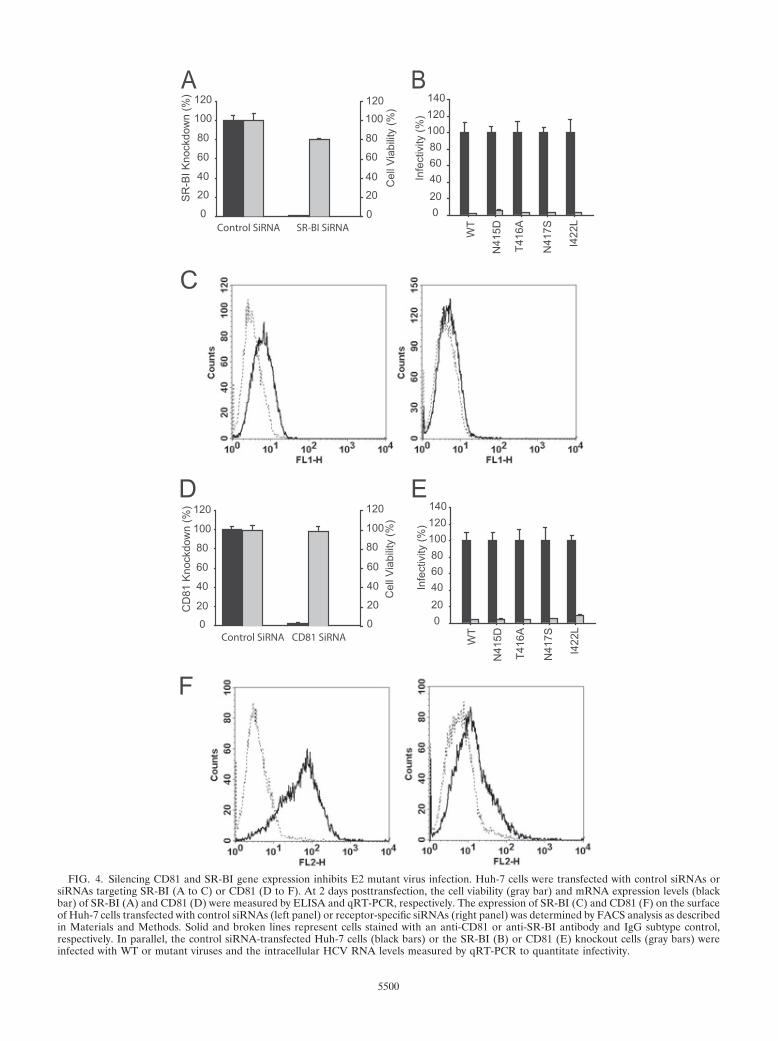
FIG. 4. Silencing CD81 and SR-BI gene expression inhibits E2 mutant virus infection. Huh-7 cells were transfected with control siRNAs orsiRNAs targeting SR-BI (A to C) or CD81 (D to F). At 2 days posttransfection, the cell viability (gray bar) and mRNA expression levels (blackbar) of SR-BI (A) and CD81 (D) were measured by ELISA and qRT-PCR, respectively. The expression of SR-BI (C) and CD81 (F) on the surfaceof Huh-7 cells transfected with control siRNAs (left panel) or receptor-specific siRNAs (right panel) was determined by FACS analysis as describedin Materials and Methods. Solid and broken lines represent cells stained with an anti-CD81 or anti-SR-BI antibody and IgG subtype control,respectively. In parallel, the control siRNA-transfected Huh-7 cells (black bars) or the SR-BI (B) or CD81 (E) knockout cells (gray bars) wereinfected with WT or mutant viruses and the intracellular HCV RNA levels measured by qRT-PCR to quantitate infectivity.
5500

the phenotype of the JFH1G418D virus in terms of virus infec-tivity, receptor affinity, and antibody neutralization. Similar toall of the adaptive mutants described above, the JFH1G418D viruswas more sensitive to neutralization by the patient IgGs, thehuman anti-E2 MAb CBH-5 and soluble hCD81-LEL, and lesssensitive to inhibition by the anti-SR-BI antiserum (Fig. 8A andTable 1). Furthermore, like JFH1N415D and JFH1N417S, this mu-tant was resistant to neutralization by both MAbs AP33 and 3/11.As with the adaptive mutants, the infectivity of JFH1G418D wasnot drastically altered (data not shown).
Amino acid sequence analysis. We next investigated thefrequency of changes, if any, of the relevant E2 residues innaturally occurring HCV isolates by sequence alignment. Thisanalysis showed that substitution from N to D at position 415was very rare in naturally occurring sequences, being found inonly a single sequence among a sample of 1,311 full-length E2protein sequences (Table 2). The substitutions from T to A atposition 416 and from N to S at position 417 were morecommon, being found 13 and 17 times, respectively, within thesame sample. Position 416 was more generally variable, with182 of the 1,311 sequences differing from this residue in theJFH1 sequence. Position 418, in contrast, was extremely con-served. Only 2 of 1,311 sequences varied from the G found inJFH1, and neither of these has the G-to-D substitution pro-duced here. The substitution from I to L at position 422 wasalso rare, being found only five times.
Analysis of the total number of naturally occurring substi-tutions at the relevant E2 positions identified the residue atposition 415 as the most variable in terms of number of resi-
dues used (eight in total), contrasting with the use of only Gand very rarely S at position 418 (Table 3). Although position416 was the most likely to be substituted in naturally occurringsequences with 182 substitutions found, it used fewer residuesthan position 415. Position 422 had almost the same overallnumber of substituted sequences as position 415 (38 versus 37)but used a far lower number of residues (4 compared to 8).
DISCUSSION
In this study, we demonstrate that several mutations locatedwithin a conserved E2 region encompassing residues 412 to 423influence the viral glycoprotein interaction(s) with cell recep-tors and neutralizing antibodies. This region carries residuesthat are critical for recognition of two broadly neutralizingantibodies used here, MAbs AP33 and 3/11 (45, 56). Theseresidues—L413, N415, G418, and W420 (AP33) and N415,W420, and H421 (3/11)—are well conserved, which is relevantfor future vaccine design. However, this requires a better un-derstanding of the epitope-antibody interaction at the struc-tural level. In this respect, studies of viral variants that escapeantibody-mediated neutralization should provide useful infor-mation and help identify the role of other residues in antigen-antibody interaction.
Three of the five HCV JFH1 E2 mutations (N415D, N417S,and G418D) described in the present study abrogated E2 re-activity to, and virus neutralization by, MAbs AP33 and 3/11.The N415D and N417S mutations arose spontaneously duringcell passaging, while the G418D was generated under AP33
FIG. 5. E2 mutant viruses have increased sensitivity to neutralization by human anti-envelope antibodies. JFH1WT (F), JFH1N415D (E),JFH1T416A (�), JFH1N417S (‚), or JFH1I422L (f) HCVcc was incubated for 1 h with different amounts of HCV-infected patient IgGs IgG17 (A) orIgG19 (B), or the human MAbs CBH-5 (C) and HC-11 (D) prior to infection of target cells. The level of virus inhibition was assayed as describedin Materials and Methods. The percent neutralization was calculated by quantifying viral infectivity in the presence of anti-HCV glycoproteinspecific antibodies relative to infection in the absence of antibodies.
VOL. 84, 2010 E2 MUTANTS AFFECTING HCV NEUTRALIZATION AND ENTRY 5501

selective pressure. Our amino acid sequence alignment showsthat N415D and G418D are extremely rare (1 and 0 occur-rences, respectively, out of a sample of 1,311 sequences) innatural sequences, whereas N417S, although by no means com-mon, is the major naturally occurring variant (17 occurrencesof 27) (Table 2). Recently, we described a MAb AP33 neutral-ization escape variant of a genotype 1a/2a chimeric HCVccfollowing repetitive rounds of antibody neutralization and am-plification in cell culture (17). This virus contained N415Y andE655G mutations in the E2 glycoprotein. The N415Y mutationalone severely attenuated MAb AP33 (and 3/11) recognitionand neutralization, but it did not enhance sensitivity to neu-tralization by other human anti-E2 MAbs and, interestingly, itsubstantially reduced viral fitness. Y at position 415 occurs 9times in natural HCV sequences. Of the 37 variant residues atthis position (Table 3) it is the third most common, found ingenotypes 1a, 1b, 4b, and 6. In contrast, the N415D mutation,although found much rarely in patient isolates, maintainedHCVcc fitness, as was the case for the other E2 cell culture-adaptive mutations characterized in the present study. This islikely due to the nature of the substituted amino acid and/orgenotypic differences in the E2 glycoprotein. The difference inthe E2 sequence could also account for the fact that the re-petitive passaging of the genotype 2a JFH1 HCVcc in thepresence of a neutralizing concentration of MAb AP33 al-lowed selection of a virus carrying the E2 G418D substitution.However, the different selection protocols used between the
previous (17) and the present study may, at least in part, beresponsible.
We previously showed that alanine replacement of the res-idue N417 in the HCV genotype 1a H77 E2 moderately re-duced MAb AP33 and 3/11 binding (56). In the present study,a change to serine at this position in the genotype 2a JFH1strain drastically reduced AP33 and 3/11 binding, renderingJFH1N417S HCVcc resistant to neutralization by either anti-body and highlighting the contribution of N417 to their bindingsites on E2. Furthermore, the residue N417 is part of an N-linked glycosylation site (22, 47), the removal of which fromgenotype 1a E2 (N417Q) has been shown to increase the sen-sitivity of HCVpp to antibody neutralization and to increaseCD81 binding (22). The latter observations are also in keepingwith our findings. Our data show that the molecular weight ofthe genotype 2a E2 N417S mutant is identical to the WTglycoprotein (Fig. 7b); however, it would be inappropriate toconclude on this basis that this site is not used for glycosylation.This is because the N417S change potentially creates a new Nglycosylation site over positions 415 to 417 (i.e., a change fromNTN to NTS, see Fig. 1b), which, if utilized, will not alter themigration of the mutant E2 in SDS-PAGE. Clearly, furtherstudies are required to clarify this issue. The N417S changeoccurs in 17 of the 27 variants at this position (Table 2),suggesting that it may be naturally selected.
Bungyoku et al. (10) previously showed that the E2 T416Amutation in a chimeric J6/JFH1 HCVcc background does not
FIG. 6. E2 mutations alter virus neutralization by MAbs AP33 and 3/11. JFH1WT (F), JFH1N415D (E), JFH1T416A (�), JFH1N417S (‚), orJFH1I422L (f) HCVcc was preincubated for 1 h with different concentrations of MAb AP33 (A) and 3/11 (B) before infecting target cells. The levelof virus inhibition was assayed as described in Materials and Methods. The percent neutralization was calculated by quantifying viral infectivity inthe presence of anti-E2 specific MAbs relative to infection in the absence of antibodies. (C and D) Reactivity of MAb AP33 or 3/11 to HCV E2.WT or mutant E2 from electroporated Huh-7 cells was normalized as described in the legend to Fig. 3 and tested for reactivity to MAb (C) AP33or (D) 3/11 by GNA-capture ELISA, and the data are presented as the averages of two independent experiments, each performed in triplicate.Reactivity is expressed as the percentage of binding relative to the WT E2.
5502 DHILLON ET AL. J. VIROL.

alter virus infectivity in the Huh-7-derived sub line Huh7.5 (7).In accordance with these data, we find here that the samemutation has no significant affect on HCVcc spread in Huh-7cells. Furthermore, we previously showed that the T416A mu-tation in the genotype 1a HCVpp system moderately reducedMAb AP33 and 3/11 recognition (56), enhanced CD81 bind-ing, and abrogated pseudoparticle infectivity (47). In contrast,we show here that this mutation in the genotype 2a JFH1HCVcc enhances E2 reactivity to MAbs AP33 and 3/11 andmaintains WT virus infectivity. Together, the different infec-tion systems, viral isolates and/or cell lines used in each studylikely account for these inconsistencies. T416A is one of sevenvariants at a locus subject to positive selection (9), and al-though occurring 13 times in our sample, is only a minorcomponent (7% of substitutions) of the extensive variability inthis position.
The I422L mutation was first isolated alongside other struc-tural and nonstructural mutations after several rounds of JFH1HCVcc passaging in Huh-7.5 cells and was shown not to altervirus infectivity, which is in agreement with our findings inHuh-7 cells (27). Consistent with our previous findings forI422A (56), the I422L mutation did not affect E2 recognitionby MAbs AP33 and 3/11, confirming that this residue is notcritical for E2 recognition by either MAb. I422L occurs fivetimes in our sample of 1311 E2 sequences, constituting 13% ofthe substitutions at this position (Table 2).
The reduced sensitivity of these mutants to inhibition by theFIG. 7. E2 mutations affect the infectivity conferred by E1E2 in theHCVpp assay. (A) HCVpp bearing WT JFH1 E2 or mutant JFH1 E2were generated in HEK-293T cells cotransfected with appropriateconstructs as described in Materials and Methods. Naive Huh-7 cellswere infected with HCVpp and infectivity was determined by measur-ing luciferase levels (top panel). Sucrose cushion enriched HCVpppreparations were Western blotted to detect virion incorporation of E2using an anti-E2 MAb (middle panel) and MLV gag proteins using agag-specific MAb (bottom panel). (B) Immunoprecipitation of radio-labeled E1 and E2 proteins expressed in HEK-293T cells was per-formed using the anti-E2 HMAb CBH-5 as described in Materials andMethods. (C and D) HCVpp bearing WT JFH1 E2 (F), JFH1 E2
carrying N415D (E), T416A (�), N417S (‚), or I422L (f) mutationwere first normalized with respect to their infectivity (luciferase) valuesand then mixed with MAbs AP33 (B) or CBH-5 (C) 1 h prior toinfecting Huh-7 cells. Virus infectivity was measured 3 days postinfec-tion by quantifying luciferase activity. The percent neutralization wascalculated by quantifying viral infectivity in the presence of anti-E2specific MAbs relative to infection in the absence of MAbs.
FIG. 8. Characterization of JFH1G418D virus. Huh7-J20 cells wereinfected with the JFH1WT or JFH1G418D virus that had been preincu-bated with different concentrations of IgG2 or IgG4, hCD81-LEL, orthe anti-E2 MAbs AP33, 3/11, or CBH-5. Alternatively, Huh7-J20 cellspreincubated with different concentrations of the anti-CD81 MAb oran anti-SR-BI antiserum were infected with JFH1WT or JFH1G418Dvirus. At 3 days postinfection the virus infectivity levels were deter-mined by measuring SEAP activity in the culture medium. The resultsare presented as the percent inhibition of JFH1G418D infection by theindicated molecules at 50% inhibitory (IC50) concentrations for theWT virus.
VOL. 84, 2010 E2 MUTANTS AFFECTING HCV NEUTRALIZATION AND ENTRY 5503

anti-SR-BI antibody was an unexpected result. In keeping withthese observations, we found that all of the mutants studiedhere were insensitive to HDL-mediated enhancement of virusinfection. The exact mechanism by which the HDL-SR-BI as-sociation facilitates HCV entry is currently unknown. Al-though no interaction between HDL and HCVpp particles hasbeen demonstrated in culture medium, the possibility of anassociation occurring at a postbinding stage cannot be dis-counted (58). More importantly, the binding of HDL toHCVcc virions has yet to be investigated. Also, it has beenpostulated that the lipid transfer events resulting from HDL-SR-BI binding, known to be essential for regulating the prop-erties of cells membranes, may affect the fusion efficiency ofthe HCV envelope with cell membranes (58). SR-BI was firstidentified as a putative HCV receptor based on its ability tobind soluble, truncated E2 (sE2) via HVR1 (53). However, sE2may not fully mimic E2 structures on the HCV virion (11, 28)and an interaction between SR-BI and the E1E2 heterodimershas yet to be confirmed. In addition, the initial binding ofserum HCV to SR-BI was found not to be mediated by HVR-1or indeed other regions of the E2 glycoprotein. Instead, the
association of VLDL with virus particles appeared to play acritical role in the primary interaction with SR-BI (40). Thus,there is much uncertainty as to how HCV utilizes this receptorduring virus entry. In the absence of definitive assays that canmeasure an interaction between SR-BI with full-length E1E2or indeed HCVcc virions, it is difficult to decipher the effectscaused by our E2 mutations to the entry process via this re-ceptor. However, the siRNA knockdown experiment showsthat SR-BI is not dispensable for the mutant virus entry.
Substitution of N415, T416, and N417 resulted in increasedE2-CD81 binding, whereas the binding of E2 I422L mutant toCD81 was unaltered. Moreover, each mutant virus, includingJFH1G418D, exhibited a significantly greater sensitivity to neu-tralization by hCD81-LEL, suggesting an increased affinity ofthe mutated glycoproteins for CD81. This suggests that theadaptive mutations improve the accessibility of CD81 bindingresidues of the E2 present on mature virions. The heightenedinhibition of these mutants by a range of human anti-HCVglycoprotein antibodies (and by the anti-E2 rodent MAbsAP33 and 3/11 in the case of mutants T416A and I422L)supports this theory.
Other studies have identified cell culture-adaptive mutationswithin the E2 glycoprotein (13, 18, 65). In particular, the mu-tation G451R has been extensively characterized (18, 65). Thissingle mutation increases the buoyant density of the virus, aswell as its ability to bind CD81. G451R also reduces SR-BIdependency and increases virus sensitivity to neutralization byE2 specific antibodies, indicating the greater availability ofepitopes on the mutant particle. The mutations characterizedin the present study have very similar phenotypes to G451R,suggesting that the E2 mutations selected in HCVcc may havearisen in response to similar selective pressures. For example,to persist during long-term culture subtle alterations to E2conformation may enhance virus-receptor interactions andmaintain spread. In line with this, it has been shown thatduring persistent infection of JFH1 in cell culture certain cellpopulations emerge that are less permissive to HCV infectiondue to a decrease in the cell surface expression of CD81 (65).Although this possibility was not investigated in our study orthe others (10, 27, 52), the increased affinity of each mutant to
TABLE 2. Naturally occurring substitutions in 1311 HCV E2 protein sequences
Position JFH1residue Varianta No.b Totalc Prop.d Accession no. (genotype)e
415 N D 1 37 0.03 EU482838 (1a)416 T A 13 182 0.07 AY956468 (1a), AY958005 (3a), EU155215 (1a), EU155249 (1a), EU155282 (1a),
EU155285 (1a), EU155288 (1a), EU155379 (1a), EU255930 (1a), EU255980(1a), EU482845 (1a), EU482836 (1a), EU643835 (6)
417 N S 17 27 0.63 EU256046 (1a), EU256031 (1a), EU255964 (1a), EU255952 (1a), EU255943 (1a),EU155354 (1a), EU155347 (1a), EU155297 (1a), EU155274 (1a), EU155215(1a), EF407468 (1b), EF407466 (1a), EF407477 (1a), EU407415 (1a),EF026073 (2/5 natural recombinant), AY957988 (3a), AM408911 (2/5)
418 G D 0 2 0.00422 I L 5 38 0.13 AB047643 (2a), AF271632 (1a), FJ828970 (1a), FJ828971 (1a), M62321 (1a)
a That is, substitutions as described in the text.b That is, the total number of sequences in a sample of 1,311 HCV E2 proteins with the same substitution.c That is, the total number of substituted residues in the sample of 1,311 (see Table 3).d Prop., the proportion of naturally occurring substitutions that are identical to the substitutions produced in the present study.e The accession number(s) of HCV sequences (genotypes are shown in parentheses) carrying the relevant variant is listed.
TABLE 3. Total number of naturally occurring substitutions in1,311 HCV E2 protein sequences
Protein sequenceResidue at position:
415 416 417 418 422
JFH1 N T N G I
Variantsa K N S S L1 Y S D V2 H A H T3 S I G4 T K T5 R R6 D
Total no.b 37 182 27 2 38
a Substitutions occurring in the sample of 1,311 E2 protein sequences.b Total number of sequences with a substitution. Position 416 is the most
polymorphic in terms of total number of substitutions at 14% (182 in 1,311sequences). However, position 415 has a greater diversity of variants (8 aminoacids used at least once, despite only 37 substitutions).
5504 DHILLON ET AL. J. VIROL.

CD81 (Fig. 3) may assist viral spread in cells presenting lessCD81.
The E2 region studied here lies immediately C-terminal tothe HVR-1, in which positive selection is active. In contrast,the region of interest has only a single site detected as posi-tively selected—that at position 416 (9). Consistent with this,the survey of 1311 E2 sequences reported here shows that 182of them are variants (ca. 14%) at position 416. At the remain-ing sites of interest, substitution is much rarer, never higherthan 4% of the total sample (Table 3). In addition, the specificmutations described here occur relatively rarely in naturalHCV sequences (Table 2). For instance, the G-to-D change atposition 418 is not found at all in our sample of 1,311 se-quences. Indeed, this position is extremely conserved, withonly two substitutions found in the full-length E2 sequencessampled (Tables 2 and 3). Both of those substitutions are G toS and occur in closely related sequences (not shown). TheN415D change is similarly very rare in our sample, occurringonly once.
The I-to-L substitution at position 422 is also rare (Table 2).Where substitution does occur naturally at position 422, it isconfined to relatively hydrophobic residues (Table 3), suggest-ing that a selective constraint for hydrophobicity applies at thisposition. In contrast, the N417S change constitutes 63% of allvariants found at that position, occurring 17 times (Table 2).The T416A change is intermediate in frequency, constituting7% of all naturally occurring substitutions at that position. Thenaturally occurring variants at all positions are found in a widerange of genotypes (Table 2). For instance, position 416 has Tto A substitution in genotypes 1a, 3a, and 6 and position 417has N-to-S substitutions in 1a, 1b, 3a, and 2/5 recombinants.The substitutions produced in the present study at positions415 to 417 and 422 are spontaneous occurrences in long-termcell passage, where selective conditions may be very differentto those found in the natural host. This is a plausible explana-tion for the relative rarity of these substitutions in naturallyoccurring sequences.
Broadly neutralizing MAbs to this conserved region holdgreat promise as therapeutics. Moreover, the epitope recog-nized by these MAbs can be considered a valid lead for futurevaccine design. However, an in-depth understanding of howthe neutralizing antibodies interact with E2 is necessary, forboth effective vaccine design and understanding the role of theepitope in receptor interaction and virus entry. An additionalchallenge for vaccine design is the emergence of viral variantsduring the course of infection that escape antibody neutraliza-tion. In vivo, the prevalence of antibodies reactive to the E2region 412-423 (QLINTNGSWHIN) is �2.5% (55). There-fore, there is no great selective pressure acting on this regiondriving the emergence of neutralization escape mutations. Ifsuch mutants do arise spontaneously, they are likely to confereither reduced virus fitness (17) or an increased vulnerability toneutralization by circulating antibodies targeting various glyco-protein regions, thereby eliminating these variants from thevirus pool. In summary, our data contribute to further definingthe role of key residues within the HCV E2 412-423 region thatinfluence virus-receptor interactions and antibody-mediatedneutralization.
ACKNOWLEDGMENTS
We are grateful to F.-L. Cosset, J. Dubuisson, M. Harris, J. A.McKeating, C. M. Rice, and T. Wakita for provision of reagents. Wethank Gabrielle Vieyres for expert technical assistance.
This study was funded by the Medical Research Council, UnitedKingdom, and European Union FP6 contract MRTN-CT-2006-035599and supported in part by PHS grants HL079381 and AI081903 toS.K.H.F.
REFERENCES
1. Allander, T., K. Drakenberg, A. Beyene, D. Rosa, S. Abrignani, M. Hough-ton, A. Widell, L. Grillner, and M. A. Persson. 2000. Recombinant humanmonoclonal antibodies against different conformational epitopes of the E2envelope glycoprotein of hepatitis C virus that inhibit its interaction withCD81. J. Gen. Virol. 81:2451–2459.
2. Barth, H., C. Schafer, M. I. Adah, F. Zhang, R. J. Linhardt, H. Toyoda, A.Kinoshita-Toyoda, T. Toida, T. H. Van Kuppevelt, E. Depla, F. Von Weiz-sacker, H. E. Blum, and T. F. Baumert. 2003. Cellular binding of hepatitis Cvirus envelope glycoprotein E2 requires cell surface heparan sulfate. J. Biol.Chem. 278:41003–41012.
3. Bartosch, B., J. Dubuisson, and F. L. Cosset. 2003. Infectious hepatitis Cvirus pseudo-particles containing functional E1–E2 envelope protein com-plexes. J. Exp. Med. 197:633–642.
4. Bartosch, B., G. Verney, M. Dreux, P. Donot, Y. Morice, F. Penin, J. M.Pawlotsky, D. Lavillette, and F. L. Cosset. 2005. An interplay betweenhypervariable region 1 of the hepatitis C virus E2 glycoprotein, the scavengerreceptor BI, and high-density lipoprotein promotes both enhancement ofinfection and protection against neutralizing antibodies. J. Virol. 79:8217–8229.
5. Bartosch, B., A. Vitelli, C. Granier, C. Goujon, J. Dubuisson, S. Pascale, E.Scarselli, R. Cortese, A. Nicosia, and F. L. Cosset. 2003. Cell entry ofhepatitis C virus requires a set of coreceptors that include the CD81 tet-raspanin and the SR-BI scavenger receptor. J. Biol. Chem. 278:41624–41630.
6. Benedicto, I., F. Molina-Jimenez, B. Bartosch, F. L. Cosset, D. Lavillette, J.Prieto, R. Moreno-Otero, A. Valenzuela-Fernandez, R. Aldabe, M. Lopez-Cabrera, and P. L. Majano. 2009. The tight junction-associated proteinoccludin is required for a postbinding step in hepatitis C virus entry andinfection. J. Virol. 83:8012–8020.
7. Blight, K. J., J. A. McKeating, and C. M. Rice. 2002. Highly permissive celllines for subgenomic and genomic hepatitis C virus RNA replication. J. Vi-rol. 76:13001–13014.
8. Broering, T. J., K. A. Garrity, N. K. Boatright, S. E. Sloan, F. Sandor,W. D. Thomas, Jr., G. Szabo, R. W. Finberg, D. M. Ambrosino, and G. J.Babcock. 16 September 2009, posting date. Identification and character-ization of broadly neutralizing human monoclonal antibodies directedagainst the E2 envelope glycoprotein of hepatitis C virus. J. Virol. doi:10.1128/JVI.01138–09.
9. Brown, R. J., V. S. Juttla, A. W. Tarr, R. Finnis, W. L. Irving, S. Hemsley,D. R. Flower, P. Borrow, and J. K. Ball. 2005. Evolutionary dynamics ofhepatitis C virus envelope genes during chronic infection. J. Gen. Virol.86:1931–1942.
10. Bungyoku, Y., I. Shoji, T. Makine, T. Adachi, K. Hayashida, M. Nagano-Fujii, Y. H. Ide, L. Deng, and H. Hotta. 2009. Efficient production of infec-tious hepatitis C virus with adaptive mutations in cultured hepatoma cells.J. Gen. Virol. 90:1681–1691.
11. Clayton, R. F., A. Owsianka, J. Aitken, S. Graham, D. Bhella, and A. H.Patel. 2002. Analysis of antigenicity and topology of E2 glycoprotein presenton recombinant hepatitis C virus-like particles. J. Virol. 76:7672–7682.
12. Cormier, E. G., R. J. Durso, F. Tsamis, L. Boussemart, C. Manix, W. C.Olson, J. P. Gardner, and T. Dragic. 2004. L-SIGN (CD209L) and DC-SIGN(CD209) mediate transinfection of liver cells by hepatitis C virus. Proc. Natl.Acad. Sci. U. S. A. 101:14067–14072.
13. Delgrange, D., A. Pillez, S. Castelain, L. Cocquerel, Y. Rouille, J. Dubuisson,T. Wakita, G. Duverlie, and C. Wychowski. 2007. Robust production ofinfectious viral particles in Huh-7 cells by introducing mutations in hepatitisC virus structural proteins. J. Gen. Virol. 88:2495–2503.
14. Dreux, M., T. Pietschmann, C. Granier, C. Voisset, S. Ricard-Blum, P. E.Mangeot, Z. Keck, S. Foung, N. Vu-Dac, J. Dubuisson, R. Bartenschlager, D.Lavillette, and F. L. Cosset. 2006. High-density lipoprotein inhibits hepatitisC virus-neutralizing antibodies by stimulating cell entry via activation of thescavenger receptor BI. J. Biol. Chem. 281:18285–18295.
15. Evans, M. J., T. von Hahn, D. M. Tscherne, A. J. Syder, M. Panis, B. Wolk,T. Hatziioannou, J. A. McKeating, P. D. Bieniasz, and C. M. Rice. 2007.Claudin-1 is a hepatitis C virus coreceptor required for a late step in entry.Nature 446:801–805.
16. Flint, M., C. Maidens, L. D. Loomis-Price, C. Shotton, J. Dubuisson, P.Monk, A. Higginbottom, S. Levy, and J. A. McKeating. 1999. Characteriza-tion of hepatitis C virus E2 glycoprotein interaction with a putative cellularreceptor, CD81. J. Virol. 73:6235–6244.
17. Gal-Tanamy, M., Z. Y. Keck, M. Yi, J. A. McKeating, A. H. Patel, S. K.
VOL. 84, 2010 E2 MUTANTS AFFECTING HCV NEUTRALIZATION AND ENTRY 5505

Foung, and S. M. Lemon. 2008. In vitro selection of a neutralization-resistanthepatitis C virus escape mutant. Proc. Natl. Acad. Sci. U. S. A. 105:19450–19455.
18. Grove, J., S. Nielsen, J. Zhong, M. F. Bassendine, H. E. Drummer, P. Balfe,and J. A. McKeating. 2008. Identification of a residue in hepatitis C virus E2glycoprotein that determines scavenger receptor BI and CD81 receptordependency and sensitivity to neutralizing antibodies. J. Virol. 82:12020–12029.
19. Haberstroh, A., E. K. Schnober, M. B. Zeisel, P. Carolla, H. Barth, H. E.Blum, F.-L. Cosset, G. Koutsoudakis, R. Bartenschlager, A. Union, E. Depla,A. Owsianka, A. H. Patel, C. Schuster, F. Stoll-Keller, M. Doffoël, M. Dreux,and T. F. Baumert. 2008. Neutralizing host responses in hepatitis C virusinfection target viral entry at post-binding steps and membrane fusion. Gas-troenterology 135:1719–1728.
20. Hadlock, K. G., R. E. Lanford, S. Perkins, J. Rowe, Q. Yang, S. Levy, P.Pileri, S. Abrignani, and S. K. Foung. 2000. Human monoclonal antibodiesthat inhibit binding of hepatitis C virus E2 protein to CD81 and recognizeconserved conformational epitopes. J. Virol. 74:10407–10416.
21. Helle, F., and J. Dubuisson. 2008. Hepatitis C virus entry into host cells. CellMol. Life Sci. 65:100–112.
22. Helle, F., A. Goffard, V. Morel, G. Duverlie, J. McKeating, Z. Y. Keck, S.Foung, F. Penin, J. Dubuisson, and C. Voisset. 2007. The neutralizing ac-tivity of anti-HCV antibodies is modulated by specific glycans on the E2envelope protein. J. Virol. 81:8101–8111.
23. Iro, M., J. Witteveldt, A. G. Angus, I. Woerz, A. Kaul, R. Bartenschlager, andA. H. Patel. 2009. A reporter cell line for rapid and sensitive evaluation ofhepatitis C virus infectivity and replication. Antivir. Res. 83:148–155.
24. Johansson, D. X., C. Voisset, A. W. Tarr, M. Aung, J. K. Ball, J. Dubuisson,and M. A. Persson. 2007. Human combinatorial libraries yield rare antibod-ies that broadly neutralize hepatitis C virus. Proc. Natl. Acad. Sci. U. S. A.104:16269–16274.
25. Kapadia, S. B., H. Barth, T. Baumert, J. A. McKeating, and F. V. Chisari.2007. Initiation of hepatitis C virus infection is dependent on cholesterol andcooperativity between CD81 and scavenger receptor B type I. J. Virol.81:374–383.
26. Katoh, K., K. Kuma, H. Toh, and T. Miyata. 2005. MAFFT version 5:improvement in accuracy of multiple sequence alignment. Nucleic AcidsRes. 33:511–518.
27. Kaul, A., I. Woerz, P. Meuleman, G. Leroux-Roels, and R. Bartenschlager.2007. Cell culture adaptation of hepatitis C virus and in vivo viability of anadapted variant. J. Virol. 81:13168–131679.
28. Keck, Z. Y., T. K. Li, J. Xia, B. Bartosch, F. L. Cosset, J. Dubuisson, andS. K. Foung. 2005. Analysis of a highly flexible conformational immunogenicdomain a in hepatitis C virus E2. J. Virol. 79:13199–13208.
29. Keck, Z. Y., T. K. Li, J. Xia, M. Gal-Tanamy, O. Olson, S. H. Li, A. H. Patel,J. K. Ball, S. M. Lemon, and S. K. Foung. 2008. Definition of a conservedimmunodominant domain on hepatitis C virus E2 glycoprotein by neutral-izing human monoclonal antibodies. J. Virol. 82:6061–6066.
30. Keck, Z. Y., A. Op De Beeck, K. G. Hadlock, J. Xia, T. K. Li, J. Dubuisson,and S. K. Foung. 2004. Hepatitis C virus E2 has three immunogenic domainscontaining conformational epitopes with distinct properties and biologicalfunctions. J. Virol. 78:9224–9232.
31. Krieger, M. 2001. Scavenger receptor class B type I is a multiligand HDLreceptor that influences diverse physiologic systems. J. Clin. Invest. 108:793–797.
32. Lavillette, D., A. W. Tarr, C. Voisset, P. Donot, B. Bartosch, C. Bain, A. H.Patel, J. Dubuisson, J. K. Ball, and F. L. Cosset. 2005. Characterization ofhost-range and cell entry properties of the major genotypes and subtypes ofhepatitis C virus. Hepatology 41:265–274.
33. Law, M., T. Maruyama, J. Lewis, E. Giang, A. W. Tarr, Z. Stamataki, P.Gastaminza, F. V. Chisari, I. M. Jones, R. I. Fox, J. K. Ball, J. A. McKeating,N. M. Kneteman, and D. R. Burton. 2008. Broadly neutralizing antibodiesprotect against hepatitis C virus quasispecies challenge. Nat. Med. 14:25–27.
34. Levy, S., S. C. Todd, and H. T. Maecker. 1998. CD81 (TAPA-1): a moleculeinvolved in signal transduction and cell adhesion in the immune system.Annu. Rev. Immunol. 16:89–109.
35. Lindenbach, B. D., M. J. Evans, A. J. Syder, B. Wolk, T. L. Tellinghuisen,C. C. Liu, T. Maruyama, R. O. Hynes, D. R. Burton, J. A. McKeating, andC. M. Rice. 2005. Complete replication of hepatitis C virus in cell culture.Science 309:623–626.
36. Liu, S., W. Yang, L. Shen, J. R. Turner, C. B. Coyne, and T. Wang. 2009.Tight junction proteins claudin-1 and occludin control hepatitis C virus entryand are downregulated during infection to prevent superinfection. J. Virol.83:2011–2014.
37. Lozach, P. Y., A. Amara, B. Bartosch, J. L. Virelizier, F. Arenzana-Seisdedos,F. L. Cosset, and R. Altmeyer. 2004. C-type lectins L-SIGN and DC-SIGNcapture and transmit infectious hepatitis C virus pseudotype particles.J. Biol. Chem. 279:32035–32045.
38. Lozach, P. Y., H. Lortat-Jacob, A. de Lacroix de Lavalette, I. Staropoli, S.Foung, A. Amara, C. Houles, F. Fieschi, O. Schwartz, J. L. Virelizier, F.Arenzana-Seisdedos, and R. Altmeyer. 2003. DC-SIGN and L-SIGN are
high-affinity binding receptors for hepatitis C virus glycoprotein E2. J. Biol.Chem. 278:20358–20366.
39. Macdonald, A., K. Crowder, A. Street, C. McCormick, K. Saksela, and M.Harris. 2003. The hepatitis C virus nonstructural NS5A protein inhibitsactivating protein-1 function by perturbing ras-ERK pathway signaling.J. Biol. Chem. 278:17775–17784.
40. Maillard, P., T. Huby, U. Andreo, M. Moreau, J. Chapman, and A. Bud-kowska. 2006. The interaction of natural hepatitis C virus with human scav-enger receptor SR-BI/ClaI is mediated by ApoB-containing lipoproteins.FASEB J. 20:735–737.
41. Molina, S., V. Castet, C. Fournier-Wirth, L. Pichard-Garcia, R. Avner, D.Harats, J. Roitelman, R. Barbaras, P. Graber, P. Ghersa, M. Smolarsky, A.Funaro, F. Malavasi, D. Larrey, J. Coste, J. M. Fabre, A. Sa-Cunha, and P.Maurel. 2007. The low-density lipoprotein receptor plays a role in the in-fection of primary human hepatocytes by hepatitis C virus. J. Hepatol.46:411–419.
42. Moradpour, D., F. Penin, and C. M. Rice. 2007. Replication of hepatitis Cvirus. Nat. Rev. Microbiol. 5:453–463.
43. Nakabayashi, H., K. Taketa, K. Miyano, T. Yamane, and J. Sato. 1982.Growth of human hepatoma cells lines with differentiated functions in chem-ically defined medium. Cancer Res. 42:3858–3863.
44. Owsianka, A., R. F. Clayton, L. D. Loomis-Price, J. A. McKeating, and A. H.Patel. 2001. Functional analysis of hepatitis C virus E2 glycoproteins andvirus-like particles reveals structural dissimilarities between different formsof E2. J. Gen. Virol. 82:1877–1883.
45. Owsianka, A., A. W. Tarr, V. S. Juttla, D. Lavillette, B. Bartosch, F. L.Cosset, J. K. Ball, and A. H. Patel. 2005. Monoclonal antibody AP33 definesa broadly neutralizing epitope on the hepatitis C virus E2 envelope glyco-protein. J. Virol. 79:11095–11104.
46. Owsianka, A. M., A. W. Tarr, Z. Y. Keck, T. K. Li, J. Witteveldt, R. Adair,S. K. Foung, J. K. Ball, and A. H. Patel. 2008. Broadly neutralizing humanmonoclonal antibodies to the hepatitis C virus E2 glycoprotein. J. Gen. Virol.89:653–659.
47. Owsianka, A. M., J. M. Timms, A. W. Tarr, R. J. Brown, T. P. Hickling, A.Szwejk, K. Bienkowska-Szewczyk, B. J. Thomson, A. H. Patel, and J. K. Ball.2006. Identification of conserved residues in the E2 envelope glycoprotein ofthe hepatitis C virus that are critical for CD81 binding. J. Virol. 80:8695–8704.
48. Patel, A. H., J. Wood, F. Penin, J. Dubuisson, and J. A. McKeating. 2000.Construction and characterization of chimeric hepatitis C virus E2 glycopro-teins: analysis of regions critical for glycoprotein aggregation and CD81binding. J. Gen. Virol. 81:2873–2883.
49. Perotti, M., N. Mancini, R. A. Diotti, A. W. Tarr, J. K. Ball, A. Owsianka, R.Adair, A. H. Patel, M. Clementi, and R. Burioni. 2008. Identification of abroadly cross-reacting and neutralizing human monoclonal antibody directedagainst the hepatitis C virus E2 protein. J. Virol. 82:1047–1052.
50. Pileri, P., Y. Uematsu, S. Campagnoli, G. Galli, F. Falugi, R. Petracca, A. J.Weiner, M. Houghton, D. Rosa, G. Grandi, and S. Abrignani. 1998. Bindingof hepatitis C virus to CD81. Science 282:938–941.
51. Ploss, A., M. J. Evans, V. A. Gaysinskaya, M. Panis, H. You, Y. P. de Jong,and C. M. Rice. 2009. Human occludin is a hepatitis C virus entry factorrequired for infection of mouse cells. Nature 457:882–886.
52. Russell, R. S., J. C. Meunier, S. Takikawa, K. Faulk, R. E. Engle, J. Bukh,R. H. Purcell, and S. U. Emerson. 2008. Advantages of a single-cycle pro-duction assay to study cell culture-adaptive mutations of hepatitis C virus.Proc. Natl. Acad. Sci. U. S. A. 105:4370–4375.
53. Scarselli, E., H. Ansuini, R. Cerino, R. M. Roccasecca, S. Acali, G. Filocamo,C. Traboni, A. Nicosia, R. Cortese, and A. Vitelli. 2002. The human scaven-ger receptor class B type I is a novel candidate receptor for the hepatitis Cvirus. EMBO J. 21:5017–5025.
54. Tamura, K., J. Dudley, M. Nei, and S. Kumar. 2007. MEGA4: MolecularEvolutionary Genetics Analysis (MEGA) software version 4.0. Mol. Biol.Evol. 24:1596–1599.
55. Tarr, A. W., A. M. Owsianka, D. Jayaraj, R. J. Brown, T. P. Hickling, W. L.Irving, A. H. Patel, and J. K. Ball. 2007. Determination of the humanantibody response to the epitope defined by the hepatitis C virus-neutralizingmonoclonal antibody AP33. J. Gen. Virol. 88:2991–3001.
56. Tarr, A. W., A. M. Owsianka, J. M. Timms, C. P. McClure, R. J. Brown, T. P.Hickling, T. Pietschmann, R. Bartenschlager, A. H. Patel, and J. K. Ball.2006. Characterization of the hepatitis C virus E2 epitope defined by thebroadly neutralizing monoclonal antibody AP33. Hepatology 43:592–601.
57. Vieyres, G., A. G. Angus, A. Haberstroh, T. F. Baumert, J. Dubuisson, andA. H. Patel. 2009. Rapid synchronization of hepatitis C virus infection bymagnetic adsorption. J. Virol. Methods 157:69–79.
58. Voisset, C., N. Callens, E. Blanchard, A. Op De Beeck, J. Dubuisson, and N.Vu-Dac. 2005. High density lipoproteins facilitate hepatitis C virus entrythrough the scavenger receptor class B type I. J. Biol. Chem. 280:7793–7799.
59. Voisset, C., A. Op de Beeck, P. Horellou, M. Dreux, T. Gustot, G. Duverlie,F. L. Cosset, N. Vu-Dac, and J. Dubuisson. 2006. High-density lipoproteinsreduce the neutralizing effect of hepatitis C virus (HCV)-infected patientantibodies by promoting HCV entry. J. Gen. Virol. 87:2577–2581.
60. von Hahn, T., B. D. Lindenbach, A. Boullier, O. Quehenberger, M. Paulson,
5506 DHILLON ET AL. J. VIROL.
C. M. Rice, and J. A. McKeating. 2006. Oxidized low-density lipoproteininhibits hepatitis C virus cell entry in human hepatoma cells. Hepatology43:932–942.
61. Wakita, T., T. Pietschmann, T. Kato, T. Date, M. Miyamoto, Z. Zhao, K.Murthy, A. Habermann, H. G. Krausslich, M. Mizokami, R. Bartenschlager,and T. J. Liang. 2005. Production of infectious hepatitis C virus in tissueculture from a cloned viral genome. Nat. Med. 11:791–796.
62. Witteveldt, J., M. J. Evans, J. Bitzegeio, G. Koutsoudakis, A. M. Owsianka,A. G. Angus, Z. Y. Keck, S. K. Foung, T. Pietschmann, C. M. Rice, and A. H.Patel. 2009. CD81 is dispensable for hepatitis C virus cell-to-cell transmis-sion in hepatoma cells. J. Gen. Virol. 90:48–58.
63. Zeisel, M. B., G. Koutsoudakis, E. K. Schnober, A. Haberstroh, H. E. Blum,
F. L. Cosset, T. Wakita, D. Jaeck, M. Doffoel, C. Royer, E. Soulier, E.Schvoerer, C. Schuster, F. Stoll-Keller, R. Bartenschlager, T. Pietschmann,H. Barth, and T. F. Baumert. 2007. Scavenger receptor class B type I is a keyhost factor for hepatitis C virus infection required for an entry step closelylinked to CD81. Hepatology 46:1722–1731.
64. Zhong, J., P. Gastaminza, G. Cheng, S. Kapadia, T. Kato, D. R. Burton, S. F.Wieland, S. L. Uprichard, T. Wakita, and F. V. Chisari. 2005. Robusthepatitis C virus infection in vitro. Proc. Natl. Acad. Sci. U. S. A. 102:9294–9299.
65. Zhong, J., P. Gastaminza, J. Chung, Z. Stamataki, M. Isogawa, G. Cheng,J. A. McKeating, and F. V. Chisari. 2006. Persistent hepatitis C virus infec-tion in vitro: coevolution of virus and host. J. Virol. 80:11082–11093.
VOL. 84, 2010 E2 MUTANTS AFFECTING HCV NEUTRALIZATION AND ENTRY 5507

MUHAMMAD NUAMAN ZAHID Impact of SR-BI and CD81 on Hepatitis
C virus entry and evasion
Résumé
Le virus de l’hépatite C (VHC) est l’une des causes majeures de cirrhose du foie et de carcinome hépatocellulaire.
Au courant de la première partie de ma thèse, nous nous sommes intéressés à caractériser plus en détail le rôle de
SR-BI dans l’infection par le VHC. Bien que les mécanismes impliquant SR-BI dans la liaison du virus à
l’hépatocyte aient été partiellement caractérisés, le rôle de SR-BI dans les étapes suivant la liaison du VHC reste
encore largement méconnu. Afin de mieux caractériser le rôle de l’interaction VHC/SR-BI dans l’infection par le
VHC, notre laboratoire à généré une nouvelle classe d’anticorps monoclonaux anti-SR-BI inhibant l’infection
virale. Nous avons pu démontrer que SR-BI humain jouait un rôle dans le processus d’entrée du virus à la fois lors
de l’étape de liaison du virus à la cellule hôte mais aussi au cours d’étapes suivant cette liaison. Ainsi il serait
intéressant de cibler cette fonction de SR-BI dans le cadre d’une stratégie antivirale pour lutter contre l’infection par
le VHC. Dans la seconde partie de ma thèse, nous avions pour but de caractériser les mécanismes moléculaires
intervenant dans la réinfection du greffon lors de la transplantation hépatique (TH). Nous avons ainsi identifiés 3
mutations adaptatives dans la glycoprotéine d’enveloppe E2 responsables de l’entrée virale augmentée du variant
hautement infectieux. Ces mutations influent sur la dépendance au récepteur CD81 du VHC résultant en une entrée
virale accrue. L’identification de ces mécanismes va nous permettre une meilleure compréhension de la
pathogénèse de l’infection par le VHC, et est un premier pas pour le développement d’une stratégie préventive
antivirale ou vaccinale.
Mot clés: virus de l’hépatite C, SR-BI, anticorps monoclonaux, CD81, transplantation hépatique, glycoprotéine d’enveloppe E2

Résumé en anglais
Hepatitis C virus (HCV) is a major cause of liver cirrhosis and hepatocellular carcinoma. In the first part of my
PhD, we aimed to further characterize the role of scavenger receptor class B type I (SR-BI) in HCV infection.
While the SR-BI determinants involved in HCV binding have been partially characterized, the post-binding
function of SR-BI remains remained largely unknown. To further explore the role of HCV-SR-BI interaction
during HCV infection, we generated a novel class of anti-SR-BI monoclonal antibodies inhibiting HCV
infection. We demonstrated that human SR-BI plays a dual role in the HCV entry process during both binding
and post-binding steps. Targeting the post-binding function of SR-BI thus represents an interesting antiviral
strategy against HCV infection. In the second part of my PhD, we aimed to characterize the molecular
mechanisms underlying HCV re-infection of the graft after liver transplantation (LT). We identified three
adaptive mutations in envelope glycoprotein E2 mediating enhanced entry and evasion of a highly infectious
escape variant. These mutations markedly modulated CD81 receptor dependency resulting in enhanced viral
entry. The identification of these mechanisms advances our understanding of the pathogenesis of HCV
infection and paves the way for the development of novel antiviral strategies and vaccines.
Keywords: hepatitis c virus, SR-BI, monoclonal antibodies, CD81, Liver transplantation, envelope glycoprotein E2