Embed Size (px)
Citation preview

©2014 International Medical Press ISSN 1359-6535
Naturally occurring dominant drug resistance mutations occur infrequently in the setting of recently acquired hepatitis C
Tanya L Applegate, Silvana Gaudieri, Anne Plauzolles, Abha Chopra, Jason Grebely, Michaela Lucas, Margaret Hellard, Fabio Luciani, Gregory J Dore, Gail V Matthews Antiviral Therapy 2014; 10.3851/IMP2821 Submission date 24th February 2014 Acceptance date 24th June 2014 Publication date 8th August 2014 For information about publishing your article in Antiviral Therapy go to http://www.intmedpress.com/index.cfm?pid=12
This provisional PDF matches the article and figures as they appeared upon acceptance. Copyedited and fully formatted PDF and full text (HTML) versions will be made available soon.
Publication: Antiviral Therapy; Type: Original article
DOI: 10.3851/IMP2821
Original article
Naturally occurring dominant drug resistance mutations occur infrequently in the setting of recently acquired hepatitis C Tanya L Applegate1*†, Silvana Gaudieri2,3†, Anne Plauzolles4, Abha Chopra3, Jason Grebely1, Michaela Lucas3,5, Margaret Hellard6, Fabio Luciani7, Gregory J Dore1, Gail V Matthews1 1The Kirby Institute, UNSW Australia, Sydney, Australia
2School of Anatomy, Physiology and Human Biology, University of Western Australia, Perth, Australia
3Institute for Immunology and Infectious Diseases, Murdoch University, Perth, Australia
4Centre for Forensic Science, University of Western Australia, Perth, Australia
5School of Medicine and School of Pathology & Laboratory Medicine, University of Western Australia,
Perth, Australia
6Macfarlane Burnett Institute for Population Health, Melbourne, Australia
7Inflammation and Infection Research Centre, UNSW Australia, Sydney, Australia
*Corresponding author e-mail: [email protected]
†Authors contributed equally to the preparation of this manuscript
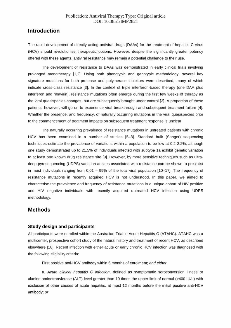
Abstract
Background: Directly Acting Antivirals (DAAs) are predicted to transform hepatitis C (HCV) therapy, yet little is known about the prevalence of naturally occurring resistance mutations in recently acquired HCV. This study aimed to determine the prevalence and frequency of drug resistance mutations in the viral quasispecies among HIV positive and negative individuals with recent HCV.
Methods: The NS3 protease, NS5A and NS5B polymerase genes were amplified from fifty genotype 1a participants of the Australian Trial in Acute Hepatitis C. Amino acid variations at sites known to be associated with possible drug resistance were analysed by ultra-deep pyrosequencing.
Results: Twelve percent of individuals harboured dominant resistance mutations, while 36% demonstrated non dominant resistant variants below that detectable by bulk sequencing (ie < 20%) but above a threshold of 1%. Resistance variants (< 1%) were observed at most sites associated with DAA resistance from all classes, with the exception of sofosbuvir.
Conclusions: Dominant resistant mutations were uncommonly observed in the setting of recent HCV. However, low level mutations to all DAA classes were observed by deep sequencing at the majority of sites, and in most individuals. The significance of these variants and impact on future treatment options remains to be determined.
Accepted 24 June 2014, published online 8 August 2014
Running head: Resistance mutations in recent HCV
Publication: Antiviral Therapy; Type: Original article
DOI: 10.3851/IMP2821
Introduction
The rapid development of directly acting antiviral drugs (DAAs) for the treatment of hepatitis C virus
(HCV) should revolutionise therapeutic options. However, despite the significantly greater potency
offered with these agents, antiviral resistance may remain a potential challenge to their use.
The development of resistance to DAAs was demonstrated in early clinical trials involving
prolonged monotherapy [1,2]. Using both phenotypic and genotypic methodology, several key
signature mutations for both protease and polymerase inhibitors were described, many of which
indicate cross-class resistance [3]. In the context of triple interferon-based therapy (one DAA plus
interferon and ribavirin), resistance mutations often emerge during the first few weeks of therapy as
the viral quasispecies changes, but are subsequently brought under control [2]. A proportion of these
patients, however, will go on to experience viral breakthrough and subsequent treatment failure [4].
Whether the presence, and frequency, of naturally occurring mutations in the viral quasispecies prior
to the commencement of treatment impacts on subsequent treatment response is unclear.
The naturally occurring prevalence of resistance mutations in untreated patients with chronic
HCV has been examined in a number of studies [5–8]. Standard bulk (Sanger) sequencing
techniques estimate the prevalence of variations within a population to be low at 0.2-2.2%, although
one study demonstrated up to 21.5% of individuals infected with subtype 1a exhibit genetic variation
to at least one known drug resistance site [9]. However, by more sensitive techniques such as ultra-
deep pyrosequencing (UDPS) variation at sites associated with resistance can be shown to pre-exist
in most individuals ranging from 0.01 – 99% of the total viral population [10–17]. The frequency of
resistance mutations in recently acquired HCV is not understood. In this paper, we aimed to
characterise the prevalence and frequency of resistance mutations in a unique cohort of HIV positive
and HIV negative individuals with recently acquired untreated HCV infection using UDPS
methodology.
Methods
Study design and participants
All participants were enrolled within the Australian Trial in Acute Hepatitis C (ATAHC). ATAHC was a
multicenter, prospective cohort study of the natural history and treatment of recent HCV, as described
elsewhere [18]. Recent infection with either acute or early chronic HCV infection was diagnosed with
the following eligibility criteria:
First positive anti-HCV antibody within 6 months of enrolment; and either
a. Acute clinical hepatitis C infection, defined as symptomatic seroconversion illness or
alanine aminotransferase (ALT) level greater than 10 times the upper limit of normal (>400 IU/L) with
exclusion of other causes of acute hepatitis, at most 12 months before the initial positive anti-HCV
antibody; or

Publication: Antiviral Therapy; Type: Original article
DOI: 10.3851/IMP2821
b. Asymptomatic hepatitis C infection with seroconversion, defined by a negative anti-HCV
antibody in the two years prior to the initial positive anti-HCV antibody.
All study participants provided written informed consent. The study protocol was approved by
St Vincent’s Hospital Human Research Ethics Committee and local ethics committees at sites. The
study was registered with clinicaltrials.gov registry (NCT00192569). Both HIV positive and negative
participants were enrolled. Due to the relationship between HCV genotype and protease inhibitor-
based resistance, only genotype 1a (GT1a) participants with a stored screening or pre-treatment
sample available, with an HCV RNA> 1000 IU/ml were included.
Detection and quantification of HCV RNA
Qualitative and quantitative HCV RNA testing was performed using the Versant TMA assay [Bayer,
Australia; <10 IU/ml] and the Versant HCV RNA 3.0 (Bayer, Australia; <615 IU/ml), respectively.
Sequencing methodology
Viral RNA extraction
Viral RNA was extracted from plasma samples using the COBAS AMPLICOR HCV Specimen
Preparation Kit v2.0 (Roche Applied Science) according to manufacturers’ instructions.
Generation of NS3 protease, NS5A and NS5B polymerase amplicons
Two initial RT-PCRs were performed to cover the non-structural regions of HCV [19]. The first-round
products were then used as templates in nested second round PCRs using generic or genotype
specific primers as previously described [19]. Alternative generic and genotype/subtype specific
primers targeting the NS3 protease, NS5A and NS5B regions were also used. The primer sequences
(5’ to 3’) were as follows: for NS3 protease 3337F GGCYTGCCYGTCTCYGC and 4035R
GTGCTCTTRCCGCTRCCNGT (modified from [7]); for NS5A/B 6158F ATGCWGCTGCCCGCGTCAC
and 6940R AGCTGGCTGGCCGAAGAGCT, 6821F TCCCTTGCGAGCCCGAACCGGA and 7522R
TGAGATCCGGGTCCCCAGGCTC, 7370F CCTTGGCCGAGCTTGCCAC and 8080R
CCCCCAGGTCAGGGTACACAAT, 8011F ACCATCATGGCGAAGAAYGA and 8683R
GAGGAGCAAGATGTTATGAGCTC. Thermocycling conditions were: one cycle for 940C for 2
minutes followed by 20 cycles at 940C for 15 seconds then annealing temperature (Tm) 1 for 30
seconds then 720C for 1 minute. This was followed by another 20 cycles at 94
0C for 15 seconds then
Tm2 for 30 seconds then 720C for 1 minute followed by a single cycle at 72
0C for 1 minute. For the
primer pair 3337F/4035R Tm1 was 560C and Tm2 was 54
0C. For the primer pair 8011F/8683R Tm1
was 530C and Tm2 was 51
0C. For the primer pairs 6158F/6940R, 6821F/7522R and 7370F/8080R
Tm1 was 610C and Tm2 was 59
0C.
FLX 454 UDPS generation and analysis
UDPS was carried-out using the 454 Life Sciences platform (GS-FLX, Roche Applied Science) for all
participants. PCR products (as described above) were quantified and then pooled for each individual.
These products were ligated to adaptors and clonally amplified on capture beads in water-in-oil
Publication: Antiviral Therapy; Type: Original article
DOI: 10.3851/IMP2821
emulsion micro-reactors. The resultant libraries were sequenced using a PicoTiterPlate and
nucleotide data collected and quality filtered using the Roche 454 software (default settings).
Following the removal of primer sequences (using an in-house program), sequence reads were
aligned using the Softgenetics NextGENe software to the H77 genotype 1a sequence (GenBank
accession number: NC004102). To ensure quality of sequence reads, an 80% nucleotide identity
threshold against the reference sequence was used for inclusion of a read for analysis. Sequences
were then imported as fasta files into the program BioEdit. Likely homopolymer errors relative to the
reference sequence and sequence errors near read ends were removed [20]. Nucleotide alignment
was then translated to appropriate amino acid sequence. Sites associated with DAA resistance were
identified and sorted based on amino acid composition. The number of reads for each amino acid at
each site associated with drug resistance was tabulated. A variation was included if it was present on
at least three sequence reads.
Results
Participants
Fifty GT1a ATAHC participants had samples available for testing. Twenty six (52%) participants were
HIV positive. Median HCV RNA was 5.3 logs (IU/ml) and the median estimated duration of HCV
infection at time of analysis was 29 weeks. Further participant characteristics at baseline, stratified by
HIV status, infection can be found in Table 1.
NS3/4 protease sequencing
Among those with available samples (n=50), sequencing of the NS3/4 protease gene was
successfully performed in 49 particpants with GT1a (Figure 1). Eleven sites known to be associated
with possible drug resistance were examined for evidence of amino acid variation including the
following site changes: V36A/M, Q41R, F43C/S/L, T54A/S, V55A, Q80K/R, S138T, R155K/Q/T,
A156S/T/V, D168A/T/V and V170A/T (Table 2). The mean coverage at each site varied from 3918-
5239 reads. Only four (8%) participants had evidence of a dominant resistant variant, one with a
V36M variation present at 99.4% and a Q80K at 99.2%, one with a sole Q80K present at 98.5%, and
two with a V55A present at 89.6% and 99.9% of total sequences, respectively. Seven additional
participants (14%) had evidence of a resistance variant present in sequences at a level above 1%, but
in all cases the prevalence of the variant was very low and never exceeded 2%. Two of these
participants had more than one resistant variant present, however, the presence of more than one
variant within a single 500bp viral sequence (present at low frequency) was not observed.
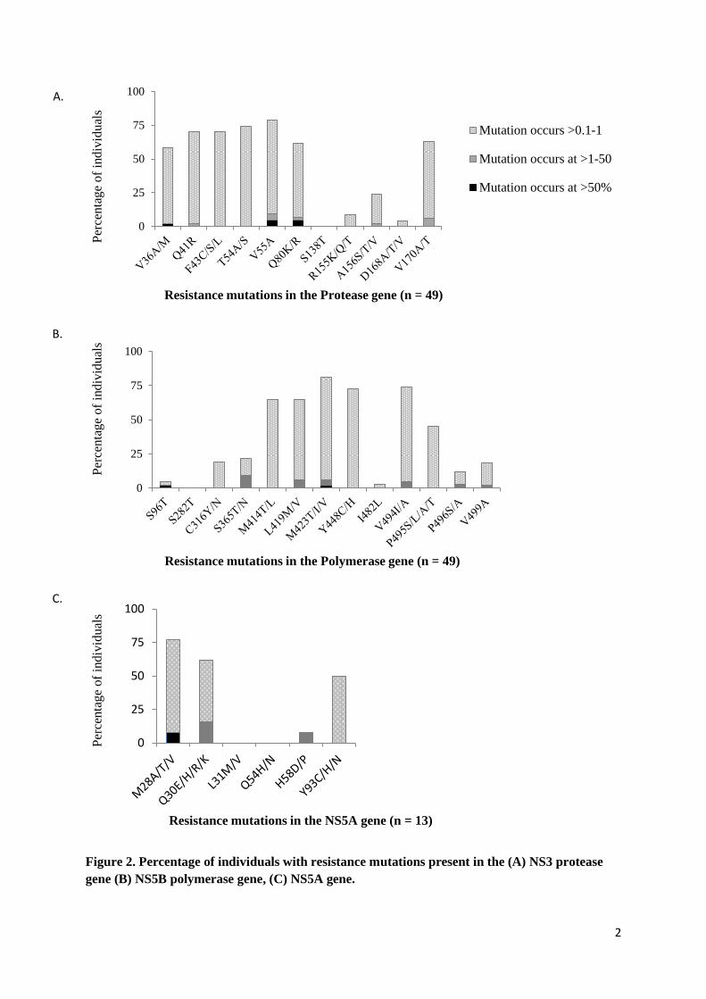
Variation above 0.1% and below 1% was seen at a much greater frequency (Figure 2a).
Evidence of resistant variants occurring at this frequency was observed at all sites except the S138T
position (< 0.1% in all participants). Some sites however were relatively conserved with low
proportions of patients with variance at this level, notably D168A/T/V (4%), and R155K/Q/T (9%),
whereas other sites such as T54A/S, V55A and Q41R demonstrated far greater variance with 74%,

Publication: Antiviral Therapy; Type: Original article
DOI: 10.3851/IMP2821
70% and 68% of participants respectively demonstrating low level resistance variants (0.1-1%) at
these sites.
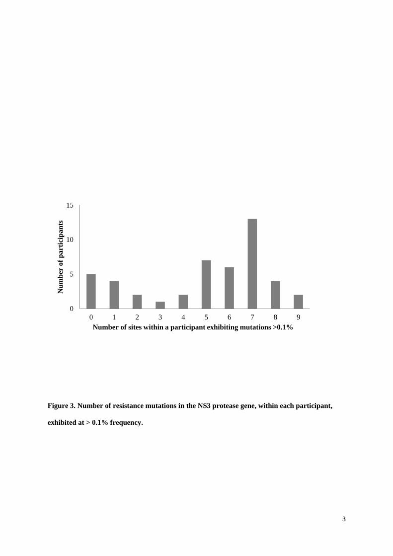
The spread of resistance variation within individual participants did not follow any particular
pattern. Around one quarter of participants (n=11) demonstrated either no or very minimal variation
(two or fewer mutations) whilst others had variation at most sites (Figure 3), however, there were no
significant differences observed in the amount of variation by either HIV status or duration of infection.
The median number of sites per subject with evidence of RAVS > 0.1% was 6. In those with 6 or more
sites affected the proportion of HIV positivity was similar to those with less than 6 sites affected (42%
vs 52%, p=0.490), and although the duration of infection was greater at 24.5 weeks versus 16 weeks,
this was non-significant, p=0.11.
NS5B polymerase sequencing
Among those with available samples (n=50), sequencing of the NS5B polymerase gene was possible
in 49 participants (Figure 1). Variation in amino acid composition was studied at 12 sites known to be
associated with resistance to polymerase inhibitors. These included the positions S282T and S96T,
P496A/S,P495S/L/A/T C316Y/N,S365 T/AL419M/V, M243 T/I/V and Y448C/H . Mean coverage at
sites ranged from 2102-6722 reads. Only one (2%) participant had evidence of a potential dominant
resistant variant, a M423I change in 98% of the viral population. A further 12 participants had
evidence of a potential resistant variant present as a minority variant at a level above 1% (Table 2).
These changes predominately involved low level variation between 1-3% at the following sites S365T
(n=4), L419M/V (n=3), M423T (n=2), V494A (n=2) and P496S (n=1). Only one participant had a non-
dominant variant at > 3% prevalence – this participant had a V499A change present at 18.8% of viral
quasispecies. The same participant also had an S365T resistant variant present at 1.38%. The
presence of multiple resistant mutations on a single viral strain was not observed (based on
approximately 500bp sequence reads).
As with the protease gene, variation at a level below 1% but above 0.1% was observed far
more frequently (Figure 2b). The amount of variation differed depending on the site. For example no
participant demonstrated any resistance variant at the S282T site and only 2% of participants’
demonstrated resistance variant at the S96T site at a level above 0.1%. At other sites variation was
much more frequent. Over two thirds of participants exhibited low level resistance variants at a
prevalence of between 0.1% and 1% at M423T, Y448C/H and Y494I/A sites (Figure 2b).
NS5A sequencing
A subgroup of thirteen genotype 1a participants also underwent sequencing of the NS5A region in
which mutations conferring resistance to the first generation NS5A inhibitors including daclatasvir and
ledipasvir are found. Six sites of interest were examined for mutational change including M28 A/T/V,
Q30 E/H/R/K, L31 M/V, Q54 H/N, H58 D/P and Y93 C/H/N (Figure 2c). Mean coverage at sites
ranged from 2583-2984 reads. Only one participant had a dominant resistance variant with a M28V
change in 99.52% of quasispecies. Two participants had variants present at between 1 and 3% of
viral quasispecies; one with a Q30R at 1.33% and an H58P change at 1.10%, while the second had

Publication: Antiviral Therapy; Type: Original article
DOI: 10.3851/IMP2821
only a Q30R mutation at 2.29% (Table 2). As with protease and polymerase regions however
resistance variants were commonly present at between 0.1 and 1% of the total virus population,
observed at the following frequencies at sites: M28V/A/T (64%), Q30R/E/R/K (46%), and Y93 C/H/N
(50%). Only the L31M/V site was highly conserved with no evidence of resistance variants at a level
above 0.01% found in any participants.
Effect of immune pressure on frequency of resistance mutations
Overlap between host T-cell immune pressure, as directed by the Human Leucocyte Antigen (HLA)
repertoire of the host, and drug pressure at specific sites in the viral genome can influence an
individual’s risk to develop drug resistance [8,21,22]. We investigated whether subjects with specific
HLA alleles, corresponding to overlapping HLA-restricted epitopes with resistance sites, correlated
with detectable circulating mutations at >1% across the three proteins of interest.
In the NS3 protease region, the HLA-A2-restricted epitope from 1073-1081 (CINGVCWTV) in
the polyprotein includes position 54 and 55 in the protease protein. Of those subjects with HLA typing
available (75% of cohort), only one had a dominant resistant variant at position 55 and that subject
carried HLA-A2 (an additional 3/16 with HLA-A2 had a resistant variant between 1-5%). No resistant
variants greater than 1% were identified in the non-A2 participants. The HLA-A24-restricted epitope
from position 1100-1108 (MYTNVDQDL) includes position 80. Of the six subjects with HLA-A24, one
had lysine (K) as the dominant amino acid whereas two out of 23 subjects without HLA-A24 had a
different dominant amino acid to glutamine (Q) and one had arginine (R) variant at 1-5%. In the NS5A
region, ten out of 14 individuals had HLA typing. For the HLA-A3-restricted epitope from 2017-2026
(GVWRGDGIMH) including position 54 in the NS5A protein, only one subject had a variant at 1.6%
and carried HLA-A3 (1/4 with HLA-A3). For the NS5B region, there were five HLA-restricted epitopes
that also spanned a RAV: HLA-A3-restricted epitope 2510-2518 (SLTPPHSAK) including position 96;
HLA-B27-restricted epitope from 2841-2849 (ARMILMTHF) including position 423; HLA-A1-restricted
epitope from 2858-2868 (QLEQALDCEIY) including position 448; HLA-B55-restricted epitope from
2898-2907 (SPGEINRVAA) including position 482; and HLA-B57-restricted epitope from 2912-2921
(LGVPPLRAWR) including positions 494, 495, 496 and 499. For the HLA-A3-restricted epitope, eight
of 30 subjects carried HLA-A3 with only one HLA-A3 subject with a variant at >1% (1.2%). For the
HLA-B27-restricted epitope, only two subjects carried HLA-B27 and neither showed variation at
position 423 at >1% while one subject without HLA-B27 showed a dominant resistant variant. For the
HLA-A1-restricted epitope, of the 12 subjects that carried HLA-A1 none had a variant at >1% or any
other of the subjects. For the HLA-B55-restricted epitope, no subjects had a variant at greater than
1% and only one subject carried HLA-B55. For the HLA-B57-restricted epitope, only one subject
carried HLA-B57 and did not have a variant at >1% for positions any variant sites. One subject had a
variant at 2.6% at position 496 but did not carry HLA-B57 and one subject had a variant at 18.8% at
499 but did not carry HLA-B57.
Publication: Antiviral Therapy; Type: Original article
DOI: 10.3851/IMP2821
Discussion
This study provides the largest and most comprehensive analysis of the prevalence of naturally
occurring DAA resistance in subjects with recent HCV. Dominant resistant variants (at levels of 50-
100%) were observed, albeit infrequently, in the setting of relatively short duration of infection.
Overall, six of 50 (12%) individuals demonstrated evidence of dominant resistance variants conferring
reduced sensitivity to one DAA treatment class. Ten percent of individuals had a dominant variant
conferring protease inhibitor resistance, one individual had a dominant variant conferring polymerase
inhibitor resistance and another had a variant conferring resistance to the NS5A inhibitor class. An
additional 18 (36% of those sequenced) demonstrated evidence of resistant variants at a level below
that detectable by bulk sequencing (ie < 20%) but above a threshold of 1%. At a lower threshold level
of between 0.1 and 1%, resistant variants were almost universal, although notably absent at a few
sites including S282T/S96T (sofosbuvir) and the L31M (daclatasavir).
Both the prevalence and significance of naturally occurring drug resistance mutations in
chronic HCV have been debated. A number of studies have examined the presence of pre-treatment
drug resistance, generally to the protease inhibitor class, with varying results [5,8,23–25]. A large
cohort of 507 individuals from US, Germany and Switzerland examined using bulk (population) based
sequencing identified either protease or polymerase dominant mutants present in 8.6% of subjects
[5]. A further large study composed of 405 individuals from the UK, Australia and Switzerland
suggested that up to 21.5% of genotype 1a subjects may have drug resistance mutations on bulk
sequencing [8]. Pre-existing mutations have been shown to influence treatment outcome as in the
PROVE1/2 studies, where all of the four participants with the R155K mutation present as the
dominant strain at baseline failed to achieve SVR [23]. Similarly, population sequencing of
participants in the Boceprevir registration studies demonstrated that the SVR rate in participants with
both baseline mutations and a <1 log decline during four weeks lead-in was 0% (compared to 28-38%
in those with wild type) [26,27]. Recently, the presence of Q80K prior to treatment with the second
generation protease inhibitor Simeprevir has been shown to negatively impact on treatment outcome,
bringing SVR rates down to 58%, similar to that of PEG/RBV alone [28]. Q80K has been
demonstrated to occur in up to 47% of untreated GT1a participants in North America [28,29], but
geographical variability is observed and the prevalence in the chronic Australian population is
unknown. In our study of recent infection Q80K was infrequent, observed in just two (4%) of untreated
participants. To date, there is insufficient data to determine if the prevalence of the Q80K in recently
acquired HCV occurs less frequently than in chronic HCV.
Given that a variant can only be detected by population sequencing when it comprises > 20%
of the viral quasispecies, there obviously exists the potential for minor drug resistant variants to be
present at levels not detected by this method. Under drug pressure these minor variants may have
the capacity to be selected due to their survival advantage in the presence of drug and fill the
replication space to become the dominant species. This phenomenon has been extensively described
Publication: Antiviral Therapy; Type: Original article
DOI: 10.3851/IMP2821
using clonal analysis in longitudinal studies of antiviral monotherapy and/or treatment failures [1,2,30–
33].
The spectrum and prevalence of pre-existing variants present using sensitive clonal or deep
sequencing methodology in treatment naïve individuals with chronic HCV has been explored in a
number of small studies [10–17]. In a study of 18 participants from the PROVE2 study who underwent
deep sequencing at baseline almost every participant had evidence of the R155K/T/Q (levels between
0.1%-7.8%), A156 S/T/V (0.l2% to 3.2%) and I170 A/T (0.1-0.5%) although evidence of other
mutations (V36 A/M, T54 A/S, V55A, Q80 R/K) were less common [10]. Other studies have also
demonstrated a low prevalence of variations (<1%) in treatment-naïve subjects [14,34]. Bartolini et al
(2013) explored the prevalence of resistance mutations in the NS3 protease in both HCV
monoinfected (n = 10) and HIV coinfected subjects (n=18), and identified pre-existing resistance
mutations to occur at a higher frequency in some individuals; V36A (26%), Q41H (45%, 19%), Q80L
(40%) [15], although this did not differ between the two groups.
The period after newly acquired HCV infection is unique in several ways. During this time
spontaneous clearance of HCV is possible, changes in the viral quasispecies occur as immune
pressure drives viral adaption, and treatment is likely to be successful [20,35–39]. Very few studies
have examined the composition of the viral quasispecies at this time, particularly with relevance to the
sites associated with drug resistance. One recent small study in 38 HIV infected individuals with acute
HCV examined regions in the protease gene by both population ultra-deep sequencing [40]. In this
study16% of individuals had a ‘dominant’ resistance variant, but deep sequencing down to a level of
<1% demonstrated every site in all samples, with the exception of T54M/L and R155Q. The authors
postulated that their higher level of resistance detection compared to monoinfected studies may be
due to a higher replication rate in HIV positive individuals, a higher mutation rate in recent HCV or a
founder effect. Our study, performed on a larger number of individuals with recently acquired HCV
infection, and including both HIV positive and negative participants, does not support any of these
hypotheses. No association was observed with either HIV status or length of infection, either when
considering the presence of dominant mutations alone or the presence of lower frequency mutations.
The data also suggest that some individuals may be more prone to variation. This could in
part be explained by the hosts own immune environment such as the influence of Human Leucocyte
Antigen (HLA) type. However, although this study was limited by the number of subjects with HLA
typing available (only 50% of cohort) and the number of subjects with each HLA type, there was no
suggestion that an individual’s HLA type either enriched, or influenced the prevalence for overlapping
DAA resistance mutation sites in these subjects.
The infrequency of mutations present at a level of >20% in our study undoubtedly reflects the
reduced fitness of most of these variants and suggests that these sites require either compensatory
mutations or a strong selective pressure (drug) to emerge. This is supported by the fact that very few
participants had more than one dominant resistance mutation and there was little evidence of multiple
mutations on single strains occurring at low frequency. However the low prevalence of resistance
mutations in this study reduced the power to detect the presence of compensatory and false negative
Publication: Antiviral Therapy; Type: Original article
DOI: 10.3851/IMP2821
results could not be excluded. There appears to be no correlation between the mutation frequency
reported in this manuscript and potential fitness deficits as described in cell culture models [41,42].
The relevance of viral strains present at between 1-20% (close to limit of Sanger or bulk
sequencing detection) and their impact on therapeutic outcome is unknown. In the setting of HIV
infection viral strains present at as low as 1% of the viral quasispecies can impact on drug efficacy. In
a study of the CCR5 antagonist vicriviroc, drug resistant strains emerged from levels of < 1% to
become the dominant virus within just two weeks, indicating that even the presence of very low
frequency variants may have significant implications [43]. At a level of between 0.1 and 1% the clinical
significance of the presence of viral variants is even less well established. As HCV replication is both
rapid and highly error prone it can be calculated that all single and double mutants are generated
multiple times a day, many of which will not survive due to inherent weakness or lack of fitness [44]. It
is interesting in our study to note that although at many sites resistance mutant variants are observed
at very low levels in the majority of individuals, in a few sites no evidence of resistance variants in any
participants were observed, even down to a threshold of 0.1%. In particular, this phenomenon was
observed at site 282 in the polymerase gene, known to be associated in vitro with resistance to the
nucleotide inhibitors including sofosbuvir, and at site 31 in the NS5A region associated with
daclatasvir resistance. This data supports the likelihood that combination regimens with these classes
of drugs are likely to be highly effective in the setting of recently acquired HCV, without the issues of
resistance which have troubled the HIV treatment field. Given that repeated courses of short duration
treatments may be required for those at ongoing risk of HCV reinfections, these data are reassuring.
Our data has several limitations. Despite the fact that it is the largest study of HCV resistance
performed in the setting of recently acquired HCV to date, the numbers of participants included are
smaller than those studied in chronic HCV using bulk sequencing, thus the confidence with which the
prevalence of dominant variants can be predicted is reduced. Secondly, the certainty around the
presence of resistance variants between 0.1 and 1% is less definite due to potential sequencing
errors creating background noise. However, the pattern of variants observed at this level, for example
from 70% for some of the more common protease inhibitor mutations to 0% for the S282T variant,
argues for a true reflection of the viral quasispecies rather than technique errors simply
overestimating diversity. In addition, although the methodology utilised here did not incorporate primer
ID type technology [45] low frequency variants observed in a smaller subsets of participants was
similar to data from a second aliquot from the same time-point and overlapped with data from a
second time-point tested, approximately after 12 weeks of the initial time-point.
In summary, this dataset provides the largest and most comprehensive analysis of the pattern
of DAA resistant variants present in the viral quasispecies at the time of recently acquired HCV from
both HIV positive and negative individuals. Our study finds a very low occurrence of dominant
resistance mutations although a high prevalence of resistant variants present at low levels of < 1%,
the significance of which are unknown. Resistance variants were observed at most of the known sites
associated with resistance to DAAs from all classes with the exception of the nucleotide agent
sofosbuvir for which no evidence of resistant variants was observed.

Publication: Antiviral Therapy; Type: Original article
DOI: 10.3851/IMP2821
Acknowledgements
We gratefully acknowledge the participants, nurses, trial coordinators and investigators of the ATAHC study for their ongoing support. We also greatly appreciate the technical help and laboratory support provided by Brendan Jacka, Francois Lamoury, Sofia Bartlett, Austin Butcher from the Viral Hepatitis and Clinical Research Program and staff at the Institute for Immunology and Infectious Diseases, Murdoch University. We would also like to acknowledge support from Pip Marks throughout the study.
Funding
This work was supported by the National Health and Medical Research Council (NHMRC) Project Grant [grant number 1006331]. The Kirby Institute for Infection and Immunity in Society is funded by the Australian Government Department of Health and Ageing and is affiliated with the Faculty of Medicine, UNSW Australia. The views expressed in this publication do not necessarily represent the position of the Australian Government. Roche Pharmaceuticals supplied financial support for pegylated IFN-alfa-2a/ribavirin for the ATAHC from which participant samples were used in this study. The funders had no role in study design, data collection and analysis, decision to publish, or preparation of the manuscript.
Previous conference presentations
1. The International Liver Congress 46th annual meeting of the European Association for the Study of the Liver (April 2012), Berlin, Germany.
2. 20th International HIV and Hepatitis Virus Drug Resistance Workshop (June 2011), Los Cabos,
Mexico.
3. 62nd
Annual Meeting of the American Association for the Study of Liver Diseases (Nov 2011), San Francisco, CA.
4. 18th International Symposium on Hepatitis C and Related Viruses (Sept 2011), Seattle, WA.
5. Australian Centre for HIV & Hepatitis Virology Research, 7th annual workshop (June 2011),
Sunshine Coast, Queensland, Australia.
Conflict of interest statement
Dr. Applegate reports grants from National Health and Medical Research Council and from National Health and Medical Research Council during the conduct of the study; Dr. Gaudieri reports grants from National Health and Medical Research Council of Australia during the conduct of the study; Ms. Plauzolles has nothing to disclose; Dr. Chopra has nothing to disclose. Dr. Grebely reports grants and personal fees from Merck, grants from Gilead, grants from Janssen, grants from Abbvie outside the submitted work; Associate Professor Lucas has nothing to disclose; Dr. Hellard reports grants from null outside the submitted work; Dr. Luciani has nothing to disclose; Professor Dore reports grants, personal fees and non-financial support from Roche, grants, personal fees and non-financial support from Merck, grants and personal fees from Janssen, grants, personal fees and non-financial support from Gilead, grants, personal fees and non-financial support from Bristol-Myers Squibb, grants and personal fees from Abbvie, grants from Vertex, grants from Boeringher Ingelheim outside the submitted work. Dr. Matthews reports grants from NHMRC during the conduct of the study; grants and other from Gilead Inc, grants and other from Janssen, other from Roche, other from BMS, grants and other from MSD outside the submitted work.
References
1. Kieffer TL, Sarrazin C, Miller JS, et al. Telaprevir and pegylated interferon-alpha-2a inhibit wild-type and resistant genotype 1 hepatitis C virus replication in patients. Hepatology 2007; 46:631–639.
2. Sarrazin C, Kieffer TL, Bartels D, et al. Dynamic hepatitis C virus genotypic and phenotypic changes in patients treated with the protease inhibitor telaprevir. Gastroenterology 2007; 132:1767–1777.
3. Thompson AJ, McHutchison JG. Antiviral resistance and specifically targeted therapy for HCV (STAT-C). J Viral Hepat 2009; 16:377–387.

Publication: Antiviral Therapy; Type: Original article
DOI: 10.3851/IMP2821
4. Rong L, Ribeiro RM, Perelson AS. Modeling quasispecies and drug resistance in hepatitis C patients treated with a protease inhibitor. Bull Math Biol 2012; 74:1789–1817.
5. Kuntzen T, Timm J, Berical A, et al. Naturally occurring dominant resistance mutations to hepatitis C virus protease and polymerase inhibitors in treatment-naive patients. Hepatology 2008; 48:1769–1778.
6. Kim AY, Timm J, Nolan BE, et al. Temporal dynamics of a predominant protease inhibitor-resistance mutation in a treatment-naive, hepatitis C virus-infected individual. J Infect Dis 2009; 199:737–741.
7. Colson P, Brouk N, Lembo F, Castellani P, Tamalet C, Gerolami R. Natural presence of substitution R155K within hepatitis C virus NS3 protease from a treatment-naive chronically infected patient. Hepatology 2008; 47:766–767.
8. Gaudieri S, Rauch A, Pfafferott K, et al. Hepatitis C virus drug resistance and immune-driven adaptations: relevance to new antiviral therapy. Hepatology 2009; 49:1069–1082.
9. Alves R, Queiroz AT, Pessoa MG, et al. The presence of resistance mutations to protease and polymerase inhibitors in Hepatitis C virus sequences from the Los Alamos databank. J Viral Hepat 2013; 20:414–421.
10. Chevaliez S, Rodriguez C, Soulier A, Ahmed-Belkacem A. Molecular characterization of HCV resistance to telaprevir by means of ultra-deep pyrosequencing: preexisting resistant variants and dynamics of resistant populations. J Hepatol 2011; 54 Supplement 1:S30.
11. Nasu A, Marusawa H, Ueda Y, et al. Genetic heterogeneity of hepatitis C virus in association with antiviral therapy determined by ultra-deep sequencing. PLoS ONE 2011; 6:e24907.
12. Lauck M, Alvarado-Mora MV, Becker EA, et al. Analysis of hepatitis C virus intrahost diversity across the coding region by ultradeep pyrosequencing. J Virol 2012; 86:3952–3960.
13. Kirst ME, Li EC, Wang CX, et al. Deep sequencing analysis of HCV NS3 resistance-associated variants and mutation linkage in liver transplant recipients. PLoS ONE 2013; 8:e69698.
14. Svarovskaia ES, Martin R, McHutchison JG, Miller MD, Mo H. Abundant drug-resistant NS3 mutants detected by deep sequencing in hepatitis C virus-infected patients undergoing NS3 protease inhibitor monotherapy. J Clin Microbiol 2012; 50:3267–3274.
15. Bartolini B, Giombini E, Zaccaro P, et al. Extent of HCV NS3 protease variability and resistance-associated mutations assessed by next generation sequencing in HCV monoinfected and HIV/HCV coinfected patients. Virus Res 2013; 177:205–208.
16. Trimoulet P, Pinson P, Papuchon J, et al. Dynamic and rapid changes in viral quasispecies by UDPS in chronic hepatitis C patients receiving telaprevir-based therapy. Antivir Ther 2013; 18:723–727.
17. Franco S, Casadella M, Noguera-Julian M, Clotet B, Tural C, Paredes R, et al. No detection of the NS5B S282T mutation in treatment-naive genotype 1 HCV/HIV-1 coinfected patients using deep sequencing. Journal of clinical virology: the official publication of the Pan American Society for Clinical Virology. 2013 Dec;58 [4]:726-9. PubMed PMID: 24140031.
18. Dore GJ, Hellard M, Matthews GV, Grebely J, Haber PS, Petoumenos K, et al. Effective treatment of injecting drug users with recently acquired hepatitis C virus infection. Gastroenterology. 2010 Jan;138 [1]:123-35 e1-2. PubMed PMID: 19782085. Pubmed Central PMCID: 2813391.
19. Rauch A, James I, Pfafferott K, et al. Divergent adaptation of hepatitis C virus genotypes 1 and 3 to human leukocyte antigen-restricted immune pressure. Hepatology 2009; 50:1017–1029.
20. Bull RA, Luciani F, McElroy K, et al. Sequential bottlenecks drive viral evolution in early acute hepatitis C virus infection. PLoS Pathog 2011; 7:e1002243.
21. John M, Moore CB, James IR, Mallal SA. Interactive selective pressures of HLA-restricted immune responses and antiretroviral drugs on HIV-1. Antivir Ther 2005; 10:551–555.
22. Tschochner M, Chopra A, Maiden TM, et al. Effects of HIV type-1 immune selection on susceptability to integrase inhibitor resistance. Antivir Ther 2009; 14:953–964.
Publication: Antiviral Therapy; Type: Original article
DOI: 10.3851/IMP2821
23. Bartels DJ, Zhou Y, Zhang EZ, et al. Natural prevalence of hepatitis C virus variants with decreased sensitivity to NS3.4A protease inhibitors in treatment-naive subjects. J Infect Dis 2008; 198:800–807.
24. Paolucci S, Fiorina L, Piralla A, et al. Naturally occurring mutations to HCV protease inhibitors in treatment-naive patients. Virol J 2012; 9:245.
25. Vicenti I, Rosi A, Saladini F, et al. Naturally occurring hepatitis C virus (HCV) NS3/4A protease inhibitor resistance-related mutations in HCV genotype 1-infected subjects in Italy. J Antimicrob Chemother 2012; 67:984–987.
26. Poordad F, McCone J, Jr., Bacon BR, et al. Boceprevir for untreated chronic HCV genotype 1 infection. N Engl J Med 2011; 364:1195–1206.
27. Merck and Co. I. FDA Antiviral Drugs Advisory Committee Meeting Boceprevir Capsules (NDA 202-258) Briefing Document. 2011.
28. Jacobson I, Dore GJ, Foster GR, Fried MW. M.P M, Marcellin P, et al. Simeprevir (TMC435) with peginterferon/ribavirin for treatment of chronic HCV genotype 1 infection in treatment-naïve patients: efficacy in difficult-to-treat patient sub-populations in the QUEST 1 and 2 phase III trial. 64th Annual Meeting of the American Association for the Study of Liver diseases, Washington, USA. 2013 November 1-5;Abstract 1122.
29. Berger KL, Triki I, Cartier M, et al. Baseline HCV NS3 Polymorphisms and their Impact on Treatment Response in Clinical Studies of the HCV NS3 Protease Inhibitor Faldaprevir. Antimicrob Agents Chemother 2013; 58:698–705.
30. Susser S, Vermehren J, Forestier N, Welker MW, Grigorian N, Fuller C, et al. Analysis of long-term persistence of resistance mutations within the hepatitis C virus NS3 protease after treatment with telaprevir or boceprevir. Journal of clinical virology: the official publication of the Pan American Society for Clinical Virology. 2011 Dec;52 [4]:321-7. PubMed PMID: 21924672.
31. Barnard RJ, McHale CM, Newhard W, et al. Emergence of resistance-associated variants after failed triple therapy with vaniprevir in treatment-experienced non-cirrhotic patients with hepatitis C-genotype 1 infection: a population and clonal analysis. Virology 2013; 443:278–284.
32. Le Pogam S, Yan JM, Chhabra M, et al. Characterization of hepatitis C virus (HCV) quasispecies dynamics upon short-term dual therapy with the HCV NS5B nucleoside polymerase inhibitor mericitabine and the NS3/4 protease inhibitor danoprevir. Antimicrob Agents Chemother 2012; 56:5494–5502.
33. Wang C, Sun JH, O'Boyle DR, II, et al. Persistence of resistant variants in hepatitis C virus-infected patients treated with the NS5A replication complex inhibitor daclatasvir. Antimicrob Agents Chemother 2013; 57:2054–2065.
34. Fonseca-Coronado S, Escobar-Gutierrez A, Ruiz-Tovar K, et al. Specific detection of naturally occurring hepatitis C virus mutants with resistance to telaprevir and boceprevir (protease inhibitors) among treatment-naive infected individuals. J Clin Microbiol 2012; 50:281–287.
35. Hajarizadeh B, Grebely J, Dore GJ. Epidemiology and natural history of HCV infection. Nature reviews. Gastroenterol Hepatol 2013; 10:553–562.
36. Grebely J, Matthews GV, Dore GJ. Treatment of acute HCV infection. Nature reviews. Gastroenterol Hepatol 2011; 8:265–274.
37. Feld JJ. Treatment indication and response to standard of care with peginterferon and ribavirin in acute and chronic HCV infection. Best Pract Res Clin Gastroenterol 2012; 26:429–444.
38. Plauzolles A, Lucas M, Gaudieri S. Hepatitis C virus adaptation to T-cell immune pressure. ScientificWorldJournal 2013; 673240.
39. Li H, Stoddard MB, Wang S, et al. Elucidation of hepatitis C virus transmission and early diversification by single genome sequencing. PLoS Pathog 2012; 8:e1002880.
40. Leggewie M, Sreenu VB, Abdelrahman T, EC ML, Wilkie GS, Klymenko T, et al. Natural NS3 resistance polymorphisms occur frequently prior to treatment in HIV-positive patients with acute hepatitis C. AIDS 2013; 27:2485–2488.

Publication: Antiviral Therapy; Type: Original article
DOI: 10.3851/IMP2821
41. Shimakami T, Welsch C, Yamane D, et al. Protease inhibitor-resistant hepatitis C virus mutants with reduced fitness from impaired production of infectious virus. Gastroenterology 2011; 140:667–675.
42. Welsch C, Shimakami T, Hartmann C, et al. Peptidomimetic escape mechanisms arise via genetic diversity in the ligand-binding site of the hepatitis C virus NS3/4A serine protease. Gastroenterology 2012; 142:654–663.
43. Tsibris AM, Korber B, Arnaout R, et al. Quantitative deep sequencing reveals dynamic HIV-1 escape and large population shifts during CCR5 antagonist therapy in vivo. PLoS ONE 2009; 4:e5683.
44. Rong L. Rapid emergence of protease inhibitor resistance in hepatitis C virus. Science Translational Medicine. 2010 [30]:30ra2.
45. Jabara CB, Jones CD, Roach J, Anderson JA, Swanstrom R. Accurate sampling and deep sequencing of the HIV-1 protease gene using a Primer ID. Proc Natl Acad Sci U S A 2011; 108:20166–20171.
Figure Legends
Figure 1. Flow chart describing the selection of ATAHC participants for inclusion in this analysis.
Figure 2. Percentage of individuals with resistance mutations present in the (A) NS3 protease gene (n=49), NS5B polymerase gene (n=49) and (C) NS5A gene (n=13).
Resistance mutations are represented as occuring at >50% (“Dominant ”, solid bars), >1-50% (grey hatched bars), >0.1-1% (light grey hatched bars). All mutations known to confer resistance at each position were catergorised together, irrespective of the amino acid change. For particpants who had evidence of multiple mutations at one site (e.g. Both M423T andM423V), the mutation seen at higher % was reported. No mutations were found to occur in the range between 3 and 88%. Dominant resistant mutations generally occurred at > 98%.
Figure 3. Number of resistance mutations in the NS3 protease gene, within each participant, exhibited at > 0.1% frequency.
The occurrence of at least one acid change known to confer resistance was included as varation at that site. Individuals demonstrated a range of the number of resistance mutations found >0.1% (0 – 9 sites).
Table 1. Participant characteristics stratified by HIV infection.
Overall HCV GT1a (n=50)
HIV uninfected (n=26)
HIV infected (n=24)
Sex, n (%) Female 11 (22%) 11 (42%) 0 (0%)
Male 39 (78%) 15 (58%) 24 (100%)
Age (yrs), mean/SD 34 (9) 31 (10) 38 (7)
Mode of HCV acquisition, n (%) IDU 34 (68%) 20 (77%) 14 (58%)
Sexual transmission 11 (22%) 2 (8%) 9 (38%)
Other 5 (10%) 4 (15%) 1 (4%)
IL28B genotype (rs12979860) TT 1 (2%) 1 (4%) 0 (0%)
CT 23 (46%) 13 (50%) 10 (42%)
CC 25 (50%) 12 (46%) 13 (54%)
Estimated duration of infection Weeks, mean/SD 29 (19) 35 (19) 23 (18)
<26weeks 29 (58%) 13 (50%) 16 (67%)
>26 weeks 21 (42%) 13 (50%) 8 (33%) Median HCV RNA (log IU/mL)*, 25%, 75% 5.3 (3.9, 6.1) 5.3 (4.0, 5.9) 5.3 (3.9, 6.4)

Publication: Antiviral Therapy; Type: Original article
DOI: 10.3851/IMP2821
Table 2. Participants with resistance mutation present at > 1% of total quasispecies population
Amino acid mutation Proportion of mutation present, %
Subject number
HIV positive
Estimated duration of infection, weeks
Protease (n = 49) Mutation at >50% V36M 99.4 127 Y 25 V55A 89.6 609 N 58 V55A 99.9 664 N 9 Q80K 98.5 804 Y 58 Q80K 99.2 127 Y 25
Mutation >1-50% Q41R 1.02 2403 Y 16
V55A 1.03 634 N 31
V55A 1.10 628 Y 12 Q80R 1.27 2016 N 20 A156S 1.99 501 N 44 I170T 1.10 2605 N 60 I170T 1.54 501 N 44 V170A 1.08 120 Y 9
Polymerase (n = 49) Mutation at >50% M423I 98 803 N 57
Mutation >1-50% S365T 1.46 653 N 20 S365T 2.62 620 Y 64 S365T 1.24 2003 N 29 S365T 1.38 120 Y 9 L419M 1.81 1402 N 21 L419M 1.19 1001 N 74 L419V 1.47 302 N 21 M423T 1.56 125 N 23 M423T 1.78 609 N 58 V494A 1.10 501 N 44 V494A 1.03 2402 Y 15 P496S 2.60 630 Y 30 V499A 18.8 120 Y 9
NS5A (n = 13) Mutation >50% M28V 99.5 302 N 21
Mutation >1-50% Q30R 2.29 120 Y 9 Q30R 1.33 627 H58P 1.10 120 Y 9

1
Figure 1. ATAHC participant selection for inclusion in this analysis.
ATAHC GT1a (n = 70)
Excluded (n = 20): • Viral load < 1000 IU/mL (n = 5)
• Insufficient sample (n=6)
• Not sequenced (n=9)
Samples available (n = 50)
Samples with
NS3/4a protease
data (n = 49)
Samples with
NS5A data (n = 13)
Samples with
NS5B polymerase
data (n = 49)
2
Figure 2. Percentage of individuals with resistance mutations present in the (A) NS3 protease
gene (B) NS5B polymerase gene, (C) NS5A gene.
0
25
50
75
100
Resistance mutations in the Protease gene (n = 49)
Mutation occurs >0.1-1
Mutation occurs at >1-50
Mutation occurs at >50%
0
25
50
75
100
Resistance mutations in the Polymerase gene (n = 49)
0
25
50
75
100
Resistance mutations in the NS5A gene (n = 13)
A.
B.
C.
Per
cen
tag
e of
indiv
idual
s
Per
centa
ge
of
indiv
idu
als
Per
cen
tag
e of
ind
ivid
ual
s
3
Figure 3. Number of resistance mutations in the NS3 protease gene, within each participant,
exhibited at > 0.1% frequency.
0
5
10
15
0 1 2 3 4 5 6 7 8 9
Nu
mb
er o
f p
art
icip
an
ts
Number of sites within a participant exhibiting mutations >0.1%