Embed Size (px)
Citation preview

eBioscience (US) Tel: +1-888-999-1371 Tel: +1-858-642-2058 eBioscience (EU) Tel: +43 1 796 40 40 304 [email protected]
Affymetrix, Inc. Tel: +1-888-362-2447 Affymetrix UK Ltd. Tel: +44-(0)1628-552550 Affymetrix Japan K.K. Tel: +81-(0)3-6430-4020
Panomics Solutions Tel: +1-877-726-6642 panomics.affymetrix.com USB Products Tel: +1-800-321-9322 usb.affymetrix.com
www.ebioscience.com Please visit our website for international distributor contact information.For Research Use Only. Not for use in diagnostic or therapeutic procedures.
IMM02507-1 NK and NKT Reagents PLF 0913©Affymetrix, Inc. All rights reserved. Affymetrix®, Axiom®, Command Console®, CytoScan®, DMET™, GeneAtlas®, GeneChip®, GeneChip-compatible™, GeneTitan®, Genotyping Console™, myDesign™, NetAffx®, OncoScan™, Powered by Affymetrix™, PrimeView®, Procarta® and QuantiGene® are trademarks or registered trademarks of Affymetrix, Inc. All other trademarks are the property of their respective owners. BestProtocols®, eBioscience®, eFluor®, Full Spectrum Cell Analysis®, InstantOne ELISA™, OneComp eBeads™, ProcartaPlex™, Ready-SET-Go!®, SAFE™ Super AquaBlue®, The New Standard of Excellence® and UltraComp eBeads™ are trademarks or registered trademarks of eBioscience, Inc. Instant ELISA® is a registered trademark of Bender MedSystems, GmbH. FlowLogic® is a registered trademark of Inivai Technologies
Products may be covered by one or more of the following patents: U.S. Patent Nos. 5,445,934; 5,744,305; 5,945,334; 6,140,044; 6,399,365; 6,420,169; 6,551,817; 6,733,977; 7,629,164; 7,790,389 and D430,024 and other U.S. or foreign patents. Products are manufactured and sold under license from OGT under 5,700,637 and 6,054,270. Cyanine (Cy) dye conjugates are covered by US Patent Nos. US5,569,587 and US5,627,027.
Natural Killer (NK) and Natural Killer T (NKT) CellsTools for Research and Results
TopicsNK Cell Receptors Ly-49, KIR and NCR Families
NK Cell Activation and Function Activate, Stimulate and Proliferate
Intracellular Flow Cytometry Staining
Immunoassay Quantitation
NK Cell Transcriptional Control
NK T Cells
NK Cell Marker Guide
Natural killer (NK) cells are lymphoid cells poised and ready to
assist in the destruction of virally infected cells and tumor cells
from the body. NK cells are part of the innate immune system
and mediate their effect in an antigen-independent manner
that, in general, does not give rise to immunological memory
or long-term protective immunity. NK cells are characterized by
the expression of CD56 (both high and low levels) and the KIR
family receptors in humans, and CD49b (DX5) and Ly-49 family
members in mice.
NK and NKT Cells

2 3
NK Cell Receptors Ly-49, KIR and NCR FamiliesNK cells, unlike Natural Killer T (NKT) cells, do not express an antigen-specific receptor. Regulation of the cytotoxic activity of NK cells is mediated by activating and inhibiting receptors expressed on the cell surface including natural cytotoxicity receptors (NCR), lectin-like receptors and CD158 family of Killer Immunoglobulin-like Receptors (KIRs). These bind to specific components present on the surface of bacteria, virally-infected cells, stressed cells or cancer cells. Rodents lack KIRs, and instead express functionally equivalent lectin-like Ly-49 receptors. These receptors can be activating or inhibiting depending upon
semi-conserved motifs (ITAM and ITIM) found in the receptor’s intracellular domain which allows a unique and controlled response by the NK cell. One important family of NK mediators is the activating Natural Cytotoxicity Receptors which include NKp30, NKp44 and NKp46. Upon stimulation, the receptors deliver potent signals to NK cells in order to kill target cells and produce inflammatory cytokines such as IFNγ. NKp46 is found on both mouse and human NK cells, while mice lack a homolog for NKp44 and contain only a pseudogene for NKp30.
Mouse CD49b (DX5) APC
Rat
lgG
2a P
E
Mouse CD49b (DX5) APC
Mou
se N
Kp
46
PE
Mouse NKp46Staining of C57Bl/6 splenocytes with Anti-Mouse CD49b (DX5) APC (cat. no. 17-5971) and Rat IgG2a Isotype Control PE (cat. no. 12-4321) (left) or Anti-Mouse NKp46 (29A1.4) PE (cat. no. 12-3351) (right). Total viable cells were analyzed.
CD56 APC
CD
337
PE
CD56 APC
CD
335
PerC
P-eF
710
CD337 PE
CD
335
PerC
P-eF
710
Human CD56 APCHum
an C
D33
5 Pe
rCP-
eFlu
or®71
0
CD56 APC
CD
337
PE
CD56 APC
CD
335
PerC
P-eF
710
CD337 PE
CD
335
PerC
P-eF
710
Human CD56 APC
Hum
an C
D33
7 PE
Human NKp30 vs NKp46Staining of normal human peripheral blood cells with Anti-Human CD56 (NCAM) APC (cat. no. 17-0567) and Anti-Human CD337 (NKp30) PE (cat. no. 12-3379) (left) or Anti-Human CD335 (NKp46) PerCP-eFluor 710 (cat. no. 46-3359) (right).
Human NK Cell Receptor Antibodies
An
tig
en
Clo
ne
Cat
. No
.
Violet Laser Blue Laser Red Laser
Puri
fied
Bio
tin
Fun
ctio
nal
G
rad
e
Green, Yellow-Green Lasers
eFlu
or®
450
FITC
PerC
P-Cy
anin
e5.5
PerC
P-eF
luo
r®71
0
PE PE-C
yan
ine7
APC
eFlu
or®
660
CD94 (NKG2)
DX22 0949 n n n
HP-3D9 5094 n
CD158a (KIR2DL1/S1) HP-MA4 1589 n n n n n n
CD158f (KIR2DL5A) UP-R1 1588 n n
CD314 (NKG2D)
1D11 5878 n n n n n n n
5C6 5879 n n
CD335 (NKp46) 9E2 3359 n n n n
CD336 (NKp44) 44.189 3369 n n n
CD337 (NKp30) AF29-4D12 3379 n n n
Mouse NK Cell Receptor Antibodies
CD94 (NKG2) 18d3 0941 n n n n n
CD314 (NKG2D)
A10 5872 n n n n
C7 5873 n n n
MI-6 5880 n n n
CX5 5882 n n n n n n
CD335 (NKp46) 29A1.4 3351 n n n n n n n n
Ly-49A A1 (Ly49A) 5856 n
Ly-49A/D 12A8 5783 n n
Ly-49D 4E5 5782 n n
Ly-49C/I/F/H 14B11 5991 n n n n
Ly-49E/F CM4 5848 n n
Ly-49G AT-8 5885 n n
Ly-49G2 4D11 5781 n n n
Ly-49H 3D10 5886 n n n n n
Ly-49I YLI-90 5895 n n n n n
Ly-108 13G3-19D 1508 n n n n n
NKG2A/C/E 20d5 5896 n n n n n
NKG2AB6 16a11 5897 n n n

4 5
Natural Killer (NK) Differentiation Profile
Cytokine Human Cat. No. Mouse Cat. No. Role
IL-214-802934-8029
14-802134-8021
Augments NK cell activity and boosts cytolytic activity by activating various kinase pathways
IL-12 p7014-812934-8129
14-812134-8121
Induces activation, stimulates cytotoxicity and production of IFNγ and TNF
IL-15/IL-15R - -14-815234-8152
Involved in proliferation, accumulation and survival
IL-1514-815934-8159
14-815334-8153
Involved in proliferation, accumulation and survival
IL-18 - - - - Upregulates NK cell cytotoxicity
Pre-NKNK
IL-2IL-12IL-15/IL-15RIL-18
MIP-1a (CCL3)MIP-1b (CCL4)RANTES (CCL5)Granzyme B IFNγIL-17AIL-22TNFαPerforin
T-betEOMES
Natural Killer Cell Maturation
NK
p44
(h)
CD
158
(h)
IL
-12R
CD56 (h)
CD49b (m)
Ly49 (m)CD161
CD11bNKp46
NK cells are activated in response to IL-2, IL-12, IL-15,
IL-15/IL-15RA complex and IL-18, and produce and secrete
a variety of cytokines, chemokines (including IFNγ, TNFα,
IL-17, and IL-22) and death-eliciting proteins (perforin and
granzymes). Similar to cytotoxic CD8+ T cells, activated NK
cells contain cytoplasmic granules that contain proteins
such as perforin and granzymes to create pores in the cell
membrane and initiate apoptosis via a caspase cascade in
target cells. Of the granzyme family, granzyme B is the best-
characterized, but granzymes A through M are also involved
and have been shown to have unique ligand specificity.
Human NK Intracellular Flow Cytometry Antibodies
An
tig
en
Clo
ne
Cat
. No
.
Violet Laser Blue Laser Red Laser
Puri
fied
Fun
ctio
nal
Gra
de
Green, Yellow-Green
Lasers
eFlu
or®
450
FITC
Ale
xaFl
uo
r® 4
88
PerC
P-Cy
anin
e5.5
PerC
P-eF
luo
r® 7
10
PE PE-C
yan
ine7
APC
eFlu
or®
660
APC
-eFl
uo
r® 7
80
Ale
xaFl
uo
r®
700
Cy5
CCL3 (MIP-1α)
PFFM3 7539 n n n n
CCL4 (MIP-1β) FL34Z3L 7540 n n
CCL5 (RANTES) VL1 9905 n
Granulysin DH2 8828 n
Granzyme B GB11 8899 n
Granzyme K G3H69 8897 n n
IL-10 JES3-9D7 7108 n n n n n
IL-22 22URTI 7229 n n n n n
IFNγ 4S.B3 7319 n n n n n n n n n n
LAP FNLAP 9829 n n n
Perforin dG9 9994 n n n n n n
TNFα MAb11 7349 n n n n n n n n n n
Mouse NK Intracellular Flow Cytometry Antibodies
Granzyme A GzA-3G8.5 5831 n n n
Granzyme B NGZB 8898 n n n n n
IL-10 JES5-16E3 7101 n n n n n n n
IL-22 IL22JOP 7222 n n n
IFNγ XMG1.2 7311 n n n n n n n n n n
LAP TW7-16B4 9821 n n n
Perforin eBioOMAK-D 9392 n n n n
TNFα MP6-XT22 7321 n n n n n n n n n n
Mouse PerforinBALB/c splenocytes were stimulated for 4 days with Mouse IL-2 Recombinant Protein (cat. no. 14-8021). Cells were harvested and stained with Anti-Mouse CD8a eFluor® 450 (cat. no. 48-0081) followed by fixation and permeabilization using the Intracellular Fixation & Permeabilization Buffer Set and protocol (cat. no. 88-8824). Cells were subsequently stained intracellularly with Rat IgG2a K Isotype Control APC (cat. no. 17-4321) (left) or Anti-Mouse Perforin APC (cat. no. 17-9392) (right). Cells in the lymphocyte gate were analyzed.
CD8 eFluor® 450 CD8 eFluor® 450
Rat
IgG
2a A
PC
Perf
orin
APC
CD8 eFluor 450
Perfo
rin A
PC
CD8 eFluor 450
rat I
gG2a
APC
mouse splenocytes were cultured with IL-2 for 4 days followed by 5 hours in the presence of monensin. Cells were thenfixed and permeabilized using the kit 88-8824 followed by staining with anti-CD8 and anti-Perforin.
CD8 eFluor 450
Perfo
rin A
PC
CD8 eFluor 450
rat I
gG2a
APC
mouse splenocytes were cultured with IL-2 for 4 days followed by 5 hours in the presence of monensin. Cells were thenfixed and permeabilized using the kit 88-8824 followed by staining with anti-CD8 and anti-Perforin.
Intracellular Flow Cytometry Staining
Cytokine detection requires the appropriate stimulation
conditions and kinetics of cytokine production that will vary
depending on the cell type and the particular cytokine being
assayed. For in vitro stimulation of cells, it is necessary to block
secretion of cytokines with protein transport inhibitors, such
as Monensin or Brefeldin A Solution, during the final hours
of the stimulation protocol. Simple buffer modifications allow
eBioscience provides high purity, low endotoxin bioactive
recombinant proteins that can be used to activate, stimulate
and proliferate NK cells. Our recombinant proteins boasts
the industry’s most demanding quality control and
performance criteria including endotoxin levels <0.01 ng/µg
(tenfold lower than other suppliers) and functional testing to
ensure consistent bioactivity with every lot.
simultaneous analysis of surface molecules and intracellular
antigens at the single-cell level by flow cytometry. Typically, cells
are fixed with formaldehyde to stabilize the cell membrane, and
then permeabilized with detergent or alcohol to create pores
in the cell membrane that require the continuous presence of
the permeabilization buffer during all subsequent steps to allow
antibodies to have access to the cytoplasm of the cell.
NK Cell Activation and Function
Activate, Stimulate and Proliferate NK Cells

6 7
Human Immunoassays
AntigenPlatinum ELISA
ELISA Ready-Set-Go!®
High Sensitivity ELISA
Instant ELISA
ELISPOT Ready Set Go!®
ProcartaPlex™ Simplex
Granzyme A BMS2026
Granzyme B BMS2027
Granzyme K 88-8379
IFNγ BMS228 88-7316 BMS228HS 88-7386 EXP010-10288
IL-10 BMS215/2 88-7106 BMS215HS BMS215INST 88-7805 EXP010-10215
IL-15 88-7158
IL-22 BMS2047 88-7522
MIP-1α (CCL3) 88-7035 BMS2029INST EXP010-12029
MIP-1β (CCL4) 88-7034 BMS2030INST EXP010-12030
RANTES (CCL5) BMS287/2INST EXP010-10287
TGFβ 1 BMS249/4 88-8350
TNFα BMS223/4 88-7346 BMS223HS BMS223INST
Mouse Immunoassays
Granzyme B BMS6029 88-8022
IFNγ BMS606 88-7314 BMS609 BMS606INST 88-7384 EXP010-20606
IL-10 BMS614/2 88-7104 BMS614INST 88-7804 EXP010-20614
IL-15/IL-15R BMS6023 88-7215
IL-22 BMS6022 88-7422
RANTES (CCL5) BMS6009INST EXP010-26009
TGFβ 1 BMS608/4 88-8350
TNFα BMS607/3 88-7324 BMS607HS BMS607/2INST EXP010-20607
NK cells are the prototypical type I Innate Lymphoid Cells (ILC). All ILC are developmentally dependent on the transcription factor inhibitor of DNA-binding 2 (Id2). NK cells express the transcription factor E4BP4 upstream of Id2.Additionally, the T-box transcription factors, Eomesodermin
Immunoassay QuantitationImmunoassay is a simple, effective assay platform used to quantitatively measure either secreted or intracellular protein biomarkers in biological samples such as serum or cell lysate. Research to gain greater insight into these biomarker profiles will ultimately produce better understandings of disease and cell biology.
(EOMES) and T-bet direct the fate and function of cytotoxic cell lineages including NK cells and CD8+ T cells. Furthermore, T-bet controls the developmental stability of immature NK cells, while EOMES regulates NK maturation.
Human Transcription Factor Antibodies
An
tig
en
Clo
ne
Cat
. No
.
Puri
fied
Bio
tin
Violet Laser Blue Laser Red LaserGreen, Yellow-
Green Lasers
eFlu
or®
450
FITC
Ale
xaFl
uo
r® 4
88
PerC
P-C
yan
ine5
.5
PerC
P-eF
luo
r®71
0
PE PE-C
yan
ine7
APC
eFlu
or®
660
E4BP4 MABA223 9812 n
EOMES21Mags8 4876 n
WD1928 4877 n n n n
GATA-3 TWAJ 9966 n n n n
Helios 22F6 9883 n n n n n
PU.1 phpu13 9819 n
Runx1 RXDMC 9816 n
Runx3 R3-5G4 9817 n n n
T-bet 4B10 5825 n n n n n
TOX TXRX10 6502 n n n
Mouse Transcription Factor Antibodies
E4BP4 S2M-E19 5927 n
EOMES21Mags8 4876 n
Dan11mag 4875 n n n n n n
GATA-3 TWAJ 9966 n n n n
Helios 22F6 9883 n n n n n
Ikaros 2A9-mIkaros 5780 n
PU.1 phpu13 9819 n
Runx1 RXDMC 9816 n
T-bet 4B10 5825 n n n n n
TOX TXRX10 6502 n n n
Mouse EOMESC57Bl/6 splenocytes were surface stained with Anti-Mouse NK1.1 APC (cat. no. 17-5941) followed by fixation and permeabilization using the Foxp3 Buffer Set (cat. no. 00-5523) and protocol. Cells were intracellularly stained with Rat IgG2a K Isotype Control eFluor® 450 (cat. no. 48-4321) (left) or Anti-Mouse EOMES eFluor® 450 (cat. no. 48-4875) (right). Cells in the lymphocyte gate were analyzed.
EOM
ES e
Flu
or®
45
0
NK1.1 APC
Rat
IgG
2a e
Flu
or®
45
0
NK1.1 APCNK1.1 APC
rat I
gG2a
eFl
uor 4
50
NK1.1 APC
EOM
ES e
Fluo
r 450
C57 mouse splenocytes stained with NK1.1 then fixed and permed with Foxp3 buffer then IC with eomes.
NK1.1 APC
rat I
gG2a
eFl
uor 4
50
NK1.1 APC
EOM
ES e
Fluo
r 450
C57 mouse splenocytes stained with NK1.1 then fixed and permed with Foxp3 buffer then IC with eomes.
eBioscience produces multiple platforms for anlayte assessment and biomarker profiling—from coat-it-yourself Ready-SET-Go!® ELISAs and traditional Platinum ELISAs, to ProcartaPlex™ Multiplex Immunoassays, using Luminex® xMAP® technology.
Ready-Set-Go!® ELISA – Keep costs low. Each affordable, coat-it-yourself ELISA kit includes
the reagents required to prepare and run the ELISA, including: ELISA-optimized antibody
matched pairs, standards, detection reagents, wash and coating buffers. Plates are optional.
Platinum ELISA – Quantitate with confidence. Pre-coated ELISA kits provide lower inter-
and intra-assay variability with ready-to-use reagents that ensure consistent data throughout
your research.
Instant ELISA® – Reduce steps and hands-on time to increase productivity.
The 1-wash ELISA introduces fewer handling steps, leaving more time for your research.
ProcartaPlex™ Multiplex Immunoassays – Quantitate more with less sample.
ProcartaPlex Multiplex Immunoassays utilize Luminex® xMAP® technology for the biomarker
profiling of up to 50 analytes in a single 25 μL sample. Gain a greater understanding of
biology and disease in the same time it takes to perform an ELISA.
Visit eBioscience.com/ProcartaPlex for information regarding characteristic assay details
and a complete listing of multiplex panels and individual analytes.
NK Cell Transcriptional Control
8 9
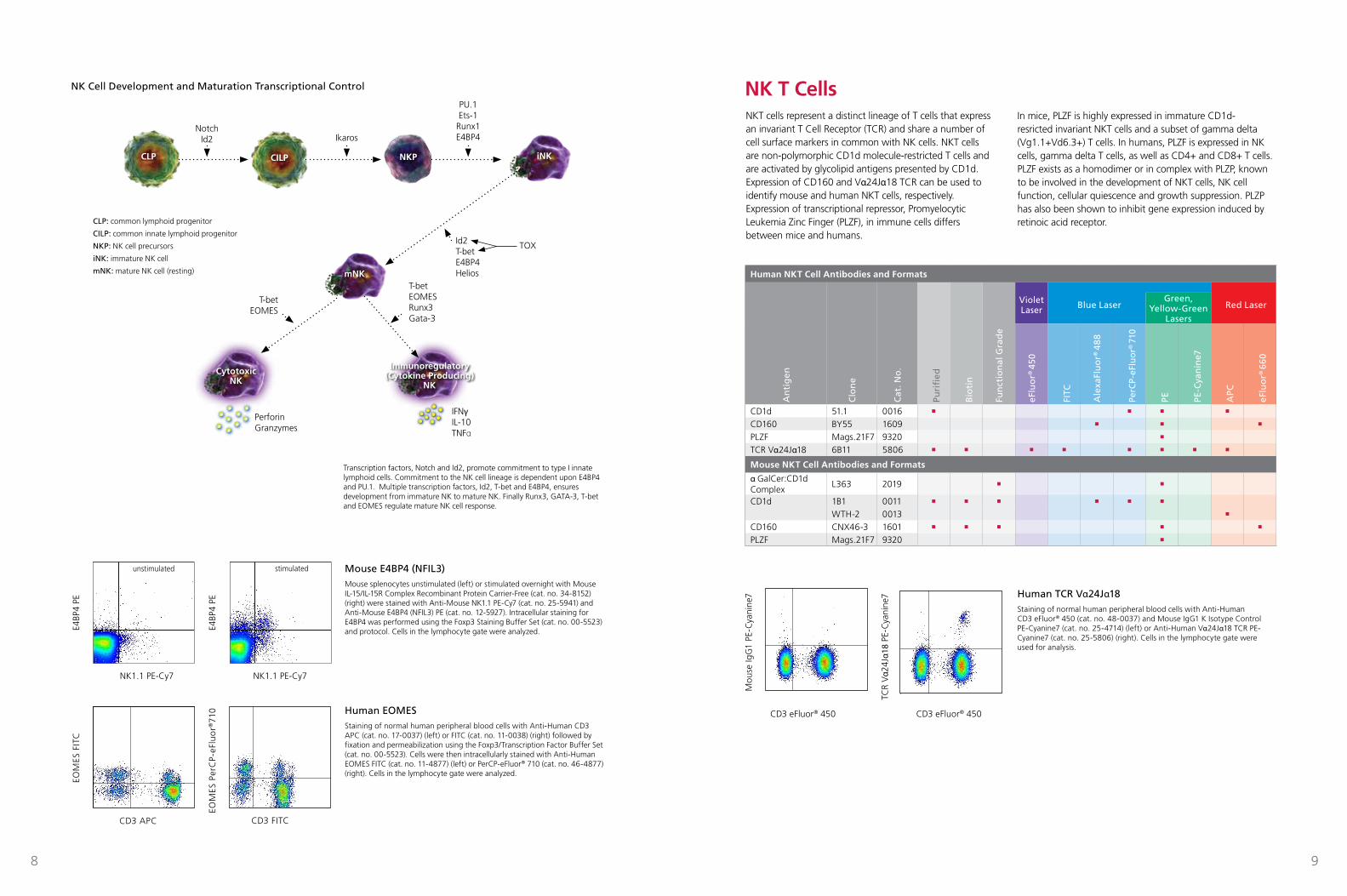
Human NKT Cell Antibodies and Formats
An
tig
en
Clo
ne
Cat
. No
.
Violet Laser Blue Laser Red Laser
Puri
fied
Bio
tin
Fun
ctio
nal
Gra
de
Green, Yellow-Green
Lasers
eFlu
or®
450
FITC
Ale
xaFl
uo
r® 4
88
PerC
P-eF
luo
r® 7
10
PE PE-C
yan
ine7
APC
eFlu
or®
660
CD1d 51.1 0016 n n n n
CD160 BY55 1609 n n n
PLZF Mags.21F7 9320 n
TCR Vα24Jα18 6B11 5806 n n n n n n n n
Mouse NKT Cell Antibodies and Formats
α GalCer:CD1d Complex
L363 2019 n n
CD1d 1B1 0011 n n n n n n
WTH-2 0013 n
CD160 CNX46-3 1601 n n n n n
PLZF Mags.21F7 9320 n
NKT cells represent a distinct lineage of T cells that express an invariant T Cell Receptor (TCR) and share a number of cell surface markers in common with NK cells. NKT cells are non-polymorphic CD1d molecule-restricted T cells and are activated by glycolipid antigens presented by CD1d. Expression of CD160 and Vα24Jα18 TCR can be used to identify mouse and human NKT cells, respectively. Expression of transcriptional repressor, Promyelocytic Leukemia Zinc Finger (PLZF), in immune cells differs between mice and humans.
In mice, PLZF is highly expressed in immature CD1d-resricted invariant NKT cells and a subset of gamma delta (Vg1.1+Vd6.3+) T cells. In humans, PLZF is expressed in NK cells, gamma delta T cells, as well as CD4+ and CD8+ T cells. PLZF exists as a homodimer or in complex with PLZP, known to be involved in the development of NKT cells, NK cell function, cellular quiescence and growth suppression. PLZP has also been shown to inhibit gene expression induced by retinoic acid receptor.
CLP CILP iNKNKP
NK Cell Development and Maturation Transcriptional Control
Human TCR Vα24Jα18Staining of normal human peripheral blood cells with Anti-Human CD3 eFluor® 450 (cat. no. 48-0037) and Mouse IgG1 K Isotype Control PE-Cyanine7 (cat. no. 25-4714) (left) or Anti-Human Vα24Jα18 TCR PE-Cyanine7 (cat. no. 25-5806) (right). Cells in the lymphocyte gate were used for analysis.
CD3 eFluor® 450
Mou
se Ig
G1
PE-C
yani
ne7
CD3 eFluor 450
v al
pha
24 J
alp
ha T
CR
PE-
Cya
nine
7
CD3 eFluor 450
Mou
se Ig
G1
PE-C
yani
ne7
CD3 eFluor® 450
TCR
Vα2
4Jα1
8 PE
-Cya
nine
7
CD3 eFluor 450
v al
pha
24 J
alp
ha T
CR
PE-
Cya
nine
7
CD3 eFluor 450
Mou
se Ig
G1
PE-C
yani
ne7
Human EOMESStaining of normal human peripheral blood cells with Anti-Human CD3 APC (cat. no. 17-0037) (left) or FITC (cat. no. 11-0038) (right) followed by fixation and permeabilization using the Foxp3/Transcription Factor Buffer Set (cat. no. 00-5523). Cells were then intracellularly stained with Anti-Human EOMES FITC (cat. no. 11-4877) (left) or PerCP-eFluor® 710 (cat. no. 46-4877) (right). Cells in the lymphocyte gate were analyzed.
CD3 FITC
Eom
es P
erC
P-e7
10
EOM
ES P
erC
P-eF
luo
r®71
0
CD3 FITCCD3 APC
Eom
es F
ITC
EOM
ES F
ITC
CD3 APC
Transcription factors, Notch and Id2, promote commitment to type I innate lymphoid cells. Commitment to the NK cell lineage is dependent upon E4BP4 and PU.1. Multiple transcription factors, Id2, T-bet and E4BP4, ensures development from immature NK to mature NK. Finally Runx3, GATA-3, T-bet and EOMES regulate mature NK cell response.
NK1.1 PE-Cy7
E4BP
4 PE
NK1.1 PE-Cy7
E4BP
4 PE
NK1.1 PE-Cy7 NK1.1 PE-Cy7
E4BP
4 PE
E4BP
4 PE
Mouse E4BP4 (NFIL3)Mouse splenocytes unstimulated (left) or stimulated overnight with Mouse IL-15/IL-15R Complex Recombinant Protein Carrier-Free (cat. no. 34-8152) (right) were stained with Anti-Mouse NK1.1 PE-Cy7 (cat. no. 25-5941) and Anti-Mouse E4BP4 (NFIL3) PE (cat. no. 12-5927). Intracellular staining for E4BP4 was performed using the Foxp3 Staining Buffer Set (cat. no. 00-5523) and protocol. Cells in the lymphocyte gate were analyzed.
NK1.1 PE-Cy7
E4BP
4 PE
NK1.1 PE-Cy7
E4BP
4 PE
mNK
Cytotoxic NK
Immunoregulatory (Cytokine Producing)
NK
unstimulated stimulated
NK T Cells
CLP: common lymphoid progenitor
CILP: common innate lymphoid progenitor
NKP: NK cell precursors
iNK: immature NK cell
mNK: mature NK cell (resting)
NotchId2 Ikaros
PU.1Ets-1
Runx1E4BP4
T-betEOMESRunx3Gata-3
PerforinGranzymes
IFNγIL-10TNFα
T-betEOMES
Id2T-betE4BP4Helios
TOX
10 11
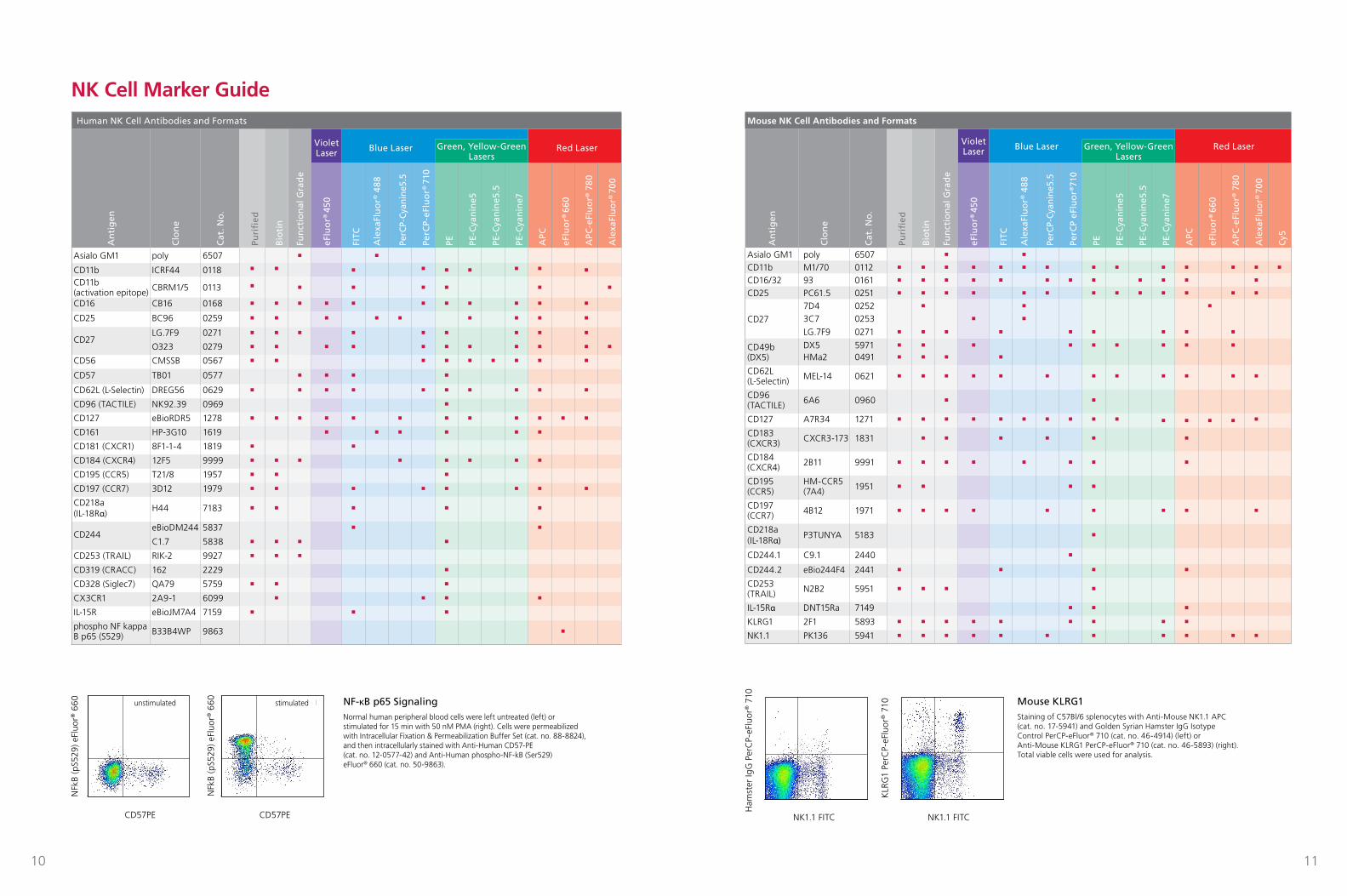
Human NK Cell Antibodies and FormatsA
nti
gen
Clo
ne
Cat
. No
.
Violet Laser Blue Laser Red Laser
Puri
fied
Bio
tin
Fun
ctio
nal
Gra
de
Green, Yellow-Green Lasers
eFlu
or®
450
FITC
Ale
xaFl
uo
r® 4
88
PerC
P-Cy
anin
e5.5
PerC
P-eF
luo
r® 7
10
PE PE-C
yan
ine5
PE-C
yan
ine5
.5
PE-C
yan
ine7
APC
eFlu
or®
660
APC
-eFl
uo
r® 7
80
Ale
xaFl
uo
r® 7
00
Asialo GM1 poly 6507 n n
CD11b ICRF44 0118 n n n n n n n n n
CD11b (activation epitope) CBRM1/5 0113 n n n n n n n
CD16 CB16 0168 n n n n n n n n n n n
CD25 BC96 0259 n n n n n n n n n
CD27LG.7F9 0271 n n n n n n n n n
O323 0279 n n n n n n n n n n n
CD56 CMSSB 0567 n n n n n n n n n
CD57 TB01 0577 n n n n
CD62L (L-Selectin) DREG56 0629 n n n n n n n n n n
CD96 (TACTILE) NK92.39 0969 n
CD127 eBioRDR5 1278 n n n n n n n n n n n n
CD161 HP-3G10 1619 n n n n n n
CD181 (CXCR1) 8F1-1-4 1819 n n
CD184 (CXCR4) 12F5 9999 n n n n n n n n
CD195 (CCR5) T21/8 1957 n n n
CD197 (CCR7) 3D12 1979 n n n n n n n n
CD218a (IL-18Rα)
H44 7183 n n n n n
CD244eBioDM244 5837 n n
C1.7 5838 n n n n
CD253 (TRAIL) RIK-2 9927 n n n
CD319 (CRACC) 162 2229 n
CD328 (Siglec7) QA79 5759 n n n
CX3CR1 2A9-1 6099 n n n n
IL-15R eBioJM7A4 7159 n n n
phospho NF kappa B p65 (S529) B33B4WP 9863 n
Mouse NK Cell Antibodies and Formats
An
tig
en
Clo
ne
Cat
. No
.
Violet Laser Blue Laser Red Laser
Puri
fied
Bio
tin
Fun
ctio
nal
Gra
de
Green, Yellow-Green Lasers
eFlu
or®
450
FITC
Ale
xaFl
uo
r® 4
88
PerC
P-Cy
anin
e5.5
PerC
P eF
luo
r®71
0
PE PE-C
yan
ine5
PE-C
yan
ine5
.5
PE-C
yan
ine7
APC
eFlu
or®
660
APC
-eFl
uo
r® 7
80
Ale
xaFl
uo
r® 7
00
Cy5
Asialo GM1 poly 6507 n n
CD11b M1/70 0112 n n n n n n n n n n n n n n
CD16/32 93 0161 n n n n n n n n n n n n
CD25 PC61.5 0251 n n n n n n n n n n n n n
CD27
7D4 0252 n n n
3C7 0253 n n
LG.7F9 0271 n n n n n n n n n
CD49b (DX5)
DX5 5971 n n n n n n n n n
HMa2 0491 n n n n
CD62L (L-Selectin) MEL-14 0621 n n n n n n n n n n n n
CD96 (TACTILE) 6A6 0960 n n
CD127 A7R34 1271 n n n n n n n n n n n n n n n
CD183 (CXCR3) CXCR3-173 1831 n n n n n n
CD184 (CXCR4) 2B11 9991 n n n n n n n n
CD195 (CCR5)
HM-CCR5 (7A4) 1951 n n n n
CD197 (CCR7) 4B12 1971 n n n n n n n n n
CD218a (IL-18Rα) P3TUNYA 5183 n
CD244.1 C9.1 2440 n
CD244.2 eBio244F4 2441 n n n n
CD253 (TRAIL) N2B2 5951 n n n n
IL-15Rα DNT15Ra 7149 n n n
KLRG1 2F1 5893 n n n n n n n n n
NK1.1 PK136 5941 n n n n n n n n n n n
Mouse KLRG1Staining of C57Bl/6 splenocytes with Anti-Mouse NK1.1 APC (cat. no. 17-5941) and Golden Syrian Hamster IgG Isotype Control PerCP-eFluor® 710 (cat. no. 46-4914) (left) or Anti-Mouse KLRG1 PerCP-eFluor® 710 (cat. no. 46-5893) (right). Total viable cells were used for analysis.
NK1.1 FITC NK1.1 FITC
Ham
ster
IgG
Per
CP-
eFlu
or® 7
10
KLR
G1
PerC
P-eF
luor
® 7
10
NK1.1 FITC
Ham
ster
IgG
Per
CP-
eFlu
or 7
10
NK1.1 FITC
KLR
G1
PerC
P-eF
luor
710
NK1.1 FITC
Ham
ster
IgG
Per
CP-
eFlu
or 7
10
NK1.1 FITC
KLR
G1
PerC
P-eF
luor
710
CD57 PE
pNF
kB (
S52
9) e
Flu
or 6
60
CD57 PE
pNF
kB (
S52
9) e
Flu
or 6
60 u n s t imu la ted s t i m u l a t e d
CD57 PE
pNF
kB (
S52
9) e
Flu
or 6
60
CD57 PE
pNF
kB (
S52
9) e
Flu
or 6
60 u n s t imu la ted s t i m u l a t e d NF-κB p65 SignalingNormal human peripheral blood cells were left untreated (left) or stimulated for 15 min with 50 nM PMA (right). Cells were permeabilized with Intracellular Fixation & Permeabilization Buffer Set (cat. no. 88-8824), and then intracellularly stained with Anti-Human CD57-PE (cat. no. 12-0577-42) and Anti-Human phospho-NF-kB (Ser529) eFluor® 660 (cat. no. 50-9863).
CD57PE CD57PE
NFk
B (p
S529
) eFl
uor®
660
NFk
B (p
S529
) eFl
uor®
660unstimulated stimulated
NK Cell Marker Guide