Embed Size (px)
Citation preview

1
Natural history, quality of life and outcome in cardiac
ATTR amyloidosis
Running Title: Outcome in cardiac ATTR amyloidosis
First Author: Dr Thirusha Lane RN PhD, et al.
Corresponding Author: Professor JD Gillmore, National Amyloidosis Centre, Division of
Medicine, University College London, Royal Free Campus, Rowland Hill Street, London
NW3 2PF, United Kingdom.
Tel: +44 (0)20 7433 2726 E-mail: [email protected]
Fax: +44 (0)20 74332844
Word count: 6813

2
Background – Cardiac transthyretin amyloidosis (ATTR-CM) is an increasingly recognised
cause of heart failure in older individuals. We sought to characterise the natural history of
ATTR-CM and compare outcomes and quality of life among patients with acquired
(ATTRwt-CM) and hereditary (hATTR-CM) forms of the disease.
Methods – We studied 711 patients with wild-type ATTRwt-CM, 205 with hereditary ATTR-
CM associated with the V1221 variant (V122I-hATTR-CM) and 118 with non-V122I-
hATTR-CM at the UK National Amyloidosis Centre between 2000 and 2017. Patients
underwent prospective protocolized evaluations comprising assessment of cardiac
parameters, functional status by 6-minute walk test, quality of life (QoL) according to Kansas
City Cardiomyopathy Questionnaire (KCCQ), and survival. Hospital service usage pre- and
post-diagnosis was established using English central health records in a subset of patients.
Results - There was substantial diagnostic delay, with patients using hospital services a
median (interquartile range) of 17 (9-27) times during the 3 years before diagnosis by which
time QoL was poor; diagnosis of ATTRwt-CM was delayed more than 4 years after
presentation with cardiac symptoms in 42% of cases. Patients with V122I-hATTR-CM were
more impaired functionally (p<0.001) and had worse measures of cardiac disease (p<0.001)
at the time of diagnosis, a greater decline in QoL, and poorer survival (p<0.001) compared to
the other subgroups.
Conclusions – ATTR-CM is an inexorably progressive and eventually fatal cardiomyopathy
associated with poor QoL. Diagnosis is often delayed for many years after symptoms
develop. Improved awareness and wider use of recently validated diagnostic imaging
methods are urgently required for patients to benefit from recent therapeutic developments.

3
Keywords: Amyloid, Transthyretin, ATTR, Cardiomyopathy
Clinical Perspective
1) What is new?
Non-biopsy diagnosis of cardiac ATTR amyloidosis through repurposed bone
scintigraphy and cardiac magnetic resonance imaging has lately led to an upsurge in
diagnoses, but still at an advanced stage in many patients.
Patients with the TTR V122I gene variant, who are mostly of Afro-Caribbean
ethnicity, typically have worse disease at diagnosis with poorer ejection fraction, renal
function and performance status, and poorer survival.
Both hereditary and acquired cardiac ATTR amyloidosis are associated with markedly
poor quality of life at the time of diagnosis, the typical patient having attended
hospital a median of 17 times during the prior three years.
2) Clinical Implications
Frequent hospital attendances with symptoms attributable to cardiac ATTR
amyloidosis or evidence of unexplained heart wall thickening provide potential
opportunities for earlier diagnosis using bone scintigraphy and cardiac magnetic
resonance imaging.
Index of suspicion of the diagnosis should be especially high among Afro-Caribbean
patients, of whom about 4 per cent possess the disease-associated TTR V1221 variant;
TTR gene sequencing is a valuable aid to diagnosis.
New disease-modifying therapies that inhibit production of TTR protein or its
conversion to ATTR amyloid show great promise, underscoring the need for
improved awareness of the disease.

4
Introduction
Transthyretin amyloidosis cardiomyopathy (ATTR-CM) has of late been increasingly
recognised as a cause of heart failure in older individuals, reflecting advances in imaging
technology and greater awareness of the disorder. Whilst it has long been known from autopsy
studies that amyloid deposits derived from plasma transthyretin (TTR) are present in the hearts
of up to 25% of elderly people,1, 2 the associated clinical syndrome, predominantly comprising
older men with restrictive cardiomyopathy that is often preceded by or associated with carpal
tunnel syndrome,3, 4 lumbar canal stenosis5 and tendinopathy,6 is still widely perceived as a rare
disease.
In addition there are far less common dominantly inherited forms of ATTR amyloidosis
(hATTR), associated with more than 130 different mutations in the TTR gene,7, 8 which present
variously from the third decade of life onwards with permutations of peripheral and autonomic
neuropathy, sometimes involvement of other organ systems, as well as causing amyloid
cardiomyopathy in most instances.9 One particular TTR gene variant, V122I, occurs in about
4% of black individuals,10 and is associated with increased susceptibility to development of
late onset predominant cardiac amyloidosis that closely mimics wild-type ATTR-CM.10, 11 The
recent upsurge in diagnosis of ATTR amyloidosis has been driven by repurposed bone
scintigraphy and cardiac MRI, both of which yield highly characteristic findings in ATTR-
CM,12, 13 but it nevertheless remains probable that heart failure, conduction disease and
arrhythmias due to the disorder are far more prevalent than currently recognised.
ATTR-CM progresses inexorably to death, typically within a few years,14 and hitherto
has not been treatable. However, several very promising new therapies are now in development
including tafamidis,15 a small molecule drug that promotes maintenance of TTR protein in its
normal soluble non-amyloid conformation, and two novel inhibitors of TTR production,
inotersen,16 an antisense oligonucleotide, and patisiran,17 a gene silencing RNA therapy.

5
In contrast with much progress in diagnostic imaging technology and therapy, there
remains a relative paucity of data on the patient pathway and natural history of ATTR-CM, as
well as its effects on quality of life (QoL). We aimed to study these themes in our cohort of
more than 1000 ATTR-CM patients enrolled into a protocolized observational study at the UK
National Amyloidosis Centre (NAC); the study included a specific aim to characterise the
pathways and phenotypes of patients with wild-type versus V122I associated ATTR-CM, in
light of inconsistent previous data on their features and relative severity.
Methods
The data that support the findings of this study are available from the corresponding author
upon reasonable request.
Patients
Patients referred to the NAC between 2000 and 2017 in whom ATTR-CM was confirmed on
the basis of validated diagnostic criteria,18 were invited to participate in a prospective
protocolized clinical follow up program comprising systematic evaluation of cardiac
parameters, functional status, QoL and survival. Briefly, the diagnosis of ATTR-CM was
established on the basis of presence of symptoms of heart failure together with a characteristic
amyloid echocardiogram and either direct endomyocardial biopsy (EMB) proof of ATTR
amyloid, or presence of ATTR amyloid in an extra-cardiac biopsy along with cardiac uptake
on 99mTc-DPD scintigraphy, or Perugini grade 2 or 3 cardiac uptake on 99mTc-DPD scintigraphy
in the absence of either an abnormal serum free light chain ratio or a monoclonal protein in the
serum or urine by immunofixation.18 All patients who were diagnosed with ATTR-CM
underwent sequencing of the TTR gene at the time of diagnosis, as previously described.8
Patients who did not consent to the protocolized clinical follow up program at NAC and those

6
who received disease modifying therapy including orthotopic liver transplantation for hATTR
amyloidosis, TTR stabilizer therapy for more than 6 months, or a TTR-lowering therapy
(within the context of a clinical trial) were excluded from the study.
Patients were managed in accordance with the Declaration of Helsinki and provided
written informed consent for retrospective analysis and publication of their data with approval
from the Royal Free Hospital ethics committee (ref: 06/Q0501/42).
Protocolized Evaluations
From the time of diagnosis, patients were systematically evaluated at the NAC on a 6-monthly
basis. Each study evaluation comprised the following; a full clinical history and examination,
routine hematology, serum and urine biochemistry including measurement of N-terminal brain
pro-natriuretic peptide (NT-proBNP), troponin T (baseline only), electrocardiography, and
detailed echocardiography.
In 2010, functional testing and assessments of quality of life were introduced into the
clinic protocol; patients were routinely asked to perform a standardized 6-minute walk test
(6MWT) and complete the Kansas City Cardiomyopathy Questionnaire (KCCQ) quality of life
assessment tool at each study evaluation. Patients who were unable to complete a 6MWT at
follow up were imputed as ‘0 meters’ for analyses of change in 6MWT distance across time.
Hospital Episode Statistics
Data on hospital episode statistics (HES) in England, defined as publicly funded hospital
service usage within the NHS in England, were obtained from NHS Digital (NHSD). Study
patients were identified on the basis of their unique NHS number. Hospital service usage
included inpatient (including surgical procedures), outpatient and A&E (emergency room)

7
services. All attendances were for medical issues and did not include those for occupational
or physical therapy.
In order to ensure robustness of the presented data, only patients in whom there was a
complete three year observation window across all three care settings pre-diagnosis of ATTR-
CM (n=534, diagnosed April 2010-August 2016) and a complete three year observation
window across all three care settings post-diagnosis of ATTR-CM (n=364, diagnosed April
2007-August 2013) were included in analyses of hospital service usage.
Access to HES data was secured via NHS Digital’s Data Access Release Service
(DARS) (ref: DARS-NIC-60624-B1R2Q) and included relevant permissions and approvals
from Research Ethics (ref: 16/LO/1065), and Confidentiality Advisory Group (CAG), for the
linkage of datasets under Section 251 of the Health and Social Care act 2014 (ref:
16/CAG/0081). The Independent Group Advising on the Release of Data (IGARD) at NHS
Digital also approved the use of HES data for this study.
99mTc-DPD scintigraphy
Patients were administered intravenously with 700 MBq of 99mTc-DPD, scanned 3 hours later
on either a General Electric (GE) Infinia Hawkeye 4 or Discovery 670 gamma camera and
whole body and cardiac a SPECT-CT images were acquired, as previously described.19
Intensity of myocardial uptake at 99mTc-DPD scintigraphy was graded 0–3 by two independent
observers according to the established Perugini criteria.20 Rare discrepancies in scoring were
resolved by consensus within a multi-disciplinary meeting attended by a panel of experts.
Histology
Biopsies were stained with Congo red by the method of Puchtler et al,21 and
immunohistochemistry was performed with a panel of antibodies including an antibody against

8
TTR, as previously described.22 After 2010, the presence of ATTR amyloid was further
corroborated by proteomic analysis, as previously described.23
Echocardiography
Echocardiography was performed at baseline and at every NAC evaluation thereafter by three
echocardiographers on GE Vivid 7 machines using EchoPac software. All echocardiograms
were reported by two independent observers and discrepancies were reviewed by a group of
clinical experts in amyloid echocardiography within a multi-disciplinary meeting.
Quality of life
After 2010, health-related QoL was measured at each evaluation by Kansas City
Cardiomyopathy Questionnaire (KCCQ) score.24 Briefly, the KCCQ is a 23-item patient-
reported measure that quantifies physical function, symptoms (frequency, severity and
stability), social function, self-efficacy and quality of life. An overall summary score is derived
from the physical and social function, symptom and quality of life domains. Scores are
represented on a scale from 0 to 100, with higher scores reflecting better health status. A
median difference of 5 points on the overall summary score is considered a clinically
significant change; a decline of 10 points is considered prognostically significant. It is
noteworthy that 88% of patients who performed the KCCQ assessment were diagnosed from
2012 onwards, and that the V122I-hATTR-CM genotypic subgroup were proportionately
under-represented (15% vs 20% in the whole cohort) and the ATTRwt-CM over-represented
(75% vs 69% in the whole cohort) amongst those completing the KCCQ assessment.
Statistical Analyses
9
All mortality data were obtained from the UK Office of National Statistics. Date of Censor
was 1st October 2017. The three genotypic sub-groups of interest were wild-type ATTR
amyloidosis (ATTRwt-CM), V122I-associated hereditary ATTR amyloidosis (V122I-hATTR-
CM) and non-V122I-associated hereditary ATTR amyloidosis (non-V122I-hATTR-CM). As
a number of the numerical variables had skewed distributions, a Kruskal Wallis test was used
to compare the distributions of each of the numerical variables at baseline in the three genotypic
subgroups. A significant result was followed by Bonferroni corrected Mann Whitney pairwise
comparisons to establish where the differences lay. A chi square test was used to compare the
proportion of males in the three groups, and this was followed by Bonferroni corrected pairwise
chi square comparisons. A mixed-effect linear regression model with main effects and
interactions was used to analyse the longitudinal data to assess the effect of genotype on the
outcome measures 6MWT, eGFR and NT-proBNP over time. Baseline variables that were
statistically significant at the 0.10 alpha level in univariable Cox proportional hazards models
were entered into a multivariable Cox model to investigate the factors independently predictive
of survival. Variables that were not significant in the initial multivariable model were removed
and an updated multivariable Cox model was re-analysed and presented. The proportional
hazards assumption was checked and confirmed where appropriate. Kaplan Meier survival
curves were drawn. All data were analysed using Stata software (StataCorp. 2017. Stata
Statistical Software: Release 15. College Station, TX: StataCorp LLC). A significance level
of 0.05 was used for all hypothesis tests unless otherwise stated.
Results
Characteristics of the cohort at baseline and comparison of genotypes
There were 1034 patients in the whole study cohort, 711 (69%) with wild-type ATTR-CM
(ATTRwt-CM), 205 (20%) with V122I-associated hATTR-CM and 118 (11%) with non-

10
V122I-associated hATTR-CM comprising 80% of all referrals who were diagnosed with
ATTR-CM at the National Amyloidosis Centre during the study period. The main reason for
exclusion from the study was a refusal to consent to the protocolized program of follow up at
the NAC. Other reasons, almost exclusively among patients with non-V122I-hATTR-CM,
included receipt of disease modifying therapy (liver transplantation, diflunisal, anti-sense
oligonucleotide therapy or RNA inhibitor therapy). There was no difference in disease-related
parameters or disease severity between patients with ATTR-CM who were included and
excluded from the study. Of the study patients with non-V122I-associated hATTR-CM, the
majority (n=97, 83%) had the T60A variant, although other pathogenic variants were
represented; 96% of non-V122I-associated hATTR-CM patients had coexistent, symptomatic
amyloid polyneuropathy at the time of diagnosis as opposed to <5% of patients within the other
genotypic subgroups. The number of new diagnoses of ATTR-CM each year stratified by
genotype are shown in Figure 1 which highlights, in particular, the exponential recent rise in
identification of ATTRwt-CM. Male gender dominated in all three subgroups: 94%, 71% and
69% respectively, although the proportion of males was significantly higher in ATTRwt-CM
than hATTR-CM (p<0.001). There was no association between gender and survival and nor
were there significant differences in baseline disease characteristics between males and
females. The characteristics of the cohort at the time of diagnosis of cardiac ATTR amyloidosis
are shown in Table 1. At diagnosis, patients with ATTRwt-CM, V122I-associated and non-
V122I-associated hATTR-CM differed significantly in terms of age, left ventricular ejection
fraction (LVEF), renal function and functional capacity (Table 1). Median age at diagnosis
was significantly higher in ATTRwt-CM (79 years) and V122I-hATTR-CM (77 years) than in
non-V122I-hATTR-CM (67 years) (p<0.001). NT-proBNP concentration was also
significantly higher at baseline in patients with ATTRwt-CM and V122I-hATTR-CM
compared to those with non-V122I-hATTR-CM (p<0.001 for both comparisons, Table 1). At

11
the time of diagnosis, LVEF and functional status as measured by 6MWT distance were
significantly worse among V122I-hATTR-CM patients compared to the two other genotypic
subgroups (Table 1).
Survival from baseline
Median patient survival from diagnosis by Kaplan-Meier analysis was 31 months in patients
with V122I-hATTR-CM compared to 69 months among patients with ATTRwt-CM
(p<0.0001) and 57 months in non-V122I hATTR-CM. NAC ATTR Disease Stage, calculated
according to previously published cutoffs in NT-proBNP (3000 ng/L) and eGFR (45 ml/min)
was, as expected, strongly predictive of survival across all genotypes (as well as within each
genotype) with median survival of 68.0, 42.4 and 25.9 months among patients with Stage I,
Stage II and Stage III disease respectively (all comparisons p<0.001). Survival of patients
diagnosed with ATTRwt-CM from 2012, the year that 99mTc-DPD scintigraphy was routinely
introduced into the diagnostic algorithm for ATTR-CM at UK NAC and following which 75%
of patients were diagnosed non-invasively, was significantly better (median 60.2 months) than
among patients diagnosed with ATTRwt-CM pre-2012 (median 46.3 months) when histology
was usually required to establish the diagnosis (63% diagnosed via biopsy, usually EMB)
(Figure 2B). Correspondingly, a higher proportion of ATTRwt-CM patients diagnosed pre-
2012 had Stage III Disease (20% vs 16%) and a lower proportion had Stage I Disease (41% vs
43%) at the time of diagnosis compared to patients with ATTRwt-CM diagnosed from 2012
onwards.
Univariable analyses showed that factors at diagnosis significantly predictive of
survival were age, NT-pro-BNP, left ventricular ejection fraction (LVEF), interventricular
septal diameter in diastole (IVSd), eGFR, 6MWT distance, modified BMI (mBMI), 24hr
urinary protein excretion, serum albumin, NAC ATTR Disease Stage, genotypic subgroup, and

12
date of diagnosis (Table 2). A multivariable model combining age, NAC ATTR Disease Stage,
LVEF, genotypic subgroup, and 6MWT distance at the time of diagnosis revealed that age
(HR, 1.037 per year; 95% CI, 1.008-1.067, p<0.011), NAC ATTR Disease Stage (HR 2.049;
CI 1.352-3.104, p=0.001 for Stage II and HR 3.705; CI 2.313-5.933, p<0.001 for Stage III
compared to Stage I), LVEF (HR 0.978 per 1% increase; 95% CI 0.963-0.993, p=0.003),
genotypic subgroup (HR 2.071; CI 1.415-3.031, p<0.001 for V122I-hATTR-CM and HR
2.727; CI 1.458-5.098, p=0.002 for non-V122I-hATTR-CM compared to ATTRwt-CM) and
6MWT distance (HR 0.881 per 50 meter increase; 95% CI 0.832-0.933, p<0.001) were
independently associated with patient survival (Table 2). Interestingly, a comparison of
outcomes between genotypic subgroups among patients with each of the three NAC ATTR
Disease Stages at the time of diagnosis showed that even within each category of Disease Stage,
V122I genotype was an independent predictor of death (HR for V122-hATTR-CM vs
ATTRwt-CM between 2 and 3, p<0.002 for all analyses). Survival among patients of African
ancestry (n=207) with ATTRwt-CM (n=13) was longer (median 40 months) than among those
with V122I-hATTR-CM (n=194) (median 31 months) but this did not reach statistical
significance (p=0.85), probably owing to the small number of patients with ATTRwt-CM. It
is noteworthy that only 6% (13/207) of the cohort who were of African ancestry had ATTRwt-
CM.
Longitudinal analyses of disease parameters and functional status during follow up
Given the prognostic importance of NT-proBNP and eGFR (the variables which determine
NAC ATTR Disease Stage) at the time of diagnosis, coupled with that of 6MWT distance,
longitudinal analyses of these three variables were undertaken with a comparison between
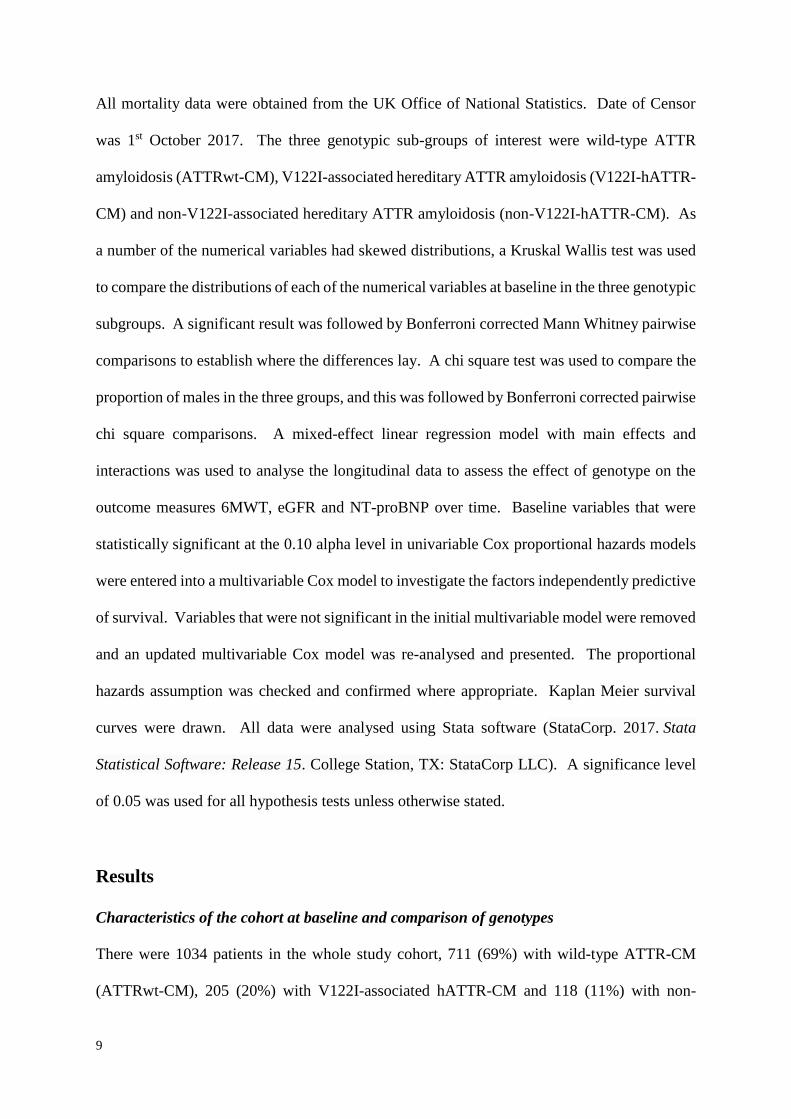
genotypic subgroups. NT-proBNP progressively increased whilst eGFR and 6MWT distance
declined during the first 2 years of follow up. There was a more rapid rate of rise in NT-

13
proBNP concentration among patients with V122I-hATTR-CM than both ATTRwt-CM
(p<0.001) and non-V122I-hATTR-CM (p=0.009) with no significant difference (p=0.884)
between the latter two genotypic subgroups (Figure 3). Mean rise in absolute NT-proBNP
concentration from diagnosis to 12 months was 842 ng/L (CI 551-1134) in patients with
ATTRwt-CM, 1043 ng/L (CI 329-1758) in patients with non-V122I-hATTR-CM and
2678 ng/L (CI 658-4698) in patients with V122I-hATTR-CM. The mean (range) rates of
decline in eGFR and decline in 6MWT distance from baseline over the course of 2 years were
remarkably consistent between different genotypic subgroups; 5 to 10 ml/min (6.5-9.9) and
approximately 100 metres (80-111) respectively (p=ns).
Hospitalisation episodes pre- and post-diagnosis of ATTR-CM
The subset of 534 English patients in whom complete data on hospital service usage were
available for the full three years prior to diagnosis of ATTR-CM attended hospital a median
(IQR) of 17 (9-27) times in the period, including a median (IQR) of 3 (1-5) inpatient hospital
admissions. The breakdown of hospital service usage year on year prior to diagnosis is shown
in Figure 4A. Median (IQR) diagnostic delay from first presentation with cardiac symptoms
was 39 (8-78) months in English patients with ATTRwt-CM with 42% waiting more than 4
years after first presentation with cardiac symptoms and a further 23% waiting between 6
months and 4 years for the diagnosis to be established. Only just over one-third of English
patients were diagnosed with ATTRwt-CM within 6 months of first presentation with cardiac
symptoms. Median (IQR) time from first presentation with cardiac symptoms to diagnosis of
hATTR-CM was 25 (4-60) months.
In the year after diagnosis of ATTR-CM (n=364), the median (IQR) number of hospital
inpatient episodes (admissions) per patient was 2 (1-5) with 30% of patients admitted as
inpatients to hospital at least 3 times during the period. The median (IQR) number of out-

14
patient and emergency room attendances per patient in the first year after diagnosis was 8 (5-
13) and 1 (1-2) respectively. The median (IQR) number of hospital inpatient admissions per
surviving patient was 2 (1-4) in the second year after diagnosis and 3 (1-5) in the third year
after diagnosis with no difference between genotypic subgroups (Figure 4B).
Quality of Life (QoL) by KCCQ
Overall KCCQ domain scores within the first 12 months of diagnosis, obtained from 158
patients, showed poor health-related QoL across all 3 genotypic subgroups of ATTR-CM
(Figure 5). The lowest scoring domains were physical limitation, social limitation and
symptom stability in all three cohorts.
The magnitude and direction of change of QoL scores in each domain were measured
in each cohort between 12 and 36 months. The direction of change of QoL scores was
overwhelmingly negative in all cohorts and for all domains. V122I-hATTR-CM patients
showed clinically significant deterioration in 7 of 10 domains, non-V122I-hATTR-CM patients
in 5 of 10 domains and ATTRwt-CM patients in 3 of 10 domains. Median changes in KCCQ
summary scores are shown in Table 3. Given that 88% of patients who completed the KCCQ
assessment were diagnosed from 2012 onwards, and that patients with ATTRwt-CM were
over-represented and those with V122I-hATTR-CM under-represented amongst those
completing the KCCQ assessment, it is likely that the overall decline in QoL following
diagnosis for the whole cohort would have been even more marked.
Discussion
This prospective observational study of more than 1000 patients with ATTR-CM undergoing
comprehensive follow-up at the National Amyloidosis Centre, which is centrally
commissioned as the single center in the UK for diagnosis and monitoring amyloidosis, has

15
yielded many new insights into the diagnosis, natural history, quality of life and outcome of
the condition. Firstly, it highlights the exponential increase during recent years in the number
of patients diagnosed with ATTR-CM, particularly ATTRwt-CM which is now diagnosed
more than twice as frequently than hATTR-CM. This presumably reflects the remarkable
sensitivity and increasingly widespread use of cardiac MRI and 99mTc-DPD (and 99mTc-PYP)
scintigraphy,12, 13 which in turn have fuelled increased awareness of the condition amongst
cardiologists. Despite this however, our data demonstrate huge delays in establishing the
diagnosis of ATTR-CM following presentation with cardiac symptoms, this taking more than
4 years in over 40% patients with ATTRwt-CM on a background of a median 17 hospital
attendances during the three years prior to diagnosis. This long delay in the face of a
progressive disease is likely to have contributed to the identified poor QoL by the time
diagnosis was finally established. On a more encouraging note, survival among patients
diagnosed with ATTRwt-CM since 2012 has been better than before this time, which in the
absence of disease-modifying therapy and coupled with the finding that a higher proportion of
such patients had NAC Stage I Disease and fewer had NAC Stage III Disease at the time of
diagnosis than patients diagnosed pre-2012, suggests that increased awareness of the disease
along with adoption of non-invasive diagnostic imaging techniques,18 is resulting in patients
being diagnosed earlier in the course of their disease. Nonetheless, given the evidence from
historical autopsy series that ATTR amyloid deposits are present up to 25% of elderly male
hearts, there seems little doubt that many individuals with ATTR-CM are currently not being
diagnosed during their lifetime. A challenge for the future will be differentiating clinically
significant from incidental myocardial ATTR amyloid deposits given the sensitivity of cardiac
scintigraphy and MRI for identifying them. In view of the prolonged survival and reduction
of hospitalizations recently reported in ATTR-CM with the TTR-stabilizing therapy, tafamidis,
(notwithstanding the comparatively low rate of hospitalizations throughout the ATTR-ACT

16
trial compared to that reported here),15 and the improvement in outcomes among patients with
hATTR amyloidosis reported with the TTR-lowering RNA inhibitor therapy, patisiran,17 there
is every prospect that awareness and early diagnosis will increase further.
Although access to healthcare in the UK is available free of charge to all residents, this
study demonstrated that patients with V122I-hATTR-CM had higher NT-proBNP, lower
LVEF, and poorer functional status than those with ATTRwt-CM and non-V122I-hATTR-CM
at the time of diagnosis, indicating that they had more advanced cardiac disease. Whilst this
may partly explain the reduced survival from the time of diagnosis in V122I-hATTR-CM
patients, our finding of a shorter delay from presentation with cardiac symptoms to diagnosis
among patients with hATTR-CM compared to ATTRwt-CM, coupled with the finding of
poorer outcomes among V122I-hATTR-CM even when stratified by NAC ATTR Disease
Stage, suggest that the disease biology may be inherently more aggressive. It is noteworthy
that overall QoL scores appeared to worsen more rapidly in hATTR-CM than ATTRwt-CM
which is likely to reflect the impact of neuropathy, present in 96% of study patients with non-
V122I-hATTR-CM, on physical performance and QoL, coupled with the generally poorer
outcomes in V122I-hATTR-CM compared to ATTRwt-CM.
The authors acknowledge a number of limitations to this study. More sensitive
echocardiographic parameters of ATTR-CM and of disease progression in the context of
ATTR-CM such as longitudinal strain by tissue doppler imaging, myocardial contraction
fraction and relative wall thickness were not included due to the fact that measurement of such
parameters were only routinely introduced into the UK National Amyloidosis Centre
echocardiography protocol within the last five years. Similarly, data on hospital episode
statistics were only available from patients living in England (excluding those from Northern
Ireland, Scotland and Wales) during the specified time period. Lastly, QoL and functional
status were available only in the subset of patients diagnosed with ATTR-CM after 2010,

17
having been introduced into the clinic protocol at that time. Nonetheless, we believe that the
data pertaining to subsets of the ATTR-CM cohort presented in this manuscript are
representative of the disease natural history in the population as a whole.
In summary, ATTR-CM is being increasingly recognized although there remains much
work to do to establish the diagnosis earlier in the course of the disease, the natural history of
which is gradual progression and death some 3-10 years from diagnosis. In this era of
promising novel therapies for ATTR amyloidosis, earlier diagnosis assumes greater importance
and argues strongly for inclusion of CMR and bone scintigraphy early in the investigative
pathway of patients with cardiac failure or cardiomyopathy of uncertain etiology.

18
VARIABLE ATTRwt
(Group 1)
V122I-hATTR
(Group 2)
Non-V122I hATTR
(Group 3)
p*
N (%)
711 (69) 205 (20) 118 (11)
Male Sex (%)
668 (94) 146 (71) 81 (69) p < 0.0001 (Gp 1 vs 2)
p < 0.0001 (Gp 1 vs 3)
Median (IQR) age (years)
79 (73-83) 77 (72-80) 67 (62–71)
p < 0.0001 (All comparisons)
Median (IQR) LVEF (mm)
58 (45-71) 49 (39-62) 53 (45-60) p < 0.0001 (Gp 1 vs 2)
p < 0.0001 (Gp 1 vs 3)
Median (IQR) IVSd (mm)
17 (15-18) 17 (16-18) 16 (15-18)
p = ns (All comparisons)
Median (IQR) NT-proBNP (ng/L)
3046 (1615-5472) 3337 (1668-6096) 2026 (871-4548) p < 0.0001 (Gp 1 vs 3)
p < 0.0001 (Gp 2 vs 3)
Median (IQR) eGFR (ml/min)
58 (45-71) 60 (47-75) 81 (62-100) p < 0.0001 (Gp 1 vs 3)
p < 0.0001 (Gp 2 vs 3)
Median (IQR) proteinuria (g/24 hr)
0.10 (0.10–0.20) 0.10 (0.10–0.20) 0.10 (0.10–0.13) p = ns (All comparisons)
Median (IQR) albumin (g/L)
44 (42-46) 42 (40-45) 43 (41-45)
p < 0.0001 (Gp 1 vs 2)
p = 0.01 (Gp 1 vs 3)
Median (IQR) mBMI
1151 (1044-1282) 1111 (976-1232) 1039 (922-1225) p < 0.0001 (Gp 1 vs 2)
p < 0.0001 (Gp 1 vs 3)
Median (IQR) 6MWT distance (meters)
345 (230-415) 260 (141-364) 374 (276-440) p < 0.0001 (Gp 1 vs 2)
p < 0.0001 (Gp 2 vs 3)
Median (IQR) 6MWT % expected for age
73 (53-88) 57 (34-76) 69 (57-84) p < 0.0002 (Gp 1 vs 2)
Table 1. Baseline characteristics of 1034 patients with cardiac transthyretin amyloidosis. ATTRwt = wild-type ATTR amyloidosis; V122I-hATTR = V122I-
associated hereditary ATTR amyloidosis; Non-V122I-hATTR = Non-V122I-associated hereditary ATTR amyloidosis; LVEF = left ventricular ejection fraction;
IVSd = intraventricular septal thickness in diastole; eGFR = estimated glomerular filtration rate (corrected for race); mBMI = modified Basal Metabolic Index;

19
MWT = 6-minute walk test. IQR = interquartile range. Gp = Group. * Comparison of numerical variables was by Kruskal Wallis test followed, where relevant,
by Bonferroni-corrected Mann Whitney pairwise comparisons.

20
UNIVARIABLE ANALYSIS
Variable HR for death 95% CI p
Age (per year increase) 1.04 1.025–1.054 <0.001
NT-proBNP (per log unit increase) 3.77 2.915–4.862 <0.001
LVEF (per 1% increase) 0.96 0.954–0.972 <0.001
IVSd (per mm increase) 1.11 1.065–1.149 <0.001
eGFR (per 10 ml/min increase) 0.82 0.776–0.863 <0.001
6MWT (per 50 meter increase) 0.81 0.767–0.0.847 <0.001
mBMI (per unit increase) 0.99 0.998-0.999 <0.001
24hr protein excretion (per 0.1 g increase) 1.07 1.039-1.095 <0.001
Serum albumin (per 1 g/L increase) 0.91 0.882-0.934 <0.001
Gender
Female
Male
1
0.91
0.692-1.204
0.519
NAC ATTR Disease Stage
Stage I
Stage II
Stage III
1
2.05
7.05
1.631-2.583
4.876-10.182
<0.001
<0.001
Genotype
ATTRwt
V122I-hATTR
Non-V122I-hATTR
1
1.91
0.80
1.526-2.385
0.582-1.100
<0.001
0.168
Date of Diagnosis
Pre-2012
2012 onwards
1
0.78
0.627-0.961
0.020
MULTIVARIABLE ANALYSIS
Variable HR for death 95% CI p
Age (per year increase) 1.04 1.008–1.067 0.011
NAC ATTR Disease Stage
Stage I
Stage II
Stage III
1
2.05
3.71
1.352-3.104
2.313-5.933
0.001
<0.001
LVEF (per 1% increase) 0.98 0.963–0.993 0.003
Genotype
ATTRwt
V122I-hATTR
Non-V122I-hATTR
1
2.07
2.73
1.415-3.031
1.458-5.098
<0.001
0.002
6MWT (per 50 meter increase in distance) 0.88 0.832–0.933 <0.001
Table 2. Univariable/Multivariable proportional hazards model of factors at the time of
diagnosis of cardiac ATTR amyloidosis that were predictive of survival. NAC ATTR

21
Disease Stage based on eGFR (< or ≥ 45 ml/min) and NT-proBNP (> or ≤ 3000 ng/L).
LVEF = left ventricular ejection fraction; IVSd = intraventricular septal thickness in diastole;
eGFR = estimated glomerular filtration rate; 6MWT = six-minute walk test; mBMI =
modified body mass index; ATTRwt = wild-type ATTR amyloidosis; V122I-hATTR =
V122I-associated hereditary ATTR amyloidosis; Non-V122I-hATTR = Non-V122I-
associated hereditary ATTR amyloidosis.

22
Clinical
Summary
Quality of Life Social
Limitation
Overall
Summary
ATTRwt 12m
Median (IQR)
Mean (CI)
64 (42-79)
61 (56-65)
58 (38-75)
57 (53-62)
50 (25-81)
51 (45-57)
57 (39-79)
58 (53-62)
ATTRwt 36m
Median (IQR)
Mean (CI)
55 (40-73)
56 (50-62)
63 (39-81)
59 (53-66)
38 (19-70)
43 (34-52)
52 (37-71)
54 (48-60)
Change ATTRwt 12m to 36m
Median (IQR)
Mean (CI)
-9 (-2 to -6)
-5 (-6 to -3)
4 (1 to 6)
2 (0 to 4)
-13 (-6 to -11)
-8 (-11 to -6)
-5 (-2 to -8)
-3 (-5 to -2)
V122I-hATTR 12m
Median (IQR)
Mean (CI)
62 (36-79)
57 (47-66)
75 (33-83)
61 (51-71)
42 (13-75)
45 (32-58)
67 (31-77)
55 (45-55)
V122I-hATTR 36m
Median (IQR)
Mean (CI)
51 (25-80)
52 (28-76)
42 (29-67)
50 (31-69)
25 (3-63)
35 (7-63)
44 (21-70)
47 (24-70)
Change V122I-hATTR 12m to 36m
Median (IQR)
Mean (CI)
-11 (-11 to 1)
-4 (-19 to 10)
-33 (-4 to -17)
-11 (-20 to -3)
-17 (-9 to -13)
-10 (-25 to 5)
-23 (-10 to -7)
-8 (-21 to 5)
Non-V122I-hATTR 12m
Median (IQR)
Mean (CI)
70 (39-84)
64 (48 to 80)
46 (23-71)
48 (30 to 67)
38 (25-66)
45 (27 to 63)
63 (31-71)
56 (40 to 72)
Non-V122I-hATTR 36m
Median (IQR)
Mean (CI)
42 (26-90)
54 (28-80)
46 (27-94)
56 (30-83)
31 (19-94)
45 (10-79)
41 (25-92)
54 (26-81)
Change Non-V122I-hATTR 12 to 36m
Median (IQR)
Mean (CI)
-28 (-13 to 5)
-10 (-21 to 0)
0 (4 to 23)
8 (0 to 16)
-6 (-6 to 28)
-1 (-17 to 16)
-22 (-6 to 21)
-3 (-14 to 9)
Table 3. Kansas City Cardiomyopathy Questionnaire summary scores at 12 and 36
months from diagnosis of cardiac ATTR amyloidosis in the three genotypic sub-groups.
Clinical summary score = physical limitation score + total symptom score; Overall summary
score = clinical summary score + Quality of Life score + Social Limitation score). A 5-point
change in the overall summary score reflects a clinically significant change in heart failure
status; a 10-point change is considered prognostically significant. Changes in score between
12 months post-diagnosis and 36 months post-diagnosis are shown. ATTRwt = wild-type
ATTR amyloidosis; V122I-hATTR = V122I-associated hereditary ATTR amyloidosis; Non-
V122I-hATTR = Non-V122I-associated hereditary ATTR amyloidosis.
23
Figure Legends
20002002
20042006
20082010
20122014
2016
0
20
40
60
80
100
120
140
160
ATTRwt
V122I-hATTR
Non-V122I-hATTR
Year
Nu
mb
er
of
ne
w d
iag
no
ses
of
AT
TR
-CM
Figure 1. Number of new diagnoses of cardiac transthyretin amyloidosis by year
according to genotypic subgroup. ATTRwt = wild-type ATTR amyloidosis; V122I-hATTR
= V122I-associated hereditary ATTR amyloidosis; Non-V122I-hATTR = Non-V122I-
associated hereditary ATTR amyloidosis.

24
A)
0 20 40 60 80 1000
10
20
30
40
50
60
70
80
90
100
Non-V122I-hATTR
V122I-hATTR
ATTRwt
Time (months)
% s
urv
iva
l
Numbers at risk
Non-V122I-hATTR 118 87 52 34 14 7
V122I-hATTR 205 122 42 18 7 3
ATTRwt 711 415 188 76 24 2
* **
Non-V122I hATTR
Figure 2. A) Kaplan-Meier survival stratified by genotype for the whole cohort
(*p<0.0001; **p<0.0001). B) Survival of patients with ATTRwt-CM stratified by year of
diagnosis (*p=0.009). ATTRwt = wild-type ATTR amyloidosis; V122I-hATTR = V122I-
associated hereditary ATTR amyloidosis; Non-V122I-hATTR = Non-V122I-associated
hereditary ATTR amyloidosis.
25

26
Figure 3. Change in NT-ProBNP, six-minute walk test distance and estimated glomerular
filtration rate from baseline to 12, 24 and 36 months stratified by genotypic subgroup. A)
Mean change (with 95% CI) in NT-proBNP concentration from baseline to 12, 24 and 36
months. The mean rate of rise in NT-proBNP concentration was significantly greater in V122I-
hATTR-CM than in both ATTRwt-CM (p<0.001) and non-V122I-hATTR-CM (p<0.009) but
was not significantly different between the latter genotypic sub-groups (p=0.884). B) Mean
change in six minute walk test (6MWT) distance from baseline to 12, 24 and 36 months
(p>0.05). C) Mean change in estimated glomerular filtration rate (eGFR) from baseline to 12,
24 and 36 months (p>0.05). ATTRwt = wild-type ATTR amyloidosis; V122I-hATTR = V122I-
associated hereditary ATTR amyloidosis; Non-V122I-hATTR = Non-V122I-associated
hereditary ATTR amyloidosis.

27
Figure 4. English National Health Service (NHS) hospital services usage. A) English NHS
hospital services usage covering emergency room (ER), inpatient admissions (IP) and
outpatient services (OP) in the three years prior to diagnosis of ATTR-CM. B) English NHS
hospital services usage, covering emergency room (ER), inpatient admissions (IP) and
outpatient services (OP) during the first 3 years after diagnosis of ATTR-CM (percentages
adjusted for surviving patients at each timepoint).

28
Figure 5. Health-related quality of life as measured by the Kansas City Cardiomyopathy
Questionnaire in 158 patients within the first 12 months of diagnosis stratified by
genotype. A score of 100 indicates perfect health. ATTRwt = wild-type ATTR amyloidosis;
V122I-hATTR = V122I-associated hereditary ATTR amyloidosis; Non-V122I-hATTR = Non-
V122I-associated hereditary ATTR amyloidosis.

29
Authors
Dr Thirusha Lane1
Dr Marianna Fontana1
Dr Ana Martinez-Naharro1
Dr Candida Cristina Quarta MD1
Dr Carol J Whelan MD1
Dr Aviva Petrie2
Dr Dorota M Rowczenio1
Ms Janet A Gilbertson1
Mr David F Hutt1
Dr Tamer Rezk1
Ms Svetla G Strehina1
Ms Joan Caringal-Galima1
Dr Richa Manwani1
Dr Faye A Sharpley1
Professor Ashutosh D Wechalekar1
Dr Helen J Lachmann1
Dr Shameem Mahmood1
Dr Sajitha Sachchithanantham1
Mr Edmund PS Drage3
Mr Harvey D Jenner3
Ms Rosie McDonald3
Dr Ottavia Bertolli3
Mr Alan Calleja3
Professor Philip N Hawkins1
Professor Julian D Gillmore1
30
Acknowledgements
We thank our many physician colleagues for referring and caring for the patients. We thank
Pui Lun Yip, Department of Medicine, Queen Elizabeth Hospital, Hong Kong SAR, for
additional data collection in response to the reviewers’ comments.
Sources of Funding
Core support for the National Amyloidosis Centre is provided by National Health Service
England, the UK National Institute for Health Research Biomedical Research Centre and Unit
Funding Scheme. For the HES analysis component of the study, IQVIA’s participation was
funded by GlaxoSmithKline, (who also fund other studies executed by IQVIA).
Disclosures
Dr Lane has received consultancy fees from Alnylam Pharmaceuticals and Eidos Therapeutics
Inc. Dr Whelan serves on advisory boards for Alnylam Pharmaceuticals and Akcea
Therapeutics. Professor Gillmore serves on advisory boards for Alnylam Pharmaceuticals,
Akcea Therapeutics, Pfizer Inc. and GlaxoSmithKline. The other authors report no conflicts.
Affiliations
1 National Amyloidosis Centre, Division of Medicine, University College London, London,
United Kingdom
2 Eastman Dental Institute, University College London, London, United Kingdom
3 IQVIA, London, United Kingdom

31
References
1. Cornwell GG, 3rd, Murdoch WL, Kyle RA, Westermark P and Pitkanen P. Frequency
and distribution of senile cardiovascular amyloid. A clinicopathologic correlation. Am J Med.
1983;75:618-623.
2. Tanskanen M, Peuralinna T, Polvikoski T, Notkola IL, Sulkava R, Hardy J, Singleton
A, Kiuru-Enari S, Paetau A, Tienari PJ and Myllykangas L. Senile systemic amyloidosis
affects 25% of the very aged and associates with genetic variation in alpha2-macroglobulin
and tau: a population-based autopsy study. Ann Med. 2008;40:232-239.
3. Pinney JH, Whelan CJ, Petrie A, Dungu J, Banypersad SM, Sattianayagam P,
Wechalekar A, Gibbs SDJ, Venner CP, Wassef N, McCarthy CA, Gilbertson JA, Rowczenio
D, Hawkins PN, Gillmore JD and Lachmann HJ. Senile Systemic Amyloidosis: Clinical
Features at Presentation and Outcome. J Am Heart Assoc. 2013;2:e000098. Published 2013
Apr 24. doi:10.1161/JAHA.113.000098.
4. Carr AS, Pelayo-Negro AL, Evans MR, Laura M, Blake J, Stancanelli C, Iodice V,
Wechalekar AD, Whelan CJ, Gillmore JD, Hawkins PN and Reilly MM. A study of the
neuropathy associated with transthyretin amyloidosis (ATTR) in the UK. J Neurol Neurosurg
Psychiatry. 2016;87:620-627.
5. Yanagisawa A, Ueda M, Sueyoshi T, Okada T, Fujimoto T, Ogi Y, Kitagawa K,
Tasaki M, Misumi Y, Oshima T, Jono H, Obayashi K, Hirakawa K, Uchida H, Westermark
P, Ando Y and Mizuta H. Amyloid deposits derived from transthyretin in the ligamentum
flavum as related to lumbar spinal canal stenosis. Mod Pathol. 2015;28:201-207.
6. Geller HI, Singh A, Alexander KM, Mirto TM and Falk RH. Association Between
Ruptured Distal Biceps Tendon and Wild-Type Transthyretin Cardiac Amyloidosis. JAMA.
2017;318:962-963.
32
7. Wallace MR, Naylor SL, Kluve-Beckerman B, Long GL, McDonald L, Shows TB
and Benson MD. Localization of the human prealbumin gene to chromosome 18. Biochem
Biophys Res Commun. 1985;129:753-758.
8. Rowczenio DM, Noor I, Gillmore JD, Lachmann HJ, Whelan C, Hawkins P, N. Obici
L, Westermark P, Grateau G and Wechalekar AD. Online registry for mutations in hereditary
amyloidosis including nomenclature recommendations. Hum Mutat. 2014;35:E2403-E2412.
9. Benson MD and Uemichi T. Transthyretin amyloidosis. Amyloid: Int J Exp Clin
Invest. 1996;3:44-56.
10. Jacobson DR, Gorevic PD and Buxbaum JN. A homozygous transthyretin variant
associated with senile systemic amyloidosis: evidence for a late-onset disease of genetic
etiology. Am J Hum Genet. 1990;47:127-136.
11. Quarta CC, Buxbaum JN, Shah AM, Falk RH, Claggett B, Kitzman DW, Mosley TH,
Butler KR, Boerwinkle E and Solomon SD. The amyloidogenic V122I transthyretin variant
in elderly black Americans. N Engl J Med. 2015;372:21-29.
12. Fontana M, Banypersad SM, Treibel TA, Maestrini V, Sado DM, White SK, Pica S,
Castelletti S, Piechnik SK, Robson MD, Gilbertson JA, Rowczenio D, Hutt DF, Lachmann
HJ, Wechalekar AD, Whelan CJ, Gillmore JD, Hawkins PN and Moon JC. Native T1
mapping in transthyretin amyloidosis. JACC Cardiovasc Imaging. 2014;7:157-165.
13. Maurer MS. Noninvasive Identification of ATTRwt Cardiac Amyloid: The Re-
emergence of Nuclear Cardiology. Am J Med. 2015;128:1275-1280.
14. Rapezzi C, Merlini G, Quarta CC, Riva L, Longhi S, Leone O, Salvi F, Ciliberti P,
Pastorelli F, Biagini E, Coccolo F, Cooke RM, Bacchi-Reggiani L, Sangiorgi D, Ferlini A,
Cavo M, Zamagni E, Fonte ML, Palladini G, Salinaro F, Musca F, Obici L, Branzi A and
Perlini S. Systemic cardiac amyloidoses: disease profiles and clinical courses of the 3 main
types. Circulation. 2009;120:1203-1212.

33
15. Maurer MS, Schwartz JH, Gundapaneni B, Elliott PM, Merlini G, Waddington-Cruz
M, Kristen AV, Grogan M, Witteles R, Damy T, Drachman BM, Shah SJ, Hanna M, Judge
DP, Barsdorf AI, Huber P, Patterson TA, Riley S, Schumacher J, Stewart M, Sultan MB and
Rapezzi C. Tafamidis Treatment for Patients with Transthyretin Amyloid Cardiomyopathy. N
Engl J Med. 2018;379:1007-1016.
16. Benson MD, Waddington-Cruz M, Berk JL, Polydefkis M, Dyck PJ, Wang AK,
Plante-Bordeneuve V, Barroso FA, Merlini G, Obici L, Scheinberg M, Brannagan TH, 3rd,
Litchy WJ, Whelan C, Drachman BM, Adams D, Heitner SB, Conceicao I, Schmidt HH, Vita
G, Campistol JM, Gamez J, Gorevic PD, Gane E, Shah AM, Solomon SD, Monia BP,
Hughes SG, Kwoh TJ, McEvoy BW, Jung SW, Baker BF, Ackermann EJ, Gertz MA and
Coelho T. Inotersen Treatment for Patients with Hereditary Transthyretin Amyloidosis. N
Engl J Med. 2018;379:22-31.
17. Adams D, Gonzalez-Duarte A, O'Riordan WD, Yang CC, Ueda M, Kristen AV,
Tournev I, Schmidt HH, Coelho T, Berk JL, Lin KP, Vita G, Attarian S, Plante-Bordeneuve
V, Mezei MM, Campistol JM, Buades J, Brannagan TH, 3rd, Kim BJ, Oh J, Parman Y,
Sekijima Y, Hawkins PN, Solomon SD, Polydefkis M, Dyck PJ, Gandhi PJ, Goyal S, Chen J,
Strahs AL, Nochur SV, Sweetser MT, Garg PP, Vaishnaw AK, Gollob JA and Suhr OB.
Patisiran, an RNAi Therapeutic, for Hereditary Transthyretin Amyloidosis. N Engl J Med.
2018;379:11-21.
18. Gillmore JD, Maurer MS, Falk RH, Merlini G, Damy T, Dispenzieri A, Wechalekar
AD, Berk JL, Quarta CC, Grogan M, Lachmann HJ, Bokhari S, Castano A, Dorbala S,
Johnson GB, Glaudemans AW, Rezk T, Fontana M, Palladini G, Milani P, Guidalotti PL,
Flatman K, Lane T, Vonberg FW, Whelan CJ, Moon JC, Ruberg FL, Miller EJ, Hutt DF,
Hazenberg BP, Rapezzi C and Hawkins PN. Nonbiopsy Diagnosis of Cardiac Transthyretin
Amyloidosis. Circulation. 2016;133:2404-2412.

34
19. Hutt DF, Quigley AM, Page J, Hall ML, Burniston M, Gopaul D, Lane T, Whelan CJ,
Lachmann HJ, Gillmore JD, Hawkins PN and Wechalekar AD. Utility and limitations of 3,3-
diphosphono-1,2-propanodicarboxylic acid scintigraphy in systemic amyloidosis. Eur Heart J
Cardiovasc Imaging. 2014;15:1289-1298.
20. Perugini E, Guidalotti PL, Salvi F, Cooke RM, Pettinato C, Riva L, Leone O, Farsad
M, Ciliberti P, Bacchi-Reggiani L, Fallani F, Branzi A and Rapezzi C. Noninvasive etiologic
diagnosis of cardiac amyloidosis using 99mTc-3,3-diphosphono-1,2-propanodicarboxylic
acid scintigraphy. J Am Coll Cardiol. 2005;46:1076-1084.
21. Puchtler H, Sweat F and Levine M. On the binding of Congo red by amyloid. J
Histochem Cytochem. 1962;10:355-364.
22. Tennent GA, Cafferty KD, Pepys MB and Hawkins PN. Congo red overlay
immunohistochemistry aids classification of amyloid deposits. In: R. A. Kyle and M. A.
Gertz, eds. Amyloid and Amyloidosis 1998 Pearl River, New York: Parthenon Publishing;
1999: 160-162.
23. Vrana JA, Gamez JD, Madden BJ, Theis JD, Bergen HR, 3rd and Dogan A.
Classification of amyloidosis by laser microdissection and mass spectrometry-based
proteomic analysis in clinical biopsy specimens. Blood. 2009;114:4957-4959.
24. Incorporated CO. The Kansas City Cardiomyopathy Questionnaie (KCCQ).
http://cvoutcomesorg/. 2016.