Embed Size (px)
Citation preview
RSC Advances
REVIEW
Ope
n A
cces
s A
rtic
le. P
ublis
hed
on 2
6 Ja
nuar
y 20
18. D
ownl
oade
d on
11/
30/2
019
5:11
:51
PM.
Thi
s ar
ticle
is li
cens
ed u
nder
a C
reat
ive
Com
mon
s A
ttrib
utio
n 3.
0 U
npor
ted
Lic
ence
.
View Article OnlineView Journal | View Issue
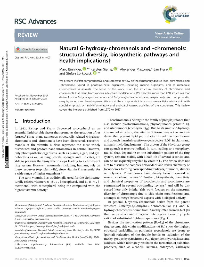
Natural 6-hydrox
aDepartment of Nutritional, Food and Consu
Sciences, Leipziger Straße 123, 36037 Fuld
hs-fulda.debAnalytiCon Discovery GmbH, Hermannswe
E-mail: [email protected] of Biological Chemistry and Nutrit
28, 70599 Stuttgart, Germany. E-mail: jan.fdInstitute of Nutrition, Friedrich Schiller Un
Jena, Germany. E-mail: stefan.lorkowski@ueCompetence Cluster for Nutrition and Ca
Jena-Leipzig, Germany
† Electronic supplementary informa10.1039/c7ra11819h
Cite this: RSC Adv., 2018, 8, 4803
Received 9th November 2017Accepted 18th January 2018
DOI: 10.1039/c7ra11819h
rsc.li/rsc-advances
This journal is © The Royal Society of C
y-chromanols and -chromenols:structural diversity, biosynthetic pathways andhealth implications†
Marc Birringer, *a Karsten Siems, b Alexander Maxones,a Jan Frank c
and Stefan Lorkowski de
We present the first comprehensive and systematic review on the structurally diverse toco-chromanols and
-chromenols found in photosynthetic organisms, including marine organisms, and as metabolic
intermediates in animals. The focus of this work is on the structural diversity of chromanols and
chromenols that result from various side chain modifications. We describe more than 230 structures that
derive from a 6-hydroxy-chromanol- and 6-hydroxy-chromenol core, respectively, and comprise di-,
sesqui-, mono- and hemiterpenes. We assort the compounds into a structure–activity relationship with
special emphasis on anti-inflammatory and anti-carcinogenic activities of the congeners. This review
covers the literature published from 1970 to 2017.
1. Introduction
In 1922, Bishop and Evans discovered a-tocopherol as anessential lipid-soluble factor that promotes the gestation of ratfetuses.1 Since then, numerous structurally related 6-hydroxy-chromanols and -chromenols have been discovered. Tocochro-manols of the vitamin E class represent the most widelydistributed and predominant chromanols in nature. However,only photosynthetic organisms, such as plants, algae, and cya-nobacteria as well as fungi, corals, sponges and tunicates, areable to perform the biosynthetic steps leading to a chromanolring system. However, mammals, including humans, rely onthese resources (esp. plant oils), since vitamin E is essential fora wide range of higher organisms.2
The term vitamin E is traditionally used for the eight struc-turally related vitamers a-, b-, g-, d-tocopherol, and a-, b-, g-, d-tocotrienol, with a-tocopherol being the compound with thehighest vitamin activity.3
mer Sciences, Fulda University of Applied
a, Germany. E-mail: marc.birringer@oe.
rder Haus 17, 14473 Potsdam, Germany.
ion, University of Hohenheim, Garbenstr.
iversity Jena, Dornburger Str. 25, 07743
ni-jena.de
rdiovascular Health (nutriCARD), Halle-
tion (ESI) available. See DOI:
hemistry 2018
Tocochromanols belong to the family of prenylquinones thatalso include plastochromanol-8, phylloquinones (vitamin K),and ubiquinones (coenzyme Q10). Due to its unique 6-hydroxy-chromanol structure, the vitamin E forms may act as antioxi-dants that prevent lipid peroxidation in cellular membranesand quench harmful reactive oxygen species (ROS) in plants andanimals (including humans). The proton of the 6-hydroxy groupcan quench a reactive radical, in turn leading to a tocopherylradical that, depending on the substitution pattern of the ringsystem, remains stable, with a half-life of several seconds, andcan be subsequently recycled by vitamin C. The review does notaim to discuss the complex antioxidant and redox chemistry oftocopherols forming corresponding radicals, quinones, dimersor polymers. These issues have already been discussed inseveral excellent reviews.4,5 Further, biosynthesis, bioactivityand chemical properties of tocopherols and tocotrienols aresummarized in several outstanding reviews,6 and will be dis-cussed here only briey. This work focuses on the structuraldiversity of chromanols due to side chain modications andattempts to merge structural aspects with biological activity.
In general, 6-hydroxy-chromanols derive from the parentstructure 2-methyl-3,4-dihydro-2H-chromen-6-ol (1) and 6-hydroxy-chromenols derive from 2-methyl-2H-chromen-6-ol (2)that comprise a class of bicyclic heterocycles formed by cycli-sation of substituted 1,4-benzoquinones (Fig. 1).
Besides the methylation pattern (R1–R3) of the chromanolring system, side chain modications (at R4) show the higheststructural variability. In particular tocotrienols are prone to(partial) reduction of the double bonds or oxidation of themethyl groups by cytochrome P450-dependent hydroxylases andoxidases, which ultimately results in the formation of oxidationproducts, such as alcohols, ketones, aldehydes, carboxylic
RSC Adv., 2018, 8, 4803–4841 | 4803
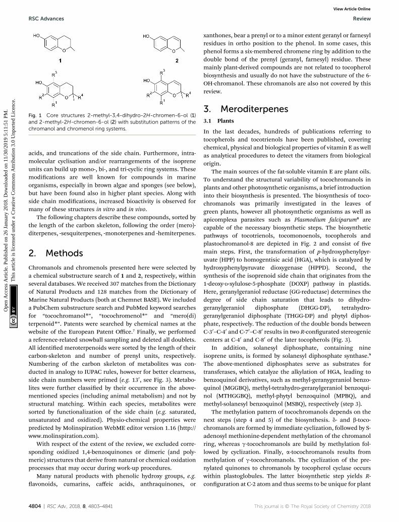
Fig. 1 Core structures 2-methyl-3,4-dihydro-2H-chromen-6-ol (1)and 2-methyl-2H-chromen-6-ol (2) with substitution patterns of thechromanol and chromenol ring systems.
RSC Advances Review
Ope
n A
cces
s A
rtic
le. P
ublis
hed
on 2
6 Ja
nuar
y 20
18. D
ownl
oade
d on
11/
30/2
019
5:11
:51
PM.
Thi
s ar
ticle
is li
cens
ed u
nder
a C
reat
ive
Com
mon
s A
ttrib
utio
n 3.
0 U
npor
ted
Lic
ence
.View Article Online
acids, and truncations of the side chain. Furthermore, intra-molecular cyclisation and/or rearrangements of the isopreneunits can build up mono-, bi-, and tri-cyclic ring systems. Thesemodications are well known for compounds in marineorganisms, especially in brown algae and sponges (see below),but have been found also in higher plant species. Along withside chain modications, increased bioactivity is observed formany of these structures in vitro and in vivo.
The following chapters describe these compounds, sorted bythe length of the carbon skeleton, following the order (mero)-diterpenes, -sesquiterpenes, -monoterpenes and -hemiterpenes.
2. Methods
Chromanols and chromenols presented here were selected bya chemical substructure search of 1 and 2, respectively, withinseveral databases. We received 307 matches from the Dictionaryof Natural Products and 128 matches from the Dictionary ofMarine Natural Products (both at Chemnet BASE). We includeda PubChem substructure search and PubMed keyword searchesfor “tocochromanol*”, “tocochromenol*” and “mero(di)terpenoid*”. Patents were searched by chemical names at thewebsite of the European Patent Office.7 Finally, we performeda reference-related snowball sampling and deleted all doublets.All identied meroterpenoids were sorted by the length of theircarbon-skeleton and number of prenyl units, respectively.Numbering of the carbon skeleton of metabolites was con-ducted in analogy to IUPAC rules, however for better clearness,side chain numbers were primed (e.g. 130, see Fig. 3). Metabo-lites were further classied by their occurrence in the above-mentioned species (including animal metabolism) and not bystructural matching. Within each species, metabolites weresorted by functionalization of the side chain (e.g. saturated,unsaturated and oxidized). Physio-chemical properties werepredicted by Molinspiration WebME editor version 1.16 (http://www.molinspiration.com).
With respect of the extent of the review, we excluded corre-sponding oxidized 1,4-benzoquinones or dimeric (and poly-meric) structures that derive from natural or chemical oxidationprocesses that may occur during work-up procedures.
Many natural products with phenolic hydroxy groups, e.g.avonoids, cumarins, caffeic acids, anthraquinones, or
4804 | RSC Adv., 2018, 8, 4803–4841
xanthones, bear a prenyl or to a minor extent geranyl or farnesylresidues in ortho position to the phenol. In some cases, thisphenol forms a six-membered chromene ring by addition to thedouble bond of the prenyl (geranyl, farnesyl) residue. Thesemainly plant-derived compounds are not related to tocopherolbiosynthesis and usually do not have the substructure of the 6-OH-chromanol. These chromanols are also not covered by thisreview.
3. Meroditerpenes3.1 Plants
In the last decades, hundreds of publications referring totocopherols and tocotrienols have been published, coveringchemical, physical and biological properties of vitamin E as wellas analytical procedures to detect the vitamers from biologicalorigin.
The main sources of the fat-soluble vitamin E are plant oils.To understand the structural variability of tocochromanols inplants and other photosynthetic organisms, a brief introductioninto their biosynthesis is presented. The biosynthesis of toco-chromanols was primarily investigated in the leaves ofgreen plants, however all photosynthetic organisms as well asapicomplexa parasites such as Plasmodium falciparum8 arecapable of the necessary biosynthetic steps. The biosyntheticpathways of tocotrienols, tocomonoenols, tocopherols andplastochromanol-8 are depicted in Fig. 2 and consist of vemain steps. First, the transformation of p-hydroxyphenylpyr-uvate (HPP) to homogentisic acid (HGA), which is catalyzed byhydroxyphenylpyruvate dioxygenase (HPPD). Second, thesynthesis of the isoprenoid side chain that originates from the1-deoxy-D-xylulose-5-phosphate (DOXP) pathway in plastids.Here, geranylgeraniol reductase (GG-reductase) determines thedegree of side chain saturation that leads to dihydro-geranylgeraniol diphosphate (DHGG-DP), tetrahydro-geranylgeraniol diphosphate (THGG-DP) and phytyl diphos-phate, respectively. The reduction of the double bonds betweenC-30–C-40 and C-70–C-80 results in two R-congurated stereogeniccenters at C-40 and C-80 of the later tocopherols (Fig. 3).
In addition, solanesyl diphosphate, containing nineisoprene units, is formed by solanesyl diphosphate synthase.9
The above-mentioned diphosphates serve as substrates fortransferases, which catalyze the alkylation of HGA, leading tobenzoquinol derivatives, such as methyl-geranygeraniol benzo-quinol (MGGBQ), methyl-tetrahydro-geranylgeraniol benzoqui-nol (MTHGGBQ), methyl-phytyl benzoquinol (MPBQ), andmethyl-solanesyl benzoquinol (MSBQ), respectively (step 3).
The methylation pattern of tocochromanols depends on thenext steps (step 4 and 5) of the biosynthesis. d- and b-toco-chromanols are formed by immediate cyclization, followed by S-adenosyl methionine-dependent methylation of the chromanolring, whereas g-tocochromanols are build by methylation fol-lowed by cyclization. Finally, a-tocochromanols results frommethylation of g-tocochromanols. The cyclization of the pre-nylated quinones to chromanols by tocopherol cyclase occurswithin plastoglobules. The latter biosynthetic step yields R-conguration at C-2 atom and thus seems to be unique for plant
This journal is © The Royal Society of Chemistry 2018
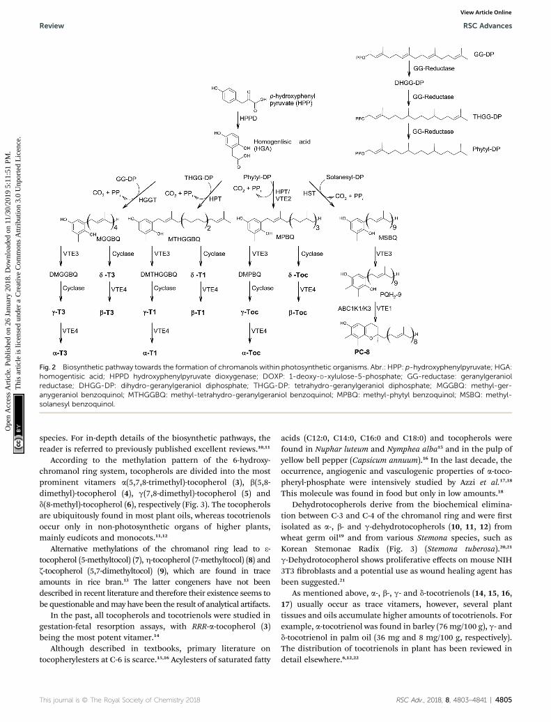
Fig. 2 Biosynthetic pathway towards the formation of chromanols within photosynthetic organisms. Abr.: HPP: p-hydroxyphenylpyruvate; HGA:homogentisic acid; HPPD hydroxyphenylpyruvate dioxygenase; DOXP: 1-deoxy-D-xylulose-5-phosphate; GG-reductase: geranylgeraniolreductase; DHGG-DP: dihydro-geranylgeraniol diphosphate; THGG-DP: tetrahydro-geranylgeraniol diphosphate; MGGBQ: methyl-ger-anygeraniol benzoquinol; MTHGGBQ: methyl-tetrahydro-geranylgeraniol benzoquinol; MPBQ: methyl-phytyl benzoquinol; MSBQ: methyl-solanesyl benzoquinol.
Review RSC Advances
Ope
n A
cces
s A
rtic
le. P
ublis
hed
on 2
6 Ja
nuar
y 20
18. D
ownl
oade
d on
11/
30/2
019
5:11
:51
PM.
Thi
s ar
ticle
is li
cens
ed u
nder
a C
reat
ive
Com
mon
s A
ttrib
utio
n 3.
0 U
npor
ted
Lic
ence
.View Article Online
species. For in-depth details of the biosynthetic pathways, thereader is referred to previously published excellent reviews.10,11
According to the methylation pattern of the 6-hydroxy-chromanol ring system, tocopherols are divided into the mostprominent vitamers a(5,7,8-trimethyl)-tocopherol (3), b(5,8-dimethyl)-tocopherol (4), g(7,8-dimethyl)-tocopherol (5) andd(8-methyl)-tocopherol (6), respectively (Fig. 3). The tocopherolsare ubiquitously found in most plant oils, whereas tocotrienolsoccur only in non-photosynthetic organs of higher plants,mainly eudicots and monocots.11,12
Alternative methylations of the chromanol ring lead to 3-tocopherol (5-methyltocol) (7), h-tocopherol (7-methyltocol) (8) andz-tocopherol (5,7-dimethyltocol) (9), which are found in traceamounts in rice bran.13 The latter congeners have not beendescribed in recent literature and therefore their existence seems tobe questionable andmay have been the result of analytical artifacts.
In the past, all tocopherols and tocotrienols were studied ingestation-fetal resorption assays, with RRR-a-tocopherol (3)being the most potent vitamer.14
Although described in textbooks, primary literature ontocopherylesters at C-6 is scarce.15,16 Acylesters of saturated fatty
This journal is © The Royal Society of Chemistry 2018
acids (C12:0, C14:0, C16:0 and C18:0) and tocopherols werefound in Nuphar luteum and Nymphea alba15 and in the pulp ofyellow bell pepper (Capsicum annuum).16 In the last decade, theoccurrence, angiogenic and vasculogenic properties of a-toco-pheryl-phosphate were intensively studied by Azzi et al.17,18
This molecule was found in food but only in low amounts.18
Dehydrotocopherols derive from the biochemical elimina-tion between C-3 and C-4 of the chromanol ring and were rstisolated as a-, b- and g-dehydrotocopherols (10, 11, 12) fromwheat germ oil19 and from various Stemona species, such asKorean Stemonae Radix (Fig. 3) (Stemona tuberosa).20,21
g-Dehydrotocopherol shows proliferative effects on mouse NIH3T3 broblasts and a potential use as wound healing agent hasbeen suggested.21
As mentioned above, a-, b-, g- and d-tocotrienols (14, 15, 16,17) usually occur as trace vitamers, however, several planttissues and oils accumulate higher amounts of tocotrienols. Forexample, a-tocotrienol was found in barley (76 mg/100 g), g- andd-tocotrienol in palm oil (36 mg and 8 mg/100 g, respectively).The distribution of tocotrienols in plant has been reviewed indetail elsewhere.6,12,22
RSC Adv., 2018, 8, 4803–4841 | 4805
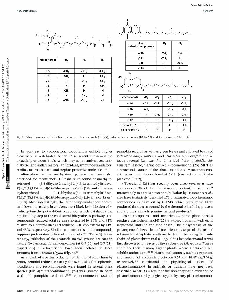
Fig. 3 Structures and substitution patterns of tocopherols (3 to 9), dehydrotocopherols (10 to 13) and tocotrienols (14 to 19).
RSC Advances Review
Ope
n A
cces
s A
rtic
le. P
ublis
hed
on 2
6 Ja
nuar
y 20
18. D
ownl
oade
d on
11/
30/2
019
5:11
:51
PM.
Thi
s ar
ticle
is li
cens
ed u
nder
a C
reat
ive
Com
mon
s A
ttrib
utio
n 3.
0 U
npor
ted
Lic
ence
.View Article Online
In contrast to tocopherols, tocotrienols exhibit higherbioactivity in vertebrates. Ashan et al. recently reviewed thebioactivity of tocotrienols, which may act as anti-cancer, anti-diabetic, anti-inammatory, antioxidant, immune-stimulatory,cardio-, neuro-, hepato- and nephro-protective molecules.23
Alternation in the methylation pattern has been alsodescribed for tocotrienols. Qureshi et al. found desmethylto-cotrienol (3,4-dihydro-2-methyl-2-(4,8,12-trimethyltrideca-30(E),70(E),110-trienyl)-2H-1-benzopyran-6-ol) (18) and didesme-thyltocotrienol (3,4-dihydro-2-(4,8,12-trimethyltrideca-30(E),70(E),110-trienyl)-2H-1-benzopyran-6-ol) (19) in rice bran24
(Fig. 3). Most interestingly, the latter compounds show choles-terol lowering activity in chicken, most likely by inhibition of 3-hydroxy-3-methylglutaryl-CoA reductase, which catalayzes therate-limiting step of the cholesterol biosynthesis pathway. Thecompounds reduced total serum cholesterol by 26% and 31%relative to a control diet and reduced LDL cholesterol by 41%and 48%, respectively. Similar to tocotrienols, both compoundssuppress proliferation B16 melanoma cells24,25 (Table 3). Inter-estingly, oxidation of the aromatic methyl groups are rare innature. Two unusual formyl-derivatives (at C-5 (20) and C-7 (21),respectively) of d-tocotrienol have been isolated in traceamounts from Garcinia virgata (Fig. 4).26
As a result of a partial reduction of the prenyl side chain bygeranylgeraniol reductase during the synthesis of tocopherols,tocodienols and tocomonoenols were found in several plantspecies (Fig. 4).11 a-Tocomonoenol (22) was isolated in palmseed and pumpkin seed oils,27,28 g-tocomonoenol (23) in
4806 | RSC Adv., 2018, 8, 4803–4841
pumpkin seed oil as well as green leaves and etiolated beans ofKalanchoe daigremontiana and Phaseolus coccineus,11,28 and d-tocomonoenol (24) was found in kiwi fruits (Actinidia chi-nensis).29 Of note, marine-derived a-tocomonoenol (25) (MDT) isa structural isomer of the above mentioned a-tocomonoenolwith a terminal double bond at C-130 (see section on Phyto-plankton (3.3.2)).
a-Tocodienol (26) has recently been discovered as a tracecompound (0.2% of the total vitamin E content) in palm oil.30
Interestingly to note is a recent publication by Hammann et al.,who have tentatively identied 170 unsaturated tocochromanolcompounds in palm oil by GC-MS, which were most likelyproduced (in trace amounts) by the thermal oil rening processand are thus unlikely genuine natural products.31
Beside tocopherols and tocotrienols, some plant speciesproduce plastochromanol-8 (27), a g-tocochromanol with eightisoprenoid units in the side chain. The biosynthesis of thepolyterpene follows that of tocotrienols except of the use ofsolanesyl-diphosphate synthase to form the elongated sidechain of plastochromanol-8 (Fig. 4).32 Plastochromanol-8 wasrst discovered in leaves of the rubber tree (Hevea brasiliensis)and since then in many higher plants, where it acts as a fat-soluble antioxidant.32–34 Nutritional sources, such as rapeseedand linseed oil, accumulate between 5.57 and 18.47 mg/100 g,respectively.34 Nutritional or physiological effects ofplastochromanol-8 in animals or humans have not beendescribed so far. As a result of the non-enzymatic oxidation ofplastochromanol-8 by singlet oxygen, hydroxy-plastochromanol
This journal is © The Royal Society of Chemistry 2018
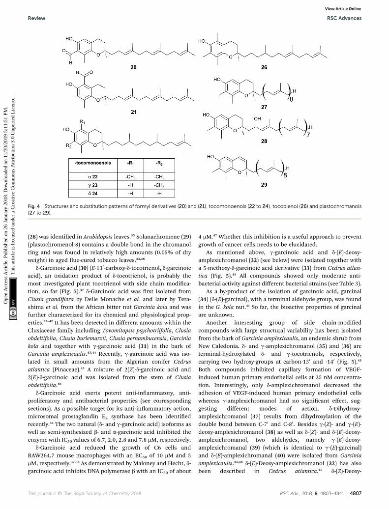
Fig. 4 Structures and substitution patterns of formyl derivatives (20) and (21), tocomonoenols (22 to 24), tocodienol (26) and plastochromanols(27 to 29).
Review RSC Advances
Ope
n A
cces
s A
rtic
le. P
ublis
hed
on 2
6 Ja
nuar
y 20
18. D
ownl
oade
d on
11/
30/2
019
5:11
:51
PM.
Thi
s ar
ticle
is li
cens
ed u
nder
a C
reat
ive
Com
mon
s A
ttrib
utio
n 3.
0 U
npor
ted
Lic
ence
.View Article Online
(28) was identied in Arabidopsis leaves.35 Solanachromene (29)(plastochromenol-8) contains a double bond in the chromanolring and was found in relatively high amounts (0.05% of dryweight) in aged ue-cured tobacco leaves.33,36
d-Garcinoic acid (30) (E-130-carboxy-d-tocotrienol, d-garcinoicacid), an oxidation product of d-tocotrienol, is probably themost investigated plant tocotrienol with side chain modica-tion, so far (Fig. 5).37 d-Garcinoic acid was rst isolated fromClusia grandiora by Delle Monache et al. and later by Tera-shima et al. from the African bitter nut Garcinia kola and wasfurther characterized for its chemical and physiological prop-erties.37–42 It has been detected in different amounts within theClusiaceae family including Tovomitopsis psychotriifolia, Clusiaobdeltifolia, Clusia burlemarxii, Clusia pernambucensis, Garciniakola and together with g-garcinoic acid (31) in the bark ofGarcinia amplexicaulis.43,44 Recently, g-garcinoic acid was iso-lated in small amounts from the Algerian conifer Cedrusatlantica (Pinaceae).45 A mixture of 2(Z)-d-garcinoic acid and2(E)-d-garcinoic acid was isolated from the stem of Clusiaobdeltifolia.46
d-Garcinoic acid exerts potent anti-inammatory, anti-proliferatory and antibacterial properties (see correspondingsections). As a possible target for its anti-inammatory action,microsomal prostaglandin E2 synthase has been identiedrecently.44 The two natural (d- and g-garcinoic acid) isoforms aswell as semi-synthesized b- and a-garcinoic acid inhibited theenzyme with IC50 values of 6.7, 2.0, 2.8 and 7.8 mM, respectively.
d-Garcinoic acid reduced the growth of C6 cells andRAW264.7 mouse macrophages with an EC50 of 10 mM and 5mM, respectively.37,38 As demonstrated by Maloney and Hecht, d-garcinoic acid inhibits DNA polymerase b with an IC50 of about
This journal is © The Royal Society of Chemistry 2018
4 mM.47 Whether this inhibition is a useful approach to preventgrowth of cancer cells needs to be elucidated.
As mentioned above, g-garcinoic acid and d-(E)-deoxy-amplexichromanol (32) (see below) were isolated together witha 5-methoxy-d-garcinoic acid derivative (33) from Cedrus atlan-tica (Fig. 5).45 All compounds showed only moderate anti-bacterial activity against different bacterial strains (see Table 3).
As a by-product of the isolation of garcinoic acid, garcinal(34) (d-(E)-garcinal), with a terminal aldehyde group, was foundin the G. kola nut.41 So far, the bioactive properties of garcinalare unknown.
Another interesting group of side chain-modiedcompounds with large structural variability has been isolatedfrom the bark of Garcinia amplexicaulis, an endemic shrub fromNew Caledonia. d- and g-amplexichromanol (35) and (36) areterminal-hydroxylated d- and g-tocotrienols, respectively,carrying two hydroxy-groups at carbon-130 and -140 (Fig. 5).43
Both compounds inhibited capillary formation of VEGF-induced human primary endothelial cells at 25 nM concentra-tion. Interestingly, only d-amplexichromanol decreased theadhesion of VEGF-induced human primary endothelial cellswhereas g-amplexichromanol had no signicant effect, sug-gesting different modes of action. d-Dihydroxy-amplexichromanol (37) results from dihydroxylation of thedouble bond between C-70 and C-80. Besides g-(Z)- and g-(E)-deoxy-amplexichromanol (38) as well as d-(Z)- and d-(E)-deoxy-amplexichromanol, two aldehydes, namely g-(E)-deoxy-amplexichromanal (39) (which is identical to g-(E)-garcinal)and d-(E)-amplexichromanal (40) were isolated from Garciniaamplexicaulis.43,48 d-(E)-Deoxy-amplexichromanol (32) has alsobeen described in Cedrus atlantica.45 d-(Z)-Deoxy-
RSC Adv., 2018, 8, 4803–4841 | 4807
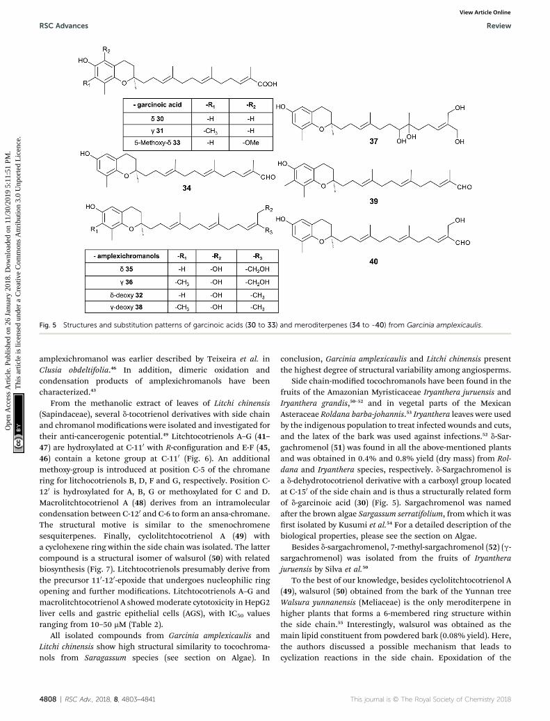
Fig. 5 Structures and substitution patterns of garcinoic acids (30 to 33) and meroditerpenes (34 to -40) from Garcinia amplexicaulis.
RSC Advances Review
Ope
n A
cces
s A
rtic
le. P
ublis
hed
on 2
6 Ja
nuar
y 20
18. D
ownl
oade
d on
11/
30/2
019
5:11
:51
PM.
Thi
s ar
ticle
is li
cens
ed u
nder
a C
reat
ive
Com
mon
s A
ttrib
utio
n 3.
0 U
npor
ted
Lic
ence
.View Article Online
amplexichromanol was earlier described by Teixeira et al. inClusia obdeltifolia.46 In addition, dimeric oxidation andcondensation products of amplexichromanols have beencharacterized.43
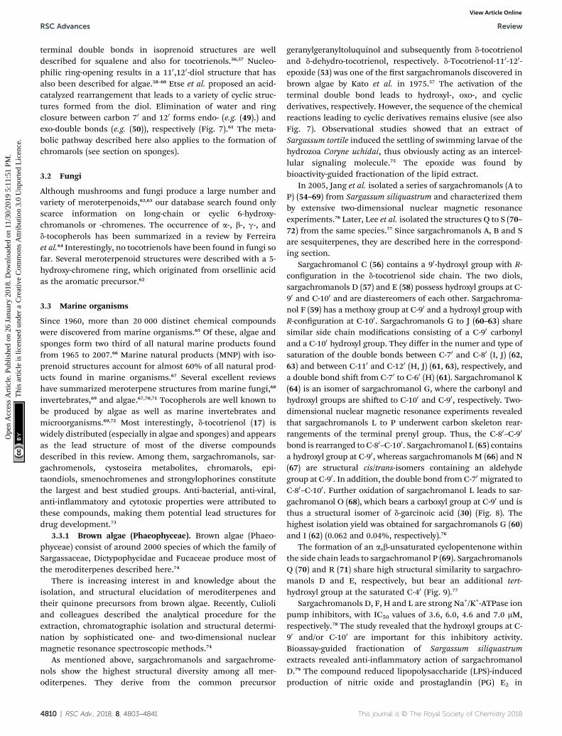
From the methanolic extract of leaves of Litchi chinensis(Sapindaceae), several d-tocotrienol derivatives with side chainand chromanol modications were isolated and investigated fortheir anti-cancerogenic potential.49 Litchtocotrienols A–G (41–47) are hydroxylated at C-110 with R-conguration and E-F (45,46) contain a ketone group at C-110 (Fig. 6). An additionalmethoxy-group is introduced at position C-5 of the chromanering for litchocotrienols B, D, F and G, respectively. Position C-120 is hydroxylated for A, B, G or methoxylated for C and D.Macrolitchtocotrienol A (48) derives from an intramolecularcondensation between C-120 and C-6 to form an ansa-chromane.The structural motive is similar to the smenochromenesesquiterpenes. Finally, cyclolitchtocotrienol A (49) witha cyclohexene ring within the side chain was isolated. The lattercompound is a structural isomer of walsurol (50) with relatedbiosynthesis (Fig. 7). Litchtocotrienols presumably derive fromthe precursor 110-120-epoxide that undergoes nucleophilic ringopening and further modications. Litchtocotrienols A–G andmacrolitchtocotrienol A showedmoderate cytotoxicity in HepG2liver cells and gastric epithelial cells (AGS), with IC50 valuesranging from 10–50 mM (Table 2).
All isolated compounds from Garcinia amplexicaulis andLitchi chinensis show high structural similarity to tocochroma-nols from Saragassum species (see section on Algae). In
4808 | RSC Adv., 2018, 8, 4803–4841
conclusion, Garcinia amplexicaulis and Litchi chinensis presentthe highest degree of structural variability among angiosperms.
Side chain-modied tocochromanols have been found in thefruits of the Amazonian Myristicaceae Iryanthera juruensis andIryanthera grandis,50–52 and in vegetal parts of the MexicanAsteraceae Roldana barba-johannis.53 Iryanthera leaves were usedby the indigenous population to treat infected wounds and cuts,and the latex of the bark was used against infections.52 d-Sar-gachromenol (51) was found in all the above-mentioned plantsand was obtained in 0.4% and 0.8% yield (dry mass) from Rol-dana and Iryanthera species, respectively. d-Sargachromenol isa d-dehydrotocotrienol derivative with a carboxyl group locatedat C-150 of the side chain and is thus a structurally related formof d-garcinoic acid (30) (Fig. 5). Sargachromenol was namedaer the brown algae Sargassum serratifolium, from which it wasrst isolated by Kusumi et al.54 For a detailed description of thebiological properties, please see the section on Algae.
Besides d-sargachromenol, 7-methyl-sargachromenol (52) (g-sargachromenol) was isolated from the fruits of Iryantherajuruensis by Silva et al.50
To the best of our knowledge, besides cyclolitchtocotrienol A(49), walsurol (50) obtained from the bark of the Yunnan treeWalsura yunnanensis (Meliaceae) is the only meroditerpene inhigher plants that forms a 6-membered ring structure withinthe side chain.55 Interestingly, walsurol was obtained as themain lipid constituent from powdered bark (0.08% yield). Here,the authors discussed a possible mechanism that leads tocyclization reactions in the side chain. Epoxidation of the
This journal is © The Royal Society of Chemistry 2018

Fig. 6 Structures and substitution patterns of litchtocotrienols (41 to 48) from Litchi chinensis, sargachromenols (51) and (52).
Fig. 7 Scheme of an acid-catalyzed cyclization cascade including final cyclization of the chromane ring according to Etse et al.61 Examples arecyclolitchtocotrienol (49) and walsurol (50).
This journal is © The Royal Society of Chemistry 2018 RSC Adv., 2018, 8, 4803–4841 | 4809
Review RSC Advances
Ope
n A
cces
s A
rtic
le. P
ublis
hed
on 2
6 Ja
nuar
y 20
18. D
ownl
oade
d on
11/
30/2
019
5:11
:51
PM.
Thi
s ar
ticle
is li
cens
ed u
nder
a C
reat
ive
Com
mon
s A
ttrib
utio
n 3.
0 U
npor
ted
Lic
ence
.View Article Online
RSC Advances Review
Ope
n A
cces
s A
rtic
le. P
ublis
hed
on 2
6 Ja
nuar
y 20
18. D
ownl
oade
d on
11/
30/2
019
5:11
:51
PM.
Thi
s ar
ticle
is li
cens
ed u
nder
a C
reat
ive
Com
mon
s A
ttrib
utio
n 3.
0 U
npor
ted
Lic
ence
.View Article Online
terminal double bonds in isoprenoid structures are welldescribed for squalene and also for tocotrienols.56,57 Nucleo-philic ring-opening results in a 110,120-diol structure that hasalso been described for algae.58–60 Etse et al. proposed an acid-catalyzed rearrangement that leads to a variety of cyclic struc-tures formed from the diol. Elimination of water and ringclosure between carbon 70 and 120 forms endo- (e.g. (49).) andexo-double bonds (e.g. (50)), respectively (Fig. 7).61 The meta-bolic pathway described here also applies to the formation ofchromarols (see section on sponges).
3.2 Fungi
Although mushrooms and fungi produce a large number andvariety of meroterpenoids,62,63 our database search found onlyscarce information on long-chain or cyclic 6-hydroxy-chromanols or -chromenes. The occurrence of a-, b-, g-, andd-tocopherols has been summarized in a review by Ferreiraet al.64 Interestingly, no tocotrienols have been found in fungi sofar. Several meroterpenoid structures were described with a 5-hydroxy-chromene ring, which originated from orsellinic acidas the aromatic precursor.62
3.3 Marine organisms
Since 1960, more than 20 000 distinct chemical compoundswere discovered from marine organisms.65 Of these, algae andsponges form two third of all natural marine products foundfrom 1965 to 2007.66 Marine natural products (MNP) with iso-prenoid structures account for almost 60% of all natural prod-ucts found in marine organisms.67 Several excellent reviewshave summarized meroterpene structures from marine fungi,68
invertebrates,69 and algae.67,70,71 Tocopherols are well known tobe produced by algae as well as marine invertebrates andmicroorganisms.69,72 Most interestingly, d-tocotrienol (17) iswidely distributed (especially in algae and sponges) and appearsas the lead structure of most of the diverse compoundsdescribed in this review. Among them, sargachromanols, sar-gachromenols, cystoseira metabolites, chromarols, epi-taondiols, smenochromenes and strongylophorines constitutethe largest and best studied groups. Anti-bacterial, anti-viral,anti-inammatory and cytotoxic properties were attributed tothese compounds, making them potential lead structures fordrug development.73
3.3.1 Brown algae (Phaeophyceae). Brown algae (Phaeo-phyceae) consist of around 2000 species of which the family ofSargassaceae, Dictypophycidae and Fucaceae produce most ofthe meroditerpenes described here.74
There is increasing interest in and knowledge about theisolation, and structural elucidation of meroditerpenes andtheir quinone precursors from brown algae. Recently, Culioliand colleagues described the analytical procedure for theextraction, chromatographic isolation and structural determi-nation by sophisticated one- and two-dimensional nuclearmagnetic resonance spectroscopic methods.74
As mentioned above, sargachromanols and sargachrome-nols show the highest structural diversity among all mer-oditerpenes. They derive from the common precursor
4810 | RSC Adv., 2018, 8, 4803–4841
geranylgeranyltoluquinol and subsequently from d-tocotrienoland d-dehydro-tocotrienol, respectively. d-Tocotrienol-110-120-epoxide (53) was one of the rst sargachromanols discovered inbrown algae by Kato et al. in 1975.57 The activation of theterminal double bond leads to hydroxyl-, oxo-, and cyclicderivatives, respectively. However, the sequence of the chemicalreactions leading to cyclic derivatives remains elusive (see alsoFig. 7). Observational studies showed that an extract ofSargassum tortile induced the settling of swimming larvae of thehydrozoa Coryne uchidai, thus obviously acting as an intercel-lular signaling molecule.75 The epoxide was found bybioactivity-guided fractionation of the lipid extract.
In 2005, Jang et al. isolated a series of sargachromanols (A toP) (54–69) from Sargassum siliquastrum and characterized themby extensive two-dimensional nuclear magnetic resonanceexperiments.76 Later, Lee et al. isolated the structures Q to S (70–72) from the same species.77 Since sargachromanols A, B and Sare sesquiterpenes, they are described here in the correspond-ing section.
Sargachromanol C (56) contains a 90-hydroxyl group with R-conguration in the d-tocotrienol side chain. The two diols,sargachromanols D (57) and E (58) possess hydroxyl groups at C-90 and C-100 and are diastereomers of each other. Sargachroma-nol F (59) has a methoxy group at C-90 and a hydroxyl group withR-conguration at C-100. Sargachromanols G to J (60–63) sharesimilar side chain modications consisting of a C-90 carbonyland a C-100 hydroxyl group. They differ in the numer and type ofsaturation of the double bonds between C-70 and C-80 (I, J) (62,63) and between C-110 and C-120 (H, J) (61, 63), respectively, anda double bond shi from C-70 to C-60 (H) (61). Sargachromanol K(64) is an isomer of sargachromanol G, where the carbonyl andhydroxyl groups are shied to C-100 and C-90, respectively. Two-dimensional nuclear magnetic resonance experiments revealedthat sargachromanols L to P underwent carbon skeleton rear-rangements of the terminal prenyl group. Thus, the C-80–C-90
bond is rearranged to C-80–C-100. Sargachromanol L (65) containsa hydroxyl group at C-90, whereas sargachromanols M (66) and N(67) are structural cis/trans-isomers containing an aldehydegroup at C-90. In addition, the double bond from C-70 migrated toC-80–C-100. Further oxidation of sargachromanol L leads to sar-gachromanol O (68), which bears a carboxyl group at C-90 und isthus a structural isomer of d-garcinoic acid (30) (Fig. 8). Thehighest isolation yield was obtained for sargachromanols G (60)and I (62) (0.062 and 0.04%, respectively).76
The formation of an a,b-unsaturated cyclopentenone withinthe side chain leads to sargachromanol P (69). SargachromanolsQ (70) and R (71) share high structural similarity to sargachro-manols D and E, respectively, but bear an additional tert-hydroxyl group at the saturated C-40 (Fig. 9).77
Sargachromanols D, F, H and L are strong Na+/K+-ATPase ionpump inhibitors, with IC50 values of 3.6, 6.0, 4.6 and 7.0 mM,respectively.78 The study revealed that the hydroxyl groups at C-90 and/or C-100 are important for this inhibitory activity.Bioassay-guided fractionation of Sargassum siliquastrumextracts revealed anti-inammatory action of sargachromanolD.79 The compound reduced lipopolysaccharide (LPS)-inducedproduction of nitric oxide and prostaglandin (PG) E2 in
This journal is © The Royal Society of Chemistry 2018

Fig. 8 Structures and substitution patterns of sargachromanols (56 to 68) from Sargassum species.
Fig. 9 Structures and substitution patterns of sargachromanols (69 to 75) from Sargassum species.
Review RSC Advances
Ope
n A
cces
s A
rtic
le. P
ublis
hed
on 2
6 Ja
nuar
y 20
18. D
ownl
oade
d on
11/
30/2
019
5:11
:51
PM.
Thi
s ar
ticle
is li
cens
ed u
nder
a C
reat
ive
Com
mon
s A
ttrib
utio
n 3.
0 U
npor
ted
Lic
ence
.View Article Online
murine RAW 264.7 macrophages and inhibited the expressionof the pro-inammatory enzymes inducible nitric oxidesynthetase (iNOS) and COX-2. In addition, the production of thepro-inammatory cytokines TNF-a, interleukin-1b (IL)-1b andIL-6 was reduced by sargachromanol D.79 Recently, sargachro-manol D was suggested as an anti-hypertensive agent, since itshowed dual antagonistic activity towards an L-type Ca2+-channel and endothelin A/B2 receptor (Table 3).80 The use ofsargachromanols is protected by several patents.81
This journal is © The Royal Society of Chemistry 2018
Sargassum siliquastrum was also used as a natural source ofsargachromanol E and G for bioactivity studies.82–87 Bothcompounds inhibited the expression of pro-inammatorycytokines in LPS-stimulated murine RAW 264.7 macro-phages.83,84,86 In addition, sargachromanol E induced apoptosisvia caspase-3 activation in promyelocytic HL-60 leukemia cells82
and inhibited ultraviolet A-induced ageing of human dermalbroblasts.88 Sargachromanol G showed anti-osteoclastogeniceffects on the expression of IL-1b-induced osteoclastogenic
RSC Adv., 2018, 8, 4803–4841 | 4811

RSC Advances Review
Ope
n A
cces
s A
rtic
le. P
ublis
hed
on 2
6 Ja
nuar
y 20
18. D
ownl
oade
d on
11/
30/2
019
5:11
:51
PM.
Thi
s ar
ticle
is li
cens
ed u
nder
a C
reat
ive
Com
mon
s A
ttrib
utio
n 3.
0 U
npor
ted
Lic
ence
.View Article Online
factors in the human osteoblast cell line MG-63 and suppressedthe activation of nuclear factor kB (NF-kB) and mitogen-activated protein kinase (MAPK) in receptor activator of NF-kBligand (RANKL)-induced RAW264.7 cells.85,86
Besides sargachromanol I and K, another two sargachro-manols, (2R)-90-oxo-d-tocotrienol (73) and (2R)-70-80-dihydro-90-oxo-d-tocotrienol (74) were isolated from Sargassum micracan-thum, however, in very low yield (Fig. 9).89
Seo et al. isolated a racemic mixture of thunbergol A (75)from Sargassum thunbergii. The compound features a 3-hydroxyhydrobenzopyran structure with a 150-carboxy groupand thus presumably derives from sargachromenol (Fig. 9).90
Cyclic sargachromanols are widely distributed in brownalgae. Taondiol (76) (Fig. 10) was the rst cyclic side chain-derivative of tocotrienol that was isolated in 0.05% yieldfrom Taonia atomaria (order Dictyotales).91 The authorsproposed an enzyme-initiated synchronous cyclizationcascade of the prenylated 1,4-hydroquinone leading to thetetracyclic ring system. We and others propose an alternativecyclization mechanism starting from 1,4-hydroquinone-14-15-epoxide (77), analogous to lanosterol synthesis92,93 (Fig. 10).The protonation of 77 via an epoxide-hydrolase enzyme wouldincrease the susceptibility of intramolecular attacks of the C-2–C-3 and C-6–C-7 double bonds. The stereochemistry of thepossible isomers of taondiol at C-2 and C-3 and C-6 and C-7has been a matter of debate. Recently, Areche et al. assignedthe stereochemistry of isoepitaondiol (78) isolated from Sty-popodium abelliforme to the formerly described isotaondiol.94
By now, the structures of taondiol, isoepitaondiol, epitaondiol(79) and 2b,3a-epitaondiol (80) (Fig. 10) have been unambig-uously assigned.94–96
Epitaondiol (79) was isolated from Stypopodium zonale andStypopodium abelliforme (both species are members of theorder Dictyotales) and its bioactivity was intensivelystudied.93,95–102 The polycyclic compound shows ichthyotoxic,anti-herpes and anti-human metapneumovirus (HMPV) activityand acts as an anti-inammatory agent in vitro and in vivo (seeTables 1–3).98,99,102 Further, epitaondiol inhibited cell prolifera-tion of human colon adenocarcinoma (Caco-2), human
Fig. 10 Proposed enzyme-catalyzed cyclization leading to cyclic sargac
4812 | RSC Adv., 2018, 8, 4803–4841
neuroblastoma (SH-SY5Y), rat basophilic leukemia (RBL-2H3)cells, and murine macrophages (RAW.267), but not of non-cancer Chinese hamster broblasts (V79) (Table 2).100 2b,3a-Epitaondiol (80) exhibited moderate neurotoxicity towardsmouse neuro-2a cells with LC50 values of 2 mM.92 Epitaondiolwas effective in the prevention of HCl/ethanol-induced gastriclesions in mice at an ED50 value of 40 mg kg�1 bodyweight.101,103
Anti-insecticide activity was found against Spodoptera frugi-perda.96 Finally, the compound induced the settlement of themussel Perna perna.98
A series of cyclic meroditerpenes was isolated from differentCystoseira species collected along the Mediterranean andcontiguous Atlantic coasts.104 According to AlgaeBase,105 morethan 289 species (and infraspecic) names were found, of which42 have been marked as currently accepted taxonomically.
It was suggested that the following cyclic diterpenes originfrom a common biosynthetic precursor, namely bifurcar-enone (81) (Fig. 11). Among them, mediterraneols C (82), D(83), and E (84) have been isolated as their trimethoxy-derivatives from Cystoseira mediterranea in high yield (0.11,0.14 and 2.0% from dry weight algae, respectively).106,107
Mediterraneols C and D are stereoisomers at C-40 andcompromise a bridged cyclooctane structure with two dienolmoieties. Mediterraneol E (84) is a tricyclic oxygen-bridgedditerpene with antineoplastic activity.107 So far, the biosyn-thesis of mediterraneols is largely unknown.106,108 Medi-terraneols have been found to inhibit the mobility of seaurchin sperm and the mitotic cell division (ED50 values of 2 mgml�1) of fertilized urchin eggs.106
Recently, cystophloroketal E (85), a meroditerpene with a 2,7-dioxabicylo[3.2.1]octane core was isolated from Cystoseira tam-ariscifolia.108 The authors assumed that ketal formation waspreceded by a Michael addition of phloroglucinol onto theunsaturated carbonyl of 4-methoxy-bifurcarenone. Thecompound showed anti-bacterial, anti-microalgal and anti-invertebrate activity (Table 3).
Another group of complex bicyclic compounds was iso-lated from Cystoseira stricta, Cystoseira mediterranea andCystoseira tamariscifolia. Cystoseirols A (86), B (87) and C (88)
hromanols (76 to 80).
This journal is © The Royal Society of Chemistry 2018

Table 1 Inhibition of inflammatory markers by chromanols and chromenolsa
Compounds Test system Effective concentrationsb References
Meroditerpenesa-Tocopherol (3) IL-1b-stimulated A549 cells 8% nitric oxide inhibition at 10 mM 279
25% iNOS inhibition at 10 mMIC50 PGE2 inhibition: > 50 mM (A549 cells)
g-Tocopherol (5) IL-1b-stimulated A549 cells IC50 PGE2 inhibition: 7.5 mM (RAW264.7) 279IC50 5-LOX inhibition: > 50 mM 245,257IC50 COX-2 (A549 cells) inhibition: > 50 mM
d-Tocopherol (6) IL-1b-stimulated A549 cells IC50 COX-2 (A549 cells)) inhibition: > 50 mM 257IC50 5-LOX inhibition: > 50 mMIC50 PGE2 inhibition: 3 mM (A549 cells)
a-Tocotrienol (14) LPS-induced RAW 264.7 cells 5% NO inhibition at 33 mM 280d-Tocotrienol (17) 31% NO inhibition at 26 mMg-Tocotrienol (16) 19% NO inhibition at 30 mM
IL-1b-stimulated A549 cells IC50 PGE2 inhibition: 1 mM (A549 cells) 245130-carboxy-a-tocopherol (205)(a-130-COOH)
LPS-induced RAW 264.7 cells Total inhibition at 2.7 mM 255IC50 NO production: 0.2–0.5 mMb
LPS-induced RAW 264.7 cells 88% NO inhibition at 5 mM 256100% iNOS inhibition at 5 mM
130-carboxy-d-tocopherol (229)(d-130-COOH)
Inhibition of COX-1 and COX-2 IC50 COX-1 (bovine) inhibition: 5.0 mM 245IC50 COX-2 (human) inhibition: 4.0 mM 245IC50 COX-2 (A549 cells)) inhibition: 4.0 mM 257
Inhibition of human recombinant 5-LOX IC50 5-LOX inhibition: 0.5–1.0 mM 253IC50 (LTB4) generation: 4–7 mMNeutrophils and promyelocytic HL-60
Leukemia cells generated LTB4 79% NO inhibition at 5 mMLPS-induced RAW 264.7 cells 56% iNOS inhibition at 5 mM 256
130-Hydroxy-a-tocopherol (204)(a-130-OH)
LPS-induced RAW 264.7 cells 49% COX-2 inhibition at 10 mM 241,25653–60% iNOS inhibition at 10 mM54% PGE2 inhibition at 10 mM44–69% NO inhibition at 10 mM
130-Hydroxy-d-tocopherol (231)(d-130-OH)
LPS-induced RAW 264.7 cells 49% NO inhibition at 10 mM 25653% iNOS inhibition at 10 mM
d-Garcinoic acid (30) Inhibition of COX-2 and 5-LOX IC50 COX-2 inhibition: 9.8 mM 257IC50 5-LOX inhibition: 1.0 mM
Inhibition of LPS-stimulated NOproduction in RAW 264.7 macrophages
IC50 NO production: 1.0 mMb 260
a-, b-,< g-,< d-Garcinoic acid(209, 232, 231, 230)
Inhibition of PGE2-synthase (PGES-1) IC50 7.8 (a-), 2.8 (b-), 2.0 (g-), 6.7 (d-) garcinoicacid
44
d-Sargachromenol (51) TPA-induced mouse ear edema IC50 edema reduction: 0.36 mg per ear 53Inhibition of COX-1 and -2 98% COX-1 inhibition at 100 ppm 52
84% COX-2 inhibition at 100 ppmLPS-induced RAW 264.7 and BV-2 cells IC50 NO production: 82 mM (RAW264.7) 129
IC50 PGE2 inhibition: 30.2 mM (RAW264.7)IC50 NO production: 1.3–2.7 mM (BV-2) 134,261
Sargachromanol D (57) LPS-induced RAW 264.7 cells IC50 NO production: 40 mMb (RAW264.7) 79IC50 PGE2 inhibition: 15 mMb (RAW264.7)
Sargachromanol E (58) LPS-induced RAW 264.7 cells IC50 NO production: 16.3 mM 83Sargachromanol G (60) LPS-induced RAW264.7 cells IC50 NO production: Ca. 15 mMb 84Chromarols (A–D) (113–116)cyclic
Inhibitors of 12- and 15-LOX 15-hLO IC50: 0.6(A), 4.0(B), 0.7(C), 1.1 mM(D) 15412-hLO IC50: all >100 mM
Epitaondiol (79) cyclic TPA-induced mouse ear edema IC50 edema reduction: 20.7 mg per ear 99IC50 myeloperoxidase activity: 17.8 mg per ear
Eicosanoid inhibition IC50 (TXB2) generation: 3.8 mM 99IC50 (LTB4) generation: 30.1 mM
MerosesquiterpenesCapillobenzopyranol (172)cyclic
LPS-induced RAW 264.7 cells 36.7% NO inhibition at 10 mM 202
90-Carboxy-d-tocopherol (206)(d-90-COOH)
Inhibition of COX-1, -2 IC50 COX-1 (bovine) inhibition: >20 mM 245IC50 COX-2 (human) inhibition: >20 mMIC50 COX-2 (A549 cells)) inhibition: 6.0 mM
This journal is © The Royal Society of Chemistry 2018 RSC Adv., 2018, 8, 4803–4841 | 4813
Review RSC Advances
Ope
n A
cces
s A
rtic
le. P
ublis
hed
on 2
6 Ja
nuar
y 20
18. D
ownl
oade
d on
11/
30/2
019
5:11
:51
PM.
Thi
s ar
ticle
is li
cens
ed u
nder
a C
reat
ive
Com
mon
s A
ttrib
utio
n 3.
0 U
npor
ted
Lic
ence
.View Article Online

Table 1 (Contd. )
Compounds Test system Effective concentrationsb References
MonoterpenesCordiachromene A (173) Inhibition of PGI2 biosynthesis IC50 8.2 mM 216
Carrageenan induced rat paw endema IC50 18.9 mM 211Inhibitor of 15-LOX IC50: 0.82 mM 214
IC50: 2 mMa-CMBHC (207) IC50 COX-1 (bovine) inhibition: 160 mM 245
IC50 COX-2 (human) inhibition: 140 mMa-CEHC (208) TNFa-stimulated NO and PGE2
production in RAEC cellsIC50 PGE2 inhibition: 59 mM 244IC50 NO production: 56 mM
g-CEHC (233) IC50 COX-1 (bovine) inhibition: 300 mM 245IC50 COX-2 (human) inhibition: 450 mMIC50 COX-2 (A549 cells)) inhibition: 35–70mMIC50 PGE2 inhibition: 30.0 mM (RAW 264.7) 279
HemiterpenesQuercinol (199) In vitro cytokine inhibition IC50 COX-1 inhibition: 4.7 mM 235
IC50 COX-2 inhibition: 0.63 mMIC50 3a-HSD inhibition: 114 mMIC50 XO inhibition: 21 mMIC50 HRP: 68 mM
a Abbreviations: thromboxane B2 (TXB2), leukotriene B4 (LTB4), cyclooxygenase (COX)-1 and -2, 3a-hydroxysteroid dehydrogenase (3a-HSD),lipoxygenase (LOX), xanthine oxidase (XO), horseradish peroxidase (HRP). b Estimated from original publication.
RSC Advances Review
Ope
n A
cces
s A
rtic
le. P
ublis
hed
on 2
6 Ja
nuar
y 20
18. D
ownl
oade
d on
11/
30/2
019
5:11
:51
PM.
Thi
s ar
ticle
is li
cens
ed u
nder
a C
reat
ive
Com
mon
s A
ttrib
utio
n 3.
0 U
npor
ted
Lic
ence
.View Article Online
possess a oxabicyclo[5:4:1]dodecane ring that results froma single methyl group displacement, supplementary bridgesand ring ssions.107,109,110 Cystoseirol A (86) inhibitedplant tumor formation in a crown-gall potato bioassay (73% at10 mM).110
Amico et al. isolated cystoketal chromane (89) from theSicilian brown alga Cystoseira balearica. Structural elucida-tion revealed a tricyclic ring system within the side chain andan epimeric mixture at C-2 (Fig. 11). Thus, the authorsproposed cystoketal chromane to be an artefact of theextraction process.111 Later, demethoxy cystoketal chromane(90) was isolated from Mediterranean Cystoseira amentaceaand recently from Cystoseira tamariscifolia.112,113 Demethoxycystoketal chromane showed cytotoxic activity with highselectively towards HepG2 cells (IC50 ¼ 14.77 mg ml�1).113 Ascreening of 55 Cystoseira species found several bicyclicmeroditerpenes, namely 14-methoxyamentol chromane (91),amentolchromane and cystoseirone, respectively.114 The lattertwo compounds were unstable and were therefore isolated astheir acetate derivatives. Interestingly, isolated amentolchro-mane acetate (92) could be transferred to cystoseirone acetate(93) by chemical oxidation with meta-chloroperoxybenzoicacid in methylenchloride. Again, it was suggested thatamentols and cystoseirone have a common biosyntheticprecursor, bifurcarenone (81) bearing the typical cis orienta-tion for the bridgehead methyls.
Finally, bifurcarenone chromane (94), the cyclizationproduct of 81, was found in Cystoseira baccata,104,115 andSargassum muticum,116 from which it was isolated as epimericmixture at C-2 (Fig. 11). The mixture showed anti-leishmanial
4814 | RSC Adv., 2018, 8, 4803–4841
activity at IC50 values of 44.9 mM and decreased the intracel-lular infection index (IC50 value of 25.0 mM).117
Sargaol (95) or dehydro-d-tocotrienol is the potentialbiosynthetic precursor for most of the chromenols found inbrown algae. It was originally isolated from Sargassum tortilecollected at the Japanese Tanabe Bay. A lipid extract of the algaeexhibited high cytotoxic activity and was used as a skin-lightening agent.118,119 Fractionation of the extract resulted inthe isolation of sargaol (95), sargadiols-I (96) and -II (97), andsargatriol (98) (Fig. 12).120,121 All compounds were moderatelycytotoxic towards murine P-388 leukemia cells with ED50 valuesof 52, 34, 41 and 42 mM, respectively (Table 2).118,120 Sargadiols(96) and (97) bear a hydroxyl group at C-60 and C-80, respectively,and sargatriol has two hydroxyl groups at C-50 and C-60. Allcompounds were suggested to be artefacts of the isolation sinceepimers at C-2 were found in all cases. In addition, heating ofthe corresponding 1,4-hydroquinones in organic solvents led tothe epimeric chromenes described in this paragraph.
Two chromenols were isolated as minor compounds fromDesmarestia menziesii collected from the Antarctic King GeorgeIsland, one bearing a hydroxy group at C-130 (99) and the othera carboxy group at C-130 (100). The latter is a structural isomer ofd-sargachromenol (51) (see below) and shares structural simi-larity with garcinoic acid (30).122,123 Again, no optical activity wasfound for the two chromenes suggesting an epimeric center atC-2. However, the authors suggested a non-enzymatic ringclosure within the living algae since no corresponding 1,4-benzoquinone was found as a potential precursor.
A C-150-aldehyde-bearing chromenol (101) with anti-leishmanial activity was found as minor compound in the
This journal is © The Royal Society of Chemistry 2018

Table 2 Cytotoxic activities of by chromanols and chromenols against cancer cellsa
Compound Isolated from Cell line/organism Effective concentrationc Reference
Meroditerpenesa-Tocopherol (3) Plant oils HepG2 cells, >100 mM 39
MDA-MB-231, MCF7 cells Not achieved 281g-Tocopherol (5) Plant oils Jurkat, HBTII, MCF7, MCF7-C3
cells>50 mM 267
d-Tocopherol (6) Plant oils Jurkat, HBTII, MCF7, MCF7-C3cells
>50 mM 267
a-Tocotrienol (14) Palm oil MDA-MB-435, MCF7, B16 cells IC50: 210 mM, 14 mM, 110 mM 25,273MDA-MB-231, MCF7 cells IC50: 24 mM, 26 mM 281
g-Tocotrienol (16) Jurkat, HBTII, MCF7, MCF7-C3cells
�50%, 35%, 30%, 35% at 50mM
267
MDA-MB-231, MCF7 cells IC50: 11 mM, 15.6 mM 281SKBR3 cells, BT474 cells IC50: 4.1 mM, 4.4 mM 275
d-Tocotrienol (17) MCF7, B16 cells IC50: 15 mM, 10 mM 25,276MDA-MB-231, MCF7 cells IC50: 17 mM, 17 mM 281
Sargaol (95) Stypopodium abelliforme Human epithelial gastric cells IC50: 18 mM 103Human broblasts IC50: 12 mM
Sargassum tortile P338 leukemia cells EC50: 52 mM 120Desmethyltocotrienol (18)(P21-tocotrienol)
Rice bran B16 cells (suppression ofproliferation)
IC50 > 1 mMb 24
Didesmethyltocotrienol (19)(P25-tocotrienol)
IC50: 0.9 mM 25
130-Carboxy-d-tocopherol (229) Semisynthetic fromGarcinia kola, humanmetabolites
Glioma C6 cells n.d. 38HepG2 cells EC50: 6.5 mM (d-13-COOH) 39THP-1 macrophages EC50: 11.1 mM (d-13-COOH) 277HCT-116 cells EC50: 8.9 mM (d-13-COOH) 257HT-29 EC50: 8.9 mM (d-13-COOH)
130-Hydroxy-a-tocopherol (204) HepG2 cells EC50 > 100 mM 39THP-1 macrophages EC50 > 100 mM 277
130-Carboxy-a-tocopherol (205) HepG2 cells EC50: 13.5 mM (a-13-COOH) 39THP-1 macrophages EC50: 7.4 mM (a-13-COOH) 277
d-Garcinoic acid (30) Garcinia kola Glioma C6 cells EC50: 10 mM 38RAW264.7 macrophages EC50: 5.5 mM 255HCT-116 cells EC50: 16 mM (d-garcinoic acid) 257HT-29 EC50: 17 mM (d-garcinoic acid)Inhibition of DNA polymerase b IC50: 4 mM 47
d-Sargachromenol (51) Sargassum sagamiamum Caspase-3 induced apoptosis inHaCaT cells
EC60: 11.8 mMb 126
Fallachromenoic acid (105) Sargassum fallax P338 leukemia cells IC50 > 27–29 mM 133d-Amplexichromanol (35) Garcinia amplexicaulis Antiangiogenicity in VEGF-
induced HUVECsEffective at 25 nM and 2.5 mM 43
g-Amplexichromanol (36)Litchtocotrienol A-G (41–47) Litchi chinensis HepG2 cells IC50: 11.1 (A), 14.2 (B), 22.7 (C),
10.7 (E), 12.3 (F), 34.1 (G) mM49
AGS cells IC50: 10.9 (A), 32 (B), 24.2 (C),26.8 (D), 27.4 (E), 49.2 (F) 43.2(G) mM
Crassumtocopherol A (134) Lobophytum crissum P338 leukemia cells IC50: 6.7 mM 169IC50: 5.2 mM
Crassumtocopherol B (135) Cytotoxicity in HT-29 cells IC50: 7.5 mMSargachromanol E (58) Sargassum siliquastrum Caspase-3 induced apoptosis in
promyelocytic HL-60 leukemiacells
EC50: 20 mMb 82
Sargatriol (98) Sargassum tortile P-338 leukemia cells EC50: 42 mM 118Sargadiol-I (96) EC50: 34 mM 120Sargadiol-II (97) EC50: 41 mMSargadiol I (96) Desmaretia menziesii Artemia salina EC50: 233 mM 123Epitaondiol (79) cyclic Stypopodium
abelliformeHuman epithelial gastric cells IC50: 29 mM 103Human broblasts IC50: 19 mMRAW 264.7 IC50: 12.7 mM 100
Isoepitaondiol (78) cyclic Human epithelial gastric cells IC50: 42 mM 103Human broblasts IC50: 65 mMNeuro-2a cell line LC50: 2 mM 92NCI-H460 LC50: 24 mM
This journal is © The Royal Society of Chemistry 2018 RSC Adv., 2018, 8, 4803–4841 | 4815
Review RSC Advances
Ope
n A
cces
s A
rtic
le. P
ublis
hed
on 2
6 Ja
nuar
y 20
18. D
ownl
oade
d on
11/
30/2
019
5:11
:51
PM.
Thi
s ar
ticle
is li
cens
ed u
nder
a C
reat
ive
Com
mon
s A
ttrib
utio
n 3.
0 U
npor
ted
Lic
ence
.View Article Online
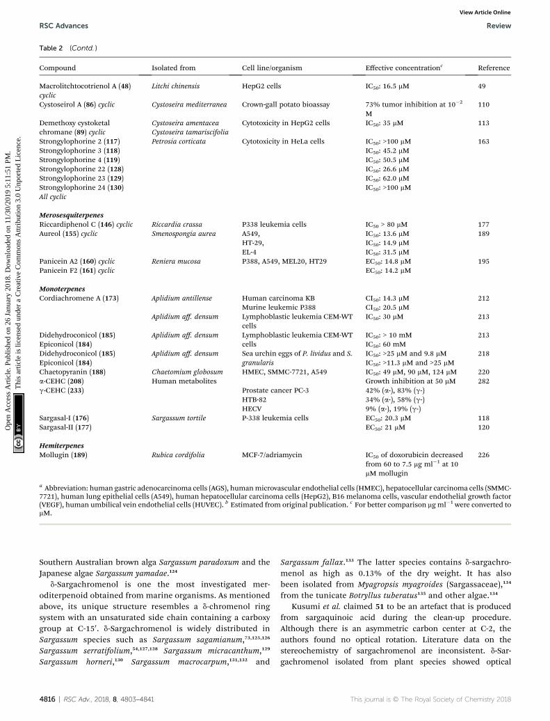
Table 2 (Contd. )
Compound Isolated from Cell line/organism Effective concentrationc Reference
Macrolitchtocotrienol A (48)cyclic
Litchi chinensis HepG2 cells IC50: 16.5 mM 49
Cystoseirol A (86) cyclic Cystoseira mediterranea Crown-gall potato bioassay 73% tumor inhibition at 10�2
M110
Demethoxy cystoketalchromane (89) cyclic
Cystoseira amentacea Cytotoxicity in HepG2 cells IC50: 35 mM 113Cystoseira tamariscifolia
Strongylophorine 2 (117) Petrosia corticata Cytotoxicity in HeLa cells IC50: >100 mM 163Strongylophorine 3 (118) IC50: 45.2 mMStrongylophorine 4 (119) IC50: 50.5 mMStrongylophorine 22 (128) IC50: 26.6 mMStrongylophorine 23 (129) IC50: 62.0 mMStrongylophorine 24 (130) IC50: >100 mMAll cyclic
MerosesquiterpenesRiccardiphenol C (146) cyclic Riccardia crassa P338 leukemia cells IC50 > 80 mM 177Aureol (155) cyclic Smenospongia aurea A549, IC50: 13.6 mM 189
HT-29, IC50: 14.9 mMEL-4 IC50: 31.5 mM
Panicein A2 (160) cyclic Reniera mucosa P388, A549, MEL20, HT29 EC50: 14.8 mM 195Panicein F2 (161) cyclic EC50: 14.2 mM
MonoterpenesCordiachromene A (173) Aplidium antillense Human carcinoma KB CI50: 14.3 mM 212
Murine leukemic P388 CI50: 20.5 mMAplidium aff. densum Lymphoblastic leukemia CEM-WT
cellsIC50: 30 mM 213
Didehydroconicol (185) Aplidium aff. densum Lymphoblastic leukemia CEM-WTcells
IC50: > 10 mM 213Epiconicol (184) IC50: 60 mMDidehydroconicol (185) Aplidium aff. densum Sea urchin eggs of P. lividus and S.
granularisIC50: >25 mM and 9.8 mM 218
Epiconicol (184) IC50: >11.3 mM and >25 mMChaetopyranin (188) Chaetomium globosum HMEC, SMMC-7721, A549 IC50: 49 mM, 90 mM, 124 mM 220a-CEHC (208) Human metabolites Growth inhibition at 50 mM 282g-CEHC (233) Prostate cancer PC-3 42% (a-), 83% (g-)
HTB-82 34% (a-), 58% (g-)HECV 9% (a-), 19% (g-)
Sargasal-I (176) Sargassum tortile P-338 leukemia cells EC50: 20.3 mM 118Sargasal-II (177) EC50: 21 mM 120
HemiterpenesMollugin (189) Rubica cordifolia MCF-7/adriamycin IC50 of doxorubicin decreased
from 60 to 7.5 mg ml�1 at 10mM mollugin
226
a Abbreviation: human gastric adenocarcinoma cells (AGS), humanmicrovascular endothelial cells (HMEC), hepatocellular carcinoma cells (SMMC-7721), human lung epithelial cells (A549), human hepatocellular carcinoma cells (HepG2), B16 melanoma cells, vascular endothelial growth factor(VEGF), human umbilical vein endothelial cells (HUVEC). b Estimated from original publication. c For better comparison mg ml�1 were converted tomM.
RSC Advances Review
Ope
n A
cces
s A
rtic
le. P
ublis
hed
on 2
6 Ja
nuar
y 20
18. D
ownl
oade
d on
11/
30/2
019
5:11
:51
PM.
Thi
s ar
ticle
is li
cens
ed u
nder
a C
reat
ive
Com
mon
s A
ttrib
utio
n 3.
0 U
npor
ted
Lic
ence
.View Article Online
Southern Australian brown alga Sargassum paradoxum and theJapanese algae Sargassum yamadae.124
d-Sargachromenol is one the most investigated mer-oditerpenoid obtained from marine organisms. As mentionedabove, its unique structure resembles a d-chromenol ringsystem with an unsaturated side chain containing a carboxygroup at C-150. d-Sargachromenol is widely distributed inSargassum species such as Sargassum sagamianum,73,125,126
Sargassum serratifolium,54,127,128 Sargassum micracanthum,129
Sargassum horneri,130 Sargassum macrocarpum,131,132 and
4816 | RSC Adv., 2018, 8, 4803–4841
Sargassum fallax.133 The latter species contains d-sargachro-menol as high as 0.13% of the dry weight. It has alsobeen isolated from Myagropsis myagroides (Sargassaceae),134
from the tunicate Botryllus tuberatus135 and other algae.134
Kusumi et al. claimed 51 to be an artefact that is producedfrom sargaquinoic acid during the clean-up procedure.Although there is an asymmetric carbon center at C-2, theauthors found no optical rotation. Literature data on thestereochemistry of sargachromenol are inconsistent. d-Sar-gachromenol isolated from plant species showed optical
This journal is © The Royal Society of Chemistry 2018
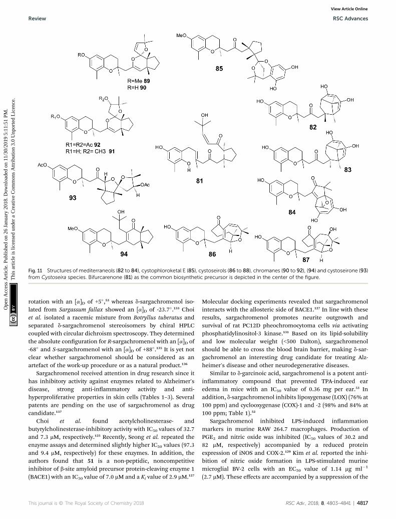
Fig. 11 Structures of mediterraneols (82 to 84), cystophloroketal E (85), cystoseirols (86 to 88), chromanes (90 to 92), (94) and cystoseirone (93)from Cystoseira species. Bifurcarenone (81) as the common biosynthetic precursor is depicted in the center of the figure.
Review RSC Advances
Ope
n A
cces
s A
rtic
le. P
ublis
hed
on 2
6 Ja
nuar
y 20
18. D
ownl
oade
d on
11/
30/2
019
5:11
:51
PM.
Thi
s ar
ticle
is li
cens
ed u
nder
a C
reat
ive
Com
mon
s A
ttrib
utio
n 3.
0 U
npor
ted
Lic
ence
.View Article Online
rotation with an [a]D of +5�,53 whereas d-sargachromenol iso-lated from Sargassum fallax showed an [a]D of -23.7�.133 Choiet al. isolated a racemic mixture from Botryllus tuberatus andseparated d-sargachromenol stereoisomers by chiral HPLCcoupled with circular dichroism spectroscopy. They determinedthe absolute conguration for R-sargachromenol with an [a]D of-68� and S-sargachromenol with an [a]D of +88�.135 It is yet notclear whether sargachromenol should be considered as anartefact of the work-up procedure or as a natural product.136
Sargachromenol received attention in drug research since ithas inhibitory activity against enzymes related to Alzheimer'sdisease, strong anti-inammatory activity and anti-hyperproliferative properties in skin cells (Tables 1–3). Severalpatents are pending on the use of sargachromenol as drugcandidate.137
Choi et al. found acetylcholinesterase- andbutyrylcholinesterase-inhibitory activity with IC50 values of 32.7and 7.3 mM, respectively.125 Recently, Seong et al. repeated theenzyme assays and determined slightly higher IC50 values (97.3and 9.4 mM, respectively) for these enzymes. In addition, theauthors found that 51 is a non-peptidic, noncompetitiveinhibitor of b-site amyloid precursor protein-cleaving enzyme 1(BACE1) with an IC50 value of 7.0 mM and a Ki value of 2.9 mM.127
This journal is © The Royal Society of Chemistry 2018
Molecular docking experiments revealed that sargachromenolinteracts with the allosteric side of BACE1.127 In line with theseresults, sargachromenol promotes neurite outgrowth andsurvival of rat PC12D pheochromocytoma cells via activatingphosphatidylinositol-3 kinase.131 Based on its lipid-solubilityand low molecular weight (<500 Dalton), sargachromenolshould be able to cross the blood brain barrier, making d-sar-gachromenol an interesting drug candidate for treating Alz-heimer's disease and other neurodegenerative diseases.
Similar to d-garcinoic acid, sargachromenol is a potent anti-inammatory compound that prevented TPA-induced earedema in mice with an IC50 value of 0.36 mg per ear.53 Inaddition, d-sargachromenol inhibits lipoxygenase (LOX) (76% at100 ppm) and cyclooxygenase (COX)-1 and -2 (98% and 84% at100 ppm; Table 1).52
Sargachromenol inhibited LPS-induced inammationmarkers in murine RAW 264.7 macrophages. Production ofPGE2 and nitric oxide was inhibited (IC50 values of 30.2 and82 mM, respectively) accompanied by a reduced proteinexpression of iNOS and COX-2.129 Kim et al. reported the inhi-bition of nitric oxide formation in LPS-stimulated murinemicroglial BV-2 cells with an EC50 value of 1.14 mg ml�1
(2.7 mM). These effects are accompanied by a suppression of the
RSC Adv., 2018, 8, 4803–4841 | 4817
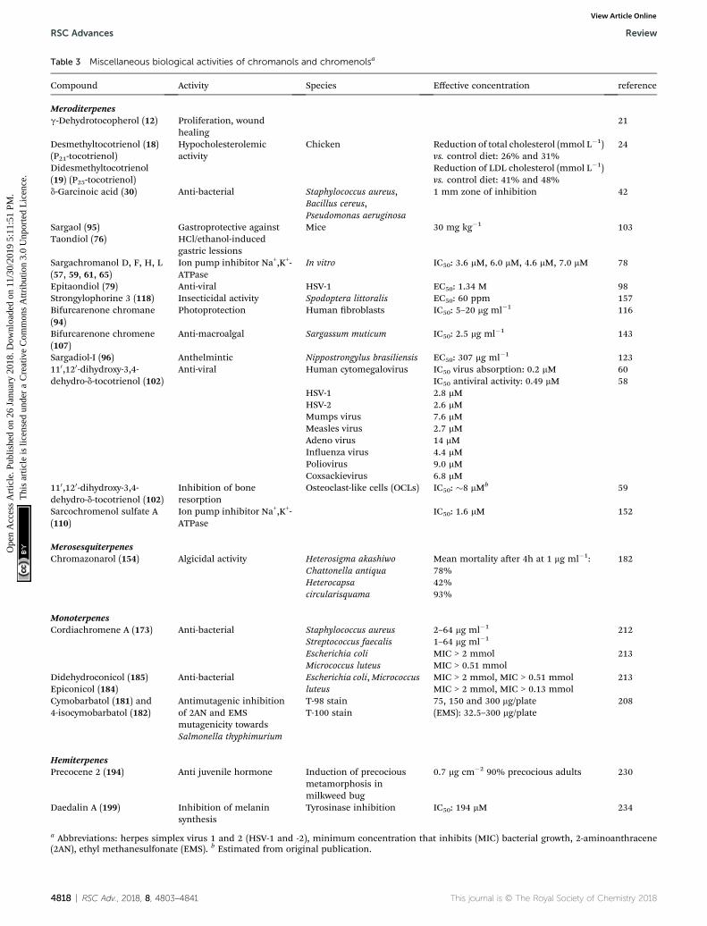
Table 3 Miscellaneous biological activities of chromanols and chromenolsa
Compound Activity Species Effective concentration reference
Meroditerpenesg-Dehydrotocopherol (12) Proliferation, wound
healing21
Desmethyltocotrienol (18)(P21-tocotrienol)
Hypocholesterolemicactivity
Chicken Reduction of total cholesterol (mmol L�1)vs. control diet: 26% and 31%
24
Didesmethyltocotrienol(19) (P25-tocotrienol)
Reduction of LDL cholesterol (mmol L�1)vs. control diet: 41% and 48%
d-Garcinoic acid (30) Anti-bacterial Staphylococcus aureus,Bacillus cereus,Pseudomonas aeruginosa
1 mm zone of inhibition 42
Sargaol (95) Gastroprotective againstHCl/ethanol-inducedgastric lessions
Mice 30 mg kg�1 103Taondiol (76)
Sargachromanol D, F, H, L(57, 59, 61, 65)
Ion pump inhibitor Na+,K+-ATPase
In vitro IC50: 3.6 mM, 6.0 mM, 4.6 mM, 7.0 mM 78
Epitaondiol (79) Anti-viral HSV-1 EC50: 1.34 M 98Strongylophorine 3 (118) Insecticidal activity Spodoptera littoralis EC50: 60 ppm 157Bifurcarenone chromane(94)
Photoprotection Human broblasts IC50: 5–20 mg ml�1 116
Bifurcarenone chromene(107)
Anti-macroalgal Sargassum muticum IC50: 2.5 mg ml�1 143
Sargadiol-I (96) Anthelmintic Nippostrongylus brasiliensis EC50: 307 mg ml�1 123110,120-dihydroxy-3,4-dehydro-d-tocotrienol (102)
Anti-viral Human cytomegalovirus IC50 virus absorption: 0.2 mM 60IC50 antiviral activity: 0.49 mM 58
HSV-1 2.8 mMHSV-2 2.6 mMMumps virus 7.6 mMMeasles virus 2.7 mMAdeno virus 14 mMInuenza virus 4.4 mMPoliovirus 9.0 mMCoxsackievirus 6.8 mM
110,120-dihydroxy-3,4-dehydro-d-tocotrienol (102)
Inhibition of boneresorption
Osteoclast-like cells (OCLs) IC50: �8 mMb 59
Sarcochromenol sulfate A(110)
Ion pump inhibitor Na+,K+-ATPase
IC50: 1.6 mM 152
MerosesquiterpenesChromazonarol (154) Algicidal activity Heterosigma akashiwo Mean mortality aer 4h at 1 mg ml�1: 182
Chattonella antiqua 78%Heterocapsacircularisquama
42%93%
MonoterpenesCordiachromene A (173) Anti-bacterial Staphylococcus aureus 2–64 mg ml�1 212
Streptococcus faecalis 1–64 mg ml�1
Escherichia coli MIC > 2 mmol 213Micrococcus luteus MIC > 0.51 mmol
Didehydroconicol (185) Anti-bacterial Escherichia coli,Micrococcusluteus
MIC > 2 mmol, MIC > 0.51 mmol 213Epiconicol (184) MIC > 2 mmol, MIC > 0.13 mmolCymobarbatol (181) and4-isocymobarbatol (182)
Antimutagenic inhibitionof 2AN and EMSmutagenicity towardsSalmonella thyphimurium
T-98 stain 75, 150 and 300 mg/plate 208T-100 stain (EMS): 32.5–300 mg/plate
HemiterpenesPrecocene 2 (194) Anti juvenile hormone Induction of precocious
metamorphosis inmilkweed bug
0.7 mg cm�2 90% precocious adults 230
Daedalin A (199) Inhibition of melaninsynthesis
Tyrosinase inhibition IC50: 194 mM 234
a Abbreviations: herpes simplex virus 1 and 2 (HSV-1 and -2), minimum concentration that inhibits (MIC) bacterial growth, 2-aminoanthracene(2AN), ethyl methanesulfonate (EMS). b Estimated from original publication.
4818 | RSC Adv., 2018, 8, 4803–4841 This journal is © The Royal Society of Chemistry 2018
RSC Advances Review
Ope
n A
cces
s A
rtic
le. P
ublis
hed
on 2
6 Ja
nuar
y 20
18. D
ownl
oade
d on
11/
30/2
019
5:11
:51
PM.
Thi
s ar
ticle
is li
cens
ed u
nder
a C
reat
ive
Com
mon
s A
ttrib
utio
n 3.
0 U
npor
ted
Lic
ence
.View Article Online
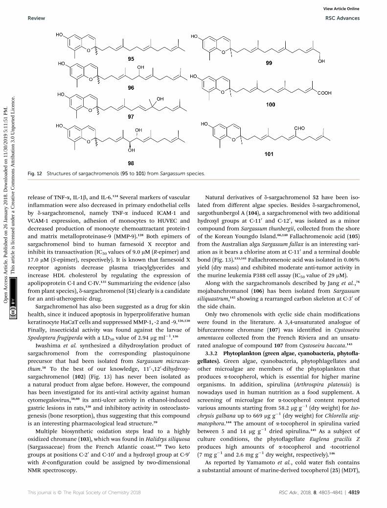
Fig. 12 Structures of sargachromenols (95 to 101) from Sargassum species.
Review RSC Advances
Ope
n A
cces
s A
rtic
le. P
ublis
hed
on 2
6 Ja
nuar
y 20
18. D
ownl
oade
d on
11/
30/2
019
5:11
:51
PM.
Thi
s ar
ticle
is li
cens
ed u
nder
a C
reat
ive
Com
mon
s A
ttrib
utio
n 3.
0 U
npor
ted
Lic
ence
.View Article Online
release of TNF-a, IL-1b, and IL-6.134 Several markers of vascularinammation were also decreased in primary endothelial cellsby d-sargachromenol, namely TNF-a induced ICAM-1 andVCAM-1 expression, adhesion of monocytes to HUVEC anddecreased production of monocyte chemoattractant protein-1and matrix metalloproteinase-9 (MMP-9).128 Both epimers ofsargachromenol bind to human farnesoid X receptor andinhibit its transactivation (IC50 values of 9.0 mM (R-epimer) and17.0 mM (S-epimer), respectively). It is known that farnesoid Xreceptor agonists decrease plasma triacylglycerides andincrease HDL cholesterol by regulating the expression ofapolipoprotein C-I and C-IV.135 Summarizing the evidence (alsofrom plant species), d-sargachromenol (51) clearly is a candidatefor an anti-atherogenic drug.
Sargachromenol has also been suggested as a drug for skinhealth, since it induced apoptosis in hyperproliferative humankeratinocyte HaCaT cells and suppressed MMP-1, -2 and -9.126,130
Finally, insecticidal activity was found against the larvae ofSpodoptera frugiperda with a LD50 value of 2.94 mg ml�1.136
Iwashima et al. synthesized a dihydroxylation product ofsargachromenol from the corresponding plastoquinoneprecursor that had been isolated from Sargassum micracan-thum.58 To the best of our knowledge, 110-,120-dihydroxy-sargachromenol (102) (Fig. 13) has never been isolated asa natural product from algae before. However, the compoundhas been investigated for its anti-viral activity against humancytomegalovirus,58,60 its anti-ulcer activity in ethanol-inducedgastric lesions in rats,138 and inhibitory activity in osteoclasto-genesis (bone resorption), thus suggesting that this compoundis an interesting pharmacological lead structure.59
Multiple biosynthetic oxidation steps lead to a highlyoxidized chromane (103), which was found in Halidrys siliquosa(Sargassaceae) from the French Atlantic coast.139 Two ketogroups at positions C-20 and C-100 and a hydroxyl group at C-90
with R-conguration could be assigned by two-dimensionalNMR spectroscopy.
This journal is © The Royal Society of Chemistry 2018
Natural derivatives of d-sargachromenol 52 have been iso-lated from different algae species. Besides d-sargachromenol,sargothunbergol A (104), a sargachromenol with two additionalhydroxyl groups at C-110 and C-120, was isolated as a minorcompound from Sargassum thunbergii, collected from the shoreof the Korean Youngdo Island.66,140 Fallachromenoic acid (105)from the Australian alga Sargassum fallax is an interesting vari-ation as it bears a chlorine atom at C-110 and a terminal doublebond (Fig. 13).133,141 Fallachromenoic acid was isolated in 0.06%yield (dry mass) and exhibited moderate anti-tumor activity inthe murine leukemia P388 cell assay (IC50 value of 29 mM).
Along with the sargachromanols described by Jang et al.,76
mojabanchromanol (106) has been isolated from Sargassumsiliquastrum,142 showing a rearranged carbon skeleton at C-30 ofthe side chain.
Only two chromenols with cyclic side chain modicationswere found in the literature. A 3,4-unsaturated analogue ofbifurcarenone chromane (107) was identied in Cystoseiraamentacea collected from the French Riviera and an unsatu-rated analogue of compound 107 from Cystoseira baccata.143
3.3.2 Phytoplankton (green algae, cyanobacteria, phytoa-gellates). Green algae, cyanobacteria, phytophlagellates andother microalgae are members of the phytoplankton thatproduces a-tocopherol, which is essential for higher marineorganisms. In addition, spirulina (Arthrospira platensis) isnowadays used in human nutrition as a food supplement. Ascreening of microalgae for a-tocopherol content reportedvarious amounts starting from 58.2 mg g�1 (dry weight) for Iso-chrysis galbana up to 669 mg g�1 (dry weight) for Chlorella stig-matophora.144 The amount of a-tocopherol in spirulina variedbetween 5 and 14 mg g�1 dried spirulina.145 As a subject ofculture conditions, the phytoagellate Euglena gracilis Zproduces high amounts of a-tocopherol and -tocotrienol(7 mg g�1 and 2.6 mg g�1 dry weight, respectively).146
As reported by Yamamoto et al., cold water sh containsa substantial amount of marine-derived tocopherol (25) (MDT),
RSC Adv., 2018, 8, 4803–4841 | 4819
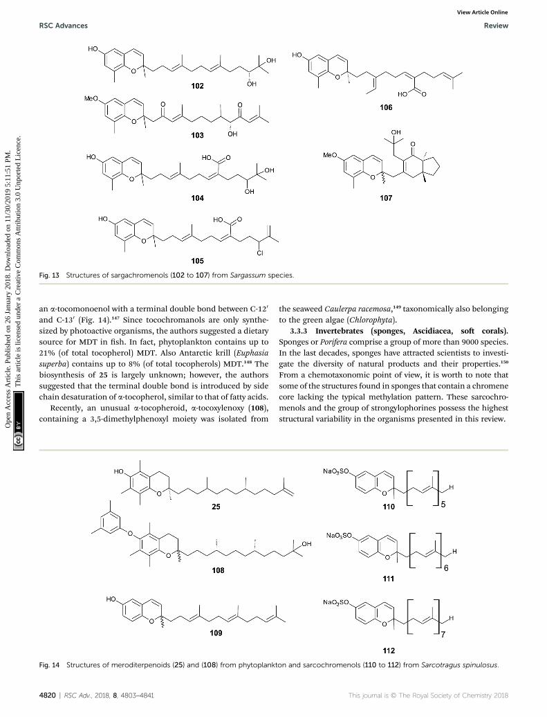
Fig. 13 Structures of sargachromenols (102 to 107) from Sargassum species.
RSC Advances Review
Ope
n A
cces
s A
rtic
le. P
ublis
hed
on 2
6 Ja
nuar
y 20
18. D
ownl
oade
d on
11/
30/2
019
5:11
:51
PM.
Thi
s ar
ticle
is li
cens
ed u
nder
a C
reat
ive
Com
mon
s A
ttrib
utio
n 3.
0 U
npor
ted
Lic
ence
.View Article Online
an a-tocomonoenol with a terminal double bond between C-120
and C-130 (Fig. 14).147 Since tocochromanols are only synthe-sized by photoactive organisms, the authors suggested a dietarysource for MDT in sh. In fact, phytoplankton contains up to21% (of total tocopherol) MDT. Also Antarctic krill (Euphasiasuperba) contains up to 8% (of total tocopherols) MDT.148 Thebiosynthesis of 25 is largely unknown; however, the authorssuggested that the terminal double bond is introduced by sidechain desaturation of a-tocopherol, similar to that of fatty acids.
Recently, an unusual a-tocopheroid, a-tocoxylenoxy (108),containing a 3,5-dimethylphenoxyl moiety was isolated from
Fig. 14 Structures of meroditerpenoids (25) and (108) from phytoplankt
4820 | RSC Adv., 2018, 8, 4803–4841
the seaweed Caulerpa racemosa,149 taxonomically also belongingto the green algae (Chlorophyta).
3.3.3 Invertebrates (sponges, Ascidiacea, so corals).Sponges or Porifera comprise a group of more than 9000 species.In the last decades, sponges have attracted scientists to investi-gate the diversity of natural products and their properties.150
From a chemotaxonomic point of view, it is worth to note thatsome of the structures found in sponges that contain a chromenecore lacking the typical methylation pattern. These sarcochro-menols and the group of strongylophorines possess the higheststructural variability in the organisms presented in this review.
on and sarcochromenols (110 to 112) from Sarcotragus spinulosus.
This journal is © The Royal Society of Chemistry 2018

Review RSC Advances
Ope
n A
cces
s A
rtic
le. P
ublis
hed
on 2
6 Ja
nuar
y 20
18. D
ownl
oade
d on
11/
30/2
019
5:11
:51
PM.
Thi
s ar
ticle
is li
cens
ed u
nder
a C
reat
ive
Com
mon
s A
ttrib
utio
n 3.
0 U
npor
ted
Lic
ence
.View Article Online
A hypothetic biosynthetic precursor of the chromene struc-ture was found in the Western Australian sponge Fasciospongiaspecies (order of Dictyoceratida, family of Thorectidae).151 Fas-cioquinol F (109) is a demethylated 3-4-dehydro-tocotrienol thatmight undergo cyclization to form complex ring systems inanalogy to taondiols (see the section on Brown algae). Thestructure is similar to sargaol (95), but lacks the methyl group atC-8 (Fig. 14). Fascioquinol F revealed moderate antibacterialactivity against Staphylococcus aureus and Bacillus subtilis (IC50
values of 13 and 30 mM, respectively).Sarcochromenols A (110), B (111) and C (112) are a group of
long-chain tocochromenols with ve, six and seven isopreneunits, respectively (Fig. 14). They were isolated from the PacicOcean sponge Sarcotragus spinulosus (Schmidt) (family ofThorectidae) and showed Na+/K+-ATPase inhibitory activitysimilar to that of the sargachromanols D, F, H and L (IC50 valuefor sarcochromenol A of 1.6 mM).78,152 The compounds have alsobeen isolated from the Indian sponge Ircinia fasciculate (Spon-gillidae).153 In addition, an un-sulfated form of sarcachromenolB was isolated in 0.1% yield.
A screening for selective human 15-LOX inhibitors from anextract of the Papua New Guinean sponge Psammocinia (order ofDictyoceratida, family of Irciniidae) revealed chromarols A to D(113 to 116; Fig. 15).154 The IC50 values for chromarols A to Dwere 0.6, 4.0, 0.7 and 1.1 mM, respectively. The authors foundhigh selectivity since the IC50 values for 12-LOX were above100 mM. The biosynthesis of the cyclohexene ring system in theside chain of chromarols presumably derives from an acid-catalyzed cyclization.
Several sponges produce a group of eight polycyclic strong-ylophorines that resemble taondiol structural motives (Fig. 16).They contain a demethylated aromatic ring and modicationsat the methyl groups at C-130 and/or C-150. They were discoveredby Braekman et al. because of their ichthyotoxic activity.155 Thebiosynthesis follows that of taondiol and is an enzyme-catalyzedcyclization cascade (see Fig. 10). Strongylophorines 2 (117), 3(118), 4 (119), and 5 (120) were isolated from Strongylophoradurissima from Maricabiin Island, Philippines,156 anda different, as yet undescribed Strongylophora species from
Fig. 15 Structures of chromarols (113 to 116) from Psammocinia specie
This journal is © The Royal Society of Chemistry 2018
Ilocos Sur, Philippines.157 These molecules contain a cycliclactone, a carboxy, an aldehyde or a hydroxyl group moiety at C-130, respectively. Strongylophorine 3, bearing a terminal carboxygroup, was isolated with 0.1% yield (dry weight).156 Further-more, the known strongylophorines 3, 9 (121) and 11 (122) wereisolated from a Taiwanese species of Strongylophora durissima.The 6-methoxy (121) and 6-acetyl (122) derivatives are struc-turally related to strongylophorine 2, which contains a cycliclactone moiety.158 Liu et al. isolated the strongylophorines 15(26R) (123) and 16 (26S) (124), respectively, from the Okinawansponge Strongylophora strongylata as epimers at the hemiacetalcarbon.159 Biosynthetic O-methylation and O-ethylation gave theacetals 26-O-methoxystrongylophorine 16 (125) and 26-O-ethoxystrongylophorine 16 (126), respectively.160,161 Noda et al.found a mixture of strongylophorines 15 and 16 to be stronginhibitors of the proteasome with IC50 values of 3.6 mM.160 Thesame study compared the proteasome-inhibitory activity ofstructurally related strongylophorines and found the followingorder: hemiacetal > acetal � carboxy > lactone > nomodication.
On their search for inhibitors of protein tyrosine phospha-tase 1B, an enzyme that plays a crucial role in the regulation ofinsulin and leptin signalling, Lee et al. found inhibitory activityfor 125, 117, 118, 123, and strongylophorine 17 (127) with IC50
values of 8.5, 24.4, 9.0, 11.9, and 14.8 mM, respectively.161
Strongylophorines 2 and 3 also inhibited hypoxia-induciblefactor-1-dependent luciferase expression in engineered U251-HRE glioma cells with EC50 values of 8 and 13 mM.162 Strong-ylophorines 22 (128), 23 (129), 24 (130), and 17 (127) were iso-lated from the Okinawan sponge Petrosia corticata anddisplayed moderate cytotoxic activity against uman cervicalcarcinoma epithelial (HeLa) cells (Table 2).163 All strong-ylophorines exhibited ichthyotoxic, insecticidal, anti-bacterial,fungicidal, and cytotoxic properties. Strongylophorine 22 andfascioquinol D are epimers at C-2 and were isolated from Fas-ciospongia sp.151 The latter compounds displayed anti-microbialactivity against Staphylococcus aureus and Bacillus subtilis withIC50 values of 25 and 2.3 mM (for strongylophorine 22) and 7.8and 2.8 mM (for fascioquinol D), respectively.
s.
RSC Adv., 2018, 8, 4803–4841 | 4821

Fig. 16 Structures of strongylophorines (117 to 130) from Strongylophora species.
RSC Advances Review
Ope
n A
cces
s A
rtic
le. P
ublis
hed
on 2
6 Ja
nuar
y 20
18. D
ownl
oade
d on
11/
30/2
019
5:11
:51
PM.
Thi
s ar
ticle
is li
cens
ed u
nder
a C
reat
ive
Com
mon
s A
ttrib
utio
n 3.
0 U
npor
ted
Lic
ence
.View Article Online
Recently, Yu et al. presented the rst semi-synthesis ofstrongylophorine 2 starting from isocupressic acid.164
3.3.4 Ascidiacea/tunicates. Ascidians, tunicates or seasquirts belong to a group of more than 3000 species, most ofthem not investigated in terms of bioactive metabolites. Ina recent review, Palanisamy et al. described almost 600 chemicalstructures found in tunicates.165 Here, we describe mer-oditerpenes, such as an epimeric mixture of R- and S-sargach-romenol and two epimeric chromenes called tuberatolide B and20-epi-tuberatolide B (131), obtained from the tunicate Botryllustuberatus.135 Tuberatolide B contains a g-lactone moiety withinthe side chain that presumably derives from a C-150-carboxy, C-60-hydroxy-precursor (Fig. 17). Both tuberatolides were strongfarnesoid X receptor agonists with IC50 values of 1.5 and 2.5 mM,respectively.135
4822 | RSC Adv., 2018, 8, 4803–4841
3.3.5 So corals. So corals (Alcyonacea) belong to theclass of Anthozoa and compromise approximately 800 speciesliving mostly in warm seawater. In recent years, the number ofnew metabolites discovered from so corals was estimated torepresent 22% of the total new marine natural products.166
Many metabolites showed anti-tumor, anti-viral, anti-foulingand anti-inammatory activities (reviewed in (ref. 167)).
Bowden et al. isolated tocotrichromenol (132), an isomer ofsargaol (95), and its dihydro derivative (133) from an unknownAustralian Nephthea species.168 The precursor quinone was alsoisolated, but did not convert into the chromenol under thework-up conditions; however, no optical activity was found at C-2.
Two a-tocopherol derivatives with three hydroxyl groups at C-80, C-110, and C-120, respectively, were isolated from Lobophytum
This journal is © The Royal Society of Chemistry 2018
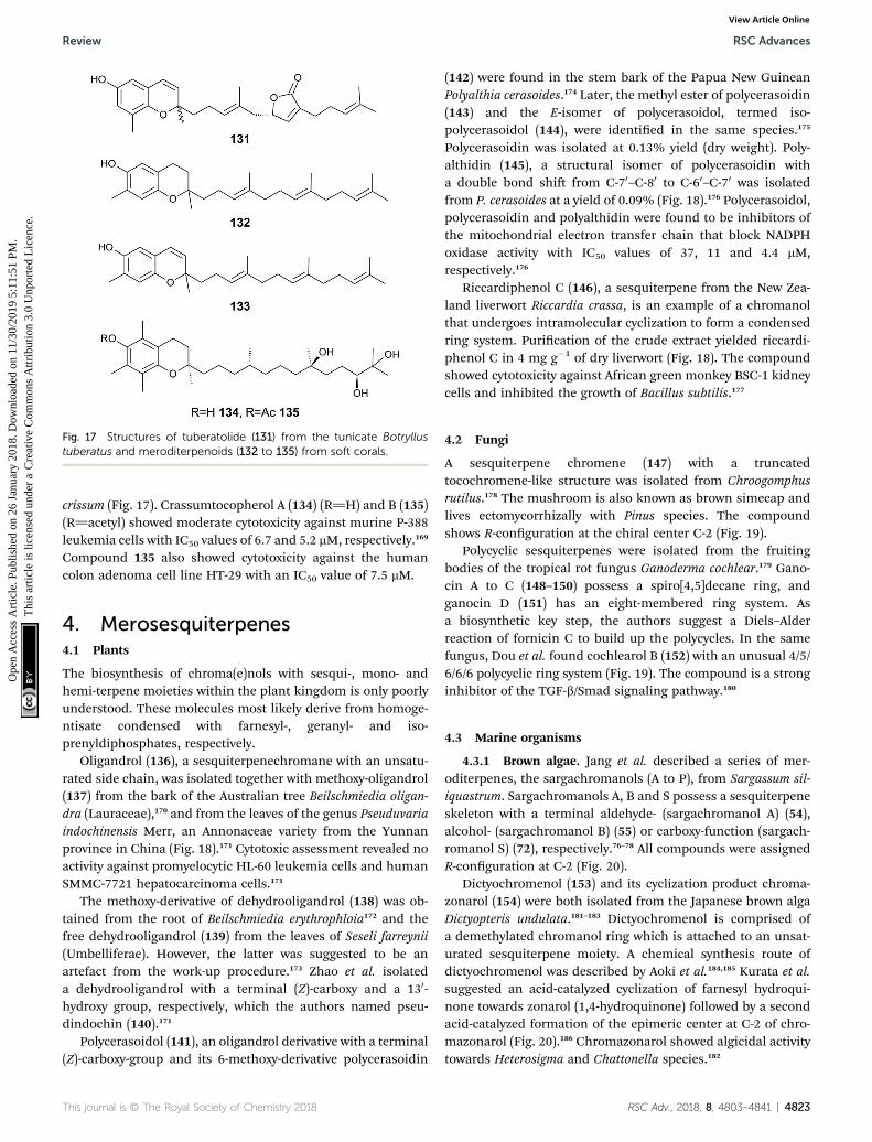
Fig. 17 Structures of tuberatolide (131) from the tunicate Botryllustuberatus and meroditerpenoids (132 to 135) from soft corals.
Review RSC Advances
Ope
n A
cces
s A
rtic
le. P
ublis
hed
on 2
6 Ja
nuar
y 20
18. D
ownl
oade
d on
11/
30/2
019
5:11
:51
PM.
Thi
s ar
ticle
is li
cens
ed u
nder
a C
reat
ive
Com
mon
s A
ttrib
utio
n 3.
0 U
npor
ted
Lic
ence
.View Article Online
crissum (Fig. 17). Crassumtocopherol A (134) (R]H) and B (135)(R]acetyl) showed moderate cytotoxicity against murine P-388leukemia cells with IC50 values of 6.7 and 5.2 mM, respectively.169
Compound 135 also showed cytotoxicity against the humancolon adenoma cell line HT-29 with an IC50 value of 7.5 mM.
4. Merosesquiterpenes4.1 Plants
The biosynthesis of chroma(e)nols with sesqui-, mono- andhemi-terpene moieties within the plant kingdom is only poorlyunderstood. These molecules most likely derive from homoge-ntisate condensed with farnesyl-, geranyl- and iso-prenyldiphosphates, respectively.
Oligandrol (136), a sesquiterpenechromane with an unsatu-rated side chain, was isolated together with methoxy-oligandrol(137) from the bark of the Australian tree Beilschmiedia oligan-dra (Lauraceae),170 and from the leaves of the genus Pseuduvariaindochinensis Merr, an Annonaceae variety from the Yunnanprovince in China (Fig. 18).171 Cytotoxic assessment revealed noactivity against promyelocytic HL-60 leukemia cells and humanSMMC-7721 hepatocarcinoma cells.171
The methoxy-derivative of dehydrooligandrol (138) was ob-tained from the root of Beilschmiedia erythrophloia172 and thefree dehydrooligandrol (139) from the leaves of Seseli farreynii(Umbelliferae). However, the latter was suggested to be anartefact from the work-up procedure.173 Zhao et al. isolateda dehydrooligandrol with a terminal (Z)-carboxy and a 130-hydroxy group, respectively, which the authors named pseu-dindochin (140).171
Polycerasoidol (141), an oligandrol derivative with a terminal(Z)-carboxy-group and its 6-methoxy-derivative polycerasoidin
This journal is © The Royal Society of Chemistry 2018
(142) were found in the stem bark of the Papua New GuineanPolyalthia cerasoides.174 Later, the methyl ester of polycerasoidin(143) and the E-isomer of polycerasoidol, termed iso-polycerasoidol (144), were identied in the same species.175
Polycerasoidin was isolated at 0.13% yield (dry weight). Poly-althidin (145), a structural isomer of polycerasoidin witha double bond shi from C-70–C-80 to C-60–C-70 was isolatedfrom P. cerasoides at a yield of 0.09% (Fig. 18).176 Polycerasoidol,polycerasoidin and polyalthidin were found to be inhibitors ofthe mitochondrial electron transfer chain that block NADPHoxidase activity with IC50 values of 37, 11 and 4.4 mM,respectively.176
Riccardiphenol C (146), a sesquiterpene from the New Zea-land liverwort Riccardia crassa, is an example of a chromanolthat undergoes intramolecular cyclization to form a condensedring system. Purication of the crude extract yielded riccardi-phenol C in 4 mg g�1 of dry liverwort (Fig. 18). The compoundshowed cytotoxicity against African green monkey BSC-1 kidneycells and inhibited the growth of Bacillus subtilis.177
4.2 Fungi
A sesquiterpene chromene (147) with a truncatedtocochromene-like structure was isolated from Chroogomphusrutilus.178 The mushroom is also known as brown simecap andlives ectomycorrhizally with Pinus species. The compoundshows R-conguration at the chiral center C-2 (Fig. 19).
Polycyclic sesquiterpenes were isolated from the fruitingbodies of the tropical rot fungus Ganoderma cochlear.179 Gano-cin A to C (148–150) possess a spiro[4,5]decane ring, andganocin D (151) has an eight-membered ring system. Asa biosynthetic key step, the authors suggest a Diels–Alderreaction of fornicin C to build up the polycycles. In the samefungus, Dou et al. found cochlearol B (152) with an unusual 4/5/6/6/6 polycyclic ring system (Fig. 19). The compound is a stronginhibitor of the TGF-b/Smad signaling pathway.180
4.3 Marine organisms
4.3.1 Brown algae. Jang et al. described a series of mer-oditerpenes, the sargachromanols (A to P), from Sargassum sil-iquastrum. Sargachromanols A, B and S possess a sesquiterpeneskeleton with a terminal aldehyde- (sargachromanol A) (54),alcohol- (sargachromanol B) (55) or carboxy-function (sargach-romanol S) (72), respectively.76–78 All compounds were assignedR-conguration at C-2 (Fig. 20).
Dictyochromenol (153) and its cyclization product chroma-zonarol (154) were both isolated from the Japanese brown algaDictyopteris undulata.181–183 Dictyochromenol is comprised ofa demethylated chromanol ring which is attached to an unsat-urated sesquiterpene moiety. A chemical synthesis route ofdictyochromenol was described by Aoki et al.184,185 Kurata et al.suggested an acid-catalyzed cyclization of farnesyl hydroqui-none towards zonarol (1,4-hydroquinone) followed by a secondacid-catalyzed formation of the epimeric center at C-2 of chro-mazonarol (Fig. 20).186 Chromazonarol showed algicidal activitytowards Heterosigma and Chattonella species.182
RSC Adv., 2018, 8, 4803–4841 | 4823

Fig. 18 Structures of plant sesquiterpenes (136 to 146).
Fig. 19 Structures of sesquiterpenes (147 to 152) from fungi species.
RSC Advances Review
Ope
n A
cces
s A
rtic
le. P
ublis
hed
on 2
6 Ja
nuar
y 20
18. D
ownl
oade
d on
11/
30/2
019
5:11
:51
PM.
Thi
s ar
ticle
is li
cens
ed u
nder
a C
reat
ive
Com
mon
s A
ttrib
utio
n 3.
0 U
npor
ted
Lic
ence
.View Article Online
4.3.2 Sponges. A structural isomer of chromazonarol, aur-eol (155), and its 5-chloro-derivative (156) were isolated from theCaribbean sponge Smenospongia aurea.187,188 It has been sug-gested that aureol results from a rearrangement of the drimaneskeleton of chromazonarol. Aureol showed moderate cytotoxicactivity against several cell lines, such as human adenocarci-nomic A549 alveolar basal epithelial cells, human colon
4824 | RSC Adv., 2018, 8, 4803–4841
adenocarcinoma HT-29 cells, and murine EL4 lymphoma cellswith IC50 values of 13.6, 14.9 and 31.5 mM, respectively.189 In2002, Nakamura et al. presented a chemical synthesis of aur-eol.190 Besides aureol, 2-epichromazonarol was isolated (2.2%dry weight) from Smenospongia aurea.187 Recently, a structurallyrelated meroterpenoid, puupehenol (157), with potent anti-microbial properties was isolated from the Hawaiian sponge
This journal is © The Royal Society of Chemistry 2018

Fig. 20 Scheme of multiple acid-catalyzed cyclizations of farnesyl hydroquinone towards chromazoranol (154) as suggested by Kurata et al.186
Structures of cyclic (157) and linear sesquiterpenoids (54, 55, 72 and 153).
Review RSC Advances
Ope
n A
cces
s A
rtic
le. P
ublis
hed
on 2
6 Ja
nuar
y 20
18. D
ownl
oade
d on
11/
30/2
019
5:11
:51
PM.
Thi
s ar
ticle
is li
cens
ed u
nder
a C
reat
ive
Com
mon
s A
ttrib
utio
n 3.
0 U
npor
ted
Lic
ence
.View Article Online
Dactylospongia sp. (Fig. 20).191 The authors suggested that thewell-known puupehenone may be a work-up artefact of thenatural precursor puupehenol.
Two epimeric sesquiterpene chromenols, named cyclo-renierin A and B (158), were found in Haliclona sp., an Indo-Pacic sponge from Vanuatu.192 The biosynthesis of the cyclo-hexenone ring system seems to follow that of walsurol (Fig. 6).
Panicein B2 (159) bears a chromene ring and an aromaticring system in the side chain (Fig. 21). It was rst isolated byCimino et al. from Haliclona panacea and later from the Medi-terranean sponge Reniera fulva.193,194 Panicein B2 was also foundin Reniera mucosa along with panicein A2 (160) and F2 (161).195
It has been suggested that the aromatic group of the side chainis formed from cyclorenierin A/B by a 1,2-methyl migration andsubsequent oxidation.193 All paniceins show racemic carboncenters at C-2 suggesting that these compounds may be arte-facts from the work-up procedure.
Faulkner et al. isolated a series of unusual ansa chromenemacrocycles from Smenospongia sp., a sponge from the Sey-chelles.196 Smenochromes A to D (162–165) were isolated with0.26% yield (dry weight) for A and 0.037% for B, C and D,respectively (Fig. 22). The compounds showed no optical activityand thus occurred as racemic mixtures. The structurally relatedlikonides A (166) and B (167) were isolated from the Kenyansponge Haytella sp. with 0.06 and 0.04% yield.197 The biosyn-thesis of ansa chromenes presumably starts from a farnesylated
This journal is © The Royal Society of Chemistry 2018
hydroquinone followed by alkylation at C-5 of the activatedhydroquinone ring or alternatively by O-alkylation of theterminal double bond.197
4.3.3 Ascidiacea/tunicates. Longithorol E (168) was isolatedas a minor metabolite from the Australian ascidian Aplidiumlongithorax (Fig. 22).198
4.3.4 Molluscs. There is emerging interest in the metabo-lites of marine nudibranchs. Since these animals completelylost their protective shell, the production or accumulation oftoxins from their prey is used as defense systems.199 Twooligandrol-like structures (169) and (170) were isolated fromCratena peregrine, and a chromenol (171) with a C-60 ketonemoiety was found in the frilled nudibranch Leminda millecrathat is only found in South Africa200 (Fig. 23).
4.3.5 So corals. Although sesquiterpenes are widelydistributed in so corals, we only found sparce information onsesquiterpene chromanes and chromenes, respectively.201 Cap-illobenzopyranol (172) was isolated from the Australian socoral Sinularia capillosa and showed moderate cytotoxicityagainst P-388 cells (ED50 values of 12.7 mM).202 Its quinoneprecursor has been isolated from Sinularia lochmodes.203 Thecompound with a terminal furanyl moiety showed in vitro anti-inammatory activity against LPS-activation in murine RAW264.7 macrophages. Protein expression of iNOS was inhibitedby 36.7% at 10 mM concentration of 172 (Table 1), howeverexpression of COX-2 was not affected.
RSC Adv., 2018, 8, 4803–4841 | 4825

Fig. 21 Structures of sesquiterpenes (155 to 161) from sponge species.
Fig. 22 Structures of ansa chromane macrocycles (162 to 167) from Smenospongia species and (168) from tunicate.
RSC Advances Review
Ope
n A
cces
s A
rtic
le. P
ublis
hed
on 2
6 Ja
nuar
y 20
18. D
ownl
oade
d on
11/
30/2
019
5:11
:51
PM.
Thi
s ar
ticle
is li
cens
ed u
nder
a C
reat
ive
Com
mon
s A
ttrib
utio
n 3.
0 U
npor
ted
Lic
ence
.View Article Online
5. Monoterpenes
Monoterpenes from plant origin have been used since ancienttimes to treat certain diseases, such as inammation or cancer.De Sousa and colleagues summarized the anti-cancer and anti-inammatory activities of monoterpenes in an outstandingrecent review.204
4826 | RSC Adv., 2018, 8, 4803–4841
5.1 Plants
The monoterpene cordiachromene A (173) was isolated fromthe heartwood of the tropical American tree Cordia alliodora(Boraginaceae) by Manners et al.205 The authors proposed ger-anyl benzoquinol as the biogenic precursor of the compound.Interestingly, the woods of Cordia alliodora are recognized fortheir durability in marine uses. Cordiachromene A was also
This journal is © The Royal Society of Chemistry 2018
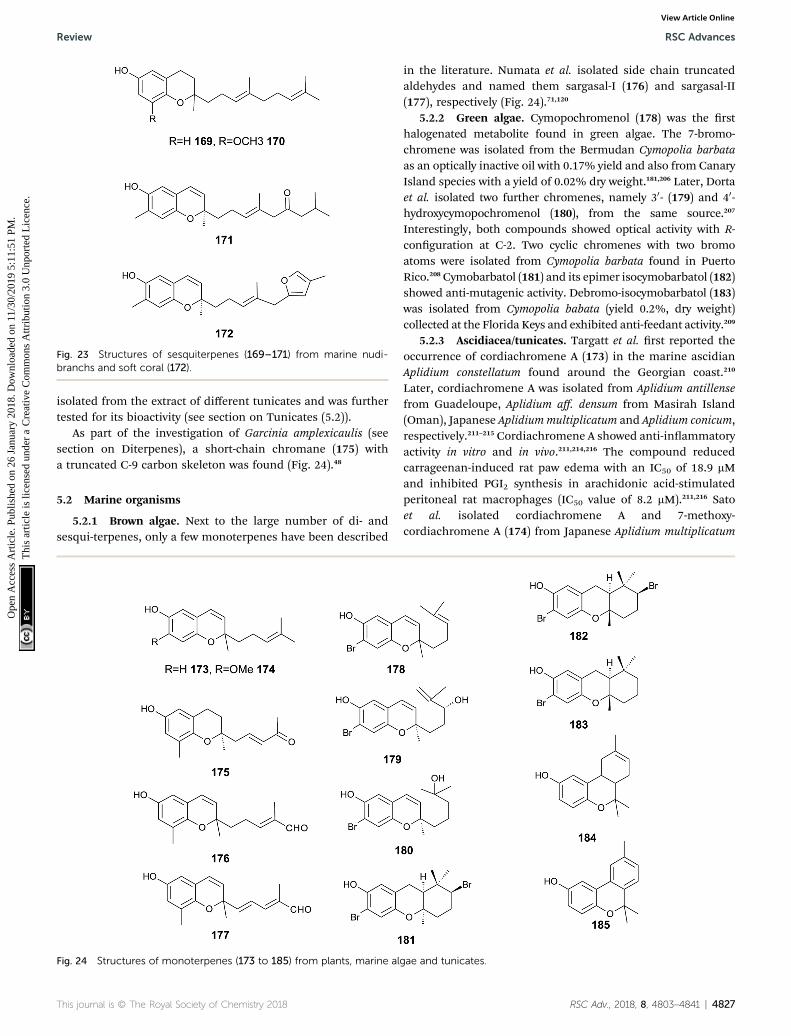
Fig. 23 Structures of sesquiterpenes (169–171) from marine nudi-branchs and soft coral (172).
Review RSC Advances
Ope
n A
cces
s A
rtic
le. P
ublis
hed
on 2
6 Ja
nuar
y 20
18. D
ownl
oade
d on
11/
30/2
019
5:11
:51
PM.
Thi
s ar
ticle
is li
cens
ed u
nder
a C
reat
ive
Com
mon
s A
ttrib
utio
n 3.
0 U
npor
ted
Lic
ence
.View Article Online
isolated from the extract of different tunicates and was furthertested for its bioactivity (see section on Tunicates (5.2)).
As part of the investigation of Garcinia amplexicaulis (seesection on Diterpenes), a short-chain chromane (175) witha truncated C-9 carbon skeleton was found (Fig. 24).48
5.2 Marine organisms
5.2.1 Brown algae. Next to the large number of di- andsesqui-terpenes, only a few monoterpenes have been described
Fig. 24 Structures of monoterpenes (173 to 185) from plants, marine al
This journal is © The Royal Society of Chemistry 2018
in the literature. Numata et al. isolated side chain truncatedaldehydes and named them sargasal-I (176) and sargasal-II(177), respectively (Fig. 24).71,120
5.2.2 Green algae. Cymopochromenol (178) was the rsthalogenated metabolite found in green algae. The 7-bromo-chromene was isolated from the Bermudan Cymopolia barbataas an optically inactive oil with 0.17% yield and also from CanaryIsland species with a yield of 0.02% dry weight.181,206 Later, Dortaet al. isolated two further chromenes, namely 30- (179) and 40-hydroxycymopochromenol (180), from the same source.207
Interestingly, both compounds showed optical activity with R-conguration at C-2. Two cyclic chromenes with two bromoatoms were isolated from Cymopolia barbata found in PuertoRico.208Cymobarbatol (181) and its epimer isocymobarbatol (182)showed anti-mutagenic activity. Debromo-isocymobarbatol (183)was isolated from Cymopolia babata (yield 0.2%, dry weight)collected at the Florida Keys and exhibited anti-feedant activity.209
5.2.3 Ascidiacea/tunicates. Targatt et al. rst reported theoccurrence of cordiachromene A (173) in the marine ascidianAplidium constellatum found around the Georgian coast.210
Later, cordiachromene A was isolated from Aplidium antillensefrom Guadeloupe, Aplidium aff. densum from Masirah Island(Oman), Japanese Aplidium multiplicatum and Aplidium conicum,respectively.211–215 Cordiachromene A showed anti-inammatoryactivity in vitro and in vivo.211,214,216 The compound reducedcarrageenan-induced rat paw edema with an IC50 of 18.9 mMand inhibited PGI2 synthesis in arachidonic acid-stimulatedperitoneal rat macrophages (IC50 value of 8.2 mM).211,216 Satoet al. isolated cordiachromene A and 7-methoxy-cordiachromene A (174) from Japanese Aplidium multiplicatum
gae and tunicates.
RSC Adv., 2018, 8, 4803–4841 | 4827
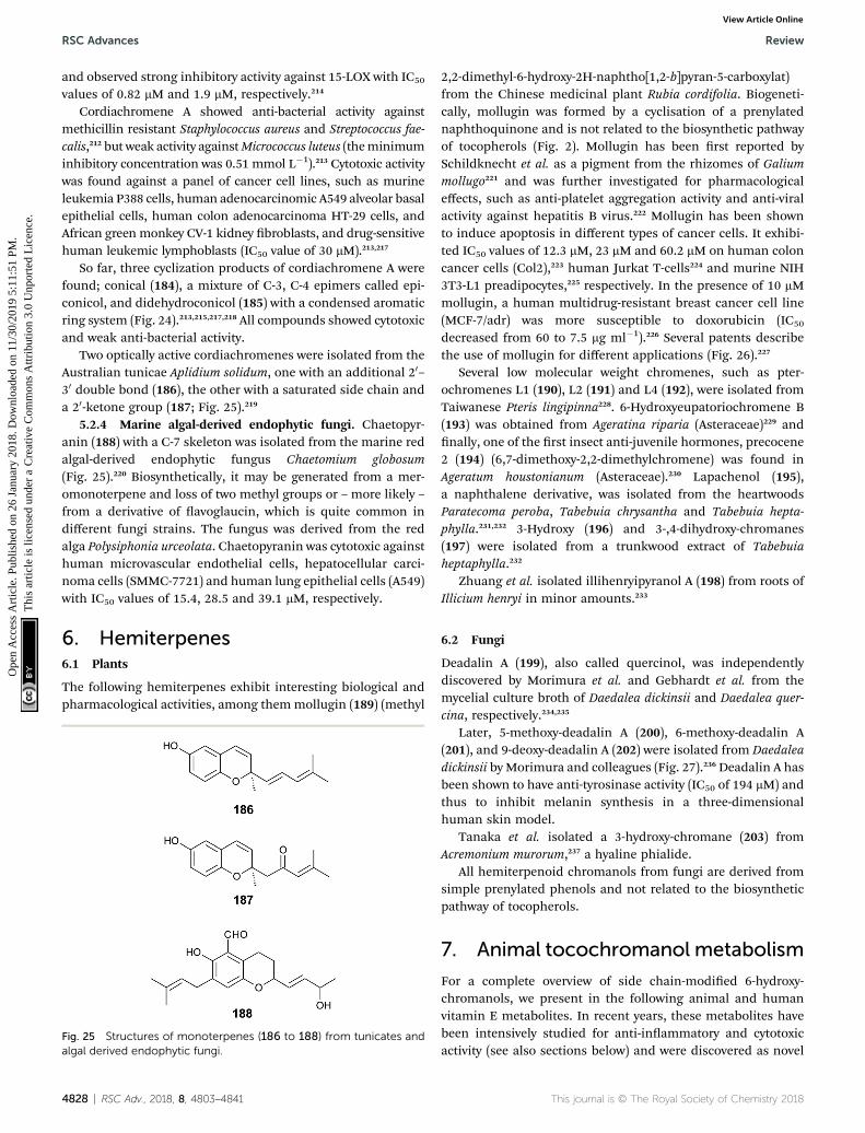
RSC Advances Review
Ope
n A
cces
s A
rtic
le. P
ublis
hed
on 2
6 Ja
nuar
y 20
18. D
ownl
oade
d on
11/
30/2
019
5:11
:51
PM.
Thi
s ar
ticle
is li
cens
ed u
nder
a C
reat
ive
Com
mon
s A
ttrib
utio
n 3.
0 U
npor
ted
Lic
ence
.View Article Online
and observed strong inhibitory activity against 15-LOX with IC50
values of 0.82 mM and 1.9 mM, respectively.214
Cordiachromene A showed anti-bacterial activity againstmethicillin resistant Staphylococcus aureus and Streptococcus fae-calis,212 but weak activity againstMicrococcus luteus (theminimuminhibitory concentration was 0.51 mmol L�1).213 Cytotoxic activitywas found against a panel of cancer cell lines, such as murineleukemia P388 cells, human adenocarcinomic A549 alveolar basalepithelial cells, human colon adenocarcinoma HT-29 cells, andAfrican green monkey CV-1 kidney broblasts, and drug-sensitivehuman leukemic lymphoblasts (IC50 value of 30 mM).213,217
So far, three cyclization products of cordiachromene A werefound; conical (184), a mixture of C-3, C-4 epimers called epi-conicol, and didehydroconicol (185) with a condensed aromaticring system (Fig. 24).213,215,217,218 All compounds showed cytotoxicand weak anti-bacterial activity.
Two optically active cordiachromenes were isolated from theAustralian tunicae Aplidium solidum, one with an additional 20–30 double bond (186), the other with a saturated side chain anda 20-ketone group (187; Fig. 25).219
5.2.4 Marine algal-derived endophytic fungi. Chaetopyr-anin (188) with a C-7 skeleton was isolated from the marine redalgal-derived endophytic fungus Chaetomium globosum(Fig. 25).220 Biosynthetically, it may be generated from a mer-omonoterpene and loss of two methyl groups or – more likely –from a derivative of avoglaucin, which is quite common indifferent fungi strains. The fungus was derived from the redalga Polysiphonia urceolata. Chaetopyranin was cytotoxic againsthuman microvascular endothelial cells, hepatocellular carci-noma cells (SMMC-7721) and human lung epithelial cells (A549)with IC50 values of 15.4, 28.5 and 39.1 mM, respectively.
6. Hemiterpenes6.1 Plants
The following hemiterpenes exhibit interesting biological andpharmacological activities, among themmollugin (189) (methyl
Fig. 25 Structures of monoterpenes (186 to 188) from tunicates andalgal derived endophytic fungi.
4828 | RSC Adv., 2018, 8, 4803–4841
2,2-dimethyl-6-hydroxy-2H-naphtho[1,2-b]pyran-5-carboxylat)from the Chinese medicinal plant Rubia cordifolia. Biogeneti-cally, mollugin was formed by a cyclisation of a prenylatednaphthoquinone and is not related to the biosynthetic pathwayof tocopherols (Fig. 2). Mollugin has been rst reported bySchildknecht et al. as a pigment from the rhizomes of Galiummollugo221 and was further investigated for pharmacologicaleffects, such as anti-platelet aggregation activity and anti-viralactivity against hepatitis B virus.222 Mollugin has been shownto induce apoptosis in different types of cancer cells. It exhibi-ted IC50 values of 12.3 mM, 23 mM and 60.2 mM on human coloncancer cells (Col2),223 human Jurkat T-cells224 and murine NIH3T3-L1 preadipocytes,225 respectively. In the presence of 10 mMmollugin, a human multidrug-resistant breast cancer cell line(MCF-7/adr) was more susceptible to doxorubicin (IC50
decreased from 60 to 7.5 mg ml�1).226 Several patents describethe use of mollugin for different applications (Fig. 26).227
Several low molecular weight chromenes, such as pter-ochromenes L1 (190), L2 (191) and L4 (192), were isolated fromTaiwanese Pteris lingipinna228. 6-Hydroxyeupatoriochromene B(193) was obtained from Ageratina riparia (Asteraceae)229 andnally, one of the rst insect anti-juvenile hormones, precocene2 (194) (6,7-dimethoxy-2,2-dimethylchromene) was found inAgeratum houstonianum (Asteraceae).230 Lapachenol (195),a naphthalene derivative, was isolated from the heartwoodsParatecoma peroba, Tabebuia chrysantha and Tabebuia hepta-phylla.231,232 3-Hydroxy (196) and 3-,4-dihydroxy-chromanes(197) were isolated from a trunkwood extract of Tabebuiaheptaphylla.232
Zhuang et al. isolated illihenryipyranol A (198) from roots ofIllicium henryi in minor amounts.233
6.2 Fungi
Deadalin A (199), also called quercinol, was independentlydiscovered by Morimura et al. and Gebhardt et al. from themycelial culture broth of Daedalea dickinsii and Daedalea quer-cina, respectively.234,235
Later, 5-methoxy-deadalin A (200), 6-methoxy-deadalin A(201), and 9-deoxy-deadalin A (202) were isolated from Daedaleadickinsii by Morimura and colleagues (Fig. 27).236 Deadalin A hasbeen shown to have anti-tyrosinase activity (IC50 of 194 mM) andthus to inhibit melanin synthesis in a three-dimensionalhuman skin model.
Tanaka et al. isolated a 3-hydroxy-chromane (203) fromAcremonium murorum,237 a hyaline phialide.
All hemiterpenoid chromanols from fungi are derived fromsimple prenylated phenols and not related to the biosyntheticpathway of tocopherols.
7. Animal tocochromanol metabolism
For a complete overview of side chain-modied 6-hydroxy-chromanols, we present in the following animal and humanvitamin E metabolites. In recent years, these metabolites havebeen intensively studied for anti-inammatory and cytotoxicactivity (see also sections below) and were discovered as novel
This journal is © The Royal Society of Chemistry 2018
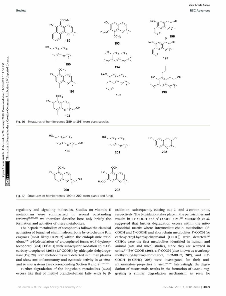
Fig. 26 Structures of hemiterpenes (189 to 198) from plant species.
Fig. 27 Structures of hemiterpenes (199 to 202) from plants and fungi.
Review RSC Advances
Ope
n A
cces
s A
rtic
le. P
ublis
hed
on 2
6 Ja
nuar
y 20
18. D
ownl
oade
d on
11/
30/2
019
5:11
:51
PM.
Thi
s ar
ticle
is li
cens
ed u
nder
a C
reat
ive
Com
mon
s A
ttrib
utio
n 3.
0 U
npor
ted
Lic
ence
.View Article Online
regulatory and signaling molecules. Studies on vitamin Emetabolism were summarized in several outstandingreviews;17,238,239 we therefore describe here only briey theformation and activities of these metabolites.
The hepatic metabolism of tocopherols follows the classicalactivation of branched chain hydrocarbons by cytochrome P450enzymes (most likely CYP4F2) within the endoplasmic retic-ulum.240 u-Hydroxylation of a-tocopherol forms a-130-hydroxy-tocopherol (204) (130-OH) with subsequent oxidation to a-130-carboxy-tocopherol (205) (130-COOH) by aldehyde dehydroge-nase (Fig. 28). Both metabolites were detected in human plasmaand show anti-inammatory and cytotoxic activity in in vitro-and in vivo systems (see corresponding Section 8 and 9).241,242
Further degradation of the long-chain metabolites (LCM)occurs like that of methyl branched-chain fatty acids by b-
This journal is © The Royal Society of Chemistry 2018
oxidation, subsequently cutting out 2- and 3-carbon units,respectively. The b-oxidation takes place in the peroxisomes andresults in 110-COOH and 90-COOH LCM.240 Mustacich et al.suggested that further degradation occurs within the mito-chondrial matrix where intermediate-chain metabolites (70-COOH and 50-COOH) and short-chain metabolites 30-COOH (orcarboxy-ethyl-hydroxy-chromanol (CEHC)) were detected.240
CEHCs were the rst metabolites identied in human andanimal (rats and mice) studies, since they are secreted inurine.243 d-90-COOH (206), a-50-COOH (also known as a-carboxy-methylbutyl-hydroxy-chromanol, a-CMBHC; 207), and a-30-COOH (a-CEHC; 208) were investigated for their anti-inammatory properties in vitro.244,245 Interestingly, the degra-dation of tocotrienols results in the formation of CEHC, sug-gesting a similar degradation mechanism as seen for
RSC Adv., 2018, 8, 4803–4841 | 4829
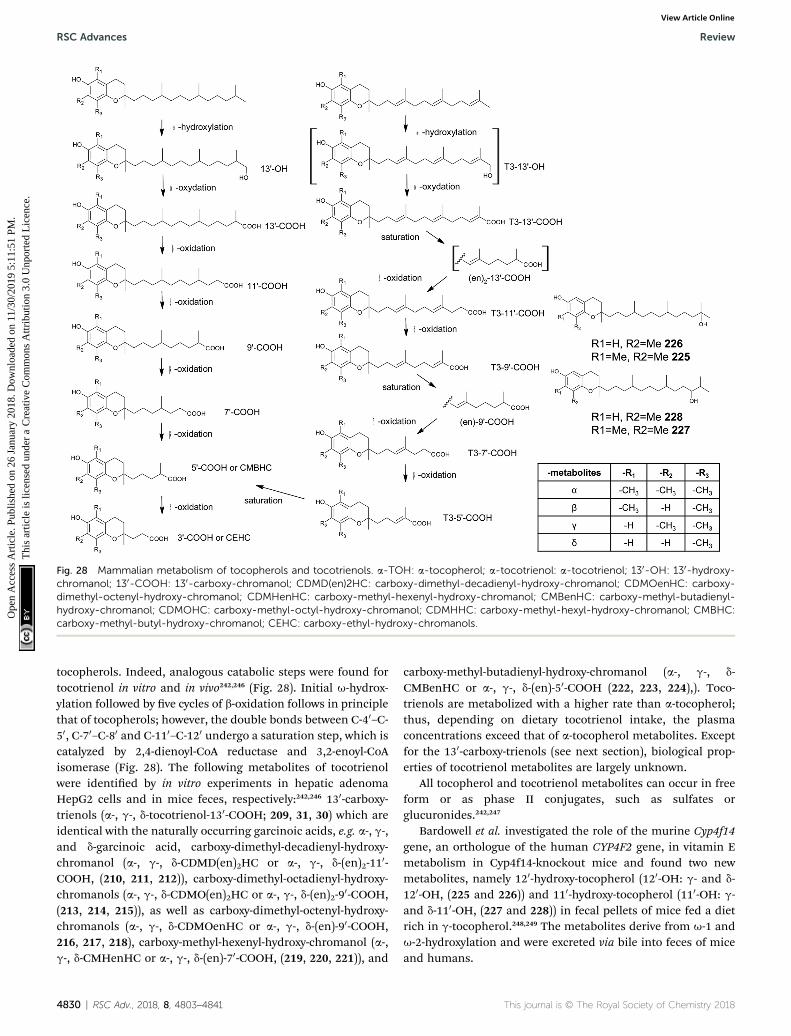
Fig. 28 Mammalian metabolism of tocopherols and tocotrienols. a-TOH: a-tocopherol; a-tocotrienol: a-tocotrienol; 130-OH: 130-hydroxy-chromanol; 130-COOH: 130-carboxy-chromanol; CDMD(en)2HC: carboxy-dimethyl-decadienyl-hydroxy-chromanol; CDMOenHC: carboxy-dimethyl-octenyl-hydroxy-chromanol; CDMHenHC: carboxy-methyl-hexenyl-hydroxy-chromanol; CMBenHC: carboxy-methyl-butadienyl-hydroxy-chromanol; CDMOHC: carboxy-methyl-octyl-hydroxy-chromanol; CDMHHC: carboxy-methyl-hexyl-hydroxy-chromanol; CMBHC:carboxy-methyl-butyl-hydroxy-chromanol; CEHC: carboxy-ethyl-hydroxy-chromanols.
RSC Advances Review
Ope
n A
cces
s A
rtic
le. P
ublis
hed
on 2
6 Ja
nuar
y 20
18. D
ownl
oade
d on
11/
30/2
019
5:11
:51
PM.
Thi
s ar
ticle
is li
cens
ed u
nder
a C
reat
ive
Com
mon
s A
ttrib
utio
n 3.
0 U
npor
ted
Lic
ence
.View Article Online
tocopherols. Indeed, analogous catabolic steps were found fortocotrienol in vitro and in vivo242,246 (Fig. 28). Initial u-hydrox-ylation followed by ve cycles of b-oxidation follows in principlethat of tocopherols; however, the double bonds between C-40–C-50, C-70–C-80 and C-110–C-120 undergo a saturation step, which iscatalyzed by 2,4-dienoyl-CoA reductase and 3,2-enoyl-CoAisomerase (Fig. 28). The following metabolites of tocotrienolwere identied by in vitro experiments in hepatic adenomaHepG2 cells and in mice feces, respectively:242,246 130-carboxy-trienols (a-, g-, d-tocotrienol-130-COOH; 209, 31, 30) which areidentical with the naturally occurring garcinoic acids, e.g. a-, g-,and d-garcinoic acid, carboxy-dimethyl-decadienyl-hydroxy-chromanol (a-, g-, d-CDMD(en)2HC or a-, g-, d-(en)2-110-COOH, (210, 211, 212)), carboxy-dimethyl-octadienyl-hydroxy-chromanols (a-, g-, d-CDMO(en)2HC or a-, g-, d-(en)2-90-COOH,(213, 214, 215)), as well as carboxy-dimethyl-octenyl-hydroxy-chromanols (a-, g-, d-CDMOenHC or a-, g-, d-(en)-90-COOH,216, 217, 218), carboxy-methyl-hexenyl-hydroxy-chromanol (a-,g-, d-CMHenHC or a-, g-, d-(en)-70-COOH, (219, 220, 221)), and
4830 | RSC Adv., 2018, 8, 4803–4841
carboxy-methyl-butadienyl-hydroxy-chromanol (a-, g-, d-CMBenHC or a-, g-, d-(en)-50-COOH (222, 223, 224),). Toco-trienols are metabolized with a higher rate than a-tocopherol;thus, depending on dietary tocotrienol intake, the plasmaconcentrations exceed that of a-tocopherol metabolites. Exceptfor the 130-carboxy-trienols (see next section), biological prop-erties of tocotrienol metabolites are largely unknown.
All tocopherol and tocotrienol metabolites can occur in freeform or as phase II conjugates, such as sulfates orglucuronides.242,247
Bardowell et al. investigated the role of the murine Cyp4f14gene, an orthologue of the human CYP4F2 gene, in vitamin Emetabolism in Cyp4f14-knockout mice and found two newmetabolites, namely 120-hydroxy-tocopherol (120-OH: g- and d-120-OH, (225 and 226)) and 110-hydroxy-tocopherol (110-OH: g-and d-110-OH, (227 and 228)) in fecal pellets of mice fed a dietrich in g-tocopherol.248,249 The metabolites derive from u-1 andu-2-hydroxylation and were excreted via bile into feces of miceand humans.
This journal is © The Royal Society of Chemistry 2018

Review RSC Advances
Ope
n A
cces
s A
rtic
le. P
ublis
hed
on 2
6 Ja
nuar
y 20
18. D
ownl
oade
d on
11/
30/2
019
5:11
:51
PM.
Thi
s ar
ticle
is li
cens
ed u
nder
a C
reat
ive
Com
mon
s A
ttrib
utio
n 3.
0 U
npor
ted
Lic
ence
.View Article Online
8. Anti-inflammatory activity of toco-chromanols and -chromenols
Many diseases, including atherosclerosis, diabetes or evencancer, are related to inammatory processes. A decreasedgrade of inammation could lead to a reduced risk for thesediseases. In the past, human clinical trials with a-tocopherol asan anti-inammatory agent revealed contradictoryresults.17,239,250,251 We here like to broaden the view to structur-ally related chromanols and chromenols and compare theiranti-inammatory in vitro activity.
The anti-inammatory activities of tocopherols and toco-trienols from the human diet are well known and are compiledin Table 1.239,245,252 In general, the chromanols with saturatedand unsaturated side chains showed good to moderate inhibi-tory activity depending on the anti-inammatory markermeasured and the in vitro system used.253,254 For example, Jianget al. investigated the inhibition of COX-2 catalyzed PGE2
synthesis in IL-1b stimulated human lung epithelial A549 cellsof a series of tocopherols and tocotrienols, respectively.245 Theinhibitory activities reached from IC50 > 50 mM for a-tocopherolto IC50 ¼ 1–3 mM for d-tocopherol and g-tocotrienol.
The LCM of tocopherols and tocotrienols with a terminal C-130-carboxy and -hydroxyl group, respectively, were found innanomolar concentration in human plasma and intensivelystudied as anti-inammatory agents. The research on the LCMwas promoted by the facile semi-syntheses from garcinoic acidthat can be efficiently isolated from Garcinia kola.38,39
a-130-Carboxy-tocopherol (205) (a-130-COOH) inhibited theexpression of iNOS by 100% at 5 mM and the formation of nitricoxide by 100% at 2.7 mM, respectively.255,256 A recent investiga-tion on the anti-inammatory activity of a-130-COOH showedstrong inhibition of recombinant 5-LOX and only moderateinhibition of COX-1, leukotriene (LT) C4 synthase, PGES-1 andepoxide hydrolase.254 Human recombinant COX-2 was notinhibited by a-130-COOH at low concentrations.
The d-tocopherol metabolite d-130-carboxy-tocopherol (229)(d-130-COOH) showed a slightly reduced activity, and inhibitediNOS protein synthesis by 56% at 5 mM and nitric oxideformation by 79% at 5 mM, respectively, in LPS-activated murineRAW264.7 macrophages. d-130-COOH further inhibited the LPS-induced upregulation of COX-2 expression in the same cellswith IC50 values of 4–5 mM.245,257 5-LOX activity was inhibited inthe range from 0.5–2.0 mM, depending on the assay used.253
The 130-Hydroxy-tocopherols of a-, and d-tocopherol (a-130-OH (204) and d-130-OH (231)) are synthetically available andhave been therefore intensively studied. a-130-OH reduced COX-2 expression (49%) and PGE2 synthesis (54%) and both alcoholsinhibited iNOS expression by 53–60% at 10 mM in LPS-inducedmurine RAW264.7 macrophages.241,256 Both alcohols can bebiochemically converted by mammalian cells to the corre-sponding acids, thus making it difficult to distinguish betweenthe activity of 130-OH and 130-COOH (unpublished results).
As described in the section on human metabolism, themetabolic truncation of the LCM leads to several medium- andshort-chain metabolites, such as 90-carboxy-tocopherols (90-
This journal is © The Royal Society of Chemistry 2018
COOH), CMBHC and CEHC, respectively. In general, the anti-inammatory activities of the medium- and short-chainmetabolites seem to decrease with the decreasing lengths ofthe side chains, resulting in higher IC50 values (Table 1).245
In summary, although the number of in vitro studies ist stilllimited and different markers of inammation cannot becompared directly, we roughly estimate the anti-inammatoryactivity of tocopherols, tocotrienols and their metabolites asfollows: a-tocopherol < non-a-tocopherol � tocotrienols � 130-OH � 130-COOH [ 90-COOH > CMBHC � CEHC. However, itmust be kept in mind that the molecular modes of action of thesemolecules seem to be quite different. It is obvious that the impactof the metabolites depends on individual metabolism rates(pharmacokinetics) of the tocochromanols from the diet. Gre-benstein et al. proposed that the affinity of vitamers towards the a-tocopherol transfer protein (a-TTP) may predict their degradationby cytochrome P450 enzymes.258 a-TTP has the strongest affinityfor a-tocopherol with Kd of 25 nM and much higher Kd values forthe other vitamin E forms, depending on their methylationpattern and side chain saturation.239,259 Accordingly, the catabo-lism of non-a-tocopherol vitamers into the corresponding LCMmay occur much faster than that of a-tocopherol, thus generatingmore anti-inammatory metabolites. As a result, a-tocopherol perse is less active than all other vitamers following the order: d-tocopherol � g-tocotrienol > g-tocopherol [ a-tocopherol.239
d-Garcinoic acid (30) is the main constituent of several Gar-cinia species, which are known for their anti-inammatoryproperties in African ethnomedicine.37 d-Garcinoic acid was re-ported to inhibit COX-2 (IC50¼ 10 mM) and, even stronger, 5-LOXwith IC50 ranging from 0.04 to 1.0 mM.257 d-Garcinoic acid down-regulated the LPS-induced expression of pro-inammatorycytokines, such as TNF-a, IL-6, IL-1b, COX-2 and iNOS inmacrophages and reduced production of nitric oxide (IC50 valueof 1 mM).255,260 A direct comparison of several carboxy-tocotrienols (tocotrienol-130-COOH metabolites) from plantorigin as inhibitors of microsomal PGE2 synthase-1 revealed thefollowing order of activity: g-garcinoic acid (31) > b-garcinoicacid (232) > d-garcinoic acid (30) > a-garcinoic acid (209);however the methylation pattern had only moderate impact.44
Structurally related forms of d-garcinoic acid, such as d-sar-gachromenol (51) with a 150-COOH group and a chromene ringsystem, showed only moderate inhibitory activity on in LPS-stimulated production nitric oxide and PGE2 in murine RAW264.7 macrophages (IC50 values of 82 mM and 30.2 mM, respec-tively);129 however, much higher activity was observed in BV-2microglial cells (IC50 value for inhibition of nitric oxideproduction of 1.3–2.7 mM).134,261
As described above, 130-OH metabolites have a similar anti-inammatory potential than the corresponding 130-COOH.Thus, natural products such as sargachromanols D (57), E (58)and G (60), respectively, with hydroxyl-groups at C-90 and C-100
are interesting intermediates. They all showed moderateinhibitory activity on nitric oxide production in LPS-stimulatedmurine RAW 264.7 cells (IC50 values of 15–40 mM).79,83,84
Cyclic meroditerpenes such as epitaondiol (79) and the chro-marols A to D (113 to 116) exhibited anti-inammatory activity invitro and in vivo (Table 1).99,154 The four chromarols A to D
RSC Adv., 2018, 8, 4803–4841 | 4831

RSC Advances Review
Ope
n A
cces
s A
rtic
le. P
ublis
hed
on 2
6 Ja
nuar
y 20
18. D
ownl
oade
d on
11/
30/2
019
5:11
:51
PM.
Thi
s ar
ticle
is li
cens
ed u
nder
a C
reat
ive
Com
mon
s A
ttrib
utio
n 3.
0 U
npor
ted
Lic
ence
.View Article Online
inhibited 15-LOX with IC50 ¼ 0.6(113), 4.0(114), 0.7(115) and 1.1mM (116), respectively, but not 12-LOX. Epitaondiol was effectivein a TPA-induced mouse ear edema study (IC50 ¼ 20.7 mg per ear)and inhibited eicosanoid synthesis with an IC50 of 3.8 mM forthromboxane B2 (TXB2) and an IC50 of 30.1 mM for LTB4.
The cyclic sesquiterpenes capillobenzopyranol (172) onlymoderately inhibited nitric oxide production in LPS-stimulatedmacrophages by 37% at 10 mM.202 The monoterpene cordiach-romene A (173) inhibited soybean 15-LOX with an IC50 of 0.82mM and lipid peroxidation with an IC50 of 2 mM.214
Only moderate anti-inammatory activity was observed forthe hemiterpene quercinol (199), whereas it inhibited COX-2expression with an IC50 of 0.63 mM.235
In Fig. 29 we postulate the structural motives that areessential for the anti-inammatory activity based on the struc-tures and properties discussed above. The most effectivecompounds described are the diterpenes 130-COOH, 130-OH,garcinoic acid and d-sargachromenol, respectively, with strongpotential as anti-inammatory drug candidates. A recent SARstudy revealed that the effects of human LCM depend on thepresence of the chromanol ring and modications in the sidechain and less on the substitution pattern at the aromaticring.256 This study is in line with the observation of Silva et al.with d-sargachromenol (51) and its precursor 1,4-benzoquinonesargaquinoic acid; the latter had less inhibitory activity towardsLOX- and COX-enzymes.52 In addition to the naturalcompounds described above, the anti-inammatory and anti-diabetic drug troglitazone exhibits a 6-hydroxy-chromane ringsystem. Troglitazone was used a PPAR-g-receptor agonist butwas withdrawn from the market since it caused hepatotox-icity.262 Obviously, anti-inammatory activity is enhanced by theoccurrence of a 6-hydroxy-chromane and -chromene moiety,respectively.
In conclusion, meroditerpenoids with a functional group(COOH, OH) at the side chain have much higher anti-inammatory activity than the parent chromanols and chro-menols, respectively.
9. Anti-proliferative and cytotoxicactivity of chromanols and chromenols
Dietary tocopherols and tocotrienols have been extensivelyinvestigated for their cancer-preventive potential in several
Fig. 29 General domains modulating the anti-inflammatory activity ofmeroterpenoids.
4832 | RSC Adv., 2018, 8, 4803–4841
human intervention trials (reviewed in263 and6), but widelyfailed to prove benecial effects.264 However, in vitro studieswith tocopherols and tocotrienols in cell cultures and in vivostudies have shown pronounced anti-neoplastic and anti-carcinogenic effects.265,266
The susceptibility of the cell lines tested for anti-carcinogenic activity varied tremendously and makes thus itdifficult to compare the compounds discussed in this section.For example, HepG2 liver cells exhibit greater resistance todrugs and toxins compared to other cells lines, since theyactively express phase I and II enzymes. As a result, higher IC50
values are expected for drug resistant cell lines, such as HepG2.Structure–activity relationship studies revealed that chem-
ical modications at C-6 of the aromatic ring (ethers or esters)magnied the cytotoxic potential of vitamin E compounds.266,267
In general, drug candidates that were ‘redox-silent’ at C-6, suchas tocopherol-succinate, showed promising results in animalstudies.268 Although most of the redox-silent compounds werechemically synthesized, distinct structure–activity relationshipshave been derived from these experiments.266 The studiesrevealed the importance of three major domains of the chro-manols tested: rst, the functional domain (I) that needs to be‘redox silent’ to exert the cytotoxic properties. Second, thesignaling domain (II) modied by the methylation pattern ofthe chromanol ring system. Third, the hydrophobic domain (III)that is mostly covered by saturated and unsaturated sidechains.267 Reviewing the structural features of the moleculespresented here, we further specify the domains that are relevantfor cytotoxicity.
Tocopherols seem to have low or moderate anti-canceractivity in the different cell model systems.267,269 a-Tocopherol(3) in particular is well-tolerated by adenoma and cancer cells insupra-physiological concentrations (above 100 mM) (see Table2).269 g-Tocopherol (5) at 25 mM, however, had anti-proliferativeactivity on colon carcinoma (CaCo-2), androgen-sensitive(LNCaP) and androgen-resistant (PC-3) prostate, lung adeno-carcinoma (A549), and osteosarcoma (SaOs-2) cells, but did notinduce apoptosis in these cell lines.269,270 Accordingly, wepostulate the following order of activity for tocopherols: g-tocopherol (5) > d-tocopherol (6) [ a-tocopherol (3).
Tocotrienols showed anti-proliferative and pro-apoptoticeffects in vitro and in vivo and are in general more potent inthe prevention of cancer than tocopherols.271 Several moleculartargets were identied for g- and d-tocotrienols (16) and (17) (g-and d-tocotrienol), respectively (summarized by272). Induction ofmitochondrial apoptosis, demonstrated by activation ofcaspase-3 and -9, along with modulation of apoptogenic genessuch as Bcl-2, Bcl-xl and Bax, respectively, has been observed formost of the tocotrienols tested.
a-Tocotrienol (14) showed low to moderate pro-apoptoticactivity against different breast cancer (MDA-MB-435, MDA-MB-231, and MCF7) and melanoma (B16) cells, respectively(Table 2).25,273,274 In contrast, g- (16) and d-tocotrienol (17)induced apoptosis at low micromolar concentrations in most ofthe cell lines tested.25,267,275,276 Sargaol (95) (3-4-dehydro-d-toco-trienol), has been tested with moderate activity in humangastric epithelial cells, human broblasts and murine
This journal is © The Royal Society of Chemistry 2018
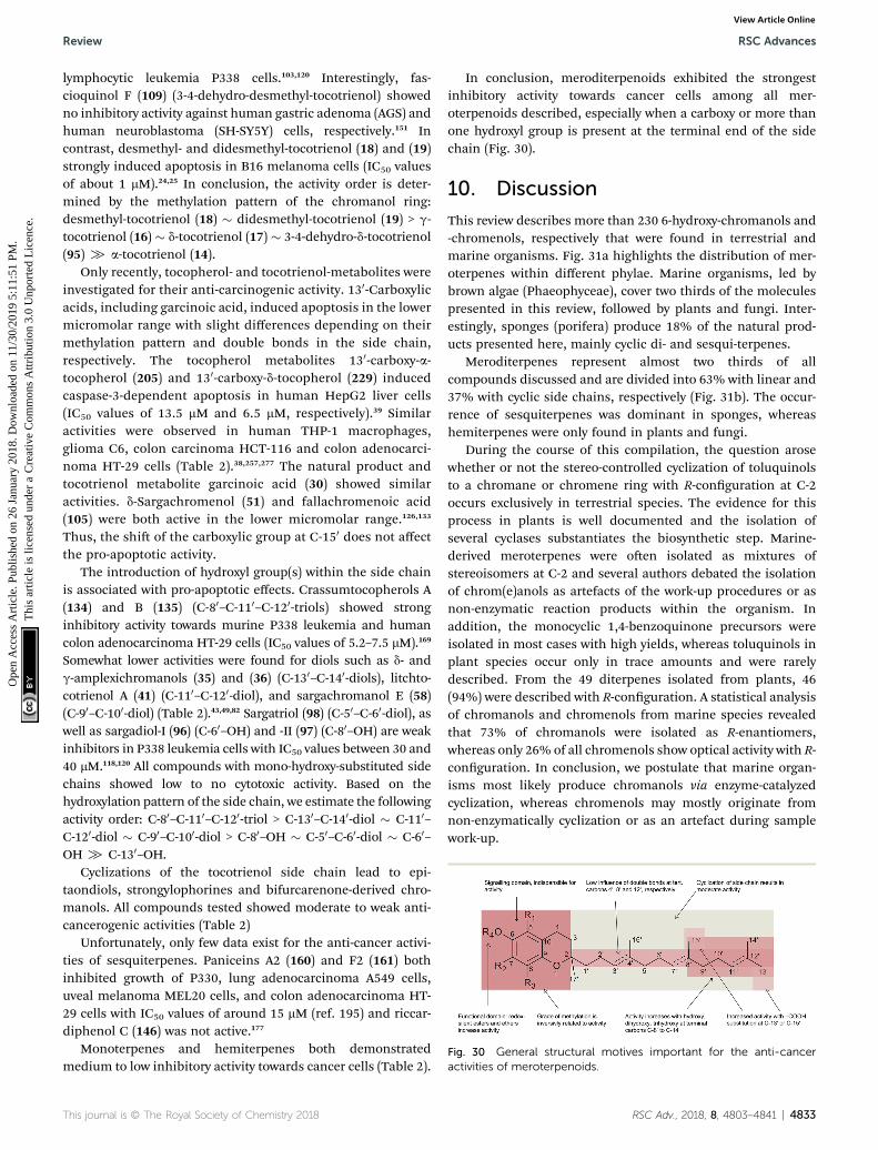
Fig. 30 General structural motives important for the anti-canceractivities of meroterpenoids.
Review RSC Advances
Ope
n A
cces
s A
rtic
le. P
ublis
hed
on 2
6 Ja
nuar
y 20
18. D
ownl
oade
d on
11/
30/2
019
5:11
:51
PM.
Thi
s ar
ticle
is li
cens
ed u
nder
a C
reat
ive
Com
mon
s A
ttrib
utio
n 3.
0 U
npor
ted
Lic
ence
.View Article Online
lymphocytic leukemia P338 cells.103,120 Interestingly, fas-cioquinol F (109) (3-4-dehydro-desmethyl-tocotrienol) showedno inhibitory activity against human gastric adenoma (AGS) andhuman neuroblastoma (SH-SY5Y) cells, respectively.151 Incontrast, desmethyl- and didesmethyl-tocotrienol (18) and (19)strongly induced apoptosis in B16 melanoma cells (IC50 valuesof about 1 mM).24,25 In conclusion, the activity order is deter-mined by the methylation pattern of the chromanol ring:desmethyl-tocotrienol (18) � didesmethyl-tocotrienol (19) > g-tocotrienol (16) � d-tocotrienol (17) � 3-4-dehydro-d-tocotrienol(95) [ a-tocotrienol (14).
Only recently, tocopherol- and tocotrienol-metabolites wereinvestigated for their anti-carcinogenic activity. 130-Carboxylicacids, including garcinoic acid, induced apoptosis in the lowermicromolar range with slight differences depending on theirmethylation pattern and double bonds in the side chain,respectively. The tocopherol metabolites 130-carboxy-a-tocopherol (205) and 130-carboxy-d-tocopherol (229) inducedcaspase-3-dependent apoptosis in human HepG2 liver cells(IC50 values of 13.5 mM and 6.5 mM, respectively).39 Similaractivities were observed in human THP-1 macrophages,glioma C6, colon carcinoma HCT-116 and colon adenocarci-noma HT-29 cells (Table 2).38,257,277 The natural product andtocotrienol metabolite garcinoic acid (30) showed similaractivities. d-Sargachromenol (51) and fallachromenoic acid(105) were both active in the lower micromolar range.126,133
Thus, the shi of the carboxylic group at C-150 does not affectthe pro-apoptotic activity.
The introduction of hydroxyl group(s) within the side chainis associated with pro-apoptotic effects. Crassumtocopherols A(134) and B (135) (C-80–C-110–C-120-triols) showed stronginhibitory activity towards murine P338 leukemia and humancolon adenocarcinoma HT-29 cells (IC50 values of 5.2–7.5 mM).169
Somewhat lower activities were found for diols such as d- andg-amplexichromanols (35) and (36) (C-130–C-140-diols), litchto-cotrienol A (41) (C-110–C-120-diol), and sargachromanol E (58)(C-90–C-100-diol) (Table 2).43,49,82 Sargatriol (98) (C-50–C-60-diol), aswell as sargadiol-I (96) (C-60–OH) and -II (97) (C-80–OH) are weakinhibitors in P338 leukemia cells with IC50 values between 30 and40 mM.118,120 All compounds with mono-hydroxy-substituted sidechains showed low to no cytotoxic activity. Based on thehydroxylation pattern of the side chain, we estimate the followingactivity order: C-80–C-110–C-120-triol > C-130–C-140-diol � C-110–C-120-diol � C-90–C-100-diol > C-80–OH � C-50–C-60-diol � C-60–OH [ C-130–OH.
Cyclizations of the tocotrienol side chain lead to epi-taondiols, strongylophorines and bifurcarenone-derived chro-manols. All compounds tested showed moderate to weak anti-cancerogenic activities (Table 2)
Unfortunately, only few data exist for the anti-cancer activi-ties of sesquiterpenes. Paniceins A2 (160) and F2 (161) bothinhibited growth of P330, lung adenocarcinoma A549 cells,uveal melanoma MEL20 cells, and colon adenocarcinoma HT-29 cells with IC50 values of around 15 mM (ref. 195) and riccar-diphenol C (146) was not active.177
Monoterpenes and hemiterpenes both demonstratedmedium to low inhibitory activity towards cancer cells (Table 2).
This journal is © The Royal Society of Chemistry 2018
In conclusion, meroditerpenoids exhibited the strongestinhibitory activity towards cancer cells among all mer-oterpenoids described, especially when a carboxy or more thanone hydroxyl group is present at the terminal end of the sidechain (Fig. 30).
10. Discussion
This review describes more than 230 6-hydroxy-chromanols and-chromenols, respectively that were found in terrestrial andmarine organisms. Fig. 31a highlights the distribution of mer-oterpenes within different phylae. Marine organisms, led bybrown algae (Phaeophyceae), cover two thirds of the moleculespresented in this review, followed by plants and fungi. Inter-estingly, sponges (porifera) produce 18% of the natural prod-ucts presented here, mainly cyclic di- and sesqui-terpenes.
Meroditerpenes represent almost two thirds of allcompounds discussed and are divided into 63% with linear and37% with cyclic side chains, respectively (Fig. 31b). The occur-rence of sesquiterpenes was dominant in sponges, whereashemiterpenes were only found in plants and fungi.
During the course of this compilation, the question arosewhether or not the stereo-controlled cyclization of toluquinolsto a chromane or chromene ring with R-conguration at C-2occurs exclusively in terrestrial species. The evidence for thisprocess in plants is well documented and the isolation ofseveral cyclases substantiates the biosynthetic step. Marine-derived meroterpenes were oen isolated as mixtures ofstereoisomers at C-2 and several authors debated the isolationof chrom(e)anols as artefacts of the work-up procedures or asnon-enzymatic reaction products within the organism. Inaddition, the monocyclic 1,4-benzoquinone precursors wereisolated in most cases with high yields, whereas toluquinols inplant species occur only in trace amounts and were rarelydescribed. From the 49 diterpenes isolated from plants, 46(94%) were described with R-conguration. A statistical analysisof chromanols and chromenols from marine species revealedthat 73% of chromanols were isolated as R-enantiomers,whereas only 26% of all chromenols show optical activity with R-conguration. In conclusion, we postulate that marine organ-isms most likely produce chromanols via enzyme-catalyzedcyclization, whereas chromenols may mostly originate fromnon-enzymatically cyclization or as an artefact during samplework-up.
RSC Adv., 2018, 8, 4803–4841 | 4833

Fig. 31 Statistical distribution of meroterpenoids.
RSC Advances Review
Ope
n A
cces
s A
rtic
le. P
ublis
hed
on 2
6 Ja
nuar
y 20
18. D
ownl
oade
d on
11/
30/2
019
5:11
:51
PM.
Thi
s ar
ticle
is li
cens
ed u
nder
a C
reat
ive
Com
mon
s A
ttrib
utio
n 3.
0 U
npor
ted
Lic
ence
.View Article Online
The structural variability of the compounds described in thisreview is remarkable. Side chain modications by oxidationand/or cyclization occur widely, especially in marine organisms.Cytochrome P450 enzymes are most likely responsible for theinitial oxidation to epoxy-, hydroxy- and carboxy-derivatives,respectively, although the corresponding enzymes werestudied only in animal vitamin E metabolism and are not fullyunderstood yet.248,249
Cyclization of the prenylated side-chain occurs via differentpathways. The rst pathway begins with an acid-catalyzedcyclization cascade between C-2–C-7, C-6–C-11 and C-10–C-15of the sesquiterpenes and diterpene backbone, respectively,that leads to di- or tricyclic 1,4-hydroquinones. This is followedby a second acid-catalyzed formation of the chromane ring asdescribed by Kurata et al (Fig. 20).186 Several examples for thiscyclization, such as chromazoranol (154) or strongylophorines(117–130), are described above.
The second cyclization pathway occurs via an epoxidation ofthe terminal double bond, followed by an acid-catalyzed cycli-zation cascade with a nal cyclization of the chromane ring, asrst described by Etse et al.61,92,93 (see also Fig. 7 and 10). Itremains unclear, if these mechanisms occur simultaneously orsequentially. Walsurol (50), cyclolitchtocotrienol A (49) or thetaondiols (76–80) are examples of the second pathway.
A third cyclization pathway occurs via the formation of the1,4-hydroquinone precursor bifurcarenone (81) by an acid-catalyzed anti-Markovnikoff cyclization between C-7 and C-11.278 Subsequent cyclization reactions lead to cystoketal chro-mane (89), mediterraneols (82–84) and cystoseirols (86, 87).
Only three meroterpenes with a cyclic side chain have beendescribed in plants, namely walsurol (50), cyclolitchtocotrienolA (49) (Fig. 7) and riccardiphenol C (146) (Fig. 19).
With some exceptions, all higher plants produce side chain-saturated tocopherols with the typical methylation pattern a-,b-, g-, and d-, respectively. Next, several algae have the ability toproduce tocopherols, although in low yields. 8-Methyl- ordesmethyl-tocotrienol moieties were found in most of thestructures described from marine organisms. Only three
4834 | RSC Adv., 2018, 8, 4803–4841
tocopherol-derivatives with a full methylation pattern (a-) wereidentied in marine organisms, namely marine-derivedtocopherol (25) from phytoplankton, a-tocoxylenoxy (108)from the green alga Caulerpa racemosa and chrassumtocopherolfrom the so coral Lobophytum crissum.
The primary biological function of the side chain modica-tions remains unclear. On the one hand, cytotoxicity, algicidaland anti-macroalgal activity was found for several metabolites.On the other hand, the settling of sea urchins and perna eggswas induced by several compounds. Thus, side chain-modiedmetabolites are presumably used as chemical protectants or assignalling molecules for intercellular communication or both.
Recent advances in the research on human vitamin Emetabolites led us to a comprehensive search for chromanol-and chromenol-structures with anti-inammatory and cytotoxicproperties (see Tables 1 and 2). The number of structurallyrelated compounds exceeded our expectations. We thereforemerged the available information on over 30 compounds andidentied structural motives that correspond to high anti-inammatory activities (Table 1). Most of the compoundsdescribed here affected arachidonic acid metabolism and alsothe synthesis of pro-inammatory cytokines. Inhibition of COX-1 and COX-2 expression, respectively, reduced prostaglandinmetabolite formation and inhibition of 5- and/or 12-LOXblocked leukotriene synthesis. Further studies will have toreveal if meroterpenoids have the potential to be developed intoanti-inammatory drug candidates.
Cytotoxicity data of approximately 50 compounds werecollected (Table 2). Like the anti-inammatory activities ofmeroterpenoids, diterpenes showed the strongest activity, ledby side chain-modied chromanols. Anti-proliferative andcytotoxic properties were modulated by the presence of hydroxyland carboxy groups. Activation of caspases-3 and -9, respec-tively, suggested that most of these compounds induce a mito-chondrial death pathway.
Rangasany et al. evaluated the drug-likeness of severalnatural products isolated from algae and found d-sargachro-menol (51) and epitaondiol (79) as good ts to Lipinski's ‘Rule
This journal is © The Royal Society of Chemistry 2018

Review RSC Advances
Ope
n A
cces
s A
rtic
le. P
ublis
hed
on 2
6 Ja
nuar
y 20
18. D
ownl
oade
d on
11/
30/2
019
5:11
:51
PM.
Thi
s ar
ticle
is li
cens
ed u
nder
a C
reat
ive
Com
mon
s A
ttrib
utio
n 3.
0 U
npor
ted
Lic
ence
.View Article Online
of Five’. This rule estimates the potential of a drug candidatebased on physio-chemical properties, such as molecular weight,number of hydrogen bond acceptors and donors, and distri-bution coefficient (log P).73,141 We screened a series ofcompounds described in this review (ESI Table 1†) and foundmany with good predicted oral bioavailability, based on thesecalculations which were conducted via Molinspiration WebMEeditor version 1.16 (http://www.molinspiration.com).
We and others tested several vitamin E metabolites for theirbiological activity in vitro and in vivo and found them to haveanti-bacterial, anti-viral, anti-inammatory and cytotoxic prop-erties (Tables 1 to 3). In general, any modication of the prenylside chain increased their biological activity.
In this review, we thoroughly described the class of 6-hydroxy-chromanols and -chromenols within living nature andsummarize their biological properties, in particular their anti-inammatory and anti-carcinogenic potential. Based on thepresented evidence, we conclude that the presence of a hydroxylor carboxy group in the side chain enhances the anti-inammatory activity of natural chromanols and chromenols.With respect to anti-proliferative and anti-cancer activities, weconclude that, among all meroterpenoids described, mer-oditerpenoids have the strongest inhibitory activity towardscancer cells, in particular when, again, bearing a carboxy ormore than one hydroxyl group at the terminal end of the sidechain. We therefore propose that the presence of a terminalhydroxyl or carboxy group in the side chain of the long-chainvitamin E metabolites warrants further investigation andmight help us to unravel the as yet unknown essential biologicalfunction(s) and modes of action of vitamin E in animals.
Conflicts of interest
There are no conicts to declare.
Acknowledgements
We acknowledge support by the Open Access Publishing Fundof Hochschule Fulda – University of Applied Sciences. The workof Marc Birringer is supportet by grants from the InternalResearch Support of Hochschule Fulda – University of AppliedSciences. The work of Stefan Lorkowski is supported by grantsfrom the Federal Ministry of Education and Research(01EA1411A), the Deutsche Forschungsgemeinscha (DFG;RTG 1715), the German Ministry of Economics and Technology(AiF 16642 BR) via AiF (the German Federation of IndustrialResearch Associations) and FEI (the Research Association of theGerman Food Industry), and by the Free State of Thuringia andthe European Social Fund (2016 FGR 0045).
References
1 H. M. Evans and K. S. Bishop, Science, 1922, 56, 650–651.2 R. Brigelius-Flohe and M. G. Traber, FASEB J., 1999, 13,1145–1155.
3 H. M. Evans, O. H. Emerson and G. A. Emerson, J. Biol.Chem., 1936, 113, 319–332.
This journal is © The Royal Society of Chemistry 2018
4 D. C. Liebler, J. A. Burr, L. Philips and A. J. Ham, Anal.Biochem., 1996, 236, 27–34.
5 L. Gille, T. Rosenau, A. V. Kozlov and W. Gregor, Biochem.Pharmacol., 2008, 76, 289–302.
6 H. Y. Peh, W. S. D. Tan, W. Liao and W. S. F. Wong,Pharmacol. Ther., 2016, 162, 152–169.
7 European Patent Office, https://www.epo.org/.8 R. A. C. Sussmann, W. L. Fotoran, E. A. Kimura andA. M. Katzin, Parasites Vectors, 2017, 10, 461.
9 (a) A. Block, R. Fristedt, S. Rogers, J. Kumar, B. Barnes,J. Barnes, C. G. Elowsky, Y. Wamboldt, S. A. Mackenzie,K. Redding, S. S. Merchant and G. J. Basset, J. Biol. Chem.,2013, 288, 27594–27606; (b) N. Yan, Y. Liu, H. Zhang,Y. Du, X. Liu and Z. Zhang, Molecules, 2017, 22, pii: E510.
10 (a) L. Spicher and F. Kessler, Curr. Opin. Plant Biol., 2015,25, 123–129; (b) J. Falk and S. Munne-Bosch, J. Exp. Bot.,2010, 61, 1549–1566; (c) P. Dormann, Planta, 2007, 225,269–276; (d) R. Szymanska and J. Kruk, Plant Physiol.Biochem., 2018, 122, 1–9.
11 J. Kruk, A. Pisarski and R. Szymanska, J. Plant Physiol., 2011,168, 2021–2027.
12 G. Horvath, L. Wessjohann, J. Bigirimana, M. Jansen,Y. Guisez, R. Caubergs and N. Horemans, Phytochemistry,2006, 67, 1185–1195.
13 (a) J. Green, D. McHale, S. Marcinkiewicz, P. Mamalis andP. R. Watt, J. Chem. Soc., 1959, 3362–3373; (b) J. Greenand S. Marcinkiewicz, Nature, 1956, 177, 86–87.
14 (a) J. Bunyan, D. McHale, J. Green and S. Marcinkiewicz, Br.J. Nutr., 1961, 15, 253–257; (b) J. Bunyan, Nature, 1961, 181,1237.
15 G. Klink, A. Buchs and F. O. Gulacar, Phytochemistry, 1994,36, 813–814.
16 S. Krauß, S. Hammann and W. Vetter, J. Agric. Food Chem.,2016, 64, 6306–6311.
17 F. Galli, A. Azzi, M. Birringer, J. M. Cook-Mills,M. Eggersdorfer, J. Frank, G. Cruciani, S. Lorkowski andN. K. Ozer, Free Radical Biol. Med., 2017, 102, 16–36.
18 J.-M. Zingg, M. Meydani and A. Azzi, BioFactors, 2012, 38,24–33.
19 W. Muller-Mulot, G. Rohrer, G. Oesterhelt, K. Schmidt,L. Allemann and R. Maurer, Fette, Seifen, Anstrichm., 1983,85, 66–71.
20 B. Brem, C. Seger, T. Pacher, M. Hartl, F. Hadacek, O. Hofer,S. Vajrodaya and H. Greger, Phytochemistry, 2004, 65, 2719–2729.
21 Y.-S. Kil, J. Park, A.-R. Han, H. A. Woo and E.-K. Seo,Molecules, 2015, 20, 5965–5974.
22 F. Shahidi and A. C. deCamargo, Int. J. Mol. Sci., 2016, 17,1745–1774.
23 H. Ashan, A. Ahad, J. Iqbal andW. A. Siddiqui, Nutr. Metab.,2014, 52, 1–22.
24 A. A. Qureshi, H. Mo, L. Packer and D. M. Peterson, J. Agric.Food Chem., 2000, 48, 3130–3140.
25 L. He, H. Mo, S. Hadisusilo, A. A. Qureshi and C. E. Elson, J.Nutr., 1997, 127, 668–674.
RSC Adv., 2018, 8, 4803–4841 | 4835

RSC Advances Review
Ope
n A
cces
s A
rtic
le. P
ublis
hed
on 2
6 Ja
nuar
y 20
18. D
ownl
oade
d on
11/
30/2
019
5:11
:51
PM.
Thi
s ar
ticle
is li
cens
ed u
nder
a C
reat
ive
Com
mon
s A
ttrib
utio
n 3.
0 U
npor
ted
Lic
ence
.View Article Online
26 J. Merza, M.-C. Aumond, D. Rondeau, V. Dumontet,A.-M. Le Ray, D. Seraphin and P. Richomme,Phytochemistry, 2004, 65, 2915–2920.
27 A. Irıas-Mata, W. Stuetz, N. Sus, S. Hammann, K. Gralla,A. Cordero-Solano, W. Vetter and J. Frank, J. Agric. FoodChem., 2017, 65, 7476–7482.
28 B. Butinar, M. Bucar-Miklavcic, C. Mariani and P. Raspor,Food Chem., 2011, 128, 505–512.
29 A. Fiorentino, C. Mastellone, B. D'Abrosca, S. Pacico,M. Scognamiglio, G. Cefarelli, R. Caputo and P. Monaco,Food Chem., 2009, 115, 187–192.
30 P. T. Gee, C. Y. Liew, M. C. Thong and M. Gay, Food Chem.,2016, 196, 367–373.
31 S. Hammann, A. Krop and W. Vetter, J. Chromatogr. A,2016, 1476, 77–87.
32 J. Kruk, R. Szymanska, J. Cela and S. Munne-Bosch,Phytochemistry, 2014, 108, 9–16.
33 K. J. Whittle, P. J. Dunphy and J. F. Pennock, Biochem. J.,1965, 96, 17C–19C.
34 A. Siger, P. Kachlicki, J. Czubinski, D. Polcyn, K. Dwieckiand M. Nogala-Kalucka, Eur. J. Lipid Sci. Technol., 2014,116, 413–422.
35 R. Szymanska and J. Kruk, Acta Biochim. Pol., 2010, 57, 105–108.
36 R. L. Rowland, J. Am. Chem. Soc., 1958, 80, 6130–6133.37 Studies in Natural Products Chemistry. Chapter 9-Garcinoic
Acid: A Promising Bioactive Natural Product for BetterUnderstanding the Physiological Functions of TocopherolMetabolites, ed. S. Kluge, M. Schubert, L. Schmolz, M.Birringer, M. Wallert and S. Lorkowski, Elsevier B.V.,2016, vol. 51.
38 F. Mazzini, M. Betti, T. Netscher, F. Galli and P. Salvadori,Chirality, 2009, 21, 519–524.
39 M. Birringer, D. Lington, S. Vertuani, S. Manfredini,D. Scharlau, M. Glei and M. Ristow, Free Radical Biol.Med., 2010, 49, 1315–1322.
40 F. D. Monache, M. Marta, M. M. Mac-Quhae andM. Nicoletti, Gazz. Chim. Ital., 1984, 114, 135–137.
41 K. Terashima, T. Shimamura, M. Tanabayashi, M. Aqil andJ. A. Akinniyi, Heterocycles, 1997, 45, 1559–1566.
42 W. N. Setzer, T. J. Green, R. O. Lawton, D. M. Moriarity,R. B. Bates, S. Caldera and W. A. Haber, Planta Med.,1995, 61, 275–276.
43 A. Lavaud, P. Richomme, M. Litaudon,R. Andriantsitohaina and D. Guilet, J. Nat. Prod., 2013, 76,2246–2252.
44 K. Alsabil, S. Suor-Cherer, A. Koeberle, G. Viault, A. Lavaud,V. Temml, B. Waltenberger, D. Schuster, M. Litaudon,S. Lorkowski, R. de Vaumas, J.-J. Helesbeux, D. Guilet,H. Stuppner, O. Werz, D. Seraphin and P. Richomme,Planta Med., 2016, 82, 1110–1116.
45 B. M. Maya, A. Abedini, S. C. Gangloff, A. Kabouche,Z. Kabouche and L. Voutquenne-Nazabadioko, Phytochem.Lett., 2017, 20, 252–258.
46 J. S. R. Teixeira, L. deMoreira, M. L. daGuedes andF. G. Cruz, J. Braz. Chem. Soc., 2006, 17, 812–815.
4836 | RSC Adv., 2018, 8, 4803–4841
47 D. J. Maloney and S. M. Hecht, Org. Lett., 2005, 7, 4297–4300.
48 A. Lavaud, P. Richomme, J. Gatto, M.-C. Aumond,C. Poullain, M. Litaudon, R. Andriantsitohaina andD. Guilet, Phytochemistry, 2015, 109, 103–110.
49 Y.-C. Lin, J.-C. Chang, S.-Y. Cheng, C.-M. Wang, Y.-L. Jhan,I.-W. Lo, Y.-M. Hsu, C.-C. Liaw, C.-C. Hwang andC.-H. Chou, J. Agric. Food Chem., 2015, 63, 2472–2478.
50 D. H. S. Silva, F. C. Pereira, M. V. B. Zanoni and M. Yoshida,Phytochemistry, 2001, 57, 437–442.
51 P. C. Vieira, O. R. Gottlieb and H. E. Gottlieb,Phytochemistry, 1983, 22, 2281–2286.
52 D. H. S. Silva, Y. Zhang, L. A. Santos, V. S. Bolzani andM. G. Nair, J. Agric. Food Chem., 2007, 55, 2569–2574.
53 A. L. Perez-Castorena, A. Arciniegas, M. T. Apan,J. L. Villasenor and A. R. de Vivar, Planta Med., 2002, 68,645–647.
54 T. Kusumi, Y. Shibata, M. Ishitsuka, T. Kinoshita andH. Kakisawa, Chem. Lett., 1979, 277–278.
55 L. X. Dong, W. U. S. Hua, M. A. Y. Bao and W. U. D. Gang,Acta Bot. Yunnanica, 2001, 23, 515–520.
56 M. Bentinger, M. Tekle, K. Brismar, T. Chojnacki,E. Swiezewska and G. Dallner, J. Biol. Chem., 2008, 283,14645–14653.
57 T. Kato, A. S. Kumanireng, I. Ichinose, Y. Kitahara,Y. Kakinuma and Y. Kato, Chem. Lett., 1975, 335–338.
58 M. Iwashima, J. Mori, X. Ting, T. Matsunaga, K. Hayashi,D. Shinoda, H. Saito, U. Sankawa and T. Hayashi, Biol.Pharm. Bull., 2005, 28, 374–377.
59 E. Komai, T. Miyahara, J. Mori, N. Obi, H. Ochiai, H. Saitoand T. Hayashi, Biol. Pharm. Bull., 2006, 29, 1980–1982.
60 K. Hayashi, J. Mori, H. Saito and T. Hayashi, Biol. Pharm.Bull., 2006, 29, 1843–1847.
61 J. T. Etse, A. I. Gray, P. G. Waterman, C. D. Bates,D. G. Watson and D. W. Thomas, J. Nat. Prod., 1988, 51,314–318.
62 R. Geris and T. J. Simpson, Nat. Prod. Rep., 2009, 26, 1063–1094.
63 Y. Matsuda and I. Abe, Nat. Prod. Rep., 2016, 33, 26–53.64 I. Ferreira, L. Barros and R. Abreu, Curr. Med. Chem., 2009,
16, 1543–1560.65 G.-P. Hu, J. Yuan, L. Sun, Z.-G. She, J.-H. Wu, X.-J. Lan,
X. Zhu, Y.-C. Lin and S.-P. Chen, Mar. Drugs, 2011, 9, 514–525.
66 J. W. Blunt, B. R. Copp, W.-P. Hu, M. H. G. Munro,P. T. Northcote and M. R. Prinsep, Nat. Prod. Rep., 2009,26, 170–244.
67 V. Amico, Phytochemistry, 1995, 39, 1257–1279.68 L. Jin, C. Quan, X. Hou and S. Fan,Mar. Drugs, 2016, 14, pii:
E76.69 M. Menna, C. Imperatore, F. D'Aniello and A. Aiello, Mar.
Drugs, 2013, 11, 1602–1643.70 S. R. Yende, U. N. Harle and B. B. Chaugule, Pharmacogn.
Rev., 2014, 8, 1–7.71 A. A. El Gamal, Saudi Pharm. J., 2010, 18, 1–25.72 A. Jensen, J. Sci. Food Agric., 1969, 20, 622–626.
This journal is © The Royal Society of Chemistry 2018
Review RSC Advances
Ope
n A
cces
s A
rtic
le. P
ublis
hed
on 2
6 Ja
nuar
y 20
18. D
ownl
oade
d on
11/
30/2
019
5:11
:51
PM.
Thi
s ar
ticle
is li
cens
ed u
nder
a C
reat
ive
Com
mon
s A
ttrib
utio
n 3.
0 U
npor
ted
Lic
ence
.View Article Online
73 K. R. R. Rengasamy, M. G. Kulkarni, W. A. Stirk and J. vanStaden, Biotechnol. Adv., 2014, 32, 1364–1381.
74 M. Gaysinski, A. Ortalo-Magne, O. P. Thomas andG. Culioli, Methods Mol. Biol., 2015, 1308, 207–223.
75 Y. Kakinuma, AIBS Bull., 1960, 10, 37.76 K. H. Jang, B. H. Lee, B. W. Choi, H.-S. Lee and J. Shin, J.
Nat. Prod., 2005, 68, 716–723.77 J. I. Lee and Y. Seo, Chem. Pharm. Bull., 2011, 59, 757–761.78 S.-C. Chung, K. H. Jang, J. Park, C.-H. Ahn, J. Shin and
K.-B. Oh, Bioorg. Med. Chem. Lett., 2011, 21, 1958–1961.79 S.-J. Heo, J. Jang, B.-R. Ye, M.-S. Kim, W.-J. Yoon, C. Oh,
D.-H. Kang, J.-H. Lee, M.-C. Kang, Y.-J. Jeon, S.-M. Kang,D. Kim and K.-N. Kim, Food Chem. Toxicol., 2014, 67, 169–175.
80 B.-G. Park, W.-S. Shin, S. Oh, G.-M. Park, N. I. Kim andS. Lee, Bioorg. Med. Chem., 2017, 25, 4649–4655.
81 (a) S. K. Kim and J. A. Kim, US Pat., US2013338221 (A1),2012; (b) J. H. Shin, T. Y. Kim, K. H. Chang, B. H. Lee andB. W. Choi, KR Pat., KR20060082214 (A), 2005.
82 S.-J. Heo, K.-N. Kim, W.-J. Yoon, C. Oh, Y.-U. Choi, A. Affan,Y.-J. Lee, H.-S. Lee and D.-H. Kang, Food Chem. Toxicol.,2011, 49, 1998–2004.
83 J.-H. Lee, J.-Y. Ko, K. Samarakoon, J.-Y. Oh, S.-J. Heo,C.-Y. Kim, J.-W. Nah, M.-K. Jang, J.-S. Lee and Y.-J. Jeon,Food Chem. Toxicol., 2013, 62, 54–60.
84 W.-J. Yoon, S.-J. Heo, S.-C. Han, H.-J. Lee, G.-J. Kang,H.-K. Kang, J.-W. Hyun, Y.-S. Koh and E.-S. Yoo, Arch.Pharm. Res., 2012, 35, 1421–1430.
85 W.-J. Yoon, S.-J. Heo, S.-C. Han, H.-J. Lee, G.-J. Kang,E.-J. Yang, S.-S. Park, H.-K. Kang and E.-S. Yoo, FoodChem. Toxicol., 2012, 50, 3273–3279.
86 W.-J. Yoon, K.-N. Kim, S.-J. Heo, S.-C. Han, J. Kim, Y.-J. Ko,H.-K. Kang and E.-S. Yoo, Biochem. Biophys. Res. Commun.,2013, 434, 892–897.
87 I. P. S. Fernando, J.-W. Nah and Y.-J. Jeon, Environ. Toxicol.Pharmacol., 2016, 48, 22–30.
88 J.-A. Kim, B.-N. Ahn, C.-S. Kong and S.-K. Kim, Br. J.Dermatol., 2013, 168, 968–976.
89 M. Makoto Iwashima, N. Tako, T. Hayakawa, T. Matsunaga,J. Mori and H. Saito, Chem. Pharm. Bull., 2008, 56, 124–128.
90 (a) J. W. Blunt, B. R. Copp, W.-P. Hu, M. H. G. Munro,P. T. Northcote and M. R. Prinsep, Nat. Prod. Rep., 2008,25, 35–94; (b) Y. Seo, K. E. Park, Y. A. Kim, H.-J. Lee,J.-S. Yoo, J.-W. Ahn and B.-J. Lee, Chem. Pharm. Bull.,2006, 54, 1730–1733.
91 A. G. Gonzalez, J. Darias and J. D. Martın, Tetrahedron Lett.,1971, 12, 2729–2732.
92 O. M. M. Sabry, S. Andrews, K. L. McPhail, D. E. Goeger,A. Yokochi, K. T. LePageh, T. F. Murray andW. H. Gerwick, J. Nat. Prod., 2005, 68, 1022–1030.
93 F. Sanchez-Ferrando and A. San-Martin, J. Org. Chem., 1995,60, 1475–1478.
94 C. Areche, A. San-Martın, J. Rovirosa, M. A. Munoz,A. Hernandez-Barragan, M. A. Bucio and P. Joseph-Nathan, J. Nat. Prod., 2010, 73, 79–82.
95 A. G. Gonzales, M. A. Alvarez, J. Darias and J. D. Martin, J.Chem. Soc., Perkin Trans. 1, 1973, 2637–2642.
This journal is © The Royal Society of Chemistry 2018
96 J. Rovirosa, M. Sepulveda, E. Quezada and A. San-Martin,Phytochemistry, 1992, 31, 2679–2681.
97 (a) W. H. Gerwick and W. Fenical, J. Org. Chem., 1981, 46,22–27; (b) A. R. Soares, B. A. P. da Gama, A. P. da Cunha,V. L. Teixeira and R. C. Pereira, Mar. Biotechnol., 2008, 10,158–165.
98 A. R. Soares, J. L. Abrantes, T. M. Lopes Souza, C. F. LeiteFontes, R. C. Pereira, I. C. de Palmer Paixao Frugulhettiand V. L. Teixeira, Planta Med., 2007, 73, 1221–1224.
99 B. Gil, M. L. Ferrandiz, M. J. Sanz, M. C. Terencio, A. Ubeda,J. Rovirosa, A. San-Martin, M. J. Alcaraz and M. Paya, LifeSci., 1995, 57, PL25–PL30.
100 D. M. Pereira, J. Cheel, C. Areche, A. San-Martin, J. Rovirosa,L. R. Silva, P. Valentao and P. B. Andrade,Mar. Drugs, 2011,9, 852–862.
101 C. Areche, A. San-Martın, J. Rovirosa and B. Sepulveda, Nat.Prod. Commun., 2011, 6, 1073–1074.
102 G. Mendes, A. R. Soares, L. Sigiliano, F. Machado, C. Kaiser,N. Romeiro, L. Gestinari, N. Santos and M. T. V. Romanos,Molecules, 2011, 16, 8437–8450.
103 C. Areche, J. Benites, A. Cornejo, L. M. Ruiz, O. Garcıa-Beltran, M. J. Simirgiotis and B. Sepulveda, Mar. Drugs,2015, 13, 1726–1738.
104 R. Valls, L. Piovetti, B. Banaigst and A. Praud,Phytochemistry, 1993, 32, 961–966.
105 M. D. Guiry and G. M. Guiry, AlgaeBase, http://www.algaebase.org.
106 C. Francisco, B. Banaigs, J. Teste and A. Cave, J. Org. Chem.,1986, 51, 1115–1120.
107 M. Fadli, J. M. Aracil, G. Jeanty, B. Banaigs, C. Francisco andS. Moreau, Tetrahedron Lett., 1991, 32, 2477–2480.
108 M. El Hattab, G. Genta-Jouve, N. Bouzidi, A. Ortalo-Magne,C. Hellio, J.-P. Marechal, L. Piovetti, O. P. Thomas andG. Culioli, J. Nat. Prod., 2015, 78, 1663–1670.
109 (a) C. Francisco, B. Banaigs, M. Rakba, J. Teste and A. Cave,J. Org. Chem., 1986, 51, 2707–2711; (b) C. Francisco,B. Banaigs, L. Codomier and A. Cave, Tetrahedron Lett.,1985, 26, 4919–4922.
110 M. Fadli, J. M. Aracil, G. Jeanty, B. Banaigs and C. Francisco,J. Nat. Prod., 1991, 54, 261–264.
111 V. Amico, F. Consulo, G. Oriente and M. Piattelli, J. Nat.Prod., 1984, 47, 947–952.
112 R. Valls, V. Mesguiche, L. Piovetti, M. Prost and G. Peiffer,Phytochemistry, 1996, 41, 1367–1371.
113 C. Vizetto-Duarte, L. Custodio, G. Acosta, J. H. G. Lago,T. R. Morais, C. Bruno de Sousa, K. N. Gangadhar,M. J. Rodrigues, H. Pereira, R. T. Lima,M. H. Vasconcelos, L. Barreira, A. P. Rauter, F. Albericioand J. Varela, PeerJ, 2016, 4, e1704.
114 G. Navarro, J. J. Fernandez and M. Norte, J. Nat. Prod., 2004,67, 495–499.
115 D. J. Faulkner, Nat. Prod. Rep., 1995, 223–269.116 E. M. Balboa, Y.-X. Li, B.-N. Ahn, S.-H. Eom, H. Domınguez,
C. Jimenez and J. Rodrıguez, J. Photochem. Photobiol., B,2015, 148, 51–58.
117 C. Bruno de Sousa, K. N. Gangadhar, T. R. Morais,G. A. A. Conserva, C. Vizetto-Duarte, H. Pereira,
RSC Adv., 2018, 8, 4803–4841 | 4837
RSC Advances Review
Ope
n A
cces
s A
rtic
le. P
ublis
hed
on 2
6 Ja
nuar
y 20
18. D
ownl
oade
d on
11/
30/2
019
5:11
:51
PM.
Thi
s ar
ticle
is li
cens
ed u
nder
a C
reat
ive
Com
mon
s A
ttrib
utio
n 3.
0 U
npor
ted
Lic
ence
.View Article Online
M. D. Laurenti, L. Campino, D. Levy, M. Uemi, L. Barreira,L. Custodio, L. F. D. Passero, J. H. G. Lago and J. Varela,Exp. Parasitol., 2017, 174, 1–9.
118 A. Numata, S. Kanbara, C. Takahashi, R. Fujiki, M. Yoneda,E. Fujita and Y. Nabeshima, Chem. Pharm. Bull., 1991, 39,2129–2131.
119 N. C. Dlova, WO Pat., WO2013072855 (A2), 2012.120 A. Numata, S. Kanbara, C. Takahashi, R. Fujiki, M. Yoneda,
Y. Usami and E. Fujita, Phytochemistry, 1992, 31, 1209–1213.
121 T. Kikuchi, Y. Mori, T. Yokoi, S. Nakazawa, H. Koruda,Y. Masada, K. Kitamura and K. Kuriyama, Chem. Pharm.Bull., 1983, 31, 106–113.
122 J. D. Faulkner, Nat. Prod. Rep., 1999, 16, 155–198.123 D. Davyt, W. Enz, E. Manta, G. Navarro and M. Norte, Nat.
Prod. Lett., 1997, 9, 305–312.124 (a) R. Brkljaca and S. Urban,Mar. Drugs, 2014, 13, 102–127;
(b) J. Kimura, C. Horie, H. Marushima, Y. Matsumoto,C. Sanjoba and Y. Osada, Japanese Patent, JP 2012–502793, 2012.
125 B. W. Choi, G. Ryu, S. H. Park, E. S. Kim, J. Shin, S. S. Roh,H. C. Shin and B. H. Lee, Phytother. Res., 2007, 21, 423–426.
126 S. Hur, H. Lee, Y. Kim, B.-H. Lee, J. Shin and T.-Y. Kim, Eur.J. Pharmacol., 2008, 582, 1–11.
127 S. H. Seong, M. Y. Ali, H.-R. Kim, H. A. Jung and J. S. Choi,Bioorg. Med. Chem., 2017, 25, 3964–3970.
128 W.-G. Gwon, E.-J. Joung, M.-S. Kwon, S.-J. Lim, T. Utsukiand H.-R. Kim, Int. Immunopharmacol., 2017, 42, 81–89.
129 E.-J. Yang, Y. M. Ham, K.-W. Yang, N. H. Lee andC.-G. Hyun, Sci. World J., 2013, 2013, 712303.
130 J.-A. Kim, B.-N. Ahn, C.-S. Kong and S.-K. Kim, Exp.Dermatol., 2012, 21, 630–631.
131 C. K. Tsang, A. Ina, T. Goto and Y. Kamei, Neuroscience,2005, 132, 633–643.
132 P. M. D. J. Fernando, M. J. Piao, S. R. K. M. Hewage,H. K. Kang, E. S. Yoo, Y. S. Koh, M. H. Ko, C. S. Ko,S. H. Byeong, S. R. Mun, N. H. Lee and J. W. Hyun,Environ. Toxicol. Pharmacol., 2016, 43, 112–119.
133 P. Reddy and S. Urban, Phytochemistry, 2009, 70, 250–255.134 S. Kim, M.-S. Lee, B. Lee, W.-G. Gwon, E.-J. Joung,
N.-Y. Yoon and H.-R. Kim, BMC Complementary Altern.Med., 2014, 14, 231.
135 H. Choi, H. Hwang, J. Chin, E. Kim, J. Lee, S.-J. Nam,B. C. Lee, B. J. Rho and H. Kang, J. Nat. Prod., 2011, 74,90–94.
136 C. L. Cespedes, P. Torres, J. C. Marın, A. Arciniegas,R. d. V. Alfonso, A. L. Perez-Castorena and E. Aranda,Phytochemistry, 2004, 65, 1963–1975.
137 (a) J. E. Simon, M. Wang, K. Gbewonyo, M. M. Ra,D. F. Acquaye and Y. Asianowa, US Pat., US2005003030(A1), 2004; (b) J. E. Simon, Q. L. Wu and W. Deng, USPat., US2012136047 (A1), 2010; (c) T. Y. Kim, J. H. Shinand B. H. Lee, US Pat., US2010168222 (A1), 2007.
138 J. Mori, T. Hayashi, M. Iwashima, T. Matsunaga andH. Saito, Biol. Pharm. Bull., 2006, 29, 1197–1201.
139 (a) J. W. Blunt, B. R. Copp, M. H. G. Munro, P. T. Northcoteand M. R. Prinsep, Nat. Prod. Rep., 2010, 27, 165–237; (b)
4838 | RSC Adv., 2018, 8, 4803–4841
G. Culioli, A. Ortalo-Magne, R. Valls, C. Hellio, A. S. Clareand L. Piovetti, J. Nat. Prod., 2008, 71, 1121–1126.
140 Y. Seo, K. E. Park and T. J. Nam, Bull. Korean Chem. Soc.,2007, 28, 1831–1833.
141 J. W. Blunt, B. R. Copp, M. H. G. Munro, P. T. Northcote andM. R. Prinsep, Nat. Prod. Rep., 2011, 28, 196–268.
142 S. H. Cho, J. Y. Cho, S. E. Kang, Y. K. Hong and D. H. Ahn, J.Environ. Biol., 2008, 29, 479–484.
143 R. Mokrini, M. B. Mesaoud, M. Daoudi, C. Hellio,J.-P. Marechal, M. El Hattab, A. Ortalo-Magne, L. Piovettiand G. Culioli, J. Nat. Prod., 2008, 71, 1806–1811.
144 H. Safafar, J. van Wagenen, P. Møller and C. Jacobsen,Mar.Drugs, 2015, 13, 7339–7356.
145 (a) D. A. Esquivel-Hernandez, V. H. Lopez, J. Rodrıguez-Rodrıguez, G. S. Aleman-Nava, S. P. Cuellar-Bermudez,M. Rostro-Alanis and R. Parra-Saldıvar, Int. J. Mol. Sci.,2016, 17, pii: E658; (b) D. J. M. Gomez-Coronado,E. Ibanez, F. J. Ruperez and C. Barbas, J. Chromatogr. A,2004, 1054, 227–233.
146 B. A. Ruggeri, R. J. Gray, T. R. Watkins and R. I. Tomlins,Appl. Environ. Microbiol., 1985, 50, 1404–1408.
147 (a) Y. Yamamoto, A. Fujisawa, A. Hara and W. C. Dunlap,Proc. Natl. Acad. Sci. U. S. A., 2001, 98, 13144–13148; (b)Y. Yamamoto, N. Maita, A. Fujisawa, J. Takashima,Y. Ishii and W. C. Dunlap, J. Nat. Prod., 1999, 62, 1685–1687.
148 W. C. Dunlap, A. Fujisawa, Y. Yamamoto, T. J. Moylan andB. D. Sidell, Comp. Biochem. Physiol., Part B: Biochem. Mol.Biol., 2002, 133, 299–305.
149 P. Yang, D.-Q. Liu, T.-J. Liang, J. Li, H.-Y. Zhang, A.-H. Liu,Y.-W. Guo and S.-C. Mao, Bioorg. Med. Chem., 2015, 23, 38–45.
150 R. A. Keyzers and M. T. Davies-Coleman, Chem. Soc. Rev.,2005, 34, 355–365.
151 H. Zhang, Z. G. Khalil and R. J. Capon, Tetrahedron, 2011,67, 2591–2595.
152 V. A. Stonik, T. N. Makarieva and A. S. Dmitrenok, J. Nat.Prod., 1992, 55, 1256–1260.
153 Y. Venkateswarlu andM. V. R. Reddy, J. Nat. Prod., 1994, 57,1286–1289.
154 R. H. Cichewicz, V. A. Kenyon, S. Whitman, N. M. Morales,J. F. Arguello, T. R. Holman and P. Crews, J. Am. Chem. Soc.,2004, 126, 14910–14920.
155 J. C. Braekman, D. Daloze, G. Hulot, B. Tursch andJ. P. Declercq, Bull. Soc. Chim. Belg., 1978, 87, 917.
156 J. Salva and D. J. Faulkner, J. Org. Chem., 1990, 55, 1941–1943.
157 M. Balbin-Oliveros, R. A. Edrada, P. Proksch, V. Wray,L. Witte and R. W. van Soest, J. Nat. Prod., 1998, 61, 948–952.
158 Y.-C. Shen, M.-C. Hung, C. V. S. Prakash and J.-J. Wang, J.Chin. Chem. Soc., 2000, 47, 567–570.
159 H. Liu, M. Namikoshi, K. Akano, H. Kobayashi, H. Nagaiand X. Yao, J. Asian Nat. Prod. Res., 2005, 7, 661–670.
160 A. Noda, E. Sakai, H. Kato, F. Losung, R. E. P. Mangindaan,N. J. de Voogd, H. Yokosawa and S. Tsukamoto, Bioorg.Med. Chem. Lett., 2015, 25, 2650–2653.
This journal is © The Royal Society of Chemistry 2018

Review RSC Advances
Ope
n A
cces
s A
rtic
le. P
ublis
hed
on 2
6 Ja
nuar
y 20
18. D
ownl
oade
d on
11/
30/2
019
5:11
:51
PM.
Thi
s ar
ticle
is li
cens
ed u
nder
a C
reat
ive
Com
mon
s A
ttrib
utio
n 3.
0 U
npor
ted
Lic
ence
.View Article Online
161 J.-S. Lee, D. B. Abdjul, H. Yamazaki, O. Takahashi,R. Kirikoshi, K. Ukai and M. Namikoshi, Bioorg. Med.Chem. Lett., 2015, 25, 3900–3902.
162 K. A. Mohammed, R. C. Jadulco, T. S. Bugni, M. K. Harper,M. Sturdy and C. M. Ireland, J. Med. Chem., 2008, 51, 1402–1405.
163 A. Hoshino, H. Mitome, H. Miyaoka, A. Shintani,Y. Yamada and R. W. M. van Soest, J. Nat. Prod., 2003, 66,1600–1605.
164 W. Yu, P. Hjerrild, J. Overgaard and T. B. Poulsen, Angew.Chem., Int. Ed., 2016, 55, 8294–8298.
165 S. K. Palanisamy, N. M. Rajendran and A. Marino, Nat. Prod.Bioprospect., 2017, 7, 1–111.
166 (a) J. W. Blunt, B. R. Copp, R. A. Keyzers, M. H. G. Munroand M. R. Prinsep, Nat. Prod. Rep., 2012, 29, 144–222; (b)J. W. Blunt, B. R. Copp, R. A. Keyzers, M. H. G. Munroand M. R. Prinsep, Nat. Prod. Rep., 2013, 30, 237–323.
167 W.-C. Wei, P.-J. Sung, C.-Y. Duh, B.-W. Chen, J.-H. Sheu andN.-S. Yang, Mar. Drugs, 2013, 11, 4083–4126.
168 B. F. Bowden and J. C. Coll, Aust. J. Chem., 1981, 34, 2677.169 S.-Y. Cheng, S.-T. Lin, S.-K. Wang and C.-Y. Duh, Bull. Chem.
Soc. Jpn., 2011, 84, 783–787.170 J. E. Baneld, D. S. Black, D. J. Collins, B. P. M. Hyland,
J. J. Lee and S. R. Pranowo, Aust. J. Chem., 1994, 47, 587.171 C.-G. Zhao, M.-J. Yao, J.-W. Yang, Y.-L. Chai, X.-D. Sun and
C.-S. Yuan, Nat. Prod. Res., 2014, 28, 169–173.172 P.-S. Yang, M.-J. Cheng, C.-F. Peng, J.-J. Chen and
I.-S. Chen, J. Nat. Prod., 2009, 72, 53–58.173 B. Muckensturm, F. Diyani, J.-P. Reduron and
M. Hildenbrand, Phytochemistry, 1997, 45, 549–550.174 M. C. Gonzalez, A. Serrano, M. C. Zafra-Polo, D. Cortes and
K. S. Rao, J. Nat. Prod., 1995, 58, 1278–1284.175 M. Carmen Gonzalez, M. A. Sentandreu, K. Sundar Rao,
M. Carmen Zafra-Polo and D. Cortes, Phytochemistry,1996, 43, 1361–1364.
176 M. C. Zafra-Polo, M. C. Gonzalez, J. R. Tormo, E. Estornelland D. Cortes, J. Nat. Prod., 1996, 59, 913–916.
177 N. B. Perry and L. M. Foster, J. Nat. Prod., 1995, 58, 1131–1135.
178 J. Luo, C. Zhang, H. Zhu, X. Jin, S. Cao, M. Jin, Z. Jiang,M. Zheng and G. Li, Nat. Prod. Res., 2015, 29, 698–702.
179 X.-R. Peng, J.-Q. Liu, L.-S. Wan, X.-N. Li, Y.-X. Yan andM.-H. Qiu, Org. Lett., 2014, 16, 5262–5265.
180 M. Dou, L. Di, L.-L. Zhou, Y.-M. Yan, X.-L. Wang, F.-J. Zhou,Z.-L. Yang, R.-T. Li, F.-F. Hou and Y.-X. Cheng, Org. Lett.,2014, 16, 6064–6067.
181 J. W. Blunt, B. R. Copp, M. H. G. Munro, P. T. Northcote andM. R. Prinsep, Nat. Prod. Rep., 2004, 21, 1–49.
182 F. Ishibashi, S. Sato, K. Sakai, S. Hirao and K. Kuwano,Biosci., Biotechnol., Biochem., 2013, 77, 1120–1122.
183 W. Fenical and O. McConnell, Experientia, 1975, 31, 1004–1005.
184 K. Aoki, M. Takahashi, M. Hashimoto, T. Okuno, K. Kurataand M. Suzuki, Biosci., Biotechnol., Biochem., 2002, 66,1915–1924.
This journal is © The Royal Society of Chemistry 2018
185 K. Aoki, M. Takahashi, M. Hashimoto, T. Okuno, K. Kurataand M. Suzuki, Biosci., Biotechnol., Biochem., 2014, 66,1915–1924.
186 K. Kurata, K. Taniguchi and M. Suzuki, Phytochemistry,1996, 41, 749–752.
187 P. Djura, D. B. Stierle, B. Sullivan, D. J. Faulkner,E. V. Arnold and J. Clardy, J. Org. Chem., 1980, 45, 1435–1441.
188 A. Aiello, E. Fattorusso and M. Menna, Z. Naturforsch., B: J.Chem. Sci., 1993, 48, 209–212.
189 R. E. Longley, O. J. McConnell, E. Essich and D. Harmody, J.Nat. Prod., 1993, 56, 915–920.
190 M. Nakamura, A. Suzuki, M. Nakatani, T. Fuchikami,M. Inoue and T. Katoh, Tetrahedron Lett., 2002, 43, 6929–6932.
191 K. Hagiwara, J. E. Garcia Hernandez, M. K. Harper,A. Carroll, C. A. Motti, J. Awaya, H.-Y. Nguyen andA. D. Wright, J. Nat. Prod., 2015, 78, 325–329.
192 M. Jaspars, P. A. Horton, L. H. Madrid and P. Crews, J. Nat.Prod., 1995, 58, 609–612.
193 G. Cimino, S. de Stefano and L. Minale, Tetrahedron, 1973,29, 2565–2570.
194 A. Casapullo, L. Minale and F. Zollo, J. Nat. Prod., 1993, 56,527–533.
195 E. Zubıa, M. J. Ortega, J. Luis Carballo and J. Salva,Tetrahedron, 1994, 50, 8153–8160.
196 Y. Venkateswarlu, D. J. Faulkner, J. L. R. Steiner,E. Corcoran and J. Clardy, J. Org. Chem., 1991, 56, 6271–6274.
197 A. Rudi, Y. Benayahu and Y. Kashman, Org. Lett., 2004, 6,4013–4016.
198 R. A. Davis, A. R. Carroll and R. J. Quinn, J. Nat. Prod., 1999,62, 1405–1409.
199 A. Putz, G. M. Konig and H. Wagele, Nat. Prod. Rep., 2010,27, 1386–1402.
200 K. L. McPhail, M. T. Davies-Coleman and J. Starmer, J. Nat.Prod., 2001, 64, 1183–1190.
201 L.-F. Liang and Y.-W. Guo, Chem. Biodiversity, 2013, 10,2161–2196.
202 S.-Y. Cheng, K.-J. Huang, S.-K. Wang, Z.-H.Wen, P.-W. Chenand C.-Y. Duh, J. Nat. Prod., 2010, 73, 771–775.
203 J. C. Coll, N. Liyanage, G. J. Stokie, I. van Altena,J. N. E. Nemorin, S. Sternhell and R. Kazlauskas, Chem.Informationsdienst, 1978, 9, 157.
204 (a) M. V. Sobral, A. L. Xavier, T. C. Lima and D. P. de Sousa,Sci. World J., 2014, 2014, 953451; (b) R. de Cassia da Silveirae Sa, L. N. Andrade and D. P. de Sousa, Molecules, 2013, 18,1227–1254.
205 G. D. Manners and L. Jurd, J. Chem. Soc., Perkin Trans. 1,1977, 405.
206 (a) H.-E. Hogberg, R. H. Thomson and T. J. King, J. Chem.Soc., Perkin Trans. 1, 1976, 1696–1701; (b) D. M. Estrada,J. D. Martın and C. Perez, J. Nat. Prod., 1987, 50, 735–737.
207 E. Dorta, J. Darias, A. SanMartın andM. Cueto, J. Nat. Prod.,2002, 65, 329–333.
RSC Adv., 2018, 8, 4803–4841 | 4839

RSC Advances Review
Ope
n A
cces
s A
rtic
le. P
ublis
hed
on 2
6 Ja
nuar
y 20
18. D
ownl
oade
d on
11/
30/2
019
5:11
:51
PM.
Thi
s ar
ticle
is li
cens
ed u
nder
a C
reat
ive
Com
mon
s A
ttrib
utio
n 3.
0 U
npor
ted
Lic
ence
.View Article Online
208 M. E. Wall, M. C. Wani, G. Manikumar, H. Taylor,T. J. Hughes, K. Gaetano, W. H. Gerwick, A. T. McPhailand D. R. McPhail, J. Nat. Prod., 1989, 52, 1092–1099.
209 M. Park, W. Fenical andM. E. Hay, Phytochemistry, 1992, 31,4115–4118.
210 N. M. Targett and W. S. Keeran, J. Nat. Prod., 1984, 47, 556–557.
211 A. F. Benslimane, Y. F. Pouchus, J.-F. Verbist, J.-Y. Petit,E. N. Khettab, L. Welin and J. D. Brion, J. Clin.Pharmacol., 1992, 32, 37–40.
212 A. F. Benslimane, Y. F. Pouchus, J. Le Boterff, J. F. Verbist,C. Roussakis and F. Monniot, J. Nat. Prod., 1988, 51, 582–583.
213 A. Simon-Levert, A. Arrault, N. Bontemps-Subielos, C. Canaland B. Banaigs, J. Nat. Prod., 2005, 68, 1412–1415.
214 A. Sato, T. Shindo, N. Kasanuki and K. Hasegawa, J. Nat.Prod., 1989, 52, 975–981.
215 L. Garrido, E. Zubıa, M. J. Ortega and J. Salva, J. Nat. Prod.,2002, 65, 1328–1331.
216 A. F. Benslimane, Y. F. Pouchus, J. F. Verbist, J. Y. Petit,J. D. Brion and L. Welin, J. Clin. Pharmacol., 1995, 35,298–301.
217 A. R. Carroll, B. F. Bowden and J. C. Coll, Aust. J. Chem.,1993, 46, 1079.
218 A. Simon-Levert, C. Menniti, L. Soulere, A.-M. Geneviere,C. Barthomeuf, B. Banaigs and A. Witczak, Mar. Drugs,2010, 8, 347–358.
219 S. J. Rochfort, R. Metzger, L. Hobbs and R. J. Capon, Aust. J.Chem., 1997, 28, 1217–1219.
220 S. Wang, X.-M. Li, F. Teuscher, D.-L. Li, A. Diesel, R. Ebel,P. Proksch and B.-G. Wang, J. Nat. Prod., 2006, 69, 1622–1625.
221 H. Schildknecht, F. Straub and V. Scheidel, Justus LiebigsAnn. Chem., 1976, 1976, 1295–1306.
222 L. K. Ho, M. J. Don, H. C. Chen, S. F. Yeh and J. M. Chen, J.Nat. Prod., 1996, 59, 330–333.
223 L. C. Chang, D. Chavez, J. J. Gills, H. H. S. Fong,J. M. Pezzuto and A. D. Kinghorn, Tetrahedron Lett., 2000,41, 7157–7162.
224 S. M. Kim, H. S. Park, D. Y. Jun, H. J. Woo, M. H. Woo,C. H. Yang and Y. H. Kim, Toxicol. Appl. Pharmacol., 2009,241, 210–220.
225 D. Y. Jun, C. R. Han, M. S. Choi, M. A. Bae, M. H. Woo andY. H. Kim, Phytother. Res., 2011, 25, 724–731.
226 T. P. Tran, H. G. Kim, J. H. Choi, M.-K. Na and H. G. Jeong,Phytomedicine, 2013, 20, 622–631.
227 (a) Y. D. Jahng, S. H. Lee, J. K. Son, Y. S. Kim andM. H.Woo,KR Pat., KR20090047718 (A), 2007; (b) C. T. Kim, KR Pat.,KR20110051476 (A), 2009; (c) J. A. Kim, Y. R. Lee,K. J. Kim, X. Wang and J. S. Lee, KR Pat., KR20100112922(A), 2009; (d) W. Kang and W. Zhang, CN Pat.,CN102875517 (A), 2012.
228 (a) N. Tanaka, M. Kudo, T. Taniguchi, T. Murakami, Y. Saikiand C. Chen, Chem. Pharm. Bull., 1978, 26, 1339–1342; (b)S. E. Drewes, F. Khan, S. F. van Vuuren and A. M. Viljoen,Phytochemistry, 2005, 66, 1812–1816.
4840 | RSC Adv., 2018, 8, 4803–4841
229 S. Banerjee, J. Jakupovic, F. Bohlmann, R. M. King andH. Robinson, Phytochemistry, 1985, 24, 2681–2683.
230 W. S. Bowers, T. Ohta, J. S. Cleere and P. A. Marsella,Science, 1976, 193, 542–547.
231 A. R. Burnett and R. H. Thomson, J. Chem. Soc. C, 1968,850–853.
232 G. Schmeda-Hirschmann and F. Papastergiou, J. Nat. Sci.,2003, 58c, 495–501.
233 P.-Y. Zhuang, G.-J. Zhang, X.-J. Wang, Y. Zhang, S.-S. Yu,S.-G. Ma, Y.-B. Liu, J. Qu, Y. Li, S. Xu, H.-N. Lu, X. Chen,L. Li, Y.-K. Si and D. Zhang, Phytochemistry, 2013, 86,176–183.
234 K. Morimura, C. Yamazaki, Y. Hattori, H. Makabe, T. KamoandM. Hirota, Biosci., Biotechnol., Biochem., 2007, 71, 2837–2840.
235 P. Gebhardt, K. Dornberger, F. A. Gollmick, U. Grafe,A. Hartl, H. Gorls, B. Schlegel and C. Hertweck, Bioorg.Med. Chem. Lett., 2007, 17, 2558–2560.
236 K. Morimura, K. Hiramatsu, C. Yamazaki, Y. Hattori,H. Makabe and M. Hirota, Biosci., Biotechnol., Biochem.,2009, 73, 627–632.
237 M. Tanaka, F. Nara, Y. Yamasato, Y. Ono and T. Ogita, J.Antibiot., 1999, 52, 827–830.
238 L. Schmolz, M. Birringer, S. Lorkowski and M. Wallert,World J. Biol. Chem., 2016, 7, 14–43.
239 Q. Jiang, Free Radical Biol. Med., 2014, 72, 76–90.240 D. J. Mustacich, S. W. Leonard, N. K. Patel andM. G. Traber,
Free Radical Biol. Med., 2010, 48, 73–81.241 S. Ciffolilli, M. Wallert, D. Bartolini, V. Krauth, O. Werz,
M. Piroddi, B. Sebastiani, P. Torquato, S. Lorkowski,M. Birringer and F. Galli, Free Radical Biol. Med., 2015,89, 952–962.
242 Y. Zhao, M.-J. Lee, C. Cheung, J.-H. Ju, Y.-K. Chen, B. Liu,L.-Q. Hu and C. S. Yang, J. Agric. Food Chem., 2010, 58,4844–4852.
243 (a) M. Schultz, M. Leist, M. Petrzika, B. Gassmann andR. Brigelius-Flohe, Am. J. Clin. Nutr., 1995, 62, 1527S–1534S; (b) S. Chiku, K. Hamamura and T. Nakamura, J.Lipid Res., 1984, 25, 40–48; (c) J. E. Swanson, R. N. Ben,G. W. Burton and R. S. Parker, J. Lipid Res., 1999, 40, 665–671.
244 P. Grammas, L. Hamdheydari, E. J. Benaksas, S. Mou,Q. N. Pye, W. J. Wechter, R. A. Floyd, C. Stewart andK. Hensley, Biochem. Biophys. Res. Commun., 2004, 319,1047–1052.
245 Q. Jiang, X. Yin, M. A. Lill, M. L. Danielson, H. Freiser andJ. Huang, Proc. Natl. Acad. Sci. U. S. A., 2008, 105, 20464–20469.
246 M. Birringer, P. Puger, D. Kluth, N. Landes andR. Brigelius-Flohe, J. Nutr., 2002, 132, 3113–3118.
247 H. Freiser and Q. Jiang, J. Nutr., 2009, 139, 884–889.248 S. A. Bardowell, X. Ding and R. S. Parker, J. Lipid Res., 2012,
53, 2667–2676.249 S. A. Bardowell, F. Duan, D. Manor, J. E. Swanson and
R. S. Parker, J. Biol. Chem., 2012, 287, 26077–26086.
This journal is © The Royal Society of Chemistry 2018
Review RSC Advances
Ope
n A
cces
s A
rtic
le. P
ublis
hed
on 2
6 Ja
nuar
y 20
18. D
ownl
oade
d on
11/
30/2
019
5:11
:51
PM.
Thi
s ar
ticle
is li
cens
ed u
nder
a C
reat
ive
Com
mon
s A
ttrib
utio
n 3.
0 U
npor
ted
Lic
ence
.View Article Online
250 H. Bruunsgaard, H. E. Poulsen, B. K. Pedersen,K. Nyyssonen, J. Kaikkonen and J. T. Salonen, J. Nutr.,2003, 133, 1170–1173.
251 S. Devaraj, R. Tang, B. Adams-Huet, A. Harris,T. Seenivasan, J. A. de Lemos and I. Jialal, Am. J. Clin.Nutr., 2007, 86, 1392–1398.
252 E. J. Goetzl, Nature, 1980, 288, 183–185.253 Z. Jiang, X. Yin and Q. Jiang, J. Immunol., 2011, 186, 1173–
1179.254 A. Koeberle, personal communication.255 M. Wallert, L. Schmolz, A. Koeberle, V. Krauth, M. Glei,
F. Galli, O. Werz, M. Birringer and S. Lorkowski, Mol.Nutr. Food Res., 2015, 59, 1524–1534.
256 L. Schmolz, M. Wallert, N. Rozzino, A. Cignarella, F. Galli,M. Glei, O. Werz, A. Koeberle, M. Birringer andS. Lorkowski, Mol. Nutr. Food Res., 2017, 61(12), DOI:10.1002/mnfr.201700562.
257 Y. Jang, N.-Y. Park, A. L. Rostgaard-Hansen, J. Huang andQ. Jiang, Free Radical Biol. Med., 2016, 95, 190–199.
258 N. Grebenstein, M. Schumacher, L. Graeve and J. Frank,Mol. Nutr. Food Res., 2014, 58, 1052–1060.
259 C. Panagabko, S. Morley, M. Hernandez, P. Cassolato,H. Gordon, R. Parsons, D. Manor and J. Atkinson,Biochemistry, 2003, 42, 6467–6474.
260 M. Wallert, personal communication.261 S.-J. Oh, E.-J. Joung, M.-S. Kwon, B. Lee, T. Utsuki, C.-W. Oh
and H.-R. Kim, J. Med. Food, 2016, 19, 1023–1031.262 (a) A. Aljada, R. Garg, H. Ghanim, P. Mohanty,
W. Hamouda, E. Assian and P. Dandona, J. Clin.Endocrinol. Metab., 2001, 86, 3250–3256; (b) V. A. Dixitand P. V. Bharatam, Chem. Res. Toxicol., 2011, 24, 1113–1122; (c) K. Kassahun, P. G. Pearson, W. Tang,I. McIntosh, K. Leung, C. Elmore, D. Dean, R. Wang,G. Doss and T. A. Baillie, Chem. Res. Toxicol., 2001, 14,62–70; (d) C. Funk, C. Ponelle, G. Scheuermann andM. Pantze, Mol. Pharmacol., 2001, 59, 627–635.
263 A. K. Smolarek and N. Suh, Nutrients, 2011, 3, 962–986.264 (a) E. A. Klein, I. M. Thompson, C. M. Tangen, J. J. Crowley,
M. S. Lucia, P. J. Goodman, L. M. Minasian, L. G. Ford,H. L. Parnes, J. M. Gaziano, D. D. Karp, M. M. Lieber,P. J. Walther, L. Klotz, J. K. Parsons, J. L. Chin,A. K. Darke, S. M. Lippman, G. E. Goodman,F. L. Meyskens and L. H. Baker, JAMA, J. Am. Med. Assoc.,2011, 306, 1549–1556; (b) C. S. Yang, N. Suh and A.-N.T. Kong, Cancer Prev. Res., 2012, 5, 701–705; (c)E. R. Miller, R. Pastor-Barriuso, D. Dalal,R. A. Riemersma, L. J. Appel and E. Guallar, Ann. Intern.Med., 2005, 142, 37–46; (d) D. Q. Pham andR. Plakogiannis, Ann. Pharmacother., 2005, 39, 1870–1878.
This journal is © The Royal Society of Chemistry 2018
265 J. Ju, S. C. Picinich, Z. Yang, Y. Zhao, N. Suh, A.-N. Kong andC. S. Yang, Carcinogenesis, 2010, 31, 533–542.
266 J. Neuzil, M. Tomasetti, Y. Zhao, L.-F. Dong, M. Birringer,X.-F. Wang, P. Low, K. Wu, B. A. Salvatore and S. J. Ralph,Mol. Pharmacol., 2007, 71, 1185–1199.
267 M. Birringer, J. H. EyTina, B. A. Salvatore and J. Neuzil, Br. J.Cancer, 2003, 88, 1948–1955.
268 (a) L.-F. Dong, G. Grant, H. Massa, R. Zobalova,E. Akporiaye and J. Neuzil, Int. J. Cancer, 2012, 131, 1052–1058; (b) L.-F. Dong, R. Freeman, J. Liu, R. Zobalova,A. Marin-Hernandez, M. Stantic, J. Rohlena, K. Valis,S. Rodriguez-Enriquez, B. Butcher, J. Goodwin,U. T. Brunk, P. K. Witting, R. Moreno-Sanchez,I. E. Scheffler, S. J. Ralph and J. Neuzil, Clin. Cancer Res.,2009, 15, 1593–1600.
269 S. E. Campbell, W. L. Stone, S. Lee, S. Whaley, H. Yang,M. Qui, P. Goforth, D. Sherman, D. McHaffie andK. Krishnan, BMC Cancer, 2006, 6, 13.
270 (a) R. Gysin, A. Azzi and T. Visarius, FASEB J., 2002, 16,1952–1954; (b) Q. Jiang, J. Wong and B. N. Ames, Ann. N.Y. Acad. Sci., 2004, 1031, 399–400.
271 (a) P. W. Sylvester, M. R. Akl, A. Malaviya, P. Parajuli,S. Ananthula, R. V. Tiwari and N. M. Ayoub, BioFactors,2014, 40, 49–58; (b) W. Xu, Y. Mi, P. He, S. He and L. Niu,Molecules, 2017, 22, pii: E1299.
272 M. M. Kanchi, M. K. Shanmugam, G. Rane, G. Sethi andA. P. Kumar, Drug Discovery Today, 2017, 12, 1765–1781.
273 N. Guthrie, A. Gapor, A. F. Chambers and K. K. Carroll, J.Nutr., 1997, 127, 544S–548S.
274 N. Guthrie, A. Gapor, A. F. Chambers and K. K. Carroll, AsiaPac. J. Clin. Nutr., 1997, 6, 41–45.
275 O. A. Alawin, R. A. Ahmed, B. A. Ibrahim, K. P. Briski andP. W. Sylvester, J. Nutr. Biochem., 2016, 27, 266–277.
276 K. Nesaretnam, R. Stephen, R. Dils and P. Darbre, Lipids,1998, 33, 461–469.
277 M. Wallert, S. Mosig, K. Rennert, H. Funke, M. Ristow,R. M. Pellegrino, G. Cruciani, F. Galli, S. Lorkowski andM. Birringer, Free Radical Biol. Med., 2014, 68, 43–51.
278 H. H. Sun, N. M. Ferrara, O. J. McConnell and W. Fenical,Tetrahedron Lett., 1980, 21, 3123–3126.
279 Q. Jiang, I. Elson-Schwab, C. Courtemanche andB. N. Ames, Proc. Natl. Acad. Sci. U. S. A., 2000, 97, 11494–11499.
280 M.-L. Yam, S. R. Abdul Had, H.-M. Cheng andK. Nesaretnam, Lipids, 2009, 44, 787–797.
281 R. Loganathan, K. R. Selvaduray, K. Nesaretnam andA. K. Radhakrishnan, Cell Proliferation, 2013, 46, 203–213.
282 F. Galli, A. M. Stabile, M. Betti, C. Conte, A. Pistilli,M. Rende, A. Floridi and A. Azzi, Arch. Biochem. Biophys.,2004, 423, 97–102.
RSC Adv., 2018, 8, 4803–4841 | 4841